

Assessing the Degradation Status of the Imperial Doors of the Ascension Church, Grindu Commune, Romania

 ,
,  , ,
, ,  ,
,  ,
,
Abstract
1. Introduction
The Church of Ascension Iconostasis
2. Materials and Methods
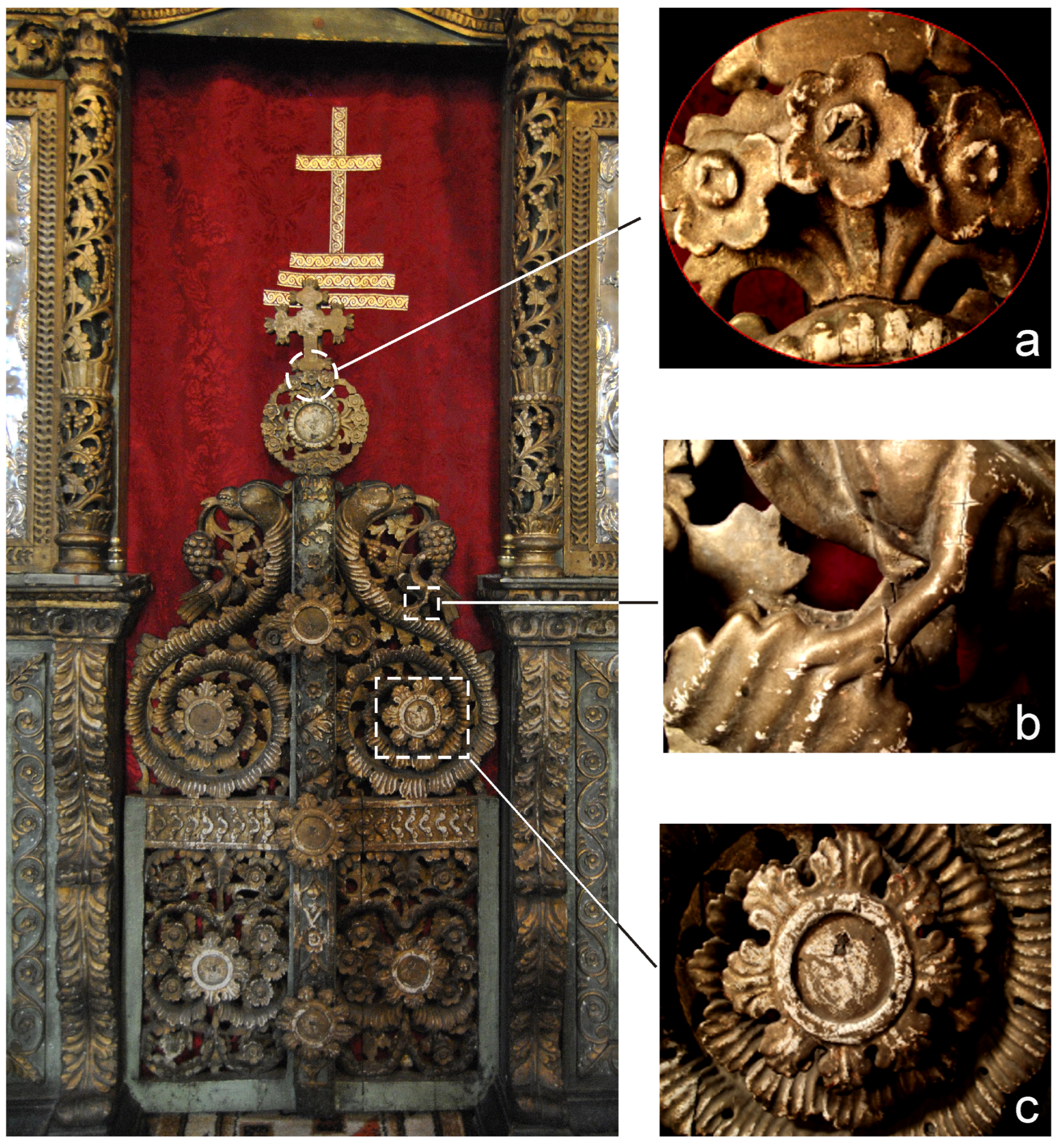
2.1. Imperial Doors
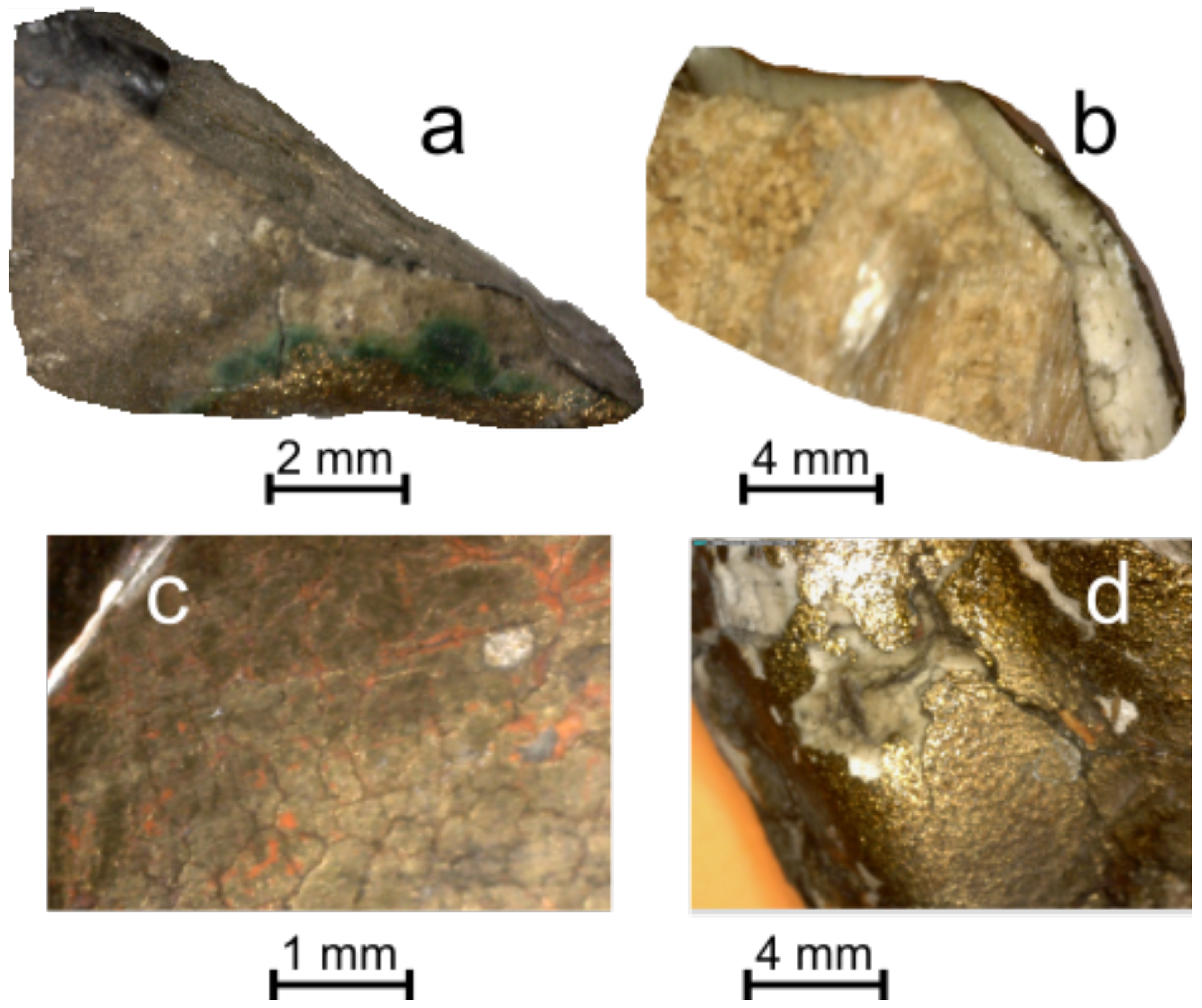
2.2. Sampling
2.3. Analytical Techniques
3. Results and Discussion
3.1. Church Microclimate
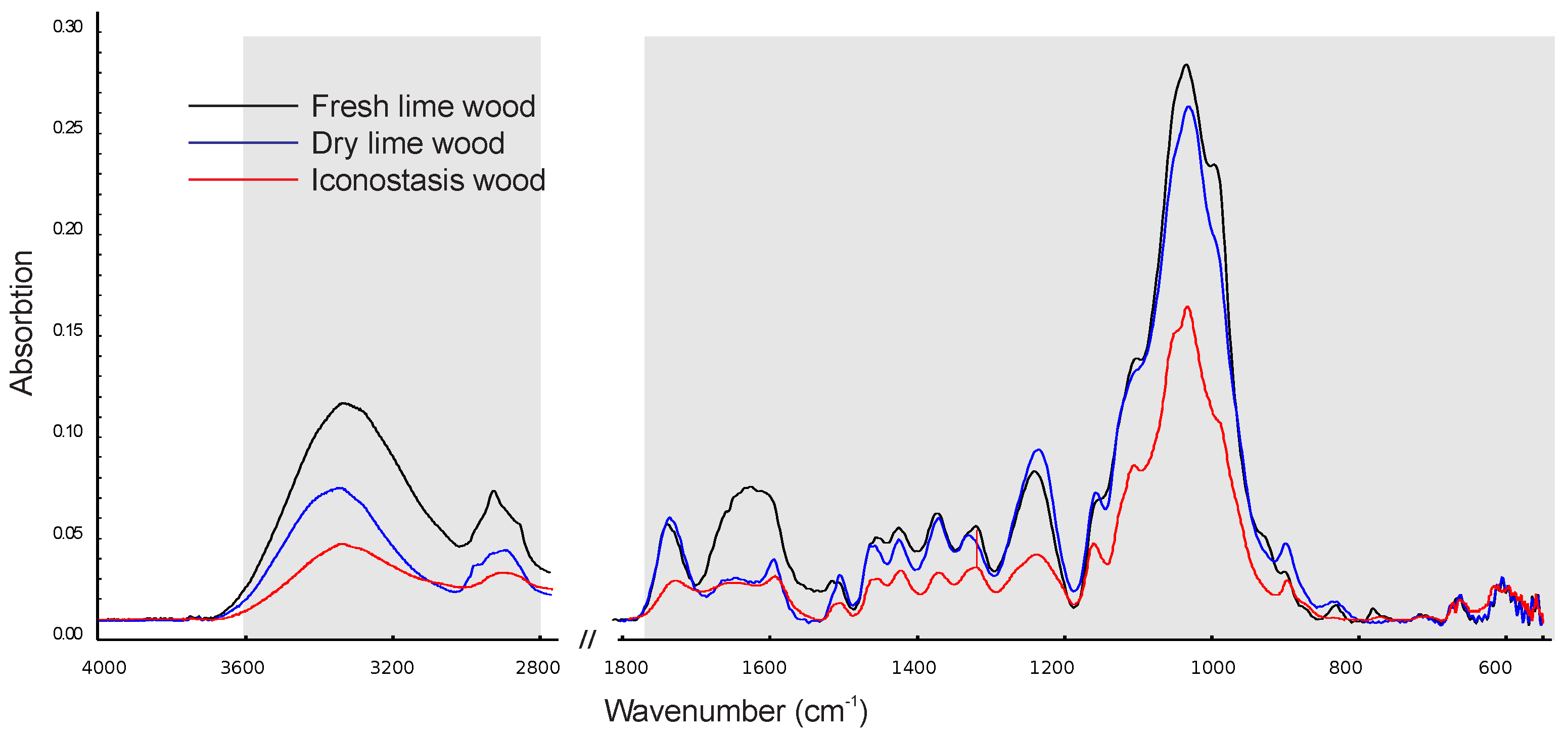
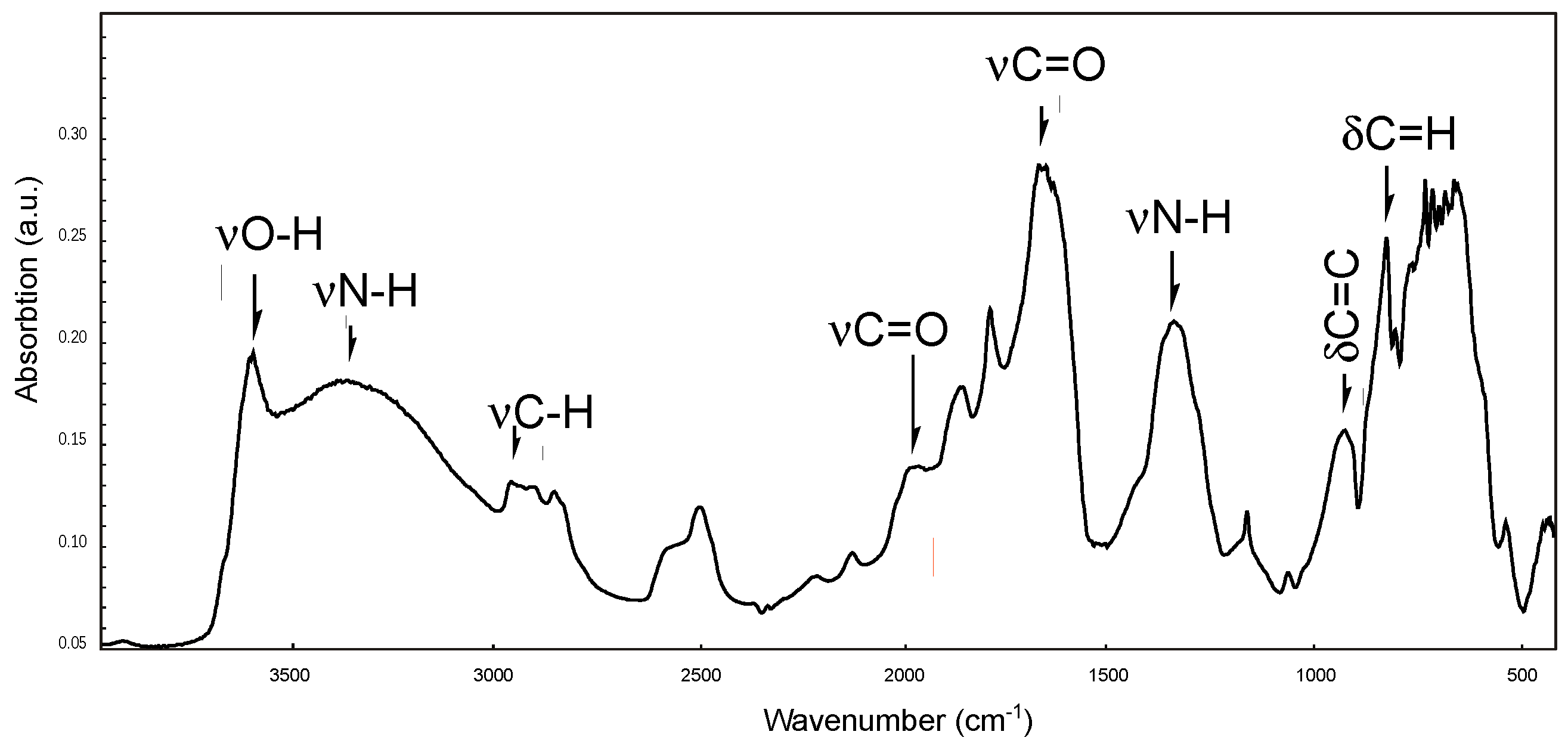
3.2. Wooden Material
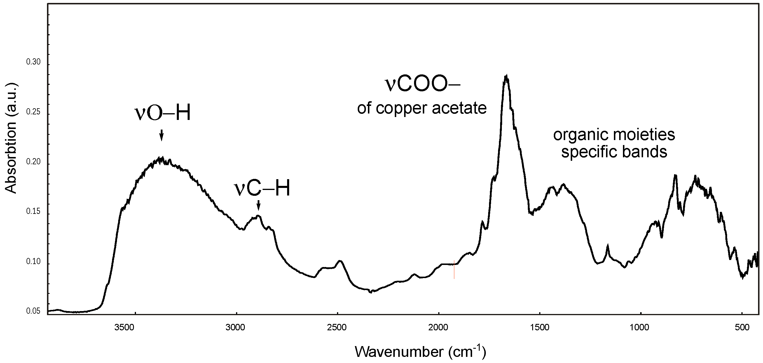
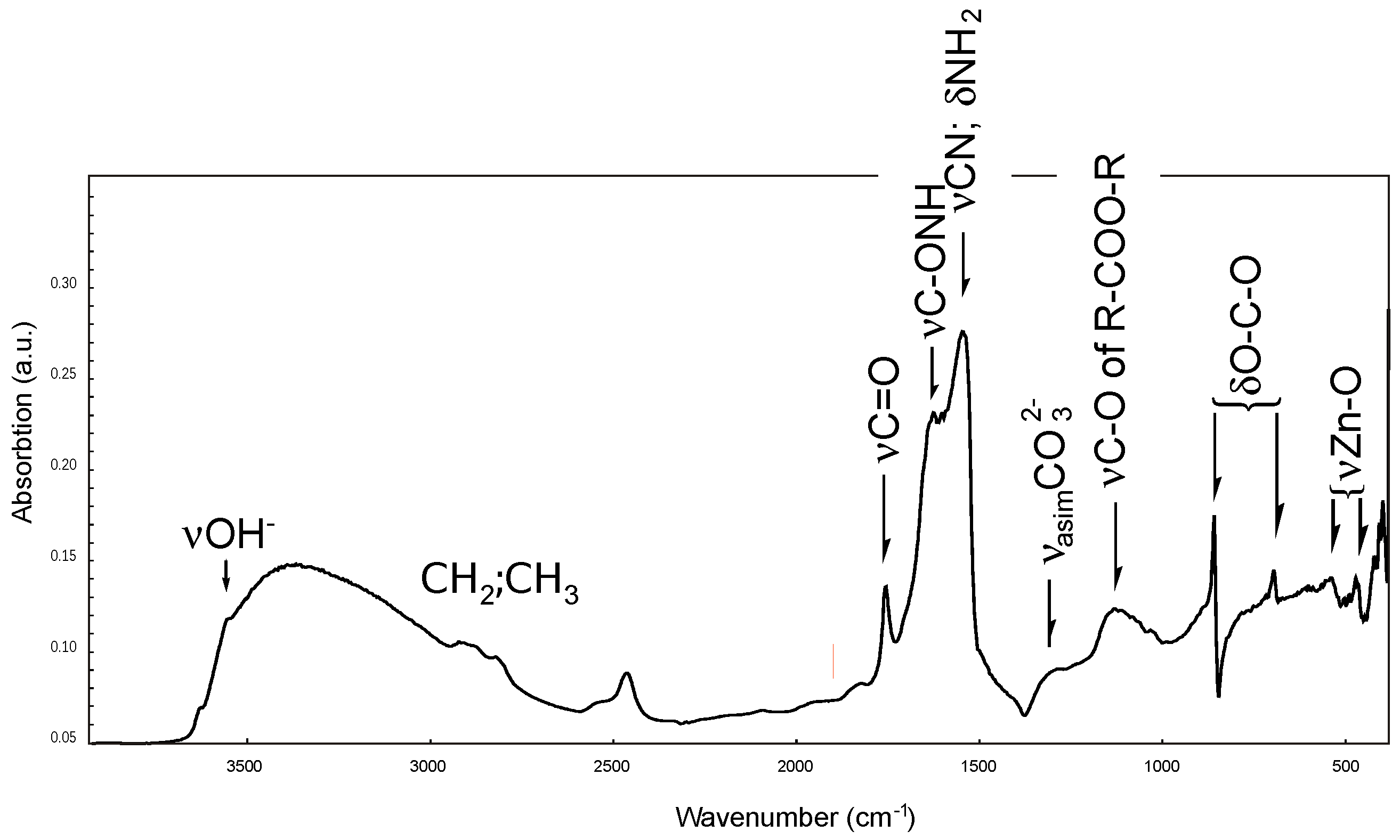
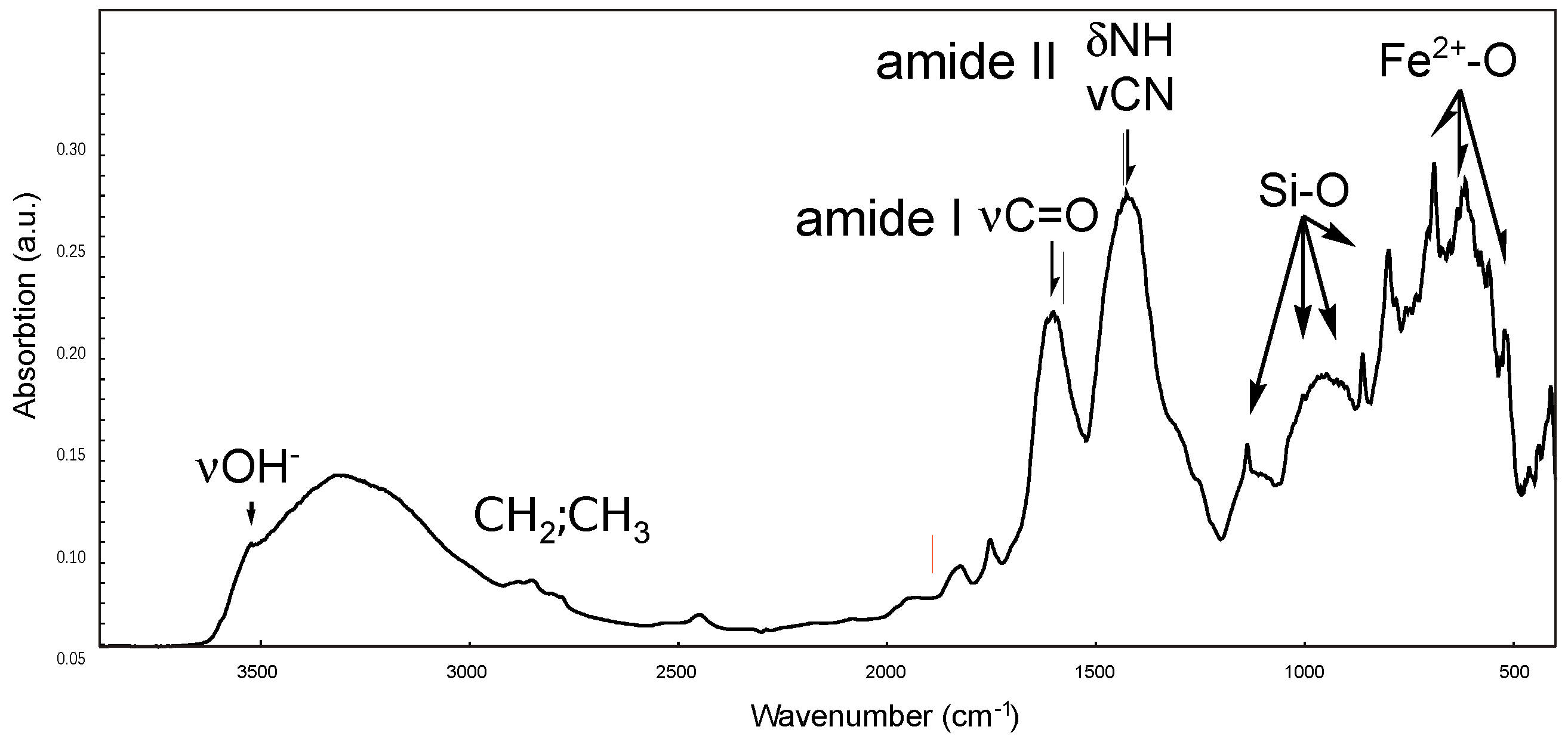
3.3. Gilded Layer

4. Conclusions
Author Contributions
Funding
Institutional Review Board Statement
Informed Consent Statement
Data Availability Statement
Acknowledgments
Conflicts of Interest
Abbreviations
| ATR | Attenuated Total Reflectance |
| FTIR | Fourier Transform Infrared Spectroscopy |
| XRF | X-Ray Fluorescence |
References
- Price, N.; Talley, M.K.; Vaccaro, A.M. (Eds.) Historical and Philosophical Issues in the Conservation of Cultural Heritage; Readings in Conservation Series; Getty Conservation Institute: Los Angeles, CA, USA, 1996; ISBN 978-0892363988. [Google Scholar]
- Legrand, S.; Vanmeert, F.; Van der Snickt, G.; Alfred, M.; De Nolf, W.; Dik, J.; Janssen, K. Examination of historical paintings by state-of-the-art hyperspectral imaging methods: From scanning infra-red spectroscopy to computed X-ray laminography. Herit. Sci. 2014, 2, 13. [Google Scholar] [CrossRef]
- Beckhoff, B.; Kanngießer, B.; Langhoff, N.; Wedell, R.; Wolf, H. (Eds.) Handbook of Practical X-ray Fluorescence Analysis; Springer: Berlin/Heidelberg, Germany, 2006; ISBN 978-3540367222. [Google Scholar]
- Badawy, W.; Duliu, O.G.; Magdy, M.; Mohamed Hadi, Q.; Sister Serafima. X-ray fluorescence spectroscopy in the investigation of works of art. In Spectroscopic and Microscopy Techniques for Archaeological and Cultural Heritage Research, 2nd ed.; Shukla, A.K., Ed.; IOP Publishing: Bristol, UK, 2023. [Google Scholar]
- Tsipali, M.; Vivdenko, S.; Tsangalidis, H.; Kostanta, A.; Mitsos, D.; Mantzana, E.; Vasileiadou, A.; Zacharias, N. Archaeometric investigation of pigments of the iconostasis from Saint Georgios church of Sohos. J. Archaeol. Sci. Rep. 2023, 52, 104235. [Google Scholar] [CrossRef]
- Annarilli, S.; Casoli, A.; Colantonio, C.; Lanteri, L.; Marsegli, A.; Pelosi, C.; Sotille, S. Multi-instrument analysis of the late 16th canvas painting, “Coronation of the Virgin with the Saints Ambrose and Jerome”, Attributed to the Tuscany-Umbria Area to Support the Possibility of Bio-Cleaning Using a Bacteria-Based System. Heritage 2022, 5, 2904–2921. [Google Scholar] [CrossRef]
- Henderson, E.J.; Helwig, K.; Stuart Read, S.; Rosendahl, S.M. Infrared chemical mapping of degradation products in cross-sections from paintings and painted objects. Herit. Sci. 2019, 7, 71. [Google Scholar] [CrossRef]
- Liu, G.L.; Kazarian, S.G. Recent advances and applications to cultural heritage using ATR-FTIR spectroscopy and ATR-FTIR spectroscopic imaging. Analyst 2022, 147, 1777–1797. [Google Scholar] [CrossRef] [PubMed]
- Pieta, E.; Olszewska-Świetlik, J.; Paluszkiewicz, C.; Zając, A.; Kwiatek, W.M. Application of ATR-FTIR mapping to identification and distribution of pigments, binders and degradation products in a 17th century painting. Vibr. Spec. 2019, 103, 102928. [Google Scholar] [CrossRef]
- Kaszowska, Z.; Kamilla Malek, K.; Magdalena Pańczyk, M.; Zajac, A.; Kwiatek, W.M. A joint application of ATR-FTIR and SEM imaging with high spatial resolution: Identification and distribution of painting materials and their degradation products in paint cross sections. Vibr. Spec. 2013, 65, 1–11. [Google Scholar] [CrossRef]
- Kalinina, K.B.; Korobov, V.A. Paul Gauguin, “Tahitian Pastorals”: Study of Painting Materials. Nanotechnol. Rep. 2022, 17, 666–675. [Google Scholar] [CrossRef]
- Masciotta, M.-G.; Ramos, L.F.; Lourenço, P.B. The importance of structural monitoring as a diagnosis and control tool in the restoration process of heritage structures: A case study in Portugal. J. Cult. Herit. 2017, 27, 36–47. [Google Scholar] [CrossRef]
- Pronti, L.; Romani, M.; Tarquini, O.; Verona-Rinati, G.; Colapietro, M.; Pifferi, A.; Marinelli, M.; Colao, F.; Guidi, M.C. Multi-sensor imaging approach to highlight hidden pentimenti and underdrawings: The case of “the Spring” painting at Chigi Palace of Ariccia. In Advanced Technologies for Cultural Heritage Monitoring and Conservation. Digital Innovations in Architecture, Engineering and Construction; Ceccarelli, S., Missori, M., Fantoni, R., Eds.; Springer: Cham, Switzerland, 2024. [Google Scholar] [CrossRef]
- Bonanni, L.; Seracini, M.; Xiao, X.; Hockenberry, M.; Cheng Costanzo, B.; Shum, S.; Teil, R.; Speranza, A.; Ishii, H. Tangible interfaces for art restoration. In Innovative Design and Creation of Visual Interfaces; Falchuk, B., Aderito, F.M., Eds.; IGI Global: Hershey, PA, USA, 2012; pp. 47–58. [Google Scholar] [CrossRef]
- Szczepanowska, H.M. Conservation of Cultural Heritage—Key Principles and Approaches; Rutlege: London, UK, 2013. [Google Scholar] [CrossRef]
- Donovan, C. State of the art in assessing research impact: Introduction to a special issue. Res. Eval. 2011, 20, 175–179. [Google Scholar] [CrossRef]
- Ogden, S. Understanding, Respect, and collaboration in cultural heritage preservation: A conservator’s developing perspective. Libr. Trends 2007, 56, 275–287. [Google Scholar] [CrossRef]
- Government of Canada; Canadian Heritage; Basic Care—Paintings, Canadian Conservation Institute. Available online: https://www.canada.ca/en/conservation-institute/services/care-objects/fine-art/basic-care-paintings.html (accessed on 30 July 2024).
- Duffey, K.; Carlson, J.; Price, L.O.; Pouliot, B.P.; Little, M.A.; Eaton, L.; Hess Norris, D.; Krill, J.; Fiske, B.; Bockrath, M.F.; et al. The Winterthur Guide to Caring for Your Collection; Winterthur Decorative Arts Series; Winterthur Museum: Winterhur, DE, USA, 2000; ISBN 978-0912724522. [Google Scholar]
- Grigorescu, S. Settlements and Monuments of Ialomita; Helis Publishing House: Slobozia, Romania, 2006; 167p. [Google Scholar]
- Carvanos, C. Guide to Byzantine Iconography; Institute for Byzantine and Modern Greek Press: Belmont, MA, USA, 1993; Volume 1, ISBN 978-0914744894. [Google Scholar]
- Cavarnos, C. Guide to Byzantine Iconography; Holy Transfiguration Monastery: Brookline, MA, USA, 2001; Volume 2, ISBN 978-0943405117. [Google Scholar]
- Florensky, P. Iconostasis; Oakwood Publications: Preston, UK, 1996; ISBN 978-0881411171. [Google Scholar]
- Vitto, F. The origin of the iconostasis in Early Christian churches in the Holy Land. In Actual Problems of Theory and History of Art: Collection of Articles; St. Petersburg University Press: St. Petersburg, Russia, 2017; Volume 7, pp. 222–231. [Google Scholar] [CrossRef]
- Djuric, I.; Stojakovic, V.; Misic, S.; Kekeljević, I. Case study of the iconostasis as the characteristic art and architectural element of the Christian Orthodox churches. In Proceedings of the 37 Education and Research in Computer Aided Architectural Design in Europe and XXIII Iberoamerican Society of Digital Graphics, Joint Conference (N. 1), Porto, Portugal, 11–13 September 2019; Available online: https://www.proceedings.blucher.com.br/article-details/34212 (accessed on 20 June 2024).
- Hradil, D.; Hradilová, J.; Bezdička, P.; Serendan, C. Late Gothic/early Renaissance gilding technology and the traditional poliment material “Armenian bole”: Truly red clay, or rather bauxite. Appl. Clay Sci. 2017, 135, 271–281. [Google Scholar] [CrossRef]
- Sandu, I.C.; Afonso, L.U.; Murta, E.; de Sa, H.M. Gilding techniques in religious art between east and west, 14th–18th centuries. Int. J. Conserv. Sci. 2010, 1, 47–62. Available online: https://ijcs.ro/pub/IJCS-10-06_Sandu.pdf (accessed on 20 June 2024).
- Mecklenburg, M.F. Some mechanical and physical properties of gilding gesso. In Gilded Wood Conservation and History; Bigelow, D., Cornu, E., Landrey, G.J., van Horne, C., Eds.; Sound View Press: Madison, CT, USA, 1991; pp. 163–170. ISBN 978-0932087218. [Google Scholar]
- Camuffo, D. Microclimate for Cultural Heritage: Measurement, Risk Assessment, Conservation, Restoration, and Maintenance of Indoor and Outdoor Monuments; Elsevier: Amsterdam, The Netherlands, 2019; ISBN 978-0444641069. [Google Scholar]
- Loupa, G.; Rapsomanikis, S. Air pollutant emission rates concentration in medieval churches during religious services. J. Atmos. Chem. 2008, 60, 169–187. [Google Scholar] [CrossRef]
- Tiano, P. Biodegradation of Cultural Heritage: Decay Mechanisms and Control Methods. In Proceedings of the ARIADNE 9 WS, Prague, Czech Republic, 4–10 February 2002; Available online: https://api.semanticscholar.org/CorpusID:15923293 (accessed on 20 June 2024).
- Pauchard, L.; Giorgiutti-Dauphine, F. Craquelures and pictorial matter. J. Cult. Herit. 2020, 46, 361–373. [Google Scholar] [CrossRef]
- Krzemień, L.; Łukomski, M.; Bratasz, L.; Kozlowski, N.; Meklenburg, M.F. Mechanism of craquelure pattern formation on panel paintings. Stud. Conserv. 2016, 61, 324–330. [Google Scholar] [CrossRef]
- Hill, C.; Hughes, M.; Gudsell, D. Environmental impact of wood modification. Coatings 2021, 11, 366. [Google Scholar] [CrossRef]
- Brischke, C.; Alfredsen, G. Wood-water relationships and their role for wood susceptibility to fungal decay. Appl. Microbiol. Biotechnol. 2020, 104, 3781–3795. [Google Scholar] [CrossRef]
- Si, S.; Tang, Q.; Xingong, L. The accelerated thermo-oxidative aging characteristics of wood fiber/polycaprolactone composite: Effect of temperature, humidity and time. J. Renew. Mater. 2021, 9, 2209–2222. [Google Scholar] [CrossRef]
- Popescu, C.M.; Vasile, C.; Popescu, M.C.; Sgurel, G. Degradation of lime wood painting supports II. Spectral characterization. Cellul. Chem. Technol. 2006, 40, 649–658. [Google Scholar]
- Ghavidel, A.; Gelbrich, J.; Kuqo, A.; Vasilache, V.; Sandu, I. Investigation of archaeological European white elm (Ulmus laevis) for identifying and characterizing the kind of biological degradation. Heritage 2020, 3, 1083–1093. [Google Scholar] [CrossRef]
- Sharma, V.; Yadav, J.; Kumar, R.; Tesarova, D.; Ekielski, A.; Mishra, P.K. On the rapid and non-destructive approach for wood identification using ATR-FTIR spectroscopy and chemometric methods. Vib. Spec. 2020, 110, 103097. [Google Scholar] [CrossRef]
- Emandi, A.; Brudugeac, P.; Emandi, I.; Stanculescu, I. The assesment of the decayed lime wood polymeric components by TG and FTIR parameters correlation. Int. J. Conserv. Sci. 2010, 1, 211–218. Available online: https://ijcs.ro/pub/IJCS-10-21-Emandi.pdf (accessed on 10 June 2024).
- Emandi, A.; Vasiliu, I.C.; Brudugeac, P.; Stamatin, I. Quantitative investigation of wood composition by integrated FTIR nd thermogravimetric methods. Cellul. Chem. Technol. 2011, 45, 579–584. Available online: https://cellulosechemtechnol.ro/pdf/CCT45,9-10(2011)/p.579-584.pdf (accessed on 10 June 2024).
- Marinescu, M.; Emandi, A.; Duliu, O.G.; Stanculescu, I.; Bercu, V.; Emandi, I. FT-IR, EPR and SEM-EDAX investigation of some accelerated aged painting binders. Vib. Spectrosc. 2014, 73, 37–44. [Google Scholar] [CrossRef]
- Avram, M.; Mateescu, G.D. Infrared Spectroscopy Applications in Organic Chemistry; Wiley: Hoboken, NJ, USA, 1978; ISBN 978-0471038450. [Google Scholar]
- Ferreira, E.S.B.; Gros, D.; Wyss, K.; Scherrer, N.C.; Zumbühl, S.; Marone, F. Faded shine.... The degradation of brass powder in two nineteenth century paintings. Herit. Sci. 2015, 3, 24. [Google Scholar] [CrossRef]
- Ricci, M.; Sebastiani, F.; Becucci, M.; Rogozny, M.; Parfenov, V. Spectroscopy-based multi-analytical approach for atudies in conservation: Decorations in theAlexander Palace (Tsarskoye Selo). Spectrosc. J. 2023, 1, 121–136. [Google Scholar] [CrossRef]
- Atanassova, V.; Dinu, M.; Polizu, S.R.; Radvan, R. Photonic applications for restoration and conservation of 19th century polychrome religious wooden artworks. Coatings 2023, 13, 1235. [Google Scholar] [CrossRef]
- Jacobson, M.C.; Hansson, H.C.; Noone, K.J.; Charlson, J.R. Organic atmospheric aerosols: Review and state of the science. Rev. Geophys. 2000, 38, 267–294. [Google Scholar] [CrossRef]
- Tudor, D.; Robinson, S.C.; Cooper, P.A. The influence of moisture content variation on fungal pigment formation in spalted wood. AMB Express 2012, 2, 69. [Google Scholar] [CrossRef] [PubMed]
- Tinti, A.; Tugnoli, V.; Bonora, S. Recent applications of vibrational mid-Infrared (IR) spectroscopy for studying soil components: A review. J. Cent. Eur. Agric. 2015, 16, 1–22. [Google Scholar] [CrossRef]
- Blando, J.D.; Porjca, R.J.; Bowman, D.; Bowman, D.; Lioy, P.J.; Turpin, B.J. Secondary formation and the Smoky Mountain organic aerosol: An examination of aerosol polarity and functional group composition during SEAVS. Environ. Sci. Technol. 1998, 32, 604–613. [Google Scholar] [CrossRef]
- Rachwał, B.; Bratasz, Ł.; Krzemień, L.; Łukomski, M.; Kozłowski, R. Fatigue damage of the gesso layer in panel paintings subjected to changing climate conditions. Strain 2012, 48, 474–481. [Google Scholar] [CrossRef]
- Vagenas, N.; Gatsouli, A.; Kontoyannis, C.G. Quantitative analysis of synthetic calcium carbonate polymorphs using FT-IR spectroscopy. Talanta 2003, 59, 831–836. [Google Scholar] [CrossRef] [PubMed]
- Vahur, S.; Teearu, A.; Peets, P.; Joosu, L.; Leito, I. ATR-FT-IR spectral collection of conservation materials in the extended region of 4000-80 cm−1. Anal. Bioanal. Chem. 2016, 408, 3373–3379. [Google Scholar] [CrossRef]
- Izzo, F.; Germinario, C.; Grifa, C.; Langella, A.; Mercurio, M. External reflectance FTIR dataset (4000–400 cm−1) for the identification of relevant mineralogical phases forming Cultural Heritage materials. Infrared Phys. Technol. 2020, 106, 103266. [Google Scholar] [CrossRef]
- Manni, A.; Matadi Boumbimba, R.; Mikdam, A.; El Bouari, A.; Addiego, F.; Meziani, J.; Wary, M. Magnesite and dolomite micro-particles: Preparation, physical properties and application in bio-based polymer composite. Polym. Bull. 2021, 79, 2149–2171. [Google Scholar] [CrossRef]
- Duliu, O.G.; Grecu, M.N.; Cristache, C. EPR and X-ray diffraction investigation of some Greek marbles and limestones. Rom. Rep. Phys. 2009, 61, 487–499. Available online: https://rrp.nipne.ro/2009_61_3/art14Duliu.pdf (accessed on 10 August 2024).
- Mposkos, E.; Baziotis, I.; Proyer, A.; Hoinkes, G. Dolomitic marbles from the ultrahigh-pressure metamorphic Kimi complex in Rhodope, N.E. Greece. Mineral. Petrol. 2006, 88, 341–362. [Google Scholar] [CrossRef]
- Meilunas, R.J.; Bentsen, J.G.; Steinberg, A. Analysis of aged paint binders by FTIR spectroscopy. Stud. Conserv. 1990, 35, 33–51. [Google Scholar] [CrossRef]
- Franceschi, E.; Locardi, F. Strontium, a new marker of the origin of gypsum in cultural heritage? J. Cult. Herit. 2014, 15, 522–527. [Google Scholar] [CrossRef]
- Sommer, M.; Kaczorek, D.; Kuzyakov, Y.; Breuer, J. Silicon pools and fluxes in soils and landscapes—A review. J. Plant Nutr. Soil Sci. 2006, 169, 310–329. [Google Scholar] [CrossRef]
- Namduri, H.; Nasrazadani, S. Quantitative analysis of iron oxides using Fourier transform infrared spectrophotometry. Corros. Sci. 2008, 50, 2493–2497. [Google Scholar] [CrossRef]
- Abendrot, M.; Chęcińsk, L.; Kusz, J.; Lisowska, K.; Zawadzka, K.; Felczak, A.; Kalinowska-Lis, U. Zinc(II) Complexes with amino acids for potential use in dermatology: Synthesis, crystal structures, and antibacterial activity. Molecules 2020, 25, 951. [Google Scholar] [CrossRef]
- Pellegrini, D.; Duce, C.; Bonaduce, I.; Biagi, S.; Ghezzi, L.; Colombini, M.P.; Tinè, M.R.; Bramanti, E. Fourier Transform Infrared Spectroscopic study of rabbit glue/inorganic pigments mixtures in fresh and aged reference paint reconstructions. Microchem. J. 2015, 124, 31–35. [Google Scholar] [CrossRef]
- De Liedekerke, M. Zinc Oxide (Zinc White). In Pigments, Inorganic. Ullmann’s Encyclopedia of Industrial Chemistry; Wiley: Hoboken, NJ, USA, 2006. [Google Scholar] [CrossRef]


| Assignments | Reference Lime Wood | Dried Wood | Iconostasis Wood | Remarks |
|---|---|---|---|---|
| O-H | 3337 i | 3345 m | 3338 w | Cellulose chain damage |
| C-H aliphatic | 2900–2800 sh | 2900–2800 w large | 2900–2800 vw | Idem |
| C-O-C cellulose and hemicellulose | 1033 i–1160 sh | 1033 m–1160 sh | 1033 w–1160 sh | Decreasing the amount of cellulose |
| C=O unconjugated hemicellulose | 1735 | 1737 | 1737 wl | Loss of hemicellulose |
| -CH2 –cellulose | 1319 m | 1319 w | 1319 vw | Drastic decrease in cellulose and hemicellulose |
| C=O embedded in aromatic skeleton | 1593 w | 1593 w | 1593 vw | Damage to lignin aromatic skeleton |
| C=O acetyl and carboxyl groups of xylan | 1503 m | 1503 m | 1503 vw | Damage to lignin aromatic matrix |
| C=O lignin and acetyl in carboxylic vibration of xylan | 1234 m | 1234 w | 1234 vw | Damage to lignin skeleton |
| C-H aromatic skeleton of lignin, splitting bands | 1456 m; 1423 m | 1456 w; 1423 w | 1456 vw; 1423 vw | Decrease in lignin amount |
| H-O-H of absorbed water | 1624 m | 1624 w | absent | Decrease in hydration degree |
Disclaimer/Publisher’s Note: The statements, opinions and data contained in all publications are solely those of the individual author(s) and contributor(s) and not of MDPI and/or the editor(s). MDPI and/or the editor(s) disclaim responsibility for any injury to people or property resulting from any ideas, methods, instructions or products referred to in the content. |
© 2024 by the authors. Licensee MDPI, Basel, Switzerland. This article is an open access article distributed under the terms and conditions of the Creative Commons Attribution (CC BY) license (https://creativecommons.org/licenses/by/4.0/).
Share and Cite
Duliu, O.G.; Emandi, A.; Marinescu, M.; Cinteza, O.; Stanculescu, I.; Ionescu, L.; Filimon, D. Assessing the Degradation Status of the Imperial Doors of the Ascension Church, Grindu Commune, Romania. Appl. Sci. 2024, 14, 7565. https://doi.org/10.3390/app14177565
Duliu OG, Emandi A, Marinescu M, Cinteza O, Stanculescu I, Ionescu L, Filimon D. Assessing the Degradation Status of the Imperial Doors of the Ascension Church, Grindu Commune, Romania. Applied Sciences. 2024; 14(17):7565. https://doi.org/10.3390/app14177565
Chicago/Turabian StyleDuliu, Octavian G., Ana Emandi, Maria Marinescu, Otilia Cinteza, Ioana Stanculescu, Liliana Ionescu, and Daniela Filimon. 2024. "Assessing the Degradation Status of the Imperial Doors of the Ascension Church, Grindu Commune, Romania" Applied Sciences 14, no. 17: 7565. https://doi.org/10.3390/app14177565
APA StyleDuliu, O. G., Emandi, A., Marinescu, M., Cinteza, O., Stanculescu, I., Ionescu, L., & Filimon, D. (2024). Assessing the Degradation Status of the Imperial Doors of the Ascension Church, Grindu Commune, Romania. Applied Sciences, 14(17), 7565. https://doi.org/10.3390/app14177565