Article

Properties of Zirconia, Lithium Disilicate Glass Ceramics, and VITA ENAMIC® Hybrid Ceramic Dental Materials Following Ultra-Short Femtosecond (30 fs) Laser Irradiation

by
Victor L. Lagunov
1,
Bakhtiar Ali
2,3,
Laurence J. Walsh
4,
Andrew B. Cameron
1,
Igor V. Litvinyuk
3,5,
Maksym Rybachuk
2,3,* and
Roy George
1,*
1
School of Medicine and Dentistry, Griffith University, Parklands Drive, Southport, QLD 4222, Australia
2
School of Engineering and Built Environment, Griffith University, 170 Kessels Rd., Nathan, QLD 4111, Australia
3
Centre for Quantum Dynamics and Australian Attosecond Science Facility, Griffith University, Science Road, Nathan, QLD 4111, Australia
4
School of Dentistry, University of Queensland, 288 Herston Rd., Herston, QLD 4006, Australia
5
School of Environment and Science, Griffith University, Nathan, QLD 4111, Australia
*
Authors to whom correspondence should be addressed.
Appl. Sci. 2024, 14(17), 7641; https://doi.org/10.3390/app14177641
Submission received: 31 July 2024 / Revised: 19 August 2024 / Accepted: 22 August 2024 / Published: 29 August 2024
(This article belongs to the Special Issue New Advances in Laser Dental Science and Biophotonics)

Abstract

:

Featured Application

This study highlights the potential application of ultra-short femtosecond lasers for modifying the surface of ceramic dental materials with high precision in a controlled manner.

Abstract

This study investigated the dose-dependent changes in the chemical composition of three dental ceramic materials—zirconia, lithium disilicate (LD), and VITA ENAMIC® hybrid composite (VITA En)—following irradiation with an ultra-short femtosecond (fs) laser (800 nm, 30 fs, 1 kHz) in an ambient air environment using average laser power (76 mW) and scanning speeds (50, 100, and 200 mm/s), simulating dental treatment processes. The chemical composition of the ablated regions was analyzed using energy dispersive spectroscopy. All irradiated samples showed increased carbon content (by up to 42%) and reduced oxygen (by up to 33%). The observed increase in C content is likely attributed to a combination of surface reactions, adsorption of carbon from the ambient environment, and carbon deposition from the laser-induced plasma, all facilitated by the high-energy conditions created by fs-laser pulses. Scanning electron microscopy revealed ablation with progressive controlled melting and recrystallization, with an absence of pile-up features typically associated with significant thermal damage. These findings demonstrate that ultra-short fs-laser irradiation induces highly controlled, dose-dependent changes in the chemical composition and surface morphology of dental ceramic materials.

1. Introduction

Dental ceramics are increasingly being used to fabricate dental restorations, driven by their advantageous mechanical properties, chemical resistance, biocompatibility, and aesthetic appeal [1,2,3]. Zirconia (ZrO2) ceramic materials are acknowledged for their high hardness and low fracture toughness [4,5,6]. Lithium disilicate (Li2Si2O5) (LD) glass ceramics and zirconia have emerged as prominent choices for all-ceramic dental restorations [7]. Hence, zirconia has become a popular ceramic material for restoring dental implants and implant fixtures.
Emerging hybrid ceramic materials such as VITA ENAMIC® (VITA En) are also of interest for subtractive manufacturing of dental restorations. Hybrid ceramics are formed by infiltrating polymers into a porous feldspathic ceramic enriched with aluminum oxide and offer several advantages, including a high resistance to fracture and higher flexural strength [8] compared to both zirconia and LD materials. VITA En has a composition of 86% ceramic and 14% polymer by weight [9].
Surface modifications of the fitting surfaces of dental ceramic restorations can enhance their capacity to adhere to teeth or abutment substrates through micromechanical interlocking or chemical bonding to the adhesive cement [10]. Feldspathic porcelains and LD-reinforced glass ceramics can be etched using hydrofluoric acid, creating surface roughness that enhances micromechanical interlocking with resin cements [11]. Acid etching is not feasible with zirconia due to its inert nature. Conventional surface modification techniques, such as grit blasting, are also not particularly effective on zirconia. Additionally, grit blasting can degrade the mechanical properties of zirconia [12]. Grit blasting using alumina and/or silica particles poses risks for crack propagation and may also leave blasting residue or surface contaminants [13]. Consequently, there is interest in using alternative methods to modify the surface of zirconia, including laser-based techniques [14].
Using lasers could be a non-contact surface processing method not only for zirconia but also for other dental ceramics and related materials, including hybrid ceramics [15]. This approach would eliminate issues such as dental tool wear and surface degradation such as cracking caused by contact between rotary cutting tools and the ceramic material [16]. Conventional dental lasers operating at wavelengths of 1064 and 10,600 nm in picosecond (ps-) or nanosecond (ns-) pulse widths have been used for marking, engraving, and cutting of ceramics and dental composites [17,18].
However, the high surface temperatures caused by photothermal interactions can cause decomposition, microcracking, and the formation of amorphous glass-like phases from melting and rapid re-solidification [19]. One solution to these challenges is to employ shorter laser pulse durations within the ultra-short femtosecond (fs) pulse width range. Ultra-short pulse laser ablation systems are technologically advanced systems capable of processing various materials at extremely high peak powers (up to a few GW) while delivering highly localized energy that enables a non-thermal ablation of even transparent and low-absorption samples with high precision and accuracy using significantly lower laser fluences compared to those employed in ns- or ps-laser processes [20].
Ultra-fast laser processing using fs-laser pulses offers several advantages over conventional ns- or ps-laser treatments. These advantages include significantly reduced laser-matter interaction time—to less than a picosecond, which leads to minimal heat dissipation into the surrounding bulk of the material [21,22]. This, in turn, reduces thermal and mechanical stresses in the surrounding area [23]. These stresses can be further mitigated by decreasing the laser beam spot size. Moreover, fs-laser interactions with materials are independent of material orientation, eliminating the need for specific sample or tool positioning prior to laser treatment. The laser beam’s effectiveness in dental treatments depends solely on its optical quality and is unaffected by material-to-tool interaction, unlike in other conventional material treatment processes. fs-lasers have been found useful for the surface processing of zirconia [15,21]. For example, Yamamuro et al. in 2022 employed 256 fs laser pulses to irradiate zirconia. They noted a partial transformation of the zirconia’s tetragonal phase into the low-intensity monoclinic phase due to thermal effects. However, with repeated laser irradiation, there is a possibility of an additional laser-induced reverse transformation from the monoclinic phase to the tetragonal phase [24]. Notably, in instances where the zirconia surface was exposed to longer ps- or ns-laser pulse durations, the laser-irradiated regions displayed melting and partial conversion to the structurally inferior monoclinic phase [25,26,27]. Given the widespread use of zirconia in implant-supported crowns [28,29] it is important to have methods to alter the surface morphology that can be controlled to a high level [30].
Using a 1030 nm, 600 fs laser irradiation on LD, Hu et al. [31] showed that it was feasible to process LD without altering its surface composition or mechanical hardness. Daskalova et al. [32] used an 800 nm, 150 fs laser radiation to alter the surface texture of alumina-toughened zirconia. They found that the process did not induce any adverse thermal side effects on the treated LD samples and preserved the tetragonal phase while reducing the monoclinic phase. Additionally, surface modifications of zirconia implants using laser processing aim to enhance roughness, wettability, and biological responses without changing the physical and chemical properties of zirconia [33,34].
Thus, processing zirconia and LD using laser light, and potentially using ultra-short fs-laser light, offers advantages over conventional processes due to its ability to achieve precise material removal with minimal thermal and mechanical damage. This method allows for highly controlled surface modifications, enabling enhanced microstructural and phase stability, which are crucial for maintaining the material’s mechanical properties. Additionally, fs-laser processing can produce fine surface textures that improve bonding strength with dental cements and other restorative materials, ultimately leading to more durable and reliable dental restorations. Nevertheless, little is known about laser interactions with emerging hybrid composite dental materials, including VITA En, which has ceramic and polymer phases, particularly regarding how laser parameters, such as pulse duration, wavelength, and energy density, affect their microstructure, surface chemistry, and mechanical properties.
The aim of the present study is to provide a pilot evaluation of dose-dependent changes in the elemental chemical composition and surface morphology of zirconia, LD glass ceramics, and VITA En hybrid composite dental materials following ultra-short femtosecond (30 fs) laser irradiation. Understanding the dose-dependent changes is essential for optimizing laser-based processing techniques. It is a necessary first step for ensuring that these materials retain their desired properties, such as biocompatibility, durability, and aesthetics, after laser treatment in clinical settings, as the latter requires a selection of key laser processing parameters, including the pulse number and scanning speed [35]. Hence, the aim of the present study is to evaluate the dose-dependent changes in elemental chemical composition and surface morphology of zirconia, LD glass ceramics, and VITA En hybrid composite materials following exposure to fs-laser irradiation.

2. Materials and Methods

2.1. Sample Preparation

Dental sample blocks for computer-aided design (CAD) and computer-aided manufacturing (CAM) of zirconia, LD glass ceramics, and VITA En were sourced from VITA Zahnfabrik H. Rauter GmbH & Co., Bad Sackingen, Germany. Zirconia in VITA YZ T manufacturing grade contained ZrO2 at 90–95 wt.%, yttria (Y2O3) at 4–6 wt.%, hafnium oxide (HfO2) at 1–3 wt.%, and alumina and pigments at <1 wt.% [36]. The LD glass ceramic in VITA AMBRIA® grade contained silica (SiO2) at 58–66 wt.%, Li2O at 12–16 wt.%, zirconia at 8–12 wt.%, alumina at 1–4 wt.%, phosphorus pentoxide (i.e., phosphorus(V) oxide, also known as phosphoric anhydride) (P2O5) at 2–6 wt.%, and other minor fractions of elements at 1 wt.% or less [37]. VITA ENAMIC® is a hybrid ceramic composed of a dual-network structure, with approximately 86% ceramic (feldspathic-based ceramic) and 14% highly cross-linked acrylate polymer by weight [38]. VITA En is reported to have a high purity level of >99%.
The samples were milled into flat oval plates with dimensions of 5 × 10 × 3 mm3, in accordance with, and exceeding the requirements of, ISO 6872:2024 [39] (Dentistry) ‘Ceramic Materials’, which specifies the preparation and surface finish for dental ceramics such as zirconia and LD glass ceramic materials. In addition, the accuracy of CAD/CAM digitized milling and finishing of test samples complied with the requirements of the ISO 12836:2015 [40] (Dentistry) ‘Digitizing Devices for CAD/CAM Systems for Indirect Dental Restorations’ standard. After milling, the samples were sintered at 1500 °C, with the zirconia samples undergoing an additional firing at 810 °C.
All samples were polished to achieve a flat surface using a stepwise approach that included the use of a Universal Polisher, Diacera HP Medium and DVA Zircon-Brite polishing paste (EVE® EVE Ernst Vetter GmbH, Keltern, Germany), and the JOTA Zir Gloss Laboratory Kit (JOTA AG Rotary Instruments, Rüthi, Switzerland). Following polishing, the samples were steam-cleaned with demineralized water to remove any residual polishing compounds. They were then cleaned with a neutral detergent solution (Extran® MA 05, Sigma Aldrich, St. Louis, MO, USA) and subjected to ultrasonic cleaning in deionized water for 10 min before being used in laser ablation experiments.

2.2. Laser Irradiation

A custom-built laser irradiation setup utilizing an ultra-short titanium-sapphire laser (FEMTOPOWER™ Compact™ PRO HE, FEMTOLASERS Produktions GmbH, Vienna, Austria) was employed to irradiate the surface of dental samples. The system delivered linearly polarized 30 fs laser pulses at a central wavelength of 800 nm, with a pulse repetition rate of 1 kHz and a pulse energy of 0.8 mJ per pulse. A protected aluminum 90° off-axis parabolic mirror (MPD149-P01, Thorlabs Inc., Newton, NJ, USA) with a focal length of 100 mm was used to focus the fs-laser beam onto the samples.
To ensure precise beam alignment, a CMOS (Complementary Metal-Oxide-Semiconductor) camera beam profiler with a resolution of 2448 × 2048 pixels (CMOS BC207VIS(M), Thorlabs Inc., Newton, NJ, USA) was utilized to diagnose the beam profile near the focal point. This setup ensured near-perfect alignment of the parabolic mirror, minimized beam aberration at the point of focus, and produced a focal spot with a 22 μm diameter, as measured using the CMOS camera at 1/e2 of the maximum intensity, with an M2 value of 1.3 for all experiments.
The sample blocks were positioned normal to the fs-laser beam and mounted on a linear translation stage, with the X-axis position controlled by a high-precision motorized stage (OptoSigma Corp., Santa Ana, CA, USA) offering a minimum incremental motion of 0.1 μm. No self-phase modulation or filamentation was observed in front of the focal point at the irradiation site during the experiments.
The samples were subjected to fs-laser irradiation at an average power of 76 mW, corresponding to laser fluence of 20 J/cm2. The laser parameters employed in this study are shown in Table 1.
Each sample was subjected to five separate fs-laser irradiation exposures under the given sample translational speeds (also known as sample speed) and laser pulse numbers. The resultant linear irradiation exposure areas/sections on the sample surface are referred to in the text as tracks.

2.3. Sample Analysis

Sample morphological examination was performed using a scanning electron microscopy (SEM) system (Phenom™ XL G2, Thermo Fisher Scientific Inc., Waltham, MA, USA) under ×2550 or higher magnifications. This system was equipped with energy dispersive spectroscopy (EDS) for elemental analysis. The EDS analysis provided mean values and standard deviations of the relative weight concentration percentages (wt.%) for each element of interest (e.g., C, O, Al, and Si) in the identified areas. EDS measurements were collected from both laser-irradiated and untreated regions.
All EDS numerical data sets were assessed employing the Kolmogorov-Smirnov non-parametric test of the equality of continuous one-dimensional probability distributions [41]. Normalized data sets were then analyzed using one-way ANOVA with post-hoc Tukey tests, utilizing Prism v10.0.3 software (GraphPad, San Diego, CA, USA). A p value < 0.05 was considered statistically significant.

3. Results

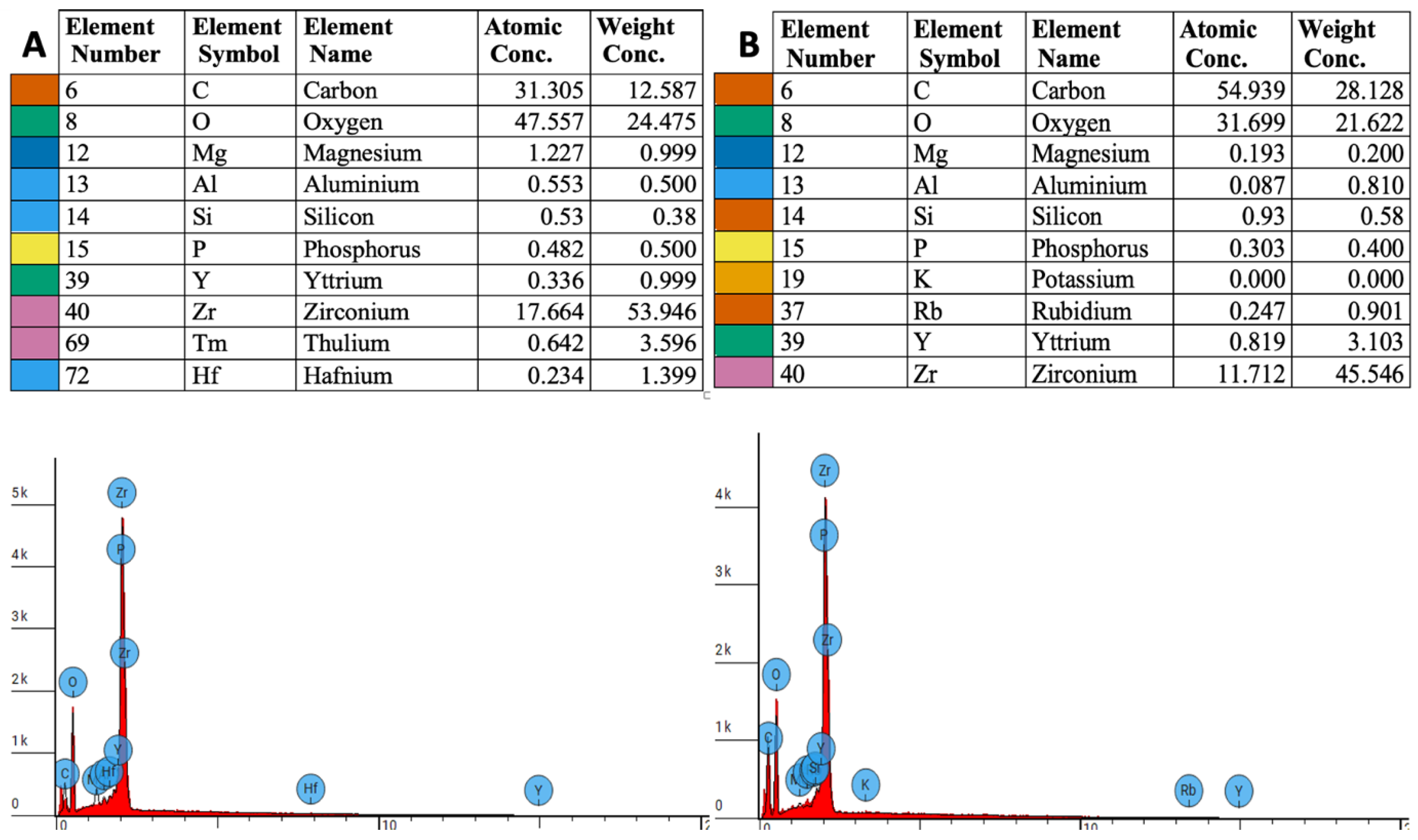
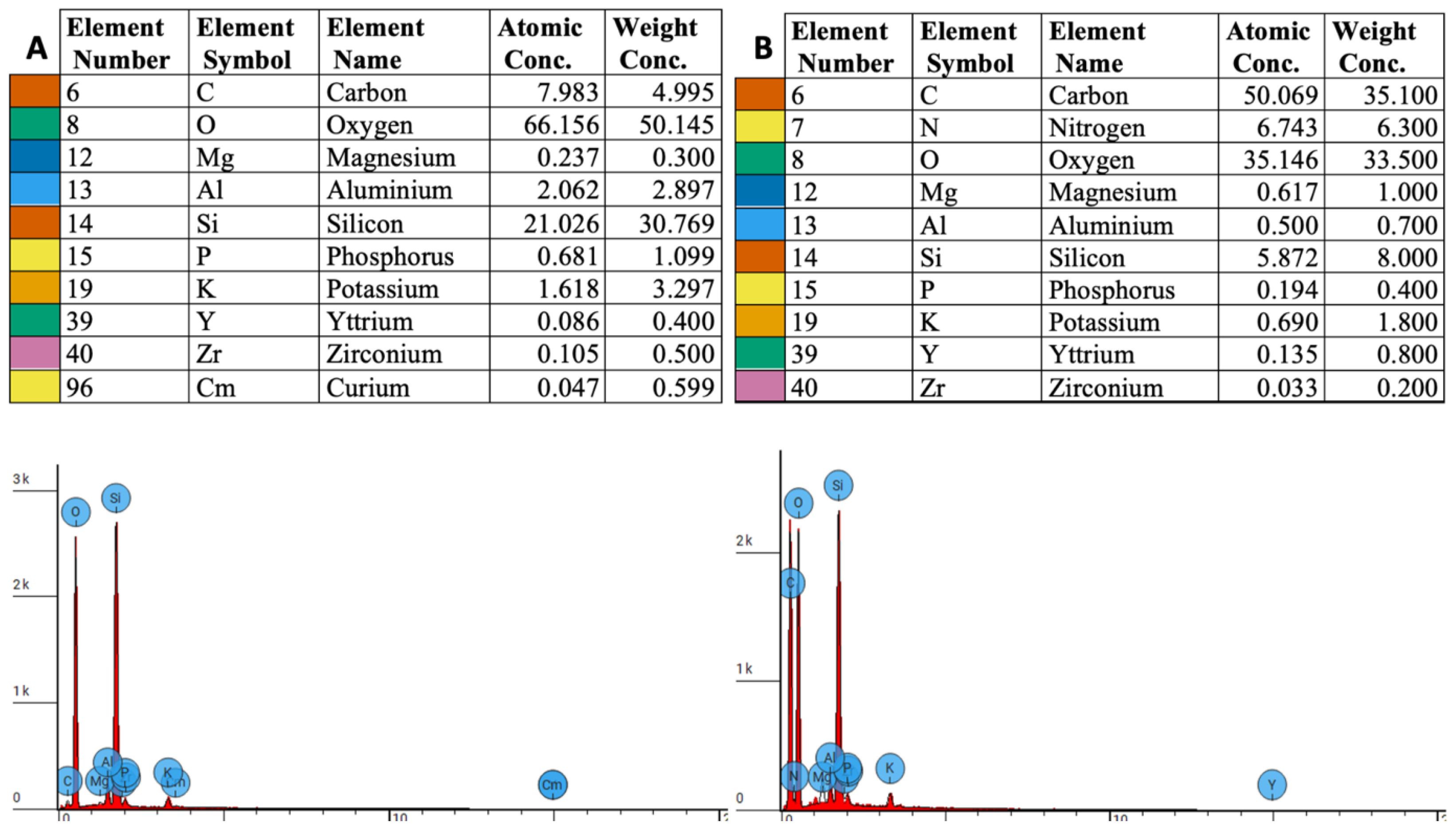
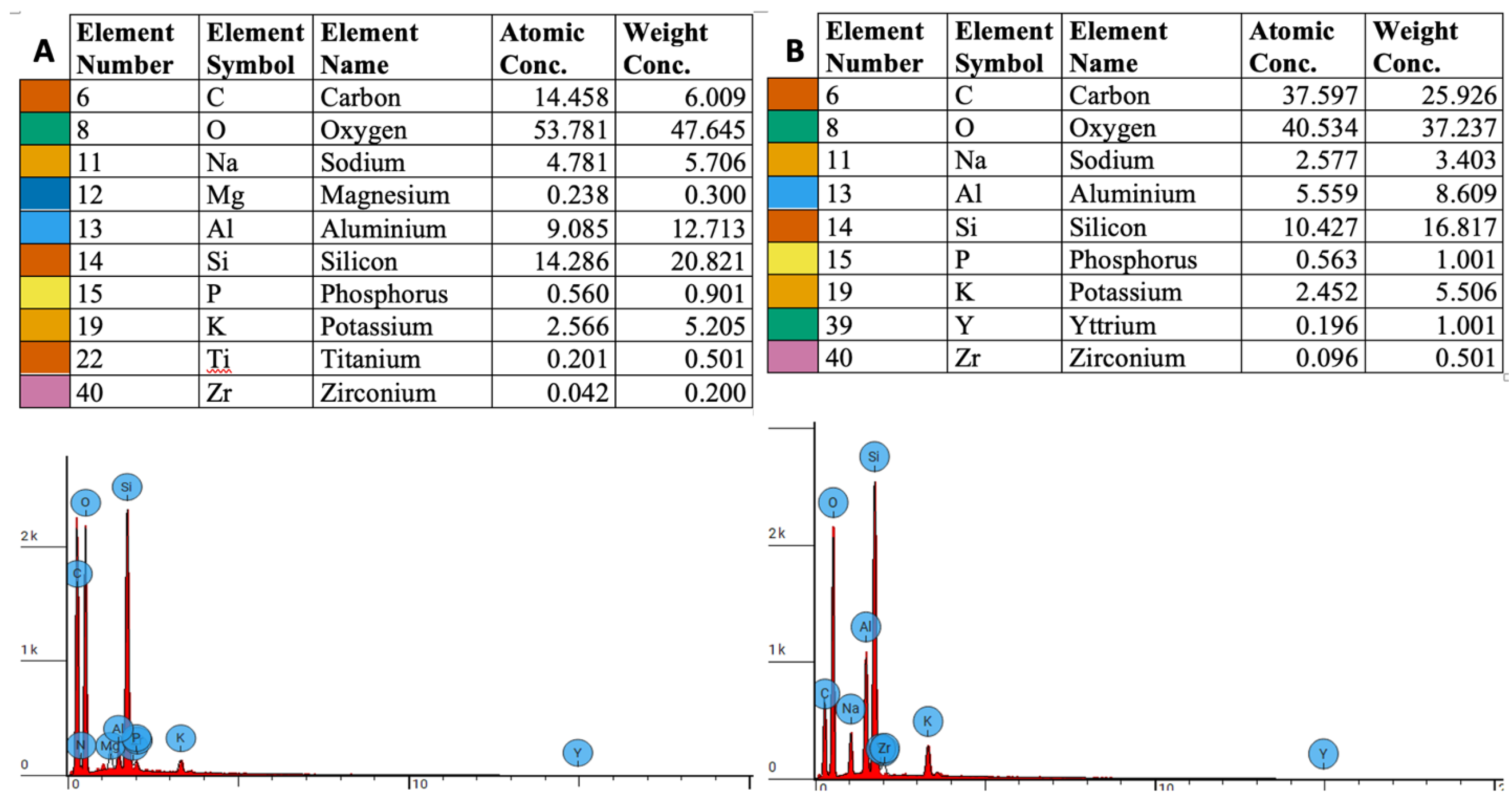
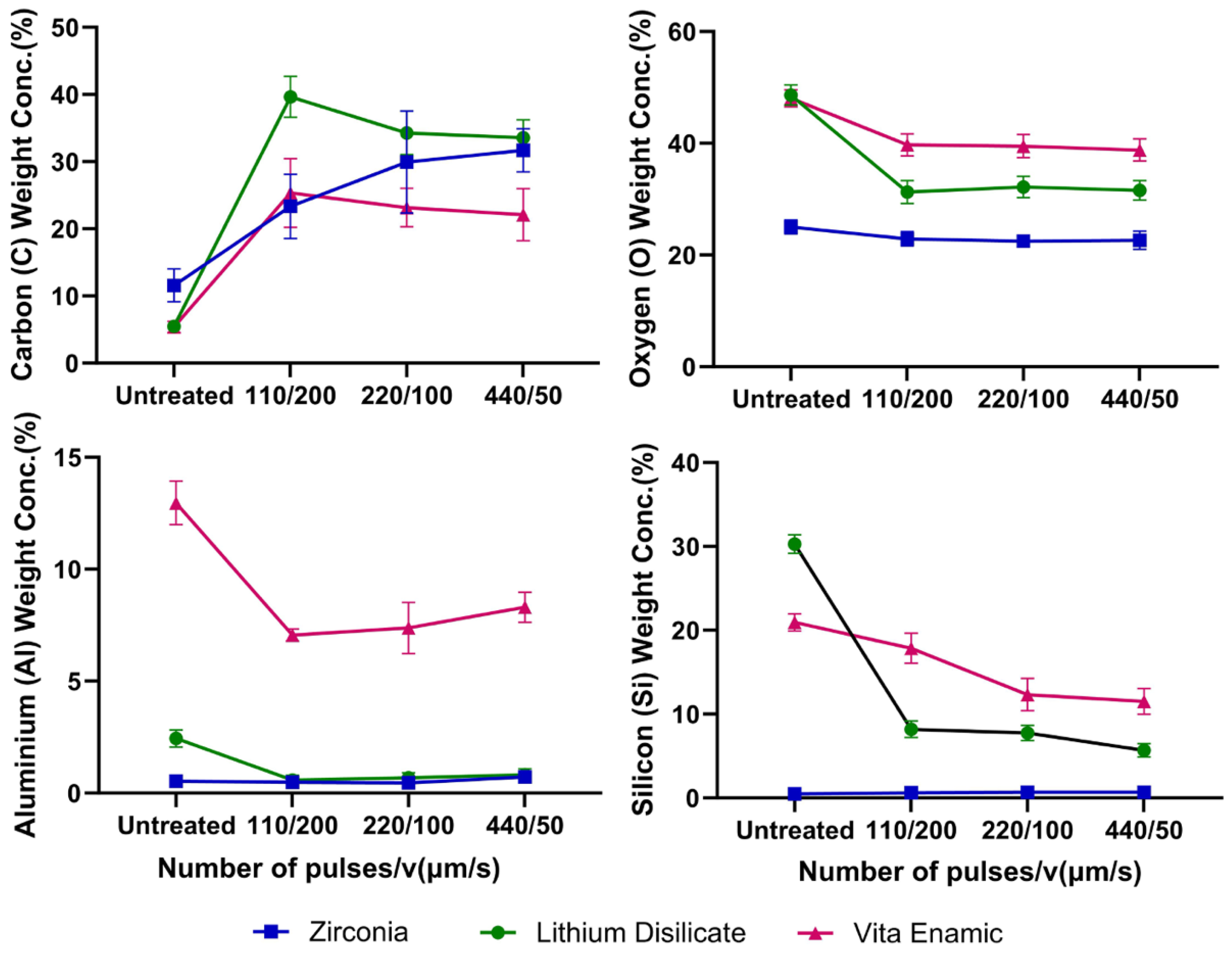
3.1. EDS Measurements

EDS results presenting chemical composition of zirconia, LD glass composite and VITA En prior to fs-laser irradiation and following fs-laser irradiation are presented in Figure 1, Figure 2 and Figure 3, respectively.

3.2. Elemental Weight Concentration Analysis

The wt.% analysis revealed significant changes in the elemental composition of constituent C, O, nitrogen (N), and constituent metals between control groups and fs-laser irradiated samples across all three types of materials studied. These results are collated and presented in Table 2, which details the types of samples and the corresponding number of fs-laser irradiation pulses. All laser treatments showed significantly reduced oxygen and increased carbon content in all three ceramic materials. Aluminum and silicon were both reduced significantly in LD and in VITA En.
There was a dose-response trend evident with a higher number of pulses, resulting in increased ablation and more marked changes in elemental composition (Figure 4). Of the three materials examined, LD showed the highest changes and zirconia the least at the same laser processing conditions.

3.3. Atomic Concentration Analysis

Data for the atomic concentration (at.%) for key elements are summarized in Table 3, which highlights changes from the untreated baseline. Laser processing caused an increase in carbon content by up to 42% and a reduction in oxygen by up to 33%. The greatest increase in carbon content occurred for LD (42%), while the increase in carbon content was 23% for zirconia and 24% for Vita En.

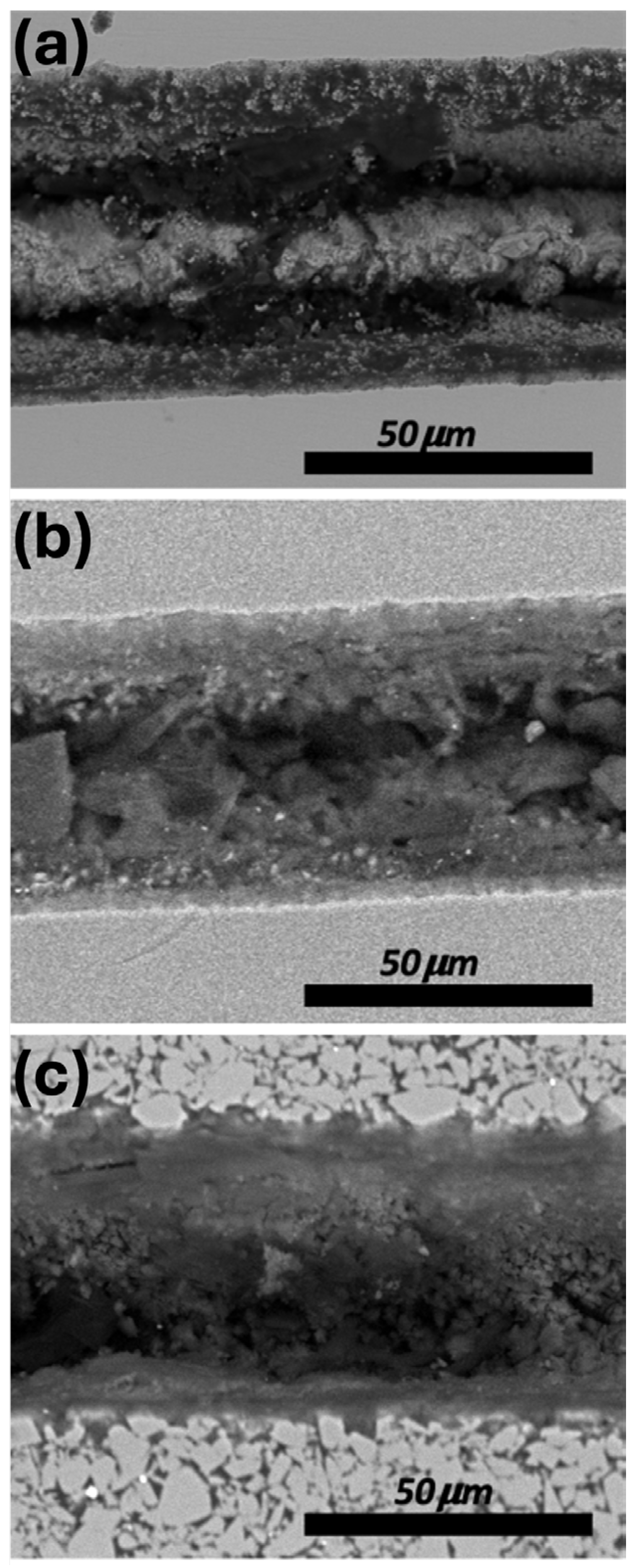
3.4. Morphological Changes of the Processed Surface

Representative SEM images are shown in Figure 5. Laser processing created a well-defined groove with regular, consistent edges. The interior surfaces of the ablated groove regions showed granular structures ranging in size from tens of nanometers to several microns. Both the side sections and planar areas of the tracks displayed granulated features and polycrystalline formations. Notably, there was an absence of surface fissures across all three types of materials investigated.
Zirconia and LD glass ceramic samples were found to display nearly identical surface morphologies across different fs-laser irradiation conditions. In contrast, VITA En samples displayed deeper irradiated regions/tracks and smoother borders, indicative of reduced surface roughness. All materials exhibited evidence of melting and recrystallization, with the effects being less pronounced in zirconia and VITA En compared to LD. The surface topography of zirconia and LD was notably smoother compared to that of VITA En.
An increase in pulse number produced deeper and wider irradiated tracks. This effect was most prominent in VITA En, which showed a pronounced increase in track width when the pulse number was increased from 220 to 440.

4. Discussion

Earlier studies have reported surface damage when using Nd:YAG and CO2 lasers, with no significant chemical alterations in zirconia and LD materials [42,43,44]. A study of materials similar to zirconia and LD glass ceramics by Kim et al. [45] on the ablation characteristics of alumina and aluminum nitride (AlN) under fs-laser ablation found that the ablation characteristics of alumina were predominantly governed by intense thermal stress stemming from elevated temperatures, resulting in minimal melting. The ablation of AlN involved melting, followed by subsequent re-solidification [45]. Notably, despite undergoing laser ablation, the original chemical composition of both alumina and AlN appeared to remain unaltered, suggesting that the alterations induced by the fs laser were primarily of a physical nature rather than chemically induced.
Zirconia is known to undergo phase transformation from tetragonal to monoclinic phase at 1043 °C, accompanied by a 3% volumetric change in crystal structure [46]. This transformation leads to the formation of microcracks within the lattice. The mechanism of stress relaxation via phase transformation might create some microcracks that can propagate through fatigue, thus impacting the total lifetime of the crown and implants and preventing zirconia from failing under the localized compressive load. This mechanism helps explain how failure of veneers can occur by chipping without exposing the zirconia support layer.
Noda et al. [47] suggested that Nd:YAG dental laser welding should not be performed on tetragonal zirconia due to the risk of inducing phase transformation and microcrack formation within the material. A recent study by Harai et al. [48] using X-ray diffraction analysis identified the presence of monoclinic crystals on surfaces treated with ns- and ps-pulse lasers. A study by Delgado-Ruiz et al. [34] used fs-laser micro-structuring on zirconia implant surfaces. Their chemical analysis revealed that the grooved and bored surfaces exhibited larger fractions of zirconium and oxygen compared to untreated control surfaces, whereas the presence of carbon and aluminum was found to be remarkably low. During fs-laser irradiation of zirconia, its tetragonal phase remains unaffected by laser irradiation, in contrast to its monoclinic phase, which can respond to light irradiation [34].
Recent studies have clearly demonstrated the advantageous characteristics of fs-laser pulses for resolving the monoclinic phase fraction in zirconia post-irradiation [25,49]. Conversely, there are several studies that have reported different outcomes regarding the monoclinic phase of zirconia. Currently, the preferred parameters for laser processing have not been decisively established [50,51,52]. Additionally, fs-laser processing has been found to affect the surface roughness but not the crystal structure of zirconia derivatives, such as alumina-toughened zirconia ceramics, without affecting the chemical or phase compositions of these materials [30,50].
Precise control over the chemical changes in materials, which may influence cell-surface interactions, has emerged as a primary focus in the advancement of new ceramic implant materials. Surface patterning techniques have enabled ongoing investigation into cell-biomaterial interactions, revealing that surface topography significantly influences cellular responses in biomaterials [53]. Cell-surface functions such as adhesion, migration, and proliferation are profoundly influenced by physical cues from the surrounding microenvironment [54]. Nevertheless, the specific responses of cells also depend on other factors, including cell type, size, the geometric configuration of the underlying topography, and the surface chemistry of ceramic materials. The influence of the ablation regimen used in the present study on cell behavior is a topic that should be addressed in future studies.
In this study, the EDS results for fs-laser surface-modified zirconia, LD glass ceramics, and VITA En materials demonstrated that fs-laser treatment facilitated the formation of carbon, and the reduction of oxygen. Gruner et al. [55] hypothesized that the formation of carbon monoxide (CO) and CO2 occurs in the irradiated area at temperatures at or above 900 °C. The formation of zirconium carbide in zirconia, along with residual oxygen content, has been observed in several recent studies [56,57,58]. The underlying mechanism is attributed to destabilized carbon and zirconium, resulting in the formation of zirconia and subsequent oxygen reduction from zirconia, which eventually leads to CO formation [59]. As the temperature increases during the carbothermal reduction of zirconia, carbon atoms substitute for oxygen atoms in the structure of zirconia [60].
According to David et al. [59], the destabilization process of zirconia proceeds until the zirconium is consumed. Heterogeneous nucleation and growth of zirconium carbon monoxide (ZrCO) occur within the carbon black agglomerates, possibly originating from the co-condensation of zirconia and CO moieties, followed by solid-state diffusion to zirconium carbon dioxide (ZrCO2). In the subsequent step, ZrCO2 is further reduced with CO until a stable carbide is ultimately formed [58]. The first step of the mechanism is explained by the observation that the ZrO2 phase is depleted during the formation of the ZrC phase [61]. The reduction in oxygen and subsequent increase in carbon in all of the fs-laser irradiated samples in this study, across all tested fs-laser parameters, can be explained by the reduction of an oxycarbide (OxCy) outer layer through both carbon and oxygen being cleaved off and subsequent CO2 formation [58,61,62]. Our work is believed to be the first study reporting on chemical changes in zirconia, LD glass ceramics, and VITA En under 30-fs laser irradiation. Our findings align with earlier findings by Yamamuro et al. [63], who used 256 fs pulses, a 1028 nm wavelength, and a 100 kHz pulse frequency. They reported minor changes for zirconium, yttrium, and oxygen fractions prior to and after fs-laser irradiation.
Notably, a key factor determining the efficiency of laser ablation of dielectric materials is the laser pulse duration [22,64,65]. Ultra-short fs-laser irradiation offers superior precision, minimal thermal effects, and enhanced processing capabilities, making it a highly effective tool for processing dielectrics, particularly when high-quality and intricate work is required [21,22,23,65]. The high peak power of fs-pulses, especially those with a pulse width of less than 50 fs in duration, induces non-linear absorption processes, such as multiphoton absorption, allowing the laser light to interact with materials that are otherwise transparent at the laser wavelength.
In longer pulse processes, such as in ns- or ps-laser processes, plasma formation on the surface of irradiated media can shield the processed material from the laser light, subsequently reducing the processing efficiency. Fs-lasers, due to their ultra-short pulse duration, often avoid significant plasma formation, leading to more efficient material processing [21,22]. Additionally, fs-lasers enable rapid material processing due to their extremely short interaction time with the material, leading to faster and improved material removal rates. The short interaction time helps to suppress thermal effects within the focal volume, preventing melting and phonon propagation into the bulk of the treated solids, even at the highest laser fluences [20]. In this study, the observed chemical changes in zirconia, LD glass ceramic, and VITA En can be attributed to a combination of confined surface reactions, the adsorption of carbon and oxygen from the ambient environment, and the intercalation of carbon from laser-induced plasma, all facilitated by the high-energy conditions created by fs-laser pulses.
However, this study has several limitations, which may affect the accuracy, interpretation, scalability, and generalized outcomes of the findings. Some of these potential limitations arise from the fact that the study probed the uppermost laser-irradiated layer only using SEM and EDS. Other surface analytical methods, such as Fourier-transformed infrared spectroscopy, were not undertaken. Hence, the present study did not capture changes occurring deeper within the sample materials. Surface artifacts or elemental entrapments (e.g., carbon deposition from the ambient environment) may not be representative of the behavior of the bulk material. Moreover, the spatial resolution of SEM and EDS might not be sufficient to detect subtle chemical changes induced by fs-laser irradiation, especially at the nanoscale.
This study used three representative dental ceramic materials of different types; however, it is not possible to directly extrapolate the findings to other dental ceramic materials, as these will differ in their chemical compositions, crystalline structures, phase compositions, and physical properties. It will be necessary to test individual materials to determine their ablation thresholds and response to fs-laser processing. The present study undertook laser processing in normal room air, so the results could be influenced by the atmospheric composition as well as by the ambient temperature and humidity. There are also issues around the intrinsic variability of experimental conditions, such as laser power stability, beam alignment, and/or sample positioning, which all could affect the outcomes. Finally, the scope of the present study excluded exploring the impacts of fs-laser processing on biological responses, such as how cells attach and spread on laser-processed dental ceramics. Such issues can be explored in future studies.

5. Conclusions

This study’s findings show that 30 fs-laser irradiation can produce defined topological and chemical surface changes in zirconia, LD glass ceramic, and VITA En materials, as shown by EDS analysis and SEM. The EDS results revealed that all irradiated samples exhibited an increase in carbon content and a reduction in oxygen content across all tested fs-laser parameters.
The present findings demonstrate that ultra-short fs-laser irradiation induces dose-dependent changes in the chemical composition and surface morphology of zirconia, LD, and VITA En materials. The observed increase in carbon content can be attributed to a combination of surface reactions, adsorption of carbon from the ambient environment, and carbon deposition from laser-induced plasma.
These findings expand earlier observations of fs-laser irradiation phenomena observed for dielectric materials, including metal oxides and carbides. SEM analysis of fs-laser irradiated regions revealed progressive controlled melting and recrystallization, with an absence of the pile-up features typically associated with significant thermal damage that accompany laser processing using ns- or ps-laser treatment processes.
These results support the use of fs-laser treatments on dental ceramic materials, demonstrating high precision and control with minimal substrate damage and no microcrack formation. The fs-laser irradiation could facilitate the modification of dental ceramic surface materials to improve bonding with natural teeth, leading to more durable restorations. Additionally, fs-laser irradiation may be employed to create microscale textures on ceramic dental implants, potentially enhancing osseointegration.

Author Contributions

Conceptualization, R.G., M.R. and V.L.L.; methodology, V.L.L. R.G., B.A. and A.B.C.; validation, B.A. and I.V.L.; formal analysis, V.L.L.; investigation, V.L.L.; resources, A.B.C., M.R. and I.V.L.; data curation, V.L.L., R.G., M.R. and L.J.W.; writing—original draft preparation, V.L.L.; writing—review and editing, V.L.L., R.G., L.J.W. and M.R.; visualization, V.L.L. and B.A.; supervision, R.G., M.R. and L.J.W.; project administration, V.L.L., B.A., M.R. and R.G. All authors have read and agreed to the published version of the manuscript.

Funding

The authors gratefully acknowledge the financial support provided by the Australian Dental Research Foundation Inc. (Grant #0309-2022), which made this research possible.

Institutional Review Board Statement

Not applicable.

Informed Consent Statement

Not applicable.

Data Availability Statement

The original contributions presented in the study are included in the article, further inquiries can be directed to the corresponding authors.

Acknowledgments

The authors acknowledge VITA Zahnfabrik H. Rauter GmbH & Co., Bad Sackingen, Germany for providing research samples.

Conflicts of Interest

The authors declare no conflicts of interest.

References

  1. Silva, L.H.D.; Lima, E.D.; Miranda, R.B.P.; Favero, S.S.; Lohbauer, U.; Cesar, P.F. Dental ceramics: A review of new materials and processing methods. Braz. Oral Res. 2017, 31 (Suppl. S1), e58. [Google Scholar] [CrossRef] [PubMed]
  2. Conejo, J.; Nueesch, R.; Vonderheide, M.; Blatz, M.B. Clinical performance of all-ceramic dental restorations. Curr. Oral Health Rep. 2017, 4, 112–123. [Google Scholar] [CrossRef]
  3. Punj, S.; Singh, J.; Singh, K. Ceramic biomaterials: Properties, state of the art and future prospectives. Ceram. Int. 2021, 47, 28059–28074. [Google Scholar] [CrossRef]
  4. Sjögren, G.; Molin, M.; Van Dijken, J.W. A 10-year prospective evaluation of CAD/CAM-manufactured (Cerec) ceramic inlays cemented with a chemically cured or dual-cured resin composite. Int. J. Prosthodont. 2004, 17, 241–246. [Google Scholar] [PubMed]
  5. Gonzaga, C.C.; Cesar, P.F.; Okada, C.Y.; Fredericci, C.; Beneduce Neto, F.; Yoshimura, H.N. Mechanical properties and porosity of dental glass-ceramics hot-pressed at different temperatures. Mater. Res. 2008, 11, 301–306. [Google Scholar] [CrossRef]
  6. Strasding, M.; Sebestyén-Hüvös, E.; Studer, S.; Lehner, C.; Jung, R.E.; Sailer, I. Long-term outcomes of all-ceramic inlays and onlays after a mean observation time of 11 years. Quintessence Int. 2020, 51, 566–576. [Google Scholar] [CrossRef] [PubMed]
  7. Mavriqi, L.; Valente, F.; Murmura, G.; Sinjari, B.; Macrì, M.; Trubiani, O.; Caputi, S.; Traini, T. Lithium disilicate and zirconia reinforced lithium silicate glass-ceramics for CAD/CAM dental restorations: Biocompatibility, mechanical and microstructural properties after crystallization. J. Dent. 2022, 119, 104054. [Google Scholar] [CrossRef]
  8. Andrade, J.; Stona, D.; Bittencourt, H.; Borges, G.; Burnett, L.; Spohr, A.M. Effect of different computer-aided design/computer-aided manufacturing (CAD/CAM) materials and thicknesses on the fracture resistance of occlusal veneers. Oper. Dent. 2018, 43, 539–548. [Google Scholar] [CrossRef]
  9. Leung, B.T.; Tsoi, J.K.; Matinlinna, J.P.; Pow, E.H. Comparison of mechanical properties of three machinable ceramics with an experimental fluorophlogopite glass ceramic. J. Prosthet. Dent. 2015, 114, 440–446. [Google Scholar] [CrossRef]
  10. Blatz, M.B.; Conejo, J.; Alammar, A.; Ayub, J. Current protocols for resin-bonded dental ceramics. Dent. Clin. N. Am. 2022, 66, 603–625. [Google Scholar] [CrossRef]
  11. Phark, J.H.; Duarte, S. Microstructural considerations for novel lithium disilicate glass ceramics: A review. J. Esthet. Restor. Dent. 2022, 34, 92–103. [Google Scholar] [CrossRef] [PubMed]
  12. Liu, D.; Matinlinna, J.P.; Pow, E.H.N. Insights into porcelain to zirconia bonding. J. Adhes. Sci. Technol. 2012, 26, 1249–1265. [Google Scholar] [CrossRef]
  13. Zaia, W.L.S.; de Figueiredo, J.L.G.; Leite, L.M.; Satake, A.; Medeiros, I.S. Bond strenght of zirconia submitted to different surface treatments. Pesqui. Bras. Odontopediatria Clínica Integr. 2015, 15, 387–398. [Google Scholar] [CrossRef]
  14. Han, J.; Zhang, F.; Van Meerbeek, B.; Vleugels, J.; Braem, A.; Castagne, S. Laser surface texturing of zirconia-based ceramics for dental applications: A review. Mater. Sci. Eng. C 2021, 123, 112034. [Google Scholar] [CrossRef]
  15. Tzanakakis, E.G.C.; Skoulas, E.; Pepelassi, E.; Koidis, P.; Tzoutzas, I.G. The use of lasers in dental materials: A review. Materials 2021, 14, 3370. [Google Scholar] [CrossRef]
  16. Lawson, N.C.; Bansal, R.; Burgess, J.O. Wear, strength, modulus and hardness of CAD/CAM restorative materials. Dent. Mat. 2016, 32, e275–e283. [Google Scholar] [CrossRef]
  17. Ergun Kunt, G.; Duran, I. Effects of laser treatments on surface roughness of zirconium oxide ceramics. BMC Oral Health 2018, 18, 222. [Google Scholar] [CrossRef]
  18. Dede, D.Ö.; Yenisey, M.; Rona, N.; Öngöz Dede, F. Effects of laser treatment on the bond strength of differently sintered zirconia ceramics. Photomed. Laser Surg. 2016, 34, 276–283. [Google Scholar] [CrossRef] [PubMed]
  19. Morales, M.; García-González, S.; Rieux, J.; Jiménez-Piqué, E. Nanosecond pulsed laser surface modification of yttria doped zirconia for solid oxide fuel cell applications: Damage and microstructural changes. J. Eur. Ceram. Soc. 2023, 43, 3396–3403. [Google Scholar] [CrossRef]
  20. Gattass, R.R.; Mazur, E. Femtosecond laser micromachining in transparent materials. Nat. Photonics 2008, 2, 219–225. [Google Scholar] [CrossRef]
  21. Ali, B.; Litvinyuk, I.V.; Rybachuk, M. Femtosecond laser micromachining of diamond: Current research status, applications and challenges. Carbon 2021, 179, 209–226. [Google Scholar] [CrossRef]
  22. Ali, B.; Xu, H.; Sang, R.T.; Litvinyuk, I.V.; Rybachuk, M. Optimised diamond to graphite conversion via a metastable sp1-bonded carbon chain formation under an ultra-short femtosecond (30 fs) laser irradiation. Carbon 2023, 204, 575–586. [Google Scholar] [CrossRef]
  23. Lagunov, V.L.; Rybachuk, M.; Itthagarun, A.; Walsh, L.J.; George, R. Modification of dental enamel, dentin by an ultra-fast femtosecond laser irradiation: A systematic review. Opt. Laser Technol. 2022, 155, 108439. [Google Scholar] [CrossRef]
  24. Yamamuro, Y.; Shimoyama, T.; Yan, J. Microscale surface patterning of zirconia by femtosecond pulsed laser irradiation. Int. J. Precis. Eng. Manuf. Green Technol. 2022, 9, 619–632. [Google Scholar] [CrossRef]
  25. Feng, D.; Shen, H. Hole quality control in underwater drilling of yttria-stabilized zirconia using a picosecond laser. Opt. Laser Technol. 2019, 113, 141–149. [Google Scholar] [CrossRef]
  26. Li, J.; Ji, L.; Hu, Y.; Bao, Y. Precise micromachining of yttria-tetragonal zirconia polycrystal ceramic using 532 nm nanosecond laser. Ceram. Int. 2016, 42, 4377–4385. [Google Scholar] [CrossRef]
  27. Panova, N.K.; Nikolova, K.T.; Dikova, T.D. Application of lasers and laser processing technologies in modern dentistry: A review. J. Chem. Technol. Metall. 2023, 58, 1116–1127. [Google Scholar] [CrossRef]
  28. Schliephake, H.; Hefti, T.; Schlottig, F.; Gédet, P.; Staedt, H. Mechanical anchorage and peri-implant bone formation of surface-modified zirconia in minipigs. J. Clin. Periodontol. 2010, 37, 818–828. [Google Scholar] [CrossRef]
  29. Saulacic, N.; Erdösi, R.; Bosshardt, D.D.; Gruber, R.; Buser, D. Acid and alkaline etching of sandblasted zirconia implants: A histomorphometric study in miniature pigs. Clin. Implant. Dent. Relat. Res. 2014, 16, 313–322. [Google Scholar] [CrossRef]
  30. Aivazi, M.; hossein Fathi, M.; Nejatidanesh, F.; Mortazavi, V.; HashemiBeni, B.; Matinlinna, J.P.; Savabi, O. The evaluation of prepared microgroove pattern by femtosecond laser on alumina-zirconia nano-composite for endosseous dental implant application. Lasers Med. Sci. 2016, 31, 1837–1843. [Google Scholar] [CrossRef]
  31. Hu, P.; Yao, L.; Zhang, M.; Nie, Z.; Ji, E.; Lue, Q.; He, Z. Femtosecond laser micro-milling dental glass ceramics: An experimental analysis and COMSOL finite element simulation. Ceram. Int. 2020, 46, 22146–22153. [Google Scholar] [CrossRef]
  32. Daskalova, A.; Angelova, L.; Carvalho, A.; Trifonov, A.; Nathala, C.; Monteiro, F.; Buchvarov, I. Effect of surface modification by femtosecond laser on zirconia based ceramics for screening of cell-surface interaction. Appl. Surf. Sci. 2020, 513, 145914. [Google Scholar] [CrossRef]
  33. Han, J.; Malek, O.; Vleugels, J.; Braem, A.; Castagne, S. Ultrashort pulsed laser ablation of zirconia-alumina composites for implant applications. J. Mater. Process. Technol. 2022, 299, 117335. [Google Scholar] [CrossRef]
  34. Delgado-Ruíz, R.; Calvo-Guirado, J.; Moreno, P.; Guardia, J.; Gomez-Moreno, G.; Mate-Sánchez, J.; Ramirez-Fernández, P.; Chiva, F. Femtosecond laser microstructuring of zirconia dental implants. J. Biomed. Mater. Res. B Appl. Biomater. 2011, 96, 91–100. [Google Scholar] [CrossRef] [PubMed]
  35. Narazaki, A.; Takada, H.; Yoshitomi, D.; Torizuka, K.; Kobayashi, Y. Ultrafast laser processing of ceramics: Comprehensive survey of laser parameters. J. Laser Appl. 2021, 33, 012009. [Google Scholar] [CrossRef]
  36. VITA YZ® SOLUTIONS Technical and Scientific Documentation. 2022. VITA Zahnfabrik H. Rauter GmbH & Co., Bad Säckingen, Germany. Available online: https://www.vita-zahnfabrik.com/VITA_YZ_en,27568,175081.html (accessed on 21 August 2024).
  37. VITA AMBRIA® PRESS SOLUTIONS Technical and Scientific Documentation. 2020. VITA Zahnfabrik H. Rauter GmbH & Co., Bad Säckingen, Germany. Available online: https://www.vita-zahnfabrik.com/en/VITA-AMBRIA-Lithium-disilicate-press-ceramic-102005.html (accessed on 21 August 2024).
  38. VITA ENAMIC® HYBRID CERAMIC. 2024. VITA Zahnfabrik H. Rauter GmbH & Co., Bad Säckingen, Germany. Available online: https://www.vita-zahnfabrik.com/index.php (accessed on 21 August 2024).
  39. ISO 6872:2024; Dentistry—Ceramic Materials. ISO: Geneva, Switzerland, 2024. Available online: https://www.iso.org/standard/81718.html (accessed on 21 August 2024).
  40. ISO 12836:2015; Dentistry—Digitizing Devices for CAD/CAM Systems for Indirect Dental Restorations—Test Methods for Assessing Accuracy. ISO: Geneva, Switzerland, 2015. Available online: https://www.iso.org/standard/68414.html (accessed on 21 August 2024).
  41. Karson, M. Handbook of Methods of Applied Statistics. Volume I: Techniques of Computation Descriptive Methods, and Statistical Inference; John Wiley: New York, NY, USA, 1967. [Google Scholar]
  42. El Gamal, A.; Fornaini, C.; Rocca, J.P.; Muhammad, O.H.; Medioni, E.; Cucinotta, A.; Brulat-Bouchard, N. The effect of CO2 and Nd: YAP lasers on CAD/CAM Ceramics: SEM, EDS and thermal studies. Laser Ther. 2016, 25, 27–34. [Google Scholar] [CrossRef] [PubMed]
  43. Rocca, J.-P.; Fornaini, C.; Brulat-Bouchard, N.; Seif, S.B.; Darque-Ceretti, E. CO2 and Nd: YAP laser interaction with lithium disilicate and Zirconia dental ceramics: A preliminary study. Opt. Laser Technol. 2014, 57, 216–223. [Google Scholar] [CrossRef]
  44. Henriques, B.; Hammes, N.; Souza, J.C.; Özcan, M.; Mesquita-Guimarães, J.; Silva, F.S.; Fredel, M.C.; Volpato, C.M.; Carvalho, Ó. Influence of ns-Nd: YAG laser surface treatment on the tensile bond strength of zirconia to resin-matrix cements. Ceram. Int. 2020, 46, 27822–27831. [Google Scholar] [CrossRef]
  45. Kim, S.H.; Balasubramani, T.; Sohn, I.-B.; Noh, Y.-C.; Lee, J.; Lee, J.B.; Jeong, S. Precision microfabrication of AlN and Al2O3 ceramics by femtosecond laser ablation. In Proceedings Volume 6879, Photon Processing in Microelectronics and Photonics VII, 68791O; SPIE: Bellingham, WA, USA, 2008; pp. 270–276. [Google Scholar] [CrossRef]
  46. Allahkarami, M.; Hanan, J.C. Mapping the tetragonal to monoclinic phase transformation in zirconia core dental crowns. Dent. Mater. 2011, 27, 1279–1284. [Google Scholar] [CrossRef]
  47. Noda, M.; Okuda, Y.; Tsuruki, J.; Minesaki, Y.; Takenouchi, Y.; Ban, S. Surface damages of zirconia by Nd: YAG dental laser irradiation. Dent. Mater. J. 2010, 29, 536–541. [Google Scholar] [CrossRef] [PubMed]
  48. Harai, T.; Mizutani, M.; Shishido, S.; Nakamura, K.; Ohmori, H.; Konno, T.J.; Kuriyagawa, T. Low-temperature degradation of yttria-stabilized zirconia treated with pulsed laser and annealing techniques. Precis. Eng. 2023, 80, 45–56. [Google Scholar] [CrossRef]
  49. Ackerl, N.; Wegener, K. Ablation characteristics of alumina and zirconia ceramics on ultra-short pulsed laser machining. J. Laser Micro Nanoeng. 2019, 14, 168–172. [Google Scholar] [CrossRef]
  50. Carvalho, A.; Grenho, L.; Fernandes, M.H.; Daskalova, A.; Trifonov, A.; Buchvarov, I.; Monteiro, F.J. Femtosecond laser microstructuring of alumina toughened zirconia for surface functionalization of dental implants. Ceram. Int. 2020, 46, 1383–1389. [Google Scholar] [CrossRef]
  51. Zhou, H.B.; Li, C.; Zhou, Z.K.; Cao, R.Y.; Chen, Y.; Zhang, S.S.; Wang, G.; Xiao, S.; Li, Z.; Xiao, P. Femtosecond laser-induced periodic surface microstructure on dental zirconia ceramic. Mater. Lett. 2018, 229, 74–77. [Google Scholar] [CrossRef]
  52. Li, Q.; Li, C.; Wang, Y. Effect of femtosecond laser ablate ultra-fine microgrooves on surface properties of dental zirconia materials. J. Mech. Behav. Biomed. Mater. 2022, 134, 105361. [Google Scholar] [CrossRef]
  53. Munro, T.; Miller, C.M.; Antunes, E.; Sharma, D. Interactions of osteoprogenitor cells with a novel zirconia implant surface. J. Funct. Biomater. 2020, 11, 50. [Google Scholar] [CrossRef]
  54. Pessková, V.; Kubies, D.; Hulejová, H.; Himmlová, L. The influence of implant surface properties on cell adhesion and proliferation. J. Mater. Sci. Mater. Med. 2007, 18, 465–473. [Google Scholar] [CrossRef]
  55. Gruner, W.; Stolle, S.; Wetzig, K. Formation of COx species during the carbothermal reduction of oxides of Zr, Si, Ti, Cr, W, and Mo. Int. J. Refract. Met. Hard Mater. 2000, 18, 137–145. [Google Scholar] [CrossRef]
  56. Gasparrini, C.; Chater, R.J.; Horlait, D.; Vandeperre, L.; Lee, W.E. Zirconium carbide oxidation: Kinetics and oxygen diffusion through the intermediate layer. J. Am. Ceram. Soc. 2018, 101, 2638–2652. [Google Scholar] [CrossRef]
  57. Liu, G.; Cheng, L.; Li, K.; Chen, Z.; Xiong, X.; Luan, X. Damage behavior of atomic oxygen on zirconium carbide coating modified carbon/carbon composite. Ceram. Int. 2020, 46, 3324–3331. [Google Scholar] [CrossRef]
  58. Kucheryavaya, A.; Lenčéš, Z.; Šajgalík, P.; Harmuth, H. Zirconium oxycarbides and oxycarbonitrides: A review. Int. J. Appl. Ceram. Technol. 2023, 20, 541–562. [Google Scholar] [CrossRef]
  59. David, J.; Trolliard, G.; Gendre, M.; Maitre, A. TEM study of the reaction mechanisms involved in the carbothermal reduction of zirconia. J. Eur. Ceram. Soc. 2013, 33, 165–179. [Google Scholar] [CrossRef]
  60. Sarkar, S.K. Solubility of Oxygen in Zirconium Carbide. Ph.D. Thesis, University of Washington, Seattle, WA, USA, 1969. Available online: https://www.proquest.com/dissertations-theses/solubility-oxygen-zirconium-carbide/docview/302441320/se-2?accountid=14723 (accessed on 21 August 2024).
  61. Sievers, M.R.; Armentrout, P.B. Oxidation of CO and reduction of CO2 by gas phase Zr+, ZrO+, and ZrO2+. Int. J. Mass Spectrom. 1999, 185–187, 117–129. [Google Scholar] [CrossRef]
  62. Liu, H.-L.; Man, Z.-Y.; Liu, J.-X.; Wang, X.-G.; Zhang, G.-J. Solid solution and densification behavior of zirconium oxycarbide (ZrCxOy) ceramics via doping ZrO2 and Zr in ZrC. J. Alloys Compd. 2017, 729, 492–497. [Google Scholar] [CrossRef]
  63. Yamamuro, Y.; Shimoyama, T.; Yan, J. Generation of nanopore structures in yttria-stabilized zirconia by femtosecond pulsed laser irradiation. J. Mater. Res. Technol. 2023, 23, 1155–1176. [Google Scholar] [CrossRef]
  64. Kaligar, A.B.; Kumar, H.A.; Ali, A.; Abuzaid, W.; Egilmez, M.; Alkhader, M.; Abed, F.; Alnaser, A.S. Femtosecond laser-based additive manufacturing: Current status and perspectives. Quantum Beam Sci. 2022, 6, 5. [Google Scholar] [CrossRef]
  65. Ali, B.; Xu, H.; Chetty, D.; Sang, R.T.; Litvinyuk, I.V.; Rybachuk, M. Laser-Induced Graphitization of Diamond Under 30 fs Laser Pulse Irradiation. J. Phys. Chem. Lett. 2022, 13, 2679–2685. [Google Scholar] [CrossRef] [PubMed]
Figure 1. Chemical composition of zirconia for selected key elements. (A) Prior to fs-laser irradiation, and (B) following laser irradiation using a 20 J/cm2 fluence and 440 pulses.
Applsci 14 07641 g001
Figure 2. Chemical composition of lithium disilicate glass ceramic for selected key elements. (A) Prior to fs-laser irradiation, and (B) following laser irradiation using a 20 J/cm2 fluence and 440 pulses.
Applsci 14 07641 g002
Figure 3. Chemical composition of VITA ENAMIC® hybrid ceramic for selected key elements. (A) Prior to fs-laser irradiation, and (B) following laser irradiation using a 20 J/cm2 fluence and 440 pulses.
Applsci 14 07641 g003
Figure 4. The effect of fs-laser pulse number/scanning speed on wt.% constituent fractions of carbon, oxygen, aluminum and silicon in zirconia, lithium disilicate glass ceramic, and VITA ENAMIC® hybrid ceramic dental materials following ultra-short femtosecond (30 fs) laser irradiation.
Applsci 14 07641 g004
Figure 5. SEM micrographs of (a) zirconia, (b) lithium disilicate glass ceramic, and (c) VITA ENAMIC® hybrid ceramic composite material processed using fs-laser irradiation using a 20 J/cm2 fluence at 440 pulses.
Applsci 14 07641 g005
Table 1. Femtosecond laser irradiation parameters employed in the study to irradiate zirconia, LD glass ceramics, and VITA En hybrid ceramic dental material samples.
Power
mW
Fluence
J/cm2
Number of Pulses
(n)
Scanning Speed
μm/s
762011050
7620220100
7620440200
Table 2. EDS data showing elemental composition in weight percent for three distinct sample groups, namely zirconia, lithium disilicate composite material, and VITA ENAMIC® hybrid material irradiated at 20 J/cm2 fluence and varying pulse number/scanning speeds.
Pulse Number/
Scanning Speed
CarbonOxygenAluminumSilicon
Zirconia
100/20023.3 (4.8) b22.9 (1.3) a0.5 (0.1) a0.6 (0.2) a
220/10029.9 (7.6) c22.5 (0.9) a0.5 (0.2) a0.7 (0.7) a
440/5031.7 (3.2) c22.7 (1.6) a0.7 (0.2) a0.7 (0.7) a
Untreated11.6 (2.4) a25.0 (1.2) b0.5 (0.1) a0.5 (0.5) a
Lithium disilicate
100/20039.7 (3.0) c31.3 (2.0) a0.6 (0.2) a8.2 (1.0) b
220/10034.3 (3.2) b32.2 (1.9) a0.7 (0.2) a7.7 (0.9) a
440/5033.5 (2.7) b31.6 (1.7) a0.8 (0.3) a5.7 (0.8) a
Untreated5.5 (0.6) a48.7 (1.8) b2.4 (0.4) b30.3 (1.1) c
VITA ENAMIC®
100/20025.3 (5.1) b39.7 (2.0) a7.1 (0.2) a17.9 (0.1) c
220/10023.2 (2.8) b39.5 (2.1) a7.4 (0.1) a12.3 (0.1) b
440/5022.1 (3.9) b38.8 (2.0) a8.3 (0.1) a11.5 (0.1) a
Untreated5.4 (0.8) a48.1 (1.4) b13.0 (0.1) b20.9 (0.2) d
Each data entry shows the mean and standard deviation, rounded to one decimal point, and the suffix letter for analysis within the particular element of the specified material. Groups with the same suffix letter are not statistically different. Data sets were derived from 5 samples per group.
Table 3. EDS data showing composition in atomic concentration for three distinct sample groups, namely zirconia, lithium disilicate composite material, and VITA ENAMIC® hybrid material irradiated at 20 J/cm2 fluence and varying pulse numbers/scanning speeds.
Pulse Number/
Scanning Speed
CarbonOxygenAluminumSilicon
Zirconia
100/20041.1 (2.2) **31.9 (2.1) ***0.2 (0.1) **NA
220/10046.9 (2.7) ***31.7 (0.8) ***0.2 (0.1) **NA
440/5054.9 (3.5) ****31.7 (2.6) ***0.1 (0.1) **NA
Untreated31.3 (4.2)47.6 (4.9)0.6 (0.2)NA
Lithium disilicate
100/20040.6 (1.8) ****32.3 (2.2) ****0.6 (0.3) **8.3 (1.5) ****
220/10042.6 (2.0) ****33.2 (2.1) ****0.6 (0.3) **7.0 (1.4) ****
440/5050.1 (4.0) ****35.2 (1.9) ****0.5 (0.3) **5.9 (1.4) ****
Untreated8.0 (2.0)66.2 (4.2)2.1 (0.7)21.0 (2.1)
VITA ENAMIC®
100/20037.0 (3.3) ****42.7 (2.6) **6.3 (1.7) *13.5 (1.7) NS
220/10038.8 (2.0) ****41.5 (3.3) ****5.5 (1.4) **12.1 (1.8) NS
440/5037.6 (4.3) ****40.9 (2.2) **5.6 (1.1) **10.4 (1.4) *
Untreated14.5 (2.4)52.5 (5.1)9.1 (1.8)14.3 (2.6)
Each data entry shows the mean and standard deviation, rounded to one decimal point. Asterisks indicate differences from the relevant untreated control sample, with * p < 0.01, ** p < 0.001, *** p < 0.0001, and **** p < 0.0001. NS = not significant. NA = not applicable. Data sets were derived from 5 samples per group.
Disclaimer/Publisher’s Note: The statements, opinions and data contained in all publications are solely those of the individual author(s) and contributor(s) and not of MDPI and/or the editor(s). MDPI and/or the editor(s) disclaim responsibility for any injury to people or property resulting from any ideas, methods, instructions or products referred to in the content.

