Article

Bioavailability of Liposomal Vitamin C in Powder Form: A Randomized, Double-Blind, Cross-Over Trial

1
Bart Ltd., 05-250 Slupno, Poland
2
AronPharma Ltd., 80-172 Gdansk, Poland
3
Department of Clinical Nutrition and Dietetics, Medical University of Gdansk, 80-211 Gdansk, Poland
4
Centre of Polymer and Carbon Materials, Polish Academy of Sciences (CPCM PAS), 41-819 Zabrze, Poland
5
Department of Food Engineering and Process Management, Institute of Food Sciences, Warsaw University of Life Sciences, 02-776 Warsaw, Poland
6
Department of Experimental and Clinical Pharmacology, Centre for Preclinical Research and Technology CEPT, Medical University of Warsaw, 02-091 Warsaw, Poland
*
Author to whom correspondence should be addressed.
Appl. Sci. 2024, 14(17), 7718; https://doi.org/10.3390/app14177718 (registering DOI)
Submission received: 30 July 2024 / Revised: 28 August 2024 / Accepted: 29 August 2024 / Published: 1 September 2024

Abstract

:
The purpose of this study was to evaluate the properties and pharmacokinetics of liposomal vitamin C in powder form obtained by a method devoid of organic solvents. The powder and liposome morphology were analyzed using scanning electron microscopy (SEM) and cryogenic transmission electron microscopy (cryo-TEM), respectively. Additionally, the carrier particle size, size distribution (STEP-Technology®; L.U.M. GmbH, Berlin, Germany), and zeta potential value were determined. The pharmacokinetic parameters of liposomal and non-liposomal vitamin C (AUC, Cmax, C10h, and C24h) were compared in a randomized, single-dose, double-blind, cross-over trial (ClinicalTrials.gov ID: NCT05843617) involving healthy adult volunteers (n = 10, 1000 mg dose). The process of spray drying used to transform liquid suspensions of the liposomes into powder form did not adversely affect the quality of the carrier particles obtained. Compared to non-encapsulated vitamin C, oral administration of the liposomal formulation resulted in significantly better absorption of ascorbic acid into the bloodstream, which equated to a higher bioavailability of the liposomal product (30% increase in AUC, p < 0.05). The duration of elevated vitamin C blood levels was also longer (C24h increase of 30%, p < 0.05). Although the results obtained are promising and suggest higher bioavailability for the liposomal form of vitamin C, the limited sample size necessitates further research with a larger cohort to confirm these findings.

1. Introduction

The main physiological and biochemical functions of vitamin C (L-ascorbic acid, 2-oxo-L-theohexono-4-lactone-2,3-enediol, C6H8O6) are due to the fact that, as a strong reductant, it is an electron donor, thus effectively preventing the oxidation of other biomolecules [1,2], including lipids, DNA, and proteins [3]. In this way, it also ensures the proper functioning of the immune system and assists the body in fighting inflammation [4]. Ascorbic acid also acts as a cofactor for enzymes involved in the synthesis of hormones, carnitine, or collagen, among others [3,4]. It also modulates the absorption, transport, and storage of iron [4].
Humans and other higher primates, guinea pigs, bats, and some species of birds and fish lost the ability to synthesize ascorbic acid de novo from glucose due to the lack of the enzyme L-gulonolactone oxidase (GLO, EC 1.1.3.8) in their bodies [3]. Hence, they need to derive vitamin C from their food [1,2,3,4].
The daily requirement for vitamin C varies according to age, gender, physiological state (pregnancy and lactation in women), and lifestyle [2]. Nevertheless, the recommended daily intake of the vitamin may also differ depending on the expert institution formulating the recommendations. Those given by the Health and Medicine Division (formerly known as the Institute of Medicine, IoM), EFSA, and FAO/WHO range from 15 to 50 mg/day for infants and children up to three years of age, and from 25 to 90–100 mg/day for older children and adolescents. For adult men, they are 45–110 mg/day, and for adult women, 45–95 mg/day. The recommended intake of vitamin C increases by a further 10 mg/day and 25–60 mg/day for pregnant and lactating adult women, respectively [5,6,7]. Increasing the intake of the vitamin by a further 35–40 mg/day is also sometimes recommended for habitual smokers [5,8].
Although the main dietary sources of vitamin C are fruits and vegetables [4], their consumption does not always guarantee an adequate supply of this active substance. The ascorbic acid content of plant materials fluctuates [1] depending largely on their type. For instance, in the case of fresh fruits, it can vary from 3.2 mg/100 g of grapes to 181.0 mg/100 g of blackcurrants, and for fresh vegetables, from 9.2 mg/100 g of lettuce (green leaf) to 133.0 mg/100 g of parsley [9].
The vitamin C content of fruits and vegetables is also impacted by the duration of transportation to the point of sale [1], the storage period and conditions, and the thermal processing parameters of the raw and/or semi-finished product [1,4,10].
The supply of vitamin C in food rations is also negatively affected by modern lifestyles, including the increased consumption of energy-dense, low-nutrient foods [11] and the more frequent consumption of meals—usually low in vegetables—when eating out or consuming take-away meals [12,13]. For seniors, especially women (≥65 years old), the average intake of vitamin C also decreases with age [14].
Therefore, vitamin C is increasingly being added to foods, beverages, and premixes [4,15,16]. The results of published studies indicate that it is also one of the most commonly supplemented micronutrients [17,18,19,20].
Undoubtedly, the growing demand for supplements containing vitamin C translates into their market supply. The development of an effective ascorbic acid-containing formula requires that the vulnerability of this active ingredient to degradation during the manufacturing and storage process be taken into account [2,10]. Ascorbic acid is highly reactive and unstable. When exposed to light, elevated temperature, air, oxygen, and alkaline environments [2,21,22], it undergoes reversible oxidation to dehydroascorbic acid (DHA), which then irreversibly hydrolyses to 2,3-diketo-L-gulonic acid [1,4,21]. Vitamin C decomposition reactions accelerate with increasing water activity (aw) [23] and in the presence of transition metal ions, particularly iron (Fe3+) [24,25] and copper (Cu2+) [25].
The breakdown of vitamin C significantly reduces its effectiveness in the human body. Accordingly, in recent decades, researchers’ interest has focused on the possibility of protecting ascorbic acid from the destabilizing effects of environmental conditions by encapsulating it in a variety of micro- and nanocarriers [2,21,26].
The results of available scientific studies indicate numerous attempts to encapsulate vitamin C in liposomes [27,28,29,30,31,32,33,34]. These lipid carriers are composed of biocompatible, biodegradable, and non-immunogenic components [35,36,37]. They also exhibit, among other things, the ability to protect sensitive active substances from degradation and increase their bioavailability [35,38,39].
Unfortunately, the widespread use of most traditional liposomal technologies in the industrial production of dietary supplements, nutraceuticals, and drugs is limited by the instability of aqueous liposomal dispersions [36,40,41,42], their high polydispersibility, and the significant amounts of organic solvents (ethanol, methanol, chloroform, ether, etc.) used to dissolve lipids that remain in the final product [42,43]. These solvents can contribute to the degradation of the encapsulated active ingredients and destabilization of the carriers themselves. They also pose a real threat to human health and the environment [42].
With this in mind, liposomal vitamin C in powder form was obtained in a procedure devoid of organic solvents. The aim of the study presented here was the evaluation of the properties of the resulting product taking into account the parameters that could significantly affect its effectiveness in vivo, as well as determination of its bioavailability compared to its non-liposomal counterpart in an in vitro study (human colon adenocarcinoma cell line Caco-2) and a randomized, double-blind, single-dose, cross-over clinical trial involving healthy volunteers.

2. Materials and Methods

2.1. Study Materials

L-ascorbic acid (Vitamin C 99%) was purchased from NHU Europe GmbH (Lünenburg, Germany), maltodextrin was purchased from AGRANA STÄRKE GmbH (Gmünd, Austria), sunflower phosphatidylcholine was purchased from Lipoid GmbH (Ludwigshafen, Germany), and pharmaceutical glycerol 99.5% was purchased from KLM Energia Sp. z o. o. (Ostróda, Poland). The water used in the process of the liposomal product preparation was purified by reversed osmosis.

2.2. Preparation of Liposomal Vitamin C in Powder Form

An aqueous solution of L-ascorbic acid, the biologically active form of vitamin C [16], was mixed with a previously prepared blend of two solvents, glycerol and sunflower phosphatidylcholine, combined in a 1:1 (w/w) ratio. The presence of the latter compound in the phospholipid mixture allowed liposomes to form spontaneously.
The mixing process, carried out under reduced pressure (below -0.5 bar), produced a liposomal suspension containing a homogeneous population of lipid carriers with a small particle size (<300 nm). A carbohydrate (maltodextrin) was added to this suspension with the aim of efficiently converting the liquid intermediate into a powder by means of a disc spray dryer, while maintaining a drying temperature not exceeding 160 °C.
The liposomal formulation obtained in this way and used in the present study contained at least 25% L-ascorbic acid (the assay was performed by HPLC with an LC-DAD; Eurofins Steins Laboratorium, Vejen, Denmark).

2.3. Verification of the Process of Obtaining Liposomal Powder Formulation

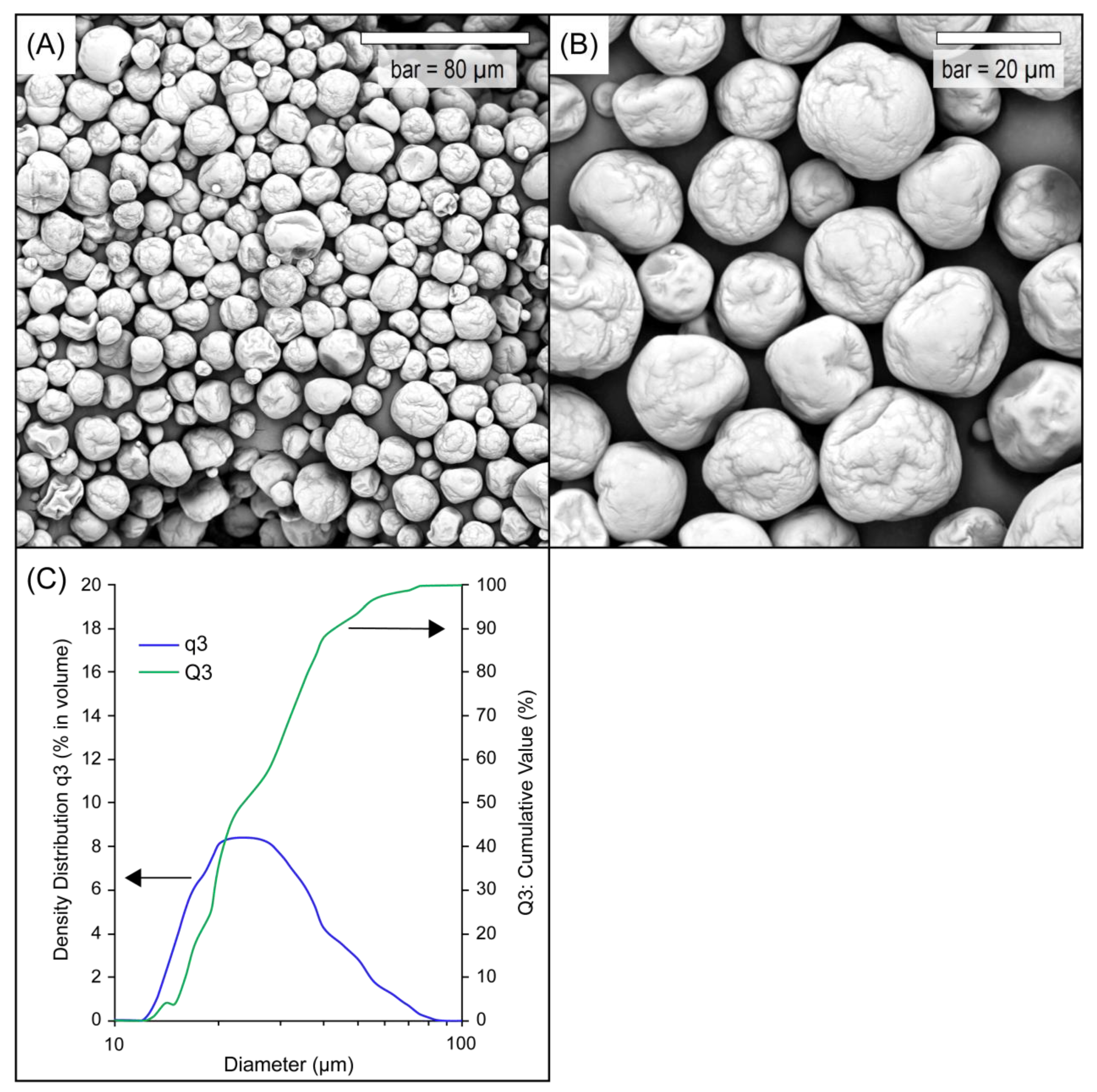
2.3.1. Scanning Electron Microscopy (SEM)

The surface morphology of liposomal vitamin C in powder form was evaluated by scanning electron microscopy (SEM) using a Phenom XL (Thermo Fisher Scientific Phenom-World BV, Eindhoven, The Netherlands), operated at 10 kV. The powder samples were directly placed on a carbon conductive tape on aluminum SEM stubs and coated with a thin gold layer using a gold-sputtering coating (Cressington, 108 auto, Watford, UK).

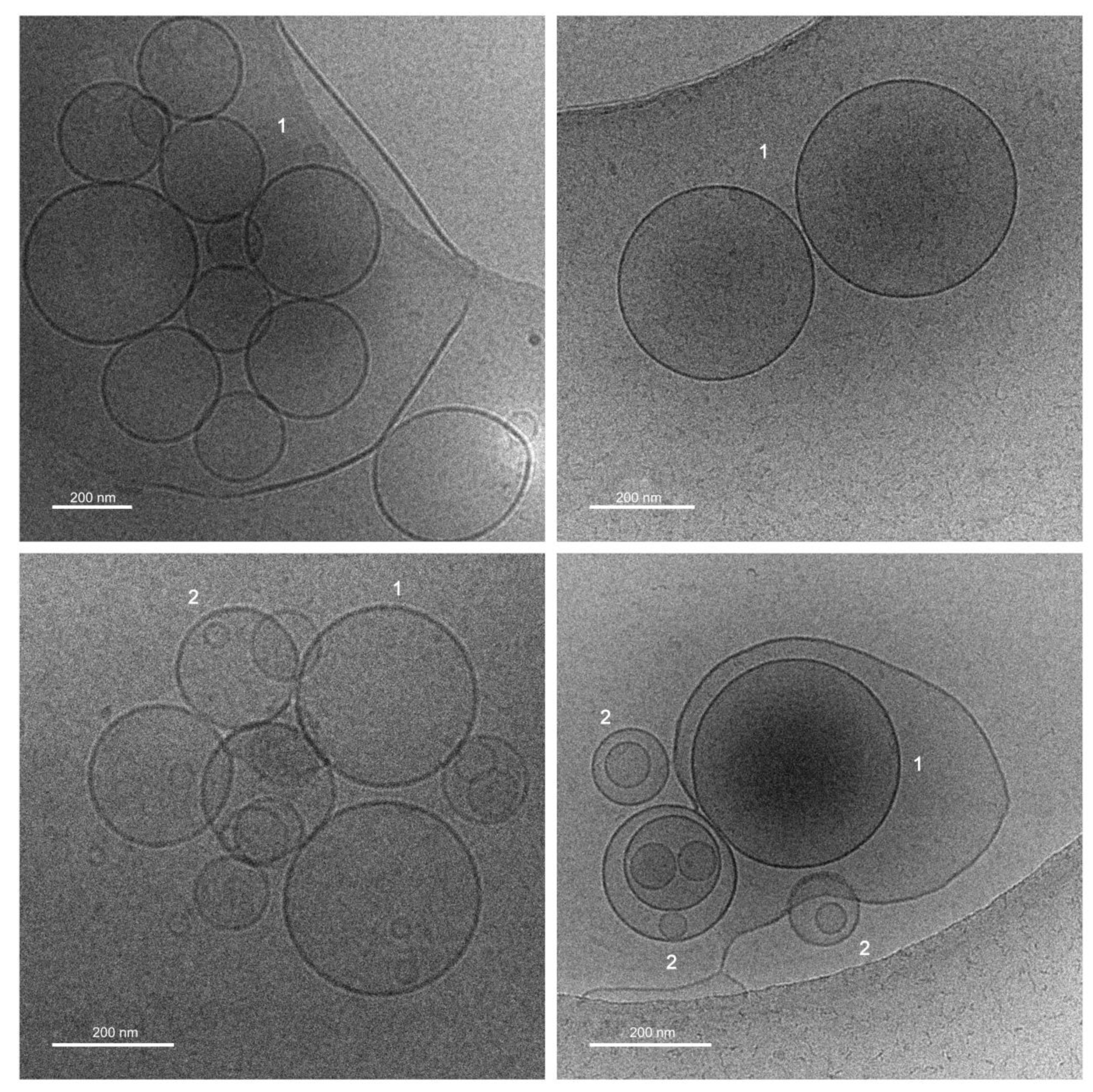
2.3.2. Cryogenic Transmission Electron Microscopy (Cryo-TEM)

Cryogenic transmission electron microscopy (cryo-TEM) images were obtained using a Tecnai F20 X TWIN microscope (FEI Company, Hillsboro, OR, USA) equipped with a field emission gun, operating at an acceleration voltage of 200 kV. Images were recorded on a Gatan Rio 16 CMOS 4k camera (Gatan Inc., Pleasanton, CA, USA) and processed with Gatan Microscopy Suite (GMS) software version 3.31.2360.0 (Gatan Inc., Pleasanton, CA, USA). The specimen was prepared by vitrification of the aqueous solutions on grids with holey carbon film (Quantifoil R 2/2; Quantifoil Micro Tools GmbH, Großlöbichau, Germany). Prior to use, the grids were activated for 15 s in oxygen plasma using a Femto plasma cleaner (Diener Electronic, Ebhausen, Germany). Cryo-samples were prepared by applying a droplet (3 μL) of the suspension to the grid, blotting with filter paper, and immediately freezing in liquid ethane using a fully automated blotting device, Vitrobot Mark IV (Thermo Fisher Scientific, Waltham, MA, USA). After preparation, the vitrified specimens were kept under liquid nitrogen until they were inserted into a cryo-TEM-holder Gatan 626 (Gatan Inc., Pleasanton, CA, USA) and analyzed in the TEM at −178 °C.

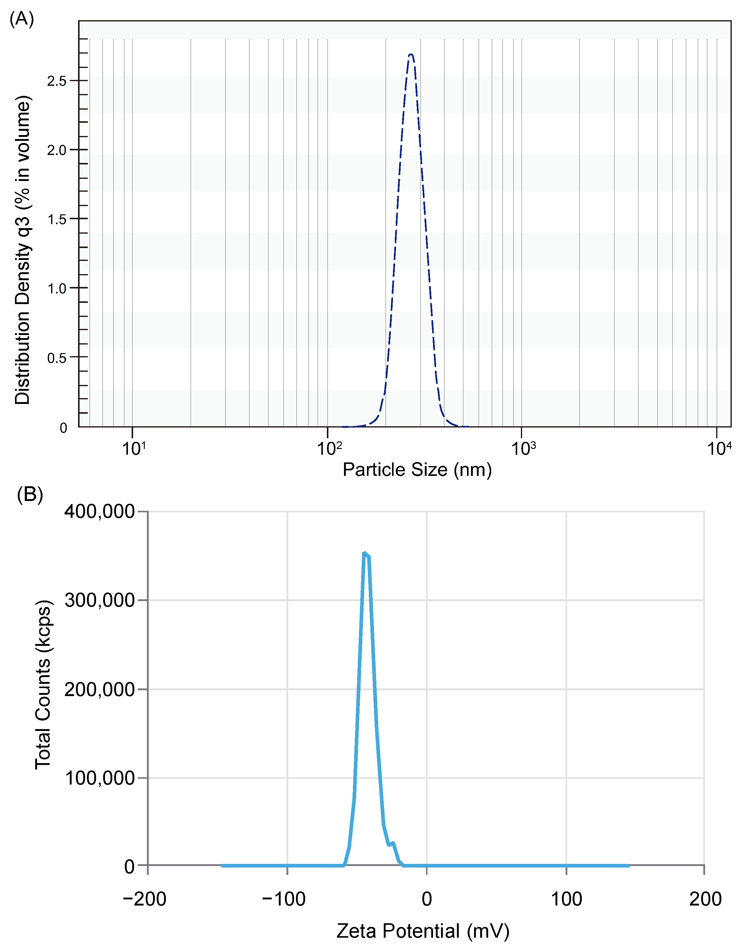
2.3.3. Measurement of Particle Size Distribution (PSD) of the Liposomal Powder and Liposomes

The PSD of the liposomal powder was measured using a laser light diffraction instrument, Cilas 1190 (Cilas, Orleans, France). Isopropanol was used as a dispersant for the powder. The mean particle diameter was recorded as the 50th (median) percentile of the cumulative PSD in volume.
The PSD of the liposomes was measured using LUMiSizer® 651 (L.U.M. GmbH, Berlin, Germany) with STEP-Technology®. The test sample was diluted 100 times in water and placed in a disposable polycarbonate cuvette with an optical path length of 10 mm. A light source with a wavelength of 470 nm was used for all the measurements.
A PSD analysis was performed in accordance with ISO13318-2 [44], using SEPView6 software. The PSD of the liposomes was characterized by a series of statistical parameters, such as harmonic mean diameter, as well as D10, D16, D50, D84, and D90. These parameters indicate the size below which 10%, 16%, 50%, 84%, and 90% of all particles are found, respectively. The width of the distribution as the span was calculated according to the following formula [45]:
(D90 − D10)/D50.

2.3.4. Measurement of Zeta Potential

Approximately 0.5 g of liposomal vitamin C powder was dissolved in 10 mL of demineralized water. The resulting solution was diluted again, yielding a final concentration of the test product of 5 mg/mL. A measurement of the zeta potential of the sample prepared in this way was carried out in a DTS1070 measuring cell, a Zetasizer Ultra Red instrument (Malvern Panalytical Ltd., Malvern, UK), at 25 °C.

2.4. In Vitro Bioavailability Study

2.4.1. Cell Culture

The Caco-2 cell line (ATCC; Manassas, VA, USA) was used to evaluate vitamin C bioavailability in vitro. This cell model is capable of forming a monolayer that mimics conditions in the small intestine. The cell culture was conducted in DMEM high-glucose medium (BioWest, Lakewood Ranch, FL, USA) supplemented with 10% FBS (fetal bovine serum; Corning, New York, NY, USA), 1% NEAA (non-essential amino acid; BioWest, Lakewood Ranch, FL, USA), and 1 mM sodium pyruvate (Sigma-Aldrich, Saint Louis, MO, USA). The medium was changed every two days, and passage was performed with 0.25% trypsin (Sigma-Aldrich, Saint Louis, MO, USA) at 80% confluency in the culture dish. Cells between passage 40 and 49 were used for this study [46,47].

2.4.2. Permeability across Human Intestinal Epithelial Cells Monolayer (Caco-2 Cells)

The cells were seeded onto insert culture dishes (Greiner, BioOne, Kremsmünster, Austria) at 3 × 10⁵ cells/insert. After 21 days of culture, the electrical resistance of the formed monolayer was measured using a Millicell ERS-2 electrode (Burlington, MA, USA). Solutions of two formulations of vitamin C at a concentration of 0.1 mg/mL were then prepared, and the cells were incubated in their presence for 2 h. After incubation, the apical and basal layers were collected, 5 mM TCEP was added to the collected solutions and filtered through a 0.22 µm PES syringe filter, and the vitamin C content was determined by ultra-performance liquid chromatography (UPLC) according to the protocol presented below.

2.4.3. Evaluation of Vitamin C Concentration

Preparation of Buffer and Wash Solutions

Pure HPLC-grade formic acid (Sigma Aldrich, St. Louis, MO, USA) was dissolved in ultrapure Millipore water (pH 2.0) or methanol (0.1%) (St. Louis, MO, USA). The mobile phase was filtered through a 0.45 µm filter each time before use. Three wash solvents, strong wash solution (acetonitrile/isopropanol/water) (5:1:1) (v/v/v), weak wash solution (water/methanol/acetonitrile) (3:1:1) (v/v/v), and seal wash solution (acetonitrile/water) (2:8) (v/v), were developed to minimize the peak disturbance. HPLC-grade acetonitrile and isopropanol were purchased from Sigma-Aldrich (St. Louis, MO, USA). A degasser was arranged to obtain high-quality results of the liquid chromatography (LC). All the chromatographic columns and samples were maintained at a temperature of 30 °C and 5 °C, respectively. UV detection was set at 270 nm.

Method Optimization

Method optimization was performed using a Waters Acquity™ (Waters Corp., Milford, MA, USA) system equipped with a binary solvent delivery pump, Acquity™ Sampler Manager, Acquity™ TUV Detector, and Acquity™ Column Heater/Cooler. The peak purity was evaluated on a Waters I-Class Plus UPLC with a PDA eλ Detector combined with an Acquity™ SM-FTN-I (Sample Manager with Flow-Through Needle) and Acquity™ BSM (Binary Solvent Manager). Ascorbic acid Certified Reference Material (CRM, PHR1008) was purchased from Supelco® by Sigma-Aldrich (St. Louis, MO, USA).

UPLC Analysis

The UPLC was carried out on 2.1 mm i.d. × 100 mm length, 1.7 µm columns (Kinetex® C18 100 Å; Phenomenex Inc., Torrance, CA, USAand Acquity UPLC® BEH C18 130 Å; Waters Corp., Milford, MA, USA) with several batches. Two mobile phases, (A) 0.1% formic acid in water and (B) 0.1% formic acid in methanol, were used for the separations. The experiments were performed using Empower3 software for instrument control, acquisition, and data analysis. The samples were programmed for isocratic elution at a flow rate of 0.05 mL/min with 1% organic modifier (B) and a 0.5 min re-equilibration time.

2.4.4. Bioavailability Expression

Bioavailability is expressed as the relative value of the difference in active ingredient content in the basal layer of the model.

2.5. Clinical Study

2.5.1. Ethical Approval

This study commenced after written approval was obtained from the Bioethics Committee at the Medical University of Warsaw (Bioethics Committee approval: KB/121/2021; approval with accepted changes: KB/36/A2022, dated 27 April 2022).

2.5.2. Trial Registration
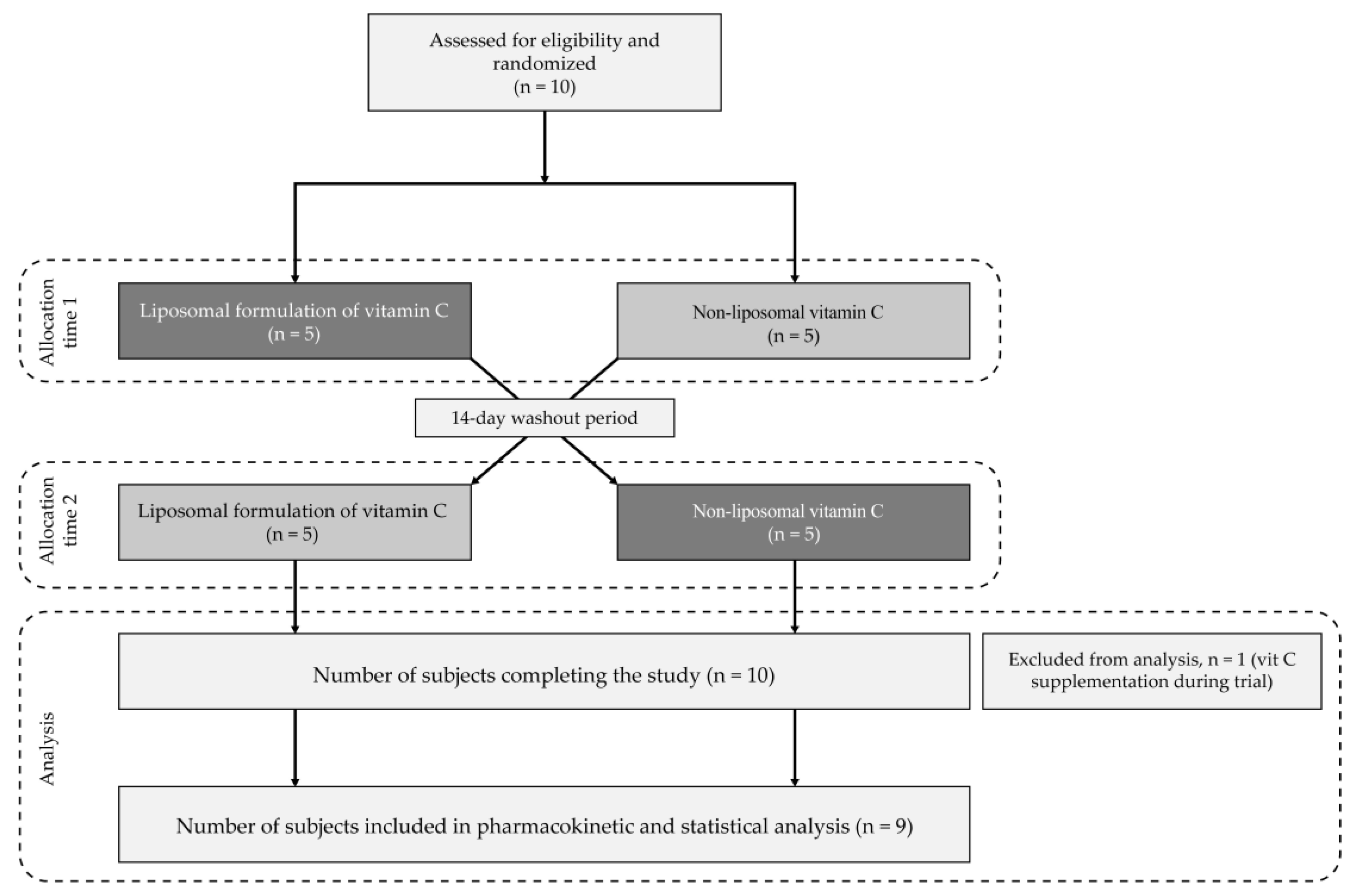
The trial was registered at ClinicalTrials.gov and received identification number (ID) NCT05843617.

2.5.3. Study Design

A randomized, single-dose, double-blind, cross-over study was conducted on a group of healthy adult human subjects under fasting conditions. The participants were enrolled based on the inclusion and exclusion criteria listed in Table 1. Informed consent was obtained from all the individual participants included in this study.
The qualified subjects were randomly allocated (sealed envelopes, simple randomization) on a 1:1 basis to two different groups: the first one received a single oral dose of 1000 mg of non-liposomal vitamin C, and the second one was administered with a single oral dose of 1000 mg of liposomal vitamin C in powder form. Both dietary supplements were given to the participants in the morning, between 7 and 8 am. The participants were under the supervision of a physician to monitor potential adverse events and ensure their safety. The flow diagram of the study design is shown in Figure 1.

2.5.4. Primary and Secondary Endpoints

The primary endpoints were defined as the bioavailability of the tested supplements based on the total doses of vitamin C absorbed into the bloodstream and the maximum concentrations (Cmax) in the blood after taking the tested forms of vitamin C. Bioavailability was defined as the area under the curve (AUC).
The secondary endpoints were defined as the times elapsed from the intake of the tested forms of vitamin C to their maximum blood concentrations (Tmax) and the vitamin C concentrations obtained in the blood 24 h after administration of the supplements (C24h).

2.5.5. Methodology

In total, 10 subjects were enrolled in this study. The participants were instructed to limit their intake of vitamin C-rich foods (primarily, raw fruits and vegetables), vitamin C-containing supplements, and pure vitamin C for a period of 72 h before and during the day of this study. On the day the experiment began, the participants followed a standardized, vitamin C-deficient diet provided by a catering company. The procedure commenced after an overnight fast of at least 8 h. Then, the volunteers orally took a single dose of 1000 mg of non-liposomal vitamin C (powder in capsules) or the liposomal formulation (powder in capsules). After a 14-day washout period, the participants were switched from their original treatment to the alternative one (liposomal or non-liposomal vitamin C formulation).
The process of preparing samples for analysis was carried out in the AronPharma laboratory (Gdansk, Poland). Venous blood was collected from each participant immediately before the administration of the studied substance (T0) and 30 min and 1, 2, 3, 4, 6, 8, 10, and 24 h post-administration. All the samples were collected in pre-labeled K2EDTA vacutainers from the forearm vein using an indwelling cannula, and the heparin-lock technique was applied to prevent blood clotting inside them. Before the blood sample was drawn, 0.5 mL of blood was discarded to prevent the saline-diluted blood and heparin from interfering with the analysis. The cannula was removed after 24 h or earlier as needed or if blocked. The vacutainer tubes were positioned vertically in a stack and were kept on ice. The blood samples collected from all the participants from all time points were first centrifuged at 2800× g for 8 min at 4 °C to isolate the plasma. Centrifugation was carried out within 30 min of the collection of all the samples at each time point. The resulting supernatant was immediately added to an equal volume of 10% (w/v) metaphosphoric acid (MPA) in 2 mmol/L of ethylenediaminetetraacetic acid disodium salt. The samples were left on ice for 5 min and centrifuged (16,000× g, 10 min, 4 °C) after adding 5 mmol/L of tris(2-carboxyethyl)phosphine hydrochloride. Each sample was filtered with a 0.22 µm polyethersulfone membrane, and the ascorbic acid level was analyzed on the same day by ultra-performance liquid chromatography (UPLC) according to the protocol described in Section 2.4.3., “Evaluation of vitamin C concentration”. The samples from the clinical trials were programmed for isocratic elution at a flow rate of 0.1 mL/min with 1% organic modifier and a 0.5 min re-equilibration time. The method for determining the concentration of ascorbic acid in the blood samples was validated at the R&D Department of AronPharma. The standard curve was developed by using a peak-areas linear regression equation from six ascorbic acid standards made up in MPA/EDTA and plasma. Each sample run included a plasma quality control sample. The samples were coded for the analytical procedure.

2.6. Statistical Analysis

The results are presented as the mean value ± standard deviation (SD). In the randomized clinical trial, differences between the groups were analyzed using Student’s t-test for dependent samples with GraphPad Prism 9.0.1 software (GraphPad Software Inc., San Diego, CA, USA). G*Power 3.9.1.7 software was used to calculate the sample size required for this study and to conduct the post hoc power analysis. Values of p < 0.05 were considered statistically significant and marked on the charts with the symbol (*); p < 0.01 was marked with (**). In line with the study design, the samples were decoded only after the analysis.

3. Results and Discussion

3.1. Properties of Liposomal Vitamin C in Powder Form

The hydrolysis of the ester bonds and/or the oxidation of the unsaturated acyl residues of the phospholipids, as well as changes in the average particle size and size distribution due to aggregation or carrier fusion—occurring in liquid liposomal suspensions—not infrequently lead to leakage of the entrapped active ingredient [36,41], translating into short shelf-lives of the product and a reduction in its in vivo effectiveness [36].
An effective way to improve the stability, bioavailability, and bioactivity of liquid liposomal suspensions is to subject them to additional “post-processing techniques”, including drying [48]. Dehydration of the liquid intermediate product significantly extends the shelf life of the product, makes it easier to transport, and reduces distribution costs, as the cold chain is not mandatory [40].
Spray drying, used in liposomal vitamin C powder technology, is more convenient and economical compared to popular freeze drying [40,48]. Spray drying, unlike lyophilization, allows for direct transformation of a liquid feed into powder without the need for grinding, and importantly, it does not generate the risk of damaging the integrity of the lipid bilayer by ice crystals formed during freezing of the semi-finished product with a liquid consistency [49]. The process parameters we chose allowed for rapid dehydration of the liposomal suspension. The effectiveness of the performed process of the microencapsulation of liposomal particles was confirmed in the analysis of the morphology of the powder obtained under a scanning electron microscope (SEM) (Figure 2A,B). This analysis highlighted the intact surface of the microcapsules formed with carbohydrate material and the liposomal particles entrapped inside. Their mean diameter (median distribution), determined using the laser light diffraction method, was 23.4 ± 1.2 µm (Figure 2C).
The intact/undamaged surface of liposomal vitamin C powder particles obtained by nanofiber weaving (Zeal) technology was visualized under the SEM in a study described by Jacob et al. [50]. However, the authors did not determine the value of the median particle distribution.
The microcapsules obtained in this study, composed of carbohydrate material, further protected the vitamin C encapsulated in the liposomes from the adverse effects of various environmental factors. They also minimized the risk of the destructive effects of high temperature and high shear forces on the integrity of the lipid vesicle structure (see ref. [40]).
The latter was highlighted in cryogenic electron microscope (cryo-TEM) images, allowing visualization of the shape and internal structure (lamellarity) of the liposomes, with sizes ranging from 5 to about 500 nm [51].
The images obtained by cryogenic transmission electron microscopy (cryo-TEM) yielded liposomes, mostly large unilamellar vesicles (LUVs), with a spherical shape and smooth surface (Figure 3). The continuity of the lipid vesicle walls indicates that the liquid liposomal intermediate was carried out in powder form without violating the continuity of the phospholipid bilayer.
The presence of multivesicular vesicles (MVVs) next to the LUVs confirms earlier reports of a larger population of unilamellar liposomes with fractions of bi- or multiwalled vesicles in cryo-TEM imaging [51,53]. This reflects the fact that the technological process does not achieve a uniform, monodisperse formulation of liposomal carriers [54]. In the discussed case, it cannot be excluded that changes in the structure of liposomes occurring during spray drying led to the formation of bi- and multilamellar vesicles [55]. Finally, in some cases, the bilayer carriers seen in cryo-TEM may actually be invaginated single-walled structures formed during sample preparation for microscopic analysis [51,53].
The cryo-TEM technique was not used to measure the size of the resulting liposomal particles in the powder because of its limited applicability in the quantitative analysis of the liposome carrier population [51].
A prerequisite for obtaining a reliable measurement of the size of liposomes is the study of a representative population of these carriers [56]. For cryo-TEM microscopy, this is difficult to achieve because a very small volume of a diluted sample is taken for measurement (3 µL in this experiment). Thus, only a small proportion of the total liposomes were analyzed.
In addition, making quantitative measurements by cryo-TEM can be hampered by, among other things, the variable thickness of the liposome aqueous solution layer inside a hole of a holey carbon film, which leads to separation (also known as sorting) of vesicles according to their size. This means that larger liposome particles tend to accumulate close to the edge of a hole, while small liposomes are located in the thinnest area of the solution layer [51,56].
The specificity of the sample preparation procedures for analysis and of the image analysis itself translates into a lack of consistency between liposome particle size measurement results obtained using the cryo-TEM technique and those obtained using other measurement techniques, e.g., dynamic light scattering (DLS) [56].
Taking into account the information above, dedicated STEP-Technology® applied in the LUMiSizer® (L.U.M GmbH, Berlin, Germany) was used to determine the particle size distribution (PSD) of the liposomes. The mean size of the lipid carriers measured in this way was 262 ± 49.8 nm (Table 2) and fell within the lower range of standard sizes (25–2500 nm) for this type of carrier [36].
This is advantageous because any reduction in the particle size of liposomes results in an improved ratio of surface area to volume, and this in turn translates directly into increased bioavailability [57]. The size of the carrier is also a key parameter determining its half-life in the bloodstream: smaller liposomes generally last longer in the bloodstream than large liposomes [36].
The particle size distribution of liposomes is an important parameter for assessing both the quality (homogeneity) of the product and its flow behavior and predicting its efficacy in vivo [58,59]. The narrow width of the distribution of liposomes in the obtained powder (span = 0.500) (Figure 4A) and their relatively small diameter prove that the drying process did not lead to the fusion and disintegration of the carriers. In contrast, the high absolute value of the zeta potential (−41.44 ± 0.30 mV, that is >30 mV) obtained for the hydrated powder (Figure 4B) demonstrates the low tendency of liposomes to aggregate or flocculate, and thus the high stability of the resulting colloidal suspension [42,58]. In other words, the process of spray drying used to transform liquid suspensions of liposomes into powder form did not adversely affect the quality of the carrier particles obtained.

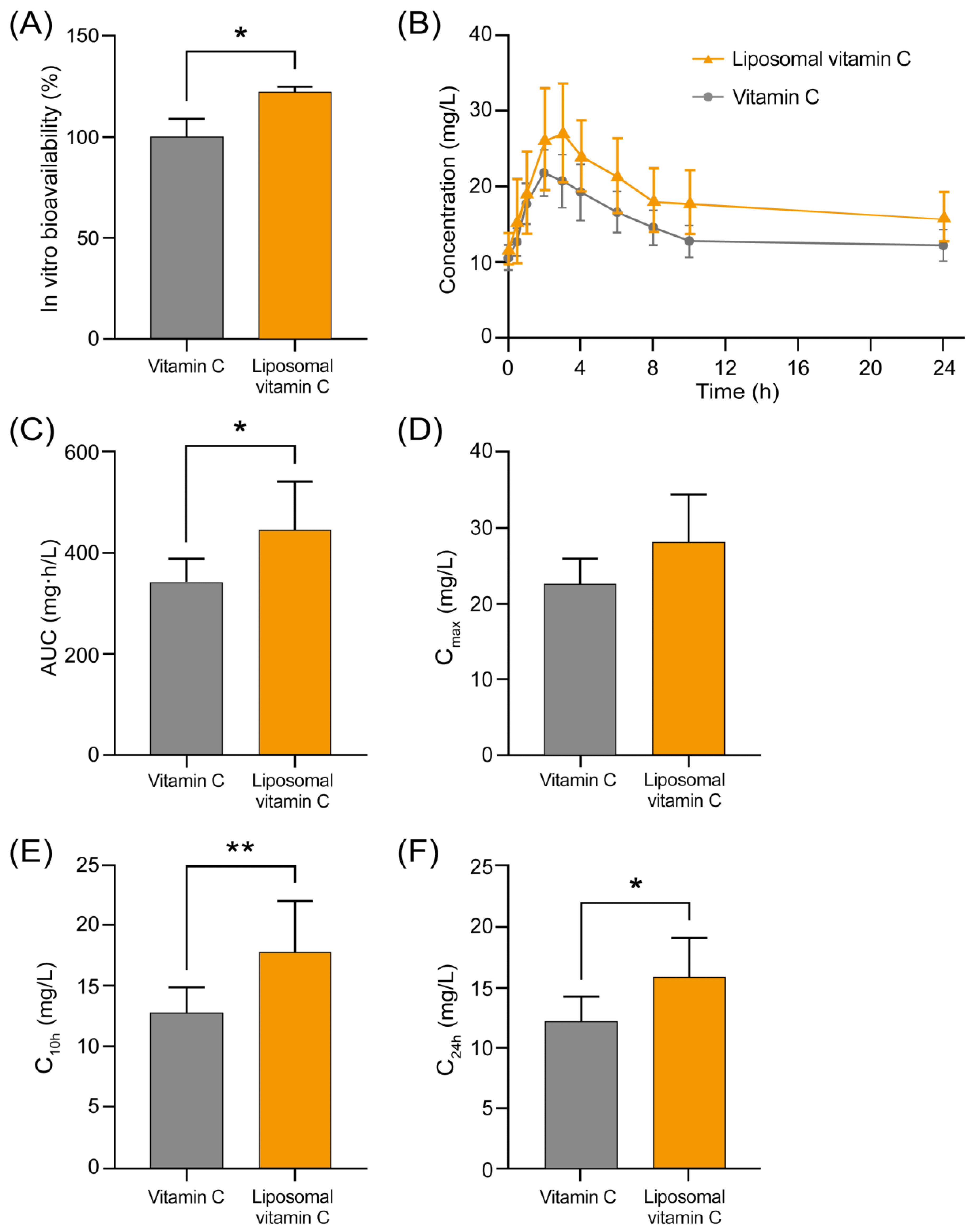
3.2. Comparison of Bioavailability of Non-Liposomal and Liposomal Ascorbic Acid in an In Vitro Cell Model

Bioavailability is the basic pharmacokinetic term that defines the amount of an ingested dose of an active substance that reaches the bloodstream unchanged. In vitro pharmacokinetic models are often used to rapidly assess the oral bioavailability of bioactive ingredients before performing expensive and time-consuming in vivo studies, including clinical trials [60]. To assess the permeability and uptake of various ascorbic acid formulations, we utilized the Caco-2 cellular model. This standard model, derived from human colon adenocarcinoma cells, is employed to effectively mimic the conditions of the small intestine [46,61] and is widely used for studying intestinal absorption and transport mechanisms of various nutrients and pharmaceuticals [62,63,64]. In this experiment, the bioavailability, expressed as the relative difference in active ingredient content in the basal layer of the Caco-2 model, was 22.28% higher for liposomal vitamin C compared to the non-liposomal form (p < 0.05) (Figure 5A). Given that the Caco-2 model is a reliable predictor of human intestinal absorption, this 22.28% increase is practically significant. Enhanced bioavailability suggests more efficient absorption, potentially leading to higher and more sustained plasma vitamin C levels. This can improve clinical outcomes, particularly in immune support and oxidative stress reduction, while allowing for lower dosages to minimize side effects and improve patient compliance. Therefore, the promising results observed in the Caco-2 model prompted us to test liposomal vitamin C powder in a clinical trial to further assess the advantages of the liposomal form in terms of higher bioavailability.

3.3. Clinical Trial Results

3.3.1. Study Population

A total of 10 subjects (5 men and 5 women) of Caucasian race participated in this study. Their demographic data are presented in Table 3. The average age of the volunteers was 36.8 years and ranged from 22 to 59 years. Their mean body weight, height, and BMI were 83.2 kg, 172 cm, and 28.3 kg/m2, respectively.
None of the participants enrolled in this study withdrew before it was completed. One participant violated the dietary recommendations, i.e., consumed a dietary supplement containing 200 mg of vitamin C during and immediately before this study. The data of this individual had to be excluded from further analysis.

3.3.2. Results of Analysis of Pharmacokinetic Properties of the Tested Formulations

Figure 5B highlights the changes in the average plasma concentrations of vitamin C within 24 h of administration of the dietary supplements tested.
The most important primary parameter compared in this study was the total absorbed dose of the active ingredient, defined as the area under the curve (AUC). For the liposomal vitamin C powder, the AUC value was 445 ± 97 mg·h/L and was statistically significantly higher (p = 0.0131) than the value obtained for non-liposomal ascorbic acid (AUC = 342 ± 45 mg·h/L) (Figure 5C). The mean of differences for AUC was 6170 (95% CI, 1691 to 10,650; dz =1.22), indicating that the liposomal formulation was superior to the reference formulation with respect to this parameter.
The value of the second primary parameter, i.e., the maximum vitamin C concentration achieved in plasma (Cmax), was numerically but not statistically higher for the liposomal form (28.0 ± 6.5 mg/L) than for its non-liposomal counterpart (22.5 ± 3.4 mg/L) (p = 0.0573) (Figure 5D).
For the liposomal formulation, the C24h value was 15.85 ± 3.26 mg/L and was statistically significantly higher (p = 0.0199) than the concentration obtained for the non-encapsulated ascorbic acid (12.18 ± 2.07 mg/L) (Figure 5F). Moreover, the concentration of vitamin C reached in the blood 10 h after administration of both forms of vitamin C (C10h) (a parameter not subject to analysis planned in the clinical trial protocol) was also statistically significantly higher (p = 0.004) for the liposomal formulation (17.8 ± 4.2 mg/L). For non-liposomal ascorbic acid, C10h was 12.7 ± 2.1 mg/L (Figure 5E).
The mean of differences for C10h and C24h was 5.1 (95%CI, 2.24–8.1; dz = 1.38) and 3.67 (95% CI, 0.75–6.58; dz = 1.28), respectively. These results demonstrate that the liposomal formulation surpassed the reference formulation in each of the aforementioned parameters.
There was no statistical difference (p = 0.4468) between the Tmax values obtained for the liposomal (2.56 ± 0.53 h) and non-liposomal forms of ascorbic acid (2.33 ± 0.70 h).
This study shows that compared to non-liposomal vitamin C, oral administration of liposomal vitamin C powder to healthy volunteers resulted in a significantly better absorption of ascorbic acid into the bloodstream, which equated to its higher bioavailability (30% increase in AUC, p < 0.05). The duration of elevated vitamin C blood levels was also longer (C24h increase of 30%, p < 0.05), which can reflect a prolonged effect of supplementation.
There are studies indicating that liposomal vitamin C has higher bioavailability compared to the non-liposomal form, as reflected in higher AUC values, Cmax, and Tmax. A clinical bioavailability study using an identical 1000 mg dose of liposomal vitamin C but in liquid form was conducted by Gopi and Balakrishnan [31]. In this case, the liquid suspension of liposomes containing vitamin C had a 1.77 times higher bioavailability (55.86 mg·h/dL compared to 31.53 mg·h/dL) and was absorbed 2.41 times faster than its conventional counterpart. In our study, we demonstrated that the AUC for liposomal vitamin C powder was 1.3 times higher. Similarly, Davis et al. [29] reported a 1.35-fold increase in bioavailability (10.3 ± 0.9 mg·h/L compared to 7.6 ± 0.4 mg·h/L) but when 4.25 g of sodium ascorbate (equivalent to 4 g of vitamin C) was administered in liposomal form. In another study, supplementation with 10 g of liposomal vitamin C was shown to enhance ascorbate bioavailability by 1.79 times, with a Cmax that was 1.67 times higher (300 μM compared to 180 μM) for the liposomal form than for the equivalent dose of the non-liposomal form [30].
Similar to our study, the Tmax value obtained by Gopi and Balakrishnan [31] for liposomal vitamin C was non-significantly higher than that obtained for the conventional form of this active ingredient (3.51 h vs. 3.42 h). Statistically insignificant differences in Tmax values were also reported in a randomized, single-dose, cross-over oral bioavailability study comparing powdered liposomal vitamin C obtained by nanofiber weaving (Zeal) technology (a dose of 150 mg of ascorbic acid) with its conventional counterpart (3.1 ± 1.15 h vs. 3.6 ± 0.62 h, p = 0.388) [50].
However, as noted by Gopi and Balakrishnan [31], an analysis of the results of a number of published studies of liposomal vitamin C bioavailability shows significant discrepancies in the pharmacokinetic parameters reported. The authors also emphasize the difficulty of comparing these studies due to factors such as differing production methods, particle sizes, dosages, and the various forms of tested vitamin C, including ascorbic acid, sodium ascorbate, and calcium ascorbate.
Indeed, the properties and quality of the liposomes obtained can vary significantly depending on the formulation [65]. The mere change in the length of the acyl chain and thus the phase transition temperature of phospholipids leads to carriers with different release profiles of the entrapped active substance, and thus different in vivo properties [66].
In turn, with the use of different production methods—impacting, among other things, the size of the carrier particles—the same chemical composition of liposomal products can translate into a lack of comparability in their in vitro properties and in vivo efficacy [67].
Not to be overlooked is the wide individual variability in vitamin C absorption [31,68], including the existence of “poor” and “good” absorbers of ascorbic acid [68]. Differences in the analytical procedures used to measure plasma vitamin C concentrations are also not insignificant to the experimental results obtained [31].
With regard to the results of our clinical trial, it should be noted that they confirmed the preliminary conclusions derived from the properties of the developed liposomal product. As expected, the narrow size distribution of liposomes in the powder and their relatively small diameter and high absolute zeta potential value translated into improved pharmacokinetic properties of vitamin C and its increased in vivo bioavailability.

4. Limitations

The main limitation of this study is the small group of volunteers participating in the clinical evaluation of the bioavailability of liposomal vitamin C powder. Therefore, it should be considered that this trial was a pilot one. The study’s strength lies in its cross-over design, which remains a significant advantage despite the small sample size. Cross-over studies allow each participant to serve as their own control, thereby enhancing the statistical power and reliability of the results. The authors are well aware of the fact that confirmation of the beneficial pharmacokinetic properties of the liposomal product demonstrated in the presented experiment might require replication of the intervention with a larger number of participants in the future. However, numerous examples in the literature demonstrate that pilot studies of similar scope and complexity have been conducted by other research teams. For instance, studies such as Bolisetty et al. [69] and Allen et al. [70] involved small sample sizes (n = 5–7 and n = 4, respectively) but included detailed analyses that provided valuable insights.

5. Conclusions

In brief, using technology devoid of organic solvents, a liposomal vitamin C suspension was obtained, which, after mixing with maltodextrin, was dehydrated in a disc spray dryer. The resulting powder-like microcapsules, with an average particle diameter of 23.4 ± 1.2 µm, were composed of a carbohydrate material encapsulating liposomal vitamin C particles. Visible under the SEM, the intact surface of the microcapsules provided a barrier protecting both the liposomes and the encapsulated vitamin C from the undesirable effects of external stressors.
In cryogenic transmission electron microscopy (cryo-TEM) observations, we confirmed the presence in the hydrated powder of liposomes (mainly LUV) with preserved integrity of the phospholipid bilayer and a spherical shape and smooth surface. The liposomes we obtained were characterized by a relatively small particle size (262 ± 49.8 nm) and the narrow width of distribution (span = 0.500), as well as the high absolute value of the zeta potential (-41.44 ± 0.30 mV, that is >30 mV) obtained for the hydrated powder, further confirming the lack of negative effects of spray drying on product quality.
In the present study, the main emphasis was laid on demonstrating the absence of negative effects of spray drying on the properties and pharmacokinetic profile of the obtained liposomal vitamin C powder. This was important because dehydration of lipid carriers significantly prolongs the shelf life of the product, eliminates the need to include preservatives in its composition (this is necessary in the case of liquid liposomal suspensions), reduces transport and distribution costs, and, at the same time, broadens the possibilities of its practical applications.
The liposomal vitamin C powder we obtained is, by design, intended for use in the production of dietary supplements (tablets, capsules with powder, vitamin premixes, jellies, etc.) and nutraceuticals but also functional foods.
Due to the limited thermal stability of ascorbic acid, the use of liposomal vitamin C in high-temperature-processed products may be difficult and result in the need to modify the manufacturing process of vitamin C-fortified foods.
An additional difficulty in the case of inclusion of liposomal vitamin C in the form of powder in formulations of complex composition may be the presence of components such as protein or fat, the removal of which during the procedure of sample preparation for observation under a cryogenic electron microscope (cryo-TEM) may realistically disturb the result of the determination aimed at confirming the presence of intact liposomes in the finished product.
Given the limitations above, further experiments would be needed to determine the effects of various technological processes, including thermal food preservation, extrusion, high-pressure homogenization, and selected matrices, on the stability of incorporated liposomal vitamin C. It would also be important to determine the effect of powdered liposomal vitamin C on the sensory properties of enriched foods [2].
The main principle behind liposomal technology is to enhance the absorption of encapsulated active substances, thereby increasing bioavailability. In the initial stage of our study, using the widely recognized Caco-2 cell model, we compared the bioavailability of non-liposomal and liposomal ascorbic acid. The results show that liposomal vitamin C had 22.28% higher bioavailability compared to non-liposomal vitamin C, as measured in the basal layer of the Caco-2 cell model. Given that these results were obtained under controlled laboratory conditions, which are inherently predictive, liposomal vitamin C powder was also evaluated in a clinical trial. In the clinical study, we assessed the pharmacokinetic properties of the tested formulations, including AUC, Cmax, C10h, C24h, and Tmax. Although the results are promising and suggest higher bioavailability for the liposomal form of vitamin C, the limited sample size necessitates further research with a larger cohort to confirm these findings. Future studies would benefit from an increased sample size to ensure statistical significance and broader applicability. Including a diverse participant pool could also provide insights into how different demographics respond to the formulation. While vitamin C is rapidly metabolized and its blood levels are transient, incorporating a follow-up could be valuable to evaluate long-term outcomes, such as sustained health benefits or effects on biomarkers related to vitamin C deficiency.

Author Contributions

Conceptualization, P.Ż., B.K., K.C., K.L., M.F. and M.P.; methodology, P.Ż., B.K., K.C., M.F., A.F., E.D. and M.P.; software, K.L., A.F. and E.D; validation, M.D. and J.G.; formal analysis, P.Ż., B.K., K.C., M.D. and P.K.; investigation, P.Ż., B.K., K.C., M.D., J.G., M.F. and M.P.; resources, M.K.; writing—original draft preparation, M.K., B.K., P.Ż., A.F. and E.D.; writing—review and editing, M.K., B.K., M.F., E.D. and P.K.; visualization, B.K., P.K., A.F. and E.D.; supervision, M.P., K.L. and M.F. All authors have read and agreed to the published version of the manuscript.

Funding

This research and the APC were funded by Bart Limited.

Institutional Review Board Statement

The RCT described in this paper was approved by the Bioethics Committee at the Medical University of Warsaw (Bioethics Committee approval: KB/121/2021; approval with accepted changes: KB/36/A2022, dated 27 April 2022).

Informed Consent Statement

Informed consent was obtained from all the subjects involved in this study.

Data Availability Statement

The raw data supporting the conclusions of this article are available from the corresponding author upon reasonable request.

Conflicts of Interest

P.Ż., M.K. and K.C. are employees of Bart Limited. B.K., M.D., J.G., P.K. and K.L. are employed by AronPharma Limited. M.F., A.F., E.D. and M.P. confirm that they have no conflicts of interest with respect to the work described in this manuscript.

References

  1. Padayatty, S.J.; Katz, A.; Wang, Y.; Eck, P.; Kwon, O.; Lee, J.H.; Chen, S.; Corpe, C.; Dutta, A.; Dutta, S.K.; et al. Vitamin C as an antioxidant: Evaluation of its role in disease prevention. J. Am. Coll. Nutr. 2003, 22, 18–35. [Google Scholar] [CrossRef]
  2. Maurya, V.K.; Shakya, A.; McClements, D.J.; Srinivasan, R.; Bashir, K.; Ramesh, T.; Lee, J.; Sathiyamoorthi, E. Vitamin C fortification: Need and recent trends in encapsulation technologies. Front. Nutr. 2023, 10, 1229243. [Google Scholar] [CrossRef]
  3. Steinberg, F.M.; Rucker, R.B. Vitamin C. In Encyclopedia of Biological Chemistry, 2nd ed.; Lennarz, W.J., Daniel Lane, M., Eds.; Elsevier: Amsterdam, The Netherlands, 2013; pp. 530–534. [Google Scholar] [CrossRef]
  4. Calder, P.C.; Albers, R.; Antoine, J.M.; Blum, S.; Bourdet-Sicard, R.; Ferns, G.A.; Folkerts, G.; Friedmann, P.S.; Frost, G.S.; Guarner, F.; et al. Inflammatory disease processes and interactions with nutrition. Br. J. Nutr. 2009, 101, S1–S45. [Google Scholar] [CrossRef] [PubMed]
  5. Institute of Medicine (US) Panel on Dietary Antioxidants and Related Compounds. 5. Vitamin C. In Dietary Reference Intakes for Vitamin C, Vitamin E, Selenium, and Carotenoids; National Academies Press (US): Washington, DC, USA, 2000. Available online: https://www.ncbi.nlm.nih.gov/books/NBK225480/ (accessed on 20 August 2024).
  6. EFSA NDA Panel (EFSA Panel on Dietetic Products, Nutrition and Allergies). Scientific Opinion on Dietary Reference Values for vitamin C. EFSA J. 2013, 11, 3418. [Google Scholar] [CrossRef]
  7. World Health Organization. Vitamin and Mineral Requirements in Human Nutrition: Report of a Joint FAO/WHO Expert Consultation, Bangkok, Thailand, 21–30 September 1998, 2nd ed.; World Health Organization: Geneva, Switzerland, 2004; Available online: https://iris.who.int/handle/10665/42716 (accessed on 20 August 2024).
  8. Blomhoff, R.; Andersen, R.; Arnesen, E.K.; Christensen, J.J.; Eneroth, H.; Erkkola, M.; Gudanaviciene, I.; Halldorsson, T.I.; Høyer-Lund, A.; Lemming, E.W.; et al. Nordic Nutrition Recommendations 2023; Nordic Council of Ministers: Copenhagen, Denmark, 2023. [Google Scholar] [CrossRef]
  9. Haytowitz, D.B.; Ahuja, J.K.C.; Showell, B.; Somanchi, M.; Nickle, M.; Nguyen, Q.A. Composition of Foods Raw, Processed, Prepared USDA National Nutrient Database for Standard Reference, Release 28; Nutrient Data Laboratory, Beltsville Human Nutrition Research Center, ARS, USDA: Beltsville, ML, USA, 2015. [Google Scholar] [CrossRef]
  10. Giannakourou, M.C.; Taoukis, P.S. Effect of Alternative Preservation Steps and Storage on Vitamin C Stability in Fruit and Vegetable Products: Critical Review and Kinetic Modelling Approaches. Foods 2021, 10, 2630. [Google Scholar] [CrossRef] [PubMed]
  11. Barco Leme, A.C.; Fisberg, R.M.; Veroneze de Mello, A.; Sales, C.H.; Ferrari, G.; Haines, J.; Rigotti, A.; Gómez, G.; Kovalskys, I.; Cortés Sanabria, L.Y.; et al. Food Sources of Shortfall Nutrients among Latin Americans: Results from the Latin American Study of Health and Nutrition (ELANS). Int. J. Environ. Res. Public Health 2021, 18, 4967. [Google Scholar] [CrossRef]
  12. Matsumoto, M.; Saito, A.; Okada, C.; Okada, E.; Tajima, R.; Takimoto, H. Consumption of meals prepared away from home is associated with inadequacy of dietary fiber, vitamin C and mineral intake among Japanese adults: Analysis from the 2015 National Health and Nutrition Survey. Nutr. J. 2021, 20, 40. [Google Scholar] [CrossRef] [PubMed]
  13. Lachat, C.; Nago, E.; Verstraeten, R.; Roberfroid, D.; Van Camp, J.; Kolsteren, P. Eating out of home and its association with dietary intake: A systematic review of the evidence. Obes. Rev. 2012, 13, 329–346. [Google Scholar] [CrossRef]
  14. O’Connell, M.L.; Coppinger, T.; Lacey, S.; Arsenic, T.; McCarthy, A.L. The nutritional status and dietary intake of free-living seniors: A cross-sectional study. Clin. Nutr. ESPEN 2021, 43, 478–486. [Google Scholar] [CrossRef]
  15. Hiatt, A.N.; Ferruzzi, M.G.; Taylor, L.S.; Mauer, L.J. Deliquescence Behavior and Chemical Stability of Vitamin C Forms (Ascorbic Acid, Sodium Ascorbate, and Calcium Ascorbate) and Blends. Int. J. Food Prop. 2011, 14, 1330–1348. [Google Scholar] [CrossRef]
  16. Sablani, S.S.; Al-Belushi, K.; Al-Marhubi, I.; Al-Belushi, R. Evaluating Stability of Vitamin C in Fortified Formula Using Water Activity and Glass Transition. Int. J. Food Prop. 2007, 10, 61–71. [Google Scholar] [CrossRef]
  17. Dickinson, A.; Blatman, J.; El-Dash, N.; Franco, J.C. Consumer usage and reasons for using dietary supplements: Report of a series of surveys. J. Am. Coll. Nutr. 2014, 33, 176–182. [Google Scholar] [CrossRef] [PubMed]
  18. Barnes, K.; Ball, L.; Desbrow, B.; Alsharairi, N.; Ahmed, F. Consumption and reasons for use of dietary supplements in an Australian university population. Nutrition 2016, 32, 524–530. [Google Scholar] [CrossRef] [PubMed]
  19. Wiltgren, A.R.; Booth, A.O.; Kaur, G.; Cicerale, S.; Lacy, K.E.; Thorpe, M.G.; Keast, R.S.; Riddell, L.J. Micronutrient supplement use and diet quality in university students. Nutrients 2015, 7, 1094–1107. [Google Scholar] [CrossRef]
  20. Günalan, E.; Çavak, B.Y.; Turhan, S.; Cebioğlu, İ.K.; Domínguez, R.; Sánchez-Oliver, A.J. Dietary Supplement Use of Turkish Footballers: Differences by Sex and Competition Level. Nutrients 2022, 14, 3863. [Google Scholar] [CrossRef]
  21. Yin, X.; Chen, K.; Cheng, H.; Chen, X.; Feng, S.; Song, Y.; Liang, L. Chemical Stability of Ascorbic Acid Integrated into Commercial Products: A Review on Bioactivity and Delivery Technology. Antioxidants 2022, 11, 153. [Google Scholar] [CrossRef]
  22. Nováková, L.; Solichová, D.; Pavlovicová, S.; Solich, P. Hydrophilic interaction liquid chromatography method for the determination of ascorbic acid. J. Sep. Sci. 2008, 31, 1634–1644. [Google Scholar] [CrossRef]
  23. Lee, S.H.; Labuza, T.P. Destruction of ascorbic acid as a function of water activity. J. Food Sci. 1975, 40, 370–373. [Google Scholar] [CrossRef]
  24. Herbig, A.L.; Renard, C.M. Factors that impact the stability of vitamin C at intermediate temperatures in a food matrix. Food Chem. 2017, 220, 444–451. [Google Scholar] [CrossRef]
  25. Akbıyık, T.; Sönmezoğlu, İ.; Güçlü, K.; Tor, İ.; Apak, R. Protection of Ascorbic Acid from Copper(II)−Catalyzed Oxidative Degradation in the Presence of Fruit Acids: Citric, Oxalic, Tartaric, Malic, Malonic, and Fumaric Acids. Int. J. Food Prop. 2012, 15, 398–411. [Google Scholar] [CrossRef]
  26. Comunian, T.; Babazadeh, A.; Rehman, A.; Shaddel, R.; Akbari-Alavijeh, S.; Boostani, S.; Jafari, S.M. Protection and controlled release of vitamin C by different micro/nanocarriers. Crit. Rev. Food Sci. Nutr. 2022, 62, 3301–3322. [Google Scholar] [CrossRef]
  27. Kirby, C.J.; Whittle, C.J.; Rigby, N.; Coxon, D.T.; Law, B.A. Stabilization of ascorbic acid by microencapsulation in liposomes. Int. J. Food Sci. Technol. 1991, 26, 437–449. [Google Scholar] [CrossRef]
  28. Farhang, B.; Kakuda, Y.; Corredig, M. Encapsulation of ascorbic acid in liposomes prepared with milk fat globule membrane-derived phospholipids. Dairy Sci. Technol. 2012, 92, 353–366. [Google Scholar] [CrossRef]
  29. Davis, J.L.; Paris, H.L.; Beals, J.W.; Binns, S.E.; Giordano, G.R.; Scalzo, R.L.; Schweder, M.M.; Blair, E.; Bell, C. Liposomal-encapsulated Ascorbic Acid: Influence on Vitamin C Bioavailability and Capacity to Protect Against Ischemia-Reperfusion Injury. Nutr. Metab. Insights 2016, 9, 25–30. [Google Scholar] [CrossRef] [PubMed]
  30. Łukawski, M.; Dałek, P.; Borowik, T.; Foryś, A.; Langner, M.; Witkiewicz, W.; Przybyło, M. New oral liposomal vitamin C formulation: Properties and bioavailability. J. Liposome Res. 2020, 30, 227–234. [Google Scholar] [CrossRef] [PubMed]
  31. Gopi, S.; Balakrishnan, P. Evaluation and clinical comparison studies on liposomal and non-liposomal ascorbic acid (vitamin C) and their enhanced bioavailability. J. Liposome Res. 2021, 31, 356–364. [Google Scholar] [CrossRef]
  32. Romano, E.; Palladino, R.; Cannavale, M.; Lamparelli, E.P.; Maglione, B. Enhanced Stability of Oral Vitamin C Delivery: A Novel Large-Scale Method for Liposomes Production and Encapsulation through Dynamic High-Pressure Microfluidization. Nanomaterials 2024, 14, 516. [Google Scholar] [CrossRef]
  33. Favarin, F.R.; Forrati, É.M.; Bassoto, V.A.; da Silva Gündel, S.; Velho, M.C.; Ledur, C.M.; Verdi, C.M.; Lemos, J.G.; Sagrillo, M.R.; Fagan, S.B.; et al. Ascorbic acid and ascorbyl palmitate-loaded liposomes: Development, characterization, stability evaluation, in vitro security profile, antimicrobial and antioxidant activities. Food Chem. 2024, 460, 140569. [Google Scholar] [CrossRef]
  34. Jiao, Z.; Wang, X.; Han, S.; Zha, X.; Xia, J. Preparation of vitamin C liposomes by rapid expansion of supercritical solution process: Experiments and optimization. J. Drug. Deliv. Sci. Technol. 2019, 51, 1–6. [Google Scholar] [CrossRef]
  35. Sharma, V.K.; Agrawal, M.K. A historical perspective of liposomes-a bio nanomaterial. Mater. Today Proc. 2021, 45, 2963–2966. [Google Scholar] [CrossRef]
  36. Sharma, A.; Sharma, U.S. Liposomes in drug delivery: Progress and limitations. Int. J. Pharm. 1997, 154, 123–140. [Google Scholar] [CrossRef]
  37. Dhayalan, M.; Wang, W.; Riyaz, S.U.M.; Dinesh, R.A.; Shanmugam, J.; Irudayaraj, S.S.; Stalin, A.; Giri, J.; Mallik, S.; Hu, R. Advances in functional lipid nanoparticles: From drug delivery platforms to clinical applications. 3 Biotech 2024, 14, 57. [Google Scholar] [CrossRef] [PubMed]
  38. Subramani, T.; Ganapathyswamy, H. An overview of liposomal nano-encapsulation techniques and its applications in food and nutraceutical. J. Food Sci. Technol. 2020, 57, 3545–3555. [Google Scholar] [CrossRef] [PubMed]
  39. Ashfaq, R.; Rasul, A.; Asghar, S.; Kovács, A.; Berkó, S.; Budai-Szűcs, M. Lipid Nanoparticles: An Effective Tool to Improve the Bioavailability of Nutraceuticals. Int. J. Mol. Sci. 2023, 24, 15764. [Google Scholar] [CrossRef] [PubMed]
  40. Ingvarsson, P.T.; Yang, M.; Nielsen, H.M.; Rantanen, J.; Foged, C. Stabilization of liposomes during drying. Expert Opin. Drug Deliv. 2011, 8, 375–388. [Google Scholar] [CrossRef]
  41. Grit, M.; Crommelin, D.J. Chemical stability of liposomes: Implications for their physical stability. Chem. Phys. Lipids 1993, 64, 3–18. [Google Scholar] [CrossRef]
  42. Lombardo, D.; Kiselev, M.A. Methods of Liposomes Preparation: Formation and Control Factors of Versatile Nanocarriers for Biomedical and Nanomedicine Application. Pharmaceutics 2022, 14, 543. [Google Scholar] [CrossRef]
  43. Khadke, S.; Roces, C.B.; Donaghey, R.; Giacobbo, V.; Su, Y.; Perrie, Y. Scalable solvent-free production of liposomes. J. Pharm. Pharmacol. 2020, 72, 1328–1340. [Google Scholar] [CrossRef]
  44. ISO 13318-2:2007; Determination of Particle Size Distribution by Centrifugal Liquid Sedimentation Methods-Part 2: Photocentrifuge Method. International Organization for Standardization: Geneva, Switzerland, 2007.
  45. Kovács, A.; Berkó, S.; Csányi, E.; Csóka, I. Development of nanostructured lipid carriers containing salicylic acid for dermal use based on the Quality by Design method. Eur. J. Pharm. Sci. 2017, 99, 246–257. [Google Scholar] [CrossRef]
  46. Hubatsch, I.; Ragnarsson, E.G.; Artursson, P. Determination of drug permeability and prediction of drug absorption in Caco-2 monolayers. Nat. Protoc. 2007, 2, 2111–2119. [Google Scholar] [CrossRef]
  47. van Breemen, R.B.; Li, Y. Caco-2 cell permeability assays to measure drug absorption. Expert Opin. Drug Metab. Toxicol. 2005, 1, 175–185. [Google Scholar] [CrossRef]
  48. Yu, J.Y.; Chuesiang, P.; Shin, G.H.; Park, H.J. Post-Processing Techniques for the Improvement of Liposome Stability. Pharmaceutics 2021, 13, 1023. [Google Scholar] [CrossRef]
  49. Franzé, S.; Selmin, F.; Samaritani, E.; Minghetti, P.; Cilurzo, F. Lyophilization of Liposomal Formulations: Still Necessary, Still Challenging. Pharmaceutics. 2018, 10, 139. [Google Scholar] [CrossRef]
  50. Jacob, J.; Sukumaran, N.P.; Jude, S. Fiber-Reinforced-Phospholipid Vehicle-Based Delivery of l-Ascorbic Acid: Development, Characterization, ADMET Profiling, and Efficacy by a Randomized, Single-Dose, Crossover Oral Bioavailability Study. ACS Omega 2021, 6, 5560–5568. [Google Scholar] [CrossRef] [PubMed]
  51. Almgren, M.; Edwards, K.; Karlsson, G. Cryo transmission electron microscopy of liposomes and related structures. Colloids Surf. A Physicochem. Eng. Asp. 2000, 174, 3–21. [Google Scholar] [CrossRef]
  52. Giuliano, C.B.; Cvjetan, N.; Ayache, J.; Walde, P. Multivesicular Vesicles: Preparation and Applications. ChemSystemsChem 2021, 3, e2000049. [Google Scholar] [CrossRef]
  53. Dragicevic-Curic, N.; Scheglmann, D.; Albrecht, V.; Fahr, A. Temoporfin-loaded invasomes: Development, characterization and in vitro skin penetration studies. J. Control. Release 2008, 127, 59–69. [Google Scholar] [CrossRef] [PubMed]
  54. Kuntsche, J.; Horst, J.C.; Bunjes, H. Cryogenic transmission electron microscopy (cryo-TEM) for studying the morphology of colloidal drug delivery systems. Int. J. Pharm. 2011, 417, 120–137. [Google Scholar] [CrossRef]
  55. Wessman, P.; Edwards, K.; Mahlin, D. Structural effects caused by spray- and freeze-drying of liposomes and bilayer disks. J. Pharm. Sci. 2010, 99, 2032–2048. [Google Scholar] [CrossRef]
  56. Egelhaaf, S.; Wehrli, E.; Müller, M.; Adrian, M.; Schurtenberger, P. Determination of the size distribution of lecithin liposomes: A comparative study using freeze fracture, cryoelectron microscopy and dynamic light scattering. J. Microsc. 1996, 184, 214–228. [Google Scholar] [CrossRef]
  57. Ajeeshkumar, K.K.; Aneesh, P.A.; Raju, N.; Suseela, M.; Ravishankar, C.N.; Benjakul, S. Advancements in liposome technology: Preparation techniques and applications in food, functional foods, and bioactive delivery: A review. Compr. Rev. Food Sci. Food Saf. 2021, 20, 1280–1306. [Google Scholar] [CrossRef] [PubMed]
  58. Elizondo, E.; Moreno, E.; Cabrera, I.; Córdoba, A.; Sala, S.; Veciana, J.; Ventosa, N. Liposomes and other vesicular systems: Structural characteristics, methods of preparation, and use in nanomedicine. Prog. Mol. Biol. Transl. Sci. 2011, 104, 1–52. [Google Scholar] [CrossRef]
  59. Shade, C.W. Liposomes as Advanced Delivery Systems for Nutraceuticals. Integr. Med. 2016, 15, 33–36. [Google Scholar]
  60. Ting, Y.; Zhao, Q.; Xia, C.; Huang, Q. Using in vitro and in vivo models to evaluate the oral bioavailability of nutraceuticals. J. Agric. Food Chem. 2015, 63, 1332–1338. [Google Scholar] [CrossRef]
  61. Hidalgo, I.J.; Raub, T.J.; Borchardt, R.T. Characterization of the human colon carcinoma cell line (Caco-2) as a model system for intestinal epithelial permeability. Gastroenterology 1989, 96, 736–749. [Google Scholar] [CrossRef]
  62. Chai, G.H.; Xu, Y.; Chen, S.Q.; Cheng, B.; Hu, F.Q.; You, J.; Du, Y.Z.; Yuan, H. Transport Mechanisms of Solid Lipid Nanoparticles across Caco-2 Cell Monolayers and their Related Cytotoxicology. ACS Appl. Mater. Interfaces 2016, 9, 5929–5940. [Google Scholar] [CrossRef] [PubMed]
  63. Subramanian, V.S.; Sabui, S.; Moradi, H.; Marchant, J.S.; Said, H.M. Inhibition of intestinal ascorbic acid uptake by lipopolysaccharide is mediated via transcriptional mechanisms. Biochim. Biophys. Acta (BBA) Biomembr. 2017, 1860, 556–565. [Google Scholar] [CrossRef]
  64. Frank, J.; Schiborr, C.; Kocher, A.; Meins, J.; Behnam, D.; Schubert-Zsilavecz, M.; Abdel-Tawab, M. Transepithelial Transport of Curcumin in Caco-2 Cells Is significantly Enhanced by Micellar Solubilisation. Plant Foods Hum. Nutr. 2017, 72, 48–53. [Google Scholar] [CrossRef] [PubMed]
  65. Liposome Drug Products: Chemistry, Manufacturing, and Controls; Human Pharmacokinetics and Bioavailability; and Labeling Documentation. Guidance for Industry. FDA. 2018. Available online: https://www.fda.gov/regulatory-information/search-fda-guidance-documents/liposome-drug-products-chemistry-manufacturing-and-controls-human-pharmacokinetics-and (accessed on 8 December 2023).
  66. Anderson, M.; Omri, A. The effect of different lipid components on the in vitro stability and release kinetics of liposome formulations. Drug Deliv. 2004, 11, 33–39. [Google Scholar] [CrossRef]
  67. Olson, J.A.; Schwartz, J.A.; Hahka, D.; Nguyen, N.; Bunch, T.; Jensen, G.M.; Adler-Moore, J.P. Toxicity and efficacy differences between liposomal amphotericin B formulations in uninfected and Aspergillus fumigatus infected mice. Med. Mycol. 2015, 53, 107–118. [Google Scholar] [CrossRef]
  68. Yung, S.; Mayersohn, M.; Robinson, J.B. Ascorbic acid absorption in humans: A comparison among several dosage forms. J. Pharm. Sci. 1982, 71, 282–285. [Google Scholar] [CrossRef] [PubMed]
  69. Bolisetty, S.; Naidoo, D.; Lui, K.; Koh, T.H.H.G.; Watson, D.; Whitehall, J. Antenatal supplementation of antioxidant vitamins to reduce the oxidative stress at delivery—A pilot study. Early Hum. Dev. 2002, 67, 47–53. [Google Scholar] [CrossRef] [PubMed]
  70. Allen, A.C.; Kelly, S.; Basdeo, S.A.; Kinsella, K.; Mulready, K.J.; Mills, K.H.; Tubridy, N.; Walsh, C.; Brady, J.J.; Hutchinson, M.; et al. A pilot study of the immunological effects of high-dose vitamin D in healthy volunteers. Mult. Scler. J. 2012, 18, 1797–1800. [Google Scholar] [CrossRef] [PubMed]
Figure 1. Flow diagram of the study design.
Applsci 14 07718 g001
Figure 2. Liposomal vitamin C in powder form: surface morphology in the SEM. Magnifications of (A) 1000× and (B) 3000×; (C) particle size distribution in volume.
Applsci 14 07718 g002
Figure 3. Cryogenic transmission electron microscopy (cryo-TEM)-imaged morphology of liposomes in the rehydrated powder. A relevant differentiator of the imaging technique applied is the possibility to assess the layered structure of the obtained liposomes. The photos show large unilamellar vesicles (LUVs) (1) and multivesicular vesicles (MVVs) (2). The latter contain non-concentrically arranged smaller internal vesicles entrapped inside a large carrier [52].
Applsci 14 07718 g003
Figure 4. Particle size (A) and zeta potential distribution (B) of liposomes in the powder form. Both assays were conducted after prior hydration of the tested samples.
Applsci 14 07718 g004
Figure 5. Bioavailability of non-encapsulated vitamin C and liposomal vitamin C in powder form. (A) Results of the experiment conducted in Caco-2 cell line model. The presented values are an average of three repeats of the assay. Statistical analysis was performed using Student’s t-test; p < 0.05 (*). (B) Pharmacokinetic profile. Comparison of the blood concentration of ascorbic acid in the two studied formulations; n = 9. (C) Comparison of bioavailability of the formulations based on the total amount of ascorbic acid that has been absorbed, defined as AUC (area under the curve); p < 0.05 (*), n = 9. (D) Maximum concentrations (Cmax) achieved in the blood after the administration of the two supplements; p = 0.0573, n = 9. (E) Concentrations achieved in the blood 10 h (C10h) post-administration (p < 0.01 (**), n = 9) and (F) 24 h (C24h) post-administration (p < 0.05 (*), n = 9).
Applsci 14 07718 g005
Table 1. Inclusion and exclusion criteria.
Inclusion CriteriaExclusion Criteria
  • Women and men aged 18–65 years.
  • Signed informed consent to participate in this study and consent to the processing of personal data.
  • No injuries or hospitalizations in the last 3 months.
  • Refraining from consuming any vitamin C supplements or foods additionally fortified with the vitamin for a period of 72 h preceding the test and during the test. Limited consumption of red peppers, parsley (parsley root), Brussels sprouts, broccoli, kohlrabi, tomatoes, cabbage, spinach, watercress, citrus fruits, and citrus juices.
  • Not taking acetylsalicylic acid (aspirin, Polopyrin, and Etopyrin) during the study.
  • Lack of consent to conduct this study.
  • Injuries or hospitalizations in the last 3 months.
  • Cancer diseases (current and past).
  • Renal dysfunction (eGFR < 60 mL/min).
  • Gastrointestinal disorders (including the use of antacids).
  • Smoking tobacco in any form.
  • Pregnancy/breastfeeding.
  • Post-organ transplantation, history of stroke, and taking anticoagulation and/or immunosuppressive drugs.
  • Hormone therapy (contraception).
Table 2. Parameters generated from a particle size distribution analysis using STEP-Technology® for liposomes.
Quantiles *
Harmonic mean ** 267.7 nm
D10≤223 nm
D16 ≤224 nm
D50 (SD ***) ≤262 nm (49.8 nm)
D84 ≤334 nm
D90 ≤354 nm
Span0.500
* The results presented in this table are the average values obtained from 3 independent measurements of the tested product batch. ** The harmonic mean represents the average size of the liposomes after excluding particles with exceptionally large and/or small sizes. In line with the results presented in this table, both the D50 values and the harmonic mean are around 260 nm, indicating the absence of abnormally sized particles in the sample, falsifying the observed result. *** SD—Standard deviation.
Table 3. Demographic characteristics of study subjects.
ParameterMeanSDMinMax% CV
Age (years)36.811.2225930.5
Height (cm)172.010.31601866.0
Weight (kg)83.211.9589814.3
BMI (kg/m2)28.35.221.338.218.4
Disclaimer/Publisher’s Note: The statements, opinions and data contained in all publications are solely those of the individual author(s) and contributor(s) and not of MDPI and/or the editor(s). MDPI and/or the editor(s) disclaim responsibility for any injury to people or property resulting from any ideas, methods, instructions or products referred to in the content.

