Article

Acute Effects of Soft Tissue Modalities on Muscular Ultrasound Characteristics and Isometric Performance

Human Performance Lab, Department of Health Sciences, Furman University, Greenville, SC 29613, USA
*
Author to whom correspondence should be addressed.
Appl. Sci. 2024, 14(17), 7994; https://doi.org/10.3390/app14177994 (registering DOI)
Submission received: 26 July 2024 / Revised: 30 August 2024 / Accepted: 4 September 2024 / Published: 6 September 2024
(This article belongs to the Special Issue Sports Injuries and Physical Rehabilitation)

Abstract

:
Prior to training, many athletes perform different soft-tissue preparation protocols. Many of these protocols involve stretching, foam rolling, and/or percussion massage. Many of these modalities have been studied, but not as a group to observe muscle alterations and differences in males and females. In total, 40 (20 males, 20 females) participants performed five minutes of static stretching, foam rolling, and percussion massage. Pre- and post-isometric leg strength, muscle activation and ultrasound assessments (cross-sectional area, echo intensity, pennation angle, fascicle length, and muscle thickness) were taken. The results indicate that there is no significant difference among modalities, and that they do not significantly alter any muscle characteristic or improve performance. There is a significant difference in size between males and female, with males having larger muscle and greater pennation angles than females. This allows males to generate significantly more muscle force. However, they both respond similarly to each modality. In conclusion, the muscle response to static stretching, foam rolling, and percussion massage do not differ among modalities and do not contribute to an increase or decrease in maximal isometric knee extension with similar effects between males and females.

1. Introduction

It is common to participate in a warmup routine prior to athletic performance. The goal of this warmup routine is to improve performance while also minimizing injury [1]. Physiological changes resulting from a warmup routine often stem from increased body temperature, which can allow for greater muscle metabolism and fiber conduction velocity [2]. Physiological changes also seen during warmups include increased muscle blood flow, increase neural activity, and improved oxygen uptake [1,2]. A variety of modalities have traditionally included a passive warmup like static stretching or more of an active warmup that is dynamic in nature and slowly increases in intensity [2]. In the last 20 years, passives warmups have changed to various soft-tissue modalities because of short-term weakness following static stretching [3,4] and little evidence of the belief in the injury reduction effects of static stretching [5,6]. Soft-tissue modalities prior to performance have consisted of manual massage, foam rolling, and percussion massage [7]. The biggest difference in these techniques is manual massage, as it varies drastically by practitioner and feedback [8], and has not demonstrated to have an effect on performance [9], while stretching, foam rolling, and percussion massage give the participant direct feedback and control over the modality. Due to the ease (one person) and availability of foam rolling (commercially produced) [10] and percussion massage [11], these have become popular choices for soft-tissue modalities.
Static stretching involves the lengthening of a muscle until the point of discomfort or desired sensation level and holding that position for a specified amount of time [4]. Acute static stretching has been shown to increase range of motion and flexibility [12,13,14]. Static stretching prior to performance may actually have a negative effect (~5%) on muscular performance [3,4,12,13,15,16,17], with little benefit outside of the increase ROM. Static stretching when combined with a myofascial release technique, such as massage or foam rolling, has resulted in an increased flexibility without the loss of muscular performance [14,17,18].
The development of diagnostic ultrasound has made it feasible to measure changes in soft tissue as measurements of muscle size (both in cross-sectional area and thickness), fascicle length [19], pennation angle [20], and echo intensity. Echo intensity has been used as a measure of muscle quality [21] and to measure increases in blood flow [22] and swelling [23]. There are mixed results as to whether stretching changes any ultrasound measurements, as a meta-analysis by Panidi et al. [19] concluded that stretching protocols produce trivial changes to fascicle length and muscle thickness with no changes in pennation angle or cross-sectional area. This analysis was carried out by looking at long-term stretching protocols, but when you start to investigate acute bouts of stretching, the research is much less and shows mixed results, as Dennis et al. [20] reported an increase in pennation angle accompanied by a decrease in vertical jump power, thus concluding that the change in pennation angle alters the muscle ability to produce force. Sekir et al. [24], however, found no difference in pennation angle, fascicle length, or muscle thickness following an acute bout of static stretching. Regarding stretching and echo intensity, there is very little research on this relationship. One study by Nakao et al. [25] reported that an increase in echo intensity following stretching is correlated with muscle elongation and shear modulus (muscle stiffness) [26]. As muscle stiffness increases, so does echo intensity. Echo intensity has been shown to be a valid way to measure increased blood flow to a muscle [22]. Since static stretching has been reported to increase blood flow following two minutes of stretching [27], the effect of stretching on echo intensity needs to be further studied. Overall, the effects of static stretching on muscle remain unclear, even via ultrasound measurements.
Another common type of soft-tissue modality is self-myofascial release with the use of a foam roller. It is more commonly referred to as “foam rolling”, which involves placing your body weight on a dense foam roller and producing continuous motion up and down the full length of the muscle while placing your hands on the ground for support [28]. The aim of foam rolling is to decrease pain and improve the pliability of the fascia, which is a complex network of connective tissue throughout the entire body [29]. Foam rolling has shown increases in ROM of 4–10° [28,30], without the negative effects seen with static stretching [28,31]. MacDonald et al. [30] stated that foam rolling can improve the vertical jumping performance along with muscle activation. However, the effects on performance are mixed as Healey et al. [32] reported that foam rolling had no impact on performance (vertical jump height and power, isometric force, and agility), but could reduce the “feeling” of fatigue. The justification for foam rolling as part of a warmup routine is based on the claims that foam rolling may increase tissue compliance [33], increase tissue water content [34], and/or improved neuromuscular function [10] and increase blood flow [35]. If there are changes in the muscle, one might expect these to be present in ultrasound images. Yektaei et al. [36] set out to determine this by measuring changes in pennation angle, muscle thickness, and cross-sectional area; they did not observe any changes in pennation angle, but observed a decrease in cross-sectional area following two minutes of foam rolling. This is contradictory to Brigatto et al. [37] who reported an increase in muscle thickness after three minutes of foam rolling. They speculated this as acute muscle swelling (increase blood flow and water content). Torrente et al. [38] observed 6–7° reductions in pennation angle following 7 weeks of foam rolling. They did not give a reason for why these changes took place, and angle was the only reported variable. Another proposed reason to foam roll is to increase blood flow [10]; however, no research has evaluated this idea.
Similar to foam rolling, percussion massage is a form of vibrational therapy that falls under the grouping of self-myofascial release modalities [39]. Vibrational therapy in this context refers to a hand-held piece of equipment called a percussion massage gun that massages the muscle by rapidly vibrating the muscle using different attachments meant to target different muscles or connective tissues [40].Vibrational therapy has been shown to improve range of motion and increase muscle performance (horizonal jumping, t-test, balance, peak torque) by 4–7% [41,42]. Multiple studies have found that using vibrational therapy pre-exercise leads to no change in performance testing, force output, or muscle activation [11,39,40,43,44]. Due to the mixed results of previous research, it is still unclear if percussion massage influences performance in a positive way or not at all. Percussion massage has been shown to increase muscle thickness following two minutes of intervention [36], and this contributes to tissue hydration. Trainer et al. [45] observed changes in muscle thickness (11 mm) for those who reported a negative response (increase in muscle soreness) to 20 min of percussion massage, but there were no changes in pennation angle. Changes in muscle thickness have contributed to an increase in blood flow (acute swelling/tissue hydration) [45] following percussive massage. Echo intensity is also affected by blood flow and swelling, so it may be another marker of percussion-reduced swelling; however, no studies have analyzed whether echo intensity values change following percussion massage.
One other aspect that is missing from the literature is the effect of these modalities between sexes. As males and females have different muscle sizes [46] and strength [47], it is still undetermined how they responded to these modalities. Of all the research presented on soft-tissue modalities, there are very few that have female participants. In a recent review, Behm et al. [48] reported that women have a different muscle stiffness and ROM at baseline, but only two studies have compared males to females after any soft-tissue modality. They make note that more females are needed as participants in these studies to better understand sex differences. One of the few studies by Cornell et al. [49] reported different responses between sexes in force output following foam rolling.
In summary, the current research on the effects of these modalities are mixed on exactly what is happening mechanically. It ranges from increased water content [45] to neurological changes [30]. It may even vary by sex, as results have differed between males and females [49]. It is still unclear if these modalities are eliciting the same or different effects on the muscle, as stretching, foam rolling, and percussion massage are very different in how they are applied. Although many studies have addressed these modalities individually, there has not been a single study addressing these modalities in the same population in the same study. Therefore, the purpose of this study was to address if soft-tissue modalities consisting of static stretching, foam rolling, and percussion massage have an effect on muscular ultrasound characteristics and performance and if there is a difference in response between sexes. This is key to understanding the mechanisms that these modalities may alter, as they may have positive effects or detrimental effects to performance and recovery.

2. Materials and Methods

2.1. Participants

A convenient sample of 40 participants volunteered for the study, 20 Males (70.85 ± 2.41 cm, 82.15 ± 13.15 kg, age: 24.15 ± 9.66 years) and 20 females (64.57 ± 3.56 cm, 66.14 ± 14.93 kg, age: 26.84 ± 15.5 years). The participants were not currently using any of the modalities and would be described as recreationally active (2–4 days of exercise) with an average time of 88.2 ± 45.6 min of exercise per week. Participants read and signed an informed consent form and answered a health history questionnaire. All participants were free of any neurological disease or musculoskeletal injuries and were not on any medications that would alter the muscle response to exercise. This study was approved by the Institutional Review Board for the protection of human participants in accordance with the Helsinki Declaration.

2.2. Protocol

The participants visited the lab on four separate occasions and were randomly assigned one of the following modalities each day: a control, static stretching, foam rolling or percussion massage. All participants were familiar with the protocol prior to all testing. Before and after each modality, an ultrasound image of their vastus lateralis (VL) was taken, as well as three maximal isometric contractions of a unilateral leg extension on an isokinetic dynamometer (Biodex Systems 2, Biodex, Corp., New York, NY, USA). Each modality consisted of 60 s of intervention followed by 30 s of rest. This was repeated five total times for a total duration of 5 min of intervention. Brigatto et al. [37] suggested a duration of greater than 3 min to elicit changes in muscle properties. The control group of participants remained seated for seven minutes.

2.3. Modalities

2.3.1. Static Stretching

The standing upright quad stretch was performed with the participant standing vertically, and then the right knee was flexed to a point at which the unilateral hand could grasp the foot, and the leg was pulled posterior until maximal tolerable discomfort (stretch) was felt in the quadricep muscle group [27].

2.3.2. Foam Rolling

Participants started in prone position with the body weight supported by their arms and feet. The participants were then instructed to lower their maximal tolerated body weight onto a commercially available foam roller that was textured in high-density foam and was 13.97 cm in diameter. They then performed a back-and-forth motion starting at the distal part of the muscle and moving superior [50].

2.3.3. Percussion Massage

Participants sat upright with their knees bent to 90 degrees in a similar position to the MVC testing. The percussion massage intervention was applied by the investigator using a Hypervolt device (Hyperice, CA, USA). The spherical trigger head (diameter of 5 cm) was used as the massage point. The investigator used a sweeping pattern moving distal to superior on the quadricep muscle group. Pressure was supplied by the investigator at the highest, tolerable amount. The device was set at 53 Hz [43].

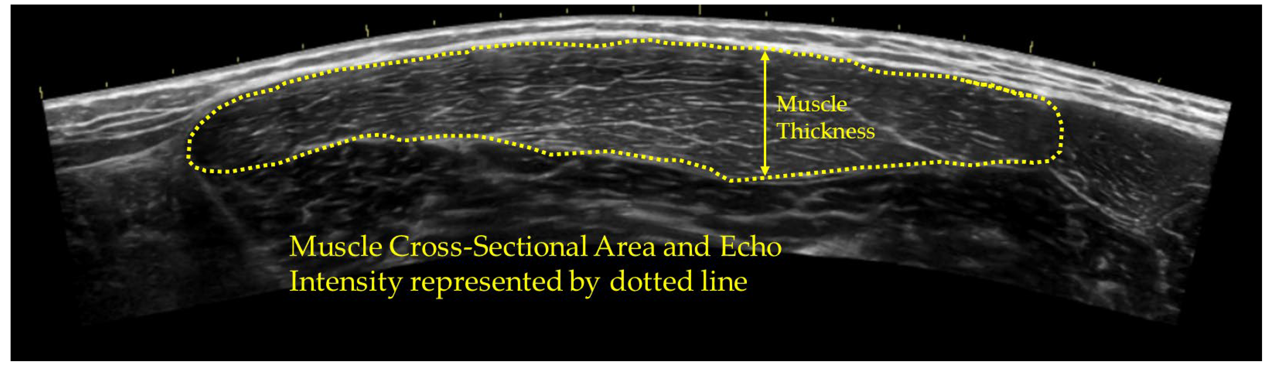
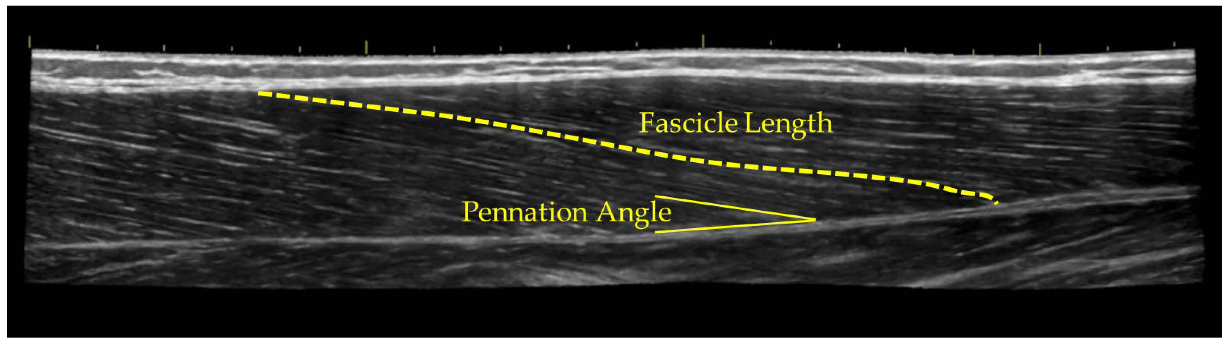
2.4. Ultrasound Assessment

Ultrasound (US) images were taken with a portable B-mode imaging device (GE Logiq e BT12, GE Healthcare, Milwaukee, WI, USA) and a multi-frequency linear-array probe (12 L-RS, 5–13 MHz, 38.4 mm field of view, GE Healthcare, Milwaukee, WI, USA). The panoramic function was used to obtain cross-sectional area images of the right vastus lateralis (VL) in the transverse plane. Participants were seated with their leg bent to 90 degrees, in the same position as isometric testing. Images of the VL were taken at 2/3 of the distance between the anterior superior iliac spine and the superior border of the patella. A high-density foam pad was secured around the right thigh with an adjustable strap [51]. The pennation angle and fascicle length [52] were recorded at the same location, but along the longitudinal axis. The angle of the transducer was adjusted visually to optimize the muscle fascicles, and then the panoramic function was used to track an individual muscle fiber so that the whole muscle fiber was visible in the image. US settings (frequency: 10 MHz, gain: 45 dB, dynamic range: 72) were kept consistent across participants. To ensure optimal image clarity, the scanning depth was individualized for each participant between 3.5 and 6.0 cm and remained the same throughout all testing. Three images were taken for each participant for each measurement, and the mean of these values was used for analysis. Skin was marked to ensure the measurements were taken at the exact same spot for post-modality measurements.
The US images were digitized and examined with ImageJ Software (version 1.46, National Institutes of Health, Bethesda, MD, USA). The polygon function was used to outline the border of the VL for the cross-sectional area. Then, the echo intensity was assessed with a computer-aided gray-scale analysis using the histogram function. (Figure 1) The echo intensity values were determined as the corresponding index of muscle quality ranging between 0 and 255 A.U. (black = zero, white = 255) [22]. Muscle thickness was calculated as the distance between the superficial to the deep aponeuroses (Figure 1). The pennation angle was assessed using the angle function between the fascicle and deep aponeuroses (Figure 2). The fascicle length was determined by manually tracing the fascicle from the superficial to the deep aponeuroses, and the length was recorded (Figure 2).

2.5. Isometric Testing and Surface EMG

Participants sat in an isokinetic dynamometer chair (Biodex System 2, 900–800) with restraints around the shoulders, torso, and thigh. All of the participants’ right legs (dominant) were used. The knee was positioned to 90 degrees and locked in place. Prior to testing, the subjects’ legs were cleaned and prepped with alcohol and marked with permanent marker where the EMG sensor will be placed to ensure the same placement for each test. A wireless surface EMG (Trigno, Delsys, Inc., Natick, MA, USA) was placed over the vastus lateralis at the same place as the ultrasound measurement [53] and in accordance with the Non-Invasive Assessment of Muscle project [54]. Participants were then instructed to kick as hard and as fast as they could for three seconds. This was repeated three times. Peak torque was calculated using Biodex software V2.0.10. The EMG signal was processed using EMG works (Delsys, Inc., Natick, MA, USA), and smoothed with a window size of a 0.025 s root mean square (RMS). The RMS signal was used to calculate the peak EMG amplitude. The low baseline noise and interface was visually inspected to be less than 1.0 so that a between-session comparison can be made. Previous research has demonstrated the excellent intrasession reliability [55] of surface EMG as well as good [56] intersession reliability when looking at maximal voluntary contractions.

2.6. Statistical Analysis

A factorial (sex) 2 × 4-way repeated measures ANOVA was run comparing pre/post values for the ultrasound assessment—cross-sectional area, echo intensity, pennation angle, fascicles length, and muscle thickness for the VL—as well as the isometric testing—peak torque and peak EMG—across each modality. This was used to compare males vs. females across time and between each modality. Post hoc Bonferroni-corrected paired sample t-tests were used to determine significance across time and group. All statistical analyses were performed using the Statistical Package for Social Science (IBM SPSS Statistics for Windows, Version 23.0. Armonk, NY, USA). All data were normally distributed and had equal variance. Due to the repetitive nature of running multiple ANOVAs, an adjusted Alpha level was set post hoc using the Holm p-value correction [57]. Individual alpha levels were set based on the results of the correlated values (peak torque/peak EMG, cross-sectional area/muscle thickness, and pennation angle/fascicles length). Corrected p values will be signified by cP.
Effect sizes were reported as partial Eta Squared (ηp2). A prior power analysis was conducted using G*Power version 3.1.9.7 for sample size estimation. Effect size was set to 0.6 [4], alpha was set to 0.05, and power was set to 0.95, with two groups and four measurements using a repeated measures design resulting in sample size of 8 per group.
During testing, 12 participants were selected to perform reliability testing, for which they reported to the lab on two separate days (~24 h) and had their ultrasound measurements taken. Reliability was analyzed using Model “3,1” [58] on all ultrasound variables (cross-sectional area, echo intensity, pennation angle, fascicle length, and muscle thickness). The statistics of interest were the results of a repeated measures ANOVA, Intra-class Correlation Coefficients (ICCs), Standard Error of Measurement (SEM), and Minimum Difference (MD) values.

3. Results

3.1. Ultrasound Measurements

In general, none of the ultrasound measurements yielded a significant difference between modalities, as they all had similar effects. None of the modalities had a positive or negative significant effect on the cross-sectional area, echo intensity, pennation angle, fascicle length, or muscle thickness. The only significant result was that the male participants, overall, had larger muscles than the female participants, thus larger pennation angles and fascicle length (Table 1).
For the cross-sectional area, there was no interaction effect (F = 0.97, p = 0.34, ηp2 = 0.08), and there was no modality effect (F = 0.59, p = 0.45, ηp2 = 0.16) or time difference (pre vs. post) (F = 1.84, p = 0.68, ηp2 = 0.09), but there was an effect for sex (cP = 0.009, ηp2 = 0.51), with a corrected p value of 0.009, with males having a higher cross-sectional area than females. The reliability results for the cross-sectional area are as follows: F = 0.95, p = 0.35, ICC3.1 = 0.99, SEM = 0.40 cm2, MD = 1.1 cm2. For echo intensity, there was no interaction effect (F = 01.21, p = 0.33, ηp2 = 0.12), and there was no modality effect (F = 1.77, p = 0.33, ηp2 = 0.12), time (pre vs. post) (F = 1.61, p = 0.20, ηp2 = 0.16), or sex difference (p = 0.47, ηp2 = 0.03). The reliability results for echo intensity are as follows: F = 0.19, p = 0.61, ICC3.1 = 0.98, SEM = 1.52 a.u., MD = 4.23 a.u. For the pennation angle, there was no interaction effect (F = 1.42, p = 0.24, ηp2 = 0.05), and there was no modality effect (F = 1.71, p = 0.19, ηp2 = 0.09) or time difference (pre vs. post) (F = 0.62, p = 0.65, ηp2 = 0.02), but there was an effect for sex (cP = 0.008, ηp2 = 0.22), with males having a larger pennation angle than females. The reliability results for the pennation angle are as follows: F = 0.01, p = 0.98, ICC3.1 = 0.97, SEM = 0.57°, MD = 1.58°. For the fascicle length, there was no interaction effect (F = 1.81, p = 0.19, ηp2 = 0.03), and there was no effect for modality (F = 2.98, p = 0.09, ηp2 = 0.11), time (pre vs. post) (F = 01.12, p = 0.46, ηp2 = 0.01), or sex (cP = 0.09, ηp2 = 0.31), with males having a longer fascicle length than females. The reliability results for fascicle length are as follows: F = 4.01, p = 0.09, ICC3.1 = 0.88, SEM = 0.35 cm, MD = 0.95 cm. For muscle thickness, there was no interaction effect (F = 1.71, p = 0.19, ηp2 = 0.07), and there was no effect for modality (F = 1.41, p = 0.24, ηp2 = 0.13), time (pre vs. post) (F = 1.00, p = 0.23, ηp2 = 0.02), or sex (cP = 0.25, ηp2 = 0.20). The reliability results for muscle thickness are as follows: F = 0.11, p = 0.75, ICC3.1 = 0.98, SEM = 0.04 cm, MD = 0.12 cm.

3.2. Isometric and EMG Measruments

In general, none of the isometric testing yielded a significant difference between modalities, as they all had similar effects. None of the modalities had a positive or negative significant effect on peak torque, or peak EMG. The only significant result was males being able to produce more force output, resulting in larger peak torque, max work, and average power (Table 1).
For peak torque, there was no interaction effect (F = 0.89, p = 0.58, ηp2 = 0.08), and there was no effect for modality (F = 0.35 p = 0.74, ηp2 = 0.01) or time (pre vs. post) (F = 1.94, p = 0.18, ηp2 = 0.81), but there was an effect for sex (cP = 0.08, ηp2 = 0.59.), with males having a higher peak torque than females. For peak EMG, there was no interaction effect (F = 0.31, p = 0.722, ηp2 = 0.09), and there was no effect for modality (F = 0.07 p = 0.78, ηp2 = 0.04), time (pre vs. post) (F = 2.46, p = 0.19, ηp2 = 0.08), or sex (p = 0.29, ηp2 = 0.09).

4. Discussion

Overall, the results of this study did not reveal that any one soft-tissue modality significantly differs from another. Static stretching, foam rolling, and percussion massage did not alter the cross-sectional area, echo intensity, pennation angle, fascicle length, or muscle thickness. The results agree with much of the literature in that none of the three modalities improve performance as we did not observe any significant changes in peak torque or muscle activation. The only significant difference that was observed was in the strength output and muscle size of the male participants. The males were able to generate more force and had a larger muscle mass. This was known and controlled in the study [46]; however, it is of note that regardless of size and strength, there was no significant difference in the use of all three modalities between sexes.
For static stretching, there were no statistically significant differences on all performance metrics. These results back the findings of much of the current literature in that static stretching has no effect on improving performance [4,12,13,15,16,17] or a slight decrease in performance of <5% [12]. Dennis et al. [20] reported an increase in ROM of 4.3°, corresponding to a 2° increase in pennation angle. Changes in fascicle length or pennation angles were not observed in our study. This is similar to the results reported by Panidi et al. [19]. Static stretching has been shown to decrease stiffness [13], while echo intensity is related to changes in elongation and stiffness [25] of the muscle. We, however, did not observe any differences between pre- and post-stretching echo intensity values. Even though we did not measure stiffness or ROM, we did not see any changes in muscle characteristics that would indicate that acute stretching does not alter any muscle morphology. If stiffness were to decrease, we would have seen a decrease in echo intensity, based off the work of Nakao et al. [25]. Our results suggest that changes in ROM and stiffness cannot be detected with these ultrasound measurements [24].
Our results for foam rolling agreed with other studies that found foam rolling to have little or no impact on muscle characteristics [36] or performance measures [28,32,37]. There were no changes in any of the muscle characteristics, and this is in agreement with Yektaei et al. [27], but it contradicts Brigatto et al. [37], who reported an increase in muscle thickness following foam rolling. Evidence that foam rolling increases muscle size by increased tissue hydration and blood flow [10,37] was not observed in our study. There were no increases in muscle size or echo intensity following foam rolling. Foam rolling has been speculated to improve force production by stimulating the neural system to increase its firing rate and recruitment patterning [10]; however, we did not see improvement in any of the isometric strength measurements or muscle activation, which is similar to other findings [49,59]. When addressing the sex difference, our results indicate that there were no differences between males and females on how they respond to foam rolling. This contradicts the results of Cornell et al. [49] who reported that there was an increase in peak force output for males but not females following foam rolling. They attributed these results to muscle mass differences.
For percussion massage, the results of this study build upon previous research [11,39,43,44]. There were no changes in performance testing, force output, or muscle activation following percussion massage. However, the results also contradict the findings of studies that found increases in horizontal jumping test, T drill test, balance measurements, and peak torque [41,42]. Percussion massage is believed to increase blood flow to the muscle, which could lead to increases in performance, but we did not observe any changes in muscle size or echo intensity that would lead us to believe there was an increase in blood flow to the muscle. This is different than what has been previously reported in the literature [36] that percussion massage increases muscle thickness. Overall, percussion massage had no effect on any muscle characteristics or performance outcome.
One result of this study that was significant was the size and strength differences between males and females, with males having a larger cross-sectional area, pennation angle, and peak torque. These are similar to other findings that demonstrate differences in muscle mass (14 vs. 11.5 cm2, respectively) [46] and strength [47] between sexes. Nuzzo et al. [47] reported an average 60% decrease in strength from males to females; this study’s average difference in peak torque was ~55%. These differences in males versus females are reduced when normalizing strength values to muscle size [60,61]. Although these findings support the differences between sexes, the novelty of this study is that both sexes respond similarly to the modalities. This indicates that muscle size or strength has little effect on how one would respond to these soft-tissue modalities. This furthers the research that has demonstrated no sex difference in muscle properties [62] or in response to stretching [63]. This contradicts previously reported [49] results that males and females have different responses to soft-tissue modalities. However, the results of this study and the summation of previous literature indicate that there is no sex difference in response to soft-tissue modalities.
There are limitations to this study, as most of the modality research is focused on increasing ROM. This study did not measure ROM, so we cannot confirm that our protocol increased ROM. Our protocol consisted of five minutes in duration, which matched or exceeded other studies that had shown increases in ROM. Static stretching was greater than 1 min [4], foam rolling was two minutes [28], and percussion massage was five minutes [11], and in other studies, these have been shown to increase ROM. Another limitation is that we defined performance as one’s ability to isometrically contract the knee extensor muscles, as maximal isometric contractions are common in the literature [4,13,53], while other studies have defined performance as dynamic muscle contraction, such as in vertical jumping [12], sprinting [15], or Wingate performance [18]. Our results only speak to the muscles’ ability to produce isometric force following these modalities. Another limitation is the use of ultrasound to measure fascicle length and pennation angle as it is a 2D approximation of a 3D structure [64]. We did, however, have good reliability in all our measurements: cross-section area ICC = 0.98, echo intensity ICC = 0.98, fascicle length ICC = 0.88, pennation angle ICC = 0.97, and muscle thickness ICC = 0.98. This is in line with previous literature, where ICC > 0.7 [51,65]. Future research should include different measures of performance, as the effects of these modalities may be more suited for dynamic contractions, or possible help with more anerobic type activities. Sonoelastography may shed more light on these modalities by measuring elasticity changes. Different muscles should also be addressed, as we did not find any improvement or detrimental effect for the quadricep muscle group. Muscles, such as gastrocnemius, may benefit from these modalities.

5. Conclusions

The main objective of research on soft-tissue modalities is to determine its purpose. The results of this study as well as of the previous literature have shown that static stretching, foam rolling, and percussion massage do not significantly change any mechanisms in human muscle that may lead to specific performance changes. They do not affect any mechanical or neuromuscular components of the underlying muscle tissue; thus, their role in improving performance is minimal at best. For clinical application, these modalities may be used if an increase in ROM is a desired outcome, but if the use of these modalities is to prepare the muscle for performance, there is no evidence that they will. An increase in ROM does not mean an increase in performance [54]. These modalities may be used as a psychological mechanism [2] (athletes like them) in a warmup routine, but overall, the results do not favor one modality over the others, and the focus of these modalities should not be on improving performance.

Author Contributions

Conceptualization, E.S.; software, E.S. and W.T.; validation, E.S.; methodology, R.H., T.R. and N.W.; formal analysis, W.T., R.H., T.R. and N.W.; investigation, E.S.; resources, E.S.; data curation, W.T., R.H., T.R. and N.W.; writing—original draft preparation, E.S. and W.T.; writing—review and editing, W.T., R.H., T.R. and N.W.; visualization, E.S.; supervision, E.S.; project administration, E.S.; funding acquisition, E.S. All authors have read and agreed to the published version of the manuscript.

Funding

This research received no external funding.

Institutional Review Board Statement

The study was in accordance with the Declaration of Helsinki and the design was approved by the Institutional Review Board of Furman University (FU52422).

Informed Consent Statement

Informed consent was obtained from all participants involved in this study.

Data Availability Statement

The original contributions presented in the study are included in the article, further inquiries can be directed to the corresponding author.

Acknowledgments

We want to thank Furman University’s Undergraduate Research Program who helped through their support of summer research students.

Conflicts of Interest

The authors declare no conflicts of interest.

References

  1. Shellock, F.G.; Prentice, W.E. Warming-up and stretching for improved physical performance and prevention of sports-related injuries. Sports Med. 1985, 2, 267–278. [Google Scholar] [CrossRef]
  2. McGowan, C.J.; Pyne, D.B.; Thompson, K.G.; Rattray, B. Warm-up strategies for sport and exercise: Mechanisms and applications. Sports Med. 2015, 45, 1523–1546. [Google Scholar] [CrossRef]
  3. Shrier, I. Does stretching improve performance? a systematic and critical review of the literature. Clin. J. Sport Med. 2004, 14, 267–273. [Google Scholar] [CrossRef] [PubMed]
  4. Behm, D.G.; Blazevich, A.J.; Kay, A.D.; McHugh, M. Acute effects of muscle stretching on physical performance, range of motion, and injury incidence in healthy active individuals: A systematic review. Appl. Physiol. Nutr. Metab. 2016, 41, 1–11. [Google Scholar] [CrossRef]
  5. Amako, M.; Oda, T.; Masuoka, K.; Yokoi, H.; Campisi, P. Effect of static stretching on prevention of injuries for military recruits. Mil. Med. 2003, 168, 442–446. [Google Scholar] [CrossRef]
  6. Behm, D.G.; Kay, A.D.; Trajano, G.S.; Alizadeh, S.; Blazevich, A.J. Effects of stretching on injury risk reduction and balance. J. Clin. Exerc. Physiol. 2021, 10, 106–116. [Google Scholar] [CrossRef]
  7. Herrera, E.; Osorio-Fuentealba, C. Impact of warm-up methods on strength-speed for sprinters in athletics: A mini review. Front. Sports Act. Living 2024, 6, 1360414. [Google Scholar] [CrossRef]
  8. Kerautret, Y.; Di Rienzo, F.; Eyssautier, C.; Guillot, A. Selective effects of manual massage and foam rolling on perceived recovery and performance: Current knowledge and future directions toward robotic massages. Front. Physiol. 2020, 11, 598898. [Google Scholar] [CrossRef] [PubMed]
  9. Davis, H.L.; Alabed, S.; Chico, T.J.A. Effect of sports massage on performance and recovery: A systematic review and meta-analysis. BMJ Open Sport Exerc. Med. 2020, 6, e000614. [Google Scholar] [CrossRef]
  10. Cole, G. The evidence behind foam rolling: A review. Sport Olymp. Paralympic Stud J. 2018, 3, 194–206. [Google Scholar]
  11. Konrad, A.; Glashüttner, C.; Reiner, M.M.; Bernsteiner, D.; Tilp, M. The acute effects of a percussive massage treatment with a hypervolt device on plantar flexor muscles’ range of motion and performance. J. Sports Sci. Med. 2020, 19, 690. [Google Scholar] [PubMed]
  12. Power, K.; Behm, D.; Cahill, F.; Carroll, M.; Young, W. An acute bout of static stretching: Effects on force and jumping performance. Med. Sci. Sports Exerc. 2004, 36, 1389–1396. [Google Scholar] [CrossRef] [PubMed]
  13. Reiner, M.M.; Tilp, M.; Guilhem, G.; Morales-Artacho, A.; Konrad, A. Comparison of a single vibration foam rolling and static stretching exercise on the muscle function and mechanical properties of the hamstring muscles. J. Sports Sci. Med. 2022, 21, 287. [Google Scholar] [CrossRef] [PubMed]
  14. Škarabot, J.; Beardsley, C.; Štirn, I. Comparing the effects of self-myofascial release with static stretching on ankle range-of-motion in adolescent athletes. Int. J. Sports Phys. Ther. 2015, 10, 203. [Google Scholar] [PubMed]
  15. Beckett, J.R.; Schneiker, K.T.; Wallman, K.E.; Dawson, B.T.; Guelfi, K.J. Effects of static stretching on repeated sprint and change of direction performance. Med. Sci. Sports Exerc. 2009, 41, 444–450. [Google Scholar] [CrossRef]
  16. Young, W.B.; Behm, D.G. Effects of running, static stretching and practice jumps on explosive force production and jumping performance. J. Sports Med. Phys. Fit. 2003, 43, 21–27. [Google Scholar]
  17. Yildiz, S.; Gelen, E.; Çilli, M.; Karaca, H.; Kayihan, G.; Ozkan, A.; Sayaca, C. Acute effects of static stretching and massage on flexibility and jumping performance. J. Musculoskelet. Neuron. Interact. 2020, 20, 498. [Google Scholar]
  18. Janot, J.; Malin, B.; Cook, R.; Hagenbucher, J.; Draeger, A.; Jordan, M.; Quinn, E. Effects of self-myofascial release and static stretching on anaerobic power output. J. Fit. Res. 2013, 2, 41–54. [Google Scholar]
  19. Panidi, I.; Donti, O.; Konrad, A.; Dinas, P.C.; Terzis, G.; Mouratidis, A.; Gaspari, V.; Donti, A.; Bogdanis, G.C. Muscle architecture adaptations to static stretching training: A systematic review with meta-analysis. Sports Med. Open 2023, 9, 47. [Google Scholar] [CrossRef]
  20. Dennis, L.G.; Blais, E.A.; Wagstaff, K.E.; Li, L.; Biber, D. The Effect of Static Stretching on Proprioception, Pennation Angle, and Muscle Power Production. Int. J. Biotechnol. Bioeng. 2020, 6, 37. [Google Scholar]
  21. Young, H.; Jenkins, N.T.; Zhao, Q.; Mccully, K.K. Measurement of intramuscular fat by muscle echo intensity. Muscle Nerve 2015, 52, 963–971. [Google Scholar] [CrossRef] [PubMed]
  22. Sobolewski, E.J.; Hall, A.B.; Rodriguez, G.C.; Richard, M.O. The Effect of Aerobic Exercise on Quadriceps Echo Intensity and Cross-Sectional Area. Int. J. Phys. Educ. Exerc. Sports 2020, 2, 6–9. [Google Scholar]
  23. Yitzchaki, N.; Kuehne, T.E.; Mouser, J.G.; Buckner, S.L. Can changes in echo intensity be used to detect the presence of acute muscle swelling? Physiol. Meas. 2019, 40, 045002. [Google Scholar] [CrossRef] [PubMed]
  24. Şekir, U.; Arslan, G.; İlhan, O.; Akova, B. Effects of static and dynamic stretching on muscle architecture. Turk. J. Sports Med. 2019, 54, 158–168. [Google Scholar] [CrossRef]
  25. Nakao, S.; Ikezoe, T.; Yagi, M.; Umehara, J.; Nojiri, S. Changes in echo intensity of the gastrocnemius muscle with passive ankle dorsiflexion: Can echo intensity be used to assess muscle elongation? Front. Physiol. 2023, 14, 1197503. [Google Scholar] [CrossRef]
  26. Nakamura, M.; Ikezoe, T.; Nishishita, S.; Umehara, J.; Kimura, M.; Ichihashi, N. Acute effects of static stretching on the shear elastic moduli of the medial and lateral gastrocnemius muscles in young and elderly women. Musculoskelet. Sci. Pract. 2017, 32, 98–103. [Google Scholar] [CrossRef]
  27. Matsuo, H.; Kubota, M.; Shimada, S.; Kitade, I.; Matsumura, M.; Nonoyama, T.; Koie, Y.; Naruse, H.; Takahashi, A.; Oki, H. The Effect of Static Stretching Duration on Muscle Blood Volume and Oxygenation. J. Strength Cond. Res. 2022, 36, 379–385. [Google Scholar] [CrossRef]
  28. MacDonald, G.Z.; Penney, M.D.; Mullaley, M.E.; Cuconato, A.L.; Drake, C.D.; Behm, D.G.; Button, D.C. An acute bout of self-myofascial release increases range of motion without a subsequent decrease in muscle activation or force. J. Strength Cond. Res. 2013, 27, 812–821. [Google Scholar] [CrossRef]
  29. Ajimsha, M.S.; Al-Mudahka, N.R.; Al-Madzhar, J.A. Effectiveness of myofascial release: Systematic review of randomized controlled trials. J. Bodyw. Mov. Ther. 2015, 19, 102–112. [Google Scholar] [CrossRef]
  30. Macdonald, G.Z.; Button, D.C.; Drinkwater, E.J.; Behm, D.G. Foam Rolling as a Recovery Tool after an Intense Bout of Physical Activity. Med. Sci. Sports Exerc. 2014, 46, 131–142. [Google Scholar] [CrossRef]
  31. Roylance, D.S.; George, J.D.; Hammer, A.M.; Rencher, N.; Fellingham, G.W.; Hager, R.L.; Myrer, W.J. Evaluating acute changes in joint range-of-motion using self-myofascial release, postural alignment exercises, and static stretches. Int. J. Exerc. Sci. 2013, 6, 6. [Google Scholar]
  32. Healey, K.C.; Hatfield, D.L.; Blanpied, P.; Dorfman, L.R.; Riebe, D. The effects of myofascial release with foam rolling on performance. J. Strength Cond. Res. 2014, 28, 61–68. [Google Scholar] [CrossRef] [PubMed]
  33. Chan, Y.; Wang, T.; Chang, C.; Chen, L.; Chu, H.; Lin, S.; Chang, S. Short-term effects of self-massage combined with home exercise on pain, daily activity, and autonomic function in patients with myofascial pain dysfunction syndrome. J. Phys. Ther. Sci. 2015, 27, 217–221. [Google Scholar] [CrossRef]
  34. Chaitow, L. Research in the water and fascia: Micro-tornadoes, hydrogenated diamonds & nanocrystals. Massage Today 2009, 9, 17–21. [Google Scholar]
  35. Schroeder, A.N.; Best, T.M. Is self-myofascial release an effective preexercise and recovery strategy? A literature review. Curr. Sports Med. Rep. 2015, 14, 200–208. [Google Scholar] [CrossRef]
  36. Yektaei, M.; Akkoç, O.; Devran, S.; Kurtdere, I.; Kirandi, Ö.; Bayraktar, B. Effect of Acute Foam Roller and Percussion Therapy on Muscle Architecture and Muscle Stiffness. SPORMETRE: J. Phys. Educ. Sport Sci. 2023, 21, 21–34. [Google Scholar]
  37. Brigatto, F.A.; Soares, E.G.; Braz, T.V.; DE CAMARGO, J.B.; Hartz, C.S.; Batista, D.R.; Col, L.O.; Marchetti, P.H.; Aoki, M.S.; Lopes, C.R. Acute Effect of Different Duration of Foam Rolling Protocols on Muscle Thickness, Pain Pressure Threshold, and Volume Load on Multiple Sets of Knee Extension. Int. J. Exerc. Sci. 2021, 14, 742. [Google Scholar]
  38. Torrente, Q.M.; Killingback, A.; Adds, P.J.; Robertson, C. The effect of self-myofascial release on the pennation angle of the vastus medialis oblique and the vastus lateralis in athletic male individuals: An ultrasound investigation. Int. J. Sports Phys. Ther. 2022, 17, 636. [Google Scholar] [CrossRef]
  39. Martin, J. A critical evaluation of percussion massage gun devices as a rehabilitation tool focusing on lower limb mobility: A literature review. SportRxiv 2021. [Google Scholar] [CrossRef]
  40. Lee, C.; Chu, I.; Lyu, B.; Chang, W.; Chang, N. Comparison of vibration rolling, nonvibration rolling, and static stretching as a warm-up exercise on flexibility, joint proprioception, muscle strength, and balance in young adults. J. Sports Sci. 2018, 36, 2575–2582. [Google Scholar] [CrossRef]
  41. Menek, M.Y.; Menek, B. Effects of percussion massage therapy, dynamic stretching, and static stretching on physical performance and balance. J. Back Musculoskelet. Rehabil. 2023, 37, 183–193. [Google Scholar] [CrossRef] [PubMed]
  42. Sams, L.; Langdown, B.L.; Simons, J.; Vseteckova, J. The Effect Of Percussive Therapy On Musculoskeletal Performance And Experiences Of Pain: A Systematic Literature Review. Int. J. Sports Phys. Ther. 2023, 18, 309–327. [Google Scholar] [CrossRef] [PubMed]
  43. Ferreira, R.M.; Silva, R.; Vigário, P.; Martins, P.N.; Casanova, F.; Fernandes, R.J.; Sampaio, A.R. The effects of massage guns on performance and recovery: A systematic review. J. Funct. Morphol. Kinesiol. 2023, 8, 138. [Google Scholar] [CrossRef]
  44. Leabeater, A.; Clarke, A.; James, L.; Huynh, M.; Driller, M. Under the Gun: The effect of percussive massage therapy on physical and perceptual recovery in active adults. J. Athl. Train. 2023, 59, 310–316. [Google Scholar] [CrossRef]
  45. Trainer, J.H.; Pascarella, M.; Paul, R.W.; Thomas, S.J. Acute effects of percussive therapy on the posterior shoulder muscles differ based on the athlete’s soreness response. Int. J. Sports Phys. Ther. 2022, 17, 887. [Google Scholar] [CrossRef] [PubMed]
  46. Stock, M.S.; Oranchuk, D.J.; Burton, A.M.; Phan, D.C. Age-, sex-, and region-specific differences in skeletal muscle size and quality. Appl. Physiol. Nutr. Metab. 2020, 45, 1253–1260. [Google Scholar] [CrossRef]
  47. Nuzzo, J.L. Narrative review of sex differences in muscle strength, endurance, activation, size, fiber type, and strength training participation rates, preferences, motivations, injuries, and neuromuscular adaptations. J. Strength Cond. Res. 2023, 37, 494–536. [Google Scholar] [CrossRef]
  48. Behm, D.G.; Alizadeh, S.; Daneshjoo, A.; Anvar, S.H.; Graham, A.; Zahiri, A.; Goudini, R.; Edwards, C.; Culleton, R.; Scharf, C. Acute effects of various stretching techniques on range of motion: A systematic review with meta-analysis. Sports Med. -Open 2023, 9, 107. [Google Scholar] [CrossRef]
  49. Cornell, D.J.; Ebersole, K.T. Influence of an acute bout of self-myofascial release on knee extension force output and electro-mechanical activation of the quadriceps. Int. J. Sports Phys. Ther. 2020, 15, 732. [Google Scholar] [CrossRef]
  50. Place, N.; Blum, Y.; Armand, S.; Maffiuletti, N.A.; Behm, D.G. Effects of a short proprioceptive neuromuscular facilitation stretching bout on quadriceps neuromuscular function, flexibility, and vertical jump performance. J. Strength Cond. Res. 2013, 27, 463–470. [Google Scholar] [CrossRef]
  51. Sobolewski, E.J.; Wein, L.D.; Crow, J.M.; Carpenter, K.M. Intra-rater and inter-rater reliability of the process of obtaining cross-sectional area and echo intensity measurements of muscles from ultrasound images. J. Ultrason. 2021, 21, 7–11. [Google Scholar] [CrossRef] [PubMed]
  52. Ruple, B.A.; Mesquita, P.H.; Godwin, J.S.; Sexton, C.L.; Osburn, S.C.; McIntosh, M.C.; Kavazis, A.N.; Libardi, C.A.; Young, K.C.; Roberts, M.D. Changes in vastus lateralis fibre cross-sectional area, pennation angle and fascicle length do not predict changes in muscle cross-sectional area. Exp. Physiol. 2022, 107, 1216–1224. [Google Scholar] [CrossRef] [PubMed]
  53. Stock, M.S.; Olinghouse, K.D.; Drusch, A.S.; Mota, J.A.; Hernandez, J.M.; Akalonu, C.C.; Thompson, B.J. Evidence of muscular adaptations within four weeks of barbell training in women. Hum. Mov. Sci. 2016, 45, 7–22. [Google Scholar] [CrossRef]
  54. Hermens, H.J.; Freriks, B.; Disselhorst-Klug, C.; Rau, G. Development of recommendations for SEMG sensors and sensor placement procedures. J. Electromyogr. Kinesiol. 2000, 10, 361–374. [Google Scholar] [CrossRef] [PubMed]
  55. Fauth, M.L.; Petushek, E.J.; Feldmann, C.R.; Hsu, B.E.; Garceau, L.R.; Lutsch, B.N.; Ebben, W.P. Reliability of surface electromyography during maximal voluntary isometric contractions, jump landings, and cutting. J. Strength Cond. Res. 2010, 24, 1131–1137. [Google Scholar] [CrossRef]
  56. Yang, J.F.; Winter, D.A. Electromyography reliability in maximal and submaximal isometric contractions. Arch.Phys.Med.Rehabil. 1983, 64, 417–420. [Google Scholar] [PubMed]
  57. Holm, S. A simple sequentially rejective multiple test procedure. JSTOR 1979, 2, 65–70. [Google Scholar]
  58. Weir, J.P. Quantifying test-retest reliability using the intraclass correlation coefficient and the SEM. J. Strength Cond. Res. 2005, 19, 231–240. [Google Scholar]
  59. Cheatham, S.W.; Kolber, M.J.; Cain, M.; Lee, M. The effects of self-myofascial release using a foam roll or roller massager on joint range of motion, muscle recovery, and performance: A systematic review. Int. J. Sports Phys. Ther. 2015, 10, 827. [Google Scholar]
  60. Comfort, P.; McMahon, J.J.; Lake, J.P.; Ripley, N.J.; Triplett, N.T.; Haff, G.G. Relative strength explains the differences in multi-joint rapid force production between sexes. PLoS ONE 2024, 19, e0296877. [Google Scholar] [CrossRef]
  61. Nimphius, S.; McBride, J.M.; Rice, P.E.; Goodman-Capps, C.L.; Capps, C.R. Comparison of quadriceps and hamstring muscle activity during an isometric squat between strength-matched men and women. J. Sports Sci. Med. 2019, 18, 101. [Google Scholar] [PubMed]
  62. Chino, K.; Takahashi, H. Measurement of gastrocnemius muscle elasticity by shear wave elastography: Association with passive ankle joint stiffness and sex differences. Eur. J. Appl. Physiol. 2016, 116, 823–830. [Google Scholar] [CrossRef] [PubMed]
  63. Martín-Santana, E.; Hernández-Sánchez, S.; Herrero-Alonso, A.J.; García-López, D. Effects of static-stretching and whole-body-vibration during warm-ups on bench-press kinematics in males and females college-aged. RICYDE. Rev. Int. De Cienc. Del 2015, 11, 348–359. [Google Scholar]
  64. Dick, T.J.; Hug, F. Advances in imaging for assessing the design and mechanics of skeletal muscle in vivo. J. Biomech. 2023, 155, 111640. [Google Scholar] [CrossRef]
  65. Kwah, L.K.; Pinto, R.Z.; Diong, J.; Herbert, R.D. Reliability and validity of ultrasound measurements of muscle fascicle length and pennation in humans: A systematic review. J. Appl. Physiol. 2013, 114, 761–769. [Google Scholar] [CrossRef]
Figure 1. Cross-sectional area and echo intensity measurements in ultrasound image.
Applsci 14 07994 g001
Figure 2. Pennation angle and fascicle length measurements in ultrasound image.
Applsci 14 07994 g002
Table 1. Measured variables (means).
ControlStretchFoam RollPercussion Massage
Cross-Sectional Area (cm2)PrePostPrePostPrePostPrePost
Male *16.8 ± 4.018.5 ± 5.616.7 ± 4.416.7 ± 4.318.9 ± 4.418.6 ± 5.217.7 ± 4.318.5 ± 6.0
Female10.5 ± 3.111.2 ± 2.513.7 ± 2.814.9 ± 3.312.5 ± 3.513.2 ± 3.211.0 ± 2.410.8 ± 2.0
Echo Intensity (a.u.)
Male48.2 ± 18.752.7 ± 13.560.8 ± 13.761.5 ± 10.461.8 ± 13.756.0 ± 14.757.6 ± 15.460.4 ± 14.9
Female55.9 ± 11.759.5 ± 10.058.7 ± 5.757.8 ± 6.866.1 ± 11.665.0 ± 7.662.5 ± 9.758.0 ± 10.1
Pennation Angle (°)
Male *15.3 ± 4.816.1 ± 3.314.3 ± 3.814.0 ± 4.316.0 ± 3.716.1 ± 4.215.0 ± 3.614.2 ± 2.4
Female13.5 ± 3.614.7 ± 3.311.7 ± 3.212.1 ± 3.814.5 ± 3.514.8 ± 2.812.7 ± 2.412.9 ± 2.7
Fascicle Length (cm)
Male *10.7 ± 1.610.7 ± 1.311.2 ± 2.411.5 ± 2.811.0 ± 1.710.5 ± 2.110.0 ± 1.610.4 ± 2.3
Female9.8 ± 0.99.7 ± 1.310.3 ± 1.510.5 ± 1.79.7 ± 0.99.9 ± 1.29.1 ± 1.39.5 ± 1.3
Muscle Thickness (cm)
Male *2.5 ± 0.52.6 ± 0.42.2 ± 0.52.3 ± 0.52.5 ± 0.42.3 ± 0.52.3 ± 0.32.2 ± 0.4
Female2.1 ± 0.52.2 ± 0.42.0 ± 0.52.0 ± 0.52.0 ± 0.42.0 ± 0.32.0 ± 0.81.9 ± 0.5
Peak Torque (Nm)
Male *224.1 ± 58.2228.9 ± 55.9227.9 ± 60.2226.1 ± 71.1224.8 ± 75.3241.7 ± 75.1227.2 ± 56.9235.8 ± 74.7
Female122.3 ± 54.9125.5 ± 56.3118.4 ± 48.6116.2 ± 56.6120.5 ± 70.2121.2 ± 69.5121.4 ± 51.4119.2 ± 47.3
Peak EMG (mV)
Male2.6 ± 1.61.4 ± 0.91.6 ± 0.61.8 ± 0.91.6 ± 0.81.8 ± 1.02.1 ± 1.21.7 ± 0.8
Female1.9 ± 0.71.4 ± 0.50.9 ± 0.51.5 ± 0.61.2 ± 0.81.0 ± 0.71.0 ± 0.30.9 ± 0.3
* denotes significant difference between males and females. No significant (p > 0.05) differences were found pre- vs. post- or across modality.
Disclaimer/Publisher’s Note: The statements, opinions and data contained in all publications are solely those of the individual author(s) and contributor(s) and not of MDPI and/or the editor(s). MDPI and/or the editor(s) disclaim responsibility for any injury to people or property resulting from any ideas, methods, instructions or products referred to in the content.

