Article

Biodegradation and Detoxification of Some Dyes by Crude Lignin Peroxidase Complex Produced by Escherichia coli Accession No: LR0250096.1 and Pseudomonas aeruginosa Accession No: CP031449.2

by
Sindiswa Lungile Dube
1,
Foluso Oluwagbemiga Osunsanmi
1,*,
Albert Olufemi Ikhane
1,
Rebamang Anthony Mosa
2 and
Andrew Rowland Opoku
1
1
Department of Biochemistry and Microbiology, University of Zululand, Richards Bay 3886, South Africa
2
Department of Biochemistry, Genetics and Microbiology, University of Pretoria, Private Bag x20, Hatfield 0028, South Africa
*
Author to whom correspondence should be addressed.
Appl. Sci. 2024, 14(17), 8012; https://doi.org/10.3390/app14178012 (registering DOI)
Submission received: 6 August 2024 / Revised: 3 September 2024 / Accepted: 5 September 2024 / Published: 7 September 2024

Abstract

:
Synthetic and untreated dyes discharged in wastewater effluents are a threat to an ecosystem. This study investigated dye degradation and detoxification efficiency of crude lignin peroxidase separately obtained from the cultures of Escherichia coli (LR0250096.1) and Pseudomonas aeruginosa (CP031449.2). The ability of the crude lignin peroxidase to degrade Malachite Green (MG), Remazol Brilliant Blue R (RBBR), Congo Red (CR), and Azure B (AZ) was evaluated at different operating conditions (enzyme, dye, and hydrogen peroxide concentrations; pH; temperature; and contact time). The ability of the degraded dyes to support the growth of bacteria was also investigated. The observed optimum operating conditions for lignin peroxidase extracts of the Escherichia coli on AZ were 20 mg/mL enzyme concentration, 50 mg/L dye, pH 7.0, temperature 50 °C, and 1.5 mM hydrogen peroxide within 20–50 min of incubation time and on MG were 20 mg/mL, 50 mg/L, 9.0, 30 °C, 0.1 mM, and 20 min, respectively. The enzyme extract from Pseudomonas aeruginosa on AZ demonstrated optimum operation conditions of 20 mg/mL, 50 mg/L, pH 9.0, 40 °C, 1.5 mM, and 50 min, respectively and on MG, they were 20 mg/mL, 50 mg/L, 6.0, 30 °C, 1.0 mM, and 20 min, respectively). The prepared enzyme showed an appreciable degradative effect on CR and RBBR compared with commercial lignin peroxidase. The degraded dyes were able to support the growth of two Gram-positive (Bacillus cereus and Staphylococcus aureus), and two Gram-negative (Proteus mirabilis and Escherichia coli) bacteria, indicating the efficiency and the potential use of the enzyme complexes in the clean-up of industrial dyes’ waste.

1. Introduction

Synthetic dyes such as Azure B (AZ), Congo Red (CR), Remazol Brilliant Blue R (RBBR), and Malachite Green (MG) are routinely used in the textile, cosmetics, food, and plastics industries [1]. Such lignin-mimicking dyes, due to their rigid structures and their resistance to oxidizing agents such as water and light, are also hard to degrade and thus cause problems when released untreated onto the environment [2,3]. When the effluent is contaminated by these dyes, the transparency of water decreases, therefore reducing penetration of solar radiation and decreasing photosynthesis of aquatic organisms [4]. Most of these dyes are carcinogenic and made to resist fading upon exposure to chemicals [5]. Therefore, an accumulation of synthetic dyes in water is crucial in wastewater management [6].
To reduce the toxicity of these dyes to the environment, efforts have been focused on treatment and removal from water systems. Regardless of the currently used pre-treatments, their limitations have created the search for an alternative method that provide a more specific and effective way of dye decolorization and detoxification [3].
Biodegradation by ligninolytic enzymes has been considered a better approach to reduce dye pollution during bioremediation [7,8]. Ligninolytic enzymes, including lignin peroxidase, manganese peroxidase, and versatile peroxidase, are topical due to their high redox capability and industrial application [9]. The application of ligninolytic enzymes span through industrial sectors, including bioremediation, biorefinery, energy, dermatology, pharmaceutical, cosmetology, and textile industries [10,11]. Versatile peroxidase and manganese peroxidase enzymes have been studied extensively. However, research on lignin peroxidase is lagging, despite its litany of potentials. Lignin peroxidases are versatile to degrade non-phenolic and phenolic and xenobiotics compounds like dyes [9]. These characteristics have motivated the use of lignin peroxidase in various textiles and the pharmaceutical industry [10,11,12,13,14]. Nonetheless, white rot fungi have extensively been studied in degradation of lignin compounds such as dyes [9]. However, during the course of industrial applications, fungi are not viable due to the structural hindrance caused by fungal filaments and the requirement of specific culture conditions such as aeration, humidity, pH, and temperature, which are incompatible with industrial processing environments. The requirements of a long lag period slow down lignin degradation [15]. The continuous demand for the ligninolytic enzyme in an indication of their values in various industrial sectors [12]. Hence, the exploration of this enzyme from another microorganism is inevitable.
Bacteria are classified as “workhorse industrial microorganisms” based on their production capability, safety, and exponential growth rate [16]. Moreover, various bacteria have been reported in extracellular enzyme formation [17,18,19]. Previously, some bacteria strains have been reported in the production of pectinolytic and cellulolytic enzymes [20,21,22].
Ligninolytic bacteria are sources of extracellular lignin-modifying enzymes (LMEs) like lignin peroxidase [23]. In nature, these enzymes are versatile, and they are associated with the degradation of recalcitrant or complex polymers [24,25]. Dye decolorization by various bacterial extracts and preparations, for example, Bacillus megaterium, Raoultella ornithinolytica OKOH-1, and Ensifer adhaerens NWODO-2. have been reported in literature [5,26]. In this study, the dye degradation and detoxification properties of optimized crude lignin peroxidase produced by Escherichia coli accession no: LR0250096.1 and Pseudomonas aeruginosa accession no: CP031449.2 bacterial strains are investigated.

2. Materials and Methods

2.1. Chemicals

Unless otherwise stated, all the chemicals used in this study were of analytical grade. Nutrient broth, salts, and nutrient agar were purchased from Merck, Darmstadt, Germany. The kraft lignin, purified commercial lignin peroxidase, Azure B (AZB), Congo Red (CR), Remazol Brilliant Blue R (RBBR), and Malachite Green (MG) were also supplied by Sigma Aldrich, St. Louis, MO, USA. Bacteriological agar was bought from Lasec (SA).

2.2. Isolation and Characterization of the Organisms

The isolation and characterization of Pseudomonas aeruginosa CP031449.2 and Escherichia coli LR0250096.1 from compost samples collected at Richards Bay, KwaZulu-Natal, South Africa, have been reported [27]. Briefly, bacteria isolates, identified through screening with the nutrient enrichment technique and found to grow on kraft lignin and effectively produce lignin peroxidase, were subsequently characterized through biochemical assays and 16S rDNA sequencing to mark the organisms genetically.

2.3. Enzyme Production

Lignin peroxidase production by the isolates was optimized by adjusting various culture parameters including pH, temperature, and lignin concentration for growth using the one factor at a time method. The predetermined optimal culture parameters (for E. coli, pH 7.5, temperature 40 °C, and lignin concentration 1 g/L; for P. aeruginosa, the respective conditions of 6.5, 40 °C, and 1 g/L) were used to culture the organisms for 7 days in an MSM-L-glucose medium (4.55 g/L K2HPO4, 0.53 g/L KH2PO4, 0.5 g/L MgSO4, 5 g/L NH4NO3, 1 g/L kraft lignin, 0.1 g/L yeast extract, 1 g/L glucose) in a submerged batch fermentation (2 L conical flasks). The 7-day cultures were filtered, and the filtrate was centrifuged at 15000 rpm for 5 min. The supernatant was then freeze-dried (VirTrisbencbtop K: 6KBTEL-85) to obtain the crude enzyme, which was stored at 4 °C and used for subsequent experiments.
The activity of the optimized enzyme was measured by the rate of H2O2-dependent oxidation of pyrogallol to purpurogallin [26]. To 310 μL of 5% w/v pyrogallol in 100 mM potassium phosphate buffer (pH 6), 25 μL of culture supernatant were added. The mixture, in the absence of the crude enzyme, served as the blank. The reaction was started by adding 15 μL of 0.5% v/v of H2O2 (30% w/w), and the direct increase in absorbance at 420 nm was monitored at 25 °C for 2–3 min at 30 s intervals using a SynergyHT 96-well microtitre plate reader (BioTeK Instruments, Charlotte, VT, USA). The enzyme activity was calculated from the following equation:
Units / mL   enzyme = ( Δ A 420 / 30 sec T e s t S a m p l e Δ A 420 / 30 sec B l a n k ( 3 ) ( d f ) ( 12 ) ( 0.1 )
where: 3 = volume (in milliliters) of an assay, df = dilution factor, 12.0 = extinction coefficient of 1 mg/mL of purpurogallin at 420 nm (determined internally), and 0.1= volume (in milliliters) of enzyme used. The protein concentration of the produced enzyme was determined using the Bradford method [28]. The effect of oxidoreductase inhibitors (1 mM EDTA, 1 mM sodium azide, and 1 mM FeCl3) on the lignin peroxidase activity followed the method described by Ahmedi et al. [29] and Patil [26].

2.4. Dye Decoloration

The parameters affecting dye decoloration, investigated included enzyme concentration (1–100 mg/mL); dye concentration (50–500 mg/L for AZB, MG, and RBBR, and for CR, 10–100 mg/L); pH 3–12; temperature (25–70 °C); H2O2 concentration (0.025–2.5 mM); and time interval (0–200 min). A 96-well plate was used; the uninoculated dye was used as a blank, the uninoculated dye containing controls was used as a reference, and commercial (Sigma) lignin peroxidase (10 mg/mL) was used as the positive control. In a typical assay [26,30], the plates were incubated at the determined optimal conditions on the mechanical shaker at 120 rpm for 30 min, after which the reactions were read spectrophotometrically at 650 nm for RBBR and AZB at 600 nm, for CR at 470 nm, and at 618 nm for MG. Decolorization efficiency (%D) was expressed as follows:
%D = {(Ainitial − Atest)/Ainitial} × 100
where Ainitial was the initial absorbance of untreated dye solutions (control), and Atest was the absorbance of dye solutions after enzymatic treatment.

2.5. Dye Toxicity in Bacteria Study

The two Gram-positive (Bacillus cereus (ATCC14579) and Staphylococcus aureus (LCT-SA112)) and two Gram-negative (Proteus mirabilis (H14320) and Escherichia coli (ATCC9637)) bacteria used in this study were obtained from the Department of Microbiology and Biochemistry, University of Zululand, South Africa. The toxicity of the degraded (treated) and untreated dyes on bacteria growth was examined. Overnight cultures of the bacterial isolates were prepared. An amount of 4.5 mL of dye, before and after enzymatic degradation, was added to 0.5 mL of 10× Luaria–Bertani broth (LB) and inoculated with the respective organism in a 250 mL Erlenmeyer flask. The mixture was incubated at 37 °C on the mechanical shaker (120 rpm). Aliquots were withdrawn at 0 hr and every 2 h for 12 h, then at 18 and 24 h, and bacterial growth was read at 620 nm (a medium solution was used as a blank). Absorbance versus time was plotted to give bacteria growth. Growth inhibition (GI) for each bacterium was calculated using the following formula:
%GI = {(Ainitial − Atest)/Ainitial} × 100
where Ainitial was the initial absorbance of untreated dye solutions (control) and Atest was the absorbance of dye solutions after enzymatic treatment. A graph of %GI vs. time was plotted.

2.6. Enzyme Decolorization Kinetics

The Lineweaver–Burk plot was applied to the decolorization data and a Michaelis–Menten model equation was used to obtain the enzyme kinetics (Vmax, Km, and the turnover number, kcat value (as Vmax/enzyme protein concentration).

2.7. Data Analysis

Unless stated otherwise, all experiments were triplicated, and the results were expressed as mean ± standard deviation (SD). One-way ANOVA and comparison analysis by Dunn’s post hoc was carried out using GraphPad prism (Version 6). The value p < 0.05 was considered as statistically significant.

3. Results

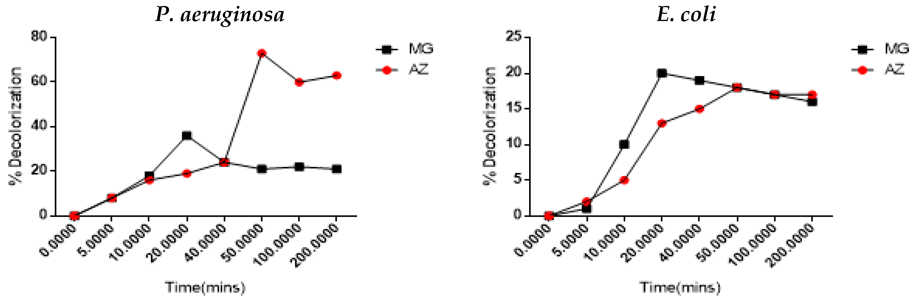
3.1. Dye Decolorization Study

The efficiency of the two crude bacterial lignin peroxidase extracts produced separately by Escherichia coli and Pseudomonas aeruginosa, and that of purified commercial (Sigma) lignin peroxidase (control) to decolorize AZ, MG, RBBR, and CR, is presented in Table 1. The enzymes (200 mg/mL for the crude preparation and 10 mg/mL for the purified preparation) were incubated at room temperature with 200 mg/L dye (50 mg/L for CR) for 30 min. The crude bacterial enzymes significantly showed appreciable decolorization of CR and RBBR in comparison with the commercial lignin peroxidase. Likewise, the crude enzymes appreciably (relative to the purified enzyme) decolorized MG and AZ. These results indicate that the crude enzymes had the potential to degrade the dyes.

3.2. Effect of Key Factors on Dye Decolorization

3.2.1. Effect of Enzyme Concentration

Since the extracts were able to effectively decolorize AZ and MG, these two dyes were used for subsequent experiments. Table 2 indicates that both enzymes were most efficient at 20 mg/mL: E. coli (AZ: 68 ± 1.88% and MG: 55 55 ± 1.78%) and P. aeruginosa (AZ: 52 2.11% and MG: 55 ± 1.66%).

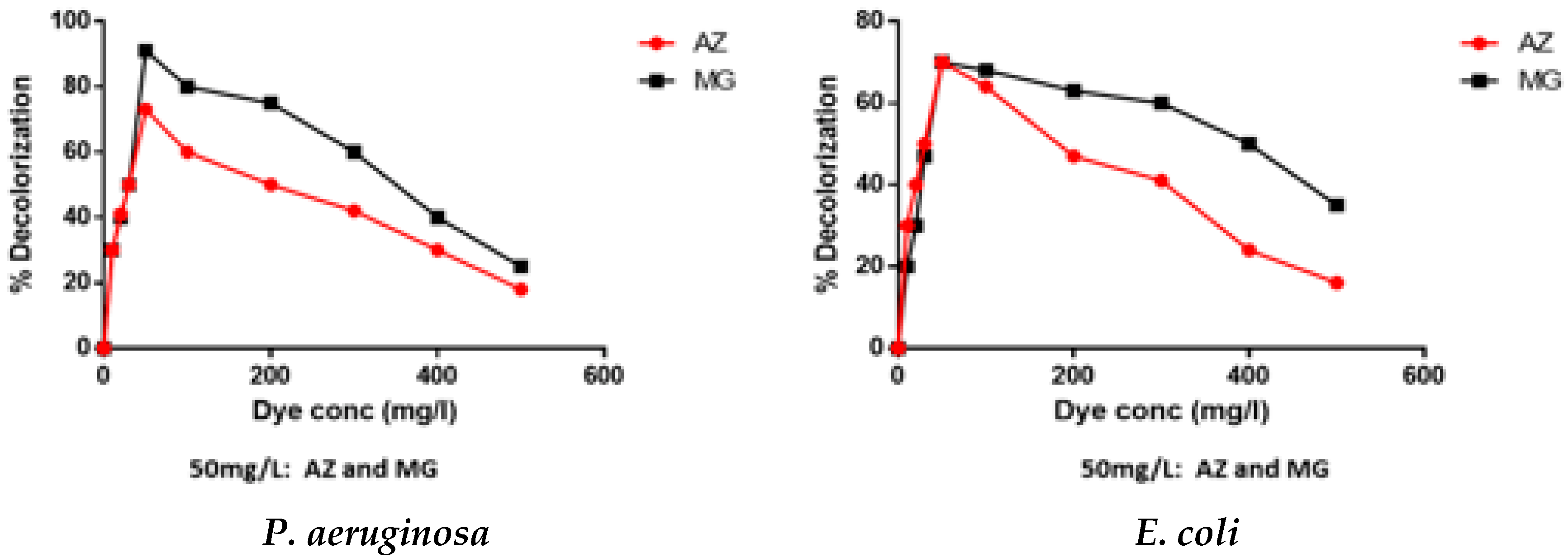
3.2.2. Effect of Dye Concentrations
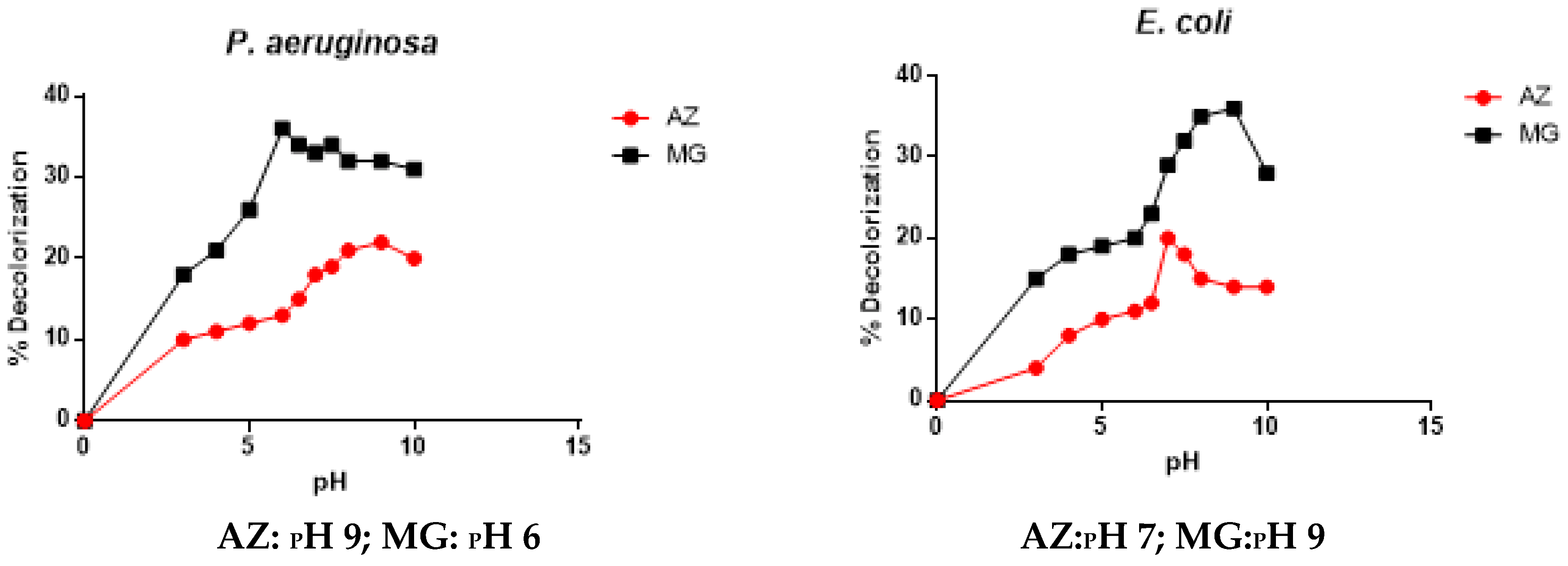
On the effect of different dye concentrations, both bacterial extracts showed high activity, 73% (AZ) and 91% (MG) for P. aeruginosa and 70% (AZ) and 70% (MG) for E. coli, at 50 mg/L on both dyes, as shown in Figure 1. Substrate (dye) inhibition of enzyme activity at higher concentrations was observed.

3.2.3. Effect of pH

The optimal pH observed (Figure 2) for the enzymes’ activity of P. aeruginosa extracts was pH 9.0 for AZ and pH 6.0 for MG, while E. coli extract showed pH 7.0 for AZ and pH 9.0 for MG, respectively.

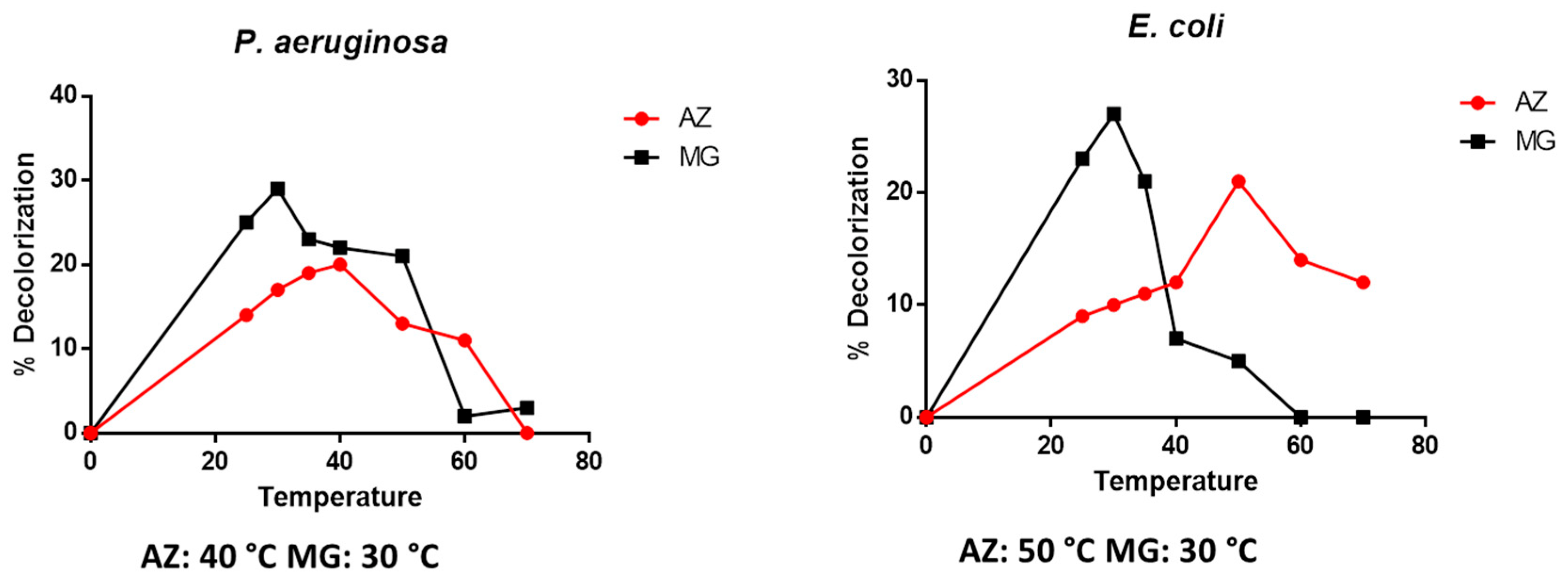
3.2.4. Effect of Temperature

The effect of temperature on the enzymes’ activity is presented in Figure 3. The extracts of P. aeruginosa showed maximum activity at 40 °C (AZ) and 30 °C (MG). The E. coli enzyme was most active at 50 °C (AZ) and 30 °C (MG).

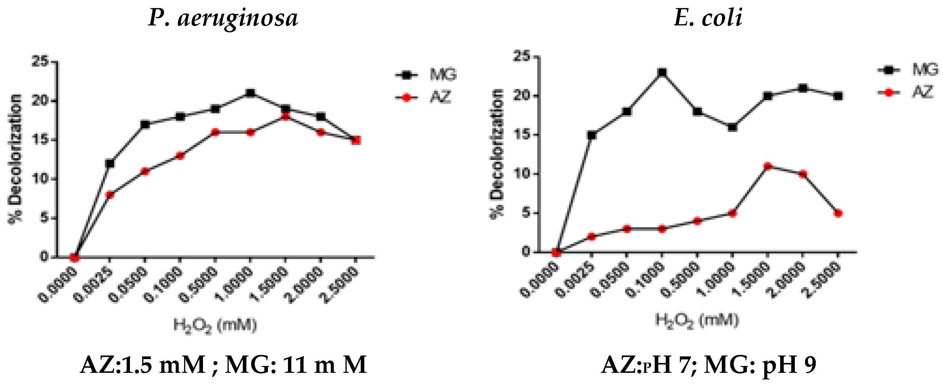
3.2.5. Effect of Substrate (H2O2) Concentration

The effect of substrate on the enzymes’ activity is presented in Figure 4. The extracts of P. aeruginosa showed maximum activity at 1.5 mM (AZ) and 11 mM (MG). The E. coli enzyme was most active at 1.5 mM (AZ) and 0.1 mM (MG).

3.2.6. Effect of Incubating Time

The time taken for both P. aeruginosa and E. coli extracts to decolorize AZ was 50 min and 20 min for MG (Figure 5).

3.2.7. Effect of Inhibitors

The inhibitors of lignin peroxidase studied showed varying effects (Table 3) on the extract of E. coli and P. aeruginosa; all three inhibitors exerted more than 50% inhibition on AZ and MG.

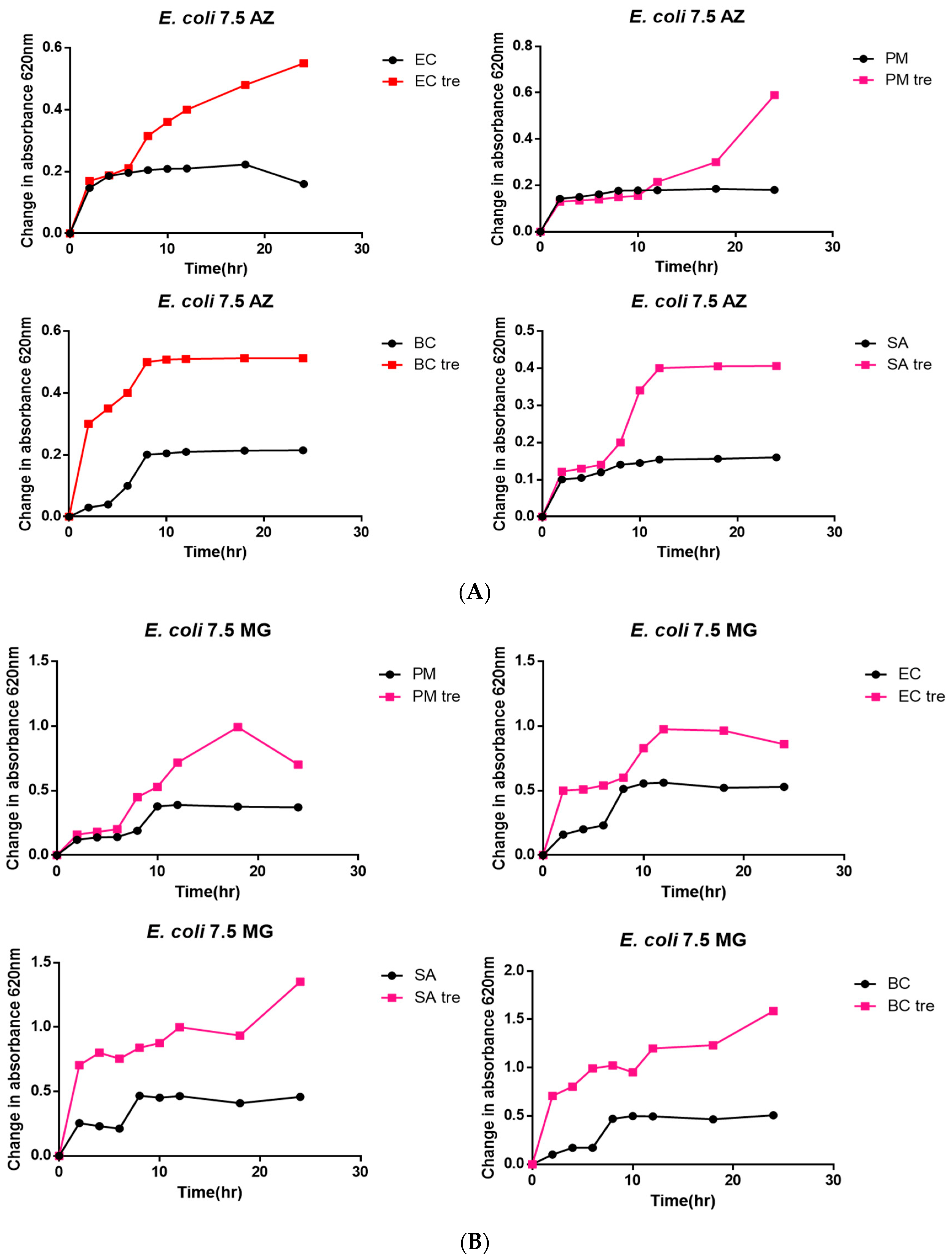
3.3. Bacterial Toxicity Test

The efficiency of the degradation (detoxification) of the dyes by the crude enzyme preparations was evaluated by growing selected bacteria (Proteus mirabilis H14320, Escherichia coli ATCC9637, Bacillus cereus ATCC14579, and Staphylococcus aureus LCT-SA112) on the treated (lignin peroxidase extracts) and untreated AZ and MG. Figure 6 and Figure 7 show that all the strains grow better on both treated dyes (AZ and MG), as an increase in absorbance at 620 nm was observed compared to the untreated dyes. Hence, the untreated dyes were toxic, since all tested bacteria shown no appreciable bacterial growth on both AZ and MG.

3.4. Determination of Decolorization Kinetics

The Lineweaver–Burk plot was applied to the decolorization data, and Table 4 presents the kinetic parameters obtained. The enzyme kinetics (Table 4) indicate that the enzyme produced by the E. coli had a higher significant (p < 0.05) affinity (Km) and catalytic efficiency (kcat/Km) for the dyes than the enzyme of P. aeruginosa.

4. Discussion

Lignin peroxidases (EC 1.11.1.14) are heme-containing enzymes that catalyze hydrogen peroxide-dependent oxidation in lignin. Dyes are structurally complex like lignin. Thus, there is a high demand for lignin peroxidase in the textile, dye manufacturing, pulp and paper, and cosmetic industries that routinely use dyes in their processes [9]. The need for lignin peroxidase by these industries cannot be over-emphasized.
The two lignin peroxidase preparations, separately obtained from E. coli and P. aeruginosa used in this study, exhibited appreciable dye decolorization potential of the synthetic dyes, Azure B (AZ) and Malachite Green (MG) (Table 1). Given that these preparations are crude enzymes, it is encouraging to note that they showed about 52–63% efficiency (the purified enzyme was 80% efficient). The inability of the lignin peroxidase or ligninolytic bacteria to effectively decolorize CR and RBBR (as observed in this study) has been reported by other researchers [3,9]. These bacterial isolates could not degrade these dyes because of the structural stability of CR and RBBR, which makes them highly resistant to biodegradation as well as their toxicity [9].
The characterization of an enzyme is crucial since factors such as substrate and enzyme concentrations, pH, and temperature play a vital role in influencing the enzymatic processes, and thus the industrial application of enzymes produced a 65 kDa lignin peroxidase from Bacillus megaterium [26,31]. In this study, the crude enzyme concentration at 50 mg/L effectively degraded the dyes (Figure 1). This was contrary to the work of Lalnunhlimi and Krishnaswamy [32], in which the dyes were degraded at 200 mg/L. This implied that at 50 mg/L, there was an increase in the nucleic acids content ratio (RNA/DNA), which promote protein synthesis [33]. These results supported the concentration efficacy of the crude enzymes to degrade the dyes. A temperature either increases or badly inhibits enzyme activity. If temperature gets too high, enzyme activity will diminish, and enzymes are denatured [34]. Likewise, a change in pH can also alter the enzyme functional group, thus influencing the enzyme capability [35]. The lignin peroxidases produced in this study seem to prefer a neutral pH environment. These observations indicated that the enzymes could also efficiently neutralize weakly acidic dyes’ waste [33]. In previous studies, Citrobacter sp. CK3 achieved the best decolorization of reactive red at a neutral pH of 6–7. In contrast, Aspergillus niger decolorized red azo dye at pH 9 [36]. The broad pH profiles (Figure 2) underscore the ability of the studied preparations to perform effectively in various industries. The mesophilic nature of the organisms reflects the optimum performance of the enzymes at 30–40 °C (Figure 3). Similar pH and temperature profiles have been reported for other lignin peroxidases [37]. It is noted that the enzyme preparations were inhibited by oxidoreductase inhibitors (Table 3). Lignin peroxidase, being a metalo-(heme)-enzyme, was inhibited by EDTA, a known metal-chelating agent [30,37,38]. Various lignin peroxidases have shown similar results [39,40,41]. The observed high survival rate of bacteria of dye treated with crude enzymes (Figure 6 and Figure 7) is an indication of the efficacy of the crude enzyme to degrade the dye without toxic effects. This suggested the potential of the crude enzymes to simultaneously degrade the dyes and detoxify the dye’s metabolite in producing no toxic products [42].
The calculated enzyme kinetics (Table 4) indicate that the enzyme produced by the E. coli had a higher affinity (Km) and catalytic efficiency (kcat/Km) for the dyes than the enzyme of P. aeruginosa. It is apparent that the E. coli enzyme was more effective on MG than on AZ. The measure of this (catalytic efficiency) is confirmed in the toxicity testing. The prepared lignin peroxidases effectively degraded and detoxified AZ and MG to allow the growth of different bacteria (Figure 6 and Figure 7). Chen et al. [43], Gokulakrishnan et al. [44], and Yang et al. [45] have all reported the reduction of toxicity of MG to E. coli and other microorganisms after decolorization.

5. Conclusions

Treating the toxicity of synthetic dyes before discharging them into wastewater effluent is one of the main challenges experienced by most industries [32]. The obtained results provide a possible and feasible way of decolorizing and degrading Azure B and Malachite Green dyes using lignin peroxidase produced from Escherichia coli LR025096.1 and Pseudomonas aeruginosa CP031449.2. These two lignin peroxidase extracts hold high potential to decolorize the synthetic dyes in wastewater, especially from industrial effluents. This study did not aim at obtaining purified lignin peroxidase. The presence and the contribution of other LMEs in the crude preparations used in the experiments cannot be ruled out. Purification of the crude lignin peroxidase from the bacteria strains will be carried out for further study.

Author Contributions

Conceptualization, S.L.D., A.R.O., R.A.M. and F.O.O.; methodology, all authors; validation, A.R.O., S.L.D., F.O.O. and R.A.M.; investigation, all authors; writing original draft preparation, S.L.D., F.O.O., A.R.O. and R.A.M.; writing—review and editing A.R.O., F.O.O., A.O.I. and S.L.D.; visualization, F.O.O., S.L.D., A.R.O. and A.O.I.; supervision, A.R.O., F.O.O. and R.A.M.; project administration, S.L.D., F.O.O., R.A.M. and A.R.O. All authors have read and agreed to the published version of the manuscript.

Funding

This research received no external funding.

Institutional Review Board Statement

The study was conducted in accordance with the Declaration of Helsinki and approved by the Research Committee, University of Zululand (UZREC 171110-030 PGM 2018/575), for studies involving bacteria.

Informed Consent Statement

Not applicable.

Data Availability Statement

Data will be made available by the corresponding author upon request.

Acknowledgments

We would like to thank the Research Office of University of Zululand for their continuous supports.

Conflicts of Interest

The authors declare no conflicts of interest.

References

  1. Tehrani-Bagha, A.R.; Holmberg, K. Solubilization of hydrophobic dyes in surfactant solutions. Mater 2013, 6, 580–608. [Google Scholar] [CrossRef] [PubMed]
  2. Levin, L.; Papinutti, L.; Forchiassin, F. Evaluation of Argentinean white rot fungi for their ability to produce lignin-modifying enzymes and decolorize industrial dyes. Bioresour. Technol. 2004, 94, 169–176. [Google Scholar] [CrossRef]
  3. Bandounas, L.; Wierckx, N.J.; de Winde, J.H.; Ruijssenaars, H.J. Isolation and characterization of novel bacterial strains exhibiting ligninolytic potential. BMC Biotechnol. 2011, 11, 94. [Google Scholar] [CrossRef]
  4. Hassaan, M.A.; El Nemr, A. Health and environmental impacts of dyes: Mini review. Am. J. Environ. Sci. Eng. 2017, 1, 64–67. [Google Scholar] [CrossRef]
  5. Mnif, I.; Fendri, R.; Ghribi, D. Malachite green bioremoval by a newly isolated strain Citrobacter sedlakii RI11; enhancement of the treatment by biosurfactant addition. Water Sci. Technol. 2015, 72, 1283–1293. [Google Scholar] [CrossRef]
  6. Lellis, B.; Fávaro-Polonio, C.Z.; Pamphile, J.A.; Polonio, J.C. Effects of textile dyes on health and the environment and bioremediation potential of living organisms. BIORI 2019, 3, 275–290. [Google Scholar] [CrossRef]
  7. Pérez, J.; Munoz-Dorado, J.; De la Rubia, T.D.L.R.; Martinez, J. Biodegradation and biological treatments of cellulose, hemicellulose and lignin: An overview. Int. Microbiol. 2002, 5, 53–63. [Google Scholar] [CrossRef] [PubMed]
  8. Sun, Y.; Cheng, J. Hydrolysis of lignocellulosic materials for ethanol production: A review. Bioresour. Technol. 2002, 83, 1–11. [Google Scholar] [CrossRef]
  9. Falade, A.O.; Eyisi, O.A.; Mabinya, L.V.; Nwodo, U.U.; Okoh, A.I. Peroxidase production and ligninolytic potentials of freshwater bacteria Raoultella ornithinolytica and Ensifer adhaerens. Biotechnol. Rep. 2017, 16, 12–17. [Google Scholar] [CrossRef]
  10. Jia, J.B.; Wang, B.Q.; Wu, A.G.; Cheng, G.J.; Li, Z.; Dong, S.J. A method to construct a third-generation horseradish peroxidase biosensor; self-assembling gold nanoparticles to three-dimensional sol-gel network. Anal. Chem. 2002, 74, 2217–2223. [Google Scholar] [CrossRef]
  11. Agostini, E.; Hernandez-Ruiz, J.; Arnao, M.B.; Milrand, S.R.; Tigier, H.A.; Acosta, M.A. Peroxidase isoenzyme secreted by turnip (Brassica napus) hairy-root culture inactivation by hydrogen peroxide and application in diagnostic kits. Appl. Biochem. Biotechnol. 2002, 35, 1–7. [Google Scholar] [CrossRef] [PubMed]
  12. Kalyani, D.C.; Phugare, S.S.; Shedbalkar, U.U.; Adhar, J.P. Purification and characterization of a bacterial peroxidase from the isolated strain Pseudomonas sp. SUK1 and its application for textile dye decolourization. Ann. Microbiol. 2011, 61, 483–491. [Google Scholar] [CrossRef]
  13. Draelos, Z.D. A split-face evaluation of a novel pigment-lightening agent compared with no treatment and hydroquinone. J. Am. Acad. Dermatol. 2015, 72, 105–107. [Google Scholar] [CrossRef]
  14. Taboada-Puig, R.; Lu-Chau, T.A.; Eibes, G.; Feijoo, G.; Moreira, M.T.; Lema, J.M. Continuous removal of endocrine disruptors by versatile peroxidase using a two-stage system. Biotechnolo. Prog. 2015, 31, 908–916. [Google Scholar] [CrossRef]
  15. Timothy, C.C.; Lars, B.; Vera, M. Something old, something new: Challenges and developments in Aspergillus niger biotechnology. Essays Biochem. 2012, 65, 213–224. [Google Scholar] [CrossRef]
  16. Schallmey, M.; Singh, A.; Ward, O.P. Developments in the use of Bacillus species for industrial production. Can. J. Microbiol. 2004, 50, 1–17. [Google Scholar] [CrossRef]
  17. Sevinc, N.; Demirkan, E. Production of protease by Bacillus sp. N-40 isolated from soil and its enzymatic properties. J. Biol. Environ. Sci. 2011, 5, 95–103. [Google Scholar]
  18. Barros, F.F.C.; Simiqueli, A.P.R.; de Andrade, C.J.; Pastore, G.M. Production of enzymes from agro industrial wastes by biosurfactant-producing strains of Bacillus subtilis. Biotechnol. Res. Int. 2013, 103960, 1–9. [Google Scholar] [CrossRef]
  19. Pant, G.; Prakash, A.; Pavani, J.V.P.; Bera, S.; Deviram, G.V.N.S.; Kumar, A.; Panchpuri, M.; Prasuna, R.G. Production, optimization and partial purification of protease from Bacillus subtilis. J. Taibah Univ. Sci. 2015, 9, 50–55. [Google Scholar] [CrossRef]
  20. Soares, M.M.C.N.; Da Silva, R.; Carmona, E.C.; Gomes, E. Pectinolytic enzyme production by Bacillus species and their potential application on juice extraction. World J. Microbiol. Biotechnol. 2001, 17, 79–82. [Google Scholar] [CrossRef]
  21. Dias, P.V.S.; Ramos, K.O.; Padilha, I.Q.M.; Araújo, D.A.M.; Santos, S.F.M.; Silva, F.L.H. Optimization of cellulase production by Bacillus sp. isolated from sugarcane cultivated soil. Chem. Eng. Trans. 2014, 38, 227–282. [Google Scholar] [CrossRef]
  22. Padilha, I.Q.M.; Carvalho, L.C.T.; Dias, P.V.S.; Grisi, T.C.S.L.; Honorato, D.A.; Silva, F.L.S.; Santos, F.M.; Araújo, D.A.M. Production and characterization of thermophilic carboxymethyl cellulase synthesized by Bacillus sp. growing on sugarcane bagasse in submerged fermentation. Braz. J. Chem. Eng. 2015, 32, 35–42. [Google Scholar] [CrossRef]
  23. Janusz, G.; Pawlik, A.; Sulej, J.; Świderska-Burek, U.; Jarosz-Wilkołazka, A.; Paszczyński, A. Lignin degradation: Microorganisms, enzymes involved, genomes analysis and evolution. FEMS Microbiol. Rev. 2017, 41, 941–962. [Google Scholar] [CrossRef]
  24. Wong, D.W. 2009. Structure and action mechanism of ligninolytic enzymes. Appl. Biochem. Biotechnol. 2009, 157, 174–209. [Google Scholar] [CrossRef] [PubMed]
  25. Chowdhary, P.A.N.K.A.J.; Saxena, G.A.U.R.A.V.; Bharagava, R.N. Role of Laccase Enzyme in Bioremediation of Industrial Wastes and Its Biotechnological Application. Bioremediation of Industrial Pollutants; Write and Print Publication: Delhi, India, 2016; pp. 307–331. [Google Scholar]
  26. Patil, S.R. Production and purification of lignin peroxidase from Bacillus megaterium and its application in bioremediation. CIB Technol. J. Microbiol. 2014, 3, 22–28. [Google Scholar] [CrossRef]
  27. Dube, S.L.; Osunsanmi, F.O.; Ngcobo, B.P.; Mkhwanazi, L.B.; Jobe, Z.Z.; Aruleba, R.T.; Mosa, R.A.; Opoku, A.R. Isolation and Characterization of Potential Lignin Peroxidase-Producing Bacteria from Compost Samples at Richards Bay (South Africa). Pol. J. Microbiol. 2023, 72, 117–124. [Google Scholar] [CrossRef]
  28. Bradford, M.M. A rapid and sensitive for the quantitation of microgram quantities of protein utilizing the principle of protein dye binding. Anal. Biochem. 1976, 72, 248–254. [Google Scholar] [CrossRef]
  29. Ahmedi, A.; Abouseoud, M.; Couvert, A.; Amrane, A. Enzymatic degradation of Congo Red by turnip (Brassica rapa) peroxidase. Z. Naturforsch C 2012, 67, 429–436. [Google Scholar] [CrossRef]
  30. Basha, S.A.; Prasada Rao, U.J. Purification and characterization of peroxidase from sprouted green gram (Vigna radiata) roots and removal of phenol and p-chlorophenol by immobilized peroxidase. J. Sci. Food Agric. 2017, 97, 3249–3260. [Google Scholar] [CrossRef]
  31. Daâssi, D.; Zouari-Mechichi, H.; Frikha, F.; Martinez, M.J.; Nasri, M.; Mechichi, T. Decolorization of the Azo Dye Acid Orange 51 by Laccase Produced in Solid Culture of a Newly Isolated Trametestrogii Strain. 3 Biotech 2013, 3, 115–125. [Google Scholar] [CrossRef]
  32. Lalnunhlimi, S.; Krishnaswamy, V. Decolorization of azo dyes (Direct Blue 151 and Direct Red 31) by moderately alkaliphilic bacterial consortium. Braz. J. Microbiol. 2016, 47, 39–46. [Google Scholar] [CrossRef] [PubMed]
  33. Roy, D.C.; Biswas, S.K.; Saha, A.K.; Sikdar, B.; Rahman, M.; Roy, A.R.; Prodhan, Z.H.; Tang, S.S. Biodegradation of crystal violet dye by bacteria isolated from textile industry effluents. PeerJ 2018, 6, 1–23. [Google Scholar] [CrossRef]
  34. Dutta, H.; Dutta, A. The microbial aspect of climate change. Energ. Ecol. Environ. 2016, 1, 209–232. [Google Scholar] [CrossRef]
  35. Robinson, P.K. Enzymes: Principles and biotechnology applications. Essays Biochem. 2015, 59, 1–41. [Google Scholar] [CrossRef]
  36. Mahmoud, M.S.; Mostafa, M.K.; Mohamed, S.A.; Sobhy, N.A.; Nasr, M. Bioremediation of red azo dye from aqueous solution by Aspergillus niger strain isolated from textile wastewater. J. Environ. Chem. Eng. 2017, 5, 547–554. [Google Scholar] [CrossRef]
  37. Falade, A.; Jaouani, A.; Mabinya, L.; Okoh, A.; Nwodo, U. Exoproduction and molecular characterization of peroxidase from Ensiferadhaerens. Appl. Sci. 2019, 9, 3121. [Google Scholar] [CrossRef]
  38. Datta, R.; Kelkar, A.; Baraniya, D.; Molaei, A.; Moulick, A.; Meena, R.S.; Formanek, P. Enzymatic degradation of lignin in soil: A review. Sustainability 2017, 9, 1163. [Google Scholar] [CrossRef]
  39. Gorjup, B.; Lampič, N.; Penca, R.; Perdih, A.; Perdih, M. Solvent effects on ligninases. Enzyme Microb. Technol. 1999, 25, 15–22. [Google Scholar] [CrossRef]
  40. Liu, A.; Huang, X.; Song, S.; Wang, D.; Lu, X.; Qu, Y.; Gao, P. Kinetics of the H2O2-dependent ligninase-catalyzed oxidation of veratryl alcohol in the presence of cationic surfactant studied by spectrophotometric technique. Spectrochim. Acta Part A Mol. Biomol. Spectrosc. 2003, 59, 2547–2551. [Google Scholar] [CrossRef]
  41. de Cassia Pereira, J.; Giese, E.C.; de Souza Moretti, M.M.; dos Santos Gomes, A.C.; Perrone, O.M.; Boscolo, M.; da Silva, R.; Gomes, E.; Martins, D.A.B. Effect of metal ions, chemical agents and organic compounds on lignocellulolytic enzymes activities. In Enzyme Inhibitors and Activators; IntechOpen: London, UK, 2017; pp. 139–164. [Google Scholar]
  42. Guo, G.; Tian, F.; Zhao, Y.; Tang, M.; Liu, W.; Liu, C.; Xue, S.; Kong, W.; Sun, Y.; Wang, S. Aerobic decolorization and detoxificationof acid scarlet GR by a newly isolated salt-tolerant yeast strain Galactomyces geotrichum GG. Int. Biodeterior. Biodegrad. 2019, 145, 104818. [Google Scholar] [CrossRef]
  43. Chen, C.H.; Chang, C.F.; Liu, S.M. Partial degradation mechanisms of malachite green and methyl violet B by Shewanella decolorationis NTOU1 under anaerobic conditions. J. Hazard. Mater. 2010, 177, 281–289. [Google Scholar] [CrossRef] [PubMed]
  44. Gokulakrishnan, S.; Parakh, P.; Prakash, H. Degradation of Malachite green by Potassium persulphate, its enhancement by 1, 8-dimethyl-1, 3, 6, 8, 10, 13-hexaazacyclotetradecane nickel (II) perchlorate complex, and removal of antibacterial activity. J. Hazard. Mater. 2012, 213, 19–27. [Google Scholar] [CrossRef] [PubMed]
  45. Yang, X.; Zheng, J.; Lu, Y.; Jia, R. Degradation and detoxification of the triphenylmethane dye malachite green catalyzed by crude manganese peroxidase from Irpexlacteus F17. ESPR 2016, 23, 9585–9597. [Google Scholar] [CrossRef] [PubMed]
Figure 1. Effect of dye concentration from lignin peroxidase extract of Pseudomonas aeruginosa and Escherichia coli on AZ and MG discoloration.
Applsci 14 08012 g001
Figure 2. Effect of pH value on decolorization of Azure B (50 mg/L) and Malachite Green (50 mg/L) by lignin peroxidase extract of Pseudomonas aeruginosa and Escherichia coli in the presence of 20 mg/mL enzyme and 2 mM H2O2 at 40 °C for 30 min.
Applsci 14 08012 g002
Figure 3. Effect of temperature on decolorization of AZ and MG by lignin peroxidase extract of Pseudomonas aeruginosa and Escherichia coli. Conditions from Figure 1 were used.
Applsci 14 08012 g003
Figure 4. Effect of substrate (H2O2) concentration by lignin peroxidase extracts of Pseudomonas aeruginosa and Escherichia coli on AZ and MG discoloration during a 60 min incubation time. Conditions as in Figure 2.
Applsci 14 08012 g004
Figure 5. Effect of incubation period (0 to 200 min) on discoloration of AZ and MG under optimized conditions.
Applsci 14 08012 g005
Figure 6. (A): Toxicity of treated (lignin peroxidase extract-E. coli) and untreated Azure B during an incubation period (0–24 h) under optimized conditions. (B) Toxicity of treated (lignin peroxidase extract-E. coli) and untreated Malachite Green during an incubation period (0–24 h) under optimized conditions. {Keys: PM: Proteus mirabilis H14320, EC: Escherichia coli ATCC9637, SA: Staphylococcus aureus LCT-SA112, BC: Bacillus cereus ATCC14579}.
Applsci 14 08012 g006
Figure 7. (A): Toxicity of treated (lignin peroxidase extract-Pseudomonas aeruginosa) and untreated Azure B during an incubation period (0–24 h) under optimized conditions. (B): Toxicity of treated (lignin peroxidase extract-Pseudomonas aeruginosa) and untreated Malachite Green during an incubation period (0–24 h) under optimized conditions {Keys: PM: Proteus mirabilis H14320, EC: Escherichia coli ATCC9637, SA: Staphylococcus aureus LCT-SA112, BC: Bacillus cereus ATCC14579 and tre: treated}.
Applsci 14 08012 g007
Table 1. Dye decolorization (%) of AZ, RBBR, CR, and MG by purified lignin peroxidase and lignin peroxidase extract of Escherichia coli and Pseudomonas aeruginosa. Values with different superscript alphabets were significant (p < 0.05).
DyesCrude Enzyme from Escherichia coliCrude Enzyme from Pseudomonas aeruginosaCommercial Lignin Peroxidase
AZ63 ± 1.34 a56 ± 2.11 b72 ± 1.45 c
RBBR11 ± 1.12 a6 ± 1.78 b0 ± 0.13 c
CR8 ± 0.45 a3 ± 1.88 b0 ± 1.77 c
MG53 ± 1.44 a52 ± 2.06 a80 ± 1.33 c
Table 2. Effect of enzyme concentration from lignin peroxidase extract of E. coli and P. aeruginosa on Azure B and Malachite Green discoloration. Different alphabets showed significant differences (p < 0.05).
Enzyme Concentrations (mg/mL)AZ (% Dye Decolorization) E. coliMG (% Dye Decolorization) E. coliAZ (% Dye Decolorization) P. aeruginosaMG (% Dye Decolorization) P. aeruginosa
10.5 ± 1.22 a9 ± 1.11 f2 ± 2.66 i6 ± 1.56 a
520 ± 0.39 b11 ± 2.23 f9.3 ± 3.56 f24 ± 2.12 j
1032 ± 2.14 c35 ± 3.12 c31 ± 1.44 c43 ± 2.88 k
2068 ± 1.88 d55 ± 1.78 g52 ± 2.11 h55 ± 1.66 g
4060 ± 2.03 e55 ± 1.48 g51 ± 3.99 h55 ± 2.33 g
5062 ± 3.11 e52 ± 2.74 h50 ± 3.22 h53 ± 2.71 h
10062 ± 2.99 e50 ± 1.32 h50 ± 0.47 h54 ± 2.31 g
Table 3. Inhibition of lignin peroxidase extract from E. coli and P. aeruginosa by various chemical substances. Different alphabets showed significant differences (p < 0.05).
% Inhibition of E. coli (AZ)% Inhibition of E. coli (MG)% Inhibition of P. aeruginosa (AZ)% Inhibition of P. aeruginosa (MG)
1 mM NaN383 ± 1.34 a68 ± 2.76 b71 ± 3.86 b63 ± 2.76 c
1 mM EDTA67 ± 3.21 b68 ± 3.36 b75 ± 2.73 d79 ± 3.22 a
1 mM FeCl360 ± 4.23 c67 ± 3.29 b55 ± 2.06 e58 ± 4.22 e
Table 4. The Vmax, Km and kcat of both lignin peroxidase extracts of E. coli and P. aeruginosa. Different alphabets showed significant differences (p < 0.05).
Enzyme KineticsE. coli (LiP Extract)P. aeruginosa (LiP Extract)
AZMGAZMG
Vmax (µmol m−3 s−1)1.4 ± 1.23 × 1043.0 ± 2.23 × 1031.5± 0.33 × 1041.0 ± 2.11 × 10⁴
Km (µmol/L)6.7 ± 0.82 × 1048 ± 1.42 × 1037 ± 2.11 × 1043.5 ± 0.32 × 10⁴
kcat (s−1)6.6 ± 1.93 × 1021.4 ± 0.27 × 1027.9 ± 0.77 × 1025.3 ± 0.11 × 10²
Catalytic efficiency = kcat/ Km(µmol−1 s−1)9.9 ± 1.44 × 1011.8 ± 0.22 × 1011.1± 0.09 × 1011.5 ± 0.07 × 101
Disclaimer/Publisher’s Note: The statements, opinions and data contained in all publications are solely those of the individual author(s) and contributor(s) and not of MDPI and/or the editor(s). MDPI and/or the editor(s) disclaim responsibility for any injury to people or property resulting from any ideas, methods, instructions or products referred to in the content.

