Systematic Review

Long-Term Outcomes of Collagen Matrix versus Subepithelial Connective Tissue in Root Coverage for Multiple Teeth: A Systematic Review and Meta-Analysis

1
Department of Orthodontics, Seoul St. Mary’s Hospital, College of Medicine, The Catholic University of Korea, Seoul 06591, Republic of Korea
2
Graduate School of Clinical Dental Science, The Catholic University of Korea, Seoul 06591, Republic of Korea
3
Medical Library, College of Medicine, The Catholic University of Korea, Seoul 06591, Republic of Korea
4
Department of Oral and Maxillofacial Surgery, Seoul St. Mary’s Hospital, College of Medicine, The Catholic University of Korea, Seoul 06591, Republic of Korea
5
Department of Periodontics, College of Medicine, The Catholic University of Korea, Seoul 06591, Republic of Korea
6
Dental Implantology, Graduate School of Clinical Dental Science, The Catholic University of Korea, Seoul 06591, Republic of Korea
7
Department of Medicine, Graduate School, The Catholic University of Korea, Seoul 06591, Republic of Korea
*
Authors to whom correspondence should be addressed.
These authors contributed equally to this work.
Appl. Sci. 2024, 14(17), 8049; https://doi.org/10.3390/app14178049
Submission received: 14 July 2024 / Revised: 1 September 2024 / Accepted: 5 September 2024 / Published: 9 September 2024

Abstract

:
Background: This study aimed to conduct a meta-analysis to assess the differences in the performance of root coverage between collagen matrix and subepithelial connective tissue. Methods: A comprehensive search using a combination of controlled vocabulary (MeSH) and free-text terms was undertaken by two reviewers to identify published systematic reviews. Three major electronic databases (Medline via PubMed, Cochrane database, and Embase) were searched up to October 2023. Results: Initially, five hundred and seventy-six articles were identified, but only nine studies met the criteria for inclusion in the meta-analysis. Meta-analysis showed that the pooled mean difference in mean root coverage of collagen matrix compared with subepithelial connective tissue was −0.48 (95% confidence interval of −0.69 to −0.26). The relative risk of complete root coverage of collagen matrix compared with subepithelial connective tissue was 0.68 (95% confidence interval, 0.49 to 0.94). The pooled mean difference in width of keratinized tissue of collagen matrix compared with subepithelial connective tissue was −0.75 (95% confidence interval of −1.25 to −0.26). Conclusions: The outcomes of this study revealed that subepithelial connective tissue generally exhibited superior performance. Nonetheless, the utilization of the coronally advanced flap with collagen matrix may yield comparable results in terms of the percentage of complete root coverage.

1. Introduction

Gingival recession is defined as the migration of the gingival margin from the white enamel border to the apical side of the tooth [1]. Dental patients frequently exhibit gingival recession problems [2]. Approximately half of adults over the age of 30 are known to have gingival recession in at least one intraoral site in various populations, and untreated defects lead to further recession and have been reported to be a factor in the esthetic decline in future patients [1]. The causes of gingival recession include periodontitis, malpositioning of the tooth, poor prosthetics, and iatrogenic causes [3].
Root coverage can be used for the patient’s esthetic needs, tooth hypersensitivity, and cervical abrasion [4]. Over the past few decades, various surgical techniques on the root surface have been used to enhance root coverage/periodontal regeneration. Techniques for root coverage can be broadly categorized as advanced flap, pedicle soft tissue graft, and free soft tissue graft [5]. Free soft tissue grafting can be further categorized into free gingival grafting and subepithelial connective tissue grafting. Subepithelial connective tissue grafting was described previously and used subepithelial connective tissue grafting to cover the exposed root [6]. The advantages of subepithelial connective tissue grafting include relatively little postoperative pain in the donor area, dual blood supply to the graft from the overlying flap and periosteum in the recipient area, good blood supply, and a good prognosis due to low graft resorption [7,8]. In addition, it has the advantage of being able to be applied to multiple gingival recessions with excellent esthetics due to its color harmony with adjacent tissues [9]. Furthermore, because of its high success rate and pleasing color, coronally advanced flap with subepithelial connective tissue grafting can be regarded as a standard treatment [10].
Usually, subepithelial connective tissue is obtained from the palatal side [11]. Minimally invasive surgical techniques have been developed to address the surgical limitations of limited autologous harvest and collagen, a tissue graft substitute, has been suggested to overcome the limitations associated with autogenous tissue transplantation [1]. In preclinical and clinical studies, treatment concepts should be based on a biological basis that takes into account not only the use of regenerative materials but also the patient’s innate healing potential, which is why we want to apply root coverage utilizing collagen, which has a decisive influence on periodontal wound healing and regeneration [12].
A previous report evaluated the efficacy of a cross-linked xenogeneic volume-stable collagen matrix in treating gingival recessions and in addition to the fact that the treatment significantly reduced oral hypersensitivity, reasonably high patient-reported pleasure and esthetics were connected with the treatment outcomes [13]. Furthermore, a previous report showed the possibility of a volume-stable collagen matrix functionalized with injectable platelet-rich fibrin for the treatment of root coverage of multiple gingival recessions with reduced morbidity [14]. Buccal gingival recession treated with porcine collagen matrix and the gingival tissues matured in a healthy manner and matched the surrounding soft tissue areas in terms of color and texture [15]. However, it should be noted that in certain cases, total root coverage could not be achieved due to extensive buccal bone loss [15]. Consequently, this study aims to conduct a meta-analysis to assess the differences in multiple root coverage for multiple teeth between collagen and subepithelial connective tissue. The null hypothesis posits that there will be no significant difference in root coverage.

2. Materials and Methods

2.1. Protocol and Eligibility Criteria

This systematic review conforms to the guidelines set forth in the Preferred Reporting Items for Systematic Reviews and Meta-Analyses (PRISMA) statement, as specified in reference [16]. Our systematic review was executed with meticulous attention to the PRISMA Statement’s research methodologies. We collected exhaustive data through rigorous analysis and transparent reporting, resulting in a comprehensive and objective evaluation of the subject under investigation. However, it should be noted that the protocol for this systematic review was not registered in a public registry.
Question: is there a difference in the treatment of multiple root coverage between collagen matrix and connective tissue?
Participants: patients who received multiple root coverage.
Interventions: collagen matrix.
Comparisons: subepithelial connective tissue obtained from the palate.
Outcomes: root coverage, change in probing depth, and change in keratinized tissue width after 3 and 6 months, and 1, 3, 5, and 9 years.
Study design: randomized controlled trials (RCTs) limited to studies focusing on adults.
The criteria for inclusion and exclusion are detailed below.
Inclusion Criteria:
  • Studies involving collagen matrix and subepithelial connective tissue;
  • Cases with multiple recessions;
  • Studies focused on natural teeth;
  • Randomized controlled trials (RCTs).
Exclusion Criteria:
  • Patients who have undergone implant procedures;
  • Patients with dentures;
  • Studies on root coverage that do not involve the use of either collagen matrix or subepithelial connective tissue;
  • Cases involving a single gingival recession.

2.2. Information Sources and Search Strategy

A thorough search was conducted by two reviewers (SHH and HJS) using a combination of controlled vocabulary (MeSH) and free-text terms to identify published systematic reviews. This search encompassed three major electronic databases: Medline via PubMed, the Cochrane database, and Embase, with the search being conducted up to 20 December 2023. A manual search was also carried out to review the references of all retrieved full-text articles for any relevant studies that might have been missed through the electronic search. The search results were exported to EndNote reference management software (Version 21, Clarivate, Philadelphia, PA, USA) for the removal of duplicates. The search approach was adapted based on the specific requirements of each database. Details on the search strategy are found in Supplementary Material Table S1.

2.3. Study Selection and Data Extraction

The titles and abstracts of the retrieved papers were blindly screened by two reviewers (SHH and HJS) for the eligibility criteria. Any disagreement was resolved by discussion with another author (WJP). Finally, full texts of the remaining articles were reviewed independently and in duplicate by two reviewers (SHH and HJS) before final inclusion. Data were extracted independently from the included studies according to the PICOS question and arranged in the following fields: general information (author name, publication year, nationality), participants (number of patients, number of teeth being treated, tooth number), intervention/comparison (type of graft material), and outcomes (root coverage, probing depth, width, and keratinized tissue after 1, 3, 6, and 12 months from the treatment).

2.4. Risk of Bias Assessment

The reviewers used the Cochrane Risk-Of-Bias (ROB 2.0) tool for randomized studies. The guideline checklist included questions on the randomization process (selection bias), deviations from the intended interventions (performance bias), missing outcome data (attrition bias), measurement of the outcome (detection bias), selection of the reported result (reporting bias), and overall bias. The risk of bias of the recruited studies was evaluated as low risk, some concerns, or high risk. The quality of the eligible studies was evaluated by 2 reviewers (SHH and WJP).

2.5. Data Synthesis and Analysis

Meta-analysis was conducted using R (Version 3.5.0; R Project for Statistical Computing). The standardized mean difference (SMD) and 95% confidence interval (CI) were used as summary statistics. A random-effect method was used for meta-analysis. The level of significance was set at 0.05. I2 and chi-square test were performed to quantify the heterogeneity across studies. This study utilized methods from the Cochrane Handbook to manage the within-patient correlation of treatment outcomes in clustered trials [17]. For studies that did not adjust for clustering, the standard error was inflated using a correlation coefficient of 0.07 [18].

3. Results

3.1. Study Selection and Data Extraction

The initial search identified 576 articles. After 241 duplicates were excluded, the article titles and abstracts were read, and 313 articles did not meet the inclusion criteria. The inclusion and exclusion criteria were applied to the 22 full-text articles. After thirteen articles that did not meet the inclusion criteria were excluded, nine studies were assessed for eligibility. The literature-screening flowchart is shown in Figure 1. Supplementary Materials Table S2 shows the excluded articles with reasons for extrusion. Table 1 provides an overview of the key attributes of the studies incorporated in this analysis. The outcome parameters of the included studies are listed in Table 2.

3.2. Risk of Bias Assessment

The summary of the risk of bias and overall risk of bias score for each field in the included articles are illustrated in Figure 2. Figure 2A indicates the risk of bias of each domain in each study, with green representing a low risk of bias, yellow signifying unclear risk of bias, and red denoting a high risk of bias. This assessment reveals that most studies exhibit a low risk of bias across the majority of domains. All studies were rated as low risk for randomization, deviation from the intended intervention, and missing outcome data; however, some concerns were noted in the measurement and selection domain. The reasons for the concerns regarding bias were unclear selection of reported results and measurement of outcomes, which are shown in Supplementary Materials Table S3. Figure 2B shows the overall risk of bias of each domain, with the length of the green rectangle indicating the number of studies assessed as having a low risk of bias. The majority of studies exhibited some concerns of bias in 88.9% of the studies. This assessment highlights that certain areas, including the selection of reported results and the measurement of outcomes, warranted some caution.

3.3. Meta-Analysis

Nine included articles (Aroca et al., 2013 [19]; Cieślik-Wegemund et al., 2016 [20]; Nahas et al., 2019 [21]; Pietruska et al., 2019 [22]; Rakasevic et al., 2020 [23]; Tonetti et al., 2021 [24]; Milinkovic et al., 2022 [25]; Molnár et al., 2022 [26]; and Skurska et al., 2022 [27]) assessed the differences in root coverage between collagen matrix and subepithelial connective tissue.

3.3.1. Type of Gingival Recession

Participants in this study included different types of gingival recessions. Specifically, Miller class I featured marginal tissue recession that did not extend to the mucogingival junction and was not accompanied by an alveolar bone loss in the interdental area [28]. Miller class II involved marginal tissue recession that extended to or beyond the mucogingival junction and was also not associated with alveolar bone loss in the interdental area. Recession type 1 was characterized by gingival recession with no loss of interproximal attachment, and the cementoenamel junction was not detectable clinically at both the mesial and distal aspects of the tooth [29]. Table 1 provides an overview of the gingival recession in the participants.

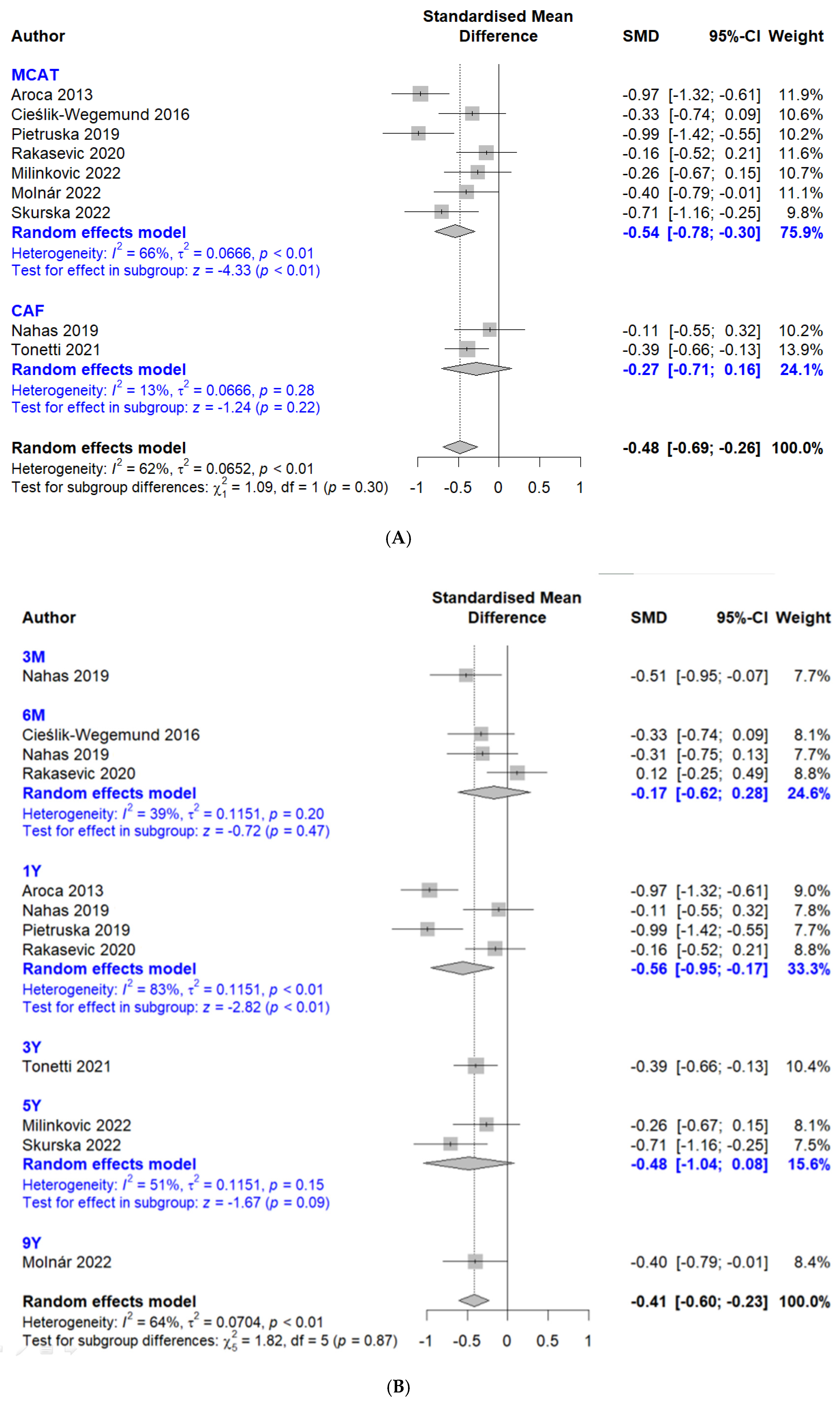
3.3.2. Mean Root Coverage

Considering the diversity in the number of subjects, research background, and research period, among other factors, a random effects model was applied. Additionally, the high I2 value (62%) and the p-value (p < 0.01) indicate the presence of significant heterogeneity among the studies. A forest plot of the meta-analysis showed that the pooled mean difference in mean root coverage of collagen matrix compared with subepithelial connective tissue was −0.48 (95% confidence interval of −0.69 to −0.26) (Figure 3A). The subgroup of modified coronally advanced flap showed that the pooled mean difference in mean root coverage of collagen matrix compared with subepithelial connective tissue was −0.27 (95% confidence interval of −0.71 to 0.16). A random effects model was applied with an I2 value of 13% and a p-value of 0.28. The subgroup of applying modified coronally advanced tunnel showed that the pooled mean difference in mean root coverage of collagen matrix compared with subepithelial connective tissue was −0.54 (95% confidence interval of −0.78 to −0.30). The forest plot favored subepithelial connective tissue with higher mean root coverage compared with collagen matrix.
The results of different follow-up periods are shown in Figure 2B. A random effects model was applied, and the high I2 value (64%) and the p-value (p < 0.01) indicate the presence of significant heterogeneity among the studies. The results of the forest plot of the meta-analysis showed that the pooled mean difference in mean root coverage of collagen matrix compared with subepithelial connective tissue was −0.41 (95% confidence interval of −0.60 to −0.23) (Figure 3B). The 6-month subgroup showed that the pooled mean difference in mean root coverage of collagen matrix compared with subepithelial connective tissue was −0.17 (95% confidence interval of −0.62 to 0.28) with an I2 value of 39% and a p-value of 0.20. The 1-year subgroup showed that the pooled mean difference in mean root coverage of collagen matrix compared with subepithelial connective tissue was −0.56 (95% confidence interval of −0.95 to −0.17).

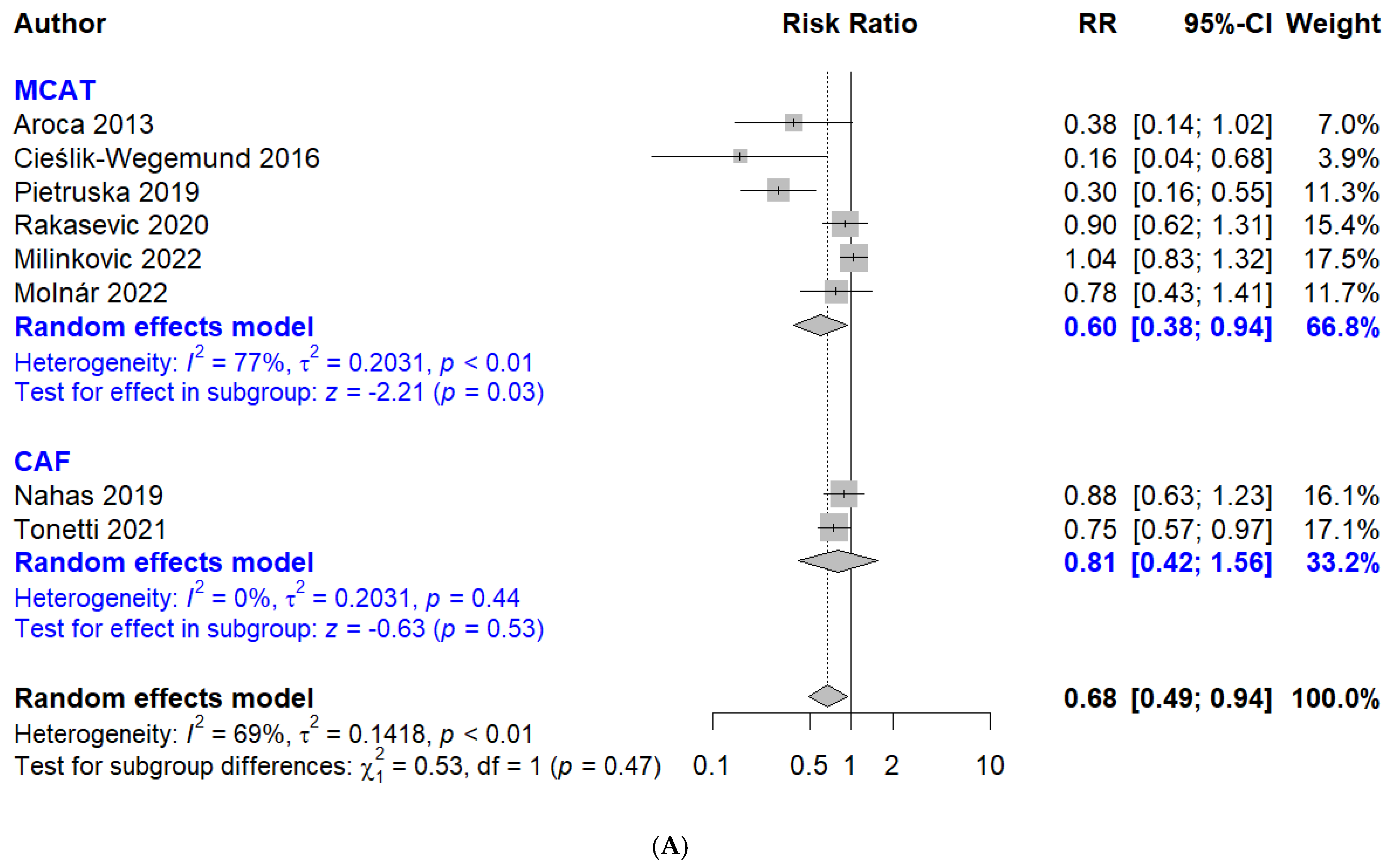
3.3.3. Complete Root Coverage

A random effects model was applied, and the high I2 value (69%) and the p-value (p < 0.01) indicate the presence of significant heterogeneity among the studies. The forest plot of the meta-analysis showed that the relative risk of complete root coverage of collagen matrix compared with subepithelial connective tissue was 0.68 (95% confidence interval, 0.49 to 0.94) (Figure 4A). There was a significant difference between collagen and connective tissue. The subgroup using coronally advanced flap showed a relative risk of complete root coverage of collagen matrix compared with subepithelial connective tissue of 0.81 (95% confidence interval, 0.42 to 1.56). There was no significant difference between collagen and connective tissue. The subgroup applying a modified coronally advanced tunnel showed a relative risk of complete root coverage of collagen matrix compared with the subepithelial connective tissue of 0.60 (95% confidence interval, 0.38 to 0.94). There was a significant difference between collagen and connective tissue, with more favorable results from subepithelial connective tissue.
The results of different follow-up periods are shown in Figure 4B. A random effects model was applied, and the high I2 value (60%) and the p-value (p < 0.01) indicate the presence of significant heterogeneity among the studies. The 3-month subgroup showed the relative risk of complete root coverage of collagen matrix compared with subepithelial connective tissue was 0.70 (95% confidence interval, 0.47 to 1.04). The 6-month subgroup showed the relative risk of complete root coverage of collagen matrix compared with subepithelial connective tissue was 0.59 (95% confidence interval, 0.32 to 1.11).

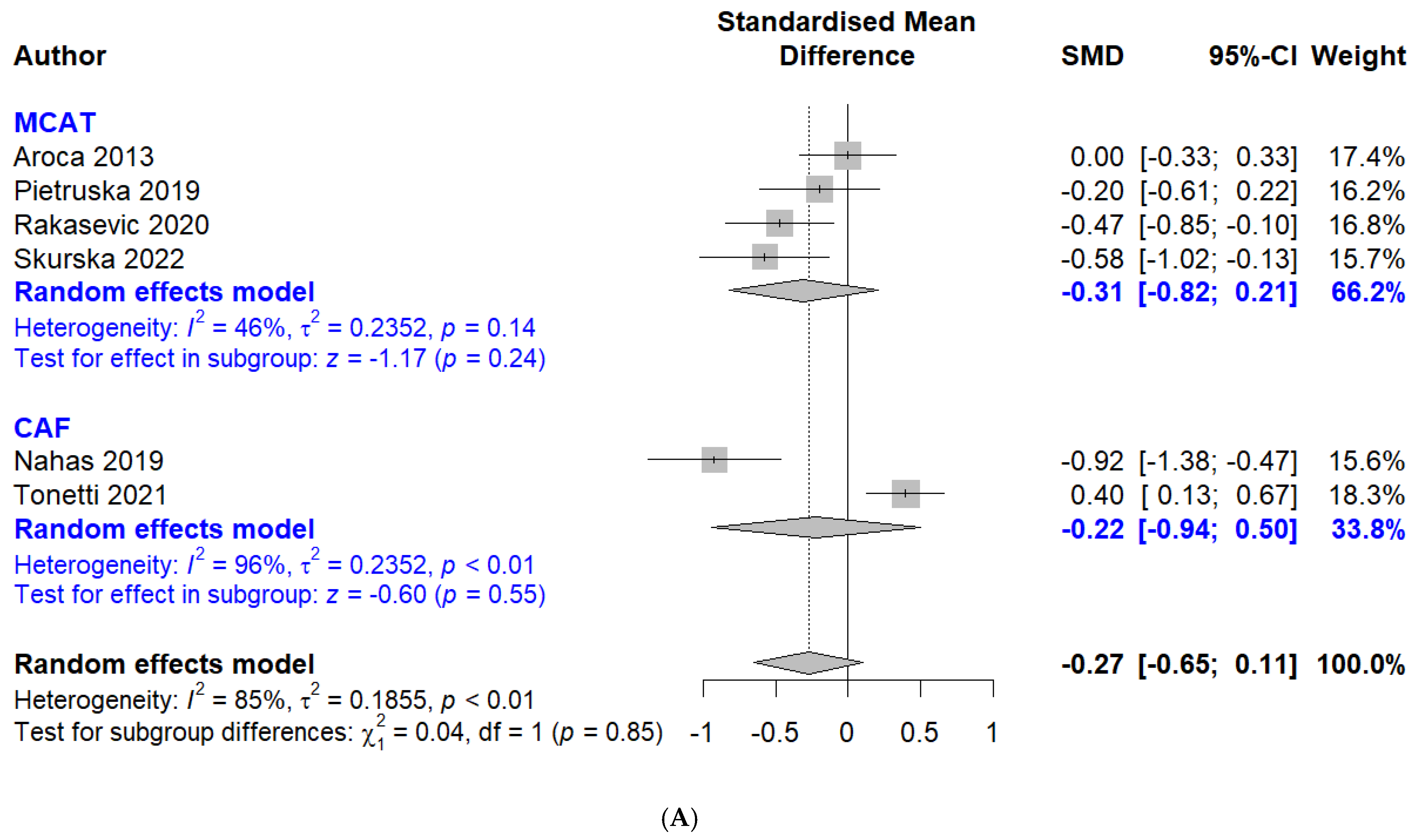
3.3.4. Width of Keratinized Tissue

The high I2 value (90%) and the p-value (p < 0.01) indicate the presence of significant heterogeneity among the studies and a random effects model was applied for the analysis. The forest plot of the meta-analysis showed that the pooled mean difference in width of keratinized tissue of collagen matrix compared with subepithelial connective tissue was −0.75 (95% confidence interval of −1.25 to −0.26) (Figure 5A). There was a significant difference between collagen and subepithelial connective tissue, favoring subepithelial connective tissue. The subgroup using coronally advanced flap showed that the pooled mean difference in width of keratinized tissue of collagen matrix compared with subepithelial connective tissue was −0.70 (95% confidence interval of −1.78 to 0.37). The results for the subgroup using modified coronally advanced tunnel showed that the pooled mean difference in width of keratinized tissue of collagen matrix compared with subepithelial connective tissue was −0.77 (95% confidence interval of −1.40 to −0.15).
The results of different follow-up periods are shown in Figure 4B. A random effects model was applied, and the high I2 value (88%) and the p-value (p < 0.01) indicate the presence of significant heterogeneity among the studies. The results of the forest plot of the meta-analysis showed that the pooled mean difference in width of keratinized tissue of collagen matrix compared with subepithelial connective tissue was −0.62 (95% confidence interval of −0.99 to −0.25) (Figure 5B). A high I2 value (73%) was found, but the p-value (p = 0.06) did not indicate statistically significant heterogeneity among the studies. The 3-month subgroup showed that the pooled mean difference in width of keratinized tissue of collagen matrix compared with subepithelial connective tissue was −0.41 (95% confidence interval of −1.33 to 0.50). The 6-month subgroup showed that the pooled mean difference in width of keratinized tissue of collagen matrix compared with subepithelial connective tissue was −0.34 (95% confidence interval of −0.98 to 0.30). The 1-year results showed that the pooled mean difference in width of keratinized tissue of collagen matrix compared with subepithelial connective tissue was −0.94 (95% confidence interval of −1.69 to −0.19).

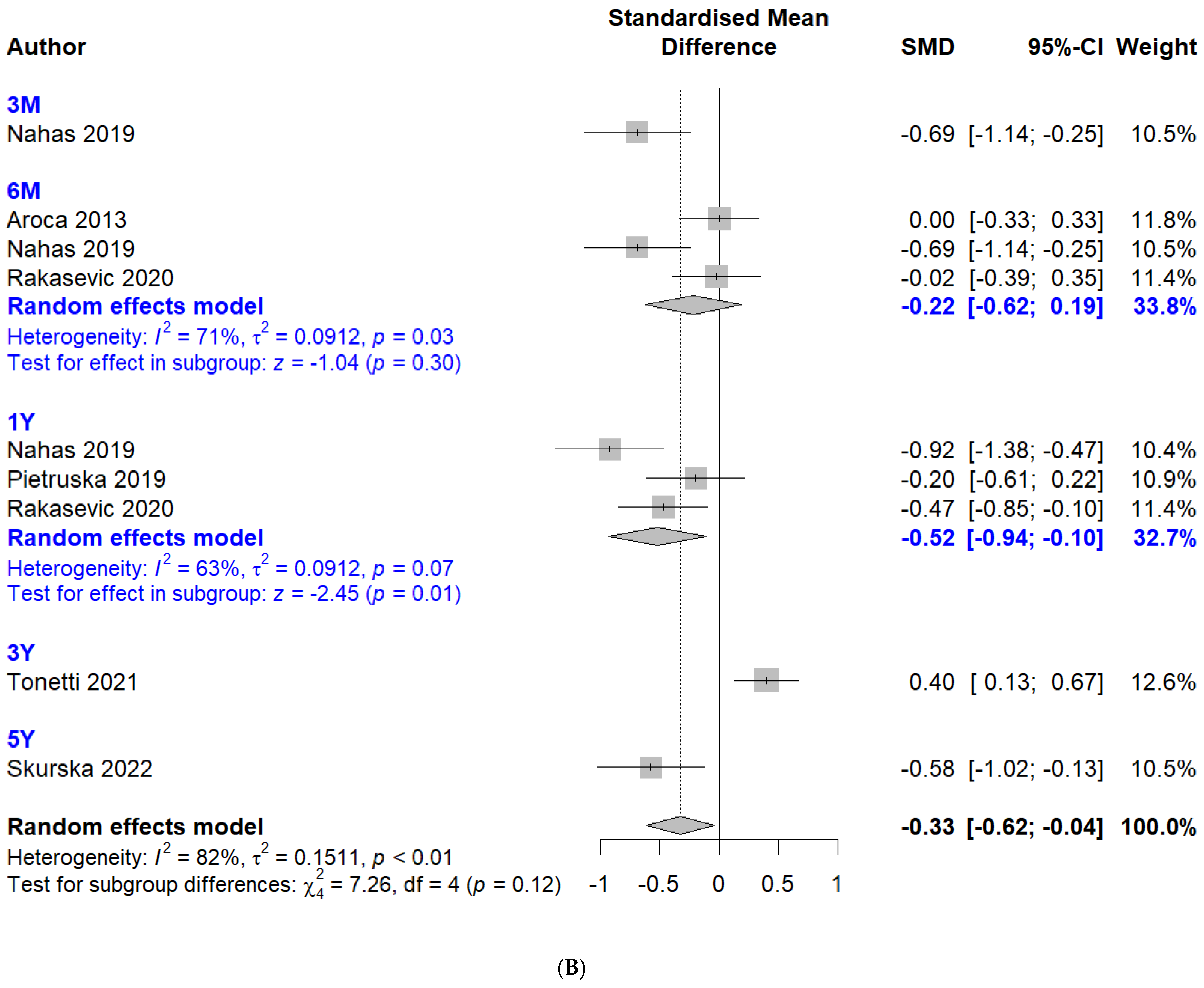
3.3.5. Probing Depth

The high I2 value (85%) and the p-value (p < 0.01) indicate the presence of significant heterogeneity among the studies and a random effects model was applied for the analysis. The forest plot of the meta-analysis showed that the pooled mean difference in probing depth of collagen matrix compared with subepithelial connective tissue was −0.27 (95% confidence interval of −0.65 to 0.11) (Figure 6A). There was no significant difference between collagen and connective tissue. The subgroup of coronally advanced flap showed the pooled mean difference in probing depth of collagen matrix compared with subepithelial connective tissue was −0.22 (95% confidence interval of −0.94 to 0.50). The results for the subgroup of modified coronally advanced tunnel showed that the pooled mean difference in probing depth of collagen matrix compared with subepithelial connective tissue was −0.31 (95% confidence interval of −0.82 to 0.21).
The results of different follow-up periods are shown in Figure 6B. A random effects model was applied, and the high I2 value (82%) and the p-value (p < 0.01) indicate the presence of significant heterogeneity among the studies. The results of the forest plot of the meta-analysis showed that the pooled mean difference in probing depth of collagen matrix compared with subepithelial connective tissue was −0.33 (95% confidence interval of −0.62 to −0.04). The high I2 value (71%) and the p-value (p = 0.03) indicate the presence of significant heterogeneity among the studies. The 6-month subgroup showed that the pooled mean difference in probing depth of collagen matrix compared with subepithelial connective tissue was −0.22 (95% confidence interval of −0.62 to 0.19). The 1-year subgroup showed that the pooled mean difference in probing depth of collagen matrix compared with subepithelial connective tissue was −0.52 (95% confidence interval of −0.94 to −0.10).

3.3.6. Sensitivity Meta-Analysis

A comprehensive meta-analysis encompassing all studies to the meta-analyses was performed after individually excluding each study (Supplementary Figure S1). When comparing the comprehensive meta-analysis encompassing all studies to the meta-analyses performed after individually excluding each study, the mean difference, confidence intervals, and heterogeneity index remained generally consistent.
The heterogeneity index showed a decrease in the case of mean root coverage and complete root coverage (Supplementary Figure S1A,B). For mean root coverage, when the Aroca et al. study (2013) [19] was excluded, the standardized mean difference (SMD) increased slightly from −0.48 to −0.41, while the 95% confidence interval (CI) remained statistically significant. Notably, the I2 value decreased to 45%, indicating a substantial reduction in heterogeneity.
For complete root coverage, when the Pietruska et al. study (2019) [22] was excluded, the I2 value decreased to 49%, indicating a substantial reduction in heterogeneity; however, the risk ratio (RR) increased from 0.68 (95% confidence interval, 0.49 to 0.94) to 0.84 (95% confidence interval, 0.71 to 1.00), and the 95% confidence interval no longer indicated statistical significance, as the upper bound of the confidence interval includes 1.00. This suggests that the Pietruska et al. (2019) [22] study had a significant impact on the overall heterogeneity and that its exclusion led to a more homogeneous set of study results. However, the shift in the RR from 0.68 to 0.84, moving closer to 1.0, implies that the effect size became less pronounced, although still suggesting some degree of risk reduction.
Similar sensitivity analyses performed on the other two data did not result in a comparable reduction in heterogeneity. The lowest I2 values observed after excluding individual studies were 85% and 67%, respectively, indicating that substantial heterogeneity persisted.

3.3.7. Publication Bias Analysis

The results of the analyses for publication bias are shown in Table 3. For mean root coverage, the trim-and-fill method did not require imputation, and the adjusted overall effect size remained consistent with the initial report. Furthermore, Egger’s regression test yielded a non-significant result for publication bias, with a p-value of 0.81. However, complete root coverage, width of keratinized tissue, and probing depth showed differences between the original and trim-and-fill analyses. Both complete root coverage and probing depth showed a significant difference, with Egger’s test p-values of 0.02 and 0.01, respectively, suggesting potential publication bias.

4. Discussion

This systematic review and meta-analysis was conducted to evaluate the performance of multiple root coverage by comparing mean root coverage, probing depth, and width of keratinized tissue between collagen matrix and subepithelial connective tissue. The findings of the present study revealed that disparities were seen between collagen matrix and subepithelial connective tissue with respect to root coverage and width of keratinized tissue. In general, subepithelial connective tissue exhibited more favorable results.
Gingival recession is generally characterized by the displacement of the gingival margin apical to the cementoenamel junction, resulting in the loss of periodontal connective tissue fibers along the root cementum and the subsequent loss of alveolar bone. The classification system developed by Miller categorizes gingival recession based on its relationship to the mucogingival junction and the extent of soft/hard tissue loss in the interproximal area [28]. There are four distinct types in this classification [28]. In Miller’s Class I, marginal tissue recession does not extend to the mucogingival junction and is not accompanied by alveolar bone loss in the interdental area. Class II is characterized by marginal tissue recession that extends to or beyond the mucogingival junction. Class III involves marginal tissue recession that extends to or beyond the mucogingival junction, along with alveolar bone loss in the interdental area. Class IV is distinguished by marginal tissue recession that extends to or beyond the mucogingival junction and is accompanied by gross alveolar bone loss in the interdental area. In general, it has been reported that complete root coverage can be achieved in both Class I and Class II cases [30]. In a more recent report, the achievement of complete root coverage in Miller class III recessions was shown to be possible through surgical approaches; however, the long-term stability of such outcomes cannot be predicted with certainty [31,32]. Recession type 1 is defined as the gingival recession with no loss of interproximal attachment and in this case, the interproximal cemento–enamel junction was clinically not detectable at both mesial and distal aspects of the tooth [29]. In this study, most of the recessions were either Miller class I or II or recession type 1 [29].
Subepithelial connective tissue grafts are widely regarded as the optimal solution for achieving root coverage due to their high predictability and excellent esthetic outcomes [33]. Subepithelial connective tissue grafts exhibit excellent tissue compatibility and facilitate the regeneration of periodontal tissues, resulting in stable, long-lasting coverage of exposed roots [34]. The use of subepithelial connective tissue grafts can be more technically challenging and time-consuming due to the requirement of harvesting tissue from the palate [35]. The outcomes of employing collagen matrix versus connective tissue graft for root coverage in gingival recession did not reveal substantial variations in vascular flow [36]. However, the connective tissue graft demonstrated a more uniform pattern compared to the collagen matrix, which displayed a secondary increase in blood flow at the 14-day mark. Alternative materials, like those derived from porcine or bovine sources, may occasionally be utilized instead of autogenous tissue [37]. Collagen matrices are often employed when patient preferences or clinical conditions preclude the use of autogenous grafts [38]. Improved patient satisfaction can be achieved through the use of collagen matrix, which reduces surgical discomfort and increases soft tissue thickness [39]. The utilization of collagen matrices frequently leads to improved patient comfort, as they typically involve only one surgical site, thereby obviating the need for multiple incisions and associated postoperative discomfort [40]. Although effective in supporting new tissue growth, studies generally indicate that collagen matrices might offer slightly lower root coverage percentages compared to subepithelial connective tissue grafts [41]. In order to ensure the safe and effective use of collagen matrices, it is crucial to investigate potential side effects such as infection, bleeding, pain, and discomfort [42]. The outcomes of this study demonstrated that subepithelial connective tissue generally exhibits superior performance. Additionally, follow-up treatment studies may be required to address any discomfort and generalize the application of collagen for improving root coverage and increasing soft tissue thickness.
The specific type of collagen matrix utilized in the studies is detailed below. The use of a xenogeneic collagen matrix (Geistlich Mucograft®: Geistlich Pharma, AG, Wolhusen Switzerland) has been proposed as an alternative treatment for obtaining promising results in comparison with coronally advanced flap alone in achieving greater root coverage and keratinized tissue [19,21,43,44]. In another study, an acellular sterilized collagen matrix product (Mucoderm, Botiss Dental, Zossen, Germany) derived from porcine dermis and consisting of collagen Types I and III and elastin, was used for the treatment [20,22,23,27,45]. Acellular dermal matrix (LifeCell, Branchburg, NJ, USA) has also been used for root coverage purposes [46]. It is a type of dermal substitute obtained by special treatment of allogeneic dermis to remove its cellular components [47]. Due to its nonimmunogenic features, good osteogenic properties, encouragement of seed cell adhesion, and maintenance of stem cell characteristics, decellularized extracellular matrix has attracted a lot of interest as a bioactive scaffold material [48]. Allogeneic dermal grafts and collagen matrices are two connective tissue substitutes that have steadily acquired acceptance in the clinical setting for lowering patient morbidity [49].
The results of this research indicate that the decision to employ a flap or tunnel technique is significant when utilizing a collagen matrix for root coverage in periodontal surgery [50]. The flap technique involves making incisions to expose the bone and reflect a flap of tissue away from the root surface, thereby providing direct access [51]. A collagen matrix can be placed under the flap to facilitate coverage of the denuded root and to function as a scaffold for the growth of new tissue. In contrast, the tunnel technique is a minimally invasive approach that utilizes specialized instruments to create a tunnel under the gingival tissue, without the need for incisions or the reflection of a flap. The collagen matrix can be inserted into the tunnel to promote root coverage. There is a potential for less postoperative pain with the tunnel technique compared to the flap technique. Nevertheless, it can be surmised that the collagen matrix in the tunnel technique is more likely to be exposed to the oral environment than in the flap technique. In a prior report, the matrix was augmented by 3 mm beyond the bone, and the flap was shifted coronally, subsequently being secured beyond the matrix [46]. However, there was a controversial result indicating that the tunnel technique demonstrated comparable primary and secondary outcomes to those achieved with connective tissue grafting in adults with gingival recessions [52]. Moreover, the type of graft (connective tissue or acellular dermal matrix) did not impact the overall findings. This study showed that the utilization of the coronally advanced flap with collagen matrix may yield comparable results in terms of the percentage of complete root coverage. The use of a modified coronally advanced tunnel combined with collagen matrix revealed inferior results when compared with the subepithelial connective tissue.
The previous report indicated that subepithelial connective tissue grafts achieved excellent outcomes in terms of keratinized tissue gain [53]. Moreover, a prior study revealed a tendency towards better clinical outcomes for subepithelial connective tissue grafts concerning keratinized tissue gain as opposed to collagen matrix [36]. Conversely, previous results prove that new collagen matrix was as effective and predictable as the connective tissue graft for attaining a band of keratinized tissue in patients with fixed prosthetic restorations, but its use was associated with significantly lower patient morbidity [54]. The findings of this research demonstrate that subepithelial connective tissue typically exhibits superior performance in terms of keratinized tissue. Furthermore, the utilization of the modified coronally advanced tunnel technique with collagen matrix yields inferior results when compared to subepithelial connective tissue graft in regards to keratinized tissue. However, previous research indicated that the collagen matrix, when employed as a soft tissue substitute to augment the width of keratinized tissue or mucosa during the second implant surgery, demonstrated comparable effectiveness and predictability to the use of free gingival grafts [55].
The utilization of the collagen matrix may yield comparable results when compared with subepithelial connective tissue in terms of probing depth. Similarly, a previous report showed that collagen biomatrix was effective in reducing the probing depth [56]. It was also shown that the probing depth was sustained at optimal levels throughout the follow-up period, which lasted up to 24 months, with the employment of matrix [46].
Despite the rigorous approach of our meta-analysis, which involved a comprehensive search strategy and strict compliance with PRISMA guidelines, several limitations need to be acknowledged. The primary limitations stem from the studies included in the qualitative and quantitative assessments. Additionally, blinding of participants may have been unfeasible due to the nature of the intervention, which required the creation of a second surgical site to obtain autogenous subepithelial connective tissue [57]. Nine of the included studies exhibited concerns related to bias, particularly in the selection of reported results, potentially affecting the objectivity and generalizability of the findings. The statistical analysis also indicated substantial heterogeneity among the included studies. Sensitivity analysis revealed that one study [22] significantly impacted heterogeneity in a specific outcome, but it did not affect the heterogeneity of the other outcomes, so it was not excluded. This suggests that the high heterogeneity observed in these analyses may be attributable to broader discrepancies among the studies. High heterogeneity in a meta-analysis warrants careful consideration, as it can undermine the external validity of the pooled effect estimates and may reflect underlying differences between the studies. Our results suggest that further analysis and randomized clinical trials with rigorous inclusion criteria are necessary to validate these findings [58]. Variations in follow-up periods and differences in patient populations must also be considered when applying these findings in clinical practice.
Collectively, this meta-analysis’s outcomes indicated that subepithelial connective tissue grafts exhibited superior root coverage, improved probabilities of attaining complete root coverage, and increased the width of keratinized tissue when compared with collagen matrix. In general, the findings suggest that subepithelial connective tissue grafts should be given prioritized consideration compared to collagen matrices.

5. Conclusions

The outcomes of this study revealed that subepithelial connective tissue generally exhibits superior performance. Nonetheless, the utilization of the coronally advanced flap technique with collagen matrix may yield comparable results in terms of the percentage of complete root coverage.

Supplementary Materials

The following supporting information can be downloaded at: https://www.mdpi.com/article/10.3390/app14178049/s1, Table S1: Search strategy for the online databases; Table S2: Studies excluded from full-text reading; Table S3: Risk of bias; Figure S1: Forest plots illustrating the results of sensitivity tests. (A) Mean root coverage. (B) Complete root coverage. (C) Width of keratinized tissue. (D) Probing depth.

Author Contributions

Conceptualization, S.-H.H., H.-J.S., N.J.K., W.-J.P., J.-B.P.; formal analysis, S.-H.H., H.-J.S., N.J.K., W.-J.P., J.-B.P.; writing—original draft preparation, S.-H.H., H.-J.S., N.J.K., W.-J.P., J.-B.P.; writing—review and editing, S.-H.H., H.-J.S., N.J.K., W.-J.P., J.-B.P. All authors have read and agreed to the published version of the manuscript.

Funding

This research received no external funding.

Institutional Review Board Statement

Not applicable.

Informed Consent Statement

Not applicable.

Data Availability Statement

Not applicable.

Conflicts of Interest

The authors declare no conflicts of interest.

References

  1. Lee, C.T.; Lange, M.; Jureidini, A.; Bittner, N.; Schulze-Späte, U. Clinical and patient-reported outcomes after recession coverage using modified vestibular incision subperiosteal tunnel access with a volume-stable collagen matrix as compared to a coronally advanced flap with a subepithelial connective tissue graft. J. Periodontal Implant. Sci. 2022, 52, 466–478. [Google Scholar] [CrossRef] [PubMed]
  2. Mythri, S.; Arunkumar, S.M.; Hegde, S.; Rajesh, S.K.; Munaz, M.; Ashwin, D. Etiology and occurrence of gingival recession—An epidemiological study. J. Indian. Soc. Periodontol. 2015, 19, 671–675. [Google Scholar] [CrossRef]
  3. Jati, A.S.; Furquim, L.Z.; Consolaro, A. Gingival recession: Its causes and types, and the importance of orthodontic treatment. Dent. Press J. Orthod. 2016, 21, 18–29. [Google Scholar] [CrossRef]
  4. Kerner, S.; Borghetti, A.; Katsahian, S.; Etienne, D.; Malet, J.; Mora, F.; Monnet-Corti, V.; Glise, J.-M.; Bouchard, P. A retrospective study of root coverage procedures using an image analysis system. J. Clin. Periodontol. 2008, 35, 346–355. [Google Scholar] [CrossRef]
  5. Tian, W.; Hu, F.; Zhou, X. Comparative Evaluation of Connective Tissue Graft with Pouch/Tunnel Technique versus Connective Tissue Graft with Coronally Advanced Tunnel Flap for the Treatment of Maxillary Recession Cases in Severe Periodontitis. Tissue Eng. Regen. Med. 2021, 18, 1001–1007. [Google Scholar] [CrossRef]
  6. Langer, B.; Langer, L. Subepithelial connective tissue graft technique for root coverage. J. Periodontol. 1985, 56, 715–720. [Google Scholar] [CrossRef] [PubMed]
  7. Lee, C.T.; Chang, P.C.; Touchan, N.; Royzman, D. Root coverage with a modified laterally positioned flap combined with a subepithelial connective tissue graft in advanced recession. J. Periodontal Implant. Sci. 2014, 44, 300–306. [Google Scholar] [CrossRef] [PubMed]
  8. Smirani, R.; Rémy, M.; Devillard, R.; Naveau, A. Use of Human Gingival Fibroblasts for Pre-Vascularization Strategies in Oral Tissue Engineering. Tissue Eng. Regen. Med. 2022, 19, 525–535. [Google Scholar] [CrossRef]
  9. Park, J.B. Treatment of multiple gingival recessions using subepithelial connective tissue grafting with a single-incision technique. J. Oral. Sci. 2009, 51, 317–321. [Google Scholar] [CrossRef]
  10. Lee, Y.; Lee, D.; Kim, S.; Ku, Y.; Rhyu, I.C. Modified tunneling technique for root coverage of anterior mandible using minimal soft tissue harvesting and volume-stable collagen matrix: A retrospective study. J. Periodontal Implant. Sci. 2021, 51, 398–408. [Google Scholar] [CrossRef]
  11. Park, J.B. Root coverage with 2 connective tissue grafts obtained from the same location using a single-incision technique. Quintessence Int. 2009, 40, 371–376. [Google Scholar]
  12. Godat, M.S.; Gruen, T.D.; Miller, P.D.; Craddock, R.D. Use of Tuberosity Connective Tissue for Root Coverage and Ridge Augmentation: Background and Technique. Compend. Contin. Educ. Dent. 2018, 39, 533–539; quiz 540. [Google Scholar]
  13. Barootchi, S.; Rodriguez, M.V.; Chan, H.L.; Kripfgans, O.D.; Wang, H.L.; Tavelli, L. Coronally Advanced Flap with Xenogeneic Collagen Matrix for the Treatment of Gingival Recessions at Sites Presenting with Cervical Restorations or Noncarious Cervical Lesions: A Clinical and Ultrasonographic Study. Int. J. Periodontics Restor. Dent. 2023, 43, 146–154. [Google Scholar] [CrossRef] [PubMed]
  14. Michels, R.; Magrin, G.L.; Cruz, A.; Magini, R.S.; Benfatti, C.A.M. Functionalization of a Volume-Stable Collagen Matrix Using Liquid Platelet-Rich Fibrin: A Case Report Presenting a New Approach for Root Coverage. Case Rep. Dent. 2023, 2023, 3929269. [Google Scholar] [CrossRef]
  15. Parma-Benfenati, S.; Mastrangelo, F.; Cardo, F.; Roncati, M.; Parma-Benfenati, L.; Aimetti, M.; Nevins, M. Evaluation of a New Porcine Collagen Matrix for the Treatment of Buccal Gingival Recessions: A 12-Month Pilot Prospective Human Study Model. Int. J. Periodontics Restor. Dent. 2023, 43, 329–335. [Google Scholar]
  16. Page, M.J.; McKenzie, J.E.; Bossuyt, P.M.; Boutron, I.; Hoffmann, T.C.; Mulrow, C.D.; Shamseer, L.; Tetzlaff, J.M.; Akl, E.A.; Brennan, S.E.; et al. The PRISMA 2020 statement: An updated guideline for reporting systematic reviews. Syst. Rev. 2021, 10, 89. [Google Scholar] [CrossRef]
  17. Higgins, J.P.T.; Green, S. Cochrane Handbook for Systematic Reviews of Interventions. 2023. Version 6.4. [Updated August 2023]. Available online: https://training.cochrane.org/handbook (accessed on 10 June 2024).
  18. Atieh, M.A.; Almatrooshi, A.; Shah, M.; Hannawi, H.; Tawse-Smith, A.; Alsabeeha, N.H.M. Airflow for initial nonsurgical treatment of peri-implantitis: A systematic review and meta-analysis. Clin. Implant. Dent. Relat. Res. 2022, 24, 196–210. [Google Scholar] [CrossRef]
  19. Aroca, S.; Molnár, B.; Windisch, P.; Gera, I.; Salvi, G.E.; Nikolidakis, D.; Sculean, A. Treatment of multiple adjacent Miller class I and II gingival recessions with a Modified Coronally Advanced Tunnel (MCAT) technique and a collagen matrix or palatal connective tissue graft: A randomized, controlled clinical trial. J. Clin. Periodontol. 2013, 40, 713–720. [Google Scholar] [CrossRef] [PubMed]
  20. Cieślik-Wegemund, M.; Wierucka-Młynarczyk, B.; Tanasiewicz, M.; Gilowski, Ł. Tunnel Technique With Collagen Matrix Compared With Connective Tissue Graft for Treatment of Periodontal Recession: A Randomized Clinical Trial. J. Periodontol. 2016, 87, 1436–1443. [Google Scholar] [CrossRef]
  21. Nahas, R.; Gondim, V.; Carvalho, C.V.; Calderero, L.M.; Rosa, E.F.; Sakiyama, T.; Neto, J.B.C.; Pannuti, C.M.; Romito, G.A. Treatment of multiple recessions with collagen matrix versus connective tissue: A randomized clinical trial. Braz. Oral. Res. 2019, 33, e123. [Google Scholar] [CrossRef]
  22. Pietruska, M.; Skurska, A.; Podlewski, Ł.; Milewski, R.; Pietruski, J. Clinical evaluation of Miller class I and II recessions treatment with the use of modified coronally advanced tunnel technique with either collagen matrix or subepithelial connective tissue graft: A randomized clinical study. J. Clin. Periodontol. 2019, 46, 86–95. [Google Scholar] [CrossRef] [PubMed]
  23. Rakasevic, D.L.; Milinkovic, I.Z.; Jankovic, S.M.; Soldatovic, I.A.; Aleksic, Z.M.; Nikolic-Jakoba, N.S. The use of collagen porcine dermal matrix and connective tissue graft with modified coronally advanced tunnel technique in the treatment of multiple adjacent type I gingival recessions: A randomized, controlled clinical trial. J. Esthet. Restor. Dent. 2020, 32, 681–690. [Google Scholar] [CrossRef] [PubMed]
  24. Tonetti, M.S.; Cortellini, P.; Bonaccini, D.; Deng, K.; Cairo, F.; Allegri, M.; Conforti, G.; Graziani, F.; Guerrero, A.; Halben, J.; et al. Autologous connective tissue graft or xenogenic collagen matrix with coronally advanced flaps for coverage of multiple adjacent gingival recession. 36-month follow-up of a randomized multicentre trial. J. Clin. Periodontol. 2021, 48, 962–969. [Google Scholar] [CrossRef] [PubMed]
  25. Milinkovic, I.; Rakasevic, D.; Nikolic-Jakoba, N.; Jankovic, S.; Aleksic, Z. Collagen tissue matrix versus connective tissue graft with modified coronally advanced tunnel technique in the multiple gingival recessions treatment: Split-mouth study with 5-year of follow-up. J. Clin. Periodontol. 2022, 49, 200. [Google Scholar]
  26. Molnár, B.; Aroca, S.; Dobos, A.; Orbán, K.; Szabó, J.; Windisch, P.; Stähli, A.; Sculean, A. Treatment of multiple adjacent RT 1 gingival recessions with the modified coronally advanced tunnel (MCAT) technique and a collagen matrix or palatal connective tissue graft: 9-year results of a split-mouth randomized clinical trial. Clin. Oral. Investig. 2022, 26, 7135–7142. [Google Scholar] [CrossRef]
  27. Skurska, A.; Milewski, R.; Pietruska, M. Long-Term Clinical Assessment of Two Modifications of Tunnel Technique in Treatment of Cairo Recession Type 1 in Lower Jaw. Int. J. Environ. Res. Public Health 2022, 19, 16444. [Google Scholar] [CrossRef]
  28. Miller, P.D., Jr. A classification of marginal tissue recession. Int. J. Periodontics Restor. Dent. 1985, 5, 8–13. [Google Scholar]
  29. Cairo, F.; Nieri, M.; Cincinelli, S.; Mervelt, J.; Pagliaro, U. The interproximal clinical attachment level to classify gingival recessions and predict root coverage outcomes: An explorative and reliability study. J. Clin. Periodontol. 2011, 38, 661–666. [Google Scholar] [CrossRef]
  30. Spahr, A.; Haegewald, S.; Tsoulfidou, F.; Rompola, E.; Heijl, L.; Bernimoulin, J.; Ring, C.; Sander, S.; Haller, B. Coverage of Miller class I and II recession defects using enamel matrix proteins versus coronally advanced flap technique: A 2-year report. J. Periodontol. 2005, 76, 1871–1880. [Google Scholar] [CrossRef]
  31. Fernández-Jiménez, A.; García-De-La-Fuente, A.M.; Estefanía-Fresco, R.; Marichalar-Mendia, X.; Aguirre-Urizar, J.M.; Aguirre-Zorzano, L.A. Complete root coverage in the treatment of Miller class III or RT2 gingival recessions: A systematic review and meta-analysis. BMC Oral. Health 2021, 21, 145. [Google Scholar]
  32. Amarante, E.S.; Leknes, K.N.; Skavland, J.; Lie, T. Coronally positioned flap procedures with or without a bioabsorbable membrane in the treatment of human gingival recession. J. Periodontol. 2000, 71, 989–998. [Google Scholar] [CrossRef] [PubMed]
  33. Dwarakanath, C.D.; Divya, B.; Sruthima, G.N.; Penmetsa, G.S. Sub-epithelial connective tissue graft for root coverage in nonsmokers and smokers: A pilot comparative clinical study. J. Indian. Soc. Periodontol. 2016, 20, 435–440. [Google Scholar] [PubMed]
  34. Roman, A.; Câmpian, R.; Domşa, I.; Soancă, A.; Gocan, H. Subepithelial connective tissue graft for root coverage: Clinical case reports and histologic evaluation. Rom. J. Morphol. Embryol. 2010, 51, 793–797. [Google Scholar]
  35. Kumari, K.; Nath, B.; Kumar, A.; Chhabada, A.K.; Kumari, R.; Prakash, G. Comparison of Root Coverage by the Subepithelial Connective Tissue Graft With and Without Root Biomodification: A Comprehensive Clinical Study. Cureus 2023, 15, e44758. [Google Scholar] [CrossRef]
  36. Tatarakis, N.; Gkranias, N.; Darbar, U.; Donos, N. Blood flow changes using a 3D xenogeneic collagen matrix or a subepithelial connective tissue graft for root coverage procedures: A pilot study. Clin. Oral. Investig. 2018, 22, 1697–1705. [Google Scholar] [CrossRef] [PubMed]
  37. Herford, A.S.; Akin, L.; Cicciu, M.; Maiorana, C.; Boyne, P.J. Use of a porcine collagen matrix as an alternative to autogenous tissue for grafting oral soft tissue defects. J. Oral. Maxillofac. Surg. 2010, 68, 1463–1470. [Google Scholar] [CrossRef]
  38. Petrie, K.; Cox, C.T.; Becker, B.C.; MacKay, B.J. Clinical applications of acellular dermal matrices: A review. Scars Burn. Heal. 2022, 8, 20595131211038313. [Google Scholar] [CrossRef]
  39. Ashurko, I.; Tarasenko, S.; Magdalyanova, M.; Bokareva, S.; Balyasin, M.; Galyas, A.; Khamidova, M.; Zhornik, M.; Unkovskiy, A. Comparative analysis of xenogeneic collagen matrix and autogenous subepithelial connective tissue graft to increase soft tissue volume around dental implants: A systematic review and meta-analysis. BMC Oral. Health 2023, 23, 741. [Google Scholar] [CrossRef]
  40. Raguse, J.D.; Gath, H.J. A metabolically active dermal replacement (Dermagraft) for vestibuloplasty. J. Oral. Rehabil. 2005, 32, 337–340. [Google Scholar] [CrossRef]
  41. De Santis, D.; Luciano, U.; Pancera, P.; Castegnaro, G.; Alberti, C.; Gelpi, F. A New Matrix for Soft Tissue Management. J. Clin. Med. 2022, 11, 4486. [Google Scholar] [CrossRef]
  42. Stefanini, M.; Mounssif, I.; Barootchi, S.; Tavelli, L.; Wang, H.L.; Zucchelli, G. An exploratory clinical study evaluating safety and performance of a volume-stable collagen matrix with coronally advanced flap for single gingival recession treatment. Clin. Oral. Investig. 2020, 24, 3181–3191. [Google Scholar] [CrossRef] [PubMed]
  43. McGuire, M.K.; Scheyer, E.T. Xenogeneic collagen matrix with coronally advanced flap compared to connective tissue with coronally advanced flap for the treatment of dehiscence-type recession defects. J. Periodontol. 2010, 81, 1108–1117. [Google Scholar] [CrossRef]
  44. Barakat, H.; Dayoub, S. Treatment of miller type I and II gingival recession defects using three-dimensional porcine collagen matrix with coronally advanced flap: A randomized clinical split-mouth trial (a 1-year follow-up). Indian. J. Dent. Res. 2020, 31, 209–216. [Google Scholar] [CrossRef]
  45. Fathiazar, A.; Shariatmadar Ahmadi, R.; Sayar, F. A Comparison between Mucoderm® and Connective Tissue Graft for Root Coverage. J. Dent. 2022, 23, 402–409. [Google Scholar]
  46. Schinini, G.; Molinari, A.; Romanelli, H. Clinical evaluation of root coverage using coronally positioned flap associated with acellular dermal matrix allograft in single-type recession defects. A retrospective study. Acta Odontol. Latinoam. 2021, 34, 136–142. [Google Scholar] [CrossRef] [PubMed]
  47. Zeng, A.; Li, H.; Liu, J.; Wu, M. The Progress of Decellularized Scaffold in Stomatology. Tissue Eng. Regen. Med. 2022, 19, 451–461. [Google Scholar] [CrossRef] [PubMed]
  48. Ahn, W.B.; Lee, Y.B.; Ji, Y.H.; Moon, K.S.; Jang, H.S.; Kang, S.W. Decellularized Human Adipose Tissue as an Alternative Graft Material for Bone Regeneration. Tissue Eng. Regen. Med. 2022, 19, 1089–1098. [Google Scholar] [CrossRef] [PubMed]
  49. Tavelli, L.; Barootchi, S.; Rodriguez, M.V.; Mancini, L.; Majzoub, J.; Travan, S.; Sugai, J.; Chan, H.L.; Kripfgans, O.; Wang, H.L.; et al. Recombinant human platelet-derived growth factor improves root coverage of a collagen matrix for multiple adjacent gingival recessions: A triple-blinded, randomized, placebo-controlled trial. J. Clin. Periodontol. 2022, 49, 1169–1184. [Google Scholar] [CrossRef]
  50. Toledano-Osorio, M.; Muñoz-Soto, E.; Toledano, M.; Vallecillo-Rivas, M.; Vallecillo, C.; Ramos-García, P.; Osorio, R. Treating Gingival Recessions Using Coronally Advanced Flap or Tunnel Techniques with Autografts or Polymeric Substitutes: A Systematic Review and Meta-Analysis. Polymers 2022, 14, 1453. [Google Scholar] [CrossRef]
  51. Shkreta, M.; Atanasovska-Stojanovska, A.; Dollaku, B.; Belazelkoska, Z. Exploring the Gingival Recession Surgical Treatment Modalities: A Literature Review. Open Access Maced. J. Med. Sci. 2018, 6, 698–708. [Google Scholar] [CrossRef]
  52. Mayta-Tovalino, F.; Barboza, J.J.; Pasupuleti, V.; Hernandez, A.V. Efficacy of Tunnel Technique (TUN) versus Coronally Advanced Flap (CAF) in the Management of Multiple Gingival Recession Defects: A Meta-Analysis. Int. J. Dent. 2023, 2023, 8671484. [Google Scholar] [CrossRef] [PubMed]
  53. Chambrone, L.; Chambrone, D.; Pustiglioni, F.E.; Chambrone, L.A.; Lima, L.A. Can subepithelial connective tissue grafts be considered the gold standard procedure in the treatment of Miller Class I and II recession-type defects? J. Dent. 2008, 36, 659–671. [Google Scholar] [CrossRef] [PubMed]
  54. Sanz, M.; Lorenzo, R.; Aranda, J.J.; Martin, C.; Orsini, M. Clinical evaluation of a new collagen matrix (Mucograft prototype) to enhance the width of keratinized tissue in patients with fixed prosthetic restorations: A randomized prospective clinical trial. J. Clin. Periodontol. 2009, 36, 868–876. [Google Scholar] [CrossRef] [PubMed]
  55. Lee, K.H.; Kim, B.O.; Jang, H.S. Clinical evaluation of a collagen matrix to enhance the width of keratinized gingiva around dental implants. J. Periodontal Implant. Sci. 2010, 40, 96–101. [Google Scholar] [CrossRef]
  56. Naomi, R.; Ardhani, R.; Hafiyyah, O.A.; Fauzi, M.B. Current Insight of Collagen Biomatrix for Gingival Recession: An Evidence-Based Systematic Review. Polymers 2020, 12, 2081. [Google Scholar] [CrossRef]
  57. Monaghan, T.F.; Agudelo, C.W.; Rahman, S.N.; Wein, A.J.; Lazar, J.M.; Everaert, K.; Dmochowski, R.R. Blinding in Clinical Trials: Seeing the Big Picture. Medicina 2021, 57, 647. [Google Scholar] [CrossRef]
  58. Fernandez, Y.G.E.; Nguyen, H.; Duan, N.; Gabler, N.B.; Kravitz, R.L. Assessing Heterogeneity of Treatment Effects: Are Authors Misinterpreting Their Results? Health Serv. Res. 2010, 45, 283–301. [Google Scholar] [CrossRef]
Figure 1. Flow chart illustrating the process of selecting the articles that have been included in the systematic review.
Applsci 14 08049 g001
Figure 2. Risk of bias. (A) Summary of the risk of bias in the included studies. (B) Overall risk of bias score for each field [19,20,21,22,23,24,25,26,27].
Applsci 14 08049 g002
Figure 3. Forest plots illustrating the comparison between collagen matrix and subepithelial connective tissue for mean root coverage. (A) Different types of surgical approaches, including coronally advanced flap (CAF) and modified coronally advanced tunnel (MCAT). (B) Different follow-up periods [19,20,21,22,23,24,25,26,27].
Applsci 14 08049 g003
Figure 4. Forest plot illustrating the comparison between collagen matrix and subepithelial connective tissue for complete root coverage. (A) Different types of surgical approaches, including coronally advanced flap (CAF) and modified coronally advanced tunnel (MCAT). (B) Different follow-up periods [19,20,21,22,23,24,25,26,27].
Applsci 14 08049 g004aApplsci 14 08049 g004b
Figure 5. Forest plot illustrating the comparison between collagen matrix and subepithelial connective tissue for the width of keratinized tissue. (A) Different types of surgical approaches, including coronally advanced flap (CAF) and modified coronally advanced tunnel (MCAT). (B) Different follow-up periods [19,20,21,22,23,24,26,27].
Applsci 14 08049 g005aApplsci 14 08049 g005b
Figure 6. Forest plot illustrating the comparison between collagen matrix and subepithelial connective tissue for probing depth. (A) Different types of surgical approaches, including coronally advanced flap (CAF) and modified coronally advanced tunnel (MCAT). (B) Different follow-up periods [19,21,22,23,24,27].
Applsci 14 08049 g006aApplsci 14 08049 g006b
Table 1. Main characteristics of the included studies.
Study
Author (Year)
Study DesignSample Size
(Test/Control)
(Participants/Tooth)
Test ProcedureControl ProcedureType of RecessionFollow-Up
Aroca (2013) [19]RCT (split-mouth)(22/78):(22/78)CM + MCATCTG + MCATMiller class I and II1Y
Cieślik-Wegemund (2016) [20]RCT (parallel)(14/59):(14/47)CM + MCATCTG + MCATMiller class I and II3M, 6M
Nahas (2019) [21] RCT (split-mouth)(15/42):(15/40)CM + CAFCTG + CAFMiller class I3M, 6M, 1Y
Pietruska (2019) [22]RCT (split-mouth)(20/46):(20/45)CM + MCATCTG + MCATMiller class I and II1Y
Rakasevic (2020) [23]RCT (split-mouth)(20/62):(20/52)CM + MCATCTG + MCATMiller class I6M, 1Y
Tonetti (2021) [24]RCT (parallel)(44/107):(45/112)CM + CAFCTG + CAFInclusion: recession ≥ 3 mm; exclusion: inter-dental attachment loss greater than 1 mm, or furcation involvement3Y
Milinkovic (2022) [25]RCT (split-mouth)(15/39):(15/57)CM + MCATCTG + MCATRecession type 15Y
Molnár (2022) [26]RCT (split-mouth)(16/57):(16/57)CM + MCATCTG + MCATRecession type 19Y
Skurska (2022) [26]RCT (split-mouth)(18/40):(18/40)CM + MCATCTG + MCATRecession type 15Y
CAF, coronally advanced flap; CM, collagen matrix; CTG, connective tissue graft; MCAT, modified coronally advanced tunnel; RCT, randomized controlled trial; M, month; Y, year; Miller class I: marginal tissue recession which does not extend to mucogingival junction and is not associated with alveolar bone loss in the interdental area; Miller class II: marginal tissue recession which extends to or beyond the mucogingival junction and is not associated with alveolar bone loss in the interdental area; Recession type 1: Gingival recession with no loss of interproximal attachment. Interproximal cementoenamel junction is clinically not detectable at both mesial and distal aspects of the tooth.
Table 2. Outcome parameters of included studies.
Study
Author (Year)
Mean Root CoverageComplete Root CoverageKeratinized Tissue WidthProbing Depth
TestControlTestControlTestControlTestControlF/U Period
Aroca (2013) [19]71.00 ± 21.0090.00 ± 18.005/2213/220.30 ± 0.590.70 ± 0.670.00 ± 0.260.00 ± 0.261Y
Cieślik-Wegemund (2016) [20]NRNRNRNR0.80 ± 1.671.00 ± 1.61NRNR3M
Cieślik-Wegemund (2016) [20]91.00 ± 13.0095.00 ± 11.002/1410/140.80 ± 1.671.00 ± 1.61NRNR6M
Nahas (2019) [21]66.67 ± 39.0285.71 ± 34.0719/4226/400.40 ± 0.901.20 ± 1.300.10 ± 0.400.40 ± 0.463M
Nahas (2019) [21]77.78 ± 39.0289.29 ± 34.0725/4232/400.40 ± 1.001.20 ± 1.000.10 ± 0.400.40 ± 0.466M
Nahas (2019) [21]77.78 ± 39.0282.14 ± 36.2525/4227/400.30 ± 0.701.20 ± 1.100.10 ± 0.400.50 ± 0.461Y
Pietruska (2019) [22]53.20 ± 32.1783.10 ± 27.639/4531/460.52 ± 0.652.78 ± 1.53−0.10 ± 0.530.01 ± 0.581Y
Rakasevic (2020) [23]86.96 ± 18.5084.60 ± 21.2037/6232/520.48 ± 1.150.27 ± 1.23−0.18 ± 0.45−0.17 ± 0.416M
Rakasevic (2020) [23]85.25 ± 14.9087.60 ± 15.1029/6227/520.85 ± 1.200.84 ± 1.00−0.16 ± 0.05−0.14 ± 0.031Y
Tonetti (2021) [24]57.69 ± 57.6976.92 ± 38.4647/10766/1120.00 ± 1.200.50 ± 1.000.00 ± 0.80−0.30 ± 0.703Y
Milinkovic (2022) [25]82.10 ± 9.1084.60 ± 9.7030/3942/57NRNRNRNR5Y
Molnár (2022) [26]23.07 ± 44.5639.30 ± 35.1714/5718/570.97 ± 0.931.25 ± 0.99NRNR9Y
Skurska (2022) [27]55.25 ± 41.9682.35 ± 33.59NRNR0.65 ± 0.762.82 ± 1.51−0.15 ± 0.490.13 ± 0.475Y
NR, Not reported; F/U, Follow-up; M, month; Y, year.
Table 3. Analysis for publication bias.
VariablesOriginal AnalysisTrim-and-Fill AnalysisEgger’s Regression Test p-Value
SMD/RR (95% CI)SMD/RR (95% CI)Added Studies/Total Studies
MRC−0.48 (−0.69 to −0.26)−0.48 (−0.69 to −0.26)0/90.81
CRC0.68 (0.49 to 0.94)0.88 (0.56 to 1.38)3/110.02
KTW−0.75 (−1.25 to −0.26)−0.58 (−1.15 to −0.01)1/90.10
PD−0.27 (−0.65 to 0.11)0.13 (−0.35 to 0.60)3/90.01
SMD, standardized mean difference; RR, relative risk; CI, confidence interval; MRC, mean root coverage; CRC, complete root coverage; KTW, width of keratinized tissue; PD, probing depth.
Disclaimer/Publisher’s Note: The statements, opinions and data contained in all publications are solely those of the individual author(s) and contributor(s) and not of MDPI and/or the editor(s). MDPI and/or the editor(s) disclaim responsibility for any injury to people or property resulting from any ideas, methods, instructions or products referred to in the content.

