Article

Detachment Energy Evaluation in Nano-Particle Cleaning Using Lateral Force Microscopy

1
Department of Mechanical and Information Science and Technology, Kyushu Institute of Technology, Iizuka 820-8502, Fukuoka, Japan
2
Department of Chemical and Biomolecular Engineering, Clarkson University, Potsdam, NY 13699, USA
3
Ebara Corporation, Ltd., Fujisawa 251-0875, Kanagawa, Japan
*
Author to whom correspondence should be addressed.
Appl. Sci. 2024, 14(18), 8145; https://doi.org/10.3390/app14188145
Submission received: 14 July 2024 / Revised: 13 August 2024 / Accepted: 21 August 2024 / Published: 10 September 2024
(This article belongs to the Section Nanotechnology and Applied Nanosciences)

Abstract

:
It has been difficult to detach abrasive particles smaller than 50 nm from polished surfaces in post-CMP cleaning. During the cleaning process, the residual nano-particles exert shear force in the inevitable shear flow. In order to understand the cleaning mechanism, it is indispensable to investigate not only the force but also the energy acting on different-sized nano-particles. In this article, we proposed the evaluation of detachment energy (the energy required to detach nano-particles) by using Lateral Force Microscopy. As an example, the dominant detachment energy of the silica nano-particle between the oxide film is the potential energy of the hydrogen bond. It suggested that the silica nano-particle detachment involves the breaking of hydrogen bonds.

1. Introduction

In recent years, abrasive particles smaller than 50 nm have been applied in Chemical Mechanical Polishing (CMP) techniques to achieve a surface with nanoscale flatness within the depth of focus in a lithography process in each layer. This advancement is crucial for realizing multilayer wiring structures and reducing wiring widths down to 7 nm in high-performance large-scale ICs [1,2,3]. Due to this, the presence of residual particles on the polished film surface can be a killer defect (short or opens in electric circuits, etc.) as a mask in the lithography process [4], causing partial convexity in the subsequent film deposition process [5], etc., and then leading to lower LSI chip yields. In particular, the oxide films that serve as interlayer insulators and are polished with smaller silica nano-particles are used in the process. Hence, detaching smaller nano-particles (abrasive particles, cleaning waste/dust, etc.) from polished surfaces has indeed posed a significant challenge in post-CMP wet cleaning processes.
In general, contact cleaning by polyvinyl alcohol (PVA) brush scrubbing and non-contact cleaning by enforcing MHz waves were applied to detach nano-particles from the surface. Although the cleaning process is being reconsidered to realize optimal and efficient cleaning [2,6], the cleaning phenomenon has not yet been clearly understood because the residual contamination on the wafer surface is typically inspected only after the cleaning process in dry conditions, rather than wet conditions using the surface scanning methods (e.g., scanning electron microscopy). Consequently, it is essential to investigate the cleaning mechanisms, which involve the detachment of the nano-particle from the surface to be cleaned, and the occasional reattachment to the surface.
Thus, we have established a direct observation method for studying the cleaning phenomena that occurred in a range of a few hundred nanometers from the surface by employing an evanescent light which generated a few hundred nanometers from the surface to be cleaned [7,8,9,10,11,12,13]. By using this observing method, size-dependent particle behaviors during cleaning were evaluated in the cleaning phenomenon [14,15,16,17], especially in the presence of inevitable shear flow [18]. As a result, the smaller particles required considerably more time to detach. This size-dependent detachability of the nano-particle is considered involvement with the physicochemical energy between the nano-particle and the substrate in the shear flow. However, the potential energy of the physicochemical interactions has not been investigated quantitatively.
Various research groups have evaluated the vertical force to pull the particle with a diameter of a few micrometers at the smallest size from the substrate surface by the force curve mode of AFM [19] or by numerically calculating the interaction (van der Waals force, repulsive forces by the electric double layer, etc.) of the nano-particle and the substrate under theoretical models [20]. Hence, the energy evaluation of the nano-particle detachment in the action shear force (lateral force) was indispensable.
In this article, we proposed an evaluation method of energy required to detach various-sized nano-particles from the substrate by using Lateral Force Microscopy (LFM) in order to investigate the dominant interactions. Distinctively, the energy quantity is not a vector quantity like the interacting force, but a scalar quantity at the nanoscale. Therefore, the phenomenon could be evaluated in terms of its scalar quantity as the physicochemical energy, and not only evaluated in terms of the acting behavior to nano-particles in one or two directions and at a certain moment.
As an example, the detachment energy of the silica nano-particle from the oxide film (SiO2 film on a silicon wafer) surface was measured such as in post-CMP cleaning as shown in Figure 1. The dominant energy to detach the particle which involves breaking the bond was discovered.

2. Measurement Method of Nano-Particle Detachment Energy Using Lateral Force Microscopy

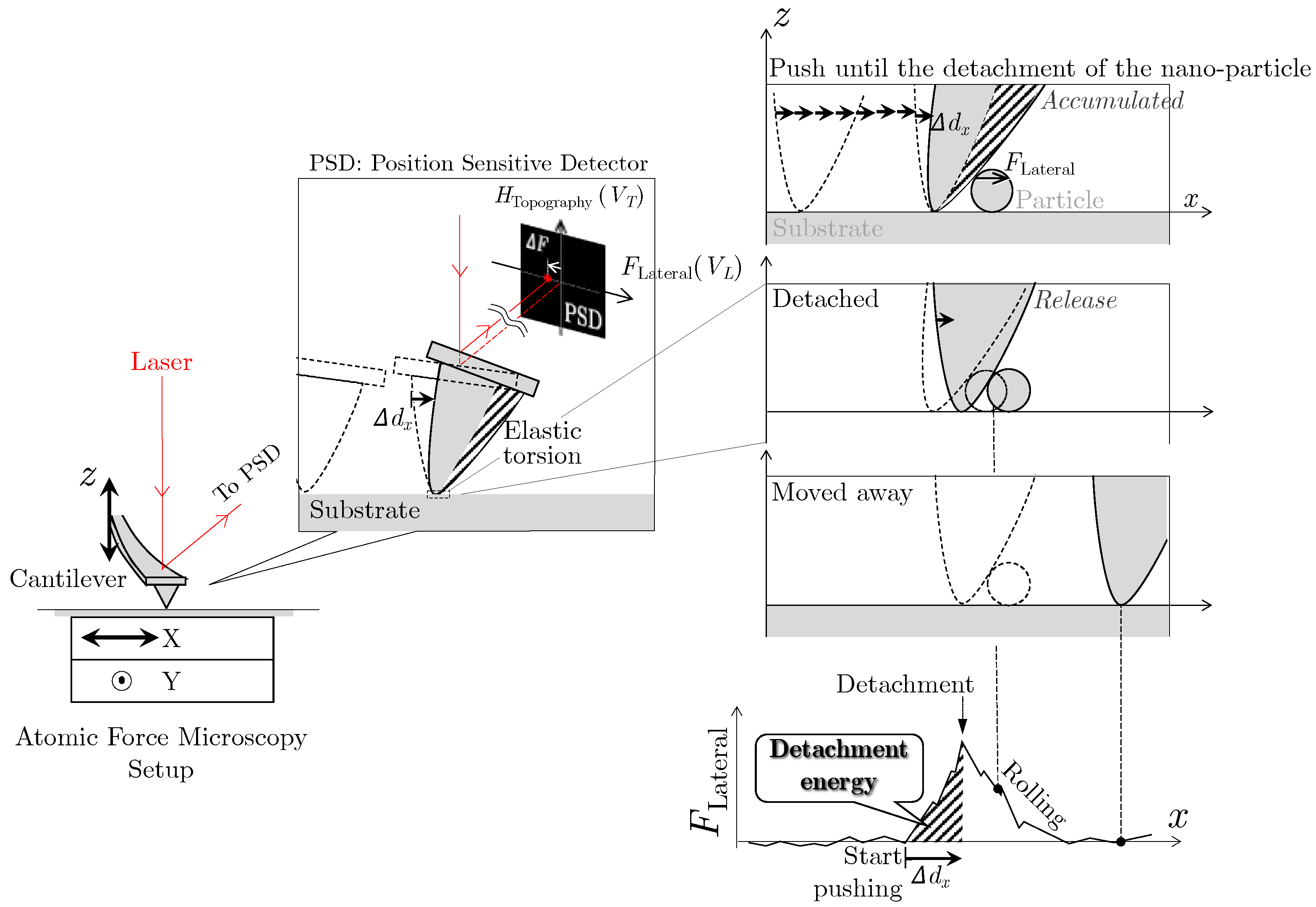
We used a method that can measure the shear action force to detach the nano-particles adhered to the substrate surface [21,22]. The position and the size (height) (Htopgraphy(VT)) of the nano-particles were determined by Atomic Force Microscopy(AFM) before and after detachment. In order to measure the force required to detach the nano-particle from the surface, we detached the nano-particle from the surface and moved it on the surface by applying a force through contact with the tip of the cantilever using LFM. (Figure 2). The elastic torsion of the cantilever was detected as a voltage signal (VL) by the position-sensitive detector (PSD). This signal indicates the variation in the lateral force FLateral acting on the particle during the probe’s movement in the x-direction and is converted to lateral force through calibration [22,23] (the calibration method is in the Supplementary Information). When the torsion is released, it can be regarded as the detachment of the particles.
The energy required to detach (detachment energy) can be calculated by the variation of lateral force and the displacement Δdx of the cantilever tip while pushing the nano-particle until it detaches. Then, the surface interrelationship between the particle and the film was quantitatively investigated.

3. Experimental Results of Detachment Energy for Various-Sized Silica Particles from an Oxide Film Surface

3.1. Measurand of the Force Required to Detach

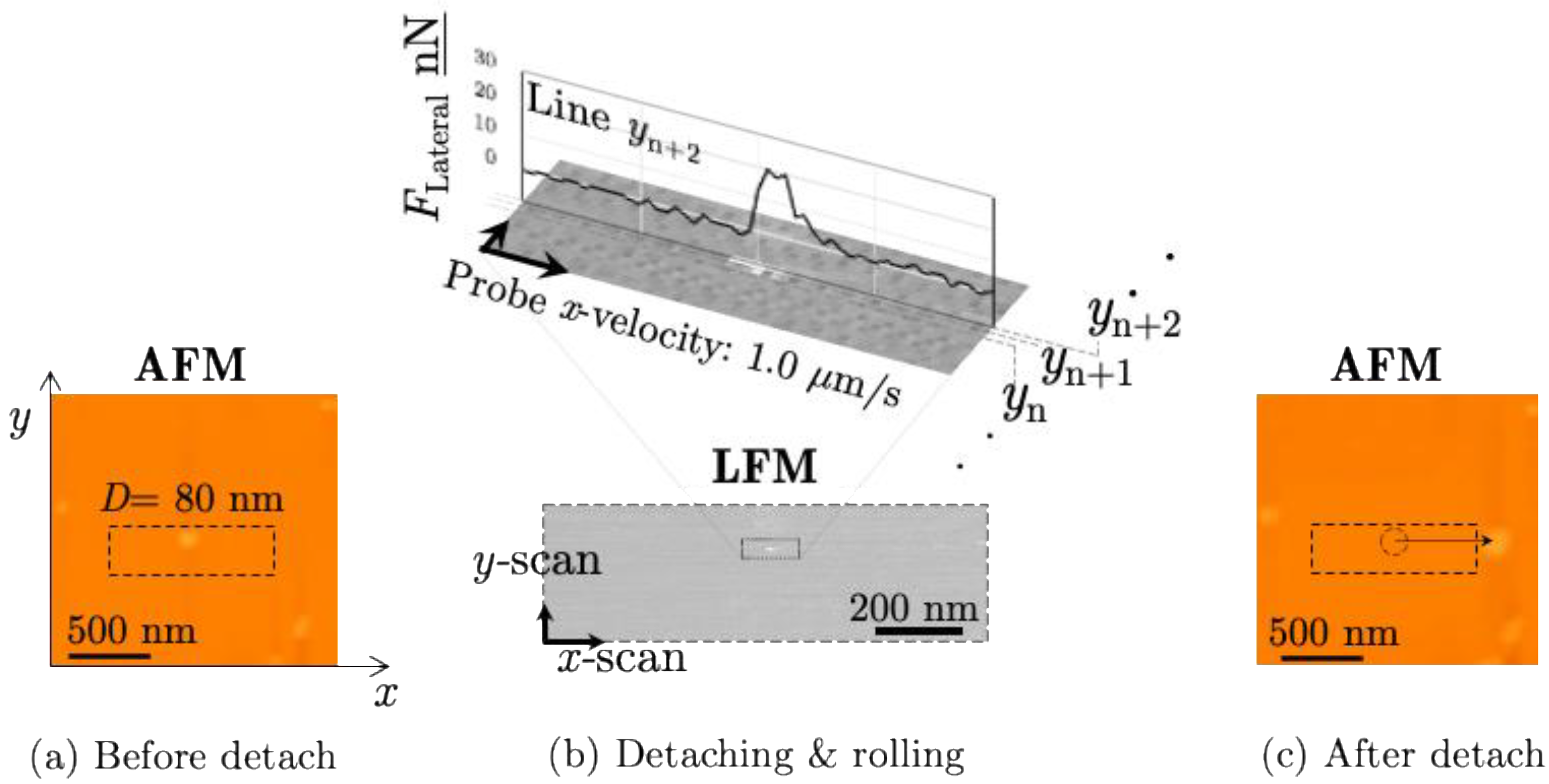
Silica standard particles with diameters of D = 52 ± 3, 80 ± 3, and 310 ± 19 nm (nanoComposix NanoXactTM Silica Nanospheres, San Diego, CA, USA) were adhered to the oxide film surface after the suspension diluted in a pH10.5 solution had naturally dried. The probe was scanned on the surface in an LFM mode (XE-300P AFM, Park Systems, Suwon, Republic of Korea) within 30 min. The scanning speed was 1000 nm/s in the x-axis, and the sampling time Δt was 1/256 s. When scanning in the x-direction at any y-coordinate is completed, the probe moves slightly in the positive y-direction and starts scanning in the x-direction from the x = 0 nm point again. The typical results of the images showed the horizontal movement of the D = 80 nm silica particle as shown in Figure 3. It was clearly confirmed that the nano-particle was detached and moved by the probe pushing.
Figure 4 shows lateral forces before acting (Line yn) and during acting on the SiO2 particles (Line yn+1~yn+2) and after detaching (Line yn+3). The timing of the nano-particle detachment was known as the lateral force maximum. The lateral force was converted from the measured voltage which indicated that the displacement of the laser spot moved on the position-sensitive detector when the cantilever was twisted. The resolution of measurable lateral force in this experiment was 2 nN (2σ) which calculated using the force variation during scanning on the oxide film in line yn. Subsequently, the work to detach Wlfm was analyzed on the line yn+2 that the particle detached from on the surface.

3.2. Measurements of the Detachment Energy

Figure 5 shows the variation in lateral forces acting on the silica particle during detachment from the oxide film surface. The work to detach the particle Wlfm that was drawn by hatching was calculated by the changes in lateral force FLateral and the displacement Δdx of the cantilever tip from the moment the probe initiated contact with the particle t0 until the particle detached at tn by Equation (1).
W l f m = i = t 0 t n F L a t e r a l ,   i · d x , i
This work is equivalent to the energy required to detach a nano-particle (detachment energy).

4. Theoretical Calculation of Interaction Energy

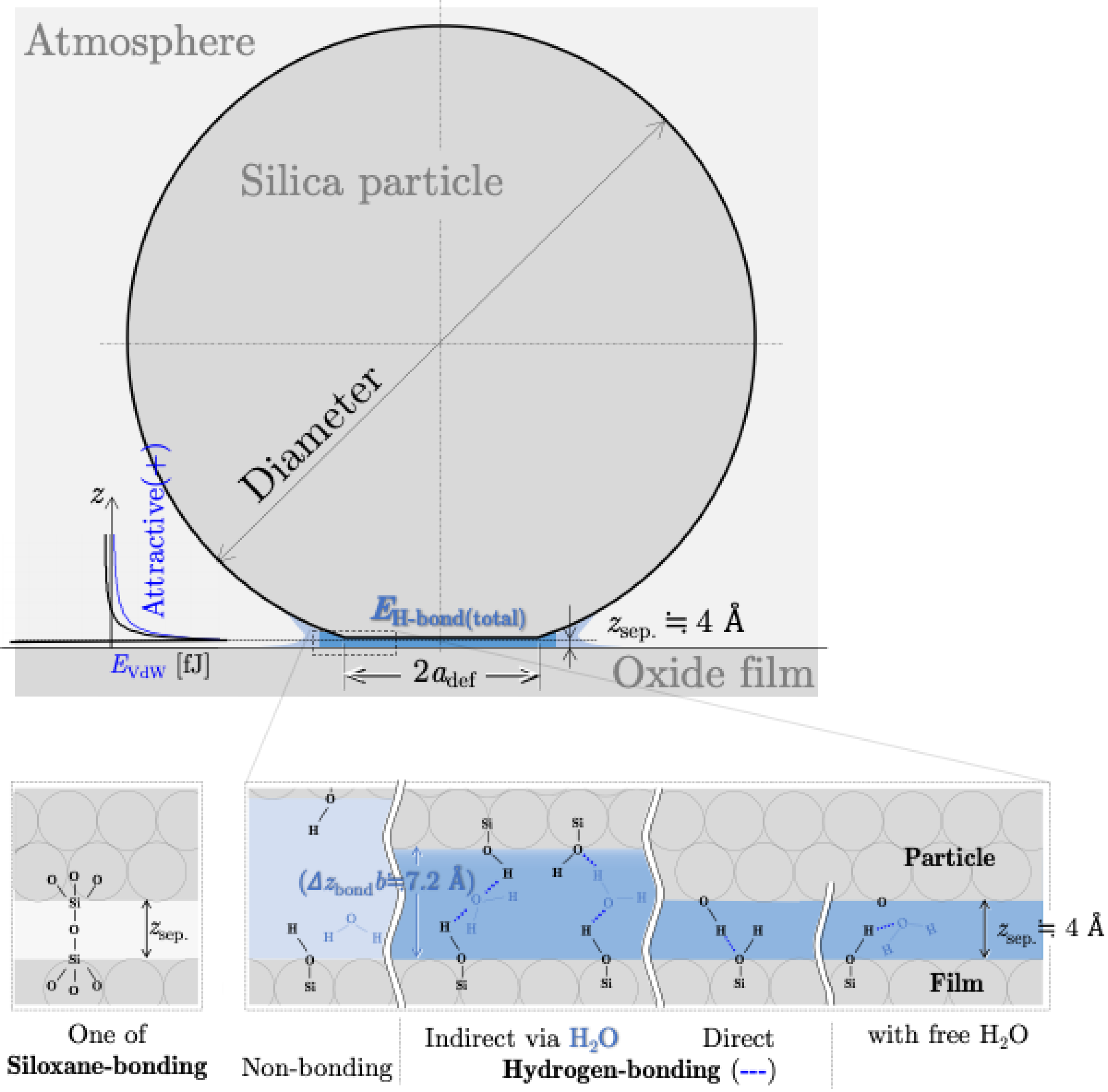
The work to detach the silica particle from the oxide surface Wlfm indicates the physicochemical interaction energies. These interaction energies (siloxane bonds, hydrogen bonds, and van der Waals interactions as shown in Figure 6) were calculated below to discover the dominant energy with physicochemical parameters as shown in Table 1.

4.1. Potential Energy of Siloxane Bonds

The SiO2 surface was hydroxylated [35], and various silanol groups related to the siloxane bridge were formed (Si-O-Si) in an environment above 200 °C. The concentration of the siloxane bridge increases when the silicas are heated under a vacuum because the OH group concentration on the surface decreases monotonically with increasing temperature. Its total siloxane bonding energy ES-bond(total) is calculated as follows [26]:
ES-bond(total) = ρOHSareaES-bond
where ρOH is the concentration of the OH group, ES-bond is the siloxane bonding energy per mole, and Sarea is the total interaction area between particle and substrate (film). The interaction area is calculated as follows:
Sarea = π(adef)2 + 2π(D/2)Δzbondb
where the first term is the deformation area of the particle (adef is the contact radius calculated by the JKR model [32]), and the second term represents the surface area of the spherical crown at a height Δzbond near the contact point with a probability b. The typical calculated results are shown in Table 2.

4.2. Potential Energy of Hydrogen Bonds

At temperatures below 200 °C, hydrogen bonding (physisorption) occurs as water remains on the surface [27]. The bond formed between a positively charged hydrogen atom and another negatively charged atom (in this paper, an oxygen atom) is called a hydrogen bond. As shown in Figure 6, hydrogen bonds include cases in which the oxygen and hydrogen atoms of the Si-OH groups on the silica nano-particle or the oxide film surface bond indirectly via water molecules or directly. The total potential energy of a hydrogen bond is calculated [26] as follows:
EH-bond(total) = ρOHSareaEH-bond
where ρOH is the concentration of the OH group, EH-bond is the hydrogen bonding energy per mole, and Sarea is the total interaction area between the nano-particle and the film. The typical calculated results are shown in Table 2.

4.3. Potential Energy of van der Waals Interaction

The van der Waals force generated between atoms and molecules is generally attractive in nature due to the motion of electrons (Figure 6). For a deformed particle of a diameter D on a flat surface, the interaction energy EVdW is given by the following [36]:
E VdW = A D 12 z s e p 1 + a d e f 2 D z s e p
where A is the Hamaker constant, zsep. is the separation distance between the particle and the substrate, and adef is the contact radius. The typical calculated results are shown in Table 3.

4.4. Dominant Interaction Energy for Detachment

Figure 7a and Table 4 show each calculated energy in the atmosphere using physicochemical properties (Table 1). As a result, the work to detach the particle from the film WlfmD showed similar characteristics to the potential energy of hydrogen bonding EH-bondD1.2. Therefore, the effective total interaction energies in the atmosphere were calculated by Equation (6) as shown in Figure 7b.
Ereq@atmosphere = EH-bond(total) + EVdW

5. Discussion

The dominant energy required to detach the particle from the surface is the energy of the hydrogen bond (Si-OH bonding) represented as EH-bond(total)between the silica particle and the oxide film. This indicates that the particle detachment involves breaking the hydrogen bond between the nano-particle and the film. The possible dominance (importance) of hydrogen bonding had already been pointed out by estimation by measuring the vertical force interaction [25]; however, our experimental results measured the lateral force in the same direction as observed in the cleaning phenomenon, and a theoretical comparison determined it clearly. Another reason for the dominance of hydrogen bonding is due to the formation of silanol groups on the surface and enabling hydrogen bonds to occur because the surfaces of the silica nano-particles and oxide film are hydrated in the alkaline solution (pH 10.5). In the case of the siloxane bond, it was significantly higher, suggesting that it required a higher temperature in the atmosphere, unrelated to the water reaction [27].
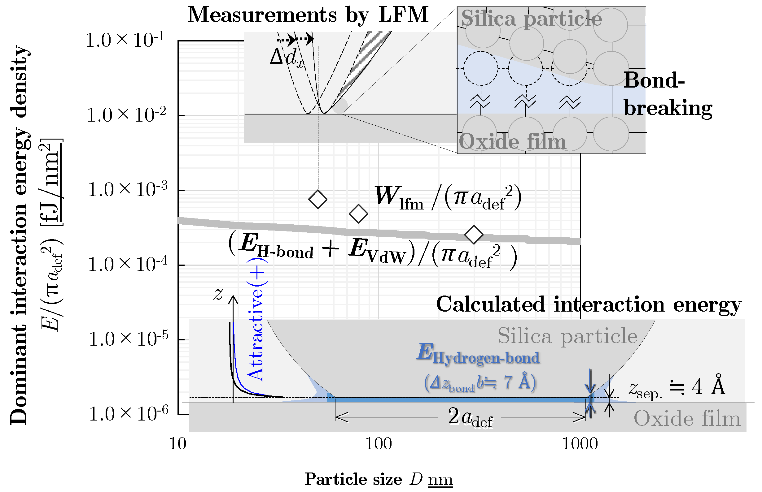
In addition, the detachment energy density was considered to evaluate the particle’s size-dependent detachability as shown in Figure 8. It was implied that the smaller particle size is difficult to detach from the surface due to higher energy density (energy per contact area).

6. Conclusions

The detachment energy evaluation method was proposed to investigate the dominant interaction in the cleaning using Lateral Force Microscopy. In this article, the detachment energies of the silica nano-particle with a diameter of 50, 70, and 300 nm from an oxide film (SiO2 film on the silicon wafer) such as in the post-CMP cleaning were measured. It was discovered that the dominant detachment energy of the silica nano-particles from the oxide film is the potential energy of hydrogen bonding. This indicates that the particle detachment involves breaking the hydrogen bond between the silica nano-particle and the oxide film.
As described above, the effectiveness of this approach was demonstrated to discover the dominant interaction which involved the physicochemical energy between the nano-particle and the substrate. It leads to the quantitative investigation of the size-dependent detachability of the nano-particle in the cleaning phenomenon.

Supplementary Materials

The following supporting information can be downloaded at: https://www.mdpi.com/article/10.3390/app14188145/s1, Figure S1: Conversion method from the measured lateral voltage to the lateral force.

Author Contributions

Conceptualization, P.K. and S.H.; methodology, P.K.; validation (conduction of the experiment and consideration of the bonding), Y.T.; formal analysis, P.K., Y.T. and S.H.; instrumentation resources, J.S.; writing—original draft preparation, Y.T.; writing—review and editing, J.S.; investigation, Y.W. and H.H. All authors have read and agreed to the published version of the manuscript.

Funding

This work was also partially supported by KAKENHI Grant-in-Aid for Scientific Research (c)16K06015, 21J14543 from the Japan Society for Promotional Science.

Institutional Review Board Statement

Not applicable.

Informed Consent Statement

Not applicable.

Data Availability Statement

The original contributions presented in this study are included in the article/Supplementary Materials, and further inquiries can be directed to the corresponding author.

Acknowledgments

The authors are grateful to Professor Emeritus S. V. Babu (https://www.clarkson.edu/news-events/clarkson-university-professor-emeritus-s-v-babu-honored-electrochemical-society-conference-and-clarkson-cmp-reunion 20 August 2024) who belonged to the Center for Advanced Materials Processing at Clarkson University for collaboration on the early stages of this work.

Conflicts of Interest

Authors Satomi Hamada, Yutaka Wada and Hirokuni Hiyama were employed by the company Ebara Corporation, Ltd. The remaining authors declare that the re-search was conducted in the absence of any commercial or financial relationships that could be construed as a potential conflict of interest.

References

  1. Yield Enhancement. International Roadmap for Device and Systemstm 2021 Update; IEEE: New York, NY, USA, 2021; pp. 3–4, 14. [Google Scholar]
  2. Reinhardt, K.A.; Kern, W. Handbook of Silicon Wafer Cleaning Technology, 3rd ed.; Elsevier: Amsterdam, The Netherlands; William Andrew: Norwich, NY, USA, 2018; pp. 254–260, 662–663. [Google Scholar]
  3. Seo, J. Challenges and solutions for post-CMP cleaning at device and interconnect levels. In Advances in Chemical Mechanical Planarization (CMP); Elsevier: Amsterdam, The Netherlands, 2022; pp. 503–532. [Google Scholar]
  4. Sanders, D.P. Advances in Patterning Materials for 193 nm Immersion Lithography. Chem. Rev. 2007, 110, 321–360. [Google Scholar] [CrossRef] [PubMed]
  5. Chen, Y.; Tang, J.; Mikhaylichenko, E.; Brown, B.; Redeker, F. Chemical mechanical cleaning for CMP defect reduction, Applied Materials. In Proceedings of the 2016 China Semiconductor Technology International Conference (CSTIC), Shanghai, China, 13–14 March 2016; IEEE: Piscataway, NJ, USA, 2016. [Google Scholar]
  6. Manzonie, A.; Buley, T.; Chu, J.-n.; Kulus, M. CMP slurry optimization for advanced nodes. Solid State Technol. 2016, 59, 20–23. [Google Scholar]
  7. Temple, P.A. Total internal reflection microscopy. a surface inspection technique. Appl. Opt. 1981, 20, 2656–2664. [Google Scholar] [CrossRef] [PubMed]
  8. Ambrose, E.J. A surface Constant Microscope for the study of Cell Movements. Nature 1956, 178, 1194. [Google Scholar] [CrossRef] [PubMed]
  9. Takahashi, S.; Nakajima, R.; Miyoshi, T.; Takaya, Y.; Takamasu, K. Development of an evanescent light measurement system for Si wafer microdefect detection. Key Eng. Mater. 2005, 295, 15–21. [Google Scholar] [CrossRef]
  10. Kimura, K.; Suzuki, K.; Khajornrungruang, P. Study on fine particle behavior in slurry flow between wafer and polishing pad as a material removal process in CMP. In Proceedings of the ICPT 2012—International Conference on Planarization/CMP Technology, Grenoble, France, 15–17 October 2012; pp. 345–350. [Google Scholar]
  11. Idei, Y.; Khajornrungruang, P.; Kimura, K. Observation of Slurry Fine Particle Motion in CMP process. In Proceedings of the JSPE Kyushu Branch, Tokyo, Japan, 22–23 November 2009; p. 11. (In Japanese). [Google Scholar]
  12. Khajornrungruang, P.; Dean, P.J.; Babu, S.V. Study on dynamic observation of sub-50 nm sized particles in water using evanescent field with a compact and mobile apparatus. In Proceedings of the ASPE2014 Annual Meeting, Boston, MA, USA, 11 September 2014; pp. 73–77. [Google Scholar]
  13. Khajornrungruang, P.; Korkmaz, S.; Angshuman, P.; Suzuki, K.; Kimura, K.; Babu, S.V. Light scattering model for individual sub-100-nm particle size determination in an evanescent field. Jpn. J. Appl. Phys. 2016, 55, 06JG02. [Google Scholar] [CrossRef]
  14. Terayama, Y.; Khajornrungruang, P.; Suzuki, K.; Kusatsu, K.; Hamada, S.; Wada, Y.; Hiyama, H. Real Time Nanoscale Cleaning Phenomenon Observation during PVA brush Scrubbing By Evanescent Field. ECS Trans. 2019, 92, 191–197. [Google Scholar] [CrossRef]
  15. Terayama, Y.; Khajornrungruang, P.; Suzuki, K.; Kusatsu, K.; Hamada, S.; Wada, Y.; Hiyama, H. Observation of Approach-to-surface of PVA brush in Cleaning by Evanescent Field. In Proceedings of the ICPT2019—International Conference on Planarization/CMP Technology, Hsinchu, Taiwan, 15–18 September 2019; pp. 60–61. [Google Scholar]
  16. Terayama, Y.; Khajornrungruang, P.; Suzuki, K.; Mori, R.; Hamada, S.; Wada, Y.; Hiyama, H. Real Time Nanoscale Cleaning Phenomenon Observation During Enforcing MHz Wave By Evanescent Field. ECS Trans. 2022, 108, 17–23. [Google Scholar] [CrossRef]
  17. Khajornrungruang, P.; Terayama, Y.; Hamada, S.; Wada, Y.; Hiyama, H. Phenomenon Observation on Activated Fluid Flow for Cleaning Sub-100 nm particles on a Silica Glass Substrate Surface by Evanescent Microscopy. In Proceedings of the ICPE—International Conference on Precision Engineering, Nara, Japan, 28 November–2 December 2022. C292. [Google Scholar]
  18. Terayama, Y.; Khajornrungruang, P.; Suzuki, K.; Fujishima, H.; Hamada, S.; Wada, Y.; Hiyama, H. Direct observation of removal of SiO2 nano-particles from silica surfaces: An evanescent field microscopy study and shear flow acting moment. Jpn. J. Appl. Phys. 2023, 62, SH8004. [Google Scholar] [CrossRef]
  19. Xu, K.; Vos, R.; Vereecke, G.; Doumen, G.; Fyen, W.; Mertens, P.W.; Heyns, M.M.; Vinckier, C.; Fransaer, J.; Kovacs, F. Fundamental study of the removal mechanisms of nano-sized particles using brush scrubber cleaning. J. Vac. Sci. Technol. B Microelectron. Nanometer Struct. 2005, 23, 2160–2175. [Google Scholar] [CrossRef]
  20. Xu, Q.; Li, M.; Zhang, L.; Niu, J.; Xia, Z. Dynamic Adhesion Forces between Microparticles and Substrates in Water. Langmuir 2014, 30, 11103–11109. [Google Scholar] [CrossRef] [PubMed]
  21. Shima, S.; Satomi, S.; Takatoh, C.; Wada, Y.; Fukunaga, A.; Sobukawa, H. Measurement of Removal Force in DI-Water for Fine Particles with Sub-nanoNewton Resolution. In Proceedings of the ICPT2017, Leuven, Belgium, 11–13 October 2017. [Google Scholar]
  22. Ranaweera, C.K.; Babu, S.V.; Hamada, S.; Seo, J. Measurement of the force required to move ceria particles from SiO2 surface using lateral force microscopy. Mater. Res. 2022, 37, 1789–1797. [Google Scholar] [CrossRef]
  23. Asay, D.B.; Kim, S.H. Direct force balance method for atomic microscopy lateral force calibration. Rev. Sci. Instrum. 2006, 77, 043903. [Google Scholar] [CrossRef]
  24. Schwaderer, P.; Funk, E.; Achenbach, F.; Weis, J.; Bräuchle, C.; Michaelis, J. Single-Molecule Measurement of the Strength of a Siloxane Bond. Langmuir 2008, 24, 1343. [Google Scholar] [CrossRef]
  25. Bowen, J.; Rossetto, H.L.; Kendall, K. Adhesion between silica surfaces due to hydrogen bonding. Surf. Topogr. Metrol. Prop. 2016, 4, 034001. [Google Scholar] [CrossRef]
  26. Wu, X.; Sacher, E.; Sacher, E. The effects of hydrogen bonds on the adhesion of inorganic oxide particles on hydrophilic silicon surfaces. J. Appl. Phys. 1999, 86, 1744–1748. [Google Scholar] [CrossRef]
  27. Bergna, H.E.; Wilmington, D. The Colloid Chemistry of Silica—Chapter 1: An overview. Adv. Chem. Am. Chem. Soc. 1994, 1–47. [Google Scholar] [CrossRef]
  28. Zhuravlev, L.T. Concentration of Hydroxyl Groups on the Surface of Amorphous Silicas. Langmuir 1987, 3, 316–318. [Google Scholar] [CrossRef]
  29. Visser, J. On Hamaker constants: A comparison between Hamaker constants and Lifshitz van der Waals constants. Adv. Colloid Interface Sci. 1972, 3, 331–363. [Google Scholar] [CrossRef]
  30. Dahneke, B. The influence of flattening on the adhesion of particles. J. Colloid Interface Sci. 1972, 40, 1–13. [Google Scholar] [CrossRef]
  31. Mittal, K.L.; Bowling, R.A. A Theoretical Review of Particle Adhesion. Part. Surf. 1988, 1, 129–142. [Google Scholar]
  32. Johnson, K.L.; Kendall, K.; Roberts, A.D. Surface energy and the contact of elastic solids. Proc. R. Soc. Lond. Ser. A 1971, 324, 301. [Google Scholar]
  33. Vo, T.; Reeder, B.; Damone, A.; Newell, P. Effect of Domain Size, Boundary, and Loading Conditions on Mechanical Properties of Amorphous Silica: A Reactive Molecular Dynamics Study. Nanomaterials 2019, 10, 54. [Google Scholar] [CrossRef] [PubMed]
  34. Sharpe, W.N.; Pulskamp, J.; Gianola, D.S.; Eberl, C.; Polcawich, R.G.; Thompson, R.J. Strain Measurements of Silicon Dioxide Microspecimens by Digital Imaging Processing. Exp. Mech. 2007, 47, 649–658. [Google Scholar] [CrossRef]
  35. Liu, G.; Zhang, C.; Zhao, M.; Guo, W.; Luo, Q. Comparison of Nanomaterials with Other Unconventional Materials Used as Additives for Soil Improvement in the Context of Sustainable Development: A Review. Nanomaterials 2021, 11, 15. [Google Scholar] [CrossRef]
  36. Tabor, D. Surface Forces and Surface Interactions. J. Colloid Interface Sci. 1977, 58, 2. [Google Scholar]
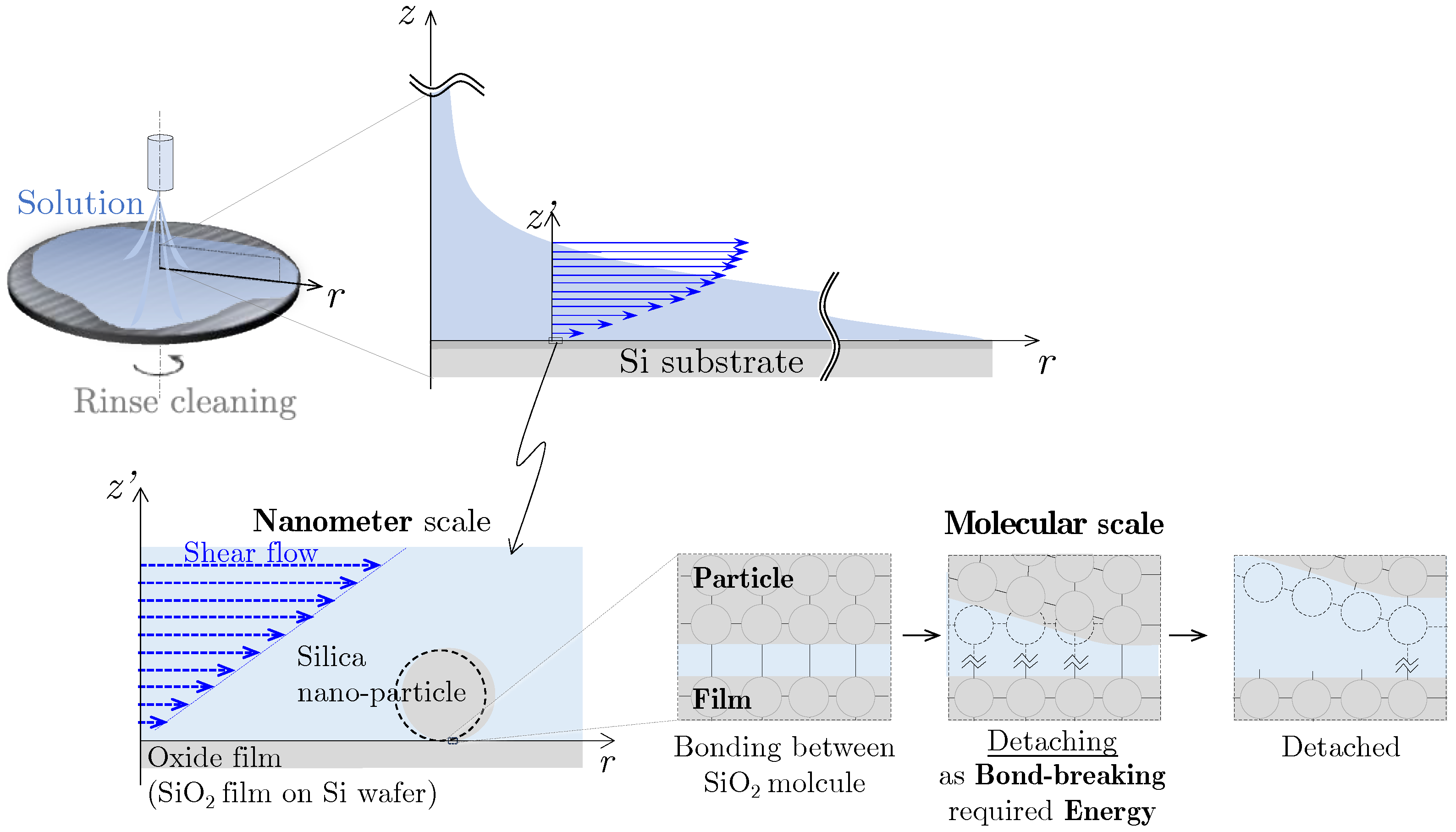
Figure 1. Nano-particle detachment involves breaking the bonding such as in post-CMP cleaning.
Applsci 14 08145 g001
Figure 2. Measurement of lateral force variation during detachment of nano-particle.
Applsci 14 08145 g002
Figure 3. D = 80 nm silica particle’s position and lateral force variation until detaching from the nano-particle.
Applsci 14 08145 g003
Figure 4. The force acting on D = 80 nm silica particle per line and particle behaviors during scanning.
Applsci 14 08145 g004
Figure 5. Lateral force variation during the detachment of various-sized silica nano-particles on the oxide film surface.
Applsci 14 08145 g005
Figure 6. Calculated interaction energies of siloxane bonds, hydrogen bonds, and van der Waals interactions between the spherical elastic silica particle and the oxide film surface.
Applsci 14 08145 g006
Figure 7. The potential energy of bonding and interaction between the silica nano-particle and the oxide film surface in the atmosphere. (a) Each calculated theoretical potential energy; (b) dominant total interaction energy.
Applsci 14 08145 g007
Figure 8. Detachment energy density between the silica particle and the oxide film surface.
Applsci 14 08145 g008
Table 1. Physicochemical parameters.
Potential Energy of Bonding Interaction
Siloxane bond energy [24]ES-bond@ ≥ 200 [°C]kJ/mol444
Hydrogen bond energy [25]EH-bond@ ≤ 200 [°C]kJ/mol20.9
OH group concentration [26,27,28]ρOH@ ≤ 200 [°C]OH/nm24.6 ± 0.6
(μmol/m2)(7.64 ± 1.0)
Total length of bond chain with forming probability b [26]
  SiO2/SiO2 interactionΔzbondbÅ≒7.2
Potential energy of van der Waals interaction
Hamaker constant [29]
  SiO2–atmosphere–SiO2A@atmosphere×10−20 J6.00
  SiO2 @atmosphereASiO2@atmosphere×10−20 J6.00
Separated distance between the particle and the film [30,31]
zsepÅ4
Contact radius calculation by JKR theory
Elastic modulus (SiO2/SiO2) [32]KN/m24.9 × 1010
  Poisson’s ratio [33]νSiO20.2
  Young’s modulus [33,34]ESiO2GPa70
Table 2. Typical calculated results of energy of siloxane bond and hydrogen bond.
Diameter [nm]ES-bond(total) [fJ]EH-bond(total) [fJ]Sarea [nm2]adef [nm]
100.130.01382.2
1001.920.0956710.4
100032.51.53959548.3
Table 3. Typical calculated results of energy of van der Waals interaction.
Diameter [nm]EVdW [fJ]adef [nm]
101.32 × 10−40.5
1001.39 × 10−32.1
10001.56 × 10−29.9
Table 4. Measured results of work to detach nano-particle and calculated potential energy.
Diameter [nm]Wlfm [fJ]Es-bond(total) [fJ]EH-bond(total) [fJ]EVdW [fJ]
500.100.840.046.81 × 10−4
800.131.470.071.11 × 10−3
3000.377.300.344.36 × 10−3
Disclaimer/Publisher’s Note: The statements, opinions and data contained in all publications are solely those of the individual author(s) and contributor(s) and not of MDPI and/or the editor(s). MDPI and/or the editor(s) disclaim responsibility for any injury to people or property resulting from any ideas, methods, instructions or products referred to in the content.