Communication

Comparison of Three Biological Control Models of Pycnoporus sanguineus on Phytopathogenic Fungi

by
Ricardo Irving Pérez-López
1,
Omar Romero-Arenas
2,*,
Conrado Parraguirre Lezama
2,
Anabel Romero López
3,
Antonio Rivera
4 and
Lilia Cedillo Ramírez
4
1
Posgrado en Manejo Sostenible de Agroecosistemas, Instituto de Ciencias, Benemérita Universidad Autónoma de Puebla, Edificio VAL 1, Km 1.7, Carretera a San Baltazar Tetela, San Pedro Zacachimalpa, Puebla 72960, Mexico
2
Centro de Agroecología, Instituto de Ciencias, Benemérita Universidad Autónoma de Puebla, Edificio VAL 1, Km 1.7, Carretera a San Baltazar Tetela, San Pedro Zacachimalpa, Puebla 72960, Mexico
3
Instituto de Física “Luis Rivera Terrazas”, Benemérita Universidad Autónoma de Puebla, Edificio VAL 2, Km 1.7, Carretera a San Baltazar Tetela, San Pedro Zacachimalpa, Puebla 72960, Mexico
4
Centro de Investigaciones en Ciencias Microbiológicas, Instituto de Ciencias, Benemérita Universidad Autónoma de Puebla, Ciudad Universitaria, Puebla 72570, Mexico
*
Author to whom correspondence should be addressed.
Appl. Sci. 2024, 14(18), 8263; https://doi.org/10.3390/app14188263
Submission received: 30 June 2024 / Revised: 9 September 2024 / Accepted: 10 September 2024 / Published: 13 September 2024
(This article belongs to the Special Issue Advances in Food Safety and Microbial Control)

Abstract

:
The genus Pycnoporus includes fungi with great potential for the production of antibiotic substances. It is necessary to develop new models to assess their effectiveness against microorganisms with an economic impact, such as phytopathogenic fungi. The objective of this study is to evaluate three models of Pycnoporus sanguineus for the growth inhibition of the phytopathogens Botrytis cinerea and Fusarium oxysporum. Model 1 involves dual tests of the antagonistic activity of P. sanguineus vs. phytopathogens, Model 2 involves antifungal effectiveness tests of cinnabarin, and Model 3 involves antifungal effectiveness tests of P. sanguineus extract. Models 2 and 3 are contrasted with products containing benomyl and captan. The results show that Model 3 is the most effective in controlling B. cinerea, with an inhibition percentage of 74.34% (p < 0.05) and a decrease in the growth rate (3.85 mm/day; p < 0.05); the same is true for F. oxysporum, with an inhibition percentage of 47.14% (p < 0.05). In general, F. oxysporum exhibits greater resistance (p < 0.05). The results of this study indicate that P. sanguineus extracts may be used as control agents for fungal species in the same way as other Pycnoporus species. Although commercial products are very efficient at inhibiting phytopathogens, one must consider the disadvantages of their use. In the short term, new models involving Pycnoporus for biological control in food production will be developed.

1. Introduction

The current food production model incorporates a multitude of challenges; one of the most pressing issues is the emergence of new and more aggressive pests and diseases affecting various commercial crops [1]. It is important to recognize that most of these threats are selectively fostered due to the excessive use of artificially synthesized agrochemical components [2], which also strongly impact biota at the rhizosphere level in the soil [3]. Therefore, it is necessary to adopt new methodologies that address these problems and generate a lower impact on biotic communities. One solution to this problem is the biological control of pests and diseases, which prioritizes the antagonistic interaction of living organisms [4]. This control method also involves the use of biopreparations derived from naturally synthesized elements by organisms through the isolation of their secondary metabolites [5], thereby reducing the risks associated with the management of live organisms [6].
The secondary metabolites, enzymes, and pigments produced by fungi have generated considerable interest in recent decades due to their biotechnological potential [7]. In this regard, various compounds extracted from basidiomycete fungi have exhibited strong antifungal activity [8]. Particularly, the compounds found in some lignicolous fungal species can biologically control phytopathogens, largely because of their ability to produce substances like those used by plants to defend against these phytopathogenic agents [3,9].
The genus Pycnoporus is a Basidiomycota fungus with extensive research potential to produce antiviral, antifungal, and antibacterial substances. This has primarily been studied through the action of cinnabarin, an orange pigment that can be obtained from its liquid or solid cultivation [10]. Its effectiveness against various bacterial species, such as Bacillus cereus, Enterococcus faecalis, Enterococcus faecium, Escherichia coli, Klebsiella pneumoniae, Listeria monocytogenes, and Lactobacillus plantarum, among others, has been previously demonstrated [11,12]. However, further research is still needed to delve into the mechanisms of action involved in interactions with these organisms [13,14]. Additionally, it is crucial to evaluate the effectiveness of the other compounds present in Pycnoporus with significant biotechnological potential [15,16,17]. Therefore, it is imperative to generate new models that incorporate a broader spectrum of components and evaluate them against other groups of economically impactful microorganisms such as phytopathogenic fungi [18,19]. Studies that have evaluated the effectiveness of Pycnoporus on these types of organisms have primarily focused on models involving the complete extraction of its fruiting bodies or on antagonistic interaction models assessing the interaction of the fungus against the growing pathogen [18,20,21]. Although the main component of interaction in these models is likely cinnabarin, models directly assessing the effect of this compound on the growth of phytopathogenic fungi are virtually non-existent. For this reason, the aim of this study is to evaluate the effect of three P. sanguineus restriction models on the in vitro growth of two phytopathogenic fungi that have a significant economic impact on vegetable cultivation: the antagonistic capacity of P. sanguineus through in vitro dual growth tests; antifungal activity tests of cinnabarin; and antifungal activity tests of full P. sanguineus extracts. These will be compared with two commercial products intended for the same purpose, allowing for the generation of high-quality biotechnological products to be directly applied in the management and control of diseases among the main commercial crops.

2. Materials and Methods

2.1. Strain Descriptions

Three growth restriction models of P. sanguineus compounds were evaluated against the phytopathogenic fungi Botrytis cinerea and Fusarium oxysporum (Ascomycota). The strains used were MA-FC220 of F. oxysporum and MA-BC20 of B. cinerea, both isolated from the roots and fruits of a strawberry crop; the sequences of these strains were deposited in the database of the National Center for Biotechnology Information (NCBI) under accession numbers OM473290.1 and OM473288.1, respectively. In addition, an isolate of P. sanguineus from an oak trunk was maintained in a Potato Dextrose Agar culture medium (PDA) (Bioxon®, Franklin Lakes, NJ, USA, EE. UU.) and designated MA-Ps1, with accession number OR622486.1 (NCBI), characterized by the Laboratory of Phytopathology 204 of the Agroecology Center of the Institute of Sciences of the Benemérita Universidad Autónoma de Puebla (BUAP).

2.2. Model 1: Dual Tests of Antagonistic Activity

The antagonistic capacity of P. sanguineus against the phytopathogens B. cinerea and F. oxysporum was evaluated through in vitro dual growth tests on PDA medium in large Petri dishes (90 mm in diameter). Mycelium circles of 5 mm in diameter of each species were placed at the end of each dish, 5 mm from the edge. A total of 5 treatments were evaluated, with 5 replications each: P. sanguineus vs. B. cinerea, P. sanguineus vs. F. oxysporum, as well as P. sanguineus, B. cinerea, and F. oxysporum grown individually as control tests. The inoculants were isolated from previously mature and growing cultures. The growth rate (GR) was assessed using the formula GR = (FR − IR)/number of days in growth, where GR = the growth rate, FR = the final radius reached by the growing strain, and IR = the initial radius of the growing strain. Simultaneously, the antagonistic capacity of P. sanguineus was assessed by the percentage inhibition of radial growth (PIRG) using the formula PIRG = (R1 − R2)/R1 × 100, where PIRG = the percentage of radial growth inhibition, R1 = the radial growth (mm) of the pathogen (B. cinerea or F. oxysporum) without the presence of P. sanguineus, and R2 = the radial growth (mm) of the pathogen (B. cinerea or F. oxysporum) in the presence of P. sanguineus. Mycelial growth was measured using a digital Vernier (CD-6 Mitutoyo, Naucalpan de Juarez, Mexico), with measurements taken every 24 h from inoculation until contact occurred between both growing mycelia.

2.3. Model 2: Antifungal Effectiveness Tests of Cinnabarin

Antifungal activity tests of cinnabarin on the phytopathogens B. cinerea and F. oxysporum were conducted under controlled conditions, where two commercial products were compared using the impregnated disc diffusion method, in which the pathogenic fungi were grown in the center of Petri dishes containing 6 mm diameter paper discs impregnated with the different treatments. These discs were placed one centimeter away from the edge of the Petri dish and arranged at the four cardinal points. For the extraction of the cinnabarin pigment, the growth of P. sanguineus was cultivated in Petri dishes with PDA medium. To facilitate the proper separation of mycelia, a layer of cellophane was placed on the gelatinized culture medium. After thirty days of growth, the mycelia were separated and completely dehydrated in an oven at 45 °C, with daily measurements taken until a constant weight was achieved. A total of 5.6 g of P. sanguineus dehydrated mycelium was obtained; subsequently, cinnabarin was isolated using extraction techniques. A total of 5.6 g of dehydrated P. sanguineus mycelium was macerated in a 10 mL solution of ethyl acetate to release the pigment, and then adjusted to a volume of 50 mL of solvent, which was allowed to macerate for 2 days for complete the pigment release. To determine the presence and optical characterization of cinnabarin, 0.5 mL of the extract was analyzed using spectrophotometry performed with a spectrometer Cary 100 UV-Vis System, Agilent at 25 °C and quartz cells with an optical path of 1 cm. UV–vis (C4H8O2): λmax (log ε) = 251 (1.09), 427 (0.19), and 446 (0.18) nm (Figure 1a). Additionally, 0.5 milliliters of the extract was analyzed using HPLC performed on a Series 200 Quaternary pump equipped with a Diode Array Detector (DAD) Perkin Elmer using Total Chrom Navigator software (TcNav version 6.2.1). Analytical HPLC analyses were performed using 0–5 min 75% CH3CN/H2O on a Pecosphera C18 150 × 4.6 (5μ) column at a flow rate of 1.0 mL/min. HPLC (CH3CN/H2O): the test showed a maximum peak at 1.5 min (Figure 1b).
The UV–vis and HPLC values coincide within the previously reported peak area for cinnabarin [22]. Additionally, cinnabarin was determined by studying the 1H NMR spectra recorded at 500 MHz using a Bruker FT-NMR spectrometer. The chemical shifts (δ) are reported in parts per million (ppm), as referenced in the 1H spectra by the signal of TMS proton resonance: 1H NMR (500 MHz, CDCl3) δ: 9.79 (1H, brs, 2-NHα), 8.50 (1H, brs, 2-NHβ), 7.46–7.04 (4H, m, H-aromatic), and 5.29 (2H, s, H-12). Along the 1H NMR spectrum of cinnabarin, two broad singlets at 9.79 and 8.50 ppm were assigned to the NH groups.
At 7.46–7.04 ppm, signals assigned to four aromatic protons were observed, while at 5.29 ppm, one simple signal appeared to integrate two protons assigned to the methylene. Subsequently, the extract was impregnated into 60 paper discs using a rotavapor at 45 °C to evaporate the solvent and pigment from the discs simultaneously. Following this, antifungal activity tests were carried out on B. cinerea and F. oxysporum on PDA medium using the impregnated discs. The cinnabarin concentration was 4 mg/mL, which was determined indirectly using an antimicrobial activity technique [12]; the method followed is described in Romero-Arenas et al. [23]. For the contrast tests, the commercial products Benomyl 50® (with benomyl C14H18N4O3 as the active ingredient) and Captan 50WP® (with captan C9H8Cl3NO2S as the active ingredient) were used, along with a control group with discs with sterile water only.
The concentrations used for the commercial products were 2 mg/mL for captan and 0.52 mg/mL for benomyl, respectively, according to the indications for their commercial use. Each one was mixed in a vortex at 1300 rpm with 60 discs, respectively. A total of 8 treatments were evaluated with 5 replications each: cinnabarin vs. B. cinerea, cinnabarin vs. F. oxysporum, captan vs. B. cinerea, captan vs. F. oxysporum, benomyl vs. B. cinerea, benomyl vs. F. oxysporum, as well as discs without any additive vs. B. cinerea and F. oxysporum, respectively. The inoculants were isolated from previously mature and growing cultures. Like the first model, the characteristics evaluated were the GR and the effectiveness of cinnabarin using the PIRG, and the same procedure was followed with the compounds captan and benomyl.

2.4. Model 3: Antifungal Effectiveness Tests of Full P. sanguineus Extract

Antifungal activity tests were conducted on the phytopathogens B. cinerea and F. oxysporum under controlled conditions using a culture medium supplemented with a complete extract of P. sanguineus mycelia, and they were compared to the commercial products. The growth, separation, and dehydration of P. sanguineus mycelia were carried out in the same way as described for the second model. A total of 6.2 g of P. sanguineus dehydrated mycelium was obtained, which was subsequently pulverized using a Nutribullet®. The obtained powder was added to the PDA at a concentration of 24.8 mg/mL, which was filtered through 0.22 μm Millipore filters and then sterilized at 120 °C for 15 min. In the same way, PDA with captan and benomyl were used as contrast treatments at concentrations of 2 mg/mL and 0.52 mg/mL, respectively, as well as a control group with PDA without any additives. Subsequently, the antifungal activity tests were carried out on B. cinerea and F. oxysporum under controlled conditions. The following treatments were performed, with 5 replications each: P. sanguineus extract vs. B. cinerea, P. sanguineus extract vs. F. oxysporum, captan vs. B. cinerea, captan vs. F. oxysporum, benomyl vs. B. cinerea, benomyl vs. F. oxysporum, as well as a culture medium without additives vs. B. cinerea and F. oxysporum, respectively. The inoculums were isolated from previously mature and growing cultures. The characteristics evaluated were the GR and the PIRG for the effectiveness of the complete extract of P. sanguineus, and the same procedure was followed for the captan and benomyl treatments.

2.5. Statistical Analysis

To determine the effectiveness of the different treatments, the results obtained from the GR and PIRG values were compared using analysis of variance (ANOVA). The PIRG variable, expressed as a percentage, was subjected to a transformation using the angular arccosine function (√x + 1). Bartlett’s homogeneity test was applied to evaluate the homogeneity of the variances, followed by the Tukey–Kramer mean comparison test, with the significance level set at p ≤ 0.05. Subsequently, t-tests were performed to recognize the variation in the resistance or susceptibility of one or both phytopathogens to each treatment based on their series of expressed values. All the statistical analyses were performed using SPSS Statistics version 17 for the Windows operating environment.

3. Results

The PIRG values among the different treatments evaluated against B. cinerea using ANOVA indicate that in the dual tests (Model 1), P. sanguineus had a very slight antagonistic effect when both grew together. According to the PIRG evaluation, B. cinerea was inhibited only 2.78% by P. sanguineus, which is the lowest inhibition percentage among the three models (p < 0.05). A higher inhibition value was observed for cinnabarin in Model 2 (16.81%, p < 0.05), while those of captan and benomyl within the same model, as well as the treatments of PDA with P. sanguineus extract and PDA with benomyl in Model 3, showed much higher rates of inhibition (84.85%, 94.83%, 74.34%, and 87.55%, respectively, p < 0.05). However, the addition of captan to PDA in Model 3 inhibited its growth completely (100%, p < 0.05; Table 1).
The PIRG values among the different treatments evaluated against F. oxysporum using ANOVA indicate that in Model 1, P. sanguineus had a moderate antagonistic effect that was similar to those found for cinnabarin and benomyl in Model 2. However, they were lower than captan in Model 2 and PDA with P. sanguineus extract in Model 3. The treatments of the PDA with captan and benomyl in Model 3 presented even higher inhibition values.
The GR values among the different treatments evaluated against B. cinerea using ANOVA indicate that B. cinerea that grew alongside P. sanguineus in Model 1 was not different from what was presented in the control treatments of Models 1, 2, and 3 (7.84 mm/day, 7.77 mm/day, and 7.61 mm/day, respectively, p < 0.05). These results were also not significantly different from those of cinnabarin in Model 2 (6.6 mm/day, p < 0.05). On the other hand, captan and benomyl in Model 2, as well as PDA with P. sanguineus extract in Model 3, reduced its growth considerably (1.94 mm/day, 1.46 mm/day, and 3.85 mm/day, respectively, p < 0.05).
Consistent with what was observed in the PIRG, the PDA with captan and benomyl almost completely inhibited its growth (0.02 mm/day and 0.93 mm/day, respectively, p < 0.05; Table 2).
The t-test results indicate a higher resistance expressed in lower PIRG percentages by F. oxysporum to the treatments of cinnabarin and benomyl in Model 2 (p = 5.75 × 10⁻⁶ and p = 1.5 × 10⁻⁸, respectively), as well as the PDA with P. sanguineus and captan in Model 3 (p = 0.004 and p = 0.0001, respectively) (Figure 2).
The GR values among the different treatments evaluated against F. oxysporum using ANOVA indicate that there were no significant differences in growth presented by Models 1 and 2, nor by the PDA control and PDA with P. sanguineus extract in Model 3, according to the mean comparison tests conducted (p < 0.05). The only decrease observed was due to the action of the PDA with captan and benomyl in Model 3 (1.22 mm/day and 0.05 mm/day, respectively, p < 0.05; Table 2).

4. Discussion

It has been demonstrated that certain components present in the Pycnoporus genus possess antifungal properties [17]. Most of the studies in this regard have used complete extracts of Pycnoporus fruiting bodies as an effective interaction model against various fungal species. These include species that are pathogenic to humans, such as Candida albicans, Candida krusei, Aspergillus fumigatus, Mucor sp., Microsporum gypseum, and Trichophyton mentagrophytes [24]. In the same way, some species impact economically important crops, including wood-degrading species, like Trametes versicolor, Trametes feei, Trametes menziezi, Lentinus sp., Lentinus sajor-caju, Lentinus strigosus, Microporus affinis, Microporus xanthopus, and Gloeophyllum trabeum [25]; species affecting various plants at the leaf level, such as Pseudocercospora griseola [26] and Mycena citricolor [27]; and species affecting different plant structures, including Botrytis cinerea, Coniophora puteana [20], and Fusarium sp. [21].
Particularly, studies evaluating the effectiveness of Pycnoporus against B. cinerea have utilized both antagonistic interaction models of the species P. coccineus [20] and complete spectrum extracts of the species P. cinnabarinus, with both solid and liquid cultures [18]. To date, only complete extract models of the species P. sanguineus have been evaluated against Fusarium sp. [21]. In all these studies, the effectiveness of Pycnoporus compounds against these phytopathogens has been demonstrated. This partially coincides with the results of the current study, which demonstrate the significant antifungal effectiveness of P. sanguineus. In the growth restriction of B. cinerea, this was observed with greater intensity under the treatment of PDA supplemented with the P. sanguineus extract, as expressed as both a higher PIRG (74.34%) and a reduction in its GR (3.85 mm/day). This was even similar to the PIRG generated by captan and benomyl in some treatments (Table 1).
However, although the treatment of PDA with the P. sanguineus extract generated a considerable PIRG against F. oxysporum (47.14%; Table 1), this was lower than what was observed against B. cinerea; overall, F. oxysporum expressed greater resistance to the different treatments (Figure 2). Nonetheless, the results of the first model evaluated (antagonistic interaction model) did not show a significant difference in growth between the dual cultures of P. sanguineus with B. cinerea and F. oxysporum compared to their respective control cultures (Table 1 and Table 2). This contrasts with the strong antagonistic effect observed by Arfi et al. [20], where the species Pycnoporus coccineus was even able to replace its competitors by growing on them. This behavior is commonly associated with highly aggressive species used for the biological control of phytopathogens, such as several Trichoderma species [28,29]. According to Arfi et al. [20], the antagonistic strategy used by P. coccineus involves the growth restriction of its competitors through the release of enzymes (mainly laccases) rather than faster resource acquisition. This can even limit the growth rate of P. coccineus itself. Although the present study cannot confirm the presence of this strategy in P. sanguineus, an increase in pigmentation of P. sanguineus mycelia was observed in the contact zone with its competitors. This may be part of a growth restriction strategy against the advancing growth of this phytopathogen. Deeper physiological analysis could clarify this phenomenon, along with the pigments and metabolites involved.
The second model of cinnabarin, and possibly to a lesser extent cinnabaric acid (Figure 1), showed the limited effectiveness of this compound in relation to the growth of the phytopathogens B. cinerea and F. oxysporum. It was not possible to observe growth restriction, as the pathogen was able to grow on the cinnabarin-impregnated discs, although it did slightly inhibit their growth, with values of 16.81% for B. cinerea and 17.79% for F. oxysporum. These PIRG values were mostly lower than those of captan and benomyl (Table 1). While there are several studies evaluating the effect of cinnabarin on the growth of bacteria, its cytotoxic effects, and even its antiviral effects [30], very few studies have specifically evaluated the action of this compound on B. cinerea and F. oxysporum or other phytopathogenic fungi. Nevertheless, Cruz-Muñóz [6] and Cruz-Muñóz [31] evaluated the effectiveness of P. sanguineus pigments against Colletotrichum fragariae, Colletotrichum gloeosporioides, and Botrytis cinerea, finding considerable antifungal activity. In any case, based on the results of the present and previous studies, the use of complete extracts of Pycnoporus may represent a more effective action model against these or other fungi due to the inclusion of different components with potential antifungal activity, such as polyporin [32], ergosterol [33], 4H-pyran-4-one, and 2,3-dihydro-3,5-dihydroxy-6-methyl [25], as well as other enzymes and compounds involved in antioxidant processes [20,34].
It is also important to highlight that the results demonstrate, as expected, the effectiveness of commercial products containing benomyl and captan, which generally resulted in the highest PIRG values among the tests conducted (Table 1 and Table 2). However, it is essential to recognize the disadvantages of their use, as they may have a more significant impact in field application with all the associated repercussions [35,36]. The results of this study highlight the importance of further research on the use of Pycnoporus compounds in biological pest and disease control. However, it is also important to consider expanding the number of phytopathogenic fungal species to be tested, delving into the compounds involved, and examining the differences that may exist between these in vitro models and in situ application contexts, which is a relatively unexplored but promising field.

5. Conclusions

Extracts from the species P. sanguineus, as reported for several species of the Pycnoporus genus, exhibit significant potential as biological control agents against fungal species. The results of this study indicate a trend in the control of the phytopathogens B. cinerea and F. oxysporum. The most effective model for controlling these phytopathogens was the complete extract of P. sanguineus added to the culture medium. Although the commercial products proved to be more effective in inhibiting phytopathogens than the P. sanguineus models, it is important to consider the disadvantages associated with their use. Undoubtedly, the development of new models that include the components present in Pycnoporus for biological control is expected in the short term. Additionally, it is crucial to explore the application of these models under field conditions, as this represents a promising but relatively unexplored area in the use of Pycnoporus as a biological control agent.

Author Contributions

Conceptualization, O.R.-A. and R.I.P.-L.; methodology, A.R.L., R.I.P.-L. and O.R.-A.; software, A.R.L. and R.I.P.-L.; validation, O.R.-A., A.R. and C.P.L.; formal analysis, C.P.L., L.C.R. and O.R.-A.; resources, O.R.-A. and R.I.P.-L.; original draft preparation, A.R., L.C.R. and O.R.-A.; writing—review and editing, C.P.L. and O.R.-A.; visualization, O.R.-A., R.I.P.-L. and A.R.L.; supervision, O.R.-A.; project administration, O.R.-A. and R.I.P.-L.; funding acquisition, O.R.-A. and R.I.P.-L. All authors have read and agreed to the published version of the manuscript.

Funding

The authors are grateful to Consejo Nacional de Humanidades, Ciencia y Tecnología (CONAHCYT) (CVU 555290 and 47406).

Institutional Review Board Statement

Not applicable.

Informed Consent Statement

Not applicable.

Data Availability Statement

The original contributions presented in the study are included in the article, further inquiries can be directed to the corresponding author.

Acknowledgments

Special thanks to CONAHCYT for the scholarship awarded to facilitate the completion of this study as part of a postdoctoral project and Laboratory 204 of the Center for Agroecology at Benémerita Universidad Autónoma of Puebla.

Conflicts of Interest

The authors declare no conflicts of interest. The funders had no role in the design of the study; in the collection, analyses, or interpretation of data; in the writing of the manuscript; or in the decision to publish the results.

References

  1. Pal, K.K.; Gardener, B.M. Biological control of plant pathogens. Plant Health Instr. 2006, 6, 1–25. [Google Scholar] [CrossRef]
  2. Serrano, L.; Galindo, E. Control biológico de organismos fitopatógenos: Un reto multidisciplinario. Ciencia 2007, 77–88. Available online: https://www.amc.edu.mx/revistaciencia/images/revista/58_1/PDF/controlBiologico.pdf (accessed on 15 January 2024).
  3. Waszczuk, U.; Zapora, E. Arboreal Fungi in Biological Control against Soil Fungi. Environ. Sci. Proc. 2021, 9, 31. [Google Scholar] [CrossRef]
  4. Rossbacher, S.; Vorburger, C. Prior adaptation of parasitoids improves biological control of symbiont-protected pests. Evol. Appl. 2020, 13, 1868–1876. [Google Scholar] [CrossRef] [PubMed]
  5. Alabouvette, C.; Olivain, C.; Steinberg, C. Biological control of plant diseases: The European situation. Eur. J. Plant Pathol. 2006, 114, 329–341. [Google Scholar] [CrossRef]
  6. Cruz-Muñoz, R. Producción de Extractos de Pycnoporus sanguineus con Actividad Antimicrobiana en Hongos y Bacterias Fitopatógenas. Master’s Thesis, Instituto Politécnico Nacional, Mexico City, México, 2012. Available online: http://www.repositoriodigital.ipn.mx/handle/123456789/15754 (accessed on 20 February 2024).
  7. Adrio, J.L.; Demain, A.L. Fungal biotechnology. Int. Microbiol. 2003, 6, 191–199. [Google Scholar] [CrossRef] [PubMed]
  8. Alves, M.; Ferreira, I.F.R.; Dias, J.; Teixeira, V.; Martins, A.; Pintado, M. A review on antimicrobial activity of mushroom (basidiomycetes) extracts and isolated compounds. Planta Med. 2012, 78, 1707–1718. [Google Scholar] [CrossRef] [PubMed]
  9. Sivanandhan, S.; Khusro, A.; Paulraj, M.G.; Ignacimuthu, S.; Al-Dhabi, N.A. Biocontrol properties of basidiomycetes: An overview. J. Fungi 2017, 3, 2. [Google Scholar] [CrossRef] [PubMed]
  10. Cruz-Muñoz, R.; Piña-Guzmán, A.B.; Yáñez-Fernández, J.; Valencia-Del Toro, G.; Bautista-Baños, S.; Villanueva-Arce, R. Producción de pigmentos de Pycnoporus sanguineus en medio de cultivo sólido. Agrociencia 2015, 49, 347–359. [Google Scholar]
  11. Smânia, A.; Delle Monache, F.; Smânia, E.F.A.; Gil, M.L.; Benchetrit, L.C.; Cruz, F.S. Antibacterial activity of a substance produced by the fungus Pycnoporus sanguineus (Fr.) Murr. J. Ethnopharmacol. 1995, 45, 177–181. [Google Scholar] [CrossRef]
  12. Smânia, E.F.A.; Smânia, A., Jr.; Loguercio-Leite, C.; Gil, M.L. Optimal parameters for Cinnabarin synthesis by Pycnoporus sanguineus. J. Chem. Technol. Biotechnol. Int. Res. Process. Environ. Clean Technol. 1997, 70, 57–59. [Google Scholar]
  13. Hwang, H.J.; Kim, S.W.; Xu, C.P.; Choi, J.W.; Yun, J.W. Morphological and rheological properties of the three different species of basidiomycetes Phellinus in submerged cultures. J. Appl. Microbiol. 2004, 96, 1296–1305. [Google Scholar] [CrossRef] [PubMed]
  14. Tuong, T.D.; Chu, D.X.; Dieu, B.T.M. Antioxidant Activity of Fruiting Body Extracts from Pycnoporus sanguineus Mushroom. Vietnam. J. Sci. Technol. 2020, 58, 143–151. [Google Scholar]
  15. Acosta-Urdapilleta, L.; Alonso Paz, G.A.; Rodríguez, A.; Adame, M.; Salgado, D.; Salgado, J.; Montiel-Peña, M.; Medrano-Vega, F.; Villegas Villarreal, E.C. Pycnoporus sanguineus, a fungus having biotechnological potential. In Hacia un Desarrollo Sostenible del Sistema de Producción-Consumo de los Hongos Comestibles y Medicinales en Latinoamérica: Avances y Perspectivas en el Siglo XXI; Martínez-Carrera, D., Curvetto, N., Sobal, M., Morales, P., Mora, V.M., Eds.; Red Latinoamericana de Hongos Comestibles y Medicinales; Porfirio MORALES: Moca, Espaillat, 2010; pp. 189–220. [Google Scholar]
  16. Lomascolo, A.; Uzan-Boukhris, E.; Herpoël-Gimbert, I.; Sigoillot, J.C.; Lesage-Meessen, L. Peculiarities of Pycnoporus species for applications in biotechnology. Appl. Microbiol. Biotechnol. 2011, 92, 1129–1149. [Google Scholar] [CrossRef] [PubMed]
  17. Pineda-Insuasti, J.A.; Gómez-Andrade, W.E.; Duarte-Trujillo, A.S.; Soto-Arroyave, C.P.; Pineda-Soto, C.A.; Fierro-Ramos, F.J.; Álvarez-Ramos, S.E. Producción de Pycnoporus spp. y sus metabolitos secundarios: Una revisión. ICIDCA Sobre Deriv. Caña Azúcar 2017, 51, 60–69. [Google Scholar]
  18. Imtiaj, A.; Lee, T.S. Screening of antibacterial and antifungal activities from Korean wild mushrooms. World J. Agric. Sci. 2007, 3, 316–321. [Google Scholar]
  19. Imtiaj, A.; Jayasinghe, C.; Lee, G.W.; Lee, T.S. Antibacterial and Antifungal Activities of Stereum ostrea, an Inedible Wild Mushroom. Mycobiology 2007, 35, 210. [Google Scholar] [CrossRef]
  20. Arfi, Y.; Levasseur, A.; Record, E. Differential gene expression in Pycnoporus coccineus during interspecific mycelial interactions with different competitors. Appl. Environ. Microbiol. 2013, 79, 6626–6636. [Google Scholar] [CrossRef]
  21. Figueiredo, Á.; Castro, E.; Silva, A. Atividade “in vitro” de extratos de Pycnoporus sanguineus e Lentinus crinitus sobre o fitopatógeno Fusarium sp. Acta Amaz. 2014, 44, 1–8. [Google Scholar] [CrossRef]
  22. Dias, D.A.; Urban, S. HPLC and NMR Studies of Phenoxazone Alkaloids from Pycnoporus cinnabarinus. Nat. Prod. Commun. 2009, 4, 489–498. [Google Scholar] [CrossRef]
  23. Romero-Arenas, O.; Jara y Rivera, A.P.; Valencia de Ita, M.A.; Lezama, C.P.; Villa-Ruano, N.; Rivera, A. In Vitro Antimicrobial Activity of Cinnabarin on Xanthomonas campestris Isolated from Bean Crops of Puebla, Mexico. Appl. Sci. 2021, 11, 5391. [Google Scholar] [CrossRef]
  24. Al-Fatimi, M.; Schröder, G.; Kreisel, H.; Lindequist, U. Biological activities of selected basidiomycetes from Yemen. Pharmazie 2013, 68, 221–226. [Google Scholar]
  25. Teoh, Y.P.; Don, M.M.; Ujang, S. Media selection for mycelia growth, antifungal activity against wood-degrading fungi, and GC-MS study by Pycnoporus sanguineus. BioResources 2011, 6, 2719–2731. [Google Scholar] [CrossRef]
  26. Viecelli, C.A.; Stangarlin, J.R.; Kuhn, O.J.; Schwan-Estrada, K.R.F. Resistance induction in bean plants against angular leaf spot by extracts from Pycnoporus sanguineus mycelium. Summa. Phytopathol. 2010, 36, 73–80. [Google Scholar]
  27. Ojeda, K.; Suescum, J. Evaluación de Cinco Cepas de Hongos Nativos como Controladores Biológicos de la Enfermedad “Ojo de Pollo”(Mycena citricolor) en café (Coffea arabica) en Condiciones in vitro. Bachelor’s Thesis, Universidad Técnica Particular de Loja, Loja, Ecuador, 2012. [Google Scholar]
  28. Nascimento, V.C.; Rodrigues-Santos, K.C.; Carvalho-Alencar, K.L.; Castro, M.B.; Kruger, R.H.; Lopes, F.A.C. Trichoderma: Biological control efficiency and perspectives for the Brazilian Midwest states and Tocantins. Braz. J. Biol. 2022, 82, e260161. [Google Scholar] [CrossRef]
  29. Mukherjee, P.K.; Mendoza-Mendoza, A.; Zeilinger, S.; Horwitz, B.A. Mycoparasitism as a mechanism of Trichoderma-mediated suppression of plant diseases. Fungal Biol. Rev. 2022, 39, 15–33. [Google Scholar] [CrossRef]
  30. Smânia, A.; Marques, C.J.S.; Smânia, E.F.A.; Zanetti, C.R.; Carobrez, S.G.; Tramonte, R.; Loguercio-Leite, C. Toxicity and Antiviral Activity of Cinnabarin Obtained from Pycnoporus sanguineus (Fr.) Murr. Phyther. Res. 2003, 17, 1069–1072. [Google Scholar] [CrossRef] [PubMed]
  31. Cruz-Muñoz, R. Caracterización de los Pigmentos de Pycnoporus sanguineus y su Citotoxicidad Sobre Hongos y Bacterias Fitopatógenas. Ph.D. Thesis, Instituto Politécnico Nacional, Mexico City, México, 2015. Available online: http://tesis.ipn.mx/bitstream/handle/123456789/19567/TesisDoctoralROCIOCRUZM.pdf?sequence=1&isAllowed=y (accessed on 12 March 2024).
  32. Henrique Rosa, L.; Gomes Machado, K.M.; Jacob, C.C.; Capelari, M.; Augusto Rosa, C.; Leomar Zani, C. Screening of Brazilian Basidiomycetes for Antimicrobial Activity. Mem. Inst. Oswaldo Cruz 2003, 98, 967–974. [Google Scholar] [CrossRef] [PubMed]
  33. Téllez-Téllez, M.; Villegas, E.; Rodriguez, A.; Acosta-Urdapilleta, M.L.; O’Donovan, A.; Diaz-Godinez, G. Mycosphere Essay 11: Fungi of Pycnoporus: Morphological and molecular identification, worldwide distribution and biotechnological potential. Mycosphere 2016, 7, 1500. [Google Scholar]
  34. Borderes, J.; Costa, A.; Guedes, A.; Tavares, L.B.B. Antioxidant activity of the extracts from Pycnoporus sanguineus mycelium. Brazilian Arch. Biol. Technol. 2011, 54, 1167–1174. [Google Scholar] [CrossRef]
  35. Potočnik, I.; Vukojević, J.; Stajić, M.; Rekanović, E.; Milijašević, S.; Stepanović, M.; Todorović, B. Toxicity of fungicides with different modes of action to Cladobotryum dendroides and Agaricus bisporus. J. Environ. Sci. Health Part B Pestic. Food Contam. Agric. Wastes 2009, 44, 823–827. [Google Scholar] [CrossRef] [PubMed]
  36. Avenot, H.F.; Morgan, D.P.; Quattrini, J.; Michailides, T.J. Resistance to Thiophanate-Methyl in Botrytis cinerea Isolates from Californian Vineyards and Pistachio and Pomegranate Orchards. Plant Dis. 2020, 104, 1069–1075. [Google Scholar] [CrossRef] [PubMed]
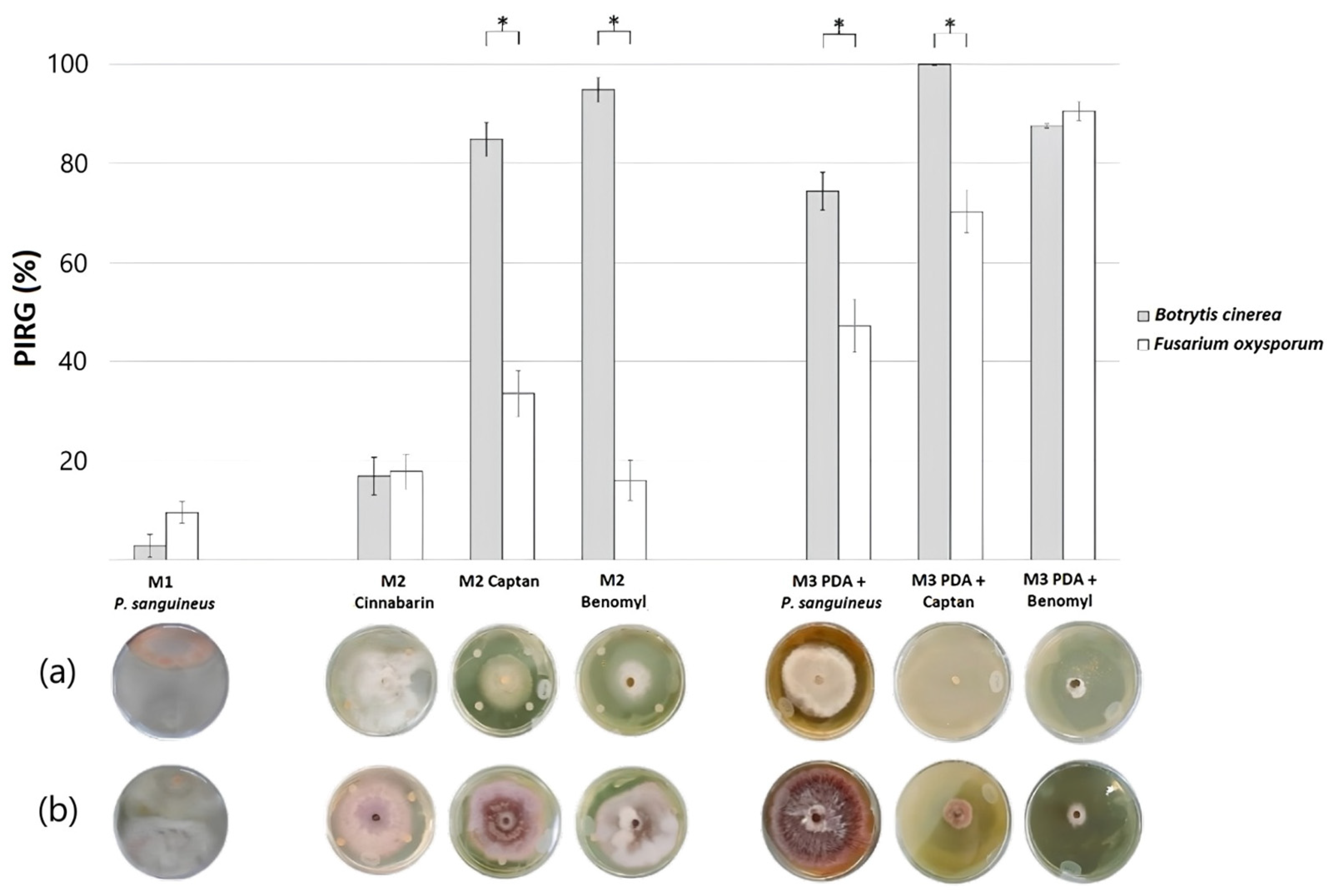
Figure 1. (a) The UV–vis spectrum (C4H8O2) and (b) high-performance liquid chromatography (CH3CN/H2O) of cinnabarin present in the extract of P. sanguineus.
Applsci 14 08263 g001
Figure 2. Percentage of inhibition of radial growth of B. cinerea (gray) and F. oxysporum (white) within treatments; dual tests of antagonistic activity with P. sanguineus (M1); antifungal effectiveness tests with cinnabarin, captan, and benomyl (M2); and antifungal effectiveness tests of culture media supplemented with P. sanguineus extract, captan, and benomyl (M3). Marks at the top indicate significant differences between B. cinerea and F. oxysporum within each treatment (t-test). Photographs in rows below each treatment (a) B. cinerea and (b) F. oxysporum are provided as visual reference of their effect. * The correlation is significant at the 0.05 level.
Applsci 14 08263 g002
Table 1. Mean values with standard error and ANOVA results for the percentage of inhibition of radial growth among the different treatments evaluated within each model.
Model’sTreatmentBotrytis cinereaFusarium oxysporum
PIRG (%)p ≤ 0.05PIRG (%)p ≤ 0.05
Model 1Pycnoporus sanguineus(±2.3) 2.78d(±2.2) 9.54d
Model 2Cinnabarin (CH2OH-COOH-NH2)(±3.8) 16.81c(±3.5) 17.79d
Captan (C9H8Cl3NO2S)(±3.4) 84.85b(±4.6) 33.50c
Benomyl (C14H18N4O3)(±2.4) 94.83b(±4.1) 15.98d
Model 3PDA + Pycnoporus sanguineus extract(±3.8) 74.34b(±5.3) 47.14c
PDA + Captan (C9H8Cl3NO2S)(±0.2) 100a(±4.2) 70.25b
PDA + Benomyl (C14H18N4O3)(±0.5) 87.55b(±1.9) 90.53a
Mean values followed by the same letter do not present statistically significant differences (p ≤ 0.05) according to Tukey’s test.
Table 2. Mean values with standard error and ANOVA results for the growth rate among the different treatments evaluated within each model.
Model’sTreatmentBotrytis cinereaFusarium oxysporum
GR (mm/day)p ≤ 0.05GR (mm/day)p ≤ 0.05
Model 1Control (PDA)(±1.5) 7.84a(±0.4) 3.47a
Pycnoporus sanguineus(±1.4) 7.02a(±0.5) 3.18a
Model 2Control (PDA)(±1.3) 7.77a(±0.8) 4.25a
Cinnabarin (CH2OH-COOH-NH2)(±1.6) 6.60ab(±0.6) 3.88a
Captan (C9H8Cl3NO2S)(±0.8) 1.94ce(±0.4) 3.16a
Benomyl (C14H18N4O3)(±0.6) 1.46ce(±0.4) 3.48a
Model 3Control (PDA)(±1.5) 7.61a(±1.2) 4.28a
PDA + Pycnoporus sanguineus extract(±0.8) 3.85bc(±0.3) 2.99a
PDA + Captan (C9H8Cl3NO2S)(±0.02) 0.02d(±0.2) 1.22b
PDA + Benomyl (C14H18N4O3)(±0.2) 0.93e(±0.2) 0.50c
Mean values followed by the same letter do not present statistically significant differences (p ≤ 0.05) according to Tukey’s test.
Disclaimer/Publisher’s Note: The statements, opinions and data contained in all publications are solely those of the individual author(s) and contributor(s) and not of MDPI and/or the editor(s). MDPI and/or the editor(s) disclaim responsibility for any injury to people or property resulting from any ideas, methods, instructions or products referred to in the content.

