Article

Abutment Tooth Formation Simulator for Naked-Eye Stereoscopy

1
Department of Fixed Prosthodontics, Kanagawa Dental University, Yokosuka 238-8580, Japan
2
Department of Liberal Arts Education, School of Dentistry, Kanagawa Dental University, Yokosuka 238-8580, Japan
3
Department of Oral Digital Science, Kanagawa Dental University, Yokosuka 238-8580, Japan
*
Author to whom correspondence should be addressed.
Appl. Sci. 2024, 14(18), 8367; https://doi.org/10.3390/app14188367
Submission received: 30 August 2024 / Revised: 12 September 2024 / Accepted: 16 September 2024 / Published: 17 September 2024
(This article belongs to the Special Issue Digital Dentistry and Oral Health)

Abstract

:
Virtual reality is considered to be useful in improving procedural skills in dental education, but systems using wearable devices such as head-mounted displays (HMDs) have many problems in terms of long-term use and hygiene, and the accuracy of stereoscopic viewing at close ranges is inadequate. We developed an abutment tooth formation simulator that utilizes a display (spatial reality display—SRD) to precisely reproduce 3D space with naked-eye stereoscopic viewing at close range. The purpose of this was to develop and validate the usefulness of an abutment tooth formation simulator using an SRD. A 3D-CG (three-dimensional computer graphics) dental model that can be cut in real time was output to the SRD, and an automatic quantitative scoring function was also implemented by comparing the cutting results with exemplars. Dentists in the department of fixed prosthodontics performed cutting operations on both a 2D display-based simulator and an SRD-based simulator and conducted a 5-point rating feedback survey. Compared to the simulator that used a 2D display, the measurements of the simulator using an SRD were significantly more accurate. The SRD-based abutment tooth formation simulator received a positive technical evaluation and high dentist satisfaction (4.37), suggesting its usefulness and raising expectations regarding its future application in dental education.

1. Introduction

The improvement of procedural skills in dentistry is a crucial aspect of dental education. Especially in the field of prosthodontics, it is essential not only to have prosthetic diagnostic skills but also good clinical skills [1,2,3,4]. In order to place a full crown on a tooth (Figure 1), it is necessary to use a rotary cutting instrument in the mouth to give the tooth an abutment morphology. Expert knowledge and sophisticated techniques are essential to ensuring the safety of the intraoral operation and to achieving a dentally ideal abutment morphology. In the modern era, to improve manual skills, dental models are mounted onto full-scale mannequins, and training is conducted using the dental instruments used in clinical practice. The dental model is made of plastic and must be replaced with a new one each training session (Figure 2a). However, the limited schedules for student practice and classroom time tend to shorten the overall training period. Achieving efficient dentistry training within a limited amount of time has become a critical issue in dental education [5,6,7,8]. In addition, there is a large cost for the equipment needed in training (dental model for prosthetic restorative practice: $200; small mannequin: $270) and an ongoing cost for replacing the dental teeth model with a new one after each training session (dental teeth model: $3).
To enhance education and the accuracy of training, technologies such as virtual reality (VR), which generates virtual reality and allows users to experience it as if it were real, and augmented reality (AR), which overlays and projects computer graphics (CG) onto real spaces, have been developed [9]. In VR, immersive virtual reality (IVR) is a technology that allows users to feel as if they exist in the real world by means of a computer-simulated environment in a three-dimensional space [10,11]. Various devices are used to create an immersive environment, but the most commonly used is the HMD [12]. Compared to 2D displays, HMDs show improved effectiveness in learning and teaching [13,14]. Additionally, the advantages of using smart glasses and head-mounted displays (HMDs) in medical education have been previously reported [15,16,17,18]. Learning about anatomical structures, such as skull models, using VR has been shown to be as effective as traditional educational methods [19]. VR-based dentistry training employs mixed reality (MR), blending AR and VR, and reproduces senses such as sight, touch, and hearing to create an environment that closely approximates actual dental practice. Additionally, VR-based local anesthesia training has been shown to be more effective than conventional training among dental students [20,21,22,23,24,25,26].
To master the technique of cavity formation in restorative dentistry, plastic teeth are conventionally used during training sessions. However, VR-based training allows unlimited practice and the same degree of proficiency as conventional methods, thereby reducing both teaching time and material costs [27]. Additionally, the use of simulations in dental education has improved educational effectiveness compared with traditional training and, in some cases, has resulted in more efficient learning, objective and reproducible feedback, improved training time, and enhanced cost-effectiveness. However, higher initial setup costs and a lack of diverse content and programs have also been reported [28]. Recently, there have been reports about the research and development of devices that utilize HMDs and 2D displays in VR-based training for dental practice (Figure 2b). Among them, the Simodont (Nissin Dental Products Inc., Kyoto, Japan) [29,30] and the Unidental MR simulator [31] have been reported to improve important indicators in the preprosthetic treatment of prosthetic dentistry. These simulators are capable of simulating abutment tooth formation.
However, wearable displays, such as HMDs, present many challenges in terms of long-term use and hygiene: HMDs are wearable devices and can provide accurate stereoscopic viewing, but the interpupillary distance must be adjusted for each wearer and then reflected in the device. It has also been reported that there are side effects, such as headaches, dizziness, and blurred vision, in VR environments that can occur when using HMDs [32,33,34], making it essential to minimize so-called VR sickness as much as possible. Two-dimensional displays and HMDs have drawbacks, struggling to grasp depth perception properly, and they diverge from the actual visual environment of abutment tooth formation. In many dental practice VR simulators, teeth occupy only a small percentage of the screen, and visual recognition via the limited field of view of the HMD differs from the clinical dental treatment environment [35]. In particular, there is a discrepancy in the perception of distance sensations at close ranges [36,37,38]. Distance perception is very important in dental practice, especially in dentistry, which requires an accuracy of 0.1 mm at distances within 1 m. VR simulators with HMDs struggle to reproduce distance accurately and faithfully.
To address these limitations, Itamiya et al. [39] constructed a VR environment using a spatial reality display (SRD) capable of stereoscopic viewing without an HMD (Figure 2c). Measuring root canal length is crucial for endodontic treatment and it is often measured visually, using cone beam computed tomography (CBCT) in clinical practice. Traditionally, root canal length is measured using images that are sliced on a 3D axis through 3D medical image decomposition software or via 3D-CG models on conventional 2D displays. However, SRD-based software has demonstrated both high accuracy and speed [40]. One of the most important items of SRD research in dental education, namely, its use in dental practice training, has not yet been studied.
The purpose of this study was to verify whether abutment formation on the SRD—which is a 3D-CG dental model that can be viewed stereoscopically with the naked eye at close range—would improve the cost-effectiveness of abutment formation practice and provide sufficient learning. This was performed by reading the digital data of dental models, obtained with an intraoral scanner, used in conventional training. This study aims to verify whether performing abutment formation on the SRD, which enables 3D-CG tooth models meant to be viewed stereoscopically with the naked eye, improves the cost-effectiveness of abutment formation practice and provides sufficient learning effects. For verification, a new abutment tooth formation simulator application using SRD was developed this time. By using this application, the accuracy of the perception of stereoscopic content without an HMD was verified. Specifically, a new abutment tooth formation simulator using a 2D display was also developed, and quantitative evaluations were conducted to compare the differences between the procedure using the naked-eye stereoscopic SRD-based abutment tooth formation simulator and the simulator with a 2D display. Through these evaluations, we analyzed the learning effect and usefulness of the 3D-CG dental teeth model on the naked eye’s stereoscopic display in abutment tooth formation and automatic evaluation.
Research issues include the accurate reproduction of a sense of distance that approximates clinical practice in training for abutment tooth formation using VR and the possibility of training with non-wearable devices.
Therefore, in this study, we will develop an abutment tooth formation simulator using a display in order to enable high-definition naked-eye stereoscopic viewing at close ranges, precisely calculated to approximate the conditions of clinical practice; improve visual recognition and design a system that does not require wearing; and clarify the usefulness of SRD in abutment tooth formation VR training.

2. Materials and Methods

2.1. Machinery and Tools

2.1.1. Software

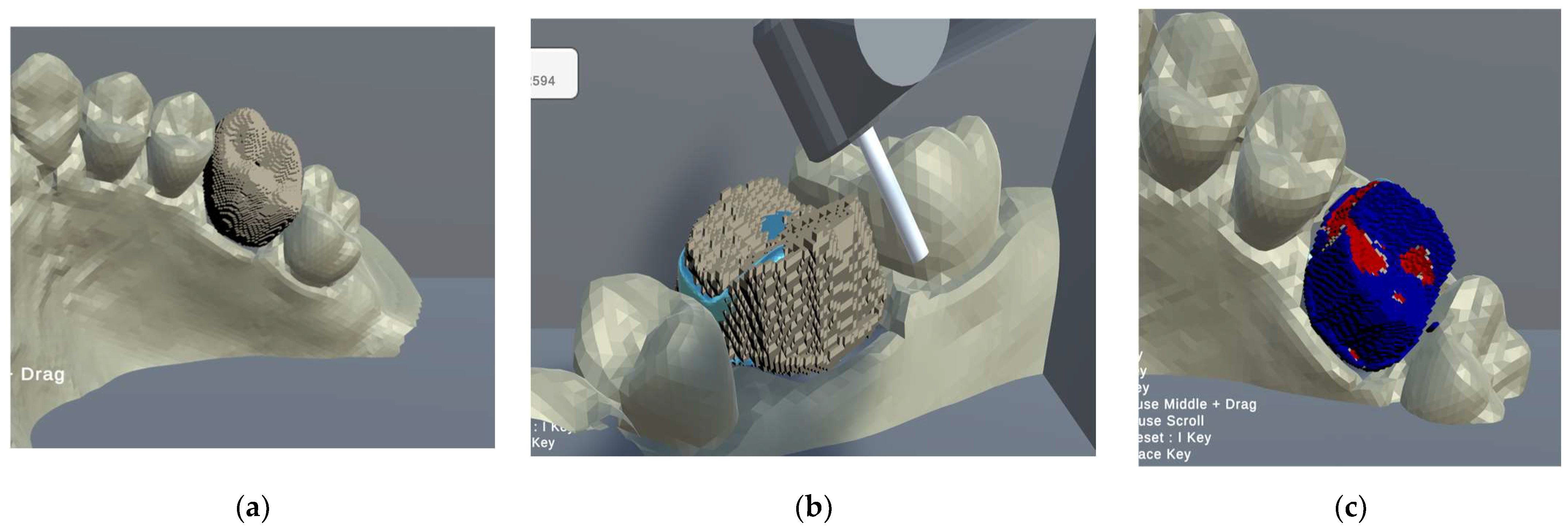
To realize an object formation function compatible with SRD, we originally employed Unity(2021.3.4f1) to develop the simulator [41], as shown in Figure 3. To replicate the procedural manipulation of traditional odontoplasty training, we used 3D Systems Touch (3D Systems Corporation, Rock Hill, SC, USA), a pen-like device with haptics that provides contact sensation [42].
The following functions were implemented to reproduce procedural manipulation during the preprosthetic treatment of prosthetic treatment with full crowns, which is high-frequency treatment:
  • A function to display the tooth model obtained through the intraoral scanner on the SRD and allows observing the 3D-CG tooth model from various angles via rotation.
  • A function that allows observing the morphology of an abutment tooth model by ooking through a 3D-CG tooth model.
It is difficult to form an ideal tooth morphology in the preprosthetic procedure if the morphology cannot be envisioned to some extent before the prosthesis is formed. The 3D-CG tooth model can be transmitted at any time to allow the observation of the abutment tooth model. Therefore, it is easy to visualize abutment teeth during tooth formation.
3.
Function to convert standard tessellation language (STL) data to morphologically deformable data.
The STL data obtained through an intraoral scanner only provide information about the surface morphology. However, to shave an object, we require the 3D spatial information of its interior. In this study, we constructed internal information based on the surface information and generated data (voxel data) that can be formed (Figure 4). Voxel data are a collection of 3D boxes, sliced at regular-distance intervals in a VR environment. They can be used to generate the internal structure of an object, similar to CBCT, in order to fill the blank spaces in the formed areas and the differences in the interior. The number of voxel faults was divided into 6 powers of 3 per side and the space was reproduced with 262,144 voxels. The number of segments was selected such that it was within the limits of current mainstream PC specifications, ensuring that the application did not have performance limitations.
4.
The real-time formation of 3D-CG tooth models and their reproduction using haptic technology.
The real-time formation of 3D-CG tooth models using voxel data. Touch feedback while shaving teeth for caries removal and preprosthetic treatment, which are frequently performed, is crucial for improving dentistry skills. Therefore, we used the 3D System Touch to reproduce the feeling of shaving teeth for preparation.
5.
Automatic objective evaluation after formation.
The 3D-CG tooth model used as an example can be compared with the voxel data obtained after tooth generation to evaluate whether tooth formation was excessive or deficient. The degrees of under-formation and over-formation, as well as agreement or disagreement with the model, were computed to conduct an objective evaluation (Figure 5).

2.1.2. Tooth Models

This study employed an epoxy model typically used in conventional training, a plastic maxillary first molar (FDI, #16), and a scan of a preformed maxillary first molar. The 3Shape TRIOS 3 Wired (J Morita Mfg. Corp., Kyoto, Japan) [43] was used to scan the plastic tooth. Subsequently, the 3D modeling software Blender (version 4.0) [44] was used to create a 3D-CG model and adjust it to only the crown portion of the tooth. This was then converted into an OBJ file.

2.2. Evaluations by Dentists

For the empirical evaluation, 20 dentists (age: 24–40; 17 females, 3 males) in the Department of Fixed Prosthodontics, with no visual impairments and no experience with SRDs, were randomly selected from the Kanagawa Dental University Hospital. The subjects had a mean clinical dental practice of 2.45 years (SD: 3.15). Subjects had acquired adequate abutment formation skills, and uniformity in basic abutment formation techniques was maintained. The participants performed abutment teeth formation simulations for the upper-left first molar on both a regular 2D display and an SRD. The measurements were conducted in a random order, and prior to the measurements, the participants were asked to observe and familiarize themselves with the exemplar abutment tooth morphology. Subsequently, the training time was measured. Subsequently, each participant was given 5 min to create an abutment tooth, and the degrees of over- or under-formation and agreement with the exemplars were evaluated through an automatic evaluation function. The was an interval of 5 min between device changes. To avoid measurement bias, participants were not allowed to view the automatic assessment results. After the measurements, they were asked to fill in a feedback questionnaire. The feedback questionnaire asked the participants to answer 15 questions on a 5-point scale. The questions included four categories, covering the usefulness of this system, the usefulness of an SRD, learning effects, and physical impairments such as dizziness and fatigue. Subjects were fully informed about the study and signed written informed consent. This study was reviewed and approved by the Ethics Committee of Kanagawa Dental University (Approval No. 970). Figure 6 shows the flow of the actual equipment verification.

2.3. Statistical Processing

For an objective evaluation, the discrepancies between the amount of over/under-formation and exemplars, employed as the automatic scoring metric for the 2D display and SRD, were recorded and their normality was verified through the Shapiro–Wilk test. The degrees of over- or under-formation and agreement with exemplars were evaluated using the Wilcoxon signed-rank sum test. Correlations between the objective assessment items, practice time, and experience were analyzed using Spearman’s rank correlation coefficient. The voxels of the exemplar model were compared to the voxels of the participant’s abutment teeth, and the number of voxels in each item was evaluated as a percentage. For the subjective evaluation, the responses to the feedback questionnaire and free-text responses were analyzed on the Likert scale (1: strongly disagree; 2: disagree; 3: neutral; 4: agree; 5: strongly agree). Figure 7 shows a dentist verifying this system. Statistical processing was performed using the software R (version 4.2.2) [45].

3. Results

3.1. Objective Evaluations

Discrepancies in SRD and under-formation in 2D displays were not normally distributed. Table 1 presents the measurement results for both displays. The discrepancy, under-formation, and over-formation items indicate the differences between the number of voxels of the exemplar abutment tooth and those created by the participants.
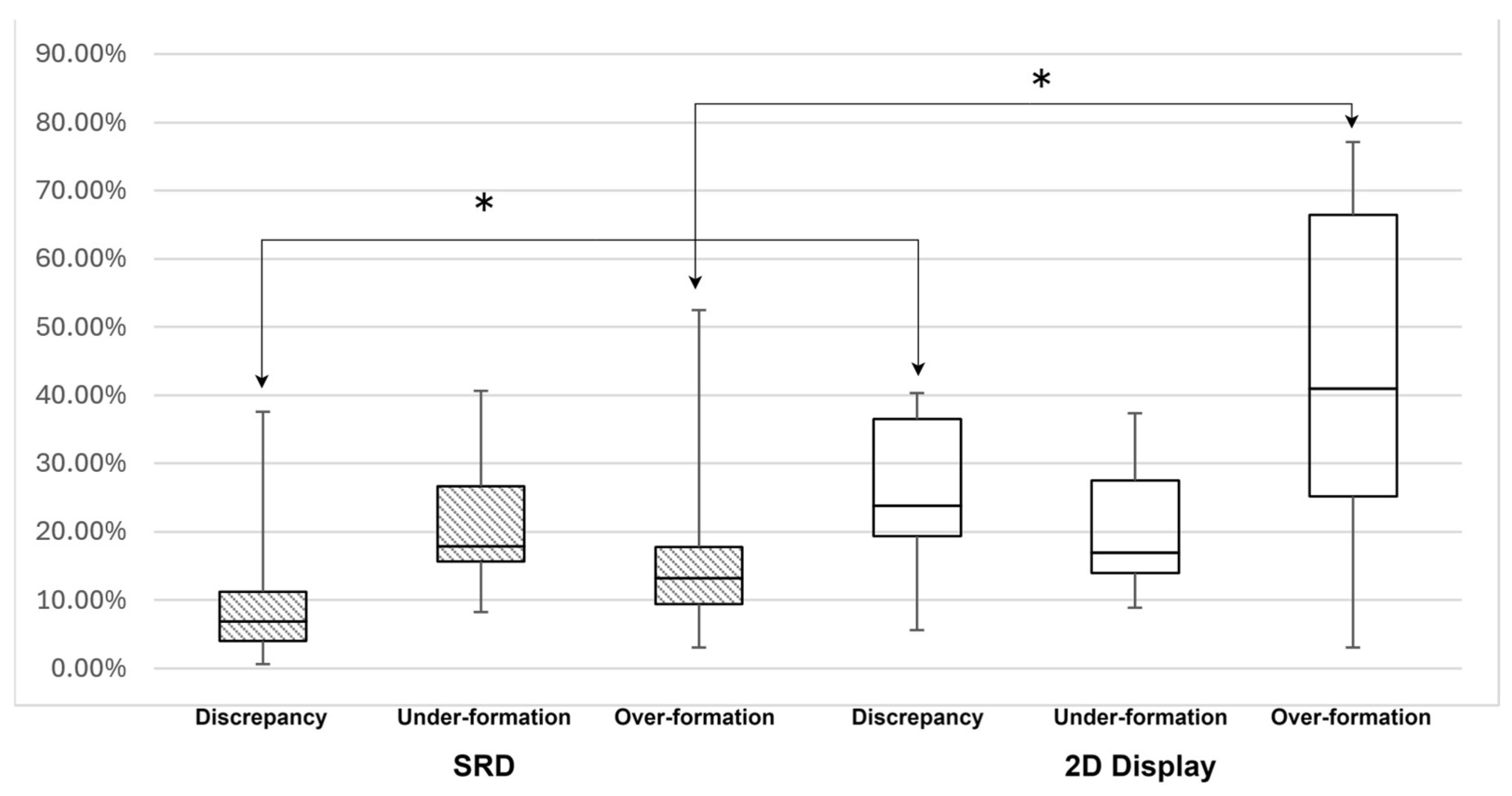
Each objective evaluation item was compared between the SRD and 2D displays and between the SRD and 2D displays. Wilcoxon’s signed-rank test was used for groups with non-normal distributions. Significant differences were observed among the smaller values of over-formation and we saw discrepancies between the SRD and the 2D display (p < 0.01), as shown in Figure 8.

3.2. Subjective Evaluations

On the Likert scale feedback questionnaire administered after the experiment, the participants provided high scores for the application (mean: 4.37/5), stereoscopic viewing (mean: 4.28/5), and learning enhancement (mean: 4.4/5), with some variations in the ratings for 3D sickness and eye fatigue (mean: 1.8/5). The results are listed in Table 2.

3.3. Correlation

The correlations between the objective endpoints for the SRD and 2D display, practice time, and dental experience were analyzed using Spearman’s rank correlation. No objective items were correlated with practice time. However, a positive correlation was found between experience and under-formation in 2D displays (p < 0.01), as listed in Table 3.

4. Discussion

In this study, digital data of dental models used in conventional training, obtained by an intraoral scanner, were loaded, and abutment formation was performed on the spatial reality display (SRD), which enables the naked-eye stereoscopic viewing of 3D-CG dental models, thereby improving the cost-effectiveness of abutment formation practice. This study examined whether performing abutment formation on the spatial reality display (SRD), which enables 3D-CG tooth models to be viewed stereoscopically with the naked eye, improves the cost-effectiveness of abutment formation practice and provides sufficient learning effects. Objective evaluations of the tooth formation and subjective evaluations, such as the feeling of using SRD, were conducted. For the objective evaluation, the upper-left first molar of a conventional mannequin-based tooth model was scanned to compare the ideal abutment tooth morphology with those created by the participants.
The discrepancy and over-formation rates were lower with the SRD than with the 2D display and statistically significant differences were observed, indicating that the use of SRD resulted in lower discrepancies in the exemplar abutment tooth morphology. This indicates that it is easier to reproduce the ideal abutment tooth morphology via simulations using the SRD.
The SRD makes it easy to obtain the depth information by changing the observation angle, which is important for obtaining the shapes of 3D objects, and thus facilitating the understanding of the 3D position of the tooth model and cutting instruments during the shaping operation. This may have led to the high accuracy of the tooth formation, demonstrating the significance of SRD for preprosthetic treatment. Previous studies have shown that SRDs can be used to accurately represent dental arch models [46]. Additionally, for root canal length measurements, the accuracy errors ranged from 0.16 to 0.62 mm [40]. Abutment tooth morphology is imperative in prosthetic pretreatment, and a taper of 2–5° is ideal for prosthetic retention [47,48]. In the proposed application, one side of the spatial resolution of the tooth model was divided by 26. One side of one of the resolved squares was approximately 0.21 mm. Based on the resolution, an ideal taper of 2–5° can be reproduced from a height of 4.2–10.5 mm. In addition, the actual formation of the abutment tooth requires a thickness that takes into consideration the material properties of the final prosthetic device (full crown) and a formation that does not damage the blood vessels and nerves (pulp) that exist inside the tooth. In other words, about 2 mm of formation is necessary to ensure the thickness of the fully encapsulated crown [49]. It is also necessary not to remove more than about 6 mm [50,51] from the occlusal surface to avoid damaging the pulp. The resolution of this application is sufficient to reproduce the results. Therefore, we believe that the accuracy of abutment tooth formation in SRD can be ensured. The automatic scoring function calculates the quantitative endpoints as volumes based on the voxel volume of the exemplars. One percent of the quantitative endpoint is approximately 3.6 mm3. The actual tooth preparation of a fully coated molar crown has a cutting volume of 250–330 mm3 [52], which is a sufficient percentage of the quantitative evaluation item to the second decimal place to maintain sufficient accuracy to perform evaluation. In addition, since visual feedback in terms of over- or under-formation can be provided by the color scheme, it is easy to evaluate the formation together with the quantitative evaluation items.
We used the first molar as it has a more complex abutment tooth morphology, and a similar validation is required for other tooth types; however, because sufficient significance was obtained for the validation of the first molar in this study, we believe that the same results can be obtained for other teeth. The correlations between the factors were tested using Spearman’s rank correlation coefficients, and no correlations were found between the practice time and objective evaluations.
None of the participants experienced abutment tooth formation using VR, the average practice time was 523.8 s, and we believe that no significant correlation was observed owing to this short practice time. Experience and objective evaluation items were positively correlated with a lack of tooth formation on 2D displays. The primary difference between the SRD and 2D display is the availability of depth observations. In cases wherein depth could not be observed, which deviated from clinical practice, the experienced dentists hesitated to form the tooth, thereby indicating a correlation between a lack of tooth formation and experience.
A Likert scale questionnaire was employed for the subjective evaluations, wherein high scores were given for the developed application, SRD, and learning improvement. Additionally, most participants found the system to be useful for prosthetics. However, a small percentage experienced 3D sickness and eye fatigue.

5. Conclusions

In this study, we developed an application that enables the simulation of an abutment tooth formation while precisely viewing 3D computer graphics with the naked eye using SRD, and verified the usefulness of the application. This application displayed a 3DCG model, obtained through a conventional intraoral scanner, which can be manipulated in real time through a tactile reproduction device. Furthermore, the differences between the mock and model models were objectively compared and automatically scored to provide an objective evaluation and comparative analysis of the accuracy of abutment tooth formation using SRD and 2D displays. The results showed that SRDs obtained higher scores and induced satisfaction among the participating dentists, with higher expectations for future dental education. Additionally, manipulations within a distance of 1 m of the participants through stereoscopic viewing without glasses was easier in the SRD than the 2D display, which did not provide depth information.
Although this study was validated with accuracy, aiming for perfect agreement with exemplars, abutment tooth formation in actual clinical practice is not a problem as long as the technical points are met. Therefore, it is necessary to adopt an evaluation method based on the identification of technical points for automatic scoring.
In addition, although this study was a comparison of visual–spatial understanding, in actual clinical practice, the method and form of abutment tooth formation need to be changed individually according to many factors such as the tactile, auditory, and intraoral environment. Therefore, we believe that validation of senses other than the vision necessary for training is also necessary.
As computational performance continues to improve in the future, the real-time deformation of 3DCG models with even higher accuracy and speed can be expected. We aim to further improve its intuitive operability and convenience for measurement operators and continue research to make it a new means of acquiring dental education using digital technology. We will verify the learning effects on dental students and continue with research aimed at introducing this system into prosthetic practice.

Author Contributions

Conceptualization, R.T. and T.I.; methodology, R.T.; software, R.T., T.I. and A.N.; validation, R.T., N.K., N.H. and K.K.; formal analysis, R.T.; investigation, R.T.; resources, R.T., T.I., A.N. and N.H.; data curation, R.T.; writing—original draft preparation, R.T.; writing—review and editing, R.T., T.I. and K.K.; visualization, R.T.; supervision, R.T., T.I. and K.K.; project administration, T.I. All authors have read and agreed to the published version of the manuscript.

Funding

This work was supported by JSPS KAKENHI Grant Number 22K10065.

Institutional Review Board Statement

The study was conducted according to the guidelines of the Declaration of Helsinki and was approved by the Institutional Ethics Committee of Kanagawa Dental University (No. 970, 5 December 2023).

Informed Consent Statement

Informed consent was obtained from all subjects involved in the study.

Data Availability Statement

The original contributions presented in the study are included in the article, further inquiries can be directed to the corresponding author.

Conflicts of Interest

The authors declare no conflicts of interest.

References

  1. Kramer, G.; Albino, J.; Andrieu, S.; Hendricson, W.; Henson, L.; Horn, B.; Neumann, L.; Young, S. Dental Student Assessment Toolbox. J. Dent. Educ. 2009, 73, 12–35. [Google Scholar] [CrossRef] [PubMed]
  2. Commission on Dental Accreditation. Accreditation Standards for Dental Education Programs 2023. Available online: https://coda.ada.org/-/media/project/ada-organization/ada/coda/files/predoc_standards.pdf (accessed on 10 September 2024).
  3. McGivney, G.P. Advanced Education in Prosthodontics. J. Prosthet. Dent. 1990, 64, 334–341. [Google Scholar] [CrossRef] [PubMed]
  4. Olley, R.C.; Frost Bem, P.M. Clinical and Academic Recommendations for Primary Dental Care Prosthodontics. Br. Dent. J. 2019, 227, 451–455. [Google Scholar] [CrossRef]
  5. Henzi, D.; Davis, E.; Jasinevicius, R.; Hendricson, W. In the Students’ Own Words: What Are the Strengths and Weaknesses of the Dental School Curriculum? J. Dent. Educ. 2007, 71, 632–645. [Google Scholar] [CrossRef]
  6. Divaris, K.; Barlow, P.J.; Chendea, S.A.; Cheong, W.S.; Dounis, A.; Dragan, I.F.; Hamlin, J.; Hosseinzadeh, L.; Kuin, D.; Mitrirattanakul, S.; et al. The Academic Environment: The Students’ Perspective. Eur. J. Dent. Educ. 2008, 12 (Suppl. 1), 120–130. [Google Scholar] [CrossRef]
  7. Uoshima, K.; Akiba, N.; Nagasawa, M. Technical Skill Training and Assessment in Dental Education. Jpn. Dent. Sci. Rev. 2021, 57, 160–163. [Google Scholar] [CrossRef] [PubMed]
  8. Eliyas, S.; Holsgrove, G.; Gallagher, J.E. Education and Training in Craft Skills Dentistry. Br. Dent. J. 2019, 226, 795–800. [Google Scholar] [CrossRef]
  9. Pottle, J. Virtual Reality and the Transformation of Medical Education. Future Healthc. J. 2019, 6, 181–185. [Google Scholar] [CrossRef]
  10. Anglin, J.M.; Sugiyama, T.; Liew, S.-L. Visuomotor Adaptation in Head-Mounted Virtual Reality versus Conventional Training. Sci. Rep. 2017, 7, 45469. [Google Scholar] [CrossRef]
  11. Muhanna, M.A. Virtual Reality and the CAVE: Taxonomy, Interaction Challenges and Research Directions. J. King Saud Univ. Comput. Inf. Sci. 2015, 27, 344–361. [Google Scholar] [CrossRef]
  12. Juliano, J.M.; Liew, S.-L. Transfer of Motor Skill between Virtual Reality Viewed Using a Head-Mounted Display and Conventional Screen Environments. J. Neuroeng. Rehabil. 2020, 17, 48. [Google Scholar] [CrossRef] [PubMed]
  13. Makri, A.; Vlachopoulos, D.; Martina, R.A. Digital Escape Rooms as Innovative Pedagogical Tools in Education: A Systematic Literature Review. Sustainability 2021, 13, 4587. [Google Scholar] [CrossRef]
  14. Peney, T.; Skarratt, P.A. Increasing the Immersivity of 360° Videos Facilitates Learning and Memory: Implications for Theory and Practice. Educ. Technol. Res. Dev. 2024. [Google Scholar] [CrossRef]
  15. Barteit, S.; Lanfermann, L.; Bärnighausen, T.; Neuhann, F.; Beiersmann, C. Augmented, Mixed, and Virtual Reality-Based Head-Mounted Devices for Medical Education: Systematic Review. JMIR Serious Games 2021, 9, e29080. [Google Scholar] [CrossRef] [PubMed]
  16. Tursø-Finnich, T.; Jensen, R.O.; Jensen, L.X.; Konge, L.; Thinggaard, E. Virtual Reality Head-Mounted Displays in Medical Education: A Systematic Review. Simul. Healthc. J. Soc. Simul. Healthc. 2023, 18, 42–50. [Google Scholar] [CrossRef]
  17. Berthold, D.P.; Muench, L.N.; Rupp, M.-C.; Siebenlist, S.; Cote, M.P.; Mazzocca, A.D.; Quindlen, K. Head-Mounted Display Virtual Reality Is Effective in Orthopaedic Training: A Systematic Review. Arthrosc. Sports Med. Rehabil. 2022, 4, e1843–e1849. [Google Scholar] [CrossRef]
  18. Lareyre, F.; Chaudhuri, A.; Adam, C.; Carrier, M.; Mialhe, C.; Raffort, J. Applications of Head-Mounted Displays and Smart Glasses in Vascular Surgery. Ann. Vasc. Surg. 2021, 75, 497–512. [Google Scholar] [CrossRef]
  19. Chen, S.; Zhu, J.; Cheng, C.; Pan, Z.; Liu, L.; Du, J.; Shen, X.; Shen, Z.; Zhu, H.; Liu, J.; et al. Can Virtual Reality Improve Traditional Anatomy Education Programmes? A Mixed-Methods Study on the Use of a 3D Skull Model. BMC Med. Educ. 2020, 20, 395. [Google Scholar] [CrossRef]
  20. Zitzmann, N.U.; Matthisson, L.; Ohla, H.; Joda, T. Digital Undergraduate Education in Dentistry: A Systematic Review. Int. J. Environ. Res. Public Health 2020, 17, 3269. [Google Scholar] [CrossRef]
  21. Cayo, C.F.; Cervantes, L.A.; Agramonte, R. VR Systems in Dental Education. Br. Dent. J. 2020, 228, 738. [Google Scholar] [CrossRef]
  22. Erdilek, D.; Gümüştaş, B.; Güray Efes, B. Digitalization Era of Dental Education: A Systematic Review. Dent. Med. Probl. 2023, 60, 513–525. [Google Scholar] [CrossRef] [PubMed]
  23. Dzyuba, N.; Jandu, J.; Yates, J.; Kushnerev, E. Virtual and Augmented Reality in Dental Education: The Good, the Bad and the Better. Eur. J. Dent. Educ. 2022; online ahead of print. [Google Scholar] [CrossRef] [PubMed]
  24. Huang, T.-K.; Yang, C.-H.; Hsieh, Y.-H.; Wang, J.-C.; Hung, C.-C. Augmented Reality (AR) and Virtual Reality (VR) Applied in Dentistry. Kaohsiung J. Med. Sci. 2018, 34, 243–248. [Google Scholar] [CrossRef] [PubMed]
  25. Hadjichristou, C.; Kokoti, M.; Bakopoulou, A. Haptics in Fixed Prosthodontics and Their Role in Dental Education: A Literature Review. J. Dent. Educ. 2024, 88, 1020–1028. [Google Scholar] [CrossRef] [PubMed]
  26. McAlpin, E.; Levine, M.; Brenner, C.; Opazo, C.; Bathini, S.; Choi, S.J.V.; Louisville, M.; Grandhi, U. Evaluating the Effectiveness of a Virtual Reality Simulation for Preclinical Local Anaesthesia Dental Education. Eur. J. Dent. Educ. 2022; online ahead of print. [Google Scholar] [CrossRef]
  27. Vincent, M.; Joseph, D.; Amory, C.; Paoli, N.; Ambrosini, P.; Mortier, É.; Tran, N. Contribution of Haptic Simulation to Analogic Training Environment in Restorative Dentistry. J. Dent. Educ. 2020, 84, 367–376. [Google Scholar] [CrossRef]
  28. Perry, S.; Bridges, S.M.; Burrow, M.F. A Review of the Use of Simulation in Dental Education. Simul. Healthc. J. Soc. Simul. Healthc. 2015, 10, 31–37. [Google Scholar] [CrossRef]
  29. Felszeghy, S.; Huhtela, O.; Manninen, K.; Mäenpää, M.; Banafa, A.; Liukkonen, M.; Mutluay, M. VR-Haptic and Phantom Head Dental Training: Does the Order Matter? A Comparative Study from a Preclinical Fixed Prostho-Dontics Course. Int. J. Comput. Dent. 2023; online ahead of print. [Google Scholar] [CrossRef]
  30. Patil, S.; Bhandi, S.; Awan, K.H.; Licari, F.W.; Di Blasio, M.; Ronsivalle, V.; Cicciù, M.; Minervini, G. Effectiveness of Haptic Feedback Devices in Preclinical Training of Dental Students—A Systematic Review. BMC Oral Health 2023, 23, 739. [Google Scholar] [CrossRef]
  31. Li, Y.; Ye, H.; Wu, S.; Zhao, X.; Liu, Y.; Lv, L.; Zhang, P.; Zhang, X.; Zhou, Y. Mixed Reality and Haptic-Based Dental Simulator for Tooth Preparation: Research, Development, and Preliminary Evaluation. JMIR Serious Games 2022, 10, e30653. [Google Scholar] [CrossRef]
  32. Moro, C.; Štromberga, Z.; Raikos, A.; Stirling, A. The Effectiveness of Virtual and Augmented Reality in Health Sciences and Medical Anatomy. Anat. Sci. Educ. 2017, 10, 549–559. [Google Scholar] [CrossRef]
  33. Chang, E.; Kim, H.T.; Yoo, B. Virtual Reality Sickness: A Review of Causes and Measurements. Int. J. Human Computer Interact. 2020, 36, 1658–1682. [Google Scholar] [CrossRef]
  34. Trinidad-Fernández, M.; Bossavit, B.; Salgado-Fernández, J.; Abbate-Chica, S.; Fernández-Leiva, A.J.; Cuesta-Vargas, A.I. Head-Mounted Display for Clinical Evaluation of Neck Movement Validation with Meta Quest 2. Sensors 2023, 23, 3077. [Google Scholar] [CrossRef] [PubMed]
  35. Kaluschke, M.; Yin, M.S.; Haddawy, P.; Suebnukarn, S.; Zachmann, G. The Effect of 3D Stereopsis and Hand-Tool Alignment on Learning Effectiveness and Skill Transfer of a VR-Based Simulator for Dental Training. PLoS ONE 2023, 18, e0291389. [Google Scholar] [CrossRef] [PubMed]
  36. Waller, D.; Richardson, A.R. Correcting Distance Estimates by Interacting with Immersive Virtual Environments: Effects of Task and Available Sensory Information. J. Exp. Psychol. Appl. 2008, 14, 61–72. [Google Scholar] [CrossRef] [PubMed]
  37. Guzsvinecz, T.; Perge, E.; Szűcs, J. Examining the Results of Virtual Reality-Based Egocentric Distance Estimation Tests Based on Immersion Level. Sensors 2023, 23, 3138. [Google Scholar] [CrossRef]
  38. Naceri, A.; Chellali, R.; Hoinville, T. Depth Perception Within Peripersonal Space Using Head-Mounted Display. Presence Teleoper. Virtual Environ. 2011, 20, 254–272. [Google Scholar] [CrossRef]
  39. Itamiya, T.; To, M.; Oguchi, T.; Fuchida, S.; Matsuo, M.; Hasegawa, I.; Kawana, H.; Kimoto, K. A Novel Anatomy Education Method Using a Spatial Reality Display Capable of Stereoscopic Imaging with the Naked Eye. Appl. Sci. 2021, 11, 7323. [Google Scholar] [CrossRef]
  40. Tsukuda, T.; Mutoh, N.; Nakano, A.; Itamiya, T.; Tani-Ishii, N. Study of Root Canal Length Estimations by 3D Spatial Reproduction with Stereoscopic Vision. Appl. Sci. 2023, 13, 8651. [Google Scholar] [CrossRef]
  41. Unity. Available online: https://unity.com (accessed on 10 September 2024).
  42. 3D System Touch. Available online: https://www.3dsystems.com/haptics-devices/touch (accessed on 10 September 2024).
  43. 3shape TRIOS 3 WIRED. Available online: https://www.3shape.com/scanners/trios-3 (accessed on 10 September 2024).
  44. Blender. Available online: https://www.blender.org (accessed on 10 September 2024).
  45. R-Project. Available online: https://www.r-project.org (accessed on 10 September 2024).
  46. Ariwa, M.; Itamiya, T.; Koizumi, S.; Yamaguchi, T. Comparison of the Observation Errors of Augmented and Spatial Reality Systems. Appl. Sci. 2021, 11, 12076. [Google Scholar] [CrossRef]
  47. Guyer, S.E. Multiple Preparations for Fixed Prosthodontics. J. Prosthet. Dent. 1970, 23, 529–553. [Google Scholar] [CrossRef] [PubMed]
  48. Jørgensen, K.D. The Relationship between Retention and Convergence Angle in Cemented Veneer Crowns. Acta Odontol. Scand. 1955, 13, 35–40. [Google Scholar] [CrossRef] [PubMed]
  49. Goodacre, C.J. Designing Tooth Preparations for Optimal Success. Dent. Clin. N. Am. 2004, 48, 359–385. [Google Scholar] [CrossRef] [PubMed]
  50. Selvakumar, H.; Kavitha, S.; Vijayakumar, R.; Eapen, T.; Bharathan, R. Study of Pulp Chamber Morphology of Primary Mandibular Molars Using Spiral Computed Tomography. J. Contemp. Dent. Pract. 2014, 15, 726–729. [Google Scholar] [CrossRef]
  51. Velmurugan, N.; Venkateshbabu, N.; Abarajithan, M.; Kandaswamy, D. Evaluation of the Pulp Chamber Size of Human Maxillary First Molars: An Institution Based in Vitro Study. Indian J. Dent. Res. 2008, 19, 92–94. [Google Scholar] [CrossRef]
  52. Sadid-Zadeh, R.; Farsaii, A.; Goodarzi, A.; Davis, E.L. Loss of Tooth Structure Associated with Preparation for Two Monolithic CAD-CAM Complete Coverage Restorations. J. Prosthet. Dent. 2018, 120, 403–408. [Google Scholar] [CrossRef]
Figure 1. Flow of abutment tooth formation.
Applsci 14 08367 g001
Figure 2. (a) Conventional dental training using mannequins; (b) conventional VR abutment formation training (using HMDs or 2D displays); and (c) VR training using naked-eye stereoscopic displays.
Applsci 14 08367 g002
Figure 3. (a) Displaying a 3D-CG model before abutment tooth formation. Exemplars can be placed in the tooth model for confirmation (light blue objects); (b) in the process of forming abutment teeth; (c) visual evaluation after automatic scoring. The amount of formation can be divided by color for feedback(Red is Over-formation, Blue is Under-formation ).
Applsci 14 08367 g003
Figure 4. (a) STL data obtained using an intraoral scanner; (b) voxelized model with added spatial information. The pink dots are the coordinates of the midpoint of each Voxel; (c) result of voxel-to-CG model conversion using the marching cubes algorithm.
Applsci 14 08367 g004
Figure 5. (a) The exemplar model is observed through a clairvoyant. The red object is an exemplar; (b) overly formed model; (c) the visualization of over- or under-formation after automatic scoring (blue: under-formed; red: over-formed).
Applsci 14 08367 g005
Figure 6. Flow of verification of actual equipment. After obtaining consent, validation will be performed on both displays, followed by automatic scoring and a questionnaire survey.
Applsci 14 08367 g006
Figure 7. (a) A view of the dentist’s hand verifying the results; (b) voxelized model with added spatial information.
Applsci 14 08367 g007
Figure 8. Comparison of objective endpoints using SRD and 2D displays (* p < 0.01).
Applsci 14 08367 g008
Table 1. Objective evaluation results for the SRD and 2D display.
DeviceEvaluation ItemsBetween the Number of Voxels of the Exemplar Abutment Tooth and Those Created by the Participants
Average(SD)MinMax
SRDDiscrepancy10.49%9.98%0.61%37.57%
Under-formation21.30%8.26%8.30%40.65%
Over-formation17.49%12.80%3.13%52.43%
2D DisplayDiscrepancy24.89%10.92%5.61%40.27%
Under-formation20.55%9.07%8.96%37.32%
Over-formation41.98%23.30%3.14%77.15%
Table 2. Likert scale ratings for the application obtained through the feedback provided by the participants.
CategoryAverage(SD)MinMax
Application4.370.234.004.90
SRD4.280.313.754.75
Physical harm1.800.811.003.00
Academic improvement4.370.274.004.87
Table 3. Correlations between experience and each objective assessment item.
Correlation Coefficientp-Value
SRDOver-formation−0.1830.440
Under-formation0.4750.034
Discrepancy0.5050.023
2D DisplayOver-formation−0.3880.090
Under-formation0.5980.005
Discrepancy−0.2210.349
Disclaimer/Publisher’s Note: The statements, opinions and data contained in all publications are solely those of the individual author(s) and contributor(s) and not of MDPI and/or the editor(s). MDPI and/or the editor(s) disclaim responsibility for any injury to people or property resulting from any ideas, methods, instructions or products referred to in the content.