Article

Authentication of a Painting Attributed to the Rembrandt School

1
Academy of Romanian Scientists (AORS), 54 Splaiul Independenței St., Sector 5, 050094 Bucharest, Romania
2
National Institute for Research and Development in Environmental Protection, 294 Splaiul Independenței, 6th District, 060031 Bucharest, Romania
3
Science Department, Interdisciplinary Research Institute, Alexandru Ioan Cuza University, 11 Carol I Bulevard, 700506 Iasi, Romania
4
Romanian Inventors Forum, 3 Sf. Petru Movilă St., L11, III/3, 700089 Iași, Romania
5
Geoscience PhD School, Geography-Geology Faculty, Alexandru Ioan Cuza University, 11 Carol I Bulevard, 700506 Iasi, Romania
6
Iasi County Police Inspectorate, Str. Mihai Costăchescu, nr. 2, 700495 Iasi, Romania
7
Faculty of Materials Science and Engineering, Gheorghe Asachi Technical University of Iasi, 67 Dimitrie Mangeron Bulevard, 700050 Iasi, Romania
*
Authors to whom correspondence should be addressed.
Appl. Sci. 2024, 14(19), 8655; https://doi.org/10.3390/app14198655
Submission received: 5 August 2024 / Revised: 4 September 2024 / Accepted: 20 September 2024 / Published: 25 September 2024
(This article belongs to the Section Materials Science and Engineering)

Abstract

:
This paper presents the archaeometric and chemometric authentication of a Renaissance-style oil painting on linen canvas, with plaster and barium sulphate-based primer preparation and bone glue as binder. By optical microscopy (OM), stereomicroscopy (SOM), scanning electron microscopy with energy dispersive X-ray spectroscopy (SEM-EDX) and attenuated total reflectance–Fourier transform infrared spectroscopy (ATR-FTIR), a series of archaeometric characteristics were identified: the grain size of the chemical components of the paint layers, the 3D porosity distribution of the pigments, the degree of penetration between the paint layers and the varnish and between the varnish and the oxidatively anchored dirt, and the typology of the craquelure (craquelure network arrangement and morphology). By correlating the results obtained, it was possible to estimate the period of the painting as 1660 ± 3 years.

1. Introduction

When attempting to authenticate an old painting, three aspects are considered: forgery—by which a total or complete forgery is made; counterfeiting or partial forgery—by which only certain characteristics are changed (addition or modification of inscriptions; removal or reforming of the structure and morphology of protective materials, film-forming substances, pictorial materials and supports; alteration of the registers of iconographic systems or sequences, on small or large surfaces—colportage forgery), and theft/illegal removal and commercialization [1,2,3,4,5].
Today, we are also witnessing an atypical phenomenon: many large collections, public or private, owning works of art or fake archaeological pieces, but which do not declare them for various reasons, particularly economic ones, which are inextricably linked to their image and value.
In general, fakes take on the defining features of a particular style, reproducing the faithful characteristics of an ‘authentic original’, and are easily convincing to buyers, art dealers, or collectors.
Forgery refers not only to the work as such or to the signature, date, etc., but also to other characteristics or alterations, such as erasures, varnishing, repainting, structural and chromatic reintegration, and other additions and deletions over time.
In practice, there are transitory situations between fake and genuine [5], such as:
modification of an original in the style of an earlier or later period and the addition of artistic elements of refinement (more expensive framing or casing, ornamentation, fastening and display elements, etc.);
structural and polychrome changes following preservation and restoration;
unjustified or insufficiently documented retouching, by depainting and repainting.
Among illicit activities, which are difficult to investigate in the case of old paintings, we mention:
falsification or plagiarism by making reproductions or copies without the consent of the author, owner, or custodian;
counterfeiting by signing, deleting, adding/completing etc.;
illegal transhumance or trafficking;
theft and substitution from collections;
destruction/vandalism;
changing contexts related to situations that allow for good value;
destroying the traces of the crime; and others.
These can be performed through a series of maneuvers (actions or operations), procedures and forms, among which we mention:
signing old works by unknown or unattributed authors with the names of famous artists;
aging new copies (by patination and cracking) and countersigning them;
failure to comply with the regime of protection of works of art of cultural heritage;
substitution or theft from collections;
false authentications, dating, and attributions by experts or qualified/authorized institutions;
knowingly facilitating the purchase of counterfeit goods;
favoring traffickers and counterfeiters through concealment, support, logistical support, etc.;
inappropriate, often aggressive, preservation and restoration interventions to highlight details, obliterate traces or remove archaeometric or artifactometric features;
hiding, destroying, or vandalizing works of art that have the status of cultural or historical assets.
The forgery and counterfeiting of old paintings is motivated by personal, sentimental, religious, truthful, onerous-to-practical, and scientific motivations [3].
In general, the purpose of these types of illicit activities can also take different forms, from the sentimental–personal to the pecuniary. As has already been said, when dealing with the reproduction of an authentic work for the purpose of trafficking and commercializing it as an original and obtaining a profit of any kind, it is a forgery and not a scientific copy, which has other purposes.
Among the main purposes that lead to forgeries are:
counterfeits produced for commercialization (from bargains to masterpieces carefully crafted following the original);
reproductions or copies produced by the author for sentimental reasons (reference works);
reproductions or copies produced by pupils for didactic, sentimental/admiring reasons and to gain artistic notoriety (reference artifacts or standards for school, mentorship, etc.);
forgeries produced to practice artistic skills and abilities (artistic styles and techniques representative of an era, school or geographical area);
reproductions or copies made for religious reasons (copies in large series based on ‘patterns’ or rendition, financial and dogmatic reasons) [6,7,8];
reproductions or copies made by specialists in order to gain artistic and scientific notoriety (fine styles and techniques, with complex elaborations that require great talent and meticulousness, forged documents related to famous works, forged archaeological research and evaluations) [9,10,11];
forgeries produced in order to discredit specialists (works that reveal mistakes in the mise en oeuvre, iconographic scenes and incoherent motifs, etc.) [12,13,14];
forgeries produced with the intention of replacing the originals for the purpose of removal, i.e., for replacement from collections, storage, or museums.
It is known that some products of human activity that have stored talent, creativity and craftsmanship have been transformed into cultural heritage assets, true historical testimonies, which, in addition to the inherent heritage elements highlighted in the work (age, value, author/workshop, artistic technique, technology of implementation, and passing of time or history), over time have taken on certain heritage functions that make them invaluable and unquestionable from an aesthetic, artistic, historical–documentary, technical–scientific, social–economic, and construction point of view, even reaching the supreme identity function of a community or an individual, i.e., the spiritual one [15,16,17].
Artwork authentication is not only about dating and establishing the author/the studio/school, but much more: determining the route from the commissioning to the collection/museum, with all its historical contexts and its structural–functional evolution/physical, aesthetic and conceptual state, together with a series of attributes related to the area of commissioning and use, ownership (owner/custodian/gallery/museum), itineraries, heritage value, etc. [18,19,20,21,22,23].
In the complex task of authenticating artwork today, the art historian or art critic, the so-called traditional expert, is powerless without the collaboration of technical–scientific experts from related fields (chemistry, physics, biology, geology, archaeology, anthropology, etc.).
By investigating certain archaeometric characteristics and evaluating chemometric ones, with archaeometric value, dating is carried out, and by aesthetic–artistic analysis, it is possible to establish the author, school, studio, etc. By using instrumental methods, the nature of the materials as well as their state of preservation or that of their structural components can be determined. By intrinsic and exhaustive analysis of the evolutionary effects of deterioration, which affect the physical state of the structural-functional elements, and those of degradation, which change the chemical nature of the component materials, it is possible to establish the two groups of attributes: heritage elements or characteristics and heritage functions, which are very important in determining authenticity [13,14].

1.1. The Role in the Authentication of Flemish Paintings of Crackle Models and the Characteristics of the Textile Support

The characteristics of the crackles and the textile support for the Flemish paintings painted in the Dutch Golden Age represent two archaeometric systems often used in authentication, which in conjunction with new ones identified following the investigation of the nature of the pictorial materials (support, preparation, polychrome layer, and varnish) and the condition of their conservation, it is possible to easily obtain original evidentiary data, which, through their sequential processing, can determine the period of the work’s creation and estimate the author, the school, or the geographical area where it was created.
Crackles can be seen as fingerprints of the painting technique and provide valuable knowledge about the painting [24,25]. More precisely, the morphology of the crack networks can be related to the mechanical properties of the painting material, or they can reveal information about the methods used by the artist or the conditions of conservation. The study of cracks in old paintings allows us to highlight the formation mechanism of both the early ones and the old ones, and thus information can be obtained regarding the role of the substrate—for example, the textile support and the thickness of the layer in the distance between the cracks.
A series of works are known [26,27,28,29] regarding the different typology of craquelures, from one geographical area to another, but also from one period to another, when discussing the distinct specificity of Flemish (Dutch), Danish, Italian, etc. craquelures.
There is also a study [30] that uses modern methods to analyse the type of cloth, the number of warp and weft in the cloth, the type of white filling and bonding material on the ground, the painting layer, the type of coloured materials, and the average colour type (the binder). Thus, it is known that the formation of cracks in an old painting results from a combination of external factors, such as the environmental conditions, but also the nature of the support, the preparation, the painting materials and the varnish, and the age. The different materials used in the layers of a painting contribute to distinctive early or antique crackle patterns. The study of crackles requires a multi-analytical approach involving imaging, elemental, and molecular analysis, along with dimensional characterization and cluster analysis methods. These studies allowed us to reveal/identify significant physical and chemical differences between artworks created in Holland and Denmark (and partly Germany) and those from France, Switzerland, or Italy, which can be attributed to different traditions of canvas preparation and painting elaboration. For example, the crackle patterns of the Flemish and Danish paintings are primarily characterized by orthogonal systems with jagged lines, an effect due over time to layers of colour containing natural earth pigments, rich in calcium carbonate and lead minium, using a textile support with high density. In contrast, the French, Swiss, and Italian paintings show cracks with diagonal networks, with larger islands, due to the predominant white content of lead and a small amount of calcium carbonate, along with other earth colours. Through the integration of the obtained data, four distinct groups of crack patterns for the four types of dominant painting materials were highlighted in all the paintings of the 17th century: rich in lead white, which is less susceptible to canvas-induced cracking compared to calcium- and barium-based pigments. Therefore, there is a correlation between the proportion of these groups of pigments, the nature of the preparation (chalk dust and gypsum), and the textile support characteristics, which lead over time to differentiated models in a geographical and temporal context. This knowledge is crucial for authentication [31,32,33,34].
The materials in each layer of a painting contribute in their own way to the formation of early or late cracks or other forms of natural aging [35,36,37]. The characteristic/specific crack patterns that develop over time in a painting depend on the type and properties of the canvas, as well as on the thickness of the pictorial layer, but also on the materials used in the elaboration of the painting through the respective technique [38,39]. Different pigments and fillers affect both the drying time and the strength of the paint film, resulting in painting layers with very different mechanical properties, which vary from weak to strong and from flexible to rigid and brittle [40,41].

1.2. Comparative Study of Rembrandt-Type Portrait Paintings with Lace Collar

With regard to artworks close to the painting under study, known works by the great artist Rembrandt Van Rijn (Leiden, 15 July 1606–Amsterdam, 14 October 1669), as well as by other masters of the period, known as the Dutch Golden Age, who created portraits, landscapes and still lifes elaborated as exceptional artistic achievements, recognized and appreciated as masterpieces of Flemish painting, have been analysed. The Netherlands was a major producer of pigments, and the great masters had easy access to lead white, ivory black, ochre, lead tin yellow, and even vermilion (mercury red). From this perspective, it is very important to establish in authentication the nature of the pictorial materials, the artistic technique, and the manner of mise en oeuvre [42].
To begin with, based on the known data related to the portraits of the great artist Rembrandt Van Rijn, who proved to be a great Baroque painter who ‘lives only through painting’ [42], a retrospective of five series of his best-known works (Figure 1) was carried out, which would allow for a comparison with the typological framing of the aesthetic–artistic elements of the painting under study.
Thus, some of Rembrandt’s paintings were retrieved from the internet and from albums [43,44,45,46,47,48,49], grouped in the following series: (i) self-portraits in the same style and appropriate periods, in terms of age; (ii) portraits and self-portraits with lace collars from different periods, in terms of age; (iii) and (iv) portraits of gentlemen different in terms of dress and age; (v) three paintings with a complex iconographic system, very well known (‘The Shipbuilder and his Wife’, ‘Anatomy Lesson of Dr. Nicolaes Tulp’ and ‘The Night Watch’) and appropriate in terms of style.
A large number of Rembrandt’s disciples are mentioned in the literature, but the most important are Gerrit Dou (1613–1675), Govaert Flink (1615–1660), and especially Carel Fabritius of Delft (1622–1654), undoubtedly the most talented of his pupils, but who died young in an accident [42].
In his studio, the great master produced numerous paintings whose style was taken up by the young painters who studied there. His pupils learned the art of Baroque painting by copying their master’s works, which led to many ‘fake Rembrandts’ entering the art trade until later. In a research program initiated and conducted by Dutch specialists [32,33], it was found that, while at the beginning of the 20th century, about a thousand works were attributed to Rembrandt, today only little more than a third of them are considered authentic works, the rest being copies or forgeries [1,2,3,42,43,44,45,46,47,48,49,50,51].
It is known that Rembrandt rarely signed his paintings. Therefore, few of his signature specimens are known; for example, he signed some paintings with an ‘R’, and a small number of paintings were signed with the signature shown in Figure 2.
From the collective’s own experience many years ago, when a painting by Rembrandt was analysed, it was signed only with ‘R’.
The majority of his paintings are masterpieces, which are quoted at major auction houses at values on the order of millions of euros. ‘Portrait of an Old Man’ was recently sold at Christie’s in London for 22 million euros [42,43,44,45,46,47,48,49,50,51], and the portrait (Figure 3) painted in the last year of his life was long considered a failed painting, but today it is noted as a masterpiece of great artistic value.
In this painting, the mastery of the biometric elements (hair, mustache, goatee under the lip, chin, ears, nose, forehead, and eyes with eyelids and dark circles) is striking, clearly showing ‘that he lived only for painting’, as Rembrandt was rightly considered to have done.
The painting under analysis has many features similar to the face of the previous painting, the difference being that the character is old.
Of these paintings, only those that had aesthetically and artistically similar elements were analysed by enlarging the image as much as possible, particularly those with ‘black areas’ either made by the artist using ivory black-based colour in oil or resulting over time from the deposits of oxidative dirt.
As they are images taken from the internet or from albums/catalogs, the presence of the old craquelure was not easy to highlight, knowing that the type of network of craquelure allows for the framing in the period and in the area (‘Flemish area’). It should be noted that most of the paintings do not bear the author’s signature.
This work aims at two important aspects of the valorization of an unknown painting: establishing the authenticity of a painting realized in the manner of the School of the Dutch painter Rembrandt Van Rijn (Leiden 15 July 1606–Amsterdam 14 October 1669) and validating chemometric characteristics by correlating them with archaeometric ones, attributed by studying the nature and state of preservation of the component materials, the distribution at the surface, and in the volume phase of polychrome stratigraphic elements of structural characteristics such as porosity, the degree of penetration between layers, the typology of the period-specific craquelure networks, the grain size and arrangement of the pictorial and preparatory materials, etc.

2. The Experimental Part

2.1. Painting Anamnesis

The painting belongs to a family from Timișoara, which was authenticated upon request, involving a series of archaeometric characteristics, in correlation with chemometric ones (gravimetric composition ratios or atomic percentages, the involvement of errors in the determination of EDX (energy dispersive X-ray spectroscopy) atomic composition in establishing the chemical congruents of the pictorial materials, etc.), validated archaeometrically.
The painting was bequeathed by the owner’s grandmother in 2021, when she was provided with a number of details about the provenance and the route (contexts) of the painting. Thus, the painting was acquired a long time ago from a granddaughter of Vasile Goldiș, from whom she knew that the work came from a collection from which it had been stolen during the Second World War (possibly from Italy).
In 1980, when it was bought by the owner’s grandfather, he framed and protected it on the back with a paper sheet, and later took it to the workshop of a museum in Timișoara to be restored. Their insufficiently justified interventions have led to many damages/shortcomings in the state of conservation and patrimonial value of the painting.
Given that the route of the painting before the last acquisition, or the existence of a conservation file containing a previous authentication expertise or other data related to the title of the painting and its author are not known with certainty, the expertise approach will involve high-resolution multispectral methods and techniques [1,2,3,4,5].
Originally, the painting was kept by the owner’s grandfather in the living room, then the owner’s mother kept it in the home’s library–storage room, which had no windows. The owner recently removed the painting from storage and brought it in for appraisal.

2.2. Experimental Protocol and Image Processing Algorithms, Sampling, and Processing of Material Samples

To begin with, an experimental protocol differentiated by a limited number of investigation methods was developed through the system of co-existence and collaboration between interdisciplinary techniques, and an algorithm was proposed for processing the reflectographic images, highlighting the pattern of cracks and the morphological characteristics of the structural elements of the textile support (thickness of the warp and weft thread, the density of the fabric, etc.) integrated with the nature of the filling materials and the binder of the preparation. Then, the nature of the pigments and the binder in the polychrome layer, but also of the varnish, allowed for the establishment of the state of conservation of the painting and the identification of new archaeometric characteristics (temporally evolving), with reference to the gradient of modification of surface structural elements and those in cross-sections (marginal structures in fractals/cracks with elevations in the roof, etc.) by corroboration with the chemometric ones, which was determined differently for the three groups of materials (preparation, polychrome layer, and varnish), as evolutionary relationships between certain atoms allowed us to quite clearly establish the age of the painting. This presents the aspects related to the role they have in the archaeometric and chemometric authentication of the network model of crackles and their morphology, along with that of the type of cloth, as a support of the materials for preparation, binders and pigments, especially the white ones (pencil, calcium carbonate, and barium sulphate mixed with earth colours).
Considering the team’s previous experience in similar paintings, the most appropriate devices available in involved laboratories were used.

2.3. Aesthetic–Artistic Analysis

The painting is executed in oil, on linen canvas support, with a primer as a base coat, bone glue as a binder, and an intimate mixture of fine plaster powder and barium sulphate as filler (Figure 4).
The painting is executed in the Renaissance style under the influence of the Flemish schools, in particular the School of Rembrandt, where the anatomy of the face is characterized by a very fine and very careful elaboration of all the biometric elements (mouth, chin, mustache, sideburns, eyebrows, eyes—with slightly legible dark circles and the lobes of the two ears). The collar is white with a diamond sheen, broadly covering the shoulders and chest, and made of a white textile (silk) with crocheted lace around the edges. The beauty and expression of the face indicate that we are faced with an intelligent character with a deep gaze and a quiet smile, his head tilted slightly to the right in a sense of endearing, gentle, friendly calm. The hat and waistcoat are faintly legible due not only to the blackened patina of age, but also to the black patches applied over the varnish with poorly anchored dirt, fine iridescence, and craquelure (which practically should have been initially devarnished), and after cleaning, the surfaces should have been re-varnished with a compatible peliculogenic. These interventions were carried out unprofessionally and insufficiently documented experimentally by the wash test.
As large surfaces of the painting have been retouched, with repainting and varnishing, without cleaning and removing the original varnish based on Damar resin, which had the role of climatic and mechanical protection, but also aesthetic properties (providing depth of colour), the painting was visibly affected, especially at the level of the face, where the new varnish interacted with the old unvarnished structures.
The author used oil colours based on lead white, ivory black, burnt umber, natural umber, azurite (pigment enamel), lead yellow, yellow ochre, red ochre, brown ochre, natural sienna, burnt sienna, and other earth colours, typical of the Flemish schools.

2.4. Conservation Status

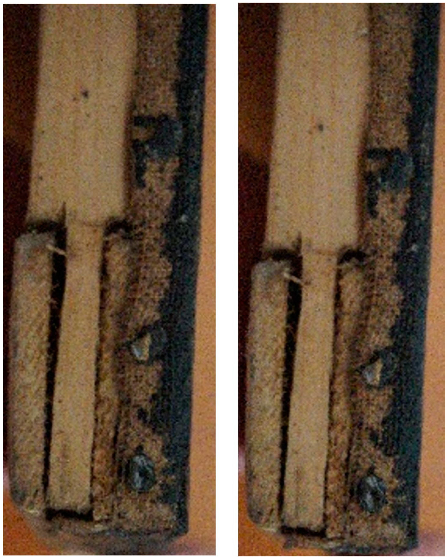
The painting was executed on a linen backing with a preparation layer of fine gypsum powder (CaSO4·2H2O) and barium sulphate (BaSO4), and the chassis is reinforced at the corners with wedges, with the ends joined together (Figure 5).
The bronze-polished frame has ornamental framing at the corners, middles, and mid-height sides (Figure 6).
Although the frame is more recent, due to storage in inadequate conditions, the ornamental framing show a lot of damage due to the lack of polish and plaster on certain areas; for example, the inner perimeter areas (the passpartu) and the outer areas towards the corners were very damaged, due to handling, display, and storage without protective measures.
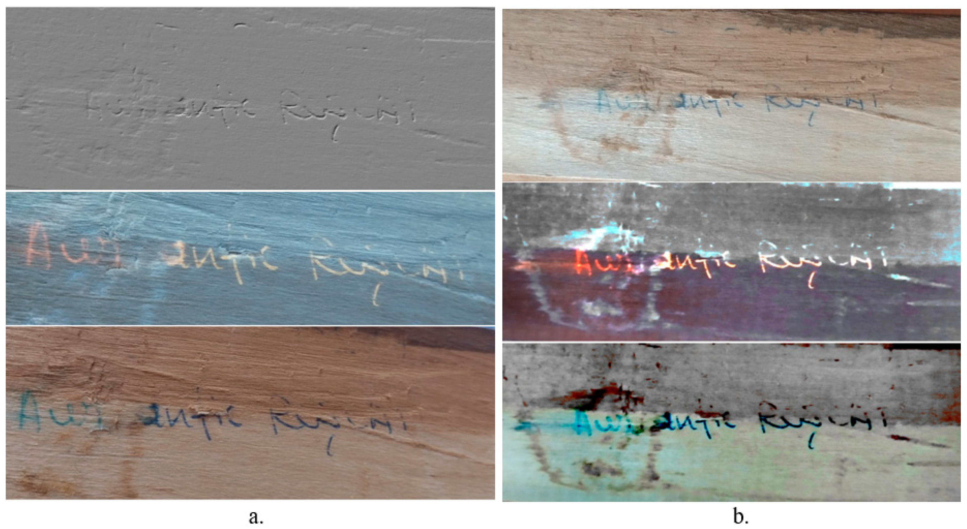
On the back there is an inscription (Figure 7), with a blue-coloured pen, belonging to the carpenter who made the frame (the difference between the whiteness of the protective paper on the back and the frame left uncovered denotes an age of the frame of approx. 50–60 years—Figure 6b).
When it comes to the images of the inscription on the back of the frame (Figure 7), it would have been desirable to undergo further processing, evaluating the inscription with the version 6 of the ‘LUCIA Forensic’ software, but this being quite recent, it is of no value in authentication, especially since the results will be approximately the same, probably with a slight note of differentiation from an aesthetic point of view for the images obtained.
The painting is in a poor state of conservation, with the grid having age craquelure on the entire surface, and perimeter craquelure and erosion resulting from handling during the period of reframing and display/storage (Figure 8).
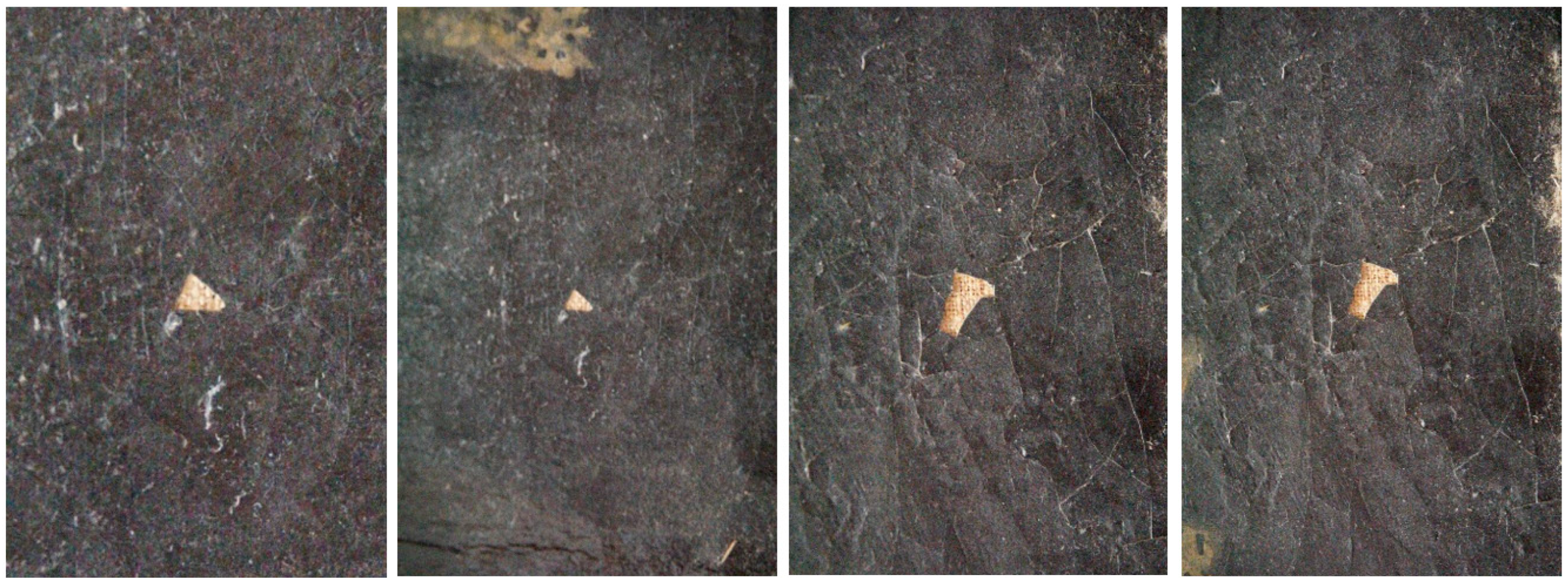
On the central surface and at the edges of the painting, in the framing area, there is a series of gaps with loss of material reaching the support, produced over time by the detachment of the pictorial layer in the craquelure network (Figure 9).
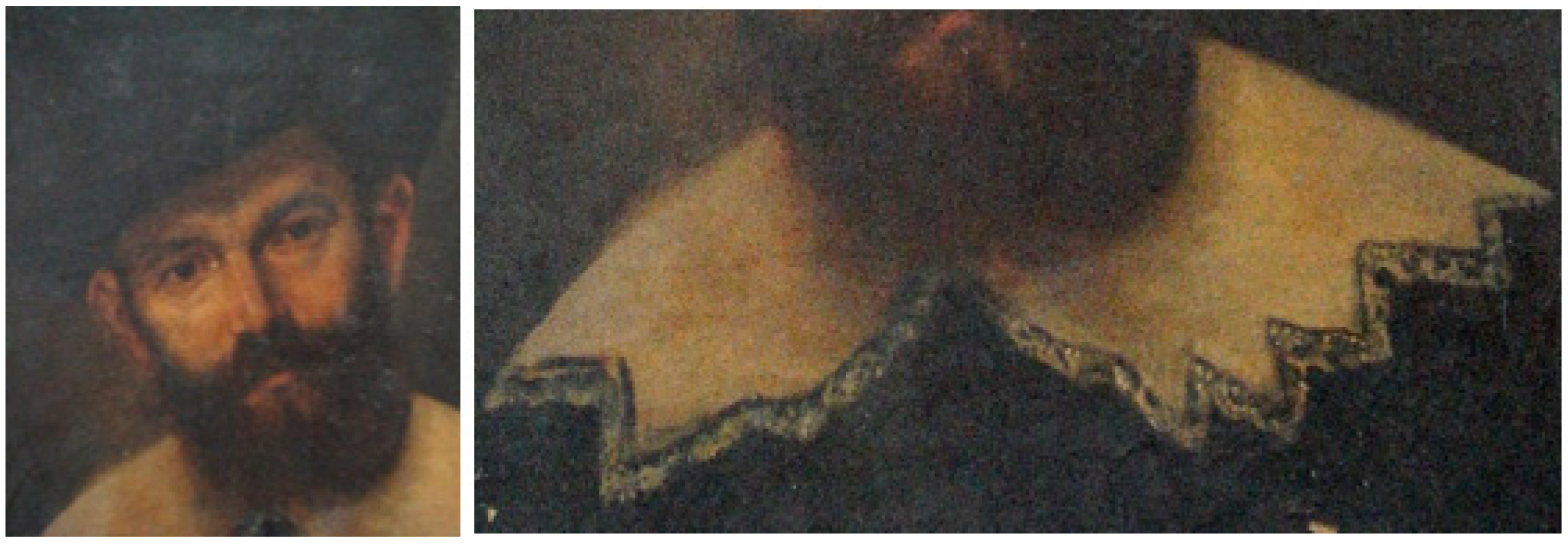
In addition to the presence of lacunar areas due to the loss of pictorial material in the craquelure network, up to the canvas support with traces of preparation, the painting shows a patina of age with areas of oxidative deposits of dirt and oxidized dirt with penetration in the varnish, which, compared to the background, allows for very poor legibility of both the clothing and the feathered hat. The white collar, ornamented at the edges with lacework (Figure 10), is poorly executed from an artistic point of view and very different from that of Rembrandt’s School (this aspect is attributed to the last restoration work carried out by an unauthorized conservator). The clothing reveals a costume of rank from the Netherlands. The owner, according to her grandmother, confessed that after the Second World War, the painting underwent a not particularly accurate restoration, involving repainting and repainting without devarnishing, which affected the aesthetics of the lacework of the collar and the anatomy of the face.
The back (Figure 4b) shows many elements penetrating the craquelure from the surface inwards. The patina of the canvas on the anterior surface of the painting denotes a poor loading and a chromatic deviation of more than 360 years. The chromatic adaptation of the back has been slightly altered over time, as the painting was originally protected, and later by interventions, by a covering of paper glued to the frame, which has been lost.
The chassis is the original, over 360 years old. It is easy to read the interventions of repainting on the chassis (Figure 11).
The frame (Figure 6) is more recent; it was changed after World War II, approximately after 1950. The frame has lost its decorative elements, plus the bronze-polished plaster ornaments, many due to mishandling and display under inappropriate conditions [12,14].
The owner knows that the painting was restored at the Banat Museum in Timisoara, about 45 years ago, when they carried out interventions to consolidate the pictorial layer, puttying of the lacunas, repainting (colour reintegration), revarnishing, and protection with paper glued on the back of the frame.

2.5. Microscopic Analysis Methods and Techniques

Initially, the components of the painting were analysed visually or with magnifying tools, and digital photography was carried out using a Sony ALPHA A6000 camera (Sony, Bangkadi Thailand) to examine certain details.
Along with visual analysis with magnification tools (hand magnifiers) and stereo magnifier (STEREO MX4, 10-220X), a series of multispectral instrumental techniques [13,14,16,17,18,19,20,21,22,23] were used in analysing the painting, involving co-assisting and corroboration between the following:
UV, Vis, and IR reflectography, using a document detector-type device with a USB port, HS525A Series, with 30× magnification power, with UVA (365 nm), UVC (254 nm), having infrared (IR), blue-white light (470 nm), and laser (980 nm) fields. The microscope is USB-powered, 1.3 MP (2.0 MP interpolated), with manual focus from 10 mm to 500 mm, frame rate: max 30 f/s, brightness under 600 l×, 8 white LEDs with adjustable illumination and magnification from 20× to 200×, AVI video format and a portable UV illuminator, with led: G5, UV 4W.
Optical microscopy (OM), carried out with a Zeiss Imager a1M microscope with an attached AXIOCAM camera and specialized software version 2018. The samples were analysed at a magnification of 50–200× in dark or bright field.
Optical Stereomicroscopy (SOM) was carried out using a STEREO MX4 microscope, made in St Petersburg, with a camera attached.
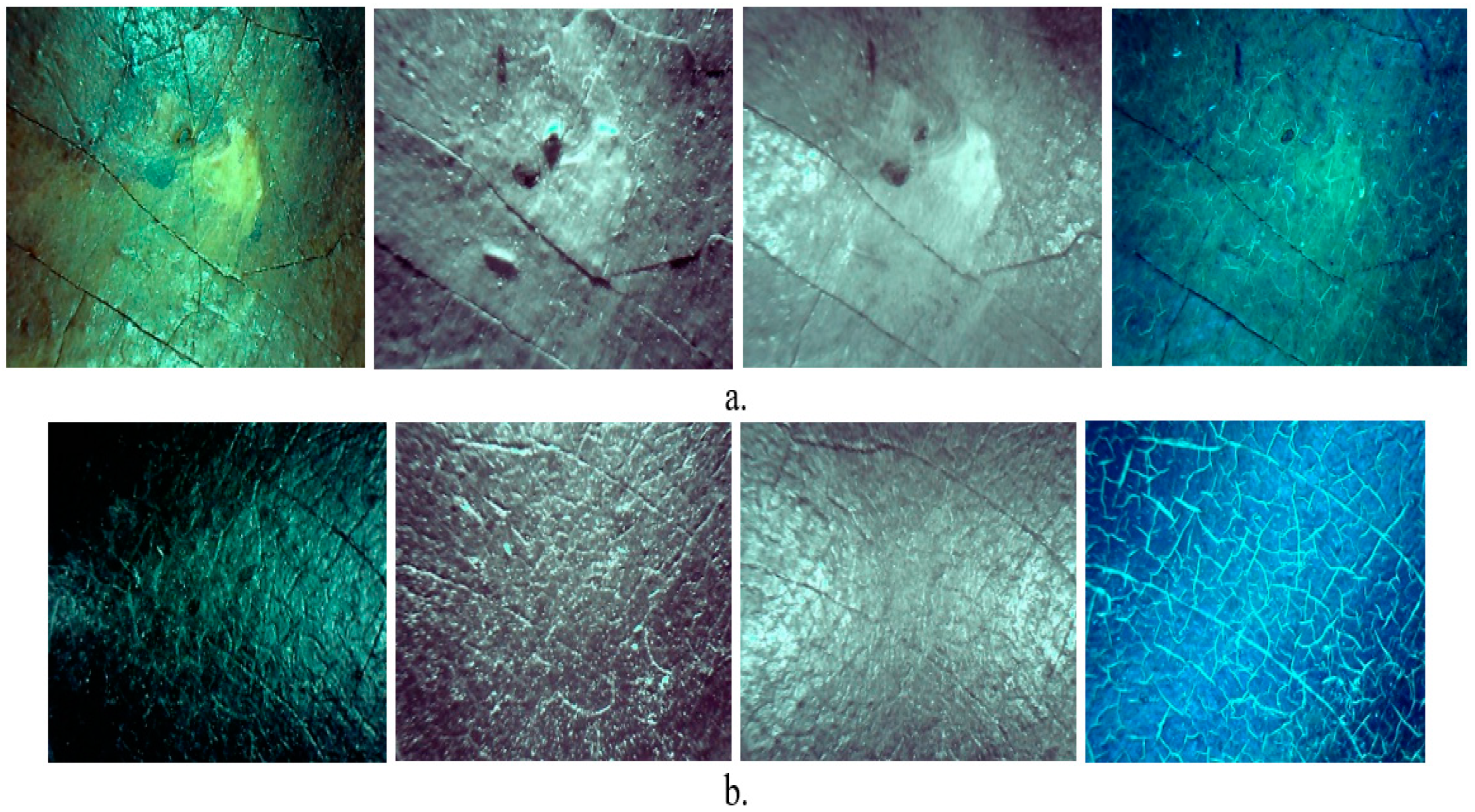
Microscopic techniques were used to identify the intervention areas: the left side of the collar, the right side of the moustache, and the upper right bottom and centre left bottom, which were analysed by dynamic UV, Vis, and IR reflectography, using four wavelengths on the three fields, which provides high readability of the crackle networks, lacunar areas, roof lifts, scaling, oxidative anchorage, etc. (Figure 12, Figure 13 and Figure 14).
Thus, the networks of UV, Vis, and IR reflectographically visible craquelure are very clearly reproduced in Figure 12a–d.
The network of craquelure is typical of 17th century Dutch paintings (Figure 13), as presented in the introduction paragraph.
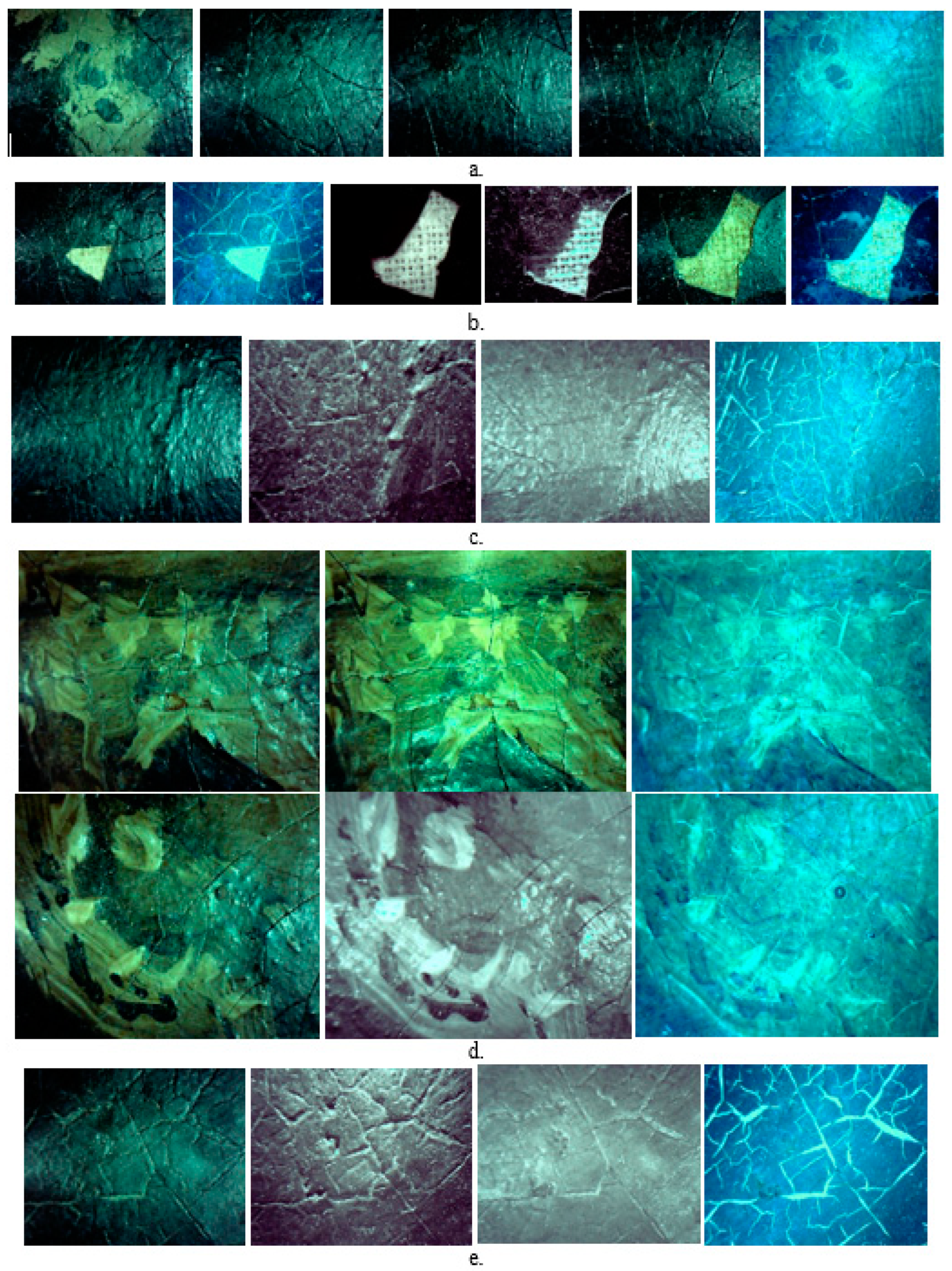
Figure 14a–e shows, by UV, Vis, and IR reflectography, the lacunar areas with loss of pictorial material in the network of movable craquelure, honeycombing and areas with anchored dirt, and the restoration of the lacework and other inappropriate retouching. In these areas, one can observe the fading by covering (repainting) of the lacework, with very few details of the lacework still preserved from the old painting.
Alongside the age craquelure and deposits of anchored dirt, Figure 14a shows insect soiling evenly distributed over large areas and punctuated. Figure 14b shows that the void areas (material losses) are deep, down to the textile backing.
The appearance of craquelure networks, typical of Dutch paintings of the 17th century, is also confirmed by the unevenness in the thickness of some of the lines of force of the craquelure (Figure 14e). In Figure 14d, the fading by chromatic reintegration of the lacework can be seen, with very few elements from the old painting.
There are also areas of roof craquelure (dynamic) and loose scales that have led to gaps in the pictorial layer (Figure 15).
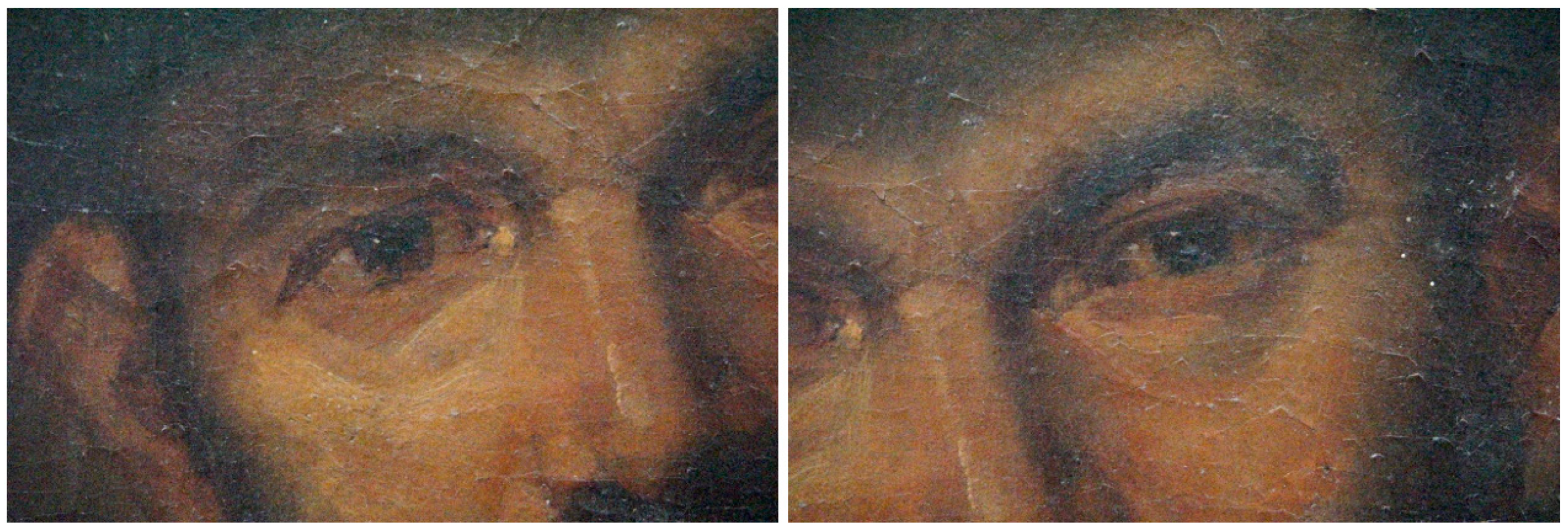
Figure 16 shows the presence of lifting craquelure in the roof at the level of the lines of force resulting from an incompatible revarnishing (the new varnish interacted with the old, varnished paint layer).

2.6. Optical Microphotography

The two techniques used in optical microscopy analysis as above [16,17,18,19,20,21,22,23] are optical microscopy (OM) and optical stereomicroscopy (SOM).
Figure 17 shows the OM images of the samples taken from the pictorial and support materials.
The white colour comes from white lead. All the pictorial materials, taken from the painting and the edge of the painting, show a summative system of pigments, varnish and anchored dirt on the front of the sample, and on the back, the linen textile used as support, weakly impregnated with a preparation based on barium white (BaSO4) and gypsum (CaSO4·2H2O).
Figure 17—S5 clearly shows both the warp and the weft of the linen backing, which has a thin warp thread (0.2–0.4 mm) with a high number of twisted fibers and a thicker weft thread (0.6–0.8 mm). The wood sample with the preparation on the edge of the chassis retains the anatomical characteristics of the old wood and the preparation based on plaster and barium sulphate. As it shows traces of preparation, it proves that the chassis is the one used in the mise en oeuvre.

2.7. SEM-EDX Analysis

A scanning electron microscope, model SEM VEGA II LSH, produced by TESCAN Czech Republic, coupled with an EDX detector type QUANTAX QX2, produced by BRUKER/ROENTEC Germany [20,21,22,23,42], was used in the SEM-EDX analysis. The microscope, completely computer controlled, has an electron gun with a tungsten filament, which can reach a resolution of 3 nm at 30 kV, with a magnification power between 30× and 1,000,000× in ‘resolution’ for the mode of operation, accelerating voltage between 200 V to 30 kV, and a scanning speed between 200 ns and 10 ms per pixel. The working pressure is less than 1 × 10⁻² Pa. The image obtained can consist of secondary electrons (SE) or back-scattered electrons (BSE). Quantax QX2 is an EDX detector used for qualitative and quantitative microanalysis. The EDX detector is a 3rd generation X-flash-type detector that does not require liquid nitrogen cooling and is approximately 10 times faster than conventional Si (Li) detectors. The technique, together with microphotograph visualization, allows for imaging with the mapping (arrangement) of atoms on the surface under investigation. Based on the X-ray spectrum, it is possible to determine the elemental composition (in gravimetric or atomic percentages) of a microstructure or selected area and to evaluate the composition variation along a vector arranged in the analysed area or section.
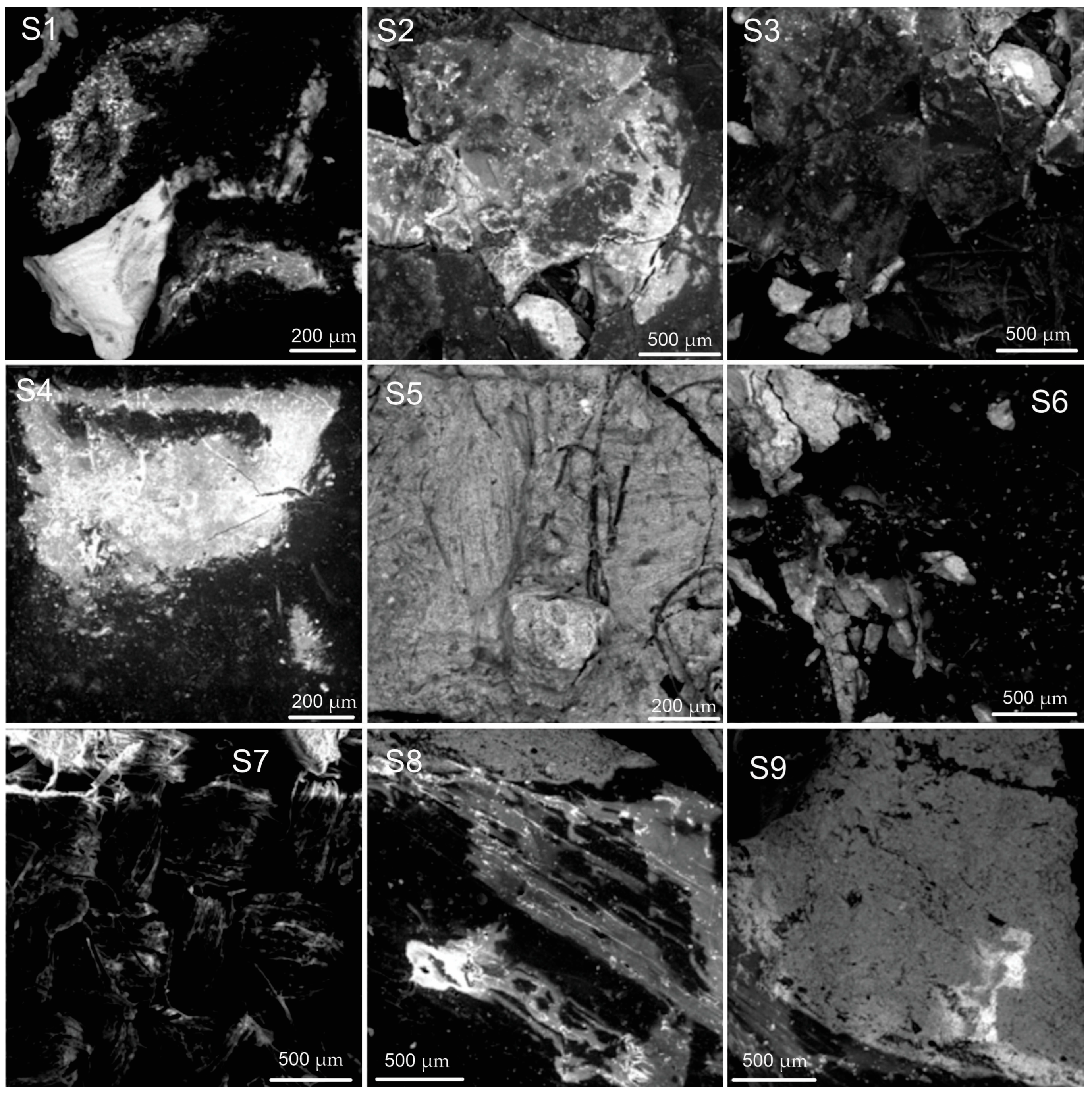
Figure 18 shows the SEM micrographs of the same samples analysed by OM, with the observation that only certain representative EDX surfaces identified/selected by OM were used in the analysis.
In correlation with the OM analysis, out of the 12 samples taken in the study, only 9 samples were analysed by SEM, for which certain areas were selected for EDX analysis. For some samples, two representative areas for a specific pictorial material were also analysed in order to allow for their differentiation by preparation (S2, S3 and S6). Two areas were also selected for the wood sample, one with the patina of time (area I) and one with traces of preparation from the mise en oeuvre (area II).
These SEM microphotographs highlight the arrangement of pigments, oil-based binders, varnish, and oxidatively anchored dirt. It is also possible to observe the granulometry of some of the pictorial materials and preparation, the morphology of the textile material, and the anatomical elements of the chassis wood, as well as the presence of fixed or mobile craquelure and gaps created over time by loss of material in pictorial roof layer.
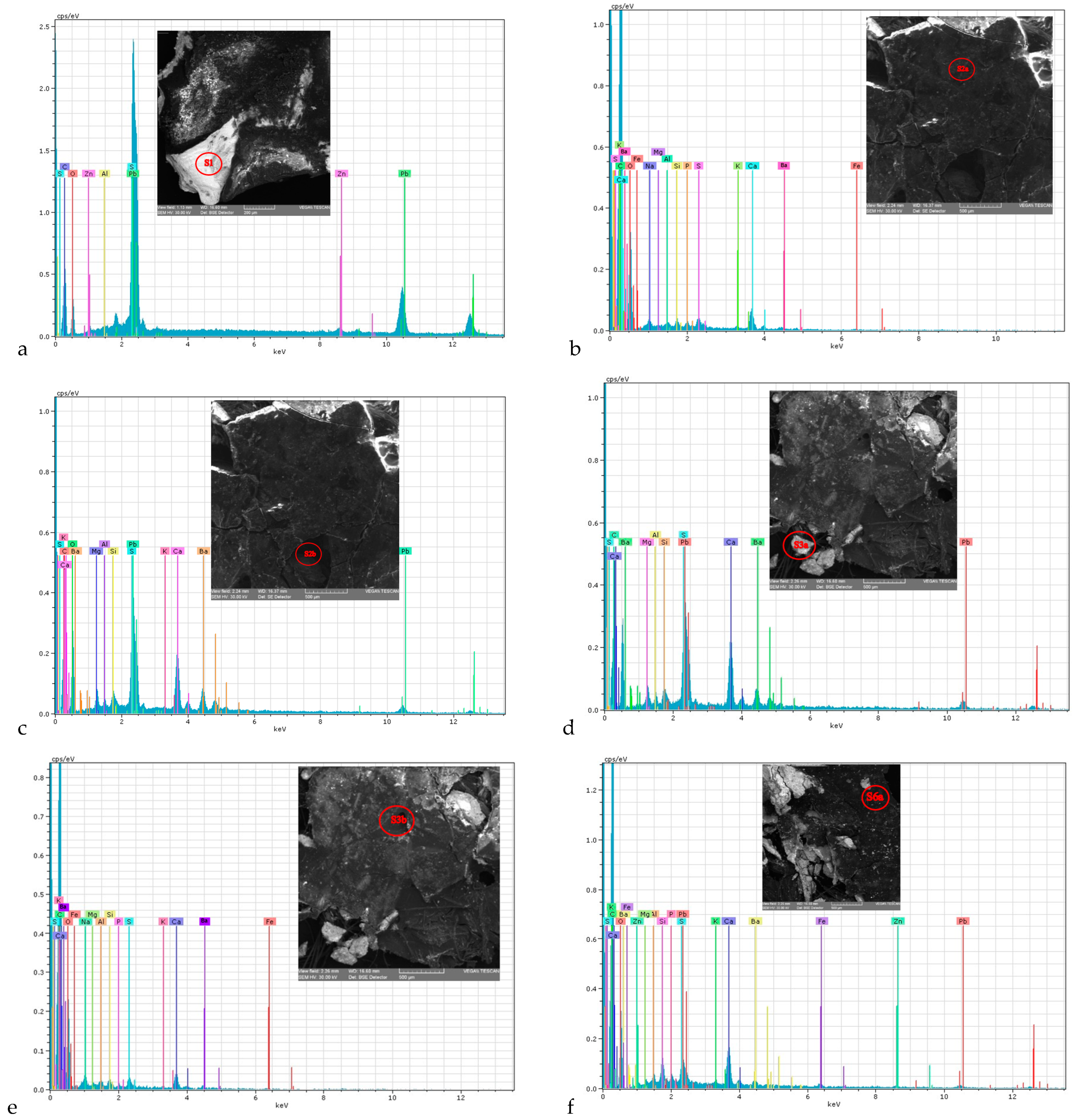
Figure 19 shows the 12 EDX spectra for the analysed areas.
EDX spectra give us information on the composition in mass (%) and atomic (%) percentages, which highlight the major and minor chemical elements of the material in the area analysed (Table 1). The errors recorded in these determinations allow for an appreciation of the chemical congruents present in the summative systems of the pictorial materials. In the evaluation of the archaeometric and chemometric (altmetrically validated) characteristics, the composition in atomic percentages (%) was used. For a clear comparative highlighting of the nine samples, either as such or duplicated in two different analysis areas (S2, S3 and S6), the compositions obtained by EDX analysis are presented in a single T (Table 1) in the order of the samples (S1–S9).
The colors in Table 1 represent for blue the atoms from white pigments, black from earth pigments and adherent dirt containing alumino-silicates, green from other earth pigments and red the oxygen atom that is found in several congruents (molecules) both from pigments and from dirt (adherence).
The net differences of the three doubled samples demonstrate the high dispersion of distinct pictorial materials from one area to another. There is some closeness/(close compositional similarity) between areas where we find ZnO retouching interventions over old lead white and coloured earths, including ultramarine (S1, S3a, S4, S6a, and S9P) or those with traces of preparation over coloured earths (S2a, S2b, S3b, S5, S6b, S7, and S8), which contain elements written in green. In contrast, the black colour and structures with anchored deposits are found in all the areas analysed (elements written in blue and black).
The chemical element oxygen is shown in red in Table 1, to emphasize that the EDX determination error is variable, ranging between 5.952 (S4) and 46.634 (S2a), which justifies several aspects related to its participation in different chemical congruents and the concentration level in the structure analysed. Thus, its concentration in comparison with the other elements in the composition is much higher, an aspect that is due to its presence in the atomic composition of several chemical congruents (pigments, boiled linseed oil binder, oxidatively anchored dirt varnish, preparation, textile backing, and wooden chassis), but also to the distribution of concentration levels in the structures analysed.
It should be noted that lead and barium, together with carbon and sulphur, show that lead white was used as pigment, and barium white (BaSO4) and plaster (CaSO4) as preparation. These pigments are found in the white shades and in the dilution of other colours, but also in the preparation, an aspect common in some Flemish paintings. In addition to these three pigments, pigments based on coloured earths, such as ultramarine, iron ochre, sienna and lead red, were also identified according to the compositions.

2.8. ATR—FTIR Spectrometry

Attenuated total reflection Fourier transform infrared (ATR-FTIR) spectra were collected with a Bruker LUMOS FTIR microscope spectrometer (Bruker Corporation, Karlsruhe, Germany) equipped with an attenuated total reflection (ATR) module and a diamond crystal. Due to the non-destructiveness of the setup used, samples were placed directly on the ATR crystal and subjected to IR investigation. All spectra were collected in the range 500–4000 cm−1, and the measurements were performed by averaging 64 scans at 2 cm−1 resolution. All spectra were recorded at room temperature [13,14,52].
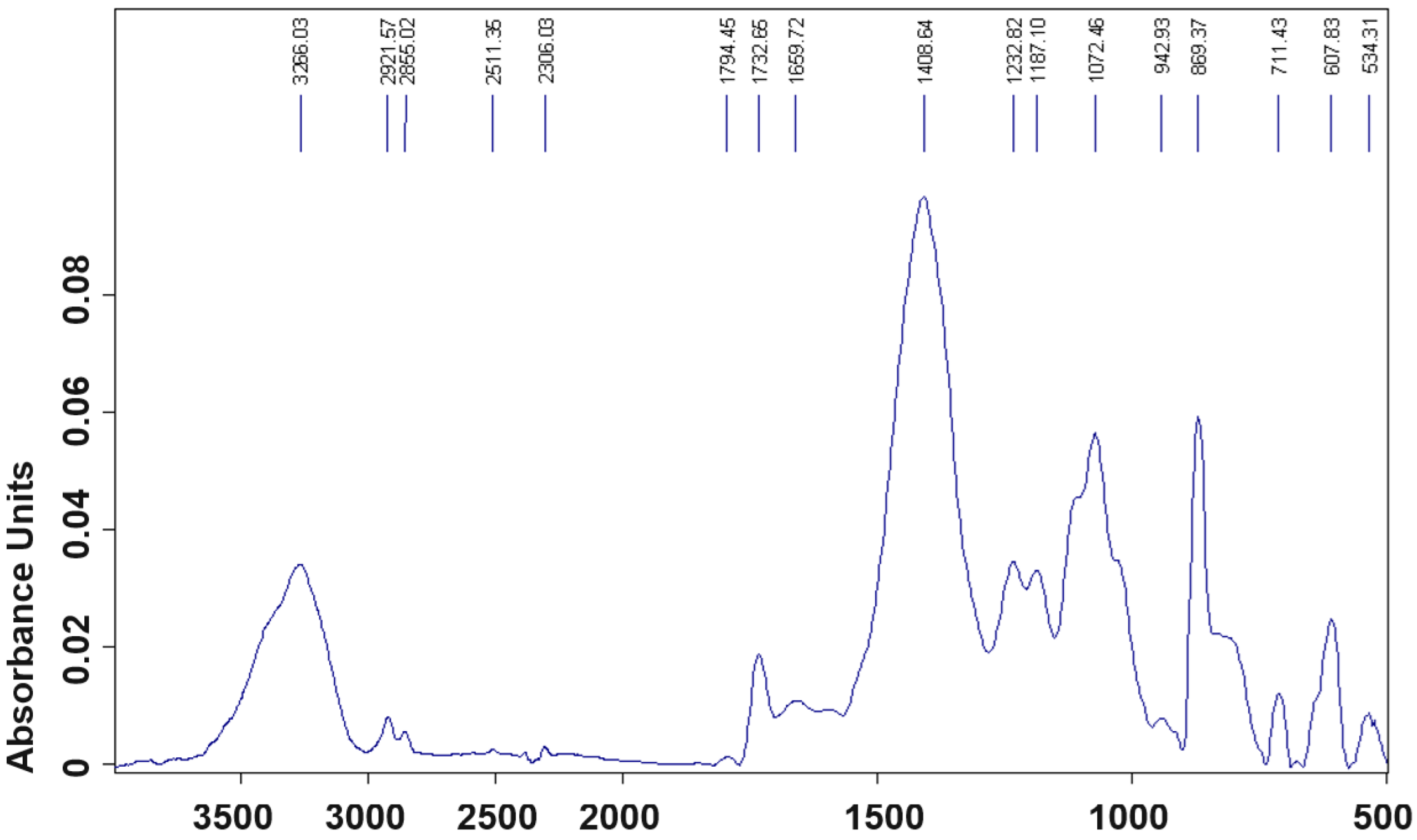
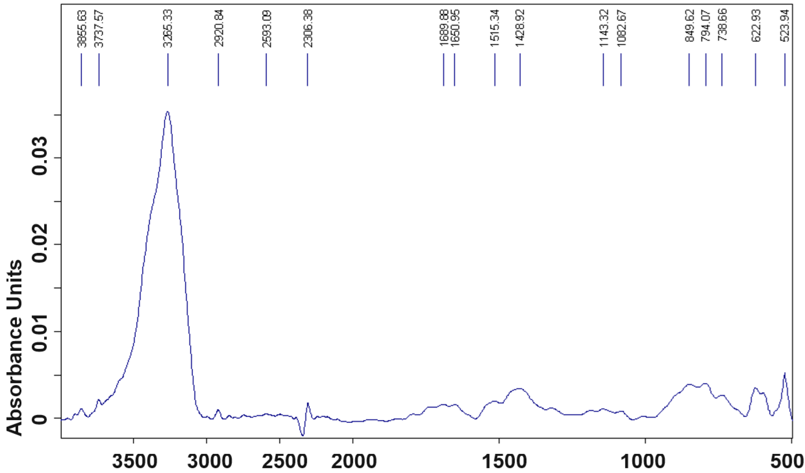
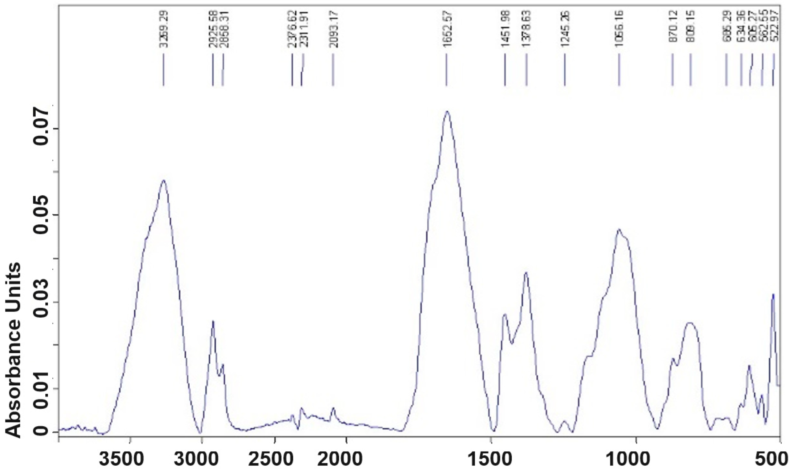
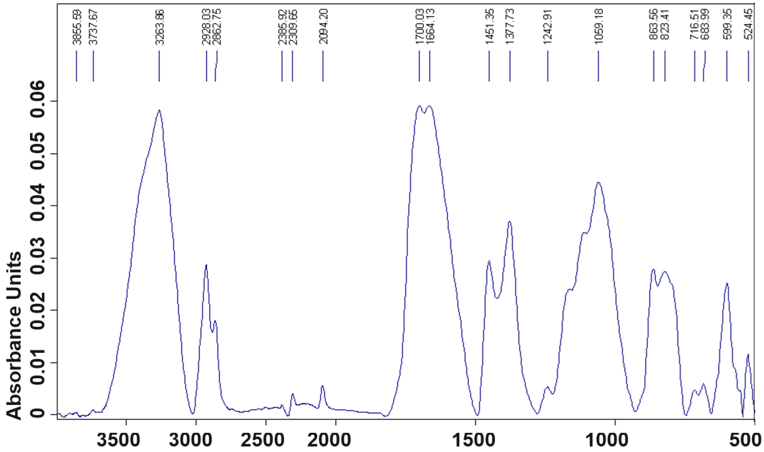
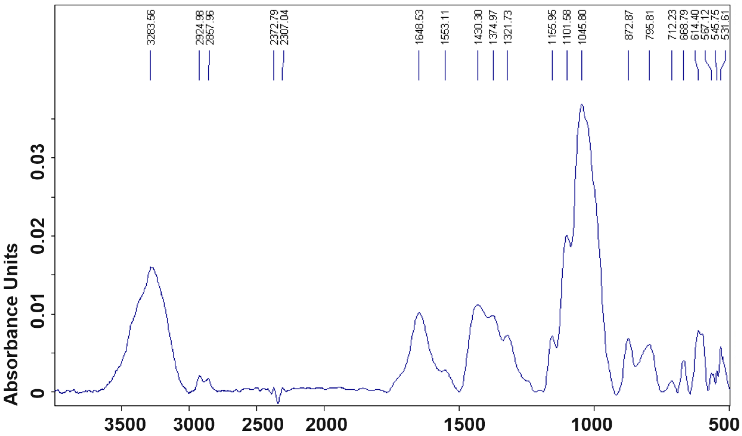
Figure 20, Figure 21, Figure 22, Figure 23, Figure 24 and Figure 25 show the ATR—FTIR spectra of the samples analysed by SEM-EDX, representing absorbance versus wavenumber (cm−1) and the specific functional group peaks with the corresponding value. Of the samples analysed by the above two techniques, only the following samples were considered: S1, S2a, S2b, S3a, S3b, and S7. Thus, samples S4 and S5, which show spectra very similar to samples S2a and S3a, which show the same pigments, and samples S8 and S9, which are on wood and are not of interest in terms of their composition in comparison with that of the pictorial materials, were not included in the study.
There are visible differences between the ATR—FTIR spectra for the six samples, except for samples S3a and S3b, which have some slightly attenuated peaks in absorbance or shifted to other wavenumbers, a higher charge, and a greater diversity of pictorial materials.
Table 2 shows the representative peaks and functional groups, by ion types and in comparison with those in the theoretical spectral bands.
The cations Pb(II, III, IV), Ba(II), Fe(II, III) and Zn(II) have their oxidation states written with Roman numerals, as they are found as solid, insoluble congruents in aqueous electrolyte systems (they are in the form of cations in the atomic network and not as aqueous ions).
IR vibration spectra of valence (ν), symmetric (νs) and antisymmetric (νas), and deformation spectra, in-plane (δ), which can be shear (δs) and pendulum (δas), and out-of-plane (γ), respectively, which in turn can be twisting (γas) and rocking (γs), both in the range 500–4000 cm−1 are theoretically assigned by group vibrations for white lead Pb3(OH)2(CO3)2 (3266, 1732, 1408, 1187, 899, 607 cm−1), BaSO4 (1180, 1108, 1063, 981, 634 și 603 cm−1), Fe(II, III) (3453/3436, 2361, 1640/1636, 1042, 915, 523 cm−1), gypsum—CaSO4 (3541, 3406, 2925, 2235, 1625, 1447/1451, 1135, 875/863 și 667/663 cm−1), ZnO (3450, 1740, 1380, 1090/1072, and 510 cm−1).
For binder (boiled linseed oil for colour and animal glue for preparation), varnish (natural resins), and oxidatively anchored dirt (dust dispersed in tars/soot, penetrating into the varnish) the vibrations (valence and deformation) corresponding to the O-H, COOH groups and various types of water (hygroscopic, crystallization, or complexation), occur in the range 3700–3300 cm−1, those corresponding to N-H groups (amides) occur in the region 3400–3100 cm−1, and those corresponding to C-O-C, C=C and C-H groups (from resins and tars) occur in the region 3200–2850; 1620–1450; 1104 and 670–590 cm−1.
The oil colours of lead white, ivory black, lead yellow, yellow ochre, red ochre, brown ochre, natural sienna, burnt sienna, ultramarine, and other earth colours are those that were identified based on the data in Table 1 and Table 2.
Rembrandt’s school had easy access to lead white, ochre, sienna, iron red, enamel, tin and lead yellow, vermilion, ultramarine, ivory black, and other pigments. Of these in the analysed painting, the following were identified: white lead, barium white, gypsum, ivory black, ochre, sienna, and ultramarine, along with other earth colours, and the presence of zinc white, which is due to later retouching, especially in recent years, which was used completely unprofessionally (completely inappropriately).
In addition to gypsum, barium sulphate (barium white), a very common composition in Flemish painting, was used in the mixture.

3. Archaeometric and Chemometric Dating Characteristics

The colours analysed, together with the protective varnish (Damar resin used initially and white rosin at the last intervention), showed a diffuse chromatic with microstructural stability provided by the oxidatively anchored dirt film, which penetrated into the original varnish (Damar resin).
Based on the composition of these elements, using the collective’s experience, the archaeometry and chemometric characteristics evolving over time have been identified and validated by previous studies [13,14].
Thus, Table 3 shows a number of the archaeometric characteristics determined from OM and SEM micro-photographs, such as: grain size of the chemical components of the pictorial layers and porosity of pigments, especially whitening pigments and fillers in the preparation, but also the typology of the craquelure (the arrangement of the networks and their morphology, the penetration depth of the oxidatively anchored dirt, its porosity, respectively. In these assessments, stratigraphic, fractal edge section areas of only those samples taken from mobile craquelure were selected. Using optical microscopy and stereomicroscopy for appropriate magnitudes, the following features were analysed microstructurally:
the way and degree of elaboration of colours in painting, when gradually (sequentially) analysing the classical system of support—preparation—drawing—colour—varnish;
summative systems of the polychrome layer (pigment + binder + varnish + anchored dirt) and of the preparation (filler and binder) with their stratigraphic arrangement, corresponding to the thin layer technique, using a small, wide brush made of pig hair and a thin-tipped one in the form of a quill made of squirrel or cat hair (the latter for the anatomical details of the face and the lacy collar);
evaluation of chemical congruences in summative systems, by involving errors in the EDX atomic composition determination, a very important tool in expert determination;
the layout and penetration of the anchored dirt areas with reference to the protective lacquer and oil colour pigments, respectively (depth ranging between 100 and 150 μm);
the arrangement and degree of stratigraphic penetration of the craquelure networks and lacunar areas, the latter resulting from loss of material during the roofing of the colour layer and preparation (craquelure networks typical of medieval paintings in The Netherlands, with depths ranging between 500 and 800 μm);
the range of variation of the thickness of the craquelure and the extension of the lines of force (craquelure aperture varying between 500 and 800 μm, craquelure depth varying between 300 and 450 μm, the length of the side of a craquelure on a force vector in the network (network mosaic side) varying from 1000 to 2500 μm);
depth of penetration of archaeometric characteristics (the degree of penetration of porosity in the volume phase of the materials analysed, varying between 1.0 and 2.5 μm);
the morphology of the pictorial surfaces, emphasizing iridescence/alveolars, texture and microtopography;
shape and arrangement of the pigment granules and preparation, respectively their particle size evaluation (between 10 and 30 μm).
Among the experimentally validated chemometric-derived archaeometric characteristics, S/C, Ca/Ba, Pb/Ba, Si/Al and Zn/Pb ratios are presented below (Table 3).
Among the archaeometric characteristics, such as porosity gradient on the penetration from the surface into the volume phase of the white colour and the penetration of oxidative dirt into the structural depth of the surfaces, were evaluated, falling in the value range of an age of ca. 360 ± 3 years. These were in agreement with the chemometric characteristics, which required experimental validation, such as elemental atomic ratios: S/C, Ca/Ba, Pb/Ba, Si/Al, and Zn/Pb, placing the painting in the period of execution around 1655–1660.
The data in Table 3 show a good correlation between the archaeometric characteristics highlighted for the samples of white (white lead), anchored dirt and preparation (porosity gradient on the surface penetration in the volume phase of the white and preparation colour varying between 1.0 and 2.5 μm, while the penetration of the surface structure of oxidatively anchored dirt at the varnish depth is between 0.5 and 1.2 μm) and the chemometric samples (from columns 2–5, Table 3).
The archaeometric characteristics correspond to an age of about 365 years (the evolution gradient of the thickness of the craquelure is about 0.0014–0.0019 μm/year, the porosity of the pigments 0.5 × 10⁻⁵–1.1 × 10⁻⁵ μm/year (considering that the painting would have been preserved in constant conditions of conservation).
The archaeometric characteristic related to the increase in porosity of the painting material and preparation (both on the front and on the back of the painting), as a result of the embrittlement of the binder by the penetration of CO2 in the presence of hygroscopic humidity (carbonation process), given by the ratios of S/C, Ca/Ba, Pb/Ba, and Zn/Pb, except the latter, also shows an evolution rate within the limit of about 365 years. The S/C value decreases about 12 times compared to the reference (elemental composition values underlined in blue and green). For Ca/Ba ratios, the values decrease by about 2 times and for the Pb/Ba ratio, the values increased by 5 times compared to the same references (white lead, barium sulphate, and gypsum samples).

4. Conclusions

From our point of view, considering all resulted data, the painting is more than 350 years old, and as an artistic technique, it belongs to the Flemish school of the second half of the 17th century.
As no signature of the painter has been identified on the whole surface of the painting, we can only make an approximate judgment on it, based on the elaboration and involvement of lights in the iconographic register and on some artistic elements specific to the School of Rembrandt Van Rijn (1606–1669). In this respect, the works of the great artist, who left his mark on the formation of many of his disciples, were used and analysed comparatively using the aesthetic–artistic characteristics (elements) close to the painting in question.
In order to date the painting, the results obtained by OM, SOM, SEM-EDX, and ATR—FTIR analyses were corroborated, which allowed us to highlight aspects related to: the nature and state of conservation (pictorial materials, preparation, textile support, chassis and frame of the painting) and to identify the archaeometric and chemometric characteristics.
Concerning the nature of the materials and their state of preservation, a new frame with plaster ornaments and polished with yellow bronze (framed about 45–50 years ago), the chassis of linden wood, the textile backing of linen cloth, the preparation based on animal glue and filler mixture of gypsum and barium sulphate, the pigments from the series—white lead, ivory black, ochre, sienna, ultramarine and other coloured earths—were all used in the mise en oeuvre, together with the protective varnish (the original varnish based on Damar resin). Among the pigments used as pictorial materials are zinc white, burnt sienna, ochre, burnt umber, ultramarine, and charcoal black, along with gypsum and barium white in the preparation. In the varnish, there are Damar and natural resins (white rosin) used 45 years ago to realize repainting and revarnishing.
For the second aspect, related to the archaeometric and chemometric characteristics used in dating, the following were taken into consideration: the grain size of the chemical components of the pictorial layers and the porosity of the pigments, especially the white pigments and fillers in the preparation, but also the typology of the craquelure (the arrangement of the networks and their morphology, the depth of penetration of the oxidatively anchored dirt, respectively).
Based on the results, we believe the picture is from Rembrandt’s school.

Author Contributions

I.S.: Conceptualization, methodology and supervision; A.D., A.V.S. and V.V.: Conceptualization, investigation, data curation and visualization; V.D., C.T.I. and I.G.S.: Conceptualization, original draft preparation, resources, formal analysis. All authors have read and agreed to the published version of the manuscript.

Funding

This research received no external funding.

Institutional Review Board Statement

Not applicable.

Informed Consent Statement

Not applicable.

Data Availability Statement

The original contributions presented in the study are included in the article, further inquiries can be directed to the corresponding authors.

Conflicts of Interest

The authors declare no conflicts of interest.

References

  1. Sandu, I.; Cotiuga, V. Cercetarea Bunurilor de Patrimoniu şi a Documentelor Falsificate; Editura AIT Laboratory: Bucuresti, Romania, 2011; ISBN 978-606-8363-09-7. [Google Scholar]
  2. Sandu, I.; Vasilache, V. Conservarea Amprentelor Formă şi a Urmelor Materiale; Editura AIT Laboratory: Bucuresti, Romania, 2011; ISBN 978-606-8363-09. [Google Scholar]
  3. Matei, G. Investigarea Criminalistică a Infracţiunilor Privind Operele de Artă şi Artefactele Arheologice; Editura Univers Juridic: Bucureşti, Romania, 2019. [Google Scholar]
  4. Baker, P. Protecting Art, Protecting Artists and Protecting Consumers Conference; Autralian Institute of Criminology: Sydney, Australia, 1999. [Google Scholar]
  5. Arnau, F. Arta Falsificatorilor–Falsificatorii Artei; Editura Meridiane: Bucureşti, Romania, 1970. [Google Scholar]
  6. Meacham, W. The Authentication of the Turin Shroud-An Issue in Archaeological Epistemology. Curr. Antropol. 1983, 24, 291–293. [Google Scholar] [CrossRef]
  7. Craig, E.A.; Bresse, R.R. Image Formation and the Shroud of Turin. J. Imaging Sci. Technol. 1994, 38, 59–67. [Google Scholar]
  8. Bowman, S.G.E.; Amber, J.C.; Lesse, M.N. Re-evaluation of British Museum radiocarbon dates issued between 1980 and 1984. Radiocarbon 1990, 32, 59–79. [Google Scholar] [CrossRef]
  9. Kilmon, J. The Shroud of Turin. Genuine artifact or manufactured relic? J. Archaeol. Inst. Am. 1997, 1, 13. [Google Scholar]
  10. Nickell, J. Inquest Day for the Shroudv of Turin; Prometheus Books: Buffalo, NY, USA, 1999. [Google Scholar]
  11. Benford, M.S.; Marino, J.G. Textile Evidence Supports Skewed Radiocarbon Date of Shroud of Turin; Worldwide Congress Sindone 2000: Orvieto, Italy, 2002. [Google Scholar]
  12. Gregory, W.K. The Dawn Man of Piltdown. Am. Mus. J. Am. Mus. Nat. Hist. 1914, 14, 189–200. [Google Scholar]
  13. Tanasa, P.O.; Sandu, I.; Vasilache, V.; Sandu, I.G.; Negru, I.C.; Sandu, A.V. Authentication of a Painting by Nicolae Grigorescu Using Modern Multi-Analytical Methods. Appl. Sci. Basel 2020, 10, 3558. [Google Scholar] [CrossRef]
  14. Sandu, I.; Tanasa, O.; Negru, I.C.; Lupascu, M.M.; Vasilache, V.; Chirila, M. Authentication of a Painting by Rene Magritte. Int. J. Conserv. Sci. 2022, 13, 1445–1462. [Google Scholar]
  15. La Nasa, J.; Doherty, B.; Rosi, F.; Braccini, C.; Broers, F.T.H.; Degano, I.; Matinero, J.M.; Miliani, C.; Modugno, F.; Sabatini, F.; et al. An integrated analytical study of crayons from the original art materials collection of the MUNCH museum in Oslo. Sci. Rep. 2021, 11, 7152. [Google Scholar] [CrossRef]
  16. Macchia, A.; Aureli, H.; Colasanti, I.A.; Rivaroli, L.; Tarquini, O.; Sabatini, M.; Dattanasio, M.; Pantoja Munoz, L.; Colapietro, M.; La Russa, M.F. In Situ Diagnostic Analysis of The Second Half of XVIII Century “Morte Di Sant’orsola” Panel Painting Coming from Chiesa Dei Santi Leonardo E Erasmo Roccagorga (Lt, Italy). Int. J. Conserv. Sci. 2012, 12, 1377–1390. [Google Scholar]
  17. Dupuis, G.; Menu, M. Quantitative characterisation of pigment mixtures used in art by fibre-optics diffuse-reflectance spectroscopy. Appl. Phys. A Mater. Sci. Process. 2006, 83, 469–474. [Google Scholar] [CrossRef]
  18. Lorusso, S.; Vandini, M.; Matteucci, C.; Tumidei, S.; Campanella, L. Anamnesistorica ed indaginediagnostica del dipinto ad olio su tavola “Madonna con bambino e santi Girolamo e Caterina da Siena” attribuibile a Domenico Beccafumi (1486–1551). Quad. Sci. 2003, 47–68. Available online: https://www.researchgate.net/publication/266796133 (accessed on 10 May 2024).
  19. Sandu, I.C.A.; Bracci, S.; Sandu, I. Instrumental analyses used in the authentification of old paintings-I. Comparison between two icons of XIXth century. Rev. Chim. 2006, 57, 796–802. [Google Scholar]
  20. Sandu, I.C.A.; Vasilache, V.; Sandu, I.; Luca, C.; Hayashi, M. Authentication of the Ancient Easel-paintings through Materials Identification from the Polychrome Layers III. Cross-section Analysis and Staining Test. Rev. Chim. 2008, 59, 855–866. [Google Scholar] [CrossRef]
  21. Sandu, I.C.A.; Bracci, S.; Sandu, I.; Lobefaro, M. Integrated Analytical Study for the Authentication of Five Russian Icons (XVI-XVII centuries). Microsc. Res. Tech. 2009, 72, 755–765. [Google Scholar] [CrossRef] [PubMed]
  22. Cristache, R.A.; Sandu, I.C.A.; Simionescu, A.E.; Vasilache, V.; Budu, A.M.; Sandu, I. Multianalytical Study of the Paint Layers Used in Authentication of Icon from XIXth Century. Rev. Chim. 2015, 66, 1034–1037. [Google Scholar]
  23. Cristache, R.A.; Sandu, I.C.A.; Budu, A.M.; Vasilache, V.; Sandu, I. Multi-analytical Study of an Ancient Icon on Wooden Panel. Rev. Chim. 2015, 66, 348–352. [Google Scholar]
  24. Slotsgaard, T.L.; Pastorelli, G.; Buti, D.; Scharff, M.; Andersen, C.K. Crack morphology and its correlation with ground materials used in paintings by Danish portrait painter Jens Juel. J. Cult. Herit. 2024, 69, 47–56. [Google Scholar] [CrossRef]
  25. Giorgiutti-Dauphiné, F.; Pauchard, L. Painting cracks: A way to investigate the pictorial matter. J. Appl. Phys. 2016, 120, 065107. [Google Scholar] [CrossRef]
  26. Andrews, I. Dutch and Flemish Old Master Paintings. Apollo-Int. Mag. Collect. 2007, 166, 104–105. [Google Scholar]
  27. Roberts, K. Dutch and Flemish Old Master Paintings and Drawings. Burlingt. Mag. 1976, 118, 664. [Google Scholar]
  28. Cornelis, B.; Museum of Fine Arts Budapest Old Masters’ Gallery. Summary catalogue, vol 2, Early Netherlandish, Dutch and Flemish paintings. Burlingt. Mag. 2001, 143, 772. [Google Scholar]
  29. Da Rugna, J.; Chareyron, G.; Pillay, R.; Joly, M. A framework for analysis of large database of old art paintings. Comput. Vis. Image Anal. Art II 2011, 7869, 786907. [Google Scholar] [CrossRef]
  30. Issa, A.A.A.K.B.; Mohie, M.A. The Conservation of an Oil Painting by Antonio Schranz, 1841 AD. Int. J. Conserv. Sci. 2021, 12, 417–428. [Google Scholar]
  31. Bucklow, S. The description of craquelure patterns. Stud. Conserv. 1997, 42, 129–140. [Google Scholar] [CrossRef]
  32. Bucklow, S. The description and classification of craquelure. Stud. Conserv. 1999, 44, 233–244. [Google Scholar] [CrossRef]
  33. Bucklow, S. Consensus in the classification of craquelure. Hamilt. Kerr Inst. Bull. 2000, 3, 61–73. [Google Scholar]
  34. Bucklow, S. The classification of craquelure patterns. In Conservation of Easel Paintings, 2nd ed.; Routledge: London, UK, 2020. [Google Scholar]
  35. Pauchard, L.; Giorgiutti-Dauphiné, F. Craquelures and pictorial matter. J. Cult. Herit. 2020, 46, 361–373. [Google Scholar] [CrossRef]
  36. Toussaint, A. Influence of pigmentation on the mechanical properties of paint films. Prog. Org. Coat. 1974, 2, 237–267. [Google Scholar] [CrossRef]
  37. Zosel, A. Mechanical behaviour of coating films. Prog. Org. Coat. 1980, 8, 47–79. [Google Scholar] [CrossRef]
  38. Mayer, R.; Smith, E. The Artist’s Handbook of Materials and Techniques, 3rd ed.; Mayer, R., Ed.; Revised and Expanded, Faber: London, UK, 1973. [Google Scholar]
  39. Karpowicz, A. A study on development of cracks on paintings. J. Am. Inst. Conserv. 1990, 29, 169–180. [Google Scholar] [CrossRef]
  40. Bratasz, Ł.; Vaziri Sereshk, M.R. Crack Saturation as a Mechanism of Acclimatization of Panel Paintings to Unstable Environments. Stud. Conserv. 2018, 63 (Suppl. 1), 22–27. [Google Scholar] [CrossRef]
  41. Bratasz, Ł.; Akoglu, K.G.; Kékicheff, P. Fracture saturation in paintings makes them less vulnerable to environmental variations in museums. Herit. Sci. 2020, 8, 11. [Google Scholar] [CrossRef]
  42. Available online: https://ro.wikipedia.org/wiki/Rembrandt (accessed on 1 January 2024).
  43. Silva, D. The Rembrandt Affair; The Library of Congress Catalog Record, G.P. Putnam’s Sons: New York, NY, USA, 2010; ISBN 9780399156588. [Google Scholar]
  44. Strauss, W.L.; van der Meulen, M. The Rembrandt Documents; New York, 1979, 150, Doc. 1638/2. Cf. S. A. C. Dudok van Heel, Peter Schatborn, Eva Ornstein-van Slooten, Dossier Rembrandt/The Rembrandt Papers (Exh. cat. Amsterdam, Museum Het Rembrandthuis) (Amsterdam, 1987). 72–73.
  45. Web Gallery of Art. Available online: http://www.wga.hu/index1.html (accessed on 1 January 2024).
  46. Available online: https://www.parool.nl/kunst-media/het-is-bijna-niet-voor-te-stellen-maar-in-vorige-eeuwen-vond-men-rembrandt-een-boerenkinkel-en-de-nachtwacht-een-mislukt-schilderij~bf2049df/?referrer=https://ro.wikipedia.org/ (accessed on 1 January 2024).
  47. Available online: https://portal.dnb.de/opac.htm?method=simpleSearch&cqlMode=true&query=nid%3D11859964X (accessed on 1 January 2024).
  48. Available online: https://www.rembrandt-van-rijn.com/quotes/ (accessed on 1 January 2024).
  49. Available online: https://www.britanica.com/biography/Rembrandt-van-rijn (accessed on 1 January 2024).
  50. Available online: https://www.rembrandtresearchproject.org/ (accessed on 14 February 2024).
  51. A Corpus of Rembrandt Paintings VI, Rembrandt’s Paintings Revisited—A Complete Survey; Springer: Dordrecht, The Netherlands, 2015; p. 748. ISBN 978-94-017-9173-1. (Hardcover).
  52. Sandu, I.C.A.; Luca, C.; Sandu, I.; Vasilache, V.; Hayashi, M. Authentication of the ancient easel paintings through materials identification from the polychrome layers-II. Analysis by means of the FT-IR spectrophotometry. Rev. De Chim. 2008, 59, 384–387. [Google Scholar] [CrossRef]
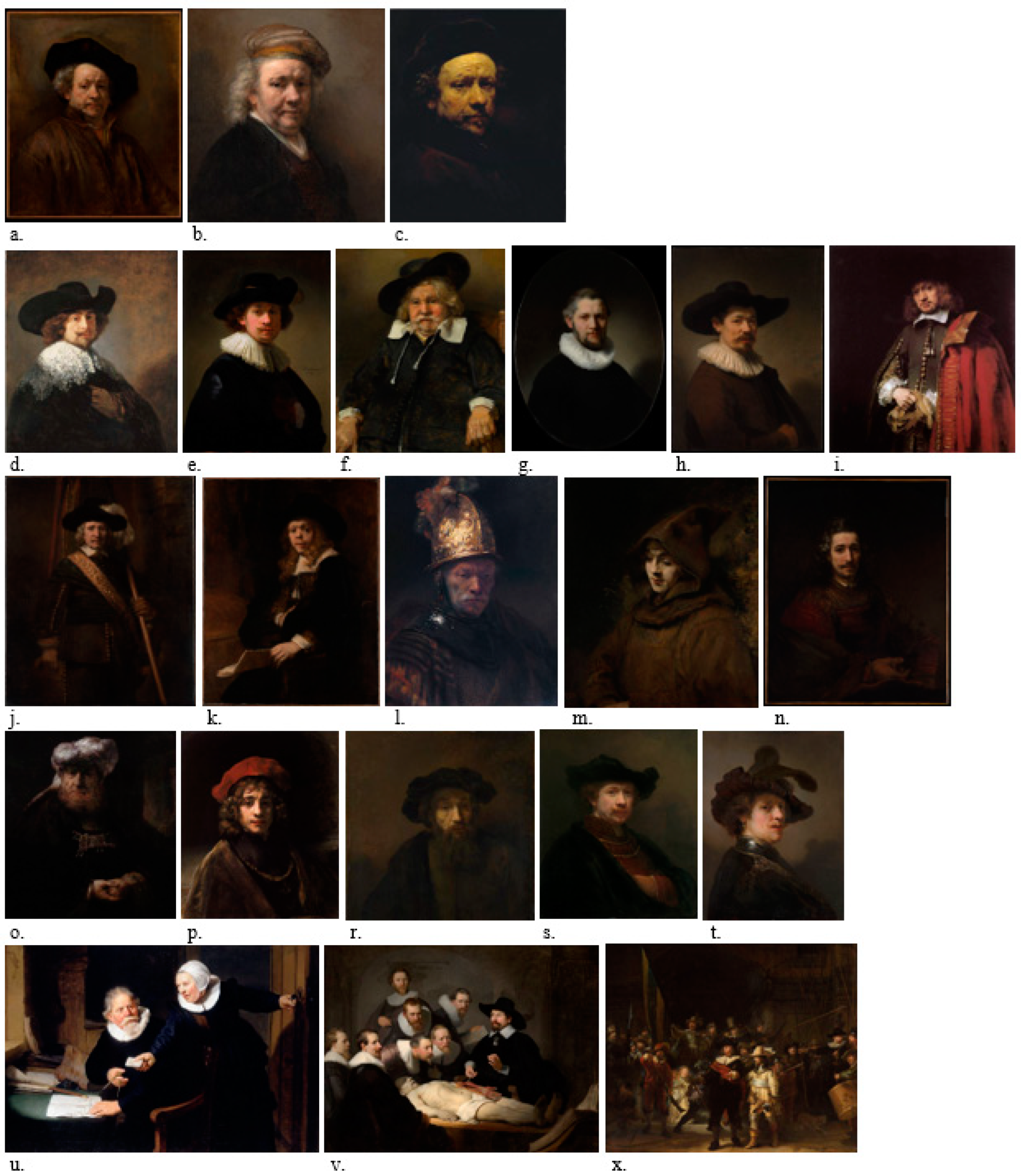
Figure 1. Portraits by Rembrandt and his School: (a) Self-portrait, 1660; (b) Self-portrait, 1669; (c) Self-portrait, aged 51, 1657; (d) Portrait of a gentleman, half-length, in a black hat and a lace falling collar, 1640; (e) Self-portrait, wearing a ruff and a black hat, 1632; (f) ‘Tronie’ of a Man with a Feathered Beret, 1635–1640; (g) Portrait of a Man, 1632; (h) Herman Doomer, 1640; (i) Portrait of Jan Six, 1654; (j) The Standard Bearer, 1654; (k) Portrait of Gerard de Lairesse, 1665–1667; (l) The Man with the Golden Helmet, 1650; (m) Titus as a Monk, 1660; (n) Man with a Magnifying Glass, 1660; (o) Head of an Oriental, 1635; (p) Portrait of Titus, 1658; (r) A Bearded Man in a Cap, 1650; (s) Self-Portrait in a Flat Cap, 1642; (t) Portrait of an Elderly Man, 1667; (u) Shipbuilder and his Wife, 1633; (v) The Anatomy Lesson of Dr. Nicolaes Tulp, 1632; (x) The Night Watch, 1642.
Applsci 14 08655 g001
Figure 2. Signature of the painter Rembrandt.
Applsci 14 08655 g002
Figure 3. Portrait of a Young Man (1669).
Applsci 14 08655 g003
Figure 4. Portrait: (a) front; (b) back.
Applsci 14 08655 g004
Figure 5. Joining the ends of the chassis platbands.
Applsci 14 08655 g005
Figure 6. The frame with ornamental framing: (a) front; (b) back (inscribed on the left side).
Applsci 14 08655 g006
Figure 7. The carpenter’s inscription on the reverse of the frame: (a) image in static reflectography (IR, UV, and vis); (b) image in dynamic reflectography. The deciphered inscription reads: ‘Antique reddish gold’, the last word being written entirely in block capitals, the cedilla of the letter ‘Ș’ is oversized.
Applsci 14 08655 g007
Figure 8. Old craquelure and erosions at the edge of the painting in the framing area.
Applsci 14 08655 g008
Figure 9. Gap in the craquelure network under the collar.
Applsci 14 08655 g009
Figure 10. Character’s face and lace collar with embroidered edge, where the repainting and varnishing interventions are legible.
Applsci 14 08655 g010
Figure 11. Loss of material at last reintegration on the chassis.
Applsci 14 08655 g011
Figure 12. Analysis in the four reflected lights: Blue-White light (470 nm), UVA (365 nm), UVC (254 nm), and IR (980 nm), which offers high readability of crack networks in a dynamic (variable) system: (a) IR, under four angles of reflection for the same area; (b) UVA, UVC and IR under two angles of reflection for the same area; (c) UVC and Blue-White light for three area; (d) UVA, UVC, IR and Blue-White light.
Applsci 14 08655 g012
Figure 13. Two areas with cracks specific to Flemish paintings analyzed in four reflected lights: Blue-White light (470 nm), UVA (365 nm), UVC (254 nm), and IR (980 nm), which offers high readability of crack networks in a dynamic (variable) system: (a) UVA, IR under two angles of reflection for the same area and UVC; (b) UVC, IR under two angles of reflection for the same area and Blue-White light.
Applsci 14 08655 g013
Figure 14. Analysis of the readability of lace restoration interventions by UV, Vis and IR reflectography: (a) analysis in UVA, IR under three different angles of reflection and Blue-White light in an area of the unrestored lace; (b) two lacunar areas (lack of pictorial material) analyzed in UVA, UVC and IR under reflection angles that offered maximum legibility; (c) an area from the collar with overlapping repainting, analyzed in UVA, UVC, IR and Blue-White light under a reflection angle that offered maximum legibility; (d) an extended area from the collar with repainting, analyzed in UVA, UVC, IR and Blue-White light under a reflection angle that offered maximum readability; (e) an area with age cracks near the collar, analyzed in UVA, IR under reflection angles that offered maximum legibility and Blue-White light.
Applsci 14 08655 g014
Figure 15. Areas with extensive craquelure network and craquelure in the roof, scaling, and detachments of the pictorial layer; in the last-but-one image, a fragment of the lacework can be seen, all highlighted in visible light and the last in UV.
Applsci 14 08655 g015
Figure 16. The legibility of revarnishing interventions at the level of the face.
Applsci 14 08655 g016
Figure 17. OM images of the samples taken from the pictorial material and support ((a)—front and (b)—back): (S1)—white pictorial material; (S2)—pictorial material taken from the edge of the painting; (S3)—black pictorial material with preparation layer, taken from the edge; (S4)—black pictorial material on the edge; (S5)—textile taken from the back of the painting; (S6)—wood with chassis edge preparation in two zones, zone I and zone II.
Applsci 14 08655 g017
Figure 18. SEM micrographs of the analysed samples (BSE image on Vega 2 Tescan at 30KV): (S1)—white pictorial material; (S2)—black pictorial material taken from the edge; (S3)—black pictorial material taken from the surface of the painting; (S4)—pictorial material with the preparation layer on the front of the sample (from the gap area); (S5)—pictorial material with preparatory layer, taken from the back; (S6)—pictorial material with preparation layer on the back of the sample; (S7)—textile material, taken from the edge; (S8)—wood material taken from the chassis with the patina of time; (S9)—wood taken from the chassis with traces of preparation.
Applsci 14 08655 g018
Figure 19. EDX spectra and elemental composition for: (a)—zone S1 from the sample of white pictorial material; (b)—zone S2a from the sample of black pictorial material, taken from the edge; (c)—zone S2b from the sample of black pictorial material, taken from the edge; (d)—S3a zone from the sample of black pictorial material taken from the painting; (e)—S3b zone from the sample of black pictorial material taken from the painting; (f)—S6a of the paint sample with the preparation layer on the face of the sample, taken from the edge of the painting (g)—S5 zone from the sample of the paint material with preparation layer on the back of the sample taken from the painting; (h)—S4 from the sample of the paint material with preparation layer on the face of the sample taken from the painting (i)—S6b of the sample of pictorial material with preparation layer on the back of the sample, taken from the edge of the painting; (j)—S7 zone from the textile sample, taken from the edge; (k)—S8 zone from wood sample taken from the chassis, zone I; (l)—S9 zone from wood sample taken from the chassis, zone II.
Applsci 14 08655 g019aApplsci 14 08655 g019b
Figure 20. FTIR spectrum of sample S1.
Applsci 14 08655 g020
Figure 21. FTIR spectrum of sample S2a.
Applsci 14 08655 g021
Figure 22. FTIR spectrum of sample S2b.
Applsci 14 08655 g022
Figure 23. FTIR spectrum of sample S3a.
Applsci 14 08655 g023
Figure 24. FTIR spectrum of sample S3b.
Applsci 14 08655 g024
Figure 25. FTIR spectrum of sample S7.
Applsci 14 08655 g025
Table 1. EDX elemental composition.
SampleChemical ElementX-ray SpectraWt (%)Normal
Wt (%)
Normal
At (%)
Error
(%)
S1
(area with white pigment)
LeadL-series37.24933.0653.4830.968
ZincK-series0.8550.7590.2530.055
AluminumK-series0.5590.4970.4020.063
CarbonK-series17.72315.73228.5882.594
SulphurK-series1.4231.2640.8600.163
OxygenK-series54.84548.68466.4148.858
Total112.654100100
S2a
(zone to stratigraphic structure)
BariumSeries—K1.0631.2490.4490.082
CarbonSeries—K15.51718.23126.1232.326
CalciumSeries—K3.6394.2751.8360.164
SulphurSeries—K2.5913.0441.6340.150
PhosphorusSeries—K1.6871.9821.1010.123
SiliconeSeries—K2.3272.7341.6750.163
AluminiumSeries—K2.6563.1201.9900.200
SodiumSeries—K6.7907.9785.9720.580
PotassiumSeries—K0.7240.8510.3750.068
MagnesiumSeries—K2.7823.2692.3150.241
IronSeries—K0.8531.0020.3090.084
OxygenSeries—K44.48652.26556.22146.634
Total85.115100100
S2b
(area towards the centre of the sample)
LeadSeries—K7.6407.6400.6540.269
BariumSeries—K4.0054.0050.5170.151
CarbonSeries—K14.57814.57821.5366.108
CalciumSeries—K3.2973.2971.4600.132
SulphurSeries—K1.5491.5490.8570.129
SiliconSeries—K0.7600.7600.4800.069
AluminiumSeries—K0.5570.5570.3660.064
PotassiumSeries—K0.2440.2440.1110.039
MagnesiumSeries—K1.8361.8361.3400.150
OxygenSeries—K65.53265.55473.65922.166
Total99.978100100
S3a
(preparation area)
LeadSeries—K8.2698.2690.6880.357
ZincSeries—K0.6760.6760.1780.067
BariumSeries—K1.6381.6380.2060.101
CarbonSeries—K21.15921.15930.3518.781
CalciumSeries—K1.3151.3150.5650.084
SulphurSeries—K0.9970.9970.5360.133
AluminiumSeries—K1.7351.7351.1080.138
SiliconSeries—K2.1362.1361.3100.142
SodiumSeries—K0.7090.7090.5310.589
PotassiumSeries—K0.8790.8790.3870.068
MagnesiumSeries—K1.1621.1620.8240.124
IronSeries—K0.7400.7400.2280.067
OxygenSeries—K58.58058.58563.08820.765
Total99.995100100
S3b
(area towards the centre of the sample)
BariumSeries—K1.0901.0900.3730.105
CarbonSeries—K20.41320.41427.8339.066
CalciumSeries—K2.4042.4040.9820.149
SulphurSeries—K1.7301.7300.8840.135
PhosphorusSeries—K0.7180.7180.3790.092
SiliconSeries—K1.6861.6860.9830.155
AluminiumSeries—K2.1742.1741.3190.205
SodiumSeries—K7.7827.7825.5440.734
PotassiumSeries—K0.4330.4330.1810.066
MagnesiumSeries—K2.3442.3441.5800.253
IronSeries—K0.9290.9290.2720.116
OxygenSeries—K58.29558.29659.67026.320
Total99.998100100
S4
(area on the front of the sample, with pictorial material, preparation and anchored dirt)
BariumSeries—K3.9284.3280.5830.179
ZincSeries—K1.8862.0780.5880.137
CarbonSeries—K16.11717.75827.3373.029
CalciumSeries—K1.9302.1260.9810.109
SulphurSeries—K2.3592.5991.4990.139
PhosphorusSeries—K1.6651.8341.0950.120
SiliconSeries—K4.0704.4842.9520.242
AluminiumSeries—K3.8674.2602.9200.261
ChlorineSeries—K2.4692.7201.4190.137
PotassiumSeries—K1.5481.7050.8060.096
SodiumSeries—K5.5216.0824.8924.400
MagnesiumSeries—K2.6102.8762.1880.228
IronSeries—K1.9292.1250.7040.113
OxygenSeries—K40.86545.02552.0365.952
Total90.764100100
S5
(area on the reverse side of the sample, with preparation and traces of pictorial material)
LeadSeries—K15.43213.8701.2720.533
BariumSeries—K4.3503.9100.5410.185
CarbonSeries—K16.00714.38622.7522.834
CalciumSeries—K4.8034.3162.0460.193
SulphurSeries—K1.4201.2760.7560.172
AluminiumSeries—K0.9370.8420.5930.097
SiliconSeries—K0.6260.5630.3800.071
MagnesiumSeries—K1.5621.4041.0970.154
OxygenSeries—K66.12859.43370.56312.233
Total111.265100100
S6a
(area on the front of the sample, with pictorial material, preparation and anchored dirt)
LeadSeries—K6.1976.1980.5020.266
ZincSeries—K0.5710.5710.1460.059
BariuSeries—K1.5731.5730.1920.090
CarbonSeries—K21.65721.65830.2408.125
CalciumSeries—K2.9962.9961.2540.128
SulphurSeries—K0.6330.6330.3310.083
PhosphorusSeries—K0.5180.5180.2800.057
SiliconSeries—K1.5651.5650.9350.109
AluminiumSeries—K0.9250.9250.5750.087
PotassiumSeries—K0.3420.3420.1470.044
MagnesiumSeries—K0.5380.5380.3710.074
IronSeries—K0.6240.6240.1870.057
OxygenSeries—K61.85961.85964.84021.162
Total99.998100100
S6b
(area on the back of the sample, with preparation and traces of pictorial material)
LeadSeries—K14.59313.5531.2770.512
BariuSeries—K6.5266.0610.8610.244
CarbonSeries—K15.13814.05822.8442.732
CalciumSeries—K5.5615.1642.5150.214
SulphurSeries—K1.6811.5610.9500.175
AluminiumSeries—K0.2840.2640.1910.056
SiliconSeries—K0.5320.4940.3430.066
MagnesiumSeries—K1.9731.8321.4710.179
OxygenSeries—K61.39057.01369.54811.828
Total107.678100100
S7
(textile fabric area)
LeadSeries—K5.8065.8060.4570.249
BariumSeries—K1.1041.1040.1310.078
CarbonSeries—K17.72317.73024.0787.087
CalciumSeries—K1.4791.4790.6020.085
SulphurSeries—K0.4590.4590.2330.086
PhosphorusSeries—K0.0350.0350.0180.031
AluminiumSeries—K0.1780.1780.1080.046
SiliconSeries—K0.1680.1680.0970.042
PotassiumSeries—K0.1090.1090.0460.036
MagnesiumSeries—K0.3540.3540.2380.065
OxygenSeries—K72.57672.57873.99224.475
Total99.991100100
S8
(zone I, on the chassis with wood patina)
CarbonSeries—K20.73220.73326.7577.341
CalciumSeries—K1.0941.0940.4230.079
SulphurSeries—K0.5130.5130.2480.061
PhosphorusSeries—K0.3730.3730.1860.057
SiliconSeries—K0.8610.8610.4750.087
AluminiumSeries—K1.2951.2950.7440.122
SodiumSeries—K3.5403.5402.3870.348
MagnesiumSeries—K1.7591.7591.1220.172
OxygenSeries—K69.83169.83267.65823.360
Total99.998100100
S9
(zone II, on the wooden chassis, with traces of preparation)
BariumSeries—K8.6977.7381.1610.310
ZincSeries—K7.7416.8882.1710.271
CarbonSeries—K6.9636.19610.6321.373
CalciumSeries—K17.80015.8388.1450.576
SulphurSeries—K4.8214.2902.7570.233
SiliconSeries—K0.5620.5000.3670.074
AluminiumSeries—K0.7760.6900.5270.096
SodiumSeries—K0.1660.1470.1320.156
MagnesiumSeries—K0.6070.5400.4580.099
OxygenSeries—K64.25557.17373.65011.046
Total112.388100100
Table 2. Representative wave numbers of functional groups.
Type IonTheoretical Spectral Bands (cm−1)Peak Present in
Samples (cm)−1
Samples Analysed
Silicate860–1175942.93; 1072.46S1
1082.67; 1143.32S2a
910.30; 1028.13; 1069.84S2b
1059.18S3a
1056.16S3b
872.87; 1101.58; 1045.80; 1155.95S7
Aluminate790–920869.37S1
849.62S2a
803.75; 869.16S2b
863.56S3a
809.15; 870.12S3b
795.81; 872.87S7
Sulphate and sulphide570–680; 960–1030607.83S1
622.93S2a
606.02S2b
599.35S3a
605.27S3b
1030.12S7
Carbonate670–745; 800–890; 1040–1100; 1320–1530; 2235–2925; 3405–3540711.43, 1408.64S1
738,66; 1428.92; 2306S2a
710.56; 1404.30S2b
716,51; 1377.73; 1451.35; 2385S3a
1378.63; 1451.98; S3b
668.79; 872.87; 2924.98; 3283.56S7
Pb (II, III, IV)607–899; 1187–1732; 3000–3266607.83: 869.37; 1072.46; 1187.10; 1408.64; 1732.66; 3266.03S1
599.35; 622.93; 794.07; 1069.18; 1428.92; 1451.35; 1700.03; 3263.86S2a and S3a
606.02; 803.75; 1069.84; 1404.38; 1626.33; 3266.38S2b and S3b
872.87; 1155.95; 1648,53; 3283.56;S7
Ba(II)603–634; 971–1063; 1108–1180634,36, 981, 1063, 1108, 1180S2b and S3b
614.40; 1101.58 S7
Fe(II, III)523; 915; 1042; 1626; 2376; 2436523, 915, 1042, 1626, 2376, 2436S2a and S3a
Zn(II)524, 1072, 1377, 1740, 3459524, 1072, 1377, 1740, 3459S1 (traces, weak/low peaks)
Acvo and hydroxocomplexes (coordination water) and crystallized water 2850–40002855.02; 2921.57; 3266.03S1
2920.84; 3265.33; 3737.57; 3855.63S2a
2857.57; 2922.49; 3266.38S2b
2862.75; 2928.033263.86S3a
2858.31; 2925.58S3b
2857.96; 2924.98; 3283.56S7
Binder (egg protein, via amide I and amide II)~1547;
1621–1690
1659.72S1
1515.34; 1650.95 S2a
1626.13S2b
1664.13S3a
1652.57S3b
1553.11; 1648.53S7
Varnish (through ester groups)~1265;
~1750
1232.82; 1732.66S1
1794.39S2a
1700.03; 1242.91S3a
1245.26S3b
Anchored dirt (carbonyl, carboxyl, ether, ethylenic and C-H groups)590–670; 1104;
1450–1620;
2930–3335;
3400–3512
605.27; 683.99; 1066.16; 1451.98; 1662.57; 2928.03; 3263.86
605.27; 685.29; 1056.16; 1451.96; 1652.57 2925.58; 3269.29
596; 667; 1104; 1458; 1619; 2934; 3237; 3335; 3398; 2512
S2a
S3a
S7
Table 3. Chemometric characteristics (C.C.) of the areas analysed.
SampleChemometric Characteristics
S/C *Ca/Ba *Pb/Ba *Si/Al **Zn/Pb **
S1
(area with white pigment)
0.080---0.023
S2a
(area to stratigraphic structure)
0.1673.423-0.876-
S2b
(area towards the centre of the sample)
0.1060.8231.9081.365-
S3a
(preparation area)
0.0470.8035.0481.2310.082
S3b
(area towards the centre of the sample)
0.0852.206-0.776-
S4
(area on the front of the sample, with pictorial material, preparation and anchored dirt)
0.1460.491-1.053-
S5
(area on the back of the sample, with preparation and traces of pictorial material)
0.0891.1043.5470.669-
S6a
(area on the front of the sample, with pictorial material, preparation and anchored dirt)
0.0291.9053.9401.6920.092
S6b
(area on the back of the sample, with preparation and traces of pictorial material)
0.1110.8522.2361.871-
S7
(textile fabric area)
0.0261.3405.2590.944-
S8
(zone I, on the chassis with patinated wood)
0.025--0.665-
S9
(zone II, on the wooden chassis, with traces of preparation)
0.6922.047-0.725-
* Valid chemometric characteristics; ** Partially valid characteristics.
Disclaimer/Publisher’s Note: The statements, opinions and data contained in all publications are solely those of the individual author(s) and contributor(s) and not of MDPI and/or the editor(s). MDPI and/or the editor(s) disclaim responsibility for any injury to people or property resulting from any ideas, methods, instructions or products referred to in the content.