Article

Green Method of Doping Photochromic TiO2

G. A. Krestov Institute of Solution Chemistry RAS, Academicheskaja Str., 1, Ivanovo 153045, Russia
*
Author to whom correspondence should be addressed.
Appl. Sci. 2024, 14(19), 8877; https://doi.org/10.3390/app14198877
Submission received: 3 September 2024 / Revised: 27 September 2024 / Accepted: 30 September 2024 / Published: 2 October 2024

Abstract

:
The doping process is a unique method of changing the microstructure of a material, influencing its mechanical, thermal, and optical properties. Typically, the doping process is carried out via chemical reagents. In this work, we present a “green” method of doping photochromic TiO2 via low-temperature plasma. The doping agent was the electrode material that was sputtered during plasma burning. The process of electrode sputtering is confirmed by the emission spectroscopy data of the plasma zone and the mass loss of the electrodes. The doping process was confirmed by X-ray, Raman spectroscopy, and XPS analyses. The role of the dopant nature and the action of diaphragm discharge in improving the photochromic properties of titanium dioxide is considered.

1. Introduction

Titanium dioxide is widely known as a photocatalyst, although TiO2 has a wide band gap (3.0–3.2 eV) [1]. The doping method is used to increase the photocatalytic properties under visible light irradiation. Both metals and nonmetals are used as doping agents [2]. In addition to its photocatalytic properties, titanium dioxide has photochromic properties [3]. The main problem with using TiO2 as a photochromic material lies in the low rate of coloring–bleaching processes under light irradiation. The structure of the resulting titanium dioxide may play a key role in this process. For example, the best photochromic characteristics appear in the amorphous structures of tungsten oxide [4]. Moreover, in experiments with TiO2, the amorphous structure has a negligibly low photochromic response [5]. Since TiO2 has a rather low photoresponse, its photochromic properties need to be improved by creating binary structures, doping, and making composites based on polymers and oxide structures. Currently, Ag, Cu, Mo, Nb, V, and W, as ions and oxides, are being intensively studied as doping agents that increase the photochromic characteristics of titanium dioxide. Silver nanoparticles are chosen as dopants because of their pronounced localized surface plasmon resonance in the visible region of the spectrum [6,7]. Copper ions are used because color changes are easily controlled by the reduction process (Cu2+→Cu+) [8]. The choice of transition metal ions (Mo, Nb, V, W) is determined by the fact that the oxide structures of these materials themselves have photochromic properties [4,9,10,11]. In a series of works by Songara et al., the influence of transition metal ions (W6+, Mo6+, and V5+) on the photochromic properties of TiO2 (anatase) was studied [12,13,14]. The bleaching time decreased in the V > Mo > W series. The authors explained this effect in terms of the charge/radius of the doped ion, charge-separation efficiency, and surface acidity. The process of discoloration (oxidation) is explained by the slow release of trapped electrons. Eglitis et al. explained the slow release of the electron by the fact that the electrons are delocalized and stabilized by the extra positive charge of Nb5+ [15].
The photochromic properties of titanium dioxide in the anatase phase or anatase/rutile mixture, which are the most stable and easily synthesized phases, have been studied. For a long time, TiO2 brookite was believed to be an unstable phase. Owing to the unique crystal lattice structure of brookite and its electronic properties, it is an ideal material for photonics, as well as for electrophotochemical applications [16,17]. Recent studies have shown that brookite can be obtained via hydrothermal methods [18].
Among the physical and chemical methods for producing TiO2, one can distinguish the sol-gel method, which allows one to obtain an oxide of a given structure (anatase, rutile, brookite, or mixed phase), particle size, and textural properties [19]. As a doping method, the use of low-temperature plasma is the most promising [20,21]. In this case, the electrode material acts as a source of ions or oxide structures.
This paper presents the results of the application of low-temperature alternating-current plasma in contact with liquid as a method for doping photochromic titanium dioxide. The electric discharge excited by an alternating current source allows for the avoidance of additional designs of the rectification circuit, which simplifies its industrial applications. In this report, the doping process was carried out via underwater AC diaphragm discharge plasma. The novelty of this work lies in the plasma treatment of photochromic sol TiO2 (pure brookite). In addition, a comparison of the effects of the doping element on the photochromic characteristics of the sols was carried out, and possible reasons for the improved photochromism were proposed.

2. Materials and Methods

2.1. Titania Sample Synthesis

TiO2 was prepared via sol-gel technology by dissolving 4.5 g of polyvinylpyrrolidone (PVP, Sigma Aldrich, St. Louis MO, USA) in 100 mL of distilled water heated to 80 °C. Next, 6 g of citric acid was added (Lenreaktive, St. Peterburg, Russia) and stirred until completely dissolved. After 2.5 mL of titanium isopropoxide (Sigma Aldrich, USA) was added, heating was stopped. The synthesis proceeded for 48 h. The complete dissolution of the white flakes and clearing of the solution indicated the end of the process.
The scheme of setup for generation of underwater AC diaphragm discharge is shown in Figure 1. Two metal electrodes were immersed in a volume of liquid. One of them was placed in a quartz ampoule with a small hole (diaphragm). The diaphragm diameter was 2 mm. Molybdenum, niobium, or tungsten wires (Shenzhen Tangda Technology Co., Ltd., Shenzhen, China) with a diameter of 0.8 mm were used as metal electrodes. The power supply for the excitation of the discharge consisted of a laboratory autotransformer and a high-voltage step-up transformer. The power supply made it possible to ignite the discharge at voltages up to 10 kV. The alternating-discharge current was 50 mA. All experiments were carried out in a glass cell with a fixed volume of 50 mL. The diaphragm discharge was initiated and propagated at the hole of the quartz ampoule in the bubble. The treatment time was 90 s. A more detailed method of plasma treatment of sol is discussed elsewhere [21]. The cell was equipped with an optical quartz window for recording the radiation spectra of the diaphragm discharge. The discharge-emission spectra (λ = 200–950 nm) were recorded using an AvaSpec ULS-3648 spectrometer (Avantes, Apeldoorn, The Netherlands) at a resolution of 0.3 nm. The ampoule with the diaphragm was placed directly opposite the optical window at a distance of 10 mm. A fiber-optic cable was brought close to the optical window to record the emission spectra.

2.2. Characterization
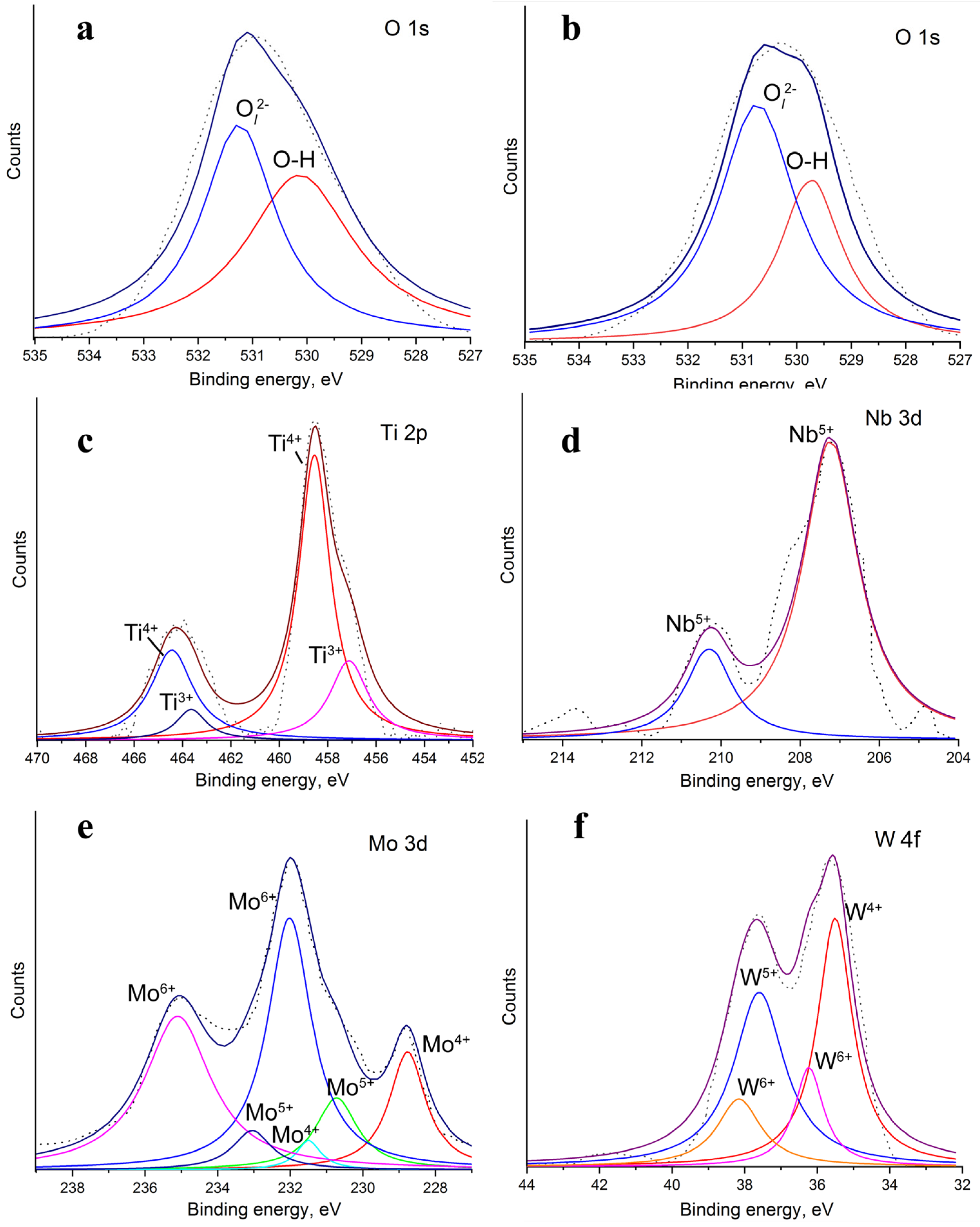
The mass of the electrodes was controlled before and after discharge via analytical balances with an accuracy of ±10−4 g (HR-150AZ, A&D Company Ltd., Tokyo, Japan).
The TiO2 phase formed during sol synthesis and after plasma treatment was determined via Raman spectroscopy (Confotec NR500 microscope (Minsk, Belarus); excitation laser with λ = 532 nm) in the range of 100–1000 cm−1. X-ray diffraction experiments were carried out on a diffractometer with a Mo-Kα source (λ = 0.07107 nm) (Bruker D8 Advance, Bruker Corporation, Billerica, MA, USA) with an attachment for registering liquid samples in the range of 2Θ = 4–40° with a step of 0.02°. The zirconium β filter was used for monochromatization of radiation. A VÅNTEC-1 position-sensitive detector (Bruker) was used to register the intensity of scattering from the samples.
The surface composition of the samples was studied via X-ray photoelectron spectroscopy (XPS) on a KRATOS AXIS ULTRA DLD photoelectron spectrometer (Kratos Analytical Ltd., Manchester, UK). The surface analysis depth was up to 3 nm, and the radiation source was an Mg monochromator (Kα = 1253.6 eV). To minimize the charge processes of the surface of the sample, the spectra of the samples were recorded using a neutralizer. The analysis area for each sample was 300 × 700 μm. The spectra were calibrated using the C 1 s 284.7 eV line. The sols were placed on a silicon substrate in a thin layer and dried to a glassy state for 24 h. Then, it is put in an input chamber for degassing for 48 h.
Sols without and after treatment by diaphragm discharge were irradiated with a UV lamp in quartz cuvettes at a distance of 10 cm from the light source with λ = 366 nm (15 W UV lamp, NDTRADE LLC, St. Peterburg, Russia). The samples were irradiated for 5, 10, or 15 min. The reversibility of the photochromic processes was studied in the dark at room temperature. The phenomenon of photocoloration and discoloration was recorded spectrophotometrically in the visible and near-IR ranges. Absorption spectra in the visible region (400–900 nm) were recorded via an SF 56 spectrophotometer, and those in the near-infrared region (900–1500 nm) were measured via an SF 256 BIK spectrophotometer (Lomo Photonika, St. Peterburg, Russia).
The intrinsic viscosity of the sols after plasma treatment was determined via an Ubbelohde dilution viscometer (Fisher Scientific, Goteborg, Sweden) fitted with a 0.56 mm capillary in a thermostat at 24 °C.
The measurements of hydrogen peroxide concentration in liquid phase at different electrode materials were carried out during the discharge action. The accumulation of H2O2 was recorded spectrophotometrically using titanium oxysulfate (TiOSO4) via the characteristic coloration with an absorption maximum at 407 nm [22].

3. Results

3.1. Characteristics of Diaphragm Discharge

Measurements of the mass of the electrodes before and after plasma treatment revealed that during the combustion of the discharge, sputtering of the electrodes occurred. The changes in the mass of the electrodes were 1.5 mg, 1.2 mg, and 0.5 mg for the Mo, Nb, and W electrodes, respectively. These values were 3.75, 3, and 1.3 wt%, respectively.
The emission spectra of a diaphragm discharge are shown in Figure 2. The spectra contain lines and bands related to the liquid phase: the OH band (280–310 nm), Hα (656 nm), O (777 nm), and titanium lines. The registered continuum is most likely associated with thermal radiation of large polymer fragments (PVPs), and it overlaps with the emission lines of the carbon atoms. The spectra also contain lines of electrode materials [23]. This also confirms the process of sputtering electrodes during burning discharge.

3.2. Sol Characteristics

Information on the phase composition of TiO2 was obtained via XRD and Raman spectroscopy (Figure 3). Figure 3a shows the diffraction patterns of the samples before and after plasma treatment. All the peaks belong to the brookite phase (JCPDS 29-1360). There are no peaks related to other phases or impurities. A change in the ratio of the main brookite peaks intensity is noted at 12.4° and 24.1°. This means the formation of lattice structure defects due to the incorporation of metal ions (doping). Therefore, plasma treatment leads to the doping of titanium dioxide.
The Raman bands in the region of 100–850 cm−1 (172, 259, 326, 396, 578, and 850 cm−1) belong to the titanium dioxide phase brookite (Figure 3b) [24]. The bands at 899 and 935 cm−1 are attributed to the PVP [25]. Plasma treatment does not result in the appearance of new bands but does cause a Raman shift in the main bands. This indicates the incorporation of metal ions into the TiO2 structure and the absence of oxide phases on its surface [14].
The chemical composition and valance states of the elements present in the undoped and doped TiO2 samples were analyzed via XPS. Figure 4a,b show the high-resolution XPS spectra of O 1s for undoped and Mo-doped TiO2 (as an example) samples. The high-resolution spectrum of O 1s for undoped TiO2 (Figure 4a) shows two components: the major peak at a binding energy 531.4 eV, which is assigned to surface hydroxyl group (O-H). The peak at 530.2 eV is attributed to the O2− 1s lattice ion. In the doped TiO2 (Mo-doped TiO2 as example), these peaks are at 530.8 eV and 529.6 eV (Figure 4b). This indicates the formation of a new charge transfer bridge during the doping process (Me-O-Ti). Figure 4c shows the XPS spectra of Ti 2p for the doped samples. The peak positions for Ti 2p1/2 and Ti 2p3/2 are 464.51 and 458.58, respectively, which correspond to Ti4+ (Ti(OOH)) [26,27]. In addition, there are peaks related to Ti3+ (most likely in the form of Ti2O3) with binding energies of 463.21 and 457.28 eV. The XPS spectra for Nb 3d for the Nb/TiO2 sample are shown in Figure 4d. Since the concentration of Nb is low, the peaks are unresolved. The major peak position is 207.31 eV, and the minor peak position is 210.2 eV. This peak is assigned to Nb 3d5/2 and 3d3/2 of Nb5+. Figure 4e presents the XPS spectra of Mo for the Mo-doped TiO2 sample. The spectra display Mo6+ 3d5/2 and 3d3/2, Mo5+ 3d5/2 and 3d3/2, and Mo4+ 3d5/2 and 3d3/2 peaks. This confirms the formation of nonstoichiometric molybdenum oxide (Mo4O11) during the plasma-doping process [28]. The W 4f XPS spectra of the W/TiO2 sample are shown in Figure 4f. The spectrum shows a resolved doublet owing to the W 4f7/2 and 4f5/2 components. Deconvolution of the peaks revealed that, in addition to the W6+ state, tungsten is also present as W5+ and W4+. This also confirms the formation of nonstoichiometric tungsten oxide (W18O49) during plasma treatment [29].

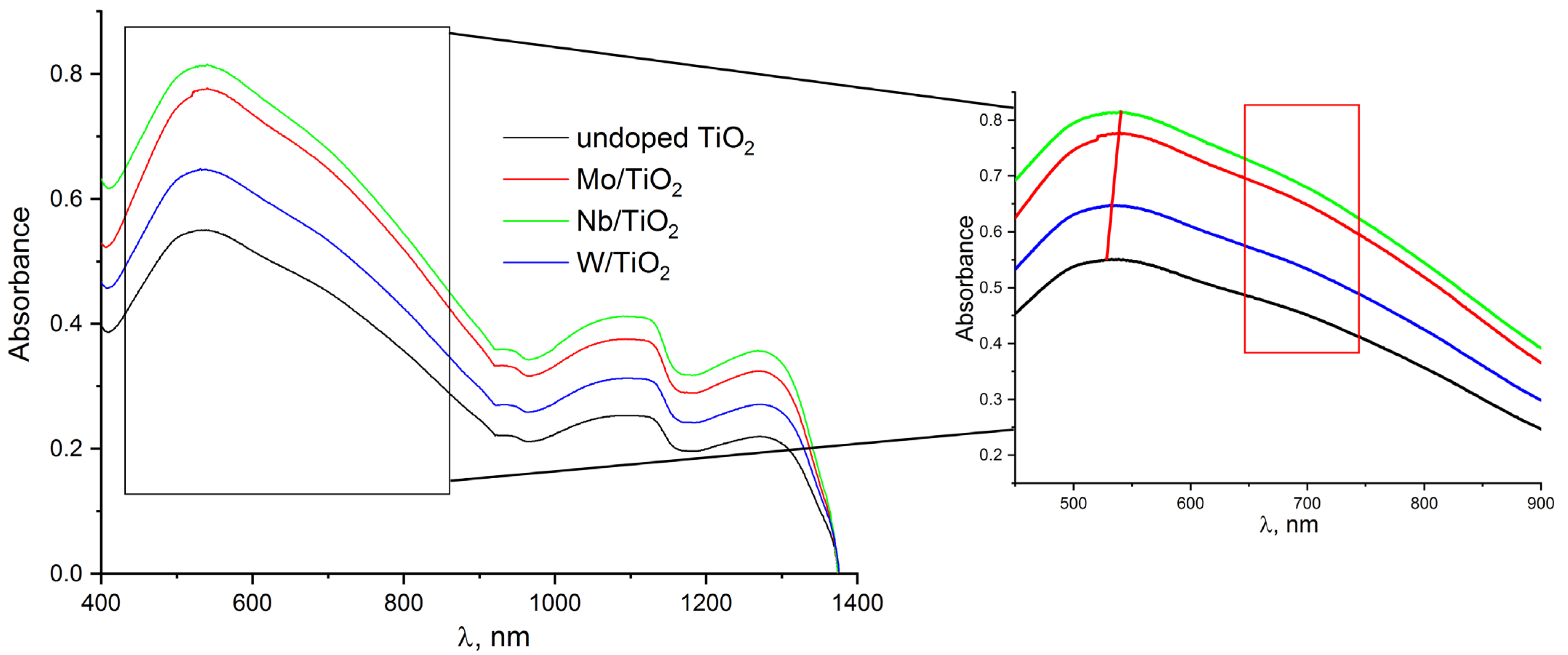
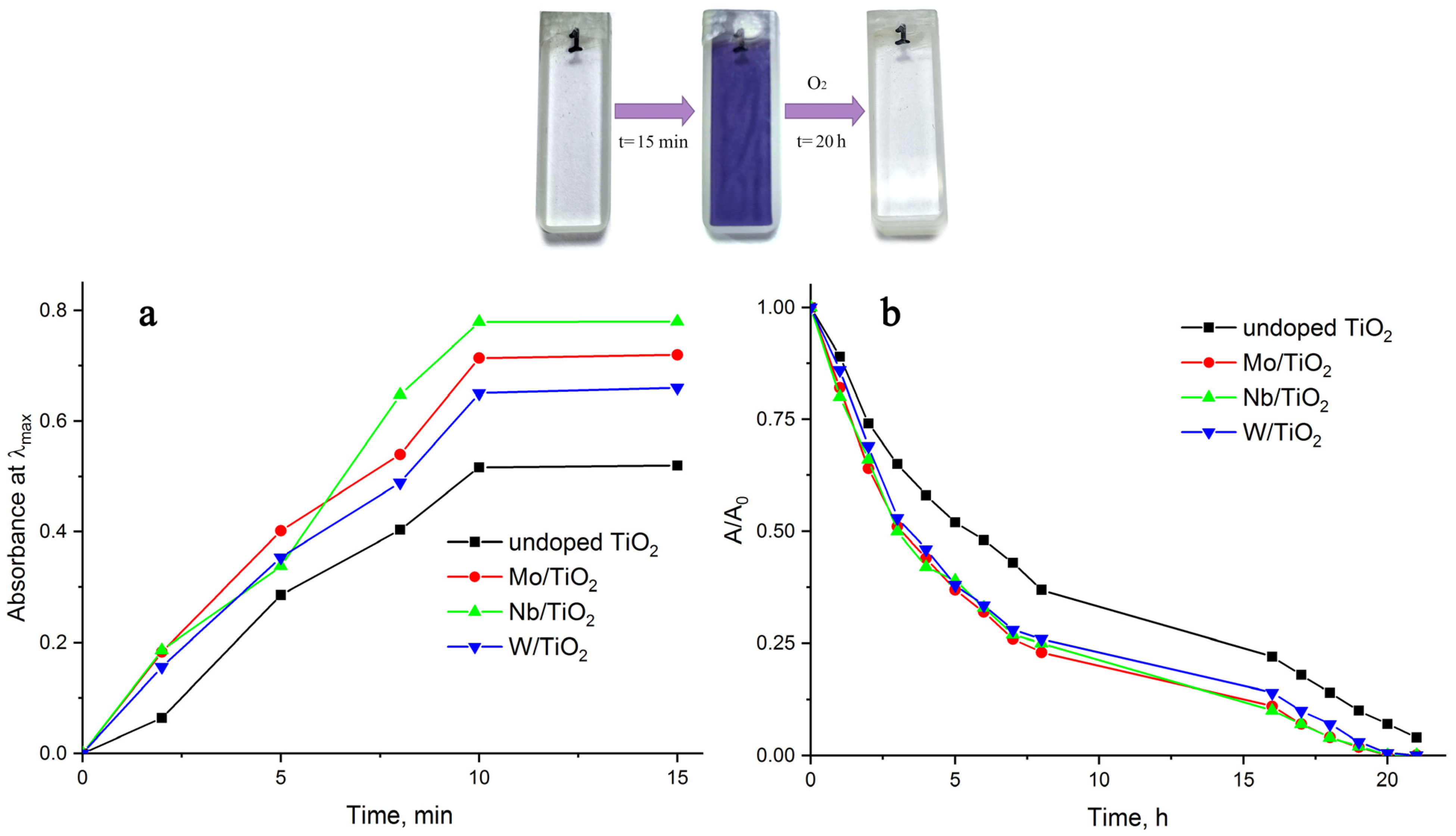
3.3. Photochromic Properties

Figure 5 shows the absorption spectra of the photocolored titanium oxide sols in the visible and near-infrared regions. In the visible region of the spectrum, the presence of a maximum of 530–540 nm and an unresolved band (highlighted fragment) in the region of 640–740 nm is registered. All these absorption bands are attributed to their own F color centers. This is inherent in an external defect arising from oxygen vacancies that capture two electrons [30,31]. Notably, the doping process shifts the absorption maximum to longer wavelengths. This is typical for structures of doped titanium dioxide [32]. Two absorption bands in the near-IR region belong to the reduced state (Ti3+). A comparison of the photocoloring intensities revealed that the influence of the dopant material on the coloring intensity can be traced. The highest coloring efficiency is recorded in sols of titanium oxide doped with niobium, despite the lower content of doped material compared with that of Mo/TiO2. As for the explanation of the increase in photochromism upon doping, we can assume the following. The key role in the photochromic process is played by localized defects, dislocations, and impurities. An important factor is the intervalence electron transfer between titanium ions and doping elements with different oxidation states (Ti4+–Mo4+, Ti4+–Nb5+, Ti4+–W5+, Ti4+–Mo5+, Ti4+–Nb6+, Ti4+–W5+, etc.) The energy level of Men+/Me(n−1)+ lies below the conduction band edge. Doping with Mo, Nb, or W ions creates energy levels below the conduction band edge that act as photoelectron traps. In addition, it is necessary to take into account that in the case of incorporation of Mo, Nb, or W ions into the TiO2 structure, longer Me–O bonds are formed compared to Ti–O [33]. This also affects the intensity of photocoloration. In the case of Nb-doped TiO2, the more intense coloration can be explained as follows. According to [34], the formation of Nb4+ is accompanied by the formation of additional oxygen vacancies near Nb4+, which act as photoelectron traps.
Figure 6 presents the kinetics of the photocoloring and bleaching of the TiO2 sols without and after plasma treatment. The results show that exposing the sol to a plasma accelerates the photocoloring process (Figure 6a). The process of decolorization of sols in the presence of atmospheric oxygen occurs more slowly (Figure 6b). Compared with those of sols of tungsten and molybdenum oxides, the dynamics of the discoloration of TiO2 sols are slower [35]. Our results revealed that approximately half of the staining in sols after exposure to plasma disappears within the first 3 h. This is faster than the results obtained previously for W6+/TiO2 (50% discoloration for 4 h) [14]. Complete discoloration was recorded after 20 h. A previous work reported that titanium dioxide doped with tungsten bleaches in 24–26 h [14] and that Mo6+/TiO2 bleaches for 3 days [13].
Our data contradict previously published results [13,14,15]. The reason for the discrepancy in our results may be a combination of several factors: (1) the phase composition of TiO2 itself, (2) the degree of oxidation of the doping agent, and (3) the modification of the sol during plasma treatment. Early studies investigated the effect of doping on the photochromic characteristics of titanium dioxide with an anatase structure or mixed-phase titanium dioxide (anatase, rutile, or brookite) [4,6,7,12,13,14]. This work demonstrates for the first time the photochromic effect of pure brookite.
On the other hand, the oxidation state of the doping agent can also affect the photochromic properties. In general, photochromicity can be described as follows:
T i O 2 + h v   T i O 2   ( e C B + h V B + )  
M e s u r f n + + e C B   M e s u r f ( n 1 ) +     o r   M e s u r f n + + h V B +   M e s u r f ( n + 1 ) +
T i 4 + + e C B   T i 3 +
When irradiated with light, an electron and a hole are formed, which accumulate in the conduction band or participate in redox processes. If the TiO2 matrix contains doping agents with maximum degrees of oxidation (for example, W6+ or Mo6+), only photogenerated electrons participate in the redox process. The electrons are delocalized near the excess positive charges of dopant ions and have less affinity to participate in oxidation reactions (bleaching process) [15]. In the case of the presence of structures in which Men+-Me(n−1)+ states exist, both a photoelectron and a photohole participate simultaneously in the oxidation-reduction process. This is confirmed by the presence of two absorption maxima in the visible region. The absorption in the region of 650 nm is responsible for trapped electrons, and the presence of an absorption maximum in the region of 430–530 nm corresponds to trapped holes [36].
It was previously established that underwater diaphragm discharge leads to the formation of structures of nonstoichiometric oxides of tungsten and molybdenum (W18O49, Mo4O11) [35]. The presence of Mo4+ in our samples was confirmed by the XPS data. This may be the reason for the increased color and accelerated discoloration (oxidation).
Plasma modification can be reflected in changes in the physicochemical properties of sols. Notably, the action of the plasma led to a decrease in the viscosity of the solutions. For PVP, which forms the basis of the sol, the molecular weight is related to the viscosity via the following relationship [37]:
η = 4.1 · 1 0 5 · M 0.85
Measurements of the viscosity of solutions after plasma treatment made it possible to estimate changes in the molecular weights of PVP (Table 1). The action of the plasma with Mo electrodes leads to a greater effect on the Mw. A decrease in the molecular weight of PVP indicates that a depolymerization process occurred, which is induced by the action of the plasma. According to a previous work [38], PVP is effectively depolymerized in the presence of H2O2. Numerous studies of the kinetics of hydrogen peroxide accumulation in plasma systems in contact with liquids have shown that the electrode material can be both a catalyst and an inhibitor of the decomposition reaction of hydrogen peroxide [39,40,41,42]. The results of measuring the concentration of hydrogen peroxide under conditions of a diaphragm discharge action in distilled water with Mo, Nb, and W electrodes are also presented in Table 1. According to the obtained results, the highest concentration of hydrogen peroxide is recorded in experiments after plasma exposure with molybdenum electrodes. The lowest concentration value accumulated when using niobium electrodes. This correlates with the molecular weight values of PVP after plasma treatment with different electrodes. Based on this, it can be concluded that the depolymerization process during plasma treatment occurs with the participation of H2O2.
Reducing the molecular weight of PVP leads to an increase in the permeability of the polymer to oxygen [43], which accelerates the bleaching process.

4. Conclusions

The process of doping by underwater diaphragm discharge was successfully carried out. The dopants were the electrode material sputtered during the discharge-burning process, which was confirmed by the decrease in the mass of the electrodes and the emission spectra of the diaphragm discharge. The doping effects of Mo, Nb, and W on the structure and optical and photochromic characteristics of TiO2 (brookite) have been studied. The nature of the dopant affects the intensity of the photocoloring, which can be explained by the degree of oxidation of the dopant. The process of plasma doping improves the photochromic properties (increase in color intensity and bleaching rate), including owing to the plasma modification of TiO2 sols.

Author Contributions

Conceptualization, A.K.; methodology, N.S.; investigation, N.S., A.E. and V.S.; writing—original draft preparation, A.K.; writing—review and editing, A.K. and N.S.; supervision, A.K.; project administration, A.K. All authors have read and agreed to the published version of the manuscript.

Funding

This study was supported by the Russian Science Foundation under grant 23-23-00051.

Institutional Review Board Statement

Not applicable.

Informed Consent Statement

Not applicable.

Data Availability Statement

Dataset available on request from the authors.

Acknowledgments

The authors express their gratitude to the Center for the collective use of scientific equipment at the “Upper Volga Regional Center for Physical–Chemical Research”. The study was partially carried out using equipment from the Center for Shared Use of the Scientific Educational Center “Physics for Solid State Nanostructures” of the Lobachevsky State University of Nizhny Novgorod. This work was partially carried out with the support of the Scientific Facility Center at the Institute of Solid State Physics of RAS.

Conflicts of Interest

The authors declare no conflicts of interest.

References

  1. Luo, C.; Ren, X.; Dai, Z.; Zhang, Y.; Qi, X.; Pan, C. Present perspectives of advanced characterization techniques in TiO2-based photocatalysts. ACS Appl. Mater. Interfaces 2017, 9, 23265–23286. [Google Scholar] [CrossRef] [PubMed]
  2. Basavarajappa, P.S.; Patil, S.B.; Ganganagappa, N.; Reddy, K.R.; Raghu, A.V.; Reddy, C.V. Recent progress in metal-doped TiO2, nonmetal doped/codoped TiO2 and TiO2 nanostructured hybrids for enhanced photocatalysis. Int. J. Hydrogen Energy 2020, 45, 7764–7778. [Google Scholar] [CrossRef]
  3. Joost, U.; Sutka, A.; Oja, M.; Smits, K.; Döbelin, N.; Loot, A.; Järvekülg, M.; Hirsimäki, M.; Valden, M.; Nõmmiste, E. Reversible photodoping of TiO2 nanoparticles for photochromic applications. Chem. Mater. 2018, 30, 8968–8974. [Google Scholar] [CrossRef]
  4. Kikuchi, E.; Iida, K.; Fujishima, A.; Itoh, K. Photochromism and photoelectrochemistry of amorphous and polycrystalline WO3 films. J. Electroanal. Chem. 1993, 351, 105–114. [Google Scholar] [CrossRef]
  5. Belhomme, L.; Duttine, M.; Labrugère, C.; Coicaud, E.; Rougier, A.; Penin, N.; Dandre, A.; Ravaine, S.; Gaudon, M. Investigation of the photochromism of WO3, TiO2, and composite WO3–TiO2 nanoparticles. Inorg. Chem. 2024, 63, 10079–10091. [Google Scholar] [CrossRef]
  6. Paul, N.; Patowary, M.; Pegu, B.; Mohanta, D. Physical properties of nanoscale TiO2 related to Ag-doping and photochromic behavior. Nanosci. Nanotechnol. Lett. 2013, 5, 452–456. [Google Scholar] [CrossRef]
  7. Crespo-Monteiro, N.; Destouches, N.; Epicier, T.; Balan, L.; Vocanson, F.; Lefkir, Y.; Michalon, J.Y. Changes in the chemical and structural properties of nanocomposite Ag: TiO2 films during photochromic transitions. J. Phys. Chem. C 2014, 118, 24055–24061. [Google Scholar] [CrossRef]
  8. Tobaldi, D.M.; Rozman, N.; Leoni, M.; Seabra, M.P.; Skapin, A.S.; Pullar, R.C.; Labrincha, J.A. Cu–TiO2 hybrid nanoparticles exhibiting tunable photochromic behavior. J. Phys. Chem. C 2015, 119, 23658–23668. [Google Scholar] [CrossRef]
  9. Morita, M.; Toyoda, S.; Kiuchi, H.; Abe, T.; Kumagai, K.; Saida, T.; Fukuda, K. Chromogenic amorphous MoO3−x nanosheets and their nanostructured films for smart window applications. ACS Appl. Nano Mater. 2021, 4, 8781–8788. [Google Scholar] [CrossRef]
  10. Miyazaki, H.; Matsuura, T.; Ota, T. Vanadium oxide-based photochromic composite film. RSC Adv. 2017, 7, 2388–2391. [Google Scholar] [CrossRef]
  11. Li, P.; Zhang, Z.; Gao, X.; Sun, H.; Peng, D.; Zou, H.; Zhang, Q.; Hao, X. Fast self-bleaching Nb2O5-based photochromics for high security dynamic anti-counterfeiting and optical storage applications. Chem. Eng. J. 2022, 435, 134801. [Google Scholar] [CrossRef]
  12. Songara, S.; Patra, M.K.; Manoth, M.; Saini, L.; Gupta, V.; Gowd, G.S.; Vadera, S.R.; Kumar, N. Synthesis and studies on photochromic properties of vanadium doped TiO2 nanoparticles. J. Photochem. Photobiol. A Chem. 2010, 209, 68–73. [Google Scholar] [CrossRef]
  13. Songara, S.; Singh, J.; Patra, M.K.; Gupta, V.; Saini, L.; Gowd, G.S.; Kumar, N. Wet chemical synthesis of molybdenum-doped amorphous TiO2 nanoparticles and studies on their photochromic properties. Int. J. Green Nanotechnol. 2012, 4, 399–407. [Google Scholar] [CrossRef]
  14. Songara, S.; Saini, L.; Gowd, G.S.; Rajpurohit, J.S.; Gupta, V.; Patra, M.K.; Vadera, S.R.; Kumar, N. Improved photochromic properties of W6+ doped nanostructured TiO2 coatings. Res. Surf. Interfaces 2024, 14, 100201. [Google Scholar] [CrossRef]
  15. Eglītis, R.; Joost, U.; Zukuls, A.; Rubenis, K.; Ignatans, R.; Avotiņa, L.; Baumane, L.; Smits, K.; Hirsimaki, M.; Kaambre, T.; et al. Strong, rapid, and reversible photochromic response of Nb doped TiO2 nanocrystal colloids in hole scavenging media. ACS Appl. Mater. Interfaces 2020, 12, 57609–57618. [Google Scholar] [CrossRef]
  16. Xu, J.; Li, K.; Wu, S.; Shi, W.; Peng, T. Preparation of brookite titania quasi nanocubes and their application in dye-sensitized solar cells. J. Mater. Chem. A 2015, 3, 7453–7462. [Google Scholar] [CrossRef]
  17. Manzoli, M.; Freyria, F.S.; Blangetti, N.; Bonelli, B. Brookite, a sometimes under evaluated TiO2 polymorph. RSC Adv. 2022, 12, 3322–3334. [Google Scholar] [CrossRef]
  18. Zou, Y.; Yu, T.; Huang, X.; Li, Y.; Guo, L.; Yan, H.; Zhou, J.; Wang, Y. Hydrothermal synthesis of Zn-doped brookite TiO2 for enhanced visible-light-responsive photocatalytic performance. Mater. Res. Express 2023, 10, 085005. [Google Scholar] [CrossRef]
  19. Arnal, P.; Corriu, R.J.P.; Leclercq, D.; Mutin, P.H.; Vioux, A. Preparation of anatase, brookite and rutile at low temperature by non-hydrolytic sol–gel methods. J. Mater. Chem. 1996, 6, 1925–1932. [Google Scholar] [CrossRef]
  20. Khlyustova, A.; Sirotkin, N.; Kusova, T.; Kraev, A.; Titov, V.; Agafonov, A. Doped TiO2: The effect of doping elements on photocatalytic activity. Mater. Adv. 2020, 1, 1193–1201. [Google Scholar] [CrossRef]
  21. Khlyustova, A.; Sirotkin, N.; Kraev, A.; Kusova, T.; Titov, V.; Agafonov, A. Mo-doped TiO2 using plasma in contact with liquids: Advantages and limitations. J. Chem. Technol. Biotechnol. 2021, 96, 1125. [Google Scholar] [CrossRef]
  22. Eisenberg, G. Colorimetric determination of hydrogen peroxide. Ind. Eng. Chem. Anal. Ed. 1943, 15, 327–328. [Google Scholar] [CrossRef]
  23. Shen, V.K.; Siderius, D.W.; Krekelberg, W.P.; Hatch, H. (Eds.) NIST Standard Reference Simulation Website, NIST Standard Reference Database Number 173; National Institute of Standards and Technology: Gaithersburg, MD, USA, 2024. Available online: https://data.nist.gov/pdr/lps/FF429BC178798B3EE0431A570681E858232 (accessed on 13 January 2024).
  24. Tompsett, G.A.; Bowmaker, G.A.; Cooney, R.P.; Metson, J.B.; Rodgers, K.A.; Seakins, J.M. The Raman spectrum of brookite, TiO2 (PBCA, Z = 8). J. Raman Spectrosc. 1995, 26, 57–62. [Google Scholar] [CrossRef]
  25. De Faria, D.L.A.; Gil, H.A.C.; De Queiroz, A.A.A. The interaction between polyvinylpyrrolidone and I2 as probed by Raman spectroscopy. J. Mol. Struct. 1999, 478, 93–98. [Google Scholar] [CrossRef]
  26. Stefanov, P.; Shipochka, M.; Stefchev, P.; Raicheva, Z.; Lazarova, V.; Spassov, L. XPS characterization of TiO2 layers deposited on quartz plates. J. Phys. Conf. Ser. 2008, 100, 012039. [Google Scholar] [CrossRef]
  27. Tandy, L.M.F.; Bueno, J.P.; Vong, Y.M.; Torres, I.Z.; Rojas, L.L.; Torres, C.J.R.; Gutierrez, H.M.; López, M.M. Reversible photochromic effect in the TiO2—Polymer hybrid system. J. Sol-Gel Sci. Technol. 2017, 82, 51–58. [Google Scholar] [CrossRef]
  28. Van Pham, D.; Patil, R.A.; Lin, J.H.; Lai, C.C.; Liou, Y.; Ma, Y.R. Doping-free bandgap tuning in one-dimensional Magnéli-phase nanorods of Mo4O11. Nanoscale 2016, 8, 5559–5566. [Google Scholar] [CrossRef]
  29. Sun, Y.; Wang, W.; Qin, J.; Zhao, D.; Mao, B.; Xiao, Y.; Cao, M. Oxygen vacancy-rich mesoporous W18O49 nanobelts with ultrahigh initial Coulombic efficiency toward high-performance lithium storage. Electrochim. Acta 2016, 187, 329–339. [Google Scholar] [CrossRef]
  30. Kuznetsov, V.N.; Glazkova, N.I.; Mikhaylov, R.V.; Serpone, N. In situ study of photo- and thermos-induced color centers in photochromic rutile TiO2 in the temperature range 90–720 K. Photochem. Photobiol. Sci. 2016, 15, 1289–1298. [Google Scholar] [CrossRef]
  31. Kuznetsov, V.N.; Serpone, N. Photoinduced coloration and photobleaching of titanium dioxide in TiO2/polymer compositions upon UV-and visible-light excitation of color centers absorption bands: Direct experimental evidence negating band-gap narrowing in anion-/cation-doped TiO2s. J. Phys. Chem. C 2007, 111, 15277–15288. [Google Scholar] [CrossRef]
  32. Khlyustova, A.; Evdokimova, A.; Shibaeva, V.; Sirotkin, N. Photochromic properties of novel composites on the base of biopolymers and plasma-modified Mo4O11 and W18O49 nanostructures. J. Vinyl Addit. Technol. 2024, 30, 585–595. [Google Scholar] [CrossRef]
  33. Onur Şahin, E.; Dai, Y.; Chan, C.K.; Tüysüz, H.; Schmidt, W.; Lim, J.; Zhang, S.; Scheu, C.; Weidenthaler, C. Monitoring the structure evolution of Titanium oxide photocatalysts: From the molecular form via the amorphous state to the crystalline phase. Chem. A Eur. J. 2021, 27, 11600–11608. [Google Scholar] [CrossRef] [PubMed]
  34. Brayner, R.; Bozon-Verduraz, F. Niobium pentoxide prepared by soft chemical routes: Morphology, structure, defects and quantum size effect. Phys. Chem. Chem. Phys. 2003, 5, 1457–1466. [Google Scholar] [CrossRef]
  35. Zhang, L.; Mohamed, H.H.; Dillert, R.; Bahnemann, D. Kinetics and mechanisms of charge transfer processes in photocatalytic systems: A review. J. Photochem. Photobiol. C 2012, 13, 263–276. [Google Scholar] [CrossRef]
  36. Miller, L.E.; Hamm, F.A. Macromolecular properties of polyvinylpyrrolidone: Molecular weight distribution. J. Phys. Chem. 1953, 57, 110–122. [Google Scholar] [CrossRef]
  37. Kaczmarek, H.; Kamińska, A.; Światek, M.; Rabek, J.F. Photo-oxidative degradation of some water-soluble polymers in the presence of accelerating agents. Angew. Makromol. Chem. 1998, 261, 109–121. [Google Scholar] [CrossRef]
  38. Kirkpatrick, M.J.; Locke, B.R. Effects of platinum electrode on hydrogen, oxygen, and hydrogen peroxide formation in aqueous phase pulsed corona electrical discharge. Ind. Eng. Chem. Res. 2006, 45, 2138–2142. [Google Scholar] [CrossRef]
  39. Kuzimicheva, L.; Titova, Y.; Maksimov, A. Generation of chemically active oxidative particles in electrolyte solutions under the action of glow and diaphragm discharges. Surf. Eng. Appl. Electrochem. 2007, 43, 90–93. [Google Scholar] [CrossRef]
  40. Holzer, F.; Locke, B.R. Influence of high voltage needle electrode material on hydrogen peroxide formation and electrode erosion in a hybrid gas–liquid series electrical discharge reactor. Plasma Chem. Plasma Process 2008, 28, 1–13. [Google Scholar] [CrossRef]
  41. Lukes, P.; Clupek, M.; Babicky, V.; Sisrova, I.; Janda, V. The catalytic role of tungsten electrode material in the plasmachemical activity of a pulsed corona discharge in water. Plasma Source Sci. Technol. 2011, 20, 034011. [Google Scholar] [CrossRef]
  42. Kuz’micheva, L.A.; Titova, Y.V.; Maksimov, A.I.; Vashurin, A.S.; Pukhovskaya, S.G. Effect of the cathode material on the accumulation of hydrogen peroxide in a plasma-solution system. Surf. Eng. Appl. Electrochem. 2013, 49, 485–487. [Google Scholar] [CrossRef]
  43. Kang, J.S.; Lee, Y.M. Effects of molecular weight of polyvinylpyrrolidone on precipitation kinetics during the formation of asymmetric polyacrylonitrile membrane. J. Appl. Polym. Sci. 2002, 85, 57–68. [Google Scholar] [CrossRef]
Figure 1. Scheme of the experimental setup.
Applsci 14 08877 g001
Figure 2. Emission spectra of diaphragm discharge burning in TiO2 sol with Mo (a), Nb (b), and W (c) electrodes.
Applsci 14 08877 g002
Figure 3. XRD patterns (a) and Raman spectra (b) of undoped and doped TiO2 sols.
Applsci 14 08877 g003
Figure 4. High-resolution XPS spectra of O 1s (for undoped TiO2 (a) and Mo-doped TiO2 as an example (b)), Ti 2p (c), Nb 3d (d), Mo 3d (e), and W 4f (f).
Applsci 14 08877 g004
Figure 5. Visible and NIR absorption spectra of the irradiated sol before and after plasma treatment.
Applsci 14 08877 g005
Figure 6. Kinetics of the photocoloration (a) and dynamics of bleaching (b) TiO2 sols before and after plasma treatment.
Applsci 14 08877 g006
Table 1. Molecular weights of the PVP and H2O2 concentrations in the samples after plasma treatment (initial MW (PVP) = 55,000 gol−1) at different electrode’s material.
Material of ElectrodeMW(PVP), gol−1C(H2O2), mM
Mo27,7241.40
Nb29,2560.91
W28,2811.38
Disclaimer/Publisher’s Note: The statements, opinions and data contained in all publications are solely those of the individual author(s) and contributor(s) and not of MDPI and/or the editor(s). MDPI and/or the editor(s) disclaim responsibility for any injury to people or property resulting from any ideas, methods, instructions or products referred to in the content.

