Review

Nanostructured Drug Delivery Systems in Immunotherapy: An Updated Overview of Nanotechnology-Based Therapeutic Innovations

by
George-Alexandru Croitoru
1,
Adelina-Gabriela Niculescu
2,3,*,
Dragoș Epistatu
1,
Dan Eduard Mihaiescu
3,
Alexandru Mihai Antohi
1,
Alexandru Mihai Grumezescu
2,3 and
Carmen-Larisa Nicolae
1
1
Faculty of Dental Medicine, Carol Davila University of Medicine and Pharmacy, 8 Eroii Sanitari Street, 050474 Bucharest, Romania
2
Research Institute of the University of Bucharest—ICUB, University of Bucharest, 050657 Bucharest, Romania
3
Faculty of Chemical Engineering and Biotechnology, National University of Science and Technology Politehnica Bucharest, 011061 Bucharest, Romania
*
Author to whom correspondence should be addressed.
Appl. Sci. 2024, 14(19), 8948; https://doi.org/10.3390/app14198948
Submission received: 11 September 2024 / Revised: 28 September 2024 / Accepted: 30 September 2024 / Published: 4 October 2024
(This article belongs to the Section Materials Science and Engineering)

Abstract

:
Using nanostructured drug delivery systems has attracted increasing interest in immunotherapeutic approaches. The intrinsic immunomodulatory properties and versatility of nanoparticles used as carriers were consistently reported to augment treatment efficiency as nanoscaled materials increase drug accumulation at the desired site, enhance cell internalization, and improve therapeutic outcomes. Thus, numerous studies have exploited the potential use of nanostructured drug delivery vehicles in delivering different cargo as a promising alternative for treating conditions like cancer, autoimmune diseases, infectious diseases, and allergic and immune disorders. In this context, this paper presents nanostructured drug delivery systems as a solid basis for immunotherapeutic innovations, highlighting their advantages for improving treatment strategies, reviewing their clinical applications, and discussing existing challenges and ways to overcome them.

1. Introduction

The immune system represents an intricate network of defense mechanisms with a fundamental role in maintaining homeostasis. These mechanisms have evolved to destroy foreign substances and pathogens, protecting the body from such threats. Nonetheless, certain imbalances may occur, generating abnormal immune responses based on exacerbated states of immunosuppression or immunostimulation [1,2,3].
The immune system comprises innate and adaptive defense mechanisms. Innate immunity serves as the first line of defense against pathogens, triggering local inflammation by activating various immune cells and producing cytokines. This response often leads to the activation of the adaptive immune system, where T and B lymphocytes, key players in antigen-specific immunity and immunologic memory, target pathogens [2,4]. Immune imbalances can manifest as autoimmunity, autoinflammation, allergy, or lymphoproliferation [5].
Misdirected immune responses, generally termed immunopathology, give rise to the immune system’s reaction that attacks host structures. Recent studies show that how antigens are presented heavily influences both inflammatory processes (immune stimulation) and immunological tolerance (lack of reaction to self or harmless foreign antigens). Improper immune stimulation can lead to adverse responses, such as autoimmune diseases, where the body’s immune system attacks its own tissues. On the other hand, allergic diseases result from hypersensitivity to typically harmless environmental substances [2]. Additionally, inadequate or insufficient immune responses can leave the host vulnerable to infections, tumors, and other harmful agents. This type of response is often characterized by the immune system’s failure to properly recognize or respond to pathogens or abnormal cells, leaving the organism unprotected against threats like chronic infections or cancer [6,7]. This highlights the delicate balance the immune system must maintain between overreaction (leading to immunopathology) and underreaction (resulting in immunodeficiency or inadequate protection).
Manipulating the immune system in a therapeutic manner holds great promise for the treatment of chronic diseases such as cancer and autoimmune disorders [8,9]. In situations that were previously thought to be fatal, immunotherapy provides the means for combating the disease and prolonging patient survival through the modulation of innate and/or adaptive immunity [10].
Although cancer immunotherapies have shown unprecedented durable responses, most patients either fail to respond (primary resistance) or relapse after an initial response (acquired resistance) (Figure 1) [11,12,13,14]. Response rates are especially low in common cancers such as breast, prostate, and colon, and even within the same patient, different tumors can exhibit varying outcomes [11].
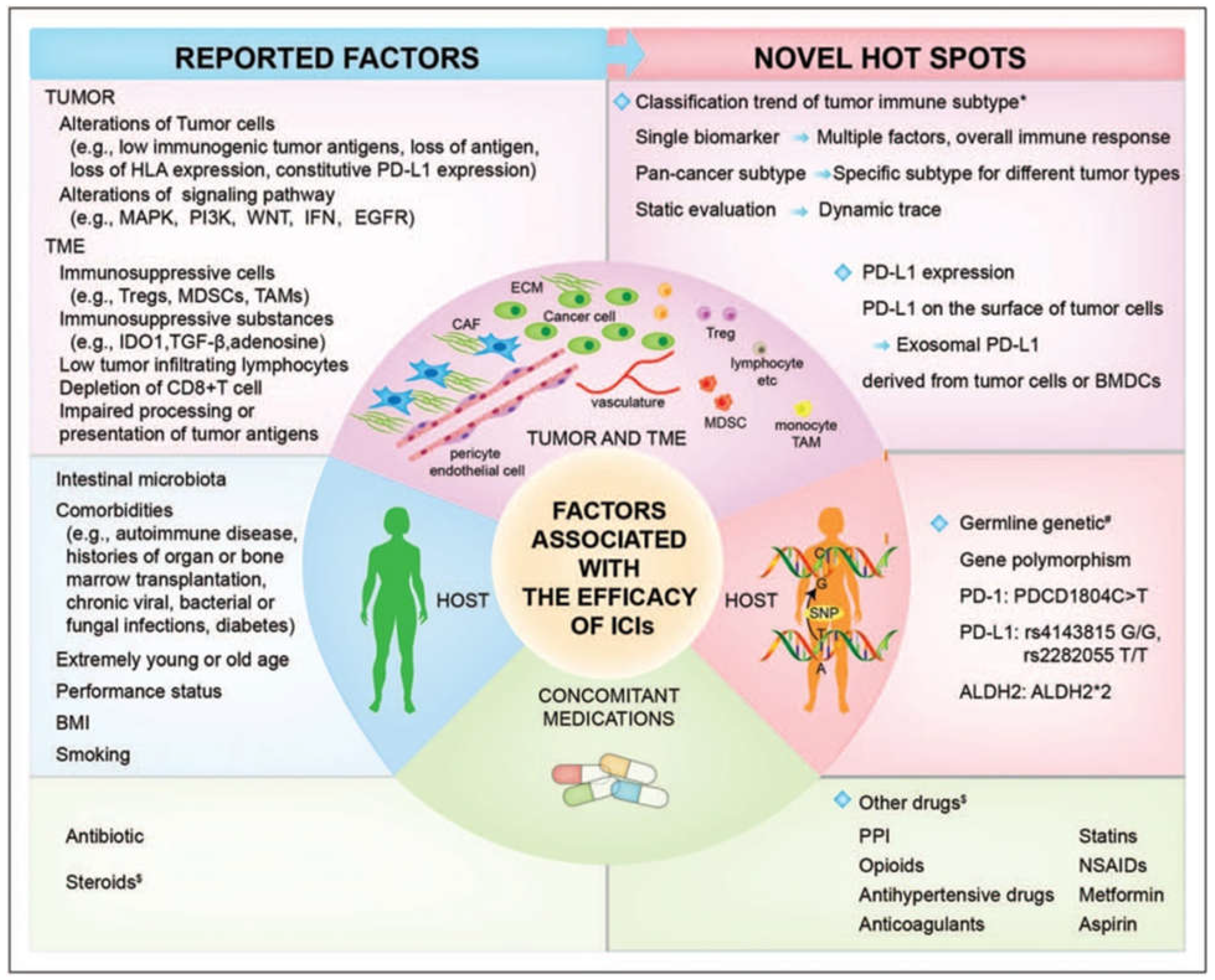
The immune system is tightly regulated by checkpoints and filters to maintain immunological tolerance and prevent autoreactive cells from causing harm. However, autoimmune disorders and malignant diseases remain common, likely due to the evolutionary trade-off between immunoprotection and immunopathology, as well as strategies employed by parasites to evade the immune response [1]. Immunotherapeutic approaches may be involved in dealing with such conditions, yet limited efficacy has been reported in certain cases where patients exhibit primary resistance to immune checkpoint inhibitors (ICIs). ICI resistance was reported to stem from either the inability of immunotherapy to produce an anticancer immune response or the failure to alleviate tumor-induced immunosuppression, with several factors being correlated with variations in ICI efficacy (Figure 2) [13].
Nevertheless, the primary obstacle in immunotherapy lies in attaining controlled regulation to guarantee therapeutic outcomes without unintended side effects. Success hinges on administering immunomodulators correctly at the exact moment and place, encompassing particular tissues, cells, and even intracellular locations. Despite the significant progress made in immunotherapy, the effectiveness of these treatments is frequently hindered by difficulties in delivering them to specific targets and controlling their release. Nanoparticles are being extensively investigated as potential means of answering these difficulties, providing enhanced control over the timing and location of immunotherapy delivery [8,9,15].
Nanoengineered delivery vehicles based on a wide range of materials have been reported in the scientific literature. Nanoparticles can be employed as carriers of interesting molecules, either counting on their intrinsic features (e.g., size, high surface area, ease of surface functionalization) or on their ability to respond to various exogenous (e.g., light, ultrasound, and electrical fields) and/or endogenous stimuli (pH, heat, and enzyme concentration) [10,15]. Moreover, nanomaterials can interact with the immune system in complex ways, involving different immune cells, signaling pathways, and cellular mechanisms [16].
In this context, this paper overviews current literature concerning nanostructured drug delivery systems in immunotherapy, highlighting the potential utility of engineered nanoplatforms and presenting their possible applications in disease management. In more detail, relevant recent English language research articles from these interconnected fields are reviewed in this work. Studies were retrieved from scientific databases, such as Science Direct, PubMed, MDPI, ClinicalTrials.gov, and Google Scholar, using combinations of various keywords, including, but not limited to, “nanotechnology”, “nanomaterials”, “nanostructured systems”, “drug delivery”, “immunotherapy”, “cancer treatment”, “infectious diseases”, “allergies”, “autoimmune diseases”, “inflammatory disorders”, and “clinical approval”.

2. Design and Engineering of Nanostructured Drug Delivery Systems

Numerous nanomaterials have been developed in recent years, significantly advancing the field of nanotechnology and broadening its application across a wide range of disciplines. In particular, nanostructured systems have been recognized as valuable tools in biomedicine, gathering increasing interest for therapeutic applications [2,17,18,19]. Biomedical nanotechnology, involving the design and engineering of sophisticated biomaterials and drug delivery systems, is particularly well-suited for the controlled targeting of the immune system [8,20].
Related to therapeutic transport, nanostructured systems offer improved delivery of otherwise insoluble drugs, maximizing their bioavailability and reducing the required dosage. Specifically, the amount of carried drug can be reduced by 100–1000 times when delivered by targeted systems to the desired site/cells. In more detail, lower amounts of active pharmaceutical ingredients can be administered as they are only released where needed, and there are no drug losses toward undesired tissues. Thus, nanocarrier approaches have the benefit of increasing local drug concentrations while minimizing side effects [21,22,23,24]. Besides of their tailored dimensions and morphologies, customized nanoparticles also exhibit enhanced permeability and retention effects, high thermal conductance, and great electromagnetic radiation absorbance [25]. Moreover, nanoparticles can combine different therapeutic strategies to create multifunctional treatment tools or encompass both therapeutic and diagnostic functions to generate theranostics [9,21,26].
Nanomaterials have extremely small sizes, with at least one dimension between 1 and 100 nm. Their sizes are comparable with some bacteria and viruses, which render them capable of interacting with the immune system in manners mimicking natural pathogens [8,27]. Their size is also advantageous for passing through normally intact physiologic barriers and reaching specific targets for releasing carried agents. Hence, nanoparticles interact at a molecular level with immune cells, leading to immunostimulatory or immunosuppressive responses, thus modulating the immune system and enabling their use for therapeutic purposes [8,18,27]. Reciprocally, immune cells also exert certain actions onto nanoparticles, determining aspects like persistence, degradation, or clearance [8].
With these advantageous properties in mind, nanomaterials have started to be explored for use in immune-based therapies targeting a plethora of diseases. Nanosystems interact with the immune system in complex ways, influencing various immune cells, such as antigen-presenting cells (APCs), B cells, and T cells, and can induce both humoral and cell-mediated immune responses. These interactions can be finely tuned by engineering the NPs with specific physicochemical properties—such as size, shape, surface area, and charge—to enhance their recognition and uptake by immune cells, and effectively modulate the immune response [3,16]. When introduced into living systems, nanoparticles (NPs) are recognized as foreign materials and can either stimulate or suppress the immune system, depending on their chemical nature. This dual capability of NPs to modulate the immune response is crucial for their application in clinical treatments, allowing for the precise control of immune activity in disease prevention and therapy [2,8].
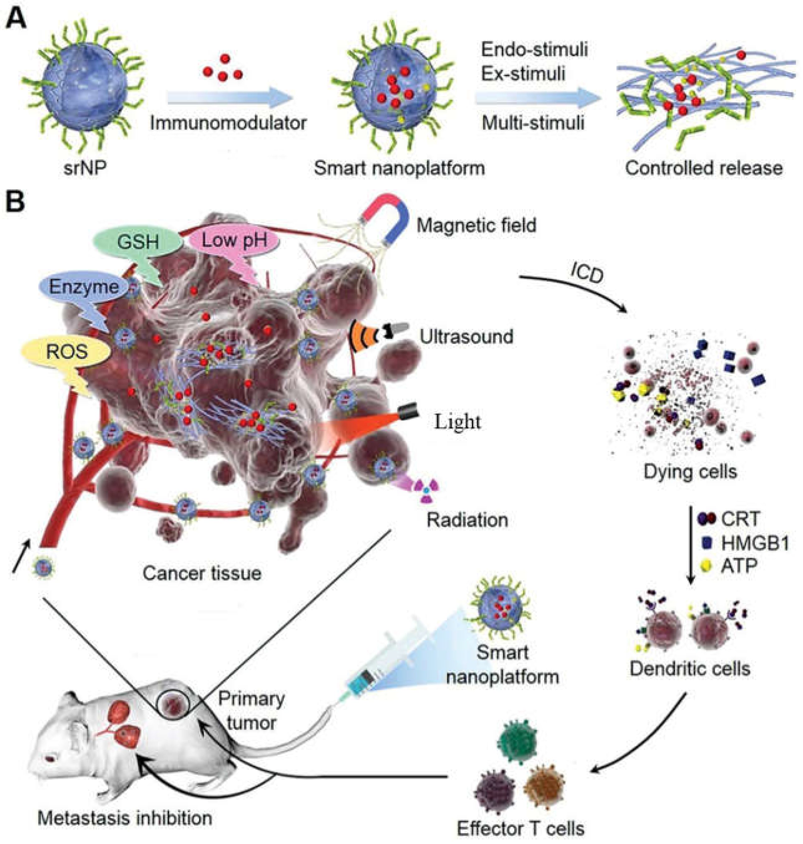
The combination of intrinsic immunomodulatory properties of nanoparticles and their versatility as nanocarriers can lead to outstanding outcomes when utilizing nanostructured systems for delivering immunotherapeutic agents. Various nanoparticles can be employed for transporting immunomodulatory drugs to specific immune cell compartments, potentially mimicking natural immune functions or activating drug release through various triggers (e.g., chemical reactions, targeting ligands, thermodynamic changes, magnetic field, irradiation at specific wavelengths, etc.) [8,28,29] (Figure 3).
Nanoparticles have potential anti-tumor activities and qualities that make them useful as therapeutic agents or vaccine adjuvants. Features like size, surface charge, functionalization, hydrophobicity or hydrophilicity, and the capacity to bind diverse biomolecules all substantially impact nanoparticle biocompatibility with target immune cells. Nanomaterials can be created to imitate specific foreign signals, modify the adaptive immune response, and develop new biological systems for use in immunotherapy. Nonetheless, certain nanoparticle properties have been demonstrated to cause considerable immunotoxicity, such as activating stress-related genes, membrane rupture, and oxidative stress, and producing pro-inflammatory cytokines [3,30,31,32]. As a result, selecting and designing materials with the appropriate qualities for immunotherapy requires a thorough approach.
Originally, silica and metal-based nanoparticles were used because of their ability to trigger a desired immunological response, either directly or indirectly, via associated antibodies or medicines. However, metal nanoparticles, such as gold nanoparticles, have been shown to be nondegradable, potentially leading to bioaccumulation, while silica nanoparticles have been observed to require surface functionalization to enhance their biocompatibility, and their effects on immune cells are reliant on their size, structure, and other physical features [3]. Thus, despite the initial popularity of these inorganic nanomaterials, more recent studies have rather focused on developing delivery systems for immunotherapeutic agents based on organic nanoparticles.
Polymers represent an appealing class of materials for fabricating nanocarriers with a wide range of polymeric nanoparticles, which are demonstrated to be biocompatible, biodegradable, soluble, stable, and versatile. Used alone, in blends, or combined with other types of materials, polymer-based nanostructures can be used as vehicles for a variety of cargos (e.g., drugs, genes, vaccines, biomolecules, imaging agents, etc.), improving their therapeutic activity while diminishing off-target toxicity [33,34]. Moreover, polymeric systems can improve immunotherapeutic approaches due to their adjustable physicochemical properties and the possibility of functionalization with different surface ligands. Polymeric nanoparticles can target dendritic cells (which are crucial for initiating immune responses), making them highly beneficial for immunotherapeutic strategies. Furthermore, studies suggest that improving the size, shape, surface charge, and hydrophobicity of nanoparticles enhances the transport of immunotherapeutic agents to malignancies. This precise control facilitates the combination of immunotherapy treatments, avoids the reoccurrence of tumors, promotes the development of long-lasting immunological memory, and improves patient outcomes by lowering toxicity and adverse events associated with the immune system [34,35].
Interesting prospects in immunotherapy are also offered by lipid nanoparticles (LNPs). LNPs have been extensively investigated because they can be manufactured on a large scale while maintaining precise manipulation of their morphology and lipid composition [36,37,38]. Their application in tumor immunotherapy has greatly enhanced the effectiveness of anti-tumor treatment while minimizing negative effects on the body as a whole. Particularly, LNPs were remarked to be valuable carriers for delivering mRNA for cancer immunotherapy. LNPs provide protection to the mRNA and further augment its internalization and distribution into cells. From a clinical perspective, LNPs have effectively enabled the transport of mRNA, as demonstrated by their involvement in COVID-19 vaccines [39,40,41].
Carbon-based nanomaterials (CBNs) have also been increasingly explored for use in transporting various therapeutics, with their unique features exploited to interfere with the immune system. CBNs have numerous forms (e.g., graphene/graphene oxide (GO) nanosheets, single/multi-walled carbon nanotubes (SWCNTs/MWCNTs), and fullerenes) of interest in nanomedicine, each with particularities concerning generated immune responses [3,18,42]. For instance, GO nanomaterials have been widely investigated for biomedical purposes, including drug delivery, cancer therapy, photodynamic therapy, vaccine antigen delivery, and more. It was reported that GO can activate the immune system through the increase in inflammatory factors and the proliferation and differentiation of lymphocytes. Nonetheless, GO displays certain cytotoxicity and can damage DNA due to its lateral size and oxidation state, thus requiring surface functionalization for better-suiting nanocarriers and adjuvants [43]. Differently, MWCNT was noted to induce cell responses by inducing oxidative stress, destabilizing lysosomal membranes, enhancing the cell permeability of 3T3 fibroblasts, bronchial epithelial cells and RAW macrophages, and eventually causing apoptosis in the targeted cells. SWCNTs were observed to act upon malignant cells by stimulating NF-kB and p38 activation and ROS production [44]. Therefore, considering the inherent potential of CBNs, they can be used together with therapeutic cargo to create synergistic outcomes.
Recently, DNA and RNA nanostructures have emerged as promising tools in biomedicine, especially in the field of drug delivery [45]. Due to the self-assembly properties of these nucleic acids, they can be designed with various architectures [46], such as polygons [47,48], nano-cubes [49,50], prisms [51,52], nano-rings [53,54,55,56], and other objects including three-way [57,58,59,60] and four-way junctions [61,62]. Among the wide range of 2D and 3D shapes, tetrahedral DNA nanostructures (TDNs) have attracted increasing attention especially due to their easy synthesis, remarkable stability, excellent biocompatibility, and facile functionalization. These advantageous properties make TDNs suitable for the transport and controlled release of chemotherapeutics, nucleic acids, imaging probes, immunotherapeutic agents, and theranostics [45].
In addition to the above-described possibilities, hybrid nanomaterials, which are composed of at least two constituents with different compositions and properties, typically organic and inorganic, have been deliberately designed to have combined activities and significant potential in enhancing cancer immunotherapy [63]. Hence, a plethora of synergistic systems have been reported in the recent literature, each with different degrees of success and specific immunotherapeutic applications.

3. Nanostructured Systems for Cancer Immunotherapy

Cancer immunotherapy, which modulates the body’s immune system to target tumors, has endowed extended survival in cases previously considered fatal [10,13]. Combinatorial approaches have particularly been rendered effective for enhancing immunotherapeutic outcomes by targeting multiple pathways within the cancer immunity cycle [64,65]. Recent advances focus on overcoming tumor-induced immune suppression, with treatments such as immune checkpoint inhibitors (ICIs) targeting CTLA-4, PD-1, and PD-L1 pathways. Such strategies represent valuable alternatives for treating solid and hematological cancers, targeting the evasion mechanisms of the tumor microenvironment [66,67,68]. Nonetheless, despite the success demonstrated throughout trials, several challenges remain. ICI-based therapies exhibit limited efficacy due to rapid drug clearance and off-target toxicity, also leading to resistance to immunotherapeutic agents in some patients [12,13,68,69]. While immunotherapy activates the host immune system, it can inadvertently trigger harmful side effects by activating non-tumor-specific immune cells [70].
Increasing evidence has been gathered in cancer immunotherapy regarding the importance of Toll-like receptors (TLRs). TLRs represent a pattern recognition receptor (PRRs) type that can detect pathogen-associated and damage-associated molecular patterns, triggering innate immune responses [10,15,71]. Particularly interesting for cancer treatment applications are the TLR7 and TLR8 present on the surface of endosomes. These receptors can induce type 1 interferons and inflammatory responses via the MyD88 pathway [10]. Thus, the administration of TLR agonists, such as imiquimod and resiquimod, can stimulate immune responses and activate cytotoxic T-cells. However, challenges such as poor solubility, side effects, and immune tolerance limit their clinical application. Moreover, the immune system can develop tolerance to TLR activation, over time diminishing the efficacy of TLR7/TLR8 agonist therapeutics [10,15,72].
In this context, nanotechnology-based drug delivery approaches have been considered promising solutions to enhance the efficacy of TLR7/8 agonists in cancer immunotherapy. The utilization of various nanocarriers, especially for small molecular immunotherapeutics, has the potential to mitigate dose-limiting toxicities and enhance treatment outcomes [10,15,25]. Nanocarriers, such as nanocapsules, nanoemulsions, and micelles, facilitate targeted delivery, enhance lymphatic transport, and extend drug release, thereby maintaining effective concentrations while reducing toxicity [10]. These nanoparticle engineering advancements enhance cancer cells’ targeted absorption and accumulation in cancerous tissues, potentially circumventing existing constraints in immunotherapy and creating mutually beneficial interactions between nanoparticles and immune cells [70,73].
One interesting possibility for delivering TLR agonists is to use poly(esteracetal)s, as these polymers have the chemical properties necessary for pH-responsive drug release [74]. For instance, Bixenmann et al. [15] developed self-assembling micelles based on amphiphilic mPEG-b-P(MDO) poly(esteracetal) block copolymers (Figure 4) able to carry an immune stimulatory TLR-7 agonist (i.e., Adifectin). This nanosystem allowed the better solubilization of the transported drug, ensuring its efficient delivery, pH-controlled release, and maintenance of receptor activity.
On a different note, Dias et al. [75] proposed the utilization of polymeric nanoparticles loaded with imiquimod as an alternative to the commercial drug formulation. The nanoparticles demonstrated significantly greater antiangiogenic activity and reduced tumor size and number compared to the commercial formulation and controls, indicating that the polymeric nanocarriers enhance treatment outcomes by increasing the skin permeation and solubility of the immunotherapeutic cargo for cutaneous malignancies. An alternative delivery vehicle for imiquimod has been developed by Gazzi et al. [76], who created pectin-based hydrogel nanocapsules for treating melanoma. This study revealed that the fabricated nanosystem allowed drug penetration in all skin layers and led to a higher cytotoxic effect of imiquimod in tested tumor cell populations, demonstrating promising potential for improving immunotherapy for this disease. Imiquimod activity can also be improved by its encapsulation into nanoemulsions, as demonstrated by Frank et al. [77]. The researchers have tested the nanoformulation against cervical cancer, proving its augmented antitumoral effect compared to free drug attributed to the combined autophagy and apoptosis mechanisms.
Several nanoformulations were also proposed for the delivery of resiquimod. For example, Rodell et al. [78] proposed encapsulating this TLR agonist into β-cyclodextrin nanoparticles to ensure its efficient delivery to tumor-associated macrophages. The authors even combined this strategy with the administration of immune checkpoint inhibitor anti-PD-1, obtaining synergistic results in terms of immunotherapy response rates. Differently, Widmer and colleagues [79] have created a resiquimod delivery system based on PLGA/mPEG-PLA nanoparticles that are internalized in dendritic cells and macrophages, which further activate anticancer immune responses. Based on in vivo tests performed on mice, the nanoformulation was proven effective for targeting lymph nodes and improving cancer immunotherapy outcomes.
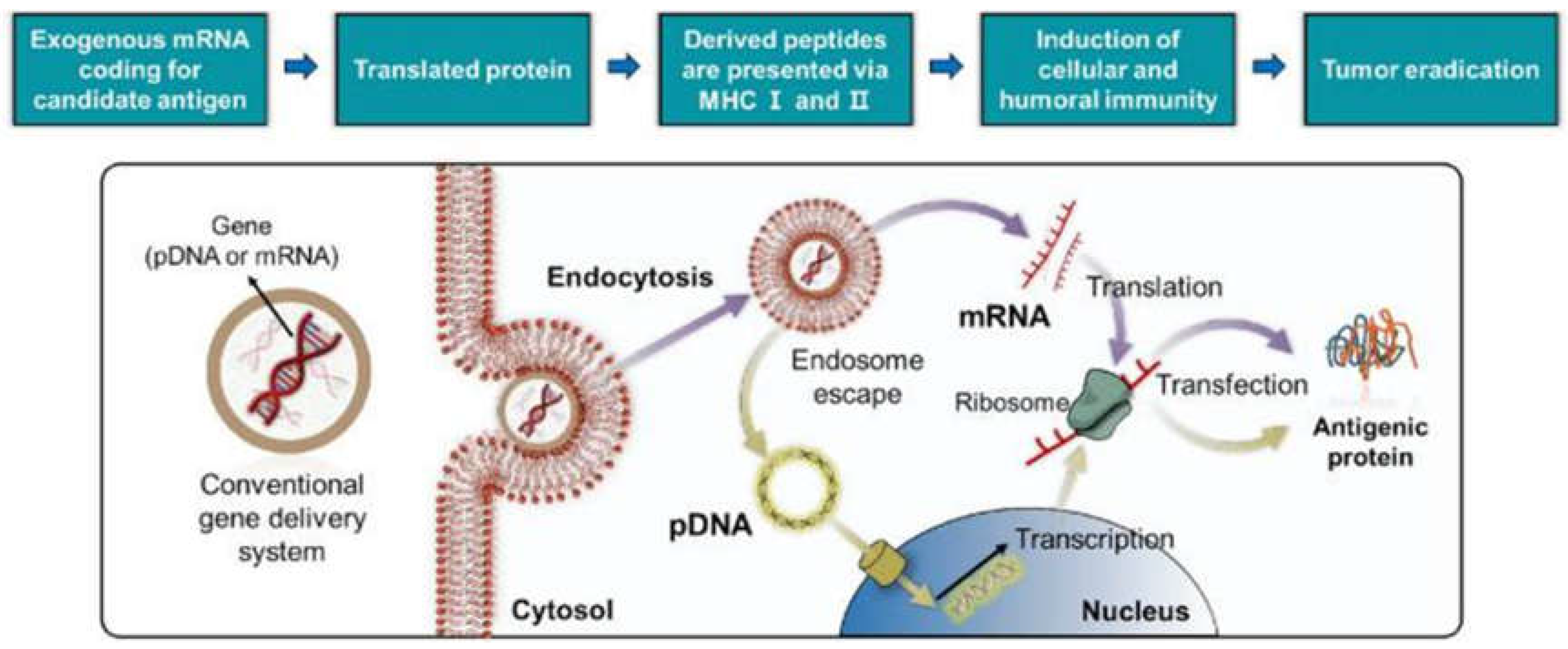
A different approach for inducing strong immune responses involves the targeted delivery of specific nucleic acids to tumor sites. Such gene delivery systems ensure the internalization of the therapeutic cargo, further reflected in the activation of cellular and humoral immune responses and eventual tumor destruction (Figure 5) [39]. Embarking on this possibility, Komura et al. [80] proposed the utilization of guanosine- and uridine-rich single-stranded RNA (GU-rich RNA) as the therapeutic agent, being recognized as an agonist of TLR7 and TLR8. In this respect, the researchers prepared a nanostructured hydrogel assembly of GU-rich RNA/DNA capable of sustained release. The delivery system was successfully internalized in murine dendritic cells and generated high immunostimulatory activity, and is thus considered a valuable adjuvant in cancer immunotherapy.
Compared to passive delivery vehicles, stimuli-triggered nanosystems allow for more accurate spatiotemporal control over TLR7/8 agonist activity in response to specific tumor microenvironment (TME) aspects, such as hypoxia, acidity, and hypoglycemia. Nanobiotechnological systems can selectively release their cargo due to temperature, pH, or redox potential changes and cross biological barriers to enhancing immunogenic cell death in therapies like chemotherapy, radiotherapy, and magnetic hyperthermia [10,70,81].
For example, Tampucci et al. [82] based their therapeutic approach on the pH-responsive character of a novel-designed nanosystem. Specifically, the researchers created nanometric self-assembling micelles made of newly synthesized fatty acid-protic ionic liquids (FA-PILs) and vitamin E for the delivery of imiquimod. The developed nanosystem allowed the controlled release of the TLR agonist in cancer tissues, which were activated by the acidic conditions of the TME and hold promise for skin cancer treatment. Kim and colleagues [83] have proposed a different pH-responsive delivery platform. The authors suggested using polymeric nanoparticles to transport a novel imidazoquinoline-based TLR7/8. The encapsulation strategy was proven effective, as it demonstrated stronger in vivo cytotoxicity and prolonged activation of NK cells compared to free drugs. Moreover, the proposed delivery alternative resulted in potent immunostimulatory activity, being a promising candidate for antibody-based cancer immunotherapy.
Another highly investigated strategy is to combine photothermal therapy with the nano-delivery of immunotherapeutic agents. In this respect, various materials and bioactive substances have been successfully mixed to create synergistic platforms able to release the therapeutic load under the application of magnetic, focused heat, or light fields [25]. For instance, Lu et al. [84] combined the advantages of photothermal therapy and immune checkpoint blockade by creating immunoadjuvant nanocarriers. The researchers fabricated pegylated polydopamine nanoparticles co-loaded with resiquimod and carbon dots that were proven effective against 4T1 breast tumors, inducing a strong antitumor immune response and inhibiting the growth of untreated distant tumors. A different thermosensitive platform for breast cancer was proposed by Jia et al. [85]. The authors reported on the fabrication of a PLEL hydrogel incorporating multifunctional nanoparticles (self-assembled from a photothermal agent (indocyanine green) and TLR-7/8 and TLR-9 agonists (i.e., resiquimod and CPG ODNs, respectively)). The system could release the therapeutic load under NIR irradiation, allowing for a sustained antitumor immune response and preventing postoperative breast cancer recurrence and metastasis. Other recently developed nanocarriers triggered photothermally include a semiconducting polymer nanoadjuvant core coated with a thermally responsive lipid shell, which was demonstrated successful in dealing with lung metastasis [68], hollow polydopamine nanoparticles coated with a bacterial outer membrane vesicle (OMV) and a B16-F10 cancer cell (CC) membrane successfully tested against melanoma [86], magnetic nanoparticles covered with a fused coating made of a murine-derived ID8 ovarian cancer cell membrane with a red blood cell membrane that displayed superior results against ovarian tumors [87], and mitochondria targeting phototherapeutic polymeric nanoparticles able to inhibit tumor metastasis [68].
Differently, Chen et al. [88] have reported on the potential use of nanotechnology in combination with photodynamic immunotherapy. Specifically, the authors employed a combination of a tumor-associated macrophage membrane (TAMM) with photosensitizer-conjugated nanoparticles. The proposed strategy managed to switch the activation of macrophages from an immunosuppressive M2-like phenotype to a more inflammatory M1-like state, producing immunogenic cell death, contributing to an enhanced efficiency against metastatic tumors, and offering new prospects toward personalized therapy.
Another strategy that has the potential to treat cancer is immune checkpoint inhibition [89]. In particular, immune checkpoint blockade (ICB), especially anti-PD1/PD-L1 and anti-CTLA-4 treatments, has been proven successful in treating solid malignancies. High-mutational-load cancers such as Hodgkin’s lymphoma, melanoma, renal cell carcinoma, non-small lung cancer carcinoma, and urothelial bladder carcinoma respond well to anti-PD1/PD-L1 antibody therapy [90]. However, this immunotherapeutic approach fails to ensure long-term remission in a significant number of patients [89]. Consequently, studies have been conducted to overcome its limitations, with important advances being reported by utilizing different delivery vehicles. For instance, Gautam et al. [64] developed a formulation based on plant virus cowpea mosaic virus (CPMV) conjugated with a multivalent display of anti-PD-1 peptides (SNTSESF) and proposed its utilization against metastatic ovarian cancer as an alternative to antibody therapies. On a different note, Neek et al. [89] augmented immune checkpoint inhibition via antigen delivery by E2 nanoparticles comprising CpG oligonucleotide 1826 (CpG) and a glycoprotein 100 (gp100) melanoma antigen epitope (CpG-gp-E2). The formulation exhibited synergistic action against tumors, with durable antitumor responses suggesting the achievement of long-lasting immune memory. Differently, Zhang et al. [72] synthesized thermosensitive liposomes encapsulating resiquimod and combined them with αPD-1, an immune checkpoint inhibitor, and ultrasound-mediated hyperthermia. The approach was demonstrated to be a viable option against the mouse mammary carcinoma model, producing tumor regression and prolonging survival. Ni and colleagues [91] also combined anti-PD-1 therapy with nanostructured delivery systems. The researchers developed a bi-adjuvant neoantigen nanovaccine (banNV) for enhanced cancer immunotherapy. The banNV co-delivers a peptide neoantigen (Adpgk) with two adjuvants, TLR7/8 agonist R848 and TLR9 agonist CpG, sensitizing T cells to PD-1 and ensuring a complete regression of 70% of neoantigen-specific tumors. Alternatively, Li et al. [92] designed a library of biomimetic nanoparticles and identified phospholipid nanoparticles (PL1) as effective carriers for delivering CD137 or OX40 mRNA to T cells, proposing it in combination with anti-PD-1 and anti-CTLA-4 antibodies to boost cancer immunotherapy.
Another interesting possibility was investigated by Wang and colleagues [93], who explored the use of polydopamine nanoparticles (Pdop-NPs) grafted with a tumor antigen, ovalbumin (OVA), forming OVA@Pdop-NPs. The OVA@Pdop-NPs were found to be non-toxic and to even slightly enhance the viability of dendritic cells (BMDCs). They showed higher cellular uptake and better migration to lymph nodes compared to free OVA. Moreover, tests performed on mice with colon tumors revealed that the fabricated nanoparticles effectively activated cytotoxic T cells, produced memory T cells and significantly suppressed tumor growth, showing promise as potential vaccine vectors in cancer immunotherapy.
Numerous recent studies have also focused on the possibility of enhancing therapeutic outcomes through a combined strategy of immunotherapy, chemotherapy, and nanomaterial-mediated delivery, especially given the potential of chemoimmunotherapy to restore drug efficacy. Smart nanoparticles have been designed for combinational therapy, delivering chemotherapeutic agents and nucleic acids either separately or simultaneously. Nonetheless, simultaneous delivery has been reported as more efficient, ensuring synchronized pharmacokinetics and precise dosing. Effective co-delivery requires ensuring that therapeutics reach tumors at desired concentrations, achieved by reducing drug leakage and employing stimuli-responsive, tumor-specific release mechanisms [28,45,94,95,96].
One example of such a synergistic system is offered by Gao et al. [97]. The authors created a dual-responsive polyplex comprising methoxypoly(ethylene glycol)-polylactide-polyhistidine-ss-oligoethylenimine (mPEG-b-PLA-PHis-ssOEI) and used it as a carrier for doxorubicin and MDR1 siRNA. The co-release of these therapeutic agents was triggered by pH/redox stimuli present in the TME, leading to enhanced MDR1 gene silence efficiency, cytotoxicity against MCF-7/ADR cells, and stronger MCF-7/ADR tumor growth inhibition. Another study by Akkin et al. [98] has proposed the utilization of a cyclodextrin nanoplex for the co-delivery of 5-fluorouracil (5-FU) and Interleukin-2 (IL-2), creating a chemo-immunotherapy tool for treating colorectal cancer. The nanoplexes allowed a synergic effect, with enhanced chemotherapeutic and immunotherapeutic activities and diminished side toxicity. Xu and colleagues [99] have also assessed the benefits of cyclodextrins as valuable nanocarriers for the dual delivery of NLG919 (i.e., a nontoxic IDO1-selective inhibitor that blocks IDO-mediated immune suppressive pathways) and paclitaxel. The proposed formulation enhanced the solubility of carried agents, significantly enhancing its anti-tumor activity. Alternatively, Zhou et al. [100] created a bone marrow mesenchymal stem cell (BM-MSC) exosome-based biosystem to deliver galectin-9 siRNA and oxaliplatin. This delivery approach resulted in augmented drug accumulation in the tumor site, efficient tumor-suppressive macrophage polarization, cytotoxic T lymphocyte recruitment, and T regulatory cells (Tregs) downregulation.
Several interesting studies have exploited the excellent biocompatibility of DNA nanostructures [40] to create tetrahedron-shaped nanocarriers of interest in chemoimmunotherapy. For instance, Shen et al. [101] introduced a tetrahedral framework nucleic acid immune adjuvant (FNAIA) designed to penetrate the skin and deliver doxorubicin and CpG oligodeoxynucleotides directly into melanoma tumors. The proposed strategy allowed for deeper tumor penetration, enhanced dendritic cell activation compared to free CpG, immunogenic cell death induction, effective tumor growth inhibition, and increased CD8+ T cell infiltration. Wang et al. [40] similarly employed DNA tetrahedrons to deliver doxorubicin, combining this chemotherapeutic load with the immunotherapeutic agent CpG oligodeoxynucleotides. The researchers also obtained synergistic therapeutic outcomes, indicating the promise of the developed system for treating cancer. Similarly, Liu et al. [102] reported on the fabrication of self-assembled tetrahedral framework nucleic acid (tFNA) nanocarriers able to transport doxorubicin and CpG. This combination produced a strong antitumor immune response, including CD8+ T cell proliferation and antitumor cytokine TNF-α and IFN-γ secretion. Moreover, when combined with PD-L1 inhibitors, it further boosted therapeutic efficacy, offering a promising strategy for clinical cancer treatment.
Besides the combination with chemotherapy, immunotherapy can also be applied in association with radiotherapy, a strategy currently used in treating lymphoma patients. Radioimmunotherapy employs complexes made of a targeting molecule (often an antibody), radionuclide chelates, and a linker. The linker may assume a nanoparticulate form, especially as nanoparticles allow multiple radionuclides and targeting groups in one complex [103,104,105]. With these aspects in mind, Shabbir et al. [103] explored Cetuximab-targeted [177Lu]-AuNPs for their ability to bind EGFR and inhibit tumor growth in Cetuximab-resistant colorectal cancer cells, showing high affinity and potential as an effective treatment to improve radioimmunotherapy results.
Despite the extensive research in nanotechnology and its potential to revolutionize cancer immunotherapy, a significant gap remains between preclinical research and clinical application. While numerous studies on nanostructured carriers have demonstrated promising outcomes regarding safety, biological performance, and enhanced therapeutic efficacy, most have not reached the clinical testing phase. Many nano-formulations are still in the early stages of safety and biological performance evaluations. As of August 2024, only two clinical studies on the ClinicalTrials.gov platform related to immunotherapeutic delivery systems for malignant diseases had been registered. Both studies, however, reported “unknown status” and had not yet posted any results, highlighting the challenges in translating experimental approaches into clinical practice. Table 1 summarizes these registered clinical trials, including details on study design, intervention types, and expected completion dates. This snapshot illustrates the current limitations in clinical progress despite the extensive research efforts focused on nanomedicine in cancer immunotherapy.
Overall, nanostructured delivery systems hold tremendous promise in augmenting cancer immunotherapy, with tens of studies demonstrating their abilities for improved targeting, enhanced cargo internalization, and synergistic effects. Moreover, recent combinatorial approaches have shown even better results, leading to encouraging outcomes against various cancer types that are otherwise difficult to manage. Nonetheless, these innovative strategies require further in-depth testing.

4. Nanostructured Systems for Immunotherapeutic Delivery in Non-Malignant Applications

Most recent research on nanostructured delivery systems for immunotherapy focuses on various cancer applications. As evidenced in the previous section, numerous studies have explored a plethora of possibilities for treating cancers in a targeted manner, leading to outstanding results when delivering therapeutic agents via nanosystems. With the advancements reported in malignant applications, interest was raised in extending the benefits of mixed nanotechnology and immunotherapy approaches for other diseases as well. In this respect, a few studies have started to emerge in the scientific literature searching for alternative solutions in treating autoimmune diseases, infectious diseases, and allergic and inflammatory disorders.

4.1. Autoimmune Diseases

Autoimmune disorders are chronic ailments in which the immune system misidentifies and destroys the body’s own cells, tissues, or organs due to a breakdown in self-tolerance and regulatory mechanisms. This imbalance, which includes complicated antigenic responses from both the adaptive and innate immune systems, can result in chronic inflammation and tissue damage. Diseases such as systemic lupus erythematosus (SLE) are usually treated with broad immunosuppressive treatments, which can slow disease development but rarely provide a cure, and frequently cause major side effects [5,21,108,109].
Recent advances in nanobiotechnology have resulted in the creation of tailored nanoparticles that target homeostatic regulatory deficiencies and promote the regeneration of therapeutic antigen-specific Tregs. These nanoparticles can target antigen-presenting cells or directly impact T cells, inducing and expanding protective Tregs of both CD4+ and CD8+ kinds. Some nanoparticles even resemble tolerogenic antigen-presenting cells. Because of their versatility, nanoparticles are being studied for autoimmune disease management, as transporters for biological agents and drugs, as anti-inflammatory substances, and as tools for developing immunological tolerance [21,109].
Several interesting studies have explored the utilization of nanostructured systems in experimental autoimmune encephalomyelitis (EAE) models. One of the first studies on this topic was conducted by Moraes et al. [110], who investigated how carbon nanotubes internalized by antigen-presenting cells (APCs) influence the development of encephalitogenic CD4+ T-cell lines, potentially altering autoimmune disease progression. Nonetheless, the authors considered that further research was needed to fully assess carbon nanoparticles’ therapeutic potential and safety due to their hydrophobic nature and unclear clearance mechanisms. A few years later, Latha et al. [111] tackled a different material for creating nanotube-based carriers. Specifically, the researchers explored the role of Ti–O-based nanomaterials such as H2Ti3O7 nanotubes (TNT) and anatase TiO2 fine particles (TFP) in models of EAE and collagen-induced arthritis (CIA), revealing the potential use of these nanomaterials in alleviating pathophysiology signs and reducing pro-inflammatory cytokine HMGB1. Based on the obtained results, the authors concluded that Ti–O-based nanomaterials can be applied in the treatment of multiple sclerosis or rheumatoid arthritis. Differently, Tosic and colleagues [112] explored the therapeutic potential of intraperitoneally administered graphene quantum dots (GQD). The nanomaterials reduced immune infiltration, demyelination, axonal damage, and cell death in the central nervous system (CNS) of EAE animals. GQD treatment also lowered the numbers of Th1 cells, the expression of the Th1 transcription factor T-bet, and proinflammatory cytokines. The protective effects were linked to the activation of MAPK/Akt signaling. GQD further protected neurons and oligodendrocytes from T cell-mediated damage, highlighting its potential to alleviate immune-mediated CNS damage during neuroinflammation.
Nanotechnological approaches may also come in handy for improving the management of inflammatory bowel diseases (IBD). IBD pathogenesis is not fully understood, but it is known to involve a complex interplay between the mucosal immune system and the commensal gut microbiome. This condition, including ulcerative colitis (UC) and Crohn’s disease (CD), is related to disrupted intestinal barrier function, microbial imbalance, and dysregulated immune responses to gut bacteria. Conventional IBD treatments are centered on managing the symptoms by suppressing immune responses. However, they often fail to address underlying issues like damage to the mucus layer and dysbiosis, which are also associated with significant systemic side effects and long-term complications, including infections, autoimmunity, and liver toxicity IBD [113,114]. To overcome these drawbacks, Lee et al. [115] proposed the utilization of GQD. After GQD intraperitoneal injection, macrophages were switched from classically activated M1 to M2, Tregs intestinal infiltration was enhanced, and excessive inflammation was alleviated through the regulation of immune cells. The results suggest the potential use of GQD in treating autoimmune disorders, including IBDs.
Among autoimmune diseases, systemic lupus erythematosus (SLE) is marked by an imbalance between effector and regulatory T cells. Limited efficacy has been reported for current treatments, which are also responsible for serious side effects. Aiming to overcome these drawbacks, Zhang et al. [116] addressed the potential use of microRNA-125a (miR-125a), an agent known to play an important role in stabilizing regulatory T cells, but it is significantly downregulated in SLE patients. Specifically, the researchers developed a nanoparticle delivery system (monomethoxy (polyethylene glycol)-poly(d,l-lactide-co-glycolide)-poly(l-lysine) (mPEG-PLGA-PLL)) to protect the cargo from degradation and deliver miR-125a into splenic T cells. This approach effectively reversed T cell imbalance and attenuated disease progression, offering a promising therapeutic strategy for SLE.
One more interesting study concerning auto-immune diseases was identified to present an alternative solution for treating vitiligo. Zhang and colleagues [117] synthesized nanoparticles containing rapamycin and the autoantigen HEL46-61 (NPHEL46-61/Rapa) to evaluate their effects on vitiligo. When TrpHEL mice bone-marrow-derived dendritic cells (BMDCs) were treated with NPHEL46-61/Rapa, expressions of the costimulatory molecules CD80 and CD86 were reduced. In vitro, these treated BMDCs inhibited antigen-specific CD4+ T cell proliferation while promoting Treg development. Administering NPHEL46-61/Rapa to TCR-TrpHEL mice reduced vitiligo, boosted Treg production, and changed cytokine production by decreasing IFN-γ and IL-6 and raising IL-10. According to the study, these nanoparticles can establish antigen-specific immune tolerance, potentially preventing vitiligo.

4.2. Infectious Diseases

Infectious diseases are posing a growing danger to both public health and medical systems. Inappropriate antimicrobial drug prescription and the overuse and/or misuse of antibiotics have contributed to the present antimicrobial resistance crisis. These factors boost microbial pathogenicity and allow bacteria to evade the host’s immune response beneath the protection of a biofilm. The rise of multidrug-resistant microbial strains has disarmed patients and doctors in the fight against severe infectious diseases. Therefore, in order to address the challenges posed by current infectious disease management approaches (e.g., insufficient efficacy, toxicity, and the development of drug tolerance), new treatment strategies have to be adopted [118,119].
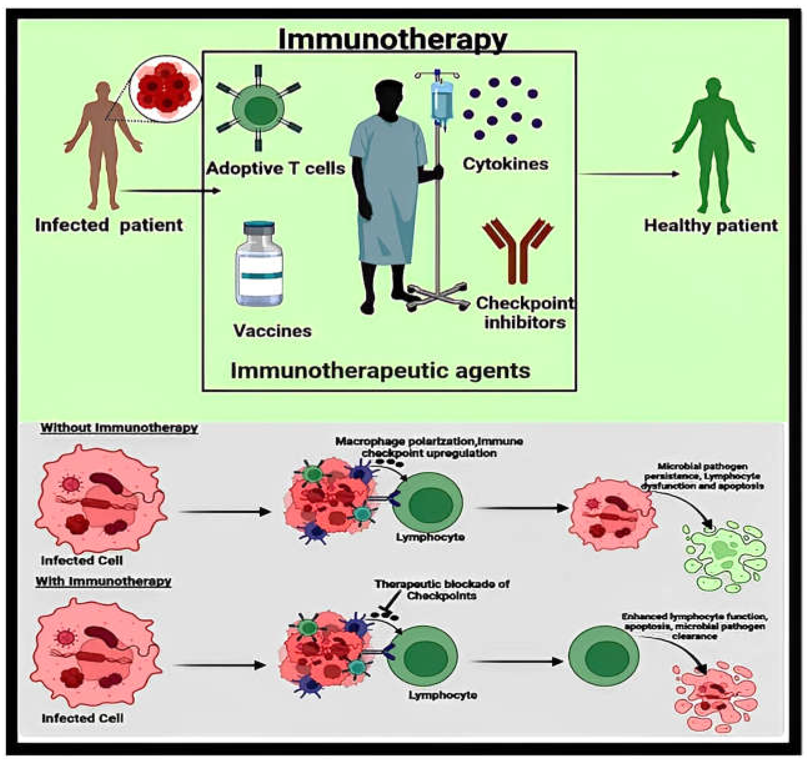
In this context, immunotherapies have been proposed as a potential solution, given their ability to enhance the host’s immune system toward combating acquired infections. These therapies, including monoclonal antibodies and vaccines, effectively modulate the host’s innate and adaptive immune responses, providing alternative strategies for treating pathogenic infections and advancing our understanding of pathogen-host immune interactions. For better clarity, Figure 6 offers a visual representation of the potential benefits of immunotherapy in treating infected patients, showing how therapeutic agents can target immune checkpoints to restore effective immune control over infections.
Conventional vaccines have benefited from certain advancements over the years, yet improvements are still necessary to avoid their associated drawbacks. Vaccines have been linked with issues like low immunogenicity, toxicity, instability, and the necessity for multiple administrations [120]. Moreover, the effective internalization of antigens by immune cells is vital for inducing a robust immunological response, indicating the advantages that different carriers may bring in delivering vaccination antigens [43].
In this context, nanotechnology has significantly progressed the creation of vaccines, which have recently been integrated into novel formulations. The utilization of different nanoparticles provides important benefits, including excellent stability, simplified administration, and improved effectiveness. Nanocarrier-based delivery systems also enhance both cellular and humoral immune responses, improve antigen immunogenicity, and enable targeted delivery and controlled release. In addition, they can be equipped with adjuvants or surface modifiers to regulate the release and dispersion of vaccine constituents [16,120,121].
The crucial role of nanotechnology in vaccine formulations for preventing severe outcomes and pathogen mutations was recently demonstrated through the success of the nanoparticle-based Moderna and Pfizer/BioNTech COVID-19 vaccines [38,122,123]. This precedent underscores the promising future of nanotechnology in vaccine development, marking the worldwide implementation of a new generation of vaccines. In more detail, COVID-19 vaccines employ LNPs encapsulated with therapeutic nucleic acids (TNA), being the most well-known current example of a clinically accepted delivery nanosystem. Nonetheless, LNPs loaded with TNA have also shown promise for managing other infectious diseases [38]. Several such conditions include, but are not limited to, influenza [124,125], cytomegalovirus [56,126], Zika virus [51], chikungunya virus [59], human immunodeficiency virus (HIV) [127,128], hepatitis B [129,130,131], hepatitis C [132,133,134], and genital herpes [135].
Other nanomaterials have also been considered valuable as carriers of vaccine antigens. Nanoparticle platforms, including lipid nanoparticles, nanoemulsions, virus-like particles (VLPs), polymeric nanoparticles, liposomes, micelles, and polymeric micelles, are valuable carriers of vaccine antigens due to their ability to enhance antigen stability and efficacy. As illustrated in Figure 7, these materials facilitate antigen delivery via improved uptake by Antigen Presenting Cells (APCs), such as neutrophils, macrophages, and dendritic cells. Once internalized via endocytosis, the antigens are processed through proteasome complexes and presented via major histocompatibility complexes (MHC) on the surface of APCs. This process triggers the activation of both CD8+ cytotoxic T cells and CD4+ helper T cells, promoting a strong adaptive immune response. Helper T cells further aid in the differentiation of memory B cells and plasma cells, resulting in the production of antigen-specific antibodies. These benefits are correlated to the particles’ extremely small size, making it easier for APCs to internalize them, allowing for effective antigen recognition, and boosting the overall vaccine efficacy [120,121].
Moreover, these nanomaterials can be surface-functionalized with different targeting moieties, enabling the precise transport of antigens to specific receptors on the cell surface producing selective and specific immune responses [120]. An interesting example of surface-modified nanoparticles for vaccine formulations has been reported by Xu and colleagues [136]. The authors developed a dual-polymer modified GO (GO–PEG–PEI) vaccine adjuvant for immunotherapy using urease B (Ure B), an antigen specific to Helicobacter pylori. The proposed nanostructured system was found to enhance dendritic cell maturation and cytokine secretion through TLR pathways while effectively delivering antigens, and inducing a stronger cellular immunity compared to free Ure B and aluminum-adjuvant-based vaccines. Therefore, the study highlights the importance of surface chemistry in nano-adjuvant design.
Differently, Vicente et al. [137] explored the possibility of nasal vaccination that generates humoral and cellular immune responses. The researchers employed a nanocarrier comprising a hydrophobic core (that can be loaded with immunostimulants) and a chitosan shell that can associate antigens. For this concept, the authors utilized imiquimod and hepatitis B surface antigens as model molecules. The nanovaccine formulation was successfully internalized by macrophages, inducing the secretion of pro-inflammatory cytokines and eliciting a specific and predominant Th1-mediated immune response.
More recently, Wu et al. [138] created delivery vehicles for antigens based on transferosomes with opposite surface charges, and transdermal formulations were administered through dissolving microneedles. In their study, cationic nanocarriers with ovalbumin as the model antigen performed better than anionic vehicles, displaying improved escape capacities from endocytic compartments, accumulating in higher amounts within lymph nodes, activating more efficiently DC maturation, and inducing Th1 immunity.

4.3. Allergic and Inflammatory Disorders

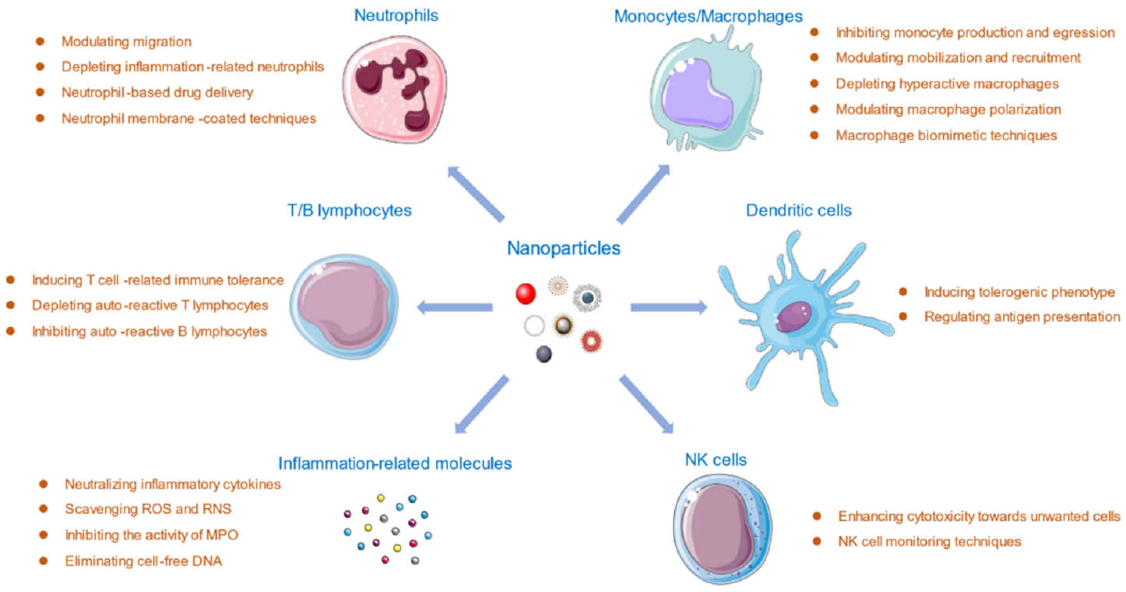
Inflammation represents a multifaceted physicochemical response of living tissues subjected to physical or chemical aggression, comprising the activation of enzymes, mithe gration of cells, the breakdown of tissues, and the release of mediators [139]. Inflammation is a common factor in numerous conditions, including obesity, diabetes, allergies, psychiatric diseases, and skin disorders [140,141,142], being an important process to target to alleviate associated symptoms. Inflammatory diseases are generally managed by administering anti-inflammatory drugs. Nonetheless, conventional anti-inflammatory agents lack tissue specificity, may produce systemic side effects, and exhibit limited efficacy in crossing biological barriers. To overcome these challenges, nanomaterials can be used to deliver anti-inflammatory agents to enhance their activity, especially as nanoparticles can interact with the components of the immune system in several ways. In more detail, the influence of nanoparticles on T/B lymphocytes, neutrophils, monocytes/macrophages, dendritic cells, natural killer (NK) cells, and inflammation-related molecules is displayed in Figure 8 [27].
For instance, Lee and colleagues [114] fabricated a nanoformulation based on hyaluronic acid and bilirubin to manage the inflammation of the colonic epithelium. The nanomedicine was tested on a murine model of acute colitis, leading to encouraging results. Namely, the proposed strategy restored the epithelium barriers, modulated gut microbiota, regulated innate immune responses, and displayed promising therapeutic efficacy against targeted conditions, thus representing a good candidate for treating inflammatory diseases.
On a different note, a few studies reported the implication of nanomaterials in allergen-specific immunotherapy as they offer advantages as both carriers and adjuvants [143,144]. For example, Wang et al. [18] have evaluated the immunological effects of intravenously administered graphene nanosheets and MWCNTs, focusing on their capacity to induce site-specific Th2 inflammatory responses through the IL-33/ST2 signaling axis. The researchers showed that both nanomaterials trigger a Th2-skewed immune response characterized by elevated levels of IL-33 and its receptor ST2, producing localized inflammation. The shift inTh1/Th2 balance may further impact host defense and exacerbate allergic diseases like asthma and anaphylaxis; these immunotoxicological effects may be attributed to the varied physicochemical properties of the delivery vehicles. Overall, the study emphasized the potential immunomodulatory effects of graphene-based and carbon nanotube nanomaterials, highlighting the importance of understanding their biological interactions to safe biomedical applications.
On the other hand, Ryan et al. [145] focused their research on fullerenes, investigating their immunotherapeutic role. The researchers have evaluated the impact of these CBNs on allergic responses, demonstrating that these nanomaterials effectively inhibit allergic reactions by modulating immune responses. The study results reveal that fullerenes can reduce the production of key pro-inflammatory cytokines and suppress immune cells’ activation in allergic pathways. These findings suggest that fullerene nanomaterials hold potential as therapeutic agents for managing allergic diseases, offering a novel approach to modulate immune function and mitigate allergic responses in mast cell-dependent conditions like asthma, inflammatory arthritis, heart disease, and multiple sclerosis.
Alternatively, the study conducted by Pali-Schöll and colleagues [146] has explored the use of biodegradable protamine nanoparticles loaded with CpG-oligodeoxynucleotides (CpG-ODN) in preventing allergen-induced Th2 immune responses in BALB/c mice. Encouraging results were obtained, with the nanoformulation effectively shifting the immune response from a Th2-dominated profile, which is associated with allergies, towards a Th1 response, characterized by reduced allergic inflammation and the enhanced production of IFN-γ. These outcomes highlight the promise these delivery systems hold for mitigating allergic diseases by modulating the immune response.
Pereira et al. [147] also reported an interesting study in this domain. The authors fabricated and tested poly(anhydride) nanoparticles loaded with cashew nut allergens as a strategy for cashew nut oral immunization. The study results reveal that the developed nanoformulation promotes a strong Th1-mediated and Treg immune response, highlighting its potential as a candidate for the oral delivery of allergens to modulate immune responses in food allergy treatments.
Differently, Garaczi et al. [148] developed DermAll, a nanomedicine designed for allergen-specific immunotherapy based on a synthetic plasmid pDNA/PEIm and ovalbumin as a model allergen. The proposed approach effectively modulated the immune response by promoting a shift towards a Th1-type response and inducing regulatory T cells, thereby reducing the Th2-mediated allergic response. These findings suggest that DermAll offers a promising alternative for allergen-specific immunotherapy, with potential benefits in terms of both efficacy and patient compliance.
Tasaniyananda et al. [149] focused their research on cat allergic rhinitis, testing the efficacy of an intranasal liposome-adjuvanted vaccine containing refined Fel d 1 on a mouse model. The obtained results demonstrate that the vaccine effectively reduces allergic symptoms and airway inflammation in sensitized mice by inducing a shift from a Th2-dominated response towards a Th1 and regulatory T-cell response. This suggests that the intranasal liposome-adjuvanted Fel d 1 vaccine could be a promising therapeutic approach for managing cat allergy, offering potential advantages in targeting local immune responses in the nasal mucosa. Aliu et al. [150] also addressed the benefits of using liposomes for delivering allergens. Specifically, the researchers evaluated neutral and cationic liposomes as delivery systems of ovalbumin for allergy treatment by sublingual immunotherapy (SLIT). The study confirms that encapsulating allergens within liposomes improves their stability and bioavailability, leading to a more robust immune response characterized by a shift towards a Th1/Th2 balance and the increased production of Tregs, offering a promising approach for more effective SLIT.
Overall, several interesting and encouraging results have been obtained concerning the nanomaterial-mediated immunotherapy of allergies and inflammatory diseases. However, most identified studies are over a decade old, and little progress has been registered since their publication. Hence, research in this field seems to have been stalled, requiring further advancements from interconnected disciplines before bringing new perspectives to clinical practice.

5. Clinically Approved Nanostructured Drug Delivery Systems

Nanostructured drug delivery systems have gained significant attention in immunotherapy, with several formulations receiving FDA approval. LNP-based and liposomal drugs are particularly prominent due to their biocompatibility, drug-loading capacity, and ability to reduce toxicity. One of the most representative examples is Doxil®, the first FDA-approved nanodrug (1995), which uses PEGylated liposomes to enhance the delivery of doxorubicin in cancer treatment. Other approved formulations using liposomes as nanocarriers, such as Myocet®, DaunoXome®, and Onivyde®, have been developed to treat various cancers, offering prolonged circulation time and reduced side effects. LNPs also played a critical role in mRNA vaccine development during the COVID-19 pandemic, with Comirnaty™ and Spikevax® demonstrating the versatility of nanotechnology in modern medicine [151,152,153,154].
Polymer-based nanostructured drug delivery systems have also been explored in immunotherapy, although fewer formulations have received regulatory authorities’ approval compared to lipid-based counterparts. One key example is Abraxane®, an albumin-bound nanoparticle formulation approved in 2005 for delivering paclitaxel to treat various cancers. Additionally, polymer micelle-based nanosystems like Genexol®, Nanoxel®, and Apealea® have been developed for delivering paclitaxel or docetaxel to treat advanced cancers, offering improved solubility, reduced toxicity, and enhanced drug delivery to tumor sites. These polymeric systems, along with lipid-based nanoparticles, emphasize the tremendous potential of using nanostructured drug delivery systems in dealing with malignant diseases, offering new perspectives for improving immunotherapeutic treatment options and raising hopes for translating under-research formulations to the clinical setting [33,151,152,154].
To provide a comprehensive overview of the current landscape of clinically approved nanostructured drug delivery systems, we have compiled a summary of key examples, including lipid- and polymer-based formulations. In this respect, Table 2 highlights critical attributes such as drug formulation, therapeutic benefits, target indications, and approval status, offering insights into the diverse nanomedicines that have successfully transitioned from research to clinical use.

6. Remaining Challenges, Emerging Trends, and Future Perspectives

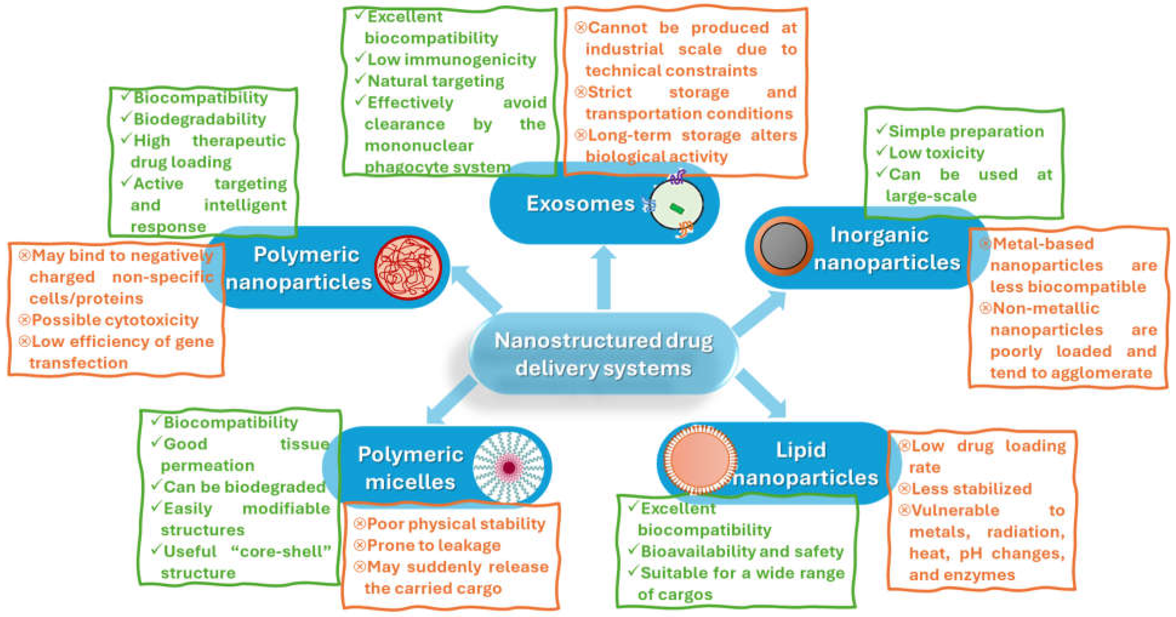
Nanostructured drug delivery systems in immunotherapy have shown immense potential in enhancing the effectiveness and specificity of treatments, particularly in cancer and autoimmune diseases. However, there are still significant limitations to address (Figure 9), emerging trends to consider, and future perspectives worth mentioning that are likely to shape the field.
One of the challenges still faced in using nanocarriers is the achievement of high specificity in targeting immune cells. For instance, targeting precision when aiming at mucosa-resident immune cells is limited by the evolution of mucosal barriers toward blocking incoming pathogens and foreign materials [174]. Moreover, the immune system exhibits heterogeneity across patients, making it impossible to engineer one-size-fits-all nanostructured delivery systems for immunotherapeutic agents [175,176]. Thus, fixing specificity issues and approaching treatments in a personalized manner are essential approaches in mitigating off-target effects and augmenting the efficacy of developed systems in each patient.
In this respect, the advances registered in fields like genomics and proteomics can be tackled in designing personalized nanoparticles that cater to the specific immunological profile of an individual patient [177]. Another possibility for reducing off-target activity is using smart nanoparticles that can adapt to the microenvironment of the tumor or inflamed tissue. This emerging trend assumes advanced targeting mechanisms like ligand-mediated and responsive delivery, and has been increasingly exploited in recent studies [8,28,29,178]. Besides this, related to the efficiency of nanoparticle-mediated immunotherapeutic systems, there is a growing trend towards developing nanoparticles that can carry multiple agents, such as drugs, genes, or imaging agents, to provide a combined therapeutic and diagnostic (theranostic) approach [81,179].
Adverse effects can also occur when using nanoparticle-mediated immunotherapies. Some nanomaterials can be immunogenic, resulting in the unwanted and unintended activation or suppression of the immune system [18,180]. The ways in which innate and adaptive immune systems interact with nanomaterials are not yet fully understood, necessitating in-depth research for better comprehending and controlling the immunogenicity of nanoparticles [181,182,183]. Until then, ensuring that nanomaterials do not elicit harmful immune responses remains a challenge.
Emerging trends for solving the biocompatibility issue revolve around creating biomimetic nanoparticles. Specifically, cell-mimicking nanoparticles may be used as carriers for immunotherapeutic agents, evading the immune system or enhancing immune targeting due to their similar features with immune cells or pathogens [178,184,185]. Another possibility is to employ natural carriers, such as exosomes and cell membrane-based nanoparticles, which benefit from inherent biocompatibility and targeting potential [178,186].
Other remaining challenges to be solved before translating nanoparticle-based delivery systems to clinical practice include the stability and shelf-life of the formulations, regulatory and safety concerns, and the scalability of the fabrication methods. In more detail, factors like storage conditions, long-term effects, safety, and efficacy standards must be clearly established before producing complex multifunctional nanoparticulated systems at an industrial scale. Additionally, at a large scale, manufacturing processes may lead to high costs, limiting their widespread adoption [185,187,188,189].
As concerns future perspectives, they should be envisaged in correlation with the existing framework and the “open threads” left from current nanoformulations proposed for immunotherapy. One possibility is to optimize the nanosystems developed years ago and “abandoned” at in vitro/in vivo testing stages despite obtaining encouraging results at the time. More in-depth research is required to address the identified challenges and move further with the progression of innovative nanomedicines toward clinical studies, eventually bringing new market options.
In connection with these aspects, artificial intelligence and machine learning can be used in predicting and optimizing the interactions between nanoparticles and the immune system. These technologies can actively contribute to engineering more effective and personalized nanostructured delivery systems, aiding in drug design through their ability to adapt surplus amounts of data available for generating meaningful insights [190].
Another appealing possibility for bringing about progress in nanoparticle-mediated immunotherapy is its integration with advanced therapies like CRISPR and gene editing. Gene editing represents a cutting-edge technology in the field of gene therapy technologies. CRISPR technology has revolutionized cancer treatment by offering new therapeutic alternatives, particularly in the field of cancer immunotherapy. Therefore, scientists are strongly driven to develop a remedy for numerous challenging disorders through the utilization of CRISPR technology. Nonetheless, several obstacles persist and require resolutions for the effective implementation of gene editing [66,191,192].
Interesting developments in the field may also be foreseen once the fabrication of uniform nanoparticles with controllable features is achieved though low-cost, energy-efficient methods. This is an essential aspect that needs to be solved to ensure nanostructured drug delivery systems’ scalability while also considering the production processes’ environmental impact. One potential solution is to use microfluidic synthesis to diminish reagent consumption, reduce waste generation, and limit batch-to-batch variations [193].
Future perspectives should also reflect upon the public perception and adoption of newly developed immunotherapeutic alternatives. Increasing the public’s awareness and understanding of nanomedicine’s benefits and risks is essential for building trust and support for these emerging technologies. Transparent communication and engagement and ensuring equitable access to nanostructured drug delivery systems across all populations, including those in low- and middle-income countries, are critical factors in implementing these new solutions and improving the quality of life of as many patients as possible.

7. Conclusions

Nanostructured drug delivery systems used in immunotherapy have shown great promise in enhancing the effectiveness and specificity of treatments, being especially exploited in cancer applications. The intrinsic immunomodulatory properties and versatility of nanoparticles used as carriers can significantly enhance the effectiveness of nanostructured systems in delivering immunotherapeutic agents, as demonstrated by numerous studies in the field. Key factors like size, surface charge, functionalization, and the ability to bind diverse biomolecules critically influence their biocompatibility with target immune cells. By mimicking specific foreign signals, nanomaterials can modify the adaptive immune response, enabling the development of innovative biological systems for immunotherapy.
Nonetheless, several challenges remain, including the following: specificity issues, the immunogenicity of different nanomaterials, uncertain long-term safety, and manufacturing costs. Yet continuous efforts are being directed toward overcoming these drawbacks, with emerging trends and perspectives suggesting that while using nanostructured drug delivery systems in immunotherapy hold great promise, the field is still evolving. Very few clinical studies have investigated the administration of immunotherapy via drug delivery systems, with this testing stage being noted for cancer and infectious diseases. Thus, upgrading the clinical trials framework and bringing new formulations to clinical testing is essential. Moreover, nanostructured drug delivery systems for other immunotherapeutic applications should be considered in future clinical trials to extend the availability of solutions for other medical conditions, such as autoimmune diseases and allergic and inflammatory disorders.
To conclude, nanostructured drug delivery systems offer a strong basis for immunotherapeutic innovations. Multidisciplinary collaboration and continued improvement will be essential to overcome current barriers and fully realize the potential of these advanced therapeutic approaches.

Funding

This research received no external funding.

Conflicts of Interest

The authors declare no conflicts of interest.

References

  1. Sorci, G.; Cornet, S.; Faivre, B. Immune Evasion, Immunopathology and the Regulation of the Immune System. Pathogens 2013, 2, 71–91. [Google Scholar] [CrossRef] [PubMed]
  2. Mitarotonda, R.; Giorgi, E.; Eufrasio-da-Silva, T.; Dolatshahi-Pirouz, A.; Mishra, Y.K.; Khademhosseini, A.; Desimone, M.F.; De Marzi, M.; Orive, G. Immunotherapeutic nanoparticles: From autoimmune disease control to the development of vaccines. Biomater. Adv. 2022, 135, 212726. [Google Scholar] [CrossRef]
  3. Azevedo, S.; Costa-Almeida, R.; Santos, S.G.; Magalhães, F.D.; Pinto, A.M. Advances in carbon nanomaterials for immunotherapy. Appl. Mater. Today 2022, 27, 101397. [Google Scholar] [CrossRef]
  4. Yan, Z.; Yang, W.; Wei, H.; Dean, M.N.; Standaert, D.G.; Cutter, G.R.; Benveniste, E.N.; Qin, H. Dysregulation of the adaptive immune system in patients with early-stage Parkinson disease. Neurol. Neuroimmunol. Neuroinflammation 2021, 8, e1036. [Google Scholar] [CrossRef] [PubMed]
  5. Long, A.; Kleiner, A.; Looney, R.J. Immune dysregulation. J. Allergy Clin. Immunol. 2023, 151, 70–80. [Google Scholar] [CrossRef]
  6. Vinay, D.S.; Ryan, E.P.; Pawelec, G.; Talib, W.H.; Stagg, J.; Elkord, E.; Lichtor, T.; Decker, W.K.; Whelan, R.L.; Kumara, H.M.C.S.; et al. Immune evasion in cancer: Mechanistic basis and therapeutic strategies. Semin. Cancer Biol. 2015, 35, S185–S198. [Google Scholar] [CrossRef]
  7. Vaillant, A.A.J.; Qurie, A. Immunodeficiency. In StatPearls; StatPearls Publishing: Treasure Island, FL, USA, 2023. [Google Scholar]
  8. Anfray, C.; Mainini, F.; Andón, F.T. Nanoparticles for Immunotherapy. Front. Nanosci. 2020, 16, 265–306. [Google Scholar]
  9. Toy, R.; Roy, K. Engineering nanoparticles to overcome barriers to immunotherapy. Bioeng. Transl. Med. 2016, 1, 47–62. [Google Scholar] [CrossRef]
  10. Varshney, D.; Qiu, S.Y.; Graf, T.P.; McHugh, K.J. Employing drug delivery strategies to overcome challenges using TLR7/8 agonists for cancer immunotherapy. AAPS J. 2021, 23, 90. [Google Scholar] [CrossRef]
  11. Sharma, P.; Hu-Lieskovan, S.; Wargo, J.A.; Ribas, A. Primary, adaptive, and acquired resistance to cancer immunotherapy. Cell 2017, 168, 707–723. [Google Scholar] [CrossRef]
  12. Dobosz, P.; Stępień, M.; Golke, A.; Dzieciątkowski, T. Challenges of the Immunotherapy: Perspectives and Limitations of the Immune Checkpoint Inhibitor Treatment. Int. J. Mol. Sci. 2022, 23, 2847. [Google Scholar] [CrossRef] [PubMed]
  13. Li, Y.-Z.; Zhang, H.-M. Recent advances in primary resistance mechanisms against immune checkpoint inhibitors. Curr. Opin. Oncol. 2022, 34, 95–106. [Google Scholar] [CrossRef] [PubMed]
  14. Kluger, H.M.; Tawbi, H.A.; Ascierto, M.L.; Bowden, M.; Callahan, M.K.; Cha, E.; Chen, H.X.; Drake, C.G.; Feltquate, D.M.; Ferris, R.L.; et al. Defining tumor resistance to PD-1 pathway blockade: Recommendations from the first meeting of the SITC Immunotherapy Resistance Taskforce. J. Immunother. Cancer 2020, 8, e000398. [Google Scholar] [CrossRef] [PubMed]
  15. Bixenmann, L.; Stickdorn, J.; Nuhn, L. Amphiphilic poly (esteracetal) s as dual pH-and enzyme-responsive micellar immunodrug delivery systems. Polym. Chem. 2020, 11, 2441–2456. [Google Scholar] [CrossRef]
  16. Croitoru, G.-A.; Pîrvulescu, D.-C.; Niculescu, A.-G.; Epistatu, D.; Rădulescu, M.; Grumezescu, A.M.; Nicolae, C.-L. Nanomaterials in Immunology: Bridging Innovative Approaches in Immune Modulation, Diagnostics, and Therapy. J. Funct. Biomater. 2024, 15, 225. [Google Scholar] [CrossRef]
  17. Salem, S.S.; Hammad, E.N.; Mohamed, A.A.; El-Dougdoug, W. A comprehensive review of nanomaterials: Types, synthesis, characterization, and applications. Biointerface Res. Appl. Chem. 2022, 13, 41. [Google Scholar]
  18. Wang, X.; Podila, R.; Shannahan, J.H.; Rao, A.M.; Brown, J.M. Intravenously delivered graphene nanosheets and multiwalled carbon nanotubes induce site-specific Th2 inflammatory responses via the IL-33/ST2 axis. Int. J. Nanomed. 2013, 8, 1733–1748. [Google Scholar] [CrossRef]
  19. Sharma, N.; Singh, S.; Behl, T.; Gupta, N.; Gulia, R.; Kanojia, N. Explicating the applications of quality by design tools in optimization of microparticles and nanotechnology based drug delivery systems. Biointerface Res. Appl. Chem. 2022, 12, 4317–4336. [Google Scholar]
  20. Panda, B.S. A review on synthesis of silver nanoparticles and their biomedical applications. Lett. Appl. NanoBioScience 2022, 11, 3218–3231. [Google Scholar]
  21. Horwitz, D.A.; Bickerton, S.; La Cava, A. Strategies to use nanoparticles to generate CD4 and CD8 regulatory T cells for the treatment of SLE and other autoimmune diseases. Front. Immunol. 2021, 12, 681062. [Google Scholar] [CrossRef]
  22. Milewska, S.; Niemirowicz-Laskowska, K.; Siemiaszko, G.; Nowicki, P.; Wilczewska, A.Z.; Car, H. Current Trends and Challenges in Pharmacoeconomic Aspects of Nanocarriers as Drug Delivery Systems for Cancer Treatment. Int. J. Nanomed. 2021, 16, 6593–6644. [Google Scholar] [CrossRef] [PubMed]
  23. Shah, A.; Aftab, S.; Nisar, J.; Ashiq, M.N.; Iftikhar, F.J. Nanocarriers for targeted drug delivery. J. Drug Deliv. Sci. Technol. 2021, 62, 102426. [Google Scholar] [CrossRef]
  24. Sultana, A.; Zare, M.; Thomas, V.; Kumar, T.S.S.; Ramakrishna, S. Nano-based drug delivery systems: Conventional drug delivery routes, recent developments and future prospects. Med. Drug Discov. 2022, 15, 100134. [Google Scholar] [CrossRef]
  25. Radzi, M.R.M.; Lim, C.K.; Sulaiman, N.; Jemon, K. Nanoparticles-based Chemo-Phototherapy Synergistic Effects for Breast Cancer Treatment: A Systematic Review. Biointerface Res. Appl. Chem. 2023, 13, 14764. [Google Scholar]
  26. Llop, J.; Lammers, T. Nanoparticles for Cancer Diagnosis, Radionuclide Therapy and Theranostics. ACS Nano 2021, 15, 16974–16981. [Google Scholar] [CrossRef]
  27. Liu, J.; Liu, Z.; Pang, Y.; Zhou, H. The interaction between nanoparticles and immune system: Application in the treatment of inflammatory diseases. J. Nanobiotechnol. 2022, 20, 127. [Google Scholar] [CrossRef]
  28. Eljack, S.; David, S.; Faggad, A.; Chourpa, I.; Allard-Vannier, E. Nanoparticles design considerations to co-deliver nucleic acids and anti-cancer drugs for chemoresistance reversal. Int. J. Pharm. X 2022, 4, 100126. [Google Scholar] [CrossRef]
  29. Zhang, J.; Lin, Y.; Lin, Z.; Wei, Q.; Qian, J.; Ruan, R.; Jiang, X.; Hou, L.; Song, J.; Ding, J.; et al. Stimuli-Responsive Nanoparticles for Controlled Drug Delivery in Synergistic Cancer Immunotherapy. Adv. Sci. 2022, 9, 2103444. [Google Scholar] [CrossRef]
  30. Mohammapdour, R.; Ghandehari, H. Mechanisms of immune response to inorganic nanoparticles and their degradation products. Adv. Drug Deliv. Rev. 2022, 180, 114022. [Google Scholar] [CrossRef]
  31. Bi, J.; Mo, C.; Li, S.; Huang, M.; Lin, Y.; Yuan, P.; Liu, Z.; Jia, B.; Xu, S. Immunotoxicity of metal and metal oxide nanoparticles: From toxic mechanisms to metabolism and outcomes. Biomater. Sci. 2023, 11, 4151–4183. [Google Scholar] [CrossRef]
  32. Ahamad, N.; Kar, A.; Mehta, S.; Dewani, M.; Ravichandran, V.; Bhardwaj, P.; Sharma, S.; Banerjee, R. Immunomodulatory nanosystems for treating inflammatory diseases. Biomaterials 2021, 274, 120875. [Google Scholar] [CrossRef] [PubMed]
  33. Niculescu, A.-G.; Grumezescu, A.M. Polymer-Based Nanosystems—A Versatile Delivery Approach. Materials 2021, 14, 6812. [Google Scholar] [CrossRef] [PubMed]
  34. Yu, Z.; Shen, X.; Yu, H.; Tu, H.; Chittasupho, C.; Zhao, Y. Smart Polymeric Nanoparticles in Cancer Immunotherapy. Pharmaceutics 2023, 15, 775. [Google Scholar] [CrossRef] [PubMed]
  35. Craparo, E.F.; Bondì, M.L. Application of polymeric nanoparticles in immunotherapy. Curr. Opin. Allergy Clin. Immunol. 2012, 12, 658–664. [Google Scholar] [CrossRef]
  36. Samaridou, E.; Heyes, J.; Lutwyche, P. Lipid nanoparticles for nucleic acid delivery: Current perspectives. Adv. Drug Deliv. Rev. 2020, 154, 37–63. [Google Scholar] [CrossRef]
  37. Aldosari, B.N.; Alfagih, I.M.; Almurshedi, A.S. Lipid nanoparticles as delivery systems for RNA-based vaccines. Pharmaceutics 2021, 13, 206. [Google Scholar] [CrossRef]
  38. Niculescu, A.-G.; Bîrcă, A.C.; Grumezescu, A.M. New Applications of Lipid and Polymer-Based Nanoparticles for Nucleic Acids Delivery. Pharmaceutics 2021, 13, 2053. [Google Scholar] [CrossRef] [PubMed]
  39. Han, J.; Lim, J.; Wang, C.J.; Han, J.H.; Shin, H.E.; Kim, S.N.; Jeong, D.; Lee, S.H.; Chun, B.H.; Park, C.G.; et al. Lipid nanoparticle-based mRNA delivery systems for cancer immunotherapy. Nano Converg. 2023, 10, 36. [Google Scholar] [CrossRef]
  40. Wang, H.-L.; Wang, Z.-G.; Liu, S.-L. Lipid Nanoparticles for mRNA Delivery to Enhance Cancer Immunotherapy. Molecules 2022, 27, 5607. [Google Scholar] [CrossRef]
  41. Wu, L.; Li, X.; Qian, X.; Wang, S.; Liu, J.; Yan, J. Lipid Nanoparticle (LNP) Delivery Carrier-Assisted Targeted Controlled Release mRNA Vaccines in Tumor Immunity. Vaccines 2024, 12, 186. [Google Scholar] [CrossRef]
  42. Papi, M.; De Spirito, M.; Palmieri, V. Nanotechnology in the COVID-19 era: Carbon-based nanomaterials as a promising solution. Carbon 2023, 210, 118058. [Google Scholar] [CrossRef] [PubMed]
  43. Cao, W.; He, L.; Cao, W.; Huang, X.; Jia, K.; Dai, J. Recent progress of graphene oxide as a potential vaccine carrier and adjuvant. Acta Biomater. 2020, 112, 14–28. [Google Scholar] [CrossRef] [PubMed]
  44. Hosseini, S.M.; Mohammadnejad, J.; Najafi-Taher, R.; Zadeh, Z.B.; Tanhaei, M.; Ramakrishna, S. Multifunctional Carbon-Based Nanoparticles: Theranostic Applications in Cancer Therapy and Diagnosis. ACS Appl. Bio Mater. 2023, 6, 1323–1338. [Google Scholar] [CrossRef] [PubMed]
  45. Sharma, A.; Vaswani, P.; Bhatia, D. Revolutionizing cancer therapy using tetrahedral DNA nanostructures as intelligent drug delivery systems. Nanoscale Adv. 2024, 6, 3714–3732. [Google Scholar] [CrossRef] [PubMed]
  46. Durbin, J.K.; Miller, D.K.; Niekamp, J.; Khisamutdinov, E.F. Modulating Immune Response with Nucleic Acid Nanoparticles. Molecules 2019, 24, 3740. [Google Scholar] [CrossRef]
  47. Khisamutdinov, E.F.; Bui, M.N.H.; Jasinski, D.; Zhao, Z.; Cui, Z.; Guo, P. Simple Method for Constructing RNA Triangle, Square, Pentagon by Tuning Interior RNA 3WJ Angle from 60° to 90° or 108°. In RNA Scaffolds: Methods and Protocols; Ponchon, L., Ed.; Springer New York: New York, NY, USA, 2015; pp. 181–193. [Google Scholar]
  48. Khisamutdinov, E.F.; Li, H.; Jasinski, D.L.; Chen, J.; Fu, J.; Guo, P. Enhancing immunomodulation on innate immunity by shape transition among RNA triangle, square and pentagon nanovehicles. Nucleic Acids Res. 2014, 42, 9996–10004. [Google Scholar] [CrossRef]
  49. Afonin, K.A.; Bindewald, E.; Yaghoubian, A.J.; Voss, N.; Jacovetty, E.; Shapiro, B.A.; Jaeger, L. In vitro assembly of cubic RNA-based scaffolds designed in silico. Nat. Nanotechnol. 2010, 5, 676–682. [Google Scholar] [CrossRef]
  50. Afonin, K.A.; Kasprzak, W.; Bindewald, E.; Puppala, P.S.; Diehl, A.R.; Hall, K.T.; Kim, T.J.; Zimmermann, M.T.; Jernigan, R.L.; Jaeger, L. Computational and Experimental Characterization of RNA Cubic Nanoscaffolds. In Therapeutic RNA Nanotechnology; Jenny Stanford Publishing: Dubai, United Arab Emirates, 2021; pp. 121–149. [Google Scholar]
  51. Iinuma, R.; Ke, Y.; Jungmann, R.; Schlichthaerle, T.; Woehrstein, J.B.; Yin, P. Polyhedra Self-Assembled from DNA Tripods and Characterized with 3D DNA-PAINT. Science 2014, 344, 65–69. [Google Scholar] [CrossRef]
  52. Khisamutdinov, E.F.; Jasinski, D.L.; Li, H.; Zhang, K.; Chiu, W.; Guo, P. Fabrication of RNA 3D nanoprism for loading and protection of small RNAs and model drugs. Adv. Mater. 2016, 28, 10079. [Google Scholar] [CrossRef]
  53. Grabow, W.W.; Zakrevsky, P.; Afonin, K.A.; Chworos, A.; Shapiro, B.A.; Jaeger, L. Self-Assembling RNA Nanorings Based on RNAI/II Inverse Kissing Complexes. In Therapeutic RNA Nanotechnology; Jenny Stanford Publishing: Dubai, United Arab Emirates, 2021; pp. 67–93. [Google Scholar]
  54. Illig, M.; Jahnke, K.; Weise, L.P.; Scheffold, M.; Mersdorf, U.; Drechsler, H.; Zhang, Y.; Diez, S.; Kierfeld, J.; Göpfrich, K. Triggered contraction of self-assembled micron-scale DNA nanotube rings. Nat. Commun. 2024, 15, 2307. [Google Scholar] [CrossRef]
  55. Glynn, A.T.; Davidson, S.R.; Qian, L. Developmental Self-Assembly of a DNA Ring with Stimulus-Responsive Size and Growth Direction. J. Am. Chem. Soc. 2022, 144, 10075–10079. [Google Scholar] [CrossRef] [PubMed]
  56. Roller, E.-M.; Khorashad, L.K.; Fedoruk, M.; Schreiber, R.; Govorov, A.O.; Liedl, T. DNA-Assembled Nanoparticle Rings Exhibit Electric and Magnetic Resonances at Visible Frequencies. Nano Lett. 2015, 15, 1368–1373. [Google Scholar] [CrossRef] [PubMed]
  57. Guo, S.; Piao, X.; Li, H.; Guo, P. Methods for construction and characterization of simple or special multifunctional RNA nanoparticles based on the 3WJ of phi29 DNA packaging motor. Methods 2018, 143, 121–133. [Google Scholar] [CrossRef] [PubMed]
  58. Nakano, K.; Sawada, T.; Mori, Y.; Morita, K.; Ishimatsu, R. Covalent Hyperbranched Polymer Self-Assemblies of Three-Way Junction DNA for Single-Molecule Devices. Langmuir 2020, 36, 10166–10174. [Google Scholar] [CrossRef] [PubMed]
  59. Vittala, S.K.; Saraswathi, S.K.; Ramesan, A.B.; Joseph, J. Nanosheets and 2D-nanonetworks by mutually assisted self-assembly of fullerene clusters and DNA three-way junctions. Nanoscale Adv. 2019, 1, 4158–4165. [Google Scholar] [CrossRef]
  60. Takezawa, Y.; Shionoya, M. Supramolecular DNA three-way junction motifs with a bridging metal center. Front. Chem. 2020, 7, 925. [Google Scholar] [CrossRef]
  61. Haque, F.; Shu, D.; Shu, Y.; Shlyakhtenko, L.S.; Rychahou, P.G.; Evers, B.M.; Guo, P. Ultrastable synergistic tetravalent RNA nanoparticles for targeting to cancers. Nano Today 2012, 7, 245–257. [Google Scholar] [CrossRef]
  62. Bi, S.; Xiu, B.; Ye, J.; Dong, Y. Target-Catalyzed DNA Four-Way Junctions for CRET Imaging of MicroRNA, Concatenated Logic Operations, and Self-Assembly of DNA Nanohydrogels for Targeted Drug Delivery. ACS Appl. Mater. Interfaces 2015, 7, 23310–23319. [Google Scholar] [CrossRef]
  63. Li, J.; Lu, W.; Yang, Y.; Xiang, R.; Ling, Y.; Yu, C.; Zhou, Y. Hybrid Nanomaterials for Cancer Immunotherapy. Adv. Sci. 2023, 10, 2204932. [Google Scholar] [CrossRef]
  64. Gautam, A.; Beiss, V.; Wang, C.; Wang, L.; Steinmetz, N.F. Plant Viral Nanoparticle Conjugated with Anti-PD-1 Peptide for Ovarian Cancer Immunotherapy. Int. J. Mol. Sci. 2021, 22, 9733. [Google Scholar] [CrossRef]
  65. Pruteanu, L.-L.; Braicu, C.; Módos, D.; Jurj, M.-A.; Raduly, L.-Z.; Zănoagă, O.; Magdo, L.; Cojocneanu, R.; Paşca, S.; Moldovan, C.; et al. Targeting Cell Death Mechanism Specifically in Triple Negative Breast Cancer Cell Lines. Int. J. Mol. Sci. 2022, 23, 4784. [Google Scholar] [CrossRef] [PubMed]
  66. Yahya, S.; Abdelmenym Mohamed, S.I.; Yahya, S. Gene Editing: A Powerful Tool for Cancer Immunotherapy. Biointerface Res. Appl. Chem. 2023, 13, 98. [Google Scholar]
  67. Chen, C.; Wang, Z.; Ding, Y.; Qin, Y. Tumor microenvironment-mediated immune evasion in hepatocellular carcinoma. Front. Immunol. 2023, 14, 1133308. [Google Scholar] [CrossRef]
  68. Zhang, Y.; He, X.; Zhang, Y.; Zhao, Y.; Lu, S.; Peng, Y.; Lu, L.; Hu, X.; Zhan, M. Native Mitochondria-Targeting polymeric nanoparticles for mild photothermal therapy rationally potentiated with immune checkpoints blockade to inhibit tumor recurrence and metastasis. Chem. Eng. J. 2021, 424, 130171. [Google Scholar] [CrossRef]
  69. Lee, C.K.; Atibalentja, D.F.; Yao, L.E.; Park, J.; Kuruvilla, S.; Felsher, D.W. Anti-PD-L1 F(ab) Conjugated PEG-PLGA Nanoparticle Enhances Immune Checkpoint Therapy. Nanotheranostics 2022, 6, 243–255. [Google Scholar] [CrossRef] [PubMed]
  70. Souza, A.O.D. Overview of nanomaterials and cellular interactions. Biointerface Res. Appl. Chem. 2023, 13, 367. [Google Scholar]
  71. Fávaro, W.J.; dos Santos, M.M.; Pereira, M.M.; Garcia, P.V.; Durán, N. Effects of P-MAPA immunotherapy associated with gemcitabine on chemically-induced pancreatic cancer in animal model: New therapeutic perspectives. Biointerface Res. Appl. Chem. 2022, 12, 7540–7555. [Google Scholar]
  72. Zhang, H.; Tang, W.-L.; Kheirolomoom, A.; Fite, B.Z.; Wu, B.; Lau, K.; Baikoghli, M.; Raie, M.N.; Tumbale, S.K.; Foiret, J.; et al. Development of thermosensitive resiquimod-loaded liposomes for enhanced cancer immunotherapy. J. Control. Release 2021, 330, 1080–1094. [Google Scholar] [CrossRef]
  73. Park, W.; Heo, Y.-J.; Han, D.K. New opportunities for nanoparticles in cancer immunotherapy. Biomater. Res. 2018, 22, 24. [Google Scholar] [CrossRef]
  74. Kumar, A.; Saha, M.; Vishwakarma, R.; Behera, K.; Trivedi, S. Green solvents tailored nanostructures of block copolymers and their potential applications in drug delivery. J. Mol. Liq. 2024, 410, 125642. [Google Scholar] [CrossRef]
  75. Dias, M.F.; de Figueiredo, B.C.P.; Teixeira-Neto, J.; Guerra, M.C.A.; Fialho, S.L.; Silva Cunha, A. In vivo evaluation of antitumoral and antiangiogenic effect of imiquimod-loaded polymeric nanoparticles. Biomed. Pharmacother. 2018, 103, 1107–1114. [Google Scholar] [CrossRef] [PubMed]
  76. Gazzi, R.P.; Frank, L.A.; Onzi, G.; Pohlmann, A.R.; Guterres, S.S. New pectin-based hydrogel containing imiquimod-loaded polymeric nanocapsules for melanoma treatment. Drug Deliv. Transl. Res. 2020, 10, 1829–1840. [Google Scholar] [CrossRef] [PubMed]
  77. Frank, L.A.; Gazzi, R.P.; Mello, P.A.; Chaves, P.; Peña, F.; Beck, R.C.R.; Buffon, A.; Pohlmann, A.R.; Guterres, S.S. Anti-HPV Nanoemulsified-Imiquimod: A New and Potent Formulation to Treat Cervical Cancer. AAPS PharmSciTech 2020, 21, 54. [Google Scholar] [CrossRef] [PubMed]
  78. Rodell, C.B.; Arlauckas, S.P.; Cuccarese, M.F.; Garris, C.S.; Li, R.; Ahmed, M.S.; Kohler, R.H.; Pittet, M.J.; Weissleder, R. TLR7/8-agonist-loaded nanoparticles promote the polarization of tumour-associated macrophages to enhance cancer immunotherapy. Nat. Biomed. Eng. 2018, 2, 578–588. [Google Scholar] [CrossRef] [PubMed]
  79. Widmer, J.; Thauvin, C.; Mottas, I.; Nguyen, V.N.; Delie, F.; Allémann, E.; Bourquin, C. Polymer-based nanoparticles loaded with a TLR7 ligand to target the lymph node for immunostimulation. Int. J. Pharm. 2018, 535, 444–451. [Google Scholar] [CrossRef]
  80. Komura, F.; Okuzumi, K.; Takahashi, Y.; Takakura, Y.; Nishikawa, M. Development of RNA/DNA Hydrogel Targeting Toll-Like Receptor 7/8 for Sustained RNA Release and Potent Immune Activation. Molecules 2020, 25, 728. [Google Scholar] [CrossRef]
  81. Niculescu, A.-G.; Grumezescu, A.M. Novel Tumor-Targeting Nanoparticles for Cancer Treatment—A Review. Int. J. Mol. Sci. 2022, 23, 5253. [Google Scholar] [CrossRef]
  82. Tampucci, S.; Guazzelli, L.; Burgalassi, S.; Carpi, S.; Chetoni, P.; Mezzetta, A.; Nieri, P.; Polini, B.; Pomelli, C.S.; Terreni, E.; et al. pH-Responsive Nanostructures Based on Surface Active Fatty Acid-Protic Ionic Liquids for Imiquimod Delivery in Skin Cancer Topical Therapy. Pharmaceutics 2020, 12, 1078. [Google Scholar] [CrossRef]
  83. Kim, H.; Khanna, V.; Kucaba, T.A.; Zhang, W.; Sehgal, D.; Ferguson, D.M.; Griffith, T.S.; Panyam, J. TLR7/8 Agonist-Loaded Nanoparticles Augment NK Cell-Mediated Antibody-Based Cancer Immunotherapy. Mol. Pharm. 2020, 17, 2109–2124. [Google Scholar] [CrossRef]
  84. Lu, Q.; Qi, S.; Li, P.; Yang, L.; Yang, S.; Wang, Y.; Cheng, Y.; Song, Y.; Wang, S.; Tan, F.; et al. Photothermally activatable PDA immune nanomedicine combined with PD-L1 checkpoint blockade for antimetastatic cancer photoimmunotherapy. J. Mater. Chem. B 2019, 7, 2499–2511. [Google Scholar] [CrossRef]
  85. Jia, Y.P.; Shi, K.; Yang, F.; Liao, J.F.; Han, R.X.; Yuan, L.P.; Hao, Y.; Pan, M.; Xiao, Y.; Qian, Z.Y.; et al. Multifunctional Nanoparticle Loaded Injectable Thermoresponsive Hydrogel as NIR Controlled Release Platform for Local Photothermal Immunotherapy to Prevent Breast Cancer Postoperative Recurrence and Metastases. Adv. Funct. Mater. 2020, 30, 2001059. [Google Scholar] [CrossRef]
  86. Wang, D.; Liu, C.; You, S.; Zhang, K.; Li, M.; Cao, Y.; Wang, C.; Dong, H.; Zhang, X. Bacterial Vesicle-Cancer Cell Hybrid Membrane-Coated Nanoparticles for Tumor Specific Immune Activation and Photothermal Therapy. ACS Appl. Mater. Interfaces 2020, 12, 41138–41147. [Google Scholar] [CrossRef] [PubMed]
  87. Xiong, J.; Wu, M.; Chen, J.; Liu, Y.; Chen, Y.; Fan, G.; Liu, Y.; Cheng, J.; Wang, Z.; Wang, S.; et al. Cancer-Erythrocyte Hybrid Membrane-Camouflaged Magnetic Nanoparticles with Enhanced Photothermal-Immunotherapy for Ovarian Cancer. ACS Nano 2021, 15, 19756–19770. [Google Scholar] [CrossRef]
  88. Chen, C.; Song, M.; Du, Y.; Yu, Y.; Li, C.; Han, Y.; Yan, F.; Shi, Z.; Feng, S. Tumor-Associated-Macrophage-Membrane-Coated Nanoparticles for Improved Photodynamic Immunotherapy. Nano Lett. 2021, 21, 5522–5531. [Google Scholar] [CrossRef]
  89. Neek, M.; Tucker, J.A.; Butkovich, N.; Nelson, E.L.; Wang, S.-W. An Antigen-Delivery Protein Nanoparticle Combined with Anti-PD-1 Checkpoint Inhibitor Has Curative Efficacy in an Aggressive Melanoma Model. Adv. Ther. 2020, 3, 2000122. [Google Scholar] [CrossRef]
  90. Crispen, P.L.; Kusmartsev, S. Mechanisms of immune evasion in bladder cancer. Cancer Immunol. Immunother. 2020, 69, 3–14. [Google Scholar] [CrossRef]
  91. Ni, Q.; Zhang, F.; Liu, Y.; Wang, Z.; Yu, G.; Liang, B.; Niu, G.; Su, T.; Zhu, G.; Lu, G.; et al. A bi-adjuvant nanovaccine that potentiates immunogenicity of neoantigen for combination immunotherapy of colorectal cancer. Sci. Adv. 2020, 6, eaaw6071. [Google Scholar] [CrossRef]
  92. Li, W.; Zhang, X.; Zhang, C.; Yan, J.; Hou, X.; Du, S.; Zeng, C.; Zhao, W.; Deng, B.; McComb, D.W.; et al. Biomimetic nanoparticles deliver mRNAs encoding costimulatory receptors and enhance T cell mediated cancer immunotherapy. Nat. Commun. 2021, 12, 7264. [Google Scholar] [CrossRef] [PubMed]
  93. Wang, N.; Yang, Y.; Wang, X.; Tian, X.; Qin, W.; Wang, X.; Liang, J.; Zhang, H.; Leng, X. Polydopamine as the Antigen Delivery Nanocarrier for Enhanced Immune Response in Tumor Immunotherapy. ACS Biomater. Sci. Eng. 2019, 5, 2330–2342. [Google Scholar] [CrossRef]
  94. Sordo-Bahamonde, C.; Lorenzo-Herrero, S.; Gonzalez-Rodriguez, A.P.; Martínez-Pérez, A.; Rodrigo, J.P.; García-Pedrero, J.M.; Gonzalez, S. Chemo-Immunotherapy: A New Trend in Cancer Treatment. Cancers 2023, 15, 2912. [Google Scholar] [CrossRef]
  95. Jiang, M.; Zeng, J.; Zhao, L.; Zhang, M.; Ma, J.; Guan, X.; Zhang, W. Chemotherapeutic drug-induced immunogenic cell death for nanomedicine-based cancer chemo–immunotherapy. Nanoscale 2021, 13, 17218–17235. [Google Scholar] [CrossRef] [PubMed]
  96. Mu, W.; Chu, Q.; Liu, Y.; Zhang, N. A Review on Nano-Based Drug Delivery System for Cancer Chemoimmunotherapy. Nano-Micro Lett. 2020, 12, 142. [Google Scholar] [CrossRef]
  97. Gao, Y.; Jia, L.; Wang, Q.; Hu, H.; Zhao, X.; Chen, D.; Qiao, M. pH/Redox Dual-Responsive Polyplex with Effective Endosomal Escape for Codelivery of siRNA and Doxorubicin against Drug-Resistant Cancer Cells. ACS Appl. Mater. Interfaces 2019, 11, 16296–16310. [Google Scholar] [CrossRef] [PubMed]
  98. Akkın, S.; Varan, G.; Aksüt, D.; Malanga, M.; Ercan, A.; Şen, M.; Bilensoy, E. A different approach to immunochemotherapy for colon Cancer: Development of nanoplexes of cyclodextrins and Interleukin-2 loaded with 5-FU. Int. J. Pharm. 2022, 623, 121940. [Google Scholar] [CrossRef] [PubMed]
  99. Xu, J.; Ren, X.; Guo, T.; Sun, X.; Chen, X.; Patterson, L.H.; Li, H.; Zhang, J. NLG919/cyclodextrin complexation and anti-cancer therapeutic benefit as a potential immunotherapy in combination with paclitaxel. Eur. J. Pharm. Sci. 2019, 138, 105034. [Google Scholar] [CrossRef]
  100. Zhou, W.; Zhou, Y.; Chen, X.; Ning, T.; Chen, H.; Guo, Q.; Zhang, Y.; Liu, P.; Zhang, Y.; Li, C.; et al. Pancreatic cancer-targeting exosomes for enhancing immunotherapy and reprogramming tumor microenvironment. Biomaterials 2021, 268, 120546. [Google Scholar] [CrossRef]
  101. Shen, F.; Sun, L.; Wang, L.; Peng, R.; Fan, C.; Liu, Z. Framework Nucleic Acid Immune Adjuvant for Transdermal Delivery Based Chemo-immunotherapy for Malignant Melanoma Treatment. Nano Lett. 2022, 22, 4509–4518. [Google Scholar] [CrossRef]
  102. Liu, M.; Hao, L.; Zhao, D.; Li, J.; Lin, Y. Self-Assembled Immunostimulatory Tetrahedral Framework Nucleic Acid Vehicles for Tumor Chemo-immunotherapy. ACS Appl. Mater. Interfaces 2022, 14, 38506–38514. [Google Scholar] [CrossRef]
  103. Shabbir, R.; Mingarelli, M.; Cabello, G.; van Herk, M.; Choudhury, A.; Smith, T.A.D. EGFR targeting of [177Lu] gold nanoparticles to colorectal and breast tumour cells: Affinity, duration of binding and growth inhibition of Cetuximab-resistant cells. J. King Saud Univ. Sci. 2021, 33, 101573. [Google Scholar] [CrossRef]
  104. Mireștean, C.C.; Iancu, R.I.; Iancu, D.T. Radiotherapy and Immunotherapy—A Future Partnership towards a New Standard. Appl. Sci. 2023, 13, 5643. [Google Scholar] [CrossRef]
  105. Rebegea, L.; Firescu, D.; Stoleriu, G.; Arbune, M.; Anghel, R.; Dumitru, M.; Mihailov, R.; Neagu, A.I.; Bacinschi, X. Radiotherapy and Immunotherapy, Combined Treatment for Unresectable Mucosal Melanoma with Vaginal Origin. Appl. Sci. 2022, 12, 7734. [Google Scholar] [CrossRef]
  106. Shun, L. NGS-Based Large-Panel in Targeted Drug Delivery and Immunotherapy of Lung Cancer. Available online: https://clinicaltrials.gov/study/NCT04159337 (accessed on 15 August 2024).
  107. Center, S.Z.M. Phase 1b Study of Pegylated Liposomal Doxorubicin and Pembrolizumab in Endocrine-Resistant Breast Cancer (KEYDOX). Available online: https://clinicaltrials.gov/study/NCT03591276 (accessed on 15 August 2024).
  108. Serra, P.; Santamaria, P. Nanoparticle-based approaches to immune tolerance for the treatment of autoimmune diseases. Eur. J. Immunol. 2018, 48, 751–756. [Google Scholar] [CrossRef] [PubMed]
  109. Khademi, Z.; Falsafi, M.; Taghdisi, S.M.; Abnous, K. Cell Membrane Surface-Engineered Nanoparticles for Autoimmune Diseases and Immunotherapy. In Cell Membrane Surface-Engineered Nanoparticles: Biomimetic Nanomaterials for Biomedical Applications; ACS Symposium Series; American Chemical Society: Washington, DC, USA, 2024; Volume 1464, pp. 217–247. [Google Scholar]
  110. Moraes, A.S.; Paula, R.F.O.; Pradella, F.; Santos, M.P.A.; Oliveira, E.C.; von Glehn, F.; Camilo, D.S.; Ceragioli, H.; Peterlevitz, A.; Baranauskas, V.; et al. The Suppressive Effect of IL-27 on Encephalitogenic Th17 Cells Induced by Multiwalled Carbon Nanotubes Reduces the Severity of Experimental Autoimmune Encephalomyelitis. CNS Neurosci. Ther. 2013, 19, 682–687. [Google Scholar] [CrossRef] [PubMed]
  111. Latha, T.S.; Lomada, D.; Dharani, P.K.; Muthukonda, S.V.; Reddy, M.C. Ti–O based nanomaterials ameliorate experimental autoimmune encephalomyelitis and collagen-induced arthritis. RSC Adv. 2016, 6, 8870–8880. [Google Scholar] [CrossRef]
  112. Tosic, J.; Stanojevic, Z.; Vidicevic, S.; Isakovic, A.; Ciric, D.; Martinovic, T.; Kravic-Stevovic, T.; Bumbasirevic, V.; Paunovic, V.; Jovanovic, S.; et al. Graphene quantum dots inhibit T cell-mediated neuroinflammation in rats. Neuropharmacology 2019, 146, 95–108. [Google Scholar] [CrossRef]
  113. Xiao, Q.; Li, X.; Li, Y.; Wu, Z.; Xu, C.; Chen, Z.; He, W. Biological drug and drug delivery-mediated immunotherapy. Acta Pharm. Sin. B 2021, 11, 941–960. [Google Scholar] [CrossRef]
  114. Lee, Y.; Sugihara, K.; Gillilland Iii, M.G.; Jon, S.; Kamada, N.; Moon, J.J. Hyaluronic acid–bilirubin nanomedicine for targeted modulation of dysregulated intestinal barrier, microbiome and immune responses in colitis. Nat. Mater. 2020, 19, 118–126. [Google Scholar] [CrossRef]
  115. Lee, B.-C.; Lee, J.Y.; Kim, J.; Yoo, J.M.; Kang, I.; Kim, J.-J.; Shin, N.; Kim, D.J.; Choi, S.W.; Kim, D.; et al. Graphene quantum dots as anti-inflammatory therapy for colitis. Sci. Adv. 2020, 6, eaaz2630. [Google Scholar] [CrossRef] [PubMed]
  116. Zhang, J.; Chen, C.; Fu, H.; Yu, J.; Sun, Y.; Huang, H.; Tang, Y.; Shen, N.; Duan, Y. MicroRNA-125a-Loaded Polymeric Nanoparticles Alleviate Systemic Lupus Erythematosus by Restoring Effector/Regulatory T Cells Balance. ACS Nano 2020, 14, 4414–4429. [Google Scholar] [CrossRef]
  117. Zhang, X.; Liu, D.; He, M.; Lin, M.; Tu, C.; Zhang, B. Polymeric nanoparticles containing rapamycin and autoantigen induce antigen-specific immunological tolerance for preventing vitiligo in mice. Hum. Vaccines Immunother. 2021, 17, 1923–1929. [Google Scholar] [CrossRef]
  118. Mercan, D.-A.; Niculescu, A.-G.; Grumezescu, A.M. Nanoparticles for Antimicrobial Agents Delivery—An Up-to-Date Review. Int. J. Mol. Sci. 2022, 23, 13862. [Google Scholar] [CrossRef] [PubMed]
  119. Qadri, H.; Shah, A.H.; Alkhanani, M.; Almilaibary, A.; Mir, M.A. Immunotherapies against human bacterial and fungal infectious diseases: A review. Front. Med. 2023, 10, 1135541. [Google Scholar] [CrossRef] [PubMed]
  120. Kheirollahpour, M.; Mehrabi, M.; Dounighi, N.M.; Mohammadi, M.; Masoudi, A. Nanoparticles and vaccine development. Pharm. Nanotechnol. 2020, 8, 6–21. [Google Scholar] [CrossRef] [PubMed]
  121. Abusalah, M.A.H.; Chopra, H.; Sharma, A.; Mustafa, S.A.; Choudhary, O.P.; Sharma, M.; Dhawan, M.; Khosla, R.; Loshali, A.; Sundriyal, A.; et al. Nanovaccines: A game changing approach in the fight against infectious diseases. Biomed. Pharmacother. 2023, 167, 115597. [Google Scholar] [CrossRef]
  122. Curley, S.M.; Putnam, D. Biological nanoparticles in vaccine development. Front. Bioeng. Biotechnol. 2022, 10, 867119. [Google Scholar] [CrossRef]
  123. Lozano, D.; Larraga, V.; Vallet-Regí, M.; Manzano, M. An Overview of the Use of Nanoparticles in Vaccine Development. Nanomaterials 2023, 13, 1828. [Google Scholar] [CrossRef] [PubMed]
  124. ModernaTX, Inc. Safety, Tolerability, and Immunogenicity of VAL-506440 in Healthy Adult Subjects; ModernaTX, Inc.: Cambridge, MA, USA, 2015. [Google Scholar]
  125. ModernaTX, Inc. Safety, Tolerability, and Immunogenicity of VAL-339851 in Healthy Adult Subjects; ModernaTX, Inc.: Cambridge, MA, USA, 2016. [Google Scholar]
  126. Scarpini, S.; Morigi, F.; Betti, L.; Dondi, A.; Biagi, C.; Lanari, M. Development of a Vaccine against Human Cytomegalovirus: Advances, Barriers, and Implications for the Clinical Practice. Vaccines 2021, 9, 551. [Google Scholar] [CrossRef] [PubMed]
  127. Melo, M.; Porter, E.; Zhang, Y.; Silva, M.; Li, N.; Dobosh, B.; Liguori, A.; Skog, P.; Landais, E.; Menis, S.; et al. Immunogenicity of RNA Replicons Encoding HIV Env Immunogens Designed for Self-Assembly into Nanoparticles. Mol. Ther. 2019, 27, 2080–2090. [Google Scholar] [CrossRef]
  128. Saunders, K.O.; Pardi, N.; Parks, R.; Santra, S.; Mu, Z.; Sutherland, L.; Scearce, R.; Barr, M.; Eaton, A.; Hernandez, G.; et al. Lipid nanoparticle encapsulated nucleoside-modified mRNA vaccines elicit polyfunctional HIV-1 antibodies comparable to proteins in nonhuman primates. NPJ Vaccines 2021, 6, 50. [Google Scholar] [CrossRef]
  129. Flisiak, R.; Jaroszewicz, J.; Łucejko, M. siRNA drug development against hepatitis B virus infection. Expert Opin. Biol. Ther. 2018, 18, 609–617. [Google Scholar] [CrossRef]
  130. Thi, E.P.; Dhillon, A.P.; Ardzinski, A.; Bidirici-Ertekin, L.; Cobarrubias, K.D.; Cuconati, A.; Kondratowicz, A.S.; Kwak, K.; Li, A.H.L.; Miller, A.; et al. ARB-1740, a RNA Interference Therapeutic for Chronic Hepatitis B Infection. ACS Infect. Dis. 2019, 5, 725–737. [Google Scholar] [CrossRef] [PubMed]
  131. Ye, X.; Tateno, C.; Thi, E.P.; Kakuni, M.; Snead, N.M.; Ishida, Y.; Barnard, T.R.; Sofia, M.J.; Shimada, T.; Lee, A.C.H. Hepatitis B Virus Therapeutic Agent ARB-1740 Has Inhibitory Effect on Hepatitis Delta Virus in a New Dually-Infected Humanized Mouse Model. ACS Infect. Dis. 2019, 5, 738–749. [Google Scholar] [CrossRef] [PubMed]
  132. Dallas, A.; Ilves, H.; Shorenstein, J.; Judge, A.; Spitler, R.; Contag, C.; Wong, S.P.; Harbottle, R.P.; MacLachlan, I.; Johnston, B.H. Minimal-length synthetic shRNAs formulated with lipid nanoparticles are potent inhibitors of hepatitis C virus IRES-linked gene expression in mice. Mol. Ther. Nucleic Acids 2013, 2, e123. [Google Scholar] [CrossRef] [PubMed]
  133. Chandra, P.K.; Kundu, A.K.; Hazari, S.; Chandra, S.; Bao, L.; Ooms, T.; Morris, G.F.; Wu, T.; Mandal, T.K.; Dash, S. Inhibition of hepatitis C virus replication by intracellular delivery of multiple siRNAs by nanosomes. Mol. Ther. 2012, 20, 1724–1736. [Google Scholar] [CrossRef] [PubMed]
  134. Moon, J.-S.; Lee, S.-H.; Kim, E.-J.; Cho, H.; Lee, W.; Kim, G.-W.; Park, H.-J.; Cho, S.-W.; Lee, C.; Oh, J.-W. Inhibition of Hepatitis C Virus in Mice by a Small Interfering RNA Targeting a Highly Conserved Sequence in Viral IRES Pseudoknot. PLoS ONE 2016, 11, e0146710. [Google Scholar] [CrossRef]
  135. Egan, K.P.; Hook, L.M.; Naughton, A.; Pardi, N.; Awasthi, S.; Cohen, G.H.; Weissman, D.; Friedman, H.M. An HSV-2 nucleoside-modified mRNA genital herpes vaccine containing glycoproteins gC, gD, and gE protects mice against HSV-1 genital lesions and latent infection. PLoS Pathog. 2020, 16, e1008795. [Google Scholar] [CrossRef]
  136. Xu, L.; Xiang, J.; Liu, Y.; Xu, J.; Luo, Y.; Feng, L.; Liu, Z.; Peng, R. Functionalized graphene oxide serves as a novel vaccine nano-adjuvant for robust stimulation of cellular immunity. Nanoscale 2016, 8, 3785–3795. [Google Scholar] [CrossRef]
  137. Vicente, S.; Peleteiro, M.; Díaz-Freitas, B.; Sanchez, A.; González-Fernández, Á.; Alonso, M.J. Co-delivery of viral proteins and a TLR7 agonist from polysaccharide nanocapsules: A needle-free vaccination strategy. J. Control. Release 2013, 172, 773–781. [Google Scholar] [CrossRef]
  138. Wu, X.; Li, Y.; Chen, X.; Zhou, Z.; Pang, J.; Luo, X.; Kong, M. A surface charge dependent enhanced Th1 antigen-specific immune response in lymph nodes by transfersome-based nanovaccine-loaded dissolving microneedle-assisted transdermal immunization. J. Mater. Chem. B 2019, 7, 4854–4866. [Google Scholar] [CrossRef]
  139. Umeyor, C.E.; Okonkwo, A.U.; Ejielo, O.D.; Umeyor, I.C.; Uronnachi, E.M.; Nwakile, C.D.; Okeke, I.J.; Attama, A.A. Formulation design and preclinical evaluations of surface modified lipid nanoparticles-coupled gel encapsulating dihydroartemisinin for treatment of localized inflammation. Lett. Appl. NanoBioSci 2021, 11, 3745–3769. [Google Scholar]
  140. Rohm, T.V.; Meier, D.T.; Olefsky, J.M.; Donath, M.Y. Inflammation in obesity, diabetes, and related disorders. Immunity 2022, 55, 31–55. [Google Scholar] [CrossRef] [PubMed]
  141. Morcuende, A.; Navarrete, F.; Nieto, E.; Manzanares, J.; Femenía, T. Inflammatory Biomarkers in Addictive Disorders. Biomolecules 2021, 11, 1824. [Google Scholar] [CrossRef] [PubMed]
  142. Widhiati, S.; Purnomosari, D.; Wibawa, T.; Soebono, H. The role of gut microbiome in inflammatory skin disorders: A systematic review. Dermatol. Rep. 2022, 14, 9188. [Google Scholar] [CrossRef] [PubMed]
  143. Krishna, S.S.; Farhana, S.A.; Tp, A.; Hussain, S.M.; Viswanad, V.; Nasr, M.H.; Sahu, R.K.; Khan, J. Modulation of immune response by nanoparticle-based immunotherapy against food allergens. Front. Immunol. 2023, 14, 1229667. [Google Scholar]
  144. Broos, S.; Lundberg, K.; Akagi, T.; Kadowaki, K.; Akashi, M.; Greiff, L.; Borrebaeck, C.A.K.; Lindstedt, M. Immunomodulatory nanoparticles as adjuvants and allergen-delivery system to human dendritic cells: Implications for specific immunotherapy. Vaccine 2010, 28, 5075–5085. [Google Scholar] [CrossRef]
  145. Ryan, J.J.; Bateman, H.R.; Stover, A.; Gomez, G.; Norton, S.K.; Zhao, W.; Schwartz, L.B.; Lenk, R.; Kepley, C.L. Fullerene Nanomaterials Inhibit the Allergic Response. J. Immunol. 2007, 179, 665–672. [Google Scholar] [CrossRef]
  146. Pali-Schöll, I.; Szöllösi, H.; Starkl, P.; Scheicher, B.; Stremnitzer, C.; Hofmeister, A.; Roth-Walter, F.; Lukschal, A.; Diesner, S.C.; Zimmer, A.; et al. Protamine nanoparticles with CpG-oligodeoxynucleotide prevent an allergen-induced Th2-response in BALB/c mice. Eur. J. Pharm. Biopharm. 2013, 85, 656–664. [Google Scholar] [CrossRef]
  147. Pereira, M.A.; de Souza Rebouças, J.; de Siqueira Ferraz-Carvalho, R.; de Redín, I.L.; Guerra, P.V.; Gamazo, C.; Brodskyn, C.I.; Irache, J.M.; Santos-Magalhães, N.S. Poly(anhydride) nanoparticles containing cashew nut proteins can induce a strong Th1 and Treg immune response after oral administration. Eur. J. Pharm. Biopharm. 2018, 127, 51–60. [Google Scholar] [CrossRef]
  148. Garaczi, E.; Szabó, K.; Francziszti, L.; Csiszovszki, Z.; Lőrincz, O.; Tőke, E.R.; Molnár, L.; Bitai, T.; Jánossy, T.; Bata-Csörgő, Z.; et al. DermAll nanomedicine for allergen-specific immunotherapy. Nanomed. Nanotechnol. Biol. Med. 2013, 9, 1245–1254. [Google Scholar] [CrossRef]
  149. Tasaniyananda, N.; Chaisri, U.; Tungtrongchitr, A.; Chaicumpa, W.; Sookrung, N. Mouse Model of Cat Allergic Rhinitis and Intranasal Liposome-Adjuvanted Refined Fel d 1 Vaccine. PLoS ONE 2016, 11, e0150463. [Google Scholar] [CrossRef]
  150. Aliu, H.; Rask, C.; Brimnes, J.; Andresen, T.L. Enhanced efficacy of sublingual immunotherapy by liposome-mediated delivery of allergen. Int. J. Nanomed. 2017, 12, 8377–8388. [Google Scholar] [CrossRef] [PubMed]
  151. Hao, Y.; Ji, Z.; Zhou, H.; Wu, D.; Gu, Z.; Wang, D.; ten Dijke, P. Lipid-based nanoparticles as drug delivery systems for cancer immunotherapy. MedComm 2023, 4, e339. [Google Scholar] [CrossRef]
  152. Gao, Y.; Joshi, M.; Zhao, Z.; Mitragotri, S. PEGylated therapeutics in the clinic. Bioeng. Transl. Med. 2024, 9, e10600. [Google Scholar] [CrossRef]
  153. Liu, X.; Cheng, Y.; Liu, Y.; Hu, X.; Wen, T. Diverse drug delivery systems for the enhancement of cancer immunotherapy: An overview. Front. Immunol. 2024, 15, 1328145. [Google Scholar] [CrossRef]
  154. Namiot, E.D.; Sokolov, A.V.; Chubarev, V.N.; Tarasov, V.V.; Schiöth, H.B. Nanoparticles in Clinical Trials: Analysis of Clinical Trials, FDA Approvals and Use for COVID-19 Vaccines. Int. J. Mol. Sci. 2023, 24, 787. [Google Scholar] [CrossRef]
  155. Barenholz, Y. Doxil®—The first FDA-approved nano-drug: Lessons learned. J. Control. Release 2012, 160, 117–134. [Google Scholar] [CrossRef] [PubMed]
  156. Zhang, X.; Wei, Z.; Ding, Z.; Lv, W.; Li, J.; Li, X.; Liu, H.; Yu, P.; Yang, X.; Gan, L. Boosting doxil-based chemoimmunotherapy via reprogramming tumor-associated macrophages. Chem. Eng. J. 2023, 451, 138971. [Google Scholar] [CrossRef]
  157. Rodríguez, F.; Caruana, P.; De la Fuente, N.; Español, P.; Gámez, M.; Balart, J.; Llurba, E.; Rovira, R.; Ruiz, R.; Martín-Lorente, C.; et al. Nano-Based Approved Pharmaceuticals for Cancer Treatment: Present and Future Challenges. Biomolecules 2022, 12, 784. [Google Scholar] [CrossRef] [PubMed]
  158. Gajera, K.; Patel, A. An overview of FDA approved liposome formulations for cancer therapy. J. Adv. Med. Pharm. Sci. 2022, 24, 1–7. [Google Scholar] [CrossRef]
  159. Leung, A.W.Y.; Amador, C.; Wang, L.C.; Mody, U.V.; Bally, M.B. What Drives Innovation: The Canadian Touch on Liposomal Therapeutics. Pharmaceutics 2019, 11, 124. [Google Scholar] [CrossRef]
  160. Dawidczyk, C.M.; Kim, C.; Park, J.H.; Russell, L.M.; Lee, K.H.; Pomper, M.G.; Searson, P.C. State-of-the-art in design rules for drug delivery platforms: Lessons learned from FDA-approved nanomedicines. J. Control. Release 2014, 187, 133–144. [Google Scholar] [CrossRef] [PubMed]
  161. Venkatakrishnan, K.; Liu, Y.; Noe, D.; Mertz, J.; Bargfrede, M.; Marbury, T.; Farbakhsh, K.; Oliva, C.; Milton, A. Pharmacokinetics and pharmacodynamics of liposomal mifamurtide in adult volunteers with mild or moderate hepatic impairment. Br. J. Clin. Pharmacol. 2014, 77, 998–1010. [Google Scholar] [CrossRef] [PubMed]
  162. Múdry, P.; Kýr, M.; Rohleder, O.; Mahdal, M.; Staniczková Zambo, I.; Ježová, M.; Tomáš, T.; Štěrba, J. Improved osteosarcoma survival with addition of mifamurtide to conventional chemotherapy—Observational prospective single institution analysis. J. Bone Oncol. 2021, 28, 100362. [Google Scholar] [CrossRef] [PubMed]
  163. Kumari, S.; Kumar, V.; Tiwari, R.K.; Ravidas, V.; Pandey, K.; Kumar, A. Amphotericin B: A drug of choice for Visceral Leishmaniasis. Acta Trop. 2022, 235, 106661. [Google Scholar] [CrossRef] [PubMed]
  164. Urits, I.; Swanson, D.; Swett, M.C.; Patel, A.; Berardino, K.; Amgalan, A.; Berger, A.A.; Kassem, H.; Kaye, A.D.; Viswanath, O. A Review of Patisiran (ONPATTRO®) for the Treatment of Polyneuropathy in People with Hereditary Transthyretin Amyloidosis. Neurol. Ther. 2020, 9, 301–315. [Google Scholar] [CrossRef] [PubMed]
  165. Rueda-Fernández, M.; Melguizo-Rodríguez, L.; Costela-Ruiz, V.J.; González-Acedo, A.; Ramos-Torrecillas, J.; Illescas-Montes, R. The current status of COVID-19 vaccines. A scoping review. Drug Discov. Today 2022, 27, 103336. [Google Scholar] [CrossRef]
  166. Timmins, P. Industry Update: The Latest Developments in the Field of Therapeutic delivery, November 2021. Ther. Deliv. 2022, 13, 141–156. [Google Scholar] [CrossRef]
  167. Wang, F.; Porter, M.; Konstantopoulos, A.; Zhang, P.; Cui, H. Preclinical development of drug delivery systems for paclitaxel-based cancer chemotherapy. J. Control. Release 2017, 267, 100–118. [Google Scholar] [CrossRef]
  168. Crintea, A.; Dutu, A.G.; Samasca, G.; Florian, I.A.; Lupan, I.; Craciun, A.M. The Nanosystems Involved in Treating Lung Cancer. Life 2021, 11, 682. [Google Scholar] [CrossRef]
  169. Wileński, S.; Koper, A.; Śledzińska, P.; Bebyn, M.; Koper, K. Innovative strategies for effective paclitaxel delivery: Recent developments and prospects. J. Oncol. Pharm. Pract. 2024, 30, 367–384. [Google Scholar] [CrossRef]
  170. Wang, X.; Dormont, F.; Lorenzato, C.; Latouche, A.; Hernandez, R.; Rouzier, R. Current perspectives for external control arms in oncology clinical trials: Analysis of EMA approvals 2016–2021. J. Cancer Policy 2023, 35, 100403. [Google Scholar] [CrossRef] [PubMed]
  171. Binkhathlan, Z.; Yusuf, O.; Ali, R.; Alomrani, A.H.; Alshamsan, A.; Alshememry, A.K.; Almomen, A.; Alkholief, M.; Aljuffali, I.A.; Alqahtani, F. Polycaprolactone–Vitamin E TPGS micelles for delivery of paclitaxel: In vitro and in vivo evaluation. Int. J. Pharm. X 2024, 7, 100253. [Google Scholar] [CrossRef] [PubMed]
  172. Serras, A.; Faustino, C.; Pinheiro, L. Functionalized Polymeric Micelles for Targeted Cancer Therapy: Steps from Conceptualization to Clinical Trials. Pharmaceutics 2024, 16, 1047. [Google Scholar] [CrossRef]
  173. Patel, P.; Vedarethinam, V.; Korsah, M.A.; Danquah, M.K.; Jeevanandam, J. Exploring the Potential of Nanoparticles in the Treatment of Breast Cancer: Current Applications and Future Directions. Appl. Sci. 2024, 14, 1809. [Google Scholar] [CrossRef]
  174. Yousefpour, P.; Ni, K.; Irvine, D.J. Targeted modulation of immune cells and tissues using engineered biomaterials. Nat. Rev. Bioeng. 2023, 1, 107–124. [Google Scholar] [CrossRef]
  175. Mitchell, M.J.; Billingsley, M.M.; Haley, R.M.; Wechsler, M.E.; Peppas, N.A.; Langer, R. Engineering precision nanoparticles for drug delivery. Nat. Rev. Drug Discov. 2021, 20, 101–124. [Google Scholar] [CrossRef]
  176. Scheetz, L.; Park, K.S.; Li, Q.; Lowenstein, P.R.; Castro, M.G.; Schwendeman, A.; Moon, J.J. Engineering patient-specific cancer immunotherapies. Nat. Biomed. Eng. 2019, 3, 768–782. [Google Scholar] [CrossRef] [PubMed]
  177. Alghamdi, M.A.; Fallica, A.N.; Virzì, N.; Kesharwani, P.; Pittalà, V.; Greish, K. The promise of nanotechnology in personalized medicine. J. Pers. Med. 2022, 12, 673. [Google Scholar] [CrossRef]
  178. Lu, Q.; Kou, D.; Lou, S.; Ashrafizadeh, M.; Aref, A.R.; Canadas, I.; Tian, Y.; Niu, X.; Wang, Y.; Torabian, P.; et al. Nanoparticles in tumor microenvironment remodeling and cancer immunotherapy. J. Hematol. Oncol. 2024, 17, 16. [Google Scholar] [CrossRef]
  179. Al-Thani, A.N.; Jan, A.G.; Abbas, M.; Geetha, M.; Sadasivuni, K.K. Nanoparticles in cancer theragnostic and drug delivery: A comprehensive review. Life Sci. 2024, 352, 122899. [Google Scholar] [CrossRef]
  180. Malachowski, T.; Hassel, A. Engineering nanoparticles to overcome immunological barriers for enhanced drug delivery. Eng. Regen. 2020, 1, 35–50. [Google Scholar] [CrossRef]
  181. Ray, P.; Haideri, N.; Haque, I.; Mohammed, O.; Chakraborty, S.; Banerjee, S.; Quadir, M.; Brinker, A.E.; Banerjee, S.K. The impact of nanoparticles on the immune system: A gray zone of nanomedicine. J. Immunol. Sci. 2021, 5, 19–33. [Google Scholar] [CrossRef]
  182. Zolnik, B.S.; González-Fernández, Á.; Sadrieh, N.; Dobrovolskaia, M.A. Minireview: Nanoparticles and the immune system. Endocrinology 2010, 151, 458–465. [Google Scholar] [CrossRef] [PubMed]
  183. Palmieri, V.; Caracciolo, G. Tuning the immune system by nanoparticle–biomolecular corona. Nanoscale Adv. 2022, 4, 3300–3308. [Google Scholar] [CrossRef] [PubMed]
  184. Sushnitha, M.; Evangelopoulos, M.; Tasciotti, E.; Taraballi, F. Cell membrane-based biomimetic nanoparticles and the immune system: Immunomodulatory interactions to therapeutic applications. Front. Bioeng. Biotechnol. 2020, 8, 627. [Google Scholar] [CrossRef] [PubMed]
  185. Wang, Z.; Wang, X.; Xu, W.; Li, Y.; Lai, R.; Qiu, X.; Chen, X.; Chen, Z.; Mi, B.; Wu, M. Translational challenges and prospective solutions in the implementation of biomimetic delivery systems. Pharmaceutics 2023, 15, 2623. [Google Scholar] [CrossRef]
  186. Sen, S.; Xavier, J.; Kumar, N.; Ahmad, M.Z.; Ranjan, O.P. Exosomes as natural nanocarrier-based drug delivery system: Recent insights and future perspectives. 3 Biotech 2023, 13, 101. [Google Scholar] [CrossRef]
  187. Đorđević, S.; Gonzalez, M.M.; Conejos-Sánchez, I.; Carreira, B.; Pozzi, S.; Acúrcio, R.C.; Satchi-Fainaro, R.; Florindo, H.F.; Vicent, M.J. Current hurdles to the translation of nanomedicines from bench to the clinic. Drug Deliv. Transl. Res. 2022, 12, 1–26. [Google Scholar] [CrossRef]
  188. Desai, N. Challenges in development of nanoparticle-based therapeutics. AAPS J. 2012, 14, 282–295. [Google Scholar] [CrossRef]
  189. Chehelgerdi, M.; Chehelgerdi, M.; Allela, O.Q.B.; Pecho, R.D.C.; Jayasankar, N.; Rao, D.P.; Thamaraikani, T.; Vasanthan, M.; Viktor, P.; Lakshmaiya, N.; et al. Progressing nanotechnology to improve targeted cancer treatment: Overcoming hurdles in its clinical implementation. Mol. Cancer 2023, 22, 169. [Google Scholar] [CrossRef]
  190. Busnatu, Ș.; Niculescu, A.-G.; Bolocan, A.; Petrescu, G.E.D.; Păduraru, D.N.; Năstasă, I.; Lupușoru, M.; Geantă, M.; Andronic, O.; Grumezescu, A.M. Clinical applications of artificial intelligence—An updated overview. J. Clin. Med. 2022, 11, 2265. [Google Scholar] [CrossRef] [PubMed]
  191. Ou, X.; Ma, Q.; Yin, W.; Ma, X.; He, Z. CRISPR/Cas9 gene-editing in cancer immunotherapy: Promoting the present revolution in cancer therapy and exploring more. Front. Cell Dev. Biol. 2021, 9, 674467. [Google Scholar] [CrossRef] [PubMed]
  192. Liu, Z.; Shi, M.; Ren, Y.; Xu, H.; Weng, S.; Ning, W.; Ge, X.; Liu, L.; Guo, C.; Duo, M.; et al. Recent advances and applications of CRISPR-Cas9 in cancer immunotherapy. Mol. Cancer 2023, 22, 35. [Google Scholar] [CrossRef]
  193. Niculescu, A.-G.; Chircov, C.; Bîrcă, A.C.; Grumezescu, A.M. Nanomaterials synthesis through microfluidic methods: An updated overview. Nanomaterials 2021, 11, 864. [Google Scholar] [CrossRef] [PubMed]
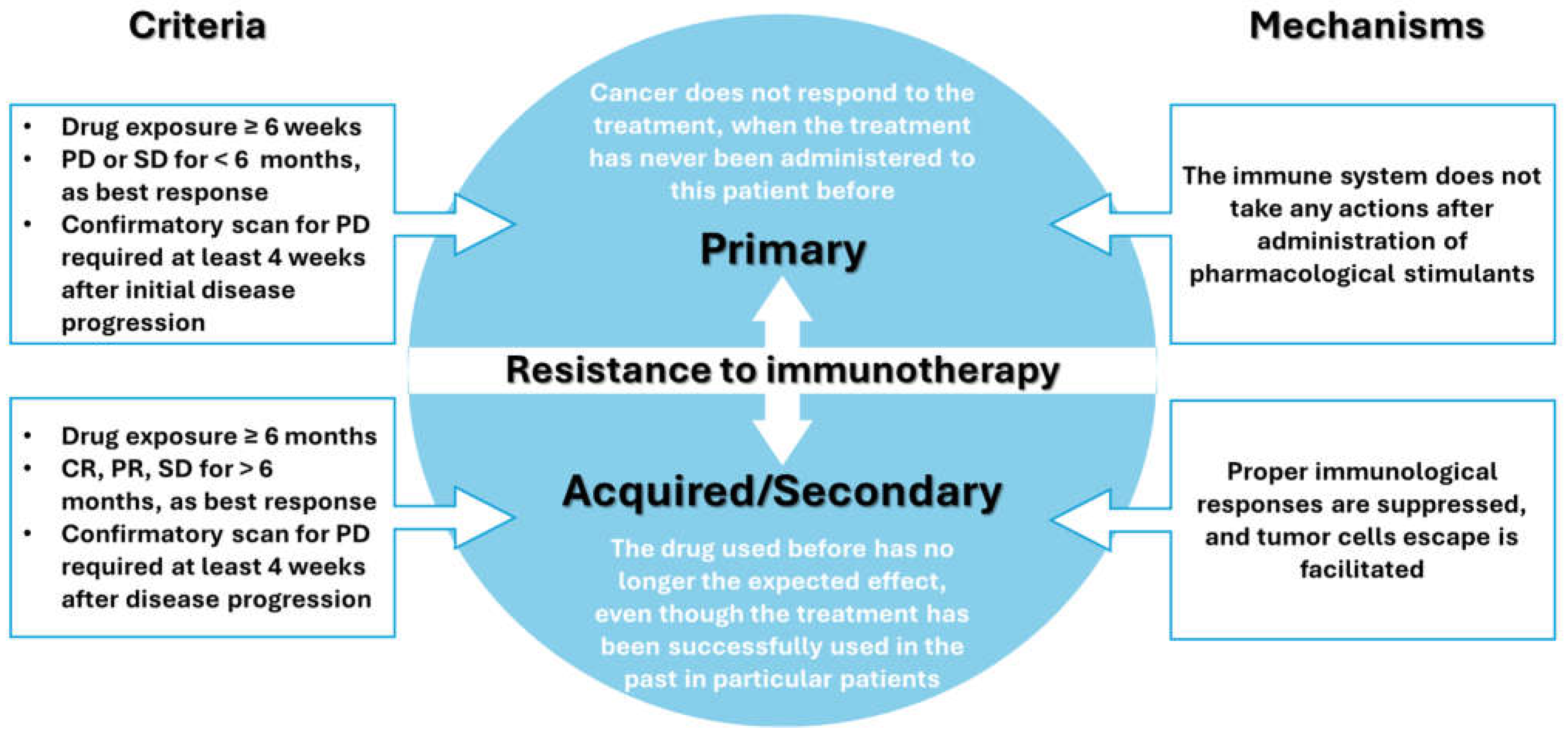
Figure 1. An at-glance comparison between primary and acquired resistance to immunotherapy. Created based on information from references [12,13,14].
Applsci 14 08948 g001
Figure 2. At-glance summary of reported factors and novel hot spots for ICI therapy. * Current methods of classification are no longer limited to traditional methods. # Germline genetics has recently become a research hotspot that affects the efficacy of immunotherapy. $ There is controversy concerning the efficacy of the mentioned drugs. Abbreviations: ICIs—immune checkpoint inhibitors; PD—progressive disease; TME—tumor microenvironment. Reprinted from an open-access source [13].
Applsci 14 08948 g002
Figure 3. Stimuli-responsive nanoparticles (srNPs) for controlled delivery in cancer immunotherapy. (A) Design of srNPs activated by endo- and/or ex-stimuli, including pH, enzyme presence, high ROS/GSH concentration, photons, US, magnetic field, and radiation, for smart drug release. (B) srNPs activated by endo- and/or ex-stimuli with controlled immunomodulator release for primary tumor treatment. Reprinted from an open-access source [29].
Applsci 14 08948 g003
Figure 4. (A) Synthesis and dual pH-responsive (or enzymatic) degradation of amphiphilic poly (esteracetal) block copolymers. (B) Schematic illustration of the concept using amphiphilic poly (esteracetal) block copolymers and their derived micelles for the pH- and lipase-responsive delivery of immunodrugs. Reprinted from an open-access source [15].
Applsci 14 08948 g004
Figure 5. Visual representation of the gene delivery process through pDNA and mRNA pathways. The use of exogenous mRNA encoding a target antigen leads to protein translation and peptide presentation through MHC Class I and II molecules, a process that activates immune responses and contributes to tumor elimination. Reprinted from an open-access source [39].
Applsci 14 08948 g005
Figure 6. Schematic representation of immunotherapy as a potential treatment for patients with infectious disease. Reprinted from an open-access source [119].
Applsci 14 08948 g006
Figure 7. Schematic representation of how nanovaccines activate immune responses. Reprinted from an open-access source [121].
Applsci 14 08948 g007
Figure 8. Schematic illustration of the interactions between nanoparticles and immune system components in managing inflammatory diseases. Reprinted from an open-access source [27].
Applsci 14 08948 g008
Figure 9. Overview of the advantages and disadvantages of different nanostructured drug delivery systems. Created based on information from [81,153,173].
Applsci 14 08948 g009
Table 1. Summary of clinical trials concerning drug delivery systems for cancer immunotherapy available on ClinicalTrials.gov as of August 2024.
ClinicalTrials.gov IDOfficial TitleStudy TypeInterventionEnrollment (Estimated)Study Completion (Estimated)Ref.
NCT04159337A Prospective Study on Companion Diagnosis by NGS-based Large-panel in Targeted Drug Delivery and Immunotherapy of Lung CancerObservationalOther: The method of gene mutation detection200031 May 2022[106]
NCT03591276A Phase 1b Study of Combination Chemo-immunotherapy With Pegylated Liposomal Doxorubicin (Doxil/Caelyx) and Pembrolizumab (Keytruda) in Metastatic Endocrine-resistant Breast CancerInterventionalDrug: Chemotherapy drugs, cancer1515 June 2021[107]
Table 2. Summary of relevant clinically approved nanostructured drug delivery systems.
Commercial NameFormulationBenefitsIndicationApproval StatusCompany/Approval HolderRefs.
Doxil®Doxorubicin hydrochloride encapsulated in PEGylated liposomes
  • Prolonged drug circulation time
  • Avoidance of the reticuloendothelial system
  • High and stable remote loading of the drug at the tumor site due to a transmembrane ammonium sulfate gradient
  • Reduced cardiotoxicity
Ovarian cancer, Multiple myeloma, AIDS-related Kaposi’s SarcomaFirst FDA-approved nano-drug (1995)Also approved by EMA (1996, marketed under the name of “Caelyx”), HC (1998), and PMDA (2009)Schering-Plough Corporation (Kenilworth, NJ, USA)[152,155,156,157]
Myocet®Doxorubicin encapsulated in liposomes
  • Reduced side-toxicity compared to free drug
  • Enhanced anti-tumor effectiveness
  • Significantly decreased cardiovascular issues and heart failure rates
Metastatic breast cancerApproved by EMA (2000) and HC (2001)Teva Pharmaceutical Industries (Tel Aviv, Israel)[153,158,159]
DaunoXome®Daunorubicin encapsulated in liposomes
  • Prolonged drug circulation time
  • Improved pharmacokinetic profile
  • Significantly reduced cardiotoxicity compared to the free drug
  • Fast uptake by the mononuclear phagocyte system
  • Enhanced tumor uptake
AIDS-related Kaposi’s SarcomaApproved by FDA, EMA, and HC (1996)Galen Pharma (Craigavon, Northern Ireland, UK)[153,157,158,160]
Onivyde®Irinotecan hydrochloride trihydrate encapsulated in PEGylated liposomes
  • Prolonged circulation time
  • Increased cargo delivery in tumors with compromised vasculature
  • Reduced side toxicity
Metastatic adenocarcinoma of the pancreasApproved by FDA (2015), EMA (2016), HC (2017), and PMDA (2020)Merrimack Pharmaceuticals (Cambridge, MA, USA)[152,153,157]
Vyxeos®Daunorubicin and cytarabine co-encapsulated in liposomes
  • Prolonged plasma half-life
  • Accumulation and persistence in high concentration in the bone marrow
Therapy-related AML or AML with myelodysplasia-related changesApproved by FDA (2015), EMA (2016), and HC (2017)Jazz Pharmaceuticals (Palo Alto, CA, USA)[157,159]
Marqibo®Vincristine encapsulated in liposomes
  • Increased drug delivery and retention
  • Prolonged drug circulation
  • Actively directed drug to target cells via fenestrations of tumor neovasculature
  • Slow drug release in the tumor microenvironment
  • Fast uptake by the mononuclear phagocyte system
Acute lymphoblastic leukemiaApproved by FDA (2012)Spectrum Pharmaceuticals (Henderson, NV, USA)[157,158,159,160]
DepoCyt®Aracytidine encapsulated in liposomes
  • Retained elevated drug levels in the cerebrospinal fluid
  • Enhanced neurological development phases
  • More effective in killing tumor cells form the meninges and cerebrospinal fluid than regular drug formulation
Neoplastic meningitisApproved by FDA, EMA, and HC (2007)Pacira Pharmaceuticals (San Diego, CA, USA)[158,159]
Mepact®Mifamurtide encapsulated in liposomes
  • Prolonged drug circulation time
  • Stimulation of innate immunity
  • Well tolerated by patients
  • Tumoricidal effect on microscopic metastases attributed to the stimulation of monocytes and macrophages
  • In vivo anti-osteosarcoma benefits attributed immunological reaction occurring in osteosarcoma lungs metastasis
High-grade, resectable, non-metastatic osteosarcomaApproved by EMA (2009)Takeda Pharmaceuticals (Tokyo, Japan)[153,158,161,162]
AmBisome®Amphotercin B encapsulated in liposomes
  • Reduced toxicity compared to free drug
  • More effective than conventional antifungal agents
  • Longer half-life than the free drug
  • TLR-4 binding induces an anti-inflammatory response
Systemic fungal infectionApproved by FDA, HC (1997), EMA (1999), and PMDA (2001)Astellas Pharma (Northbrook, Illinois, USA) and Gilead Sciences (San Dimas, CA, USA)[159,163]
Onpattro®siRNA encapsulated in LNPs
  • Harnesses the body’s natural process to silence TTR messenger RNA
  • Significantly improved the quality of life, walking, nutritional status, and performance of daily living activities of patients
Polyneuropathy of hereditary transthyretin-mediated amyloidosisApproved by FDA, EMA, HC (2018), and PMDA (2019)Alnylam Pharmaceuticals (Cambridge, MA, USA)[152,159,164]
Spikevax®mRNA encapsulated in LNPs
  • Enables the expression of the S antigen in host cells
  • Elicits an immune response through both T and B cell responses and specific antibody responses against the S antigen
COVID-19Approved by FDA (EUA in 2020, full approval in 2022), EMA, and PMDA (2021)Moderna (Cambridge, MA, USA)[152,165]
Comirnaty™mRNA encapsulated in LNPs
  • Prevents early degradation of RNA and allows it to enter host cells
  • Triggers interleukin synthesis resulting in B cells differentiation and production of numerous antibodies against S proteins
COVID-19Approved by FDA (EUA in 2020, full approval in 2021), EMA, and PMDA (2021)BioNTech/Pfizer (Mainz, Germany)[152,165]
Abraxane®Nanoparticle albumin-bound Paclitaxel
  • Overcomes the solubility issue of the drug
  • Prolonged drug circulation time
  • Relatively fast clearance rate
  • Reduced side toxicity
Metastatic breast cancer, locally advanced or metastatic NSCLC, metastatic adenocarcinoma of the pancreasApproved by FDA (2005), HC (2006), EMA (2008), and PMDA (2010)Celgene Corporation (Summit, NJ, USA)[153,160]
Fyarro®Nanoparticle albumin-bound Sirolimus
  • Preferential accumulation in solid tumors attributed to a leaky capillary system or defective lymphatic drainage of tumors
  • Enhanced uptake via endocytosis and macropinocytosis by proliferating tumor cells
  • Forty-three-fold higher sirolimus accumulation than free drug administration
Metastatic perivascular epithelioid cell tumorApproved by FDA, EMA (2021), and HC (2022)Aadi Bioscience (Pacific Palisades, CA, USA) [153,166]
Genexol®Paclitaxel encapsulated in polymer micelle
  • Well tolerated by patients
  • Remarkable efficacy in treating lung cancer
  • Potential as an effective radiosensitizer when combined with radiotherapy
  • Controllable safety profile
Metastatic breast cancer, NSCLC, ovarian cancerApproved in Republic of Korea and China (2007)Samyang Biopharm (Seoyoon Cheong, Republic of Korea)[153,167,168,169]
Apealea®Paclitaxel encapsulated in polymer micelle
  • Improved pharmacokinetic profile
  • Mitosis inhibition
  • Bioequivalent of Abraxane®
Epithelial ovarian cancer, primary peritoneal cancer, fallopian tube cancerApproved by EMA (2018)Elevar Therapeutics (Fort Lee, NJ, USA)[153,170,171]
Nanoxel®Docetaxel encapsulated in polymer micelle
  • Passive targeting of tumor cells via EPR effect
  • Selective accumulation in tumoral tissues
  • Reduced side toxicity
  • Cost-effectiveness
Metastatic esophageal squamous cell carcinomaApproved in India (2012)Samyang Biopharm (Seoyoon Cheong, Republic of Korea)[153,172]
Disclaimer/Publisher’s Note: The statements, opinions and data contained in all publications are solely those of the individual author(s) and contributor(s) and not of MDPI and/or the editor(s). MDPI and/or the editor(s) disclaim responsibility for any injury to people or property resulting from any ideas, methods, instructions or products referred to in the content.