Article

Improvement of Mechanical Properties of 3D Bioprinted Structures through Cellular Overgrowth

1
Institute of Materials Science and Engineering, Faculty of Mechanical Engineering, Lodz University of Technology, 90-537 Lodz, Poland
2
Faculty of Electrical, Electronic, Computer and Control Engineering, Lodz University of Technology, 90-537 Lodz, Poland
*
Author to whom correspondence should be addressed.
Appl. Sci. 2024, 14(19), 8977; https://doi.org/10.3390/app14198977 (registering DOI)
Submission received: 30 August 2024 / Revised: 29 September 2024 / Accepted: 2 October 2024 / Published: 5 October 2024
(This article belongs to the Special Issue Hydrogels and Microgels: Fundamentals, Fabrication and Applications)

Abstract

:
The common use of hydrogel materials in 3D bioprinting techniques is dictated by the unique properties of hydrogel bioinks, among which some of the most important in terms of sustaining vital cell functions in vitro in 3D cultures are the ability to retain large amounts of liquid and the ability to modify rigidity and mechanical properties to reproduce the structure of the natural extracellular matrix. Due to their high biocompatibility, non-immunogenicity, and the possibility of optimizing rheological properties and bioactivity at the same time, one of the most commonly used hydrogel bioink compositions are polymer solutions based on sodium alginate and gelatin. In 3D bioprinting techniques, it is necessary for hydrogel printouts to feature an appropriate geometry to ensure proper metabolic activity of the cells contained inside the printouts. The desired solution is to obtain a thin-walled printout geometry, ensuring uniform nutrient availability and gas exchange during cultivation. Within this study’s framework, tubular bioprinted structures were developed based on sodium alginate and gelatin, containing cells of the immortalized fibroblast line NIH/3T3 in their structure. Directly after the 3D printing process, such structures are characterized by extremely low mechanical strength. The purpose of this study was to perform a comparative analysis of the viability and spreading ability of the biological material contained in the printouts during their incubation for a period of 8 weeks while monitoring the effect of cellular growth on changes in the mechanical properties of the tubular structures. The observations demonstrated that the cells contained in the 3D printouts reach the ability to grow and spread in the polymer matrix after 4 weeks of cultivation, leading to obtaining a homogeneous, interconnected cell network inside the hydrogel after 6 weeks of incubation. Analysis of the mechanical properties of the printouts indicates that with the increasing time of cultivation of the structures, the degree of their overgrowth by the biological material contained inside, and the progressive degradation of the polymer matrix process, the tensile strength of tubular 3D printouts varies.

1. Introduction

3D bioprinting is a modern field of additive manufacturing classified at the intersection of biomedical science, biology, mechanical engineering, and materials science [1]. The technology is based on the manufacturing of three-dimensional structures through precise layer-by-layer deposition of a bioink that is a combination of living cells, biomaterials, and bioactive molecules [2,3,4]. Three-dimensional bioprinting techniques, thanks to the fabrication of structures that mimic native tissues, can find a variety of applications in the treatment of injuries, congenital defects, whole organ disorders, and cancer therapy. Today, 3D bioprinting technologies are the basis for in vitro drug testing and the creation of physiological tissue models of disease [5,6,7]. The dynamic development of 3D bioprinting technology started at the beginning of the 21st century, and, since then, intensive research work has been carried out on the possibility of printing various types of tissue in an effort to reduce the number of autologous transplants [3]. We can distinguish several 3D bioprinting technologies, which include extrusion-based bioprinting, droplet bioprinting, stereolithography-based bioprinting, and laser-assisted bioprinting [1,2,3,4,5]. Due to its high versatility, low cost, low material consumption, ability to use high densities of biological material, and high cell viability directly after the manufacturing process using bioinks that have the property of thinning by shearing, the most widely used 3D bioprinting technology is direct extrusion-based bioprinting [1,3,8]. The extrusion bioprinting process is carried out by extruding bioink through a nozzle using a mechanical or pneumatic drive. Continuous microfilaments are created during printing, which are deposited on the substrate and shaped into printouts with the desired geometry. The final printout structure is influenced by extrusion temperature, nozzle diameter, extrusion pressure, nozzle and extrusion speed, and path spacing between paths [1]. The numerous advantages of 3D extrusion bioprinting enable the creation of structures with compositional and structural complexity [3,8]. In the processes of 3D extrusion bioprinting, bioink compositions play a key role. Generally, hydrogels are one of the most commonly used materials for their production [5]. Their common use in 3D extrusion bioprinting techniques is dictated by their unique properties, among which the most important from the point of view of promoting the development of cellular functions in 3D in vitro cultures are their ability to retain large amounts of water and the possibility of modifying their rigidity and mechanical properties to mimic the natural extracellular matrix [9,10,11]. In addition, among the advantages of using hydrogel bioinks based on hydrogel materials in 3D bioprinting techniques, one should also emphasize their high permeability to oxygen, nutrients, and other water-soluble compounds, as well as their ability to allow free migration of the cells in any direction, unlike polymer scaffoldings [12]. The compatibility of hydrogel materials with various cell types (including fibroblasts, chondrocytes, hepatocytes, smooth muscle cells, adipocytes, and stem cells) and tissues creates the potential to create biomimetic structures that mimic the natural physiological environment, thereby increasing this way the range of their applications in the field of regenerative medicine [11,13].
Among the key parameters during the 3D bioprinting process of hydrogel bioinks, we can mention printability, mechanical properties, biodegradability, the presence of modifiable functional groups on the surface, and the way that the structures cross-link [14]. Hydrogel materials used in 3D bioprinting processes can be classified according to their origin (synthetic, natural, or hybrid/modified), ionic charge (non-ionic, cationic, anionic, or ampholytic), biodegradability (biodegradable or non-biodegradable materials), physical properties (smart or conventional) and type of cross-linking (physical, chemical or radiation cross-linking) [15,16]. In recent years, often used on their own or in combination, hydrogel bioink compositions have frequently used materials of natural origin, among which we can distinguish gelatin, sodium alginate, collagen, silk, hyaluronic acid, chitosan, fibrinogen, and agarose [17,18,19,20,21]. Control of cellular interactions in natural hydrogel materials can occur by changing the used polymers’ physical, chemical, biochemical, or physiological properties [21]. One of the most important requirements for materials intended for the 3D bioprinting process is the correct choice of bioink composition, which is characterized by suitable structural and mechanical properties; the composition must also protects cells from damage during the printout manufacturing process and provide them with an environment favorable for controlling their phenotype and maintaining their vital functions [22]. Due to their high biocompatibility, non-immunogenicity, ability to optimize rheological properties, and bioactivity, sodium alginate-based polymer solutions are one of the most widely used hydrogel compositions [9,23,24].
Sodium alginate is a naturally occurring anionic, hydrophilic polysaccharide that is obtained from brown algae or seaweed. It consists of α-L-guluronic acid (G-block) and β-D-manuronic acid (M-block), linked by 1–4 glycosidic bonds. The cross-linking process of alginate hydrogels most often occurs using divalent cations, among which calcium ions are generally used to build ionic bridges between G-units to form a thermostable three-dimensional gel network with an “egg-box” structure [17,25,26,27,28,29]. Thanks to the control of the degree of cross-linking, alginate concentration, and its molecular weight, it is possible to modify the rigidity of the hydrogel because the higher the content of G units, the higher the rigidity and porosity of the polymer matrix, which are key parameters for the viability and proliferation of cells contained inside the material [17,25,30,31]. Therefore, alginate’s physical properties can potentially be tailored directly to three-dimensional cell growth and differentiation in both in vitro and in vivo models [22]. The non-toxicity and high biocompatibility of alginate make it a hydrogel that is now widely used in various industrial fields, including tissue engineering and in the pharmaceutical and food industries. Some of the disadvantages of alginate are its slow and uncontrolled degradation in mammalian organisms and low cell bioadhesion, which generate the need for modification with adhesive ligands with the purpose of obtaining suitable biological properties [29,32,33,34]. Using alginate hydrogels in combination with other biomaterials of natural origin, such as gelatin or collagen, improves its bioactivity and modifies its physical and mechanical properties dedicated to the application requirements of 3D bioprinted structures [35,36]. Gelatin is a natural, soluble protein obtained by partial hydrolysis of collagen derived from the body’s connective tissues. We can distinguish more than a dozen types of gelatin, depending on the source of collagen and hydrolytic treatment. Due to the specific properties of gelatin, such as biodegradability, biocompatibility, non-toxicity, and low antigenicity, it has been approved by the Food and Drug Administration as a biomaterial suitable for clinical applications. Gelatins of porcine, bovine, or fish origin find their use in a wide range of regenerative medicine applications. A property that is characteristic of gelatin is its ability to undergo thermo-reversible physical cross-linking by cooling to form hydrogels by forming locally ordered regions that link with hydrogen, electrostatic, and hydrophobic bonds. This property gives gelatin-based bioinks the ability to undergo extrusion 3D printing for different shapes and structures [37,38,39]. In addition, gelatin has bioactive arginine–glycine–aspartic acid (RGD) sequences in its structure, creating a favorable microenvironment for cellular interactions that enable cell adhesion, migration, proliferation, and differentiation [9,16,39,40]. Oligopeptide sequences containing the RGD peptide also affect the more favorable controlled degradation kinetics of alginate–gelatin scaffoldings with respect to hydrogel structures produced only based on sodium alginate [39]. Therefore, appropriate compositions of alginate–gelatin hydrogels are materials that combine the most desirable features of the individual components with the purposes of obtaining synergistic effects and creating a bioink with unique properties. Thanks to the combination of sodium alginate and gelatin, it is possible to obtain high levels of cell adhesion, diffusion, spreading, migration, and proliferation, and to eliminate the limitations resulting from the poor mechanical strength of gelatin [29,39,40,41,42].
In 3D bioprinting techniques, the development of hydrogel printouts with the appropriate geometry is necessary to ensure the metabolic activity of the cells contained in the 3D structures. Obtaining thin-walled printed geometries is desirable to guarantee uniform nutrient availability and gas exchange during cultivation [43]. Using 3D bioprinting techniques, we can control the mechanical properties, porosity, permeability, and rigidity of the printouts’ hydrogel matrix [42]. The research work carried out by our team to date has enabled the development, using 3D extrusion bioprinting techniques, of three-dimensional thin-walled tubular-shaped bioprinted structures that allow for the even distribution of biological material inside the polymer matrix, as well as the diffusion of oxygen, nutrients, and metabolites from and into the cells [44,45]. Within the framework of this study, tubular printouts were developed based on sodium alginate and gelatin containing immortalized fibroblast cells of the NIH/3T3 line. Directly after the 3D bioprinting process, structures with such a geometry are characterized by extremely low mechanical properties. The aim of this research work was to perform a comparative analysis of the viability and spreading ability of the biological material contained in the printouts during their incubation for 8 weeks while monitoring the cellular proliferation effect on changes in the mechanical properties of the structures.

2. Materials and Methods
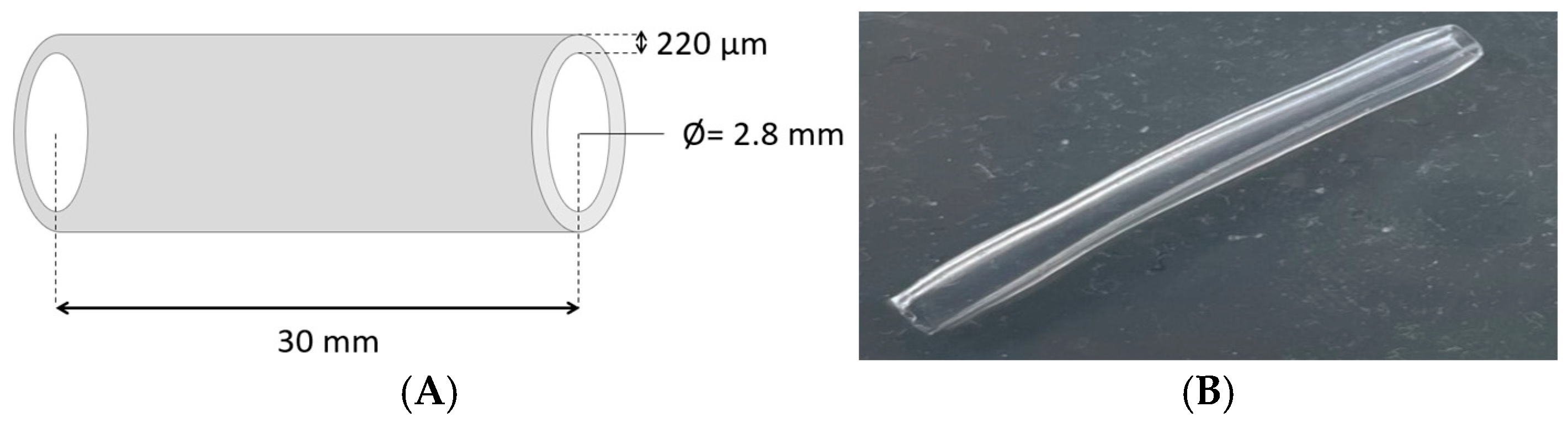
2.1. Materials

Alginic acid sodium salt from brown algae, porcine skin type B gelatin, and calcium chloride used for cross-linking hydrogel materials were purchased from Sigma Aldrich (Burlington, MA, USA). A model line of immortalized NIH/3T3 fibroblast cells from the American Type Culture Collection (ATCC, Manassas, VA, USA) was used to prepare 3D bioprinted structures. For cell cultivation, DMEM high-glucose medium (Biowest, Nuaillé, France) supplemented with the iron-fortified calf serum Calf Bovine Serum (CBS) (ATCC, Manassas, VA, USA) and a mixture of penicillin–streptomycin–neomycin (P/S/N) antibiotics (Sigma Aldrich, Burlington, MA, USA) were used. A complementary PBS buffer (Capricorn Scientific GmbH, Ebsdorfergrund, Germany) was used in cell cultivation and for rinsing hydrogel printouts. The live/dead assay kit, composed of calcein–AM and ethidium homodimer III, was obtained from Biotium (Fremont, CA, USA).

2.2. NIH/3T3 Cell Cultivation

For the preparation of the hydrogel bioinks, the NIH/3T3 model line of immortalized fibroblast cells was used. Cell cultivation was carried out in polystyrene bottles (TPP, Trasadingen, Switzerland) in DMEM high-glucose culture medium with 10% CBS serum and 1% P/S/N antibiotics addition. The culture was carried out in a CO2 incubator (NuAire, Plymouth, MN, USA) under standard conditions, i.e., at a temperature of 37 °C, at 100% relative humidity, in an atmosphere of 5% carbon dioxide and 95% air. Cell passaging, preceded by the process of trypsinization, took place regularly after obtaining a monolayer of adherent cells with 70–80% confluence. For the studies conducted, cells after the 11th passage were used.

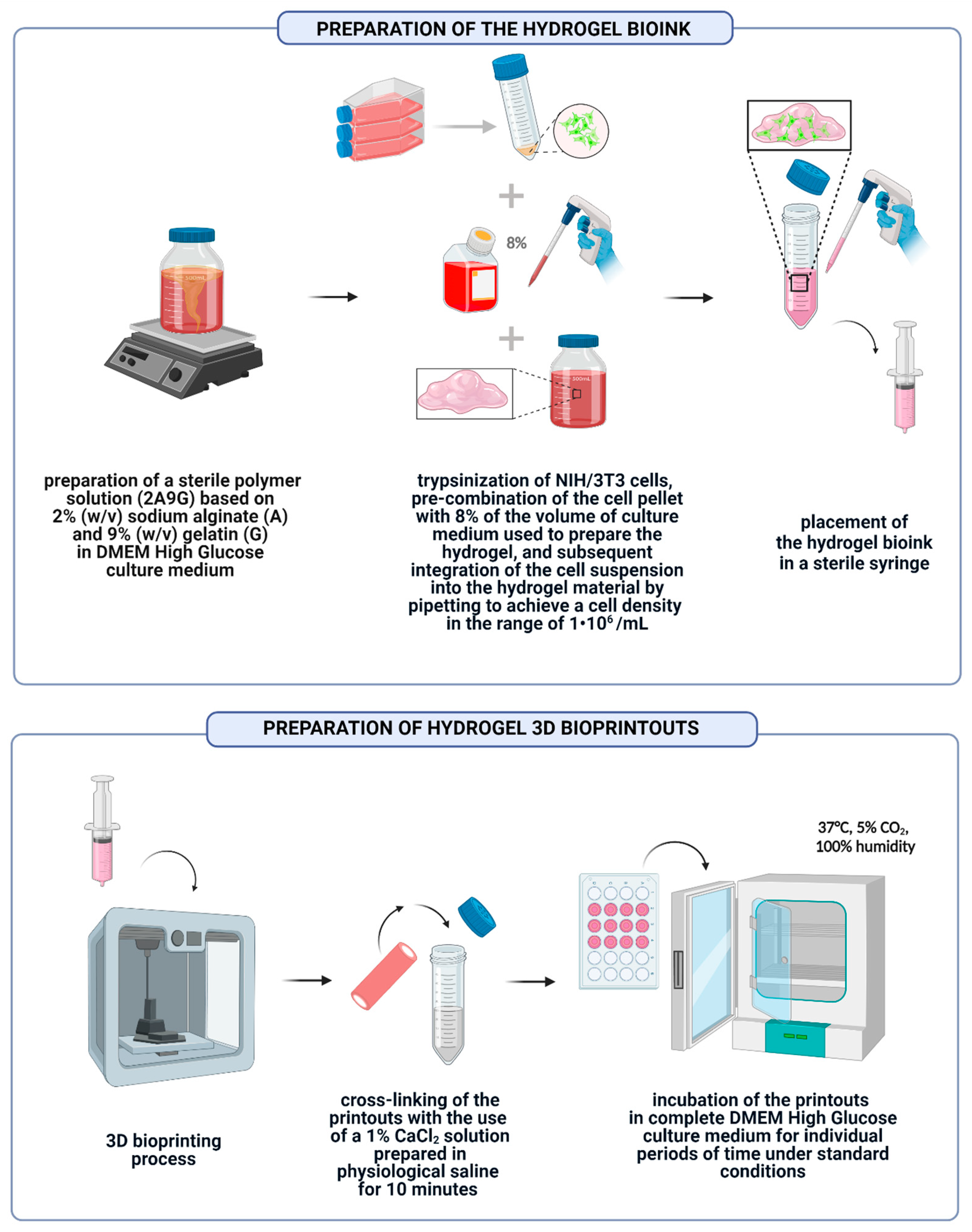
2.3. Preparation of Hydrogel Bioink and 3D Bioprinted Structures

For the preparation of 3D bioprinted structures, a composition of hydrogel materials based on alginic acid sodium salt from brown algae and gelatin from porcine skin type B was used. The hydrogel solution was prepared in the DMEM high-glucose culture medium. Before the preparation process of hydrogel solutions, appropriate amounts of sodium alginate (A) and gelatin (G) powders underwent UV radiation sterilization for 1 h. The polymer solution containing 2% sodium alginate and 9% gelatin (2A9G) was prepared in a weight–volume ratio by mixing the alginate and gelatin together in such a way that it was possible to “subtract” from the mixture of 8% culture medium that had previously been added to the cell pellet before combining it with the hydrogel. The polymer solution was mixed at a temperature not exceeding 40 °C at a speed of 200 rpm for 2 h. The missing amount of medium, equal to 8% of the solvent volume used to prepare the hydrogel, was then added to the flask containing the NIH/3T3 cell pellet. An appropriate amount of hydrogel providing a seeding density equal to 1∙106 cells/mL was added to the cell suspension. The cell suspension was combined with the hydrogel using a piston pipette. The mixing time did not exceed 1 min. The prepared hydrogel bioink was placed in a sterile syringe with a volume of 5 mL and subjected to the 3D bioprinting process of tubular structures with a length of 30 mm and an inner diameter of 2.8 mm (Figure 1). The 3D printing process was performed using a 3D printer designed and constructed at the Technical University of Lodz. The 3D printing process, during which the material is pneumatically extruded, was performed on a horizontal shaft using a conical nozzle. The height of the print layer was set at 220 µm. The 3D printing process was conducted at a temperature of 34 °C.
A 1% solution of calcium chloride prepared in saline was used to cross-link the printouts. The cross-linking time of each of the samples was 10 min. The cross-linking process was performed at room temperature. After the structures were cross-linked, each sample was rinsed three times with PBS buffer. The bioprinted structures underwent cultivation on 6-well plates in DMEM high-glucose culture medium supplemented with 10% CBS and 1% P/S/N under standard conditions for the following individual time periods: 0, 1, 4, 6, and 8 weeks. Each of the incubation periods was a separate measurement system. A schematic of the preparation of bioprinted structures is shown in Figure 2. During cultivation, the culture medium was changed every 2–3 days. The multi-well plates on which the bioprinted structures were cultivated were replaced once a week by transferring the samples to the surface of new multi-well plates.
Bioprinted structures after individual cultivation periods were subjected to assessment of the viability and proliferation of fibroblast cells contained in the hydrogel samples using a fluorescent live/dead assay, an analysis of the changes in the cell morphology using a scanning electron microscope (SEM), and an assessment of mechanical properties using a static tensile test.

2.4. Evaluation of Rheological Properties of Polymer Solutions

The rheological properties of the 2A9G polymer composition prepared as described in Section 2.3., containing no biological material, and a bioink containing 1∙106 fibroblast cells/mL were tested using an MCR702 Multidrive rheometer (Anton Paar GmbH, North Ryde, Australia). The polymer solutions were placed on the lower measurement plate of the device, and the gap between the measuring plates was set to 0.5 mm, using the geometry of a parallel plate of diameter 25 mm. The measurements were taken at a temperature of 34 °C, during which the 3D printing process takes place. The viscosity was measured in the shear rate speed range of 0.1 to 1000 1/s. Before each measurement, the polymer solutions underwent thermostating for 10 min with the purpose of obtaining an even polymer solution temperature distribution over the volume of the material.

2.5. Assessment of the Viability of Fibroblast Cells of the NIH/3T3 Line Contained in Hydrogel Printouts after Individual Periods of Cultivation of Structures Using the Fluorescent Live/Dead Assay

The analysis of the viability of the fibroblast cells contained in hydrogel printouts was carried out with live/dead staining using a mixture of dyes, with calcein–AM applied for the green labeling of live cells and ethidium homodimer III (EthD) used for the red labeling of dead cells. The cells incorporated in bioinks and hydrogel printouts were imaged using a fluorescent microscope (Nikon Eclipse LV100ND, Tokyo, Japan). The images of live and dead cells were taken in 10 randomly chosen areas of the analyzed samples. Three samples of tubular printouts were assigned for each of the cultivation time points. Based on recorded images, the number of live and dead cells was calculated using ImageJ 1.8.0 (LOCI, University of Wisconsin, WI, USA). To determine the cell viability of the NIH/3T3 line, the following relationship was used:
V i a b i l i t y   % = t h e   n u m b e r   o f   l i v i n g   c e l l s t h e   n u m b e r   o f   l i v i n g   c e l l s + t h e   n u m b e r   o f   d e a d   c e l l s     · 100 %
The results presented in the following sections show the calculated average viability of the cells incorporated in the hydrogel printouts.

2.6. Evaluation of the Proliferation of Fibroblast Cells of the NIH/3T3 Line Embedded inside the Printouts after Individual Periods of Cultivation of the Structures Using a Scanning Electron Microscope (SEM)

The bioprinted hydrogel structures were fixed with a 2.5% glutaraldehyde solution at a temperature of 4 °C for 12 h before the SEM observation process. Subsequently, the hydrogel materials were rinsed twice with PBS and subjected to a dehydration process with successively increasing concentrations of ethanol (50%, 70%, 80%, 90%, 95%, and 100%) for 10 min. The dehydrated printouts were dried in a dryer at the critical point in the LEICA EM CPD300 (Leica, Wetzlar, Germany). Finally, the samples were coated with a 5–7 nm thick conductive layer of carbon fiber using a sputtering unit SafeMatic CCU-010 LV Compact Coating Unit (SafeMatic, Zizers, Switzerland). After the applied preparation, the degree of proliferation and morphology of fibroblast cells incorporated inside the printouts after individual periods of culture of the structures were evaluated using a scanning electron microscope JSM-6610LV (Jeol, Akishima, Tokyo, Japan).

2.7. Evaluation of the Mechanical Properties of Bioprinted Hydrogel Structures Subjected to Individual Cultivation Periods Using a Static Tensile Test

The mechanical properties of the bioprinted hydrogel structures were evaluated using a static tensile test with a holder adapted to the specific geometry of the samples (Figure 3), which held the tubular printout in a stable position during the tensile test and allowed the samples to be placed in the holder without first damaging their structure. The holder was designed and printed on a 3D printer to meet the dedicated requirements of the indicated sample geometry. Static tensile tests were performed using a Bruker UMT-2 tribotester (Bruker, Billerica, MA, USA) at a constant tensile speed of 0.1 mm/s for tubular printouts containing no biological material and not subjected to incubation process and for bioprinted structures containing fibroblast cells subjected to the following individual cultivation times: 0, 1, 3, 4, 6, and 8 weeks. Five samples of tubular printouts were assigned for each time point. The analysis performed made it possible to determine the maximum breaking force for the samples tested.

3. Results and Discussion

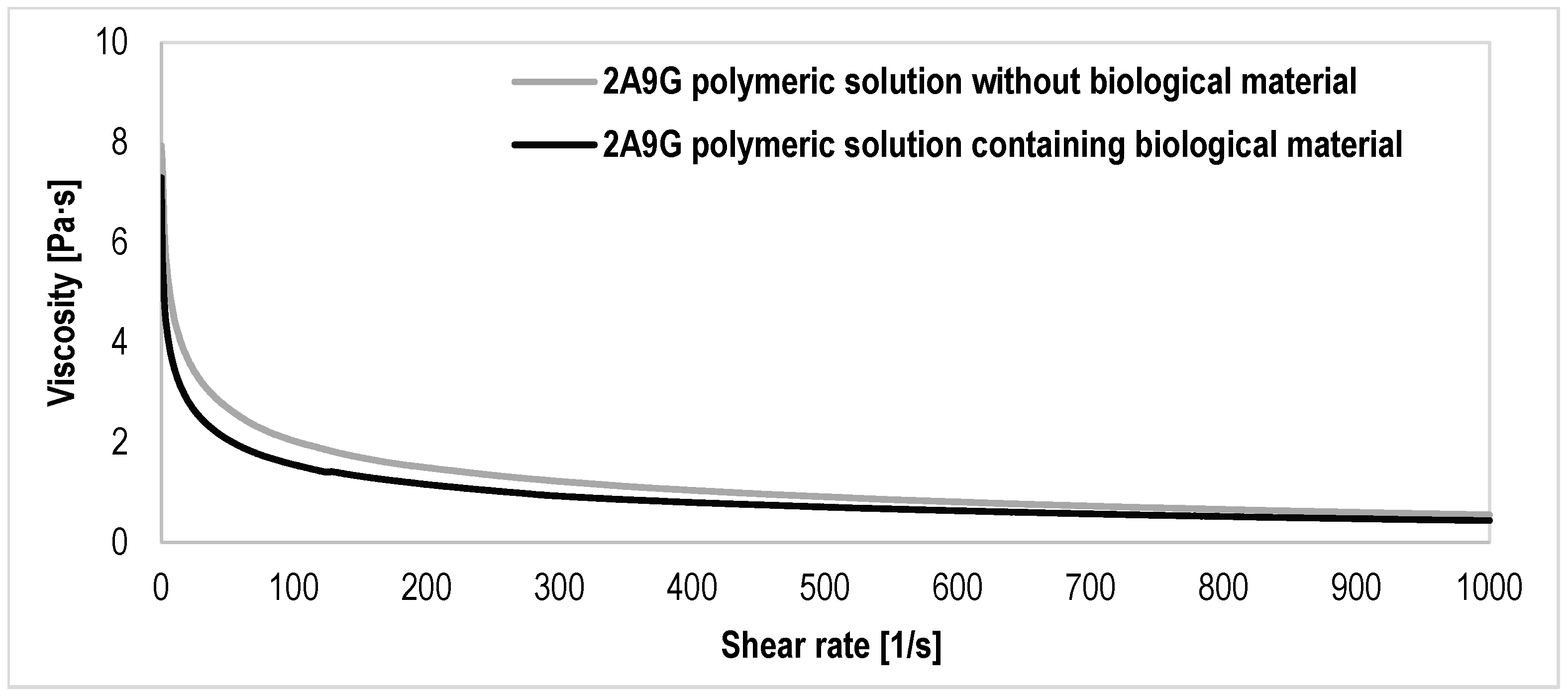
3.1. Results of the Evaluation of the Rheological Properties of the Polymer Solutions

The evaluation of the rheological properties of polymer solutions is a key parameter to determine flow properties and shape fidelity, which affect the printability of bioink. The rheological characteristics of the hydrogel composition also directly influence the rate of proliferation and differentiation of cells contained in the polymer matrix [47]. The results of the analysis of the rheological properties of polymer solutions based on sodium alginate and gelatin, shown in Figure 4, indicate that both compositions—both 2A9G supplemented with biological material and not containing cells—have the features of non-Newtonian systems and are characterized by shear-thinning ability. This property is characteristic of sodium alginate-based hydrogel solutions [48,49,50]. It enables a process of smooth printability and increased printout accuracy after the manufacturing process [47,50]. The evaluation of the rheological properties of the two polymer solutions also indicates that the content of biological material in the bioink in the form of immortalized cells of fibroblast NIH/3T3 line with a density of 1·106/mL causes a decrease in viscosity with respect to the composition containing no biological material. Research work carried out by the team of T. Gregory et al. confirms that as the density of the biological material in the sodium alginate and gelatin-based hydrogel bioink increases, a gradual decrease in the viscosity of the polymer solution occurs. Indeed, the cell content of the bioink can affect the gel microstructure by blocking inter-chain interactions in the polymer solution [47,48]. Higher viscosity hydrogel compositions require higher pressure to be applied during extrusion, which results in higher shear stresses on the cells contained in the bioink, leading to cell damage during the fabrication of bioprinted structures [49,51]. Therefore, the observed viscosity decrease in the polymer composition supplemented with biological material is an advantageous factor for obtaining high cell viability in the fabricated bioprinted hydrogel structures.

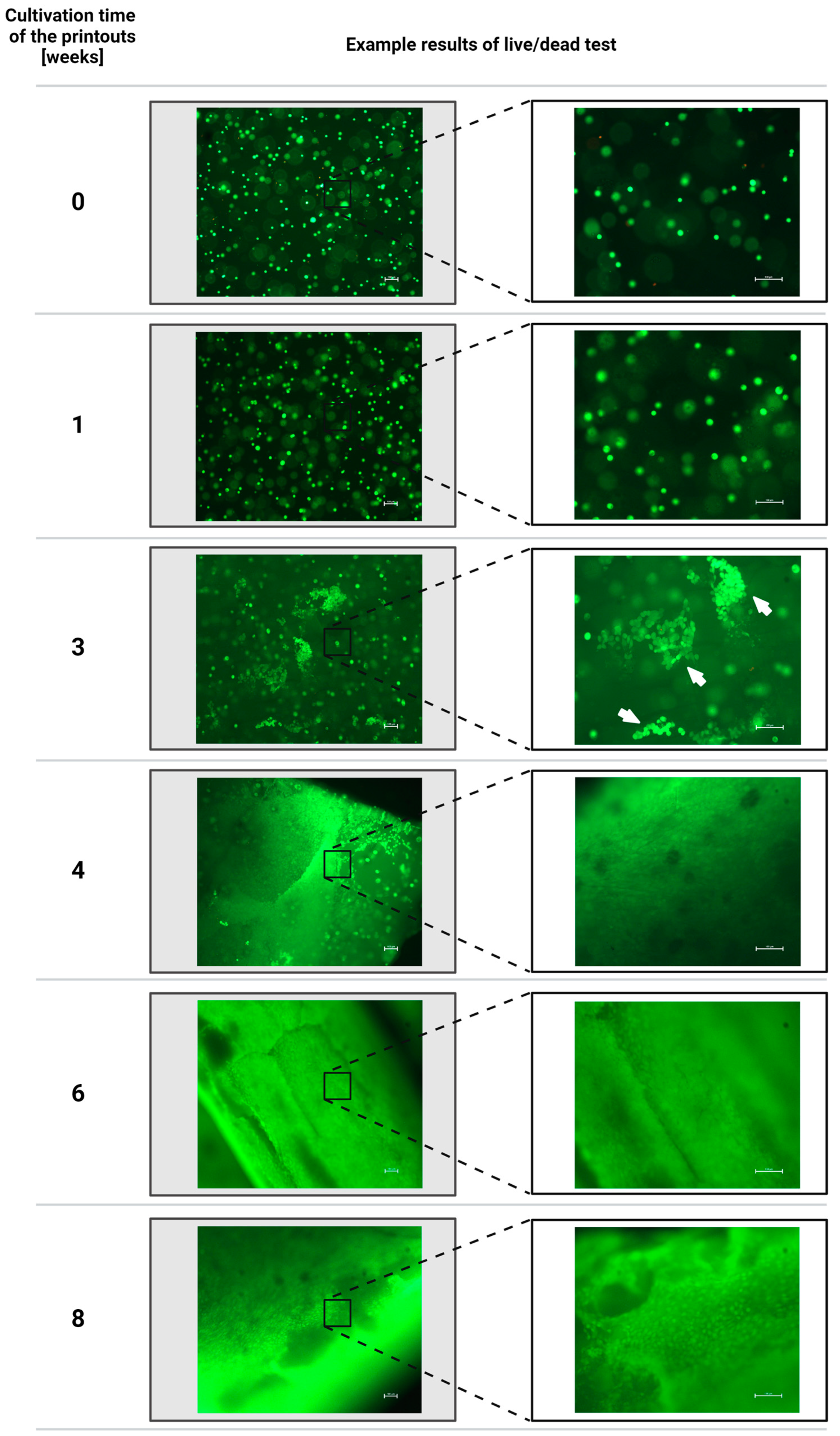
3.2. Results of the Assessment of the Viability of Fibroblast Cells of the NIH/3T3 Line Contained in Hydrogel Printouts after Individual Periods of Cultivation of Structures Using the Fluorescent Live/Dead Assay

Presented in Figure 5, the results of the cell viability assessment of the immortalized fibroblasts of the NIH/3T3 line contained in the hydrogel printouts indicate that directly after the 3D bioprinting process, the average survival rate of the biological material is equal to 88.91 ± 7.50%. This observation makes it possible to state that the very process of manufacturing the tubular structures using the 3D extrusion bioprinting technique does not have a destructive effect on the viability of model line cells incorporated into the bioink. The high viability of the cells in the printouts proves the excellent biocompatibility of the polymer matrix based on sodium alginate and gelatin. After 7-day cultivation, the viability of fibroblast cells contained in hydrogel printouts increases, reaching an average value equal to 98.73 ± 1.32%. After 3 weeks of cultivation, the viability of fibroblasts inside the bioprinted structures reaches an average value of 97.74 ± 1.84%. Observations of the degree of proliferation of the biological material contained inside the printouts after 3 weeks of cultivation indicate that the cells begin to spread in the hydrogel matrix and agglomerate toward each other, forming larger clusters. The increased number of dense clusters of biological material inside the printouts during later cultivation periods makes it impossible to clearly quantify cell viability using the live/dead assay.
The results of the qualitative evaluation of cell viability and cell proliferation inside the printouts, presented in Figure 6, prove that the structures are characterized by a high survival rate of biological material embedded in the polymer matrix each time. The morphology of the cells contained inside the polymer matrix undergoes changes during cultivation—after the 3D bioprinting process, the cells are initially characterized by a spherical, spheroidal shape, which is also observed during the first week of incubation. Then, when the cultivation time of the bioprinted structures is extended to 3 weeks, the fibroblast cells achieve the ability to proliferate, grow, and spread in the hydrogel matrix. After 4 weeks of bioprinting incubation, the presence of a more homogeneous, compacted, and interconnected network of cells with spindle-shaped morphology is observed inside the hydrogel matrix. The live/dead assay results clearly indicate excellent interaction and communication between cells. After 6 weeks of cultivation, the presence of an even, interconnected, and strongly developed fibroblast cell network is observed throughout the printout volume. Macroscopic evaluation of the samples also indicates that with the increase in the degree of cellular overgrowth, a change in the color of the tubular walls of the structures from a transparent hydrogel to a milky white tissue structure is observed. Therefore, it can be concluded that fibroblast cells remodel the hydrogel matrix during ongoing cultivation. This process may occur thanks to the suitable properties of the hydrogel material, enabling the diffusion of nutrients and metabolites, cell adhesion and proliferation, polymer matrix degradation, and also the deposition of a new extracellular matrix [52,53]. The viscoelasticity of the polymer matrix generates tension to enable mechanotransduction [54]. The printouts’ tubular shape and thin-walled geometry ensure the availability and kinetics of diffusion of nutrients and oxygen into the biological material contained in the polymer matrix. Indeed, the literature data show that the maximum diffusion distance of nutrients and oxygen that a cell can survive is limited to 200 µm [55,56]. The printed thin-walled tubular constructs, therefore, ensure even access of nutrients to the biological material contained inside the printouts, allowing the core cells to be nourished and precluding the risk of their hypoxia [57]. The spread of cells inside the printouts with progressing cultivation time results from the presence of material degradation gradients, allowing the cells to create space to grow and spread freely toward building a network structure. Gelatin, thanks to its sensitivity to ambient temperature, provides initial printout stability during the forming of the hydrogel with extrusion techniques. Degradation of gelatin-based hydrogels occurs as a result of reversible sol–gel transformation when they pass into a liquid form above the temperature of melting point (about 27–34 °C) and dissociate, which leads to gradual leakage from the hydrogel matrix under printout culture conditions [58,59,60]. Sodium alginate-based hydrogel materials incubated in physiological solutions also release Ca2+ ions cross-linking the hydrogel. This reaction occurs due to exchange with Na+ ions present in the cultivation environment of the bioprinted structures. The elevated temperature of the sample cultivation environment also amplifies the speed of this process. Calcium ions diffusing into the external environment of the samples, which are responsible for the binding of alginate blocks, cause gradual degradation of the polymer matrix [60,61]. The decomposition of the hydrogel based on sodium alginate and gelatin, occurring in the manner described, allows the cells to create spaces for growth and spreading. The literature data also indicate that fibroblast cells embedded in the collagen matrix release matrix metalloproteinases, such as collagenase and gelatinase, which also affect collagen degradation and other proteins [53]. The described phenomena indicate that an important aspect of designing 3D bioprinted structures, reflecting the native tissues of the body, is the creation of hydrogel constructs that enable control of the dynamic relations between cellular growth and the degradation process of the surrounding polymer matrix [52].

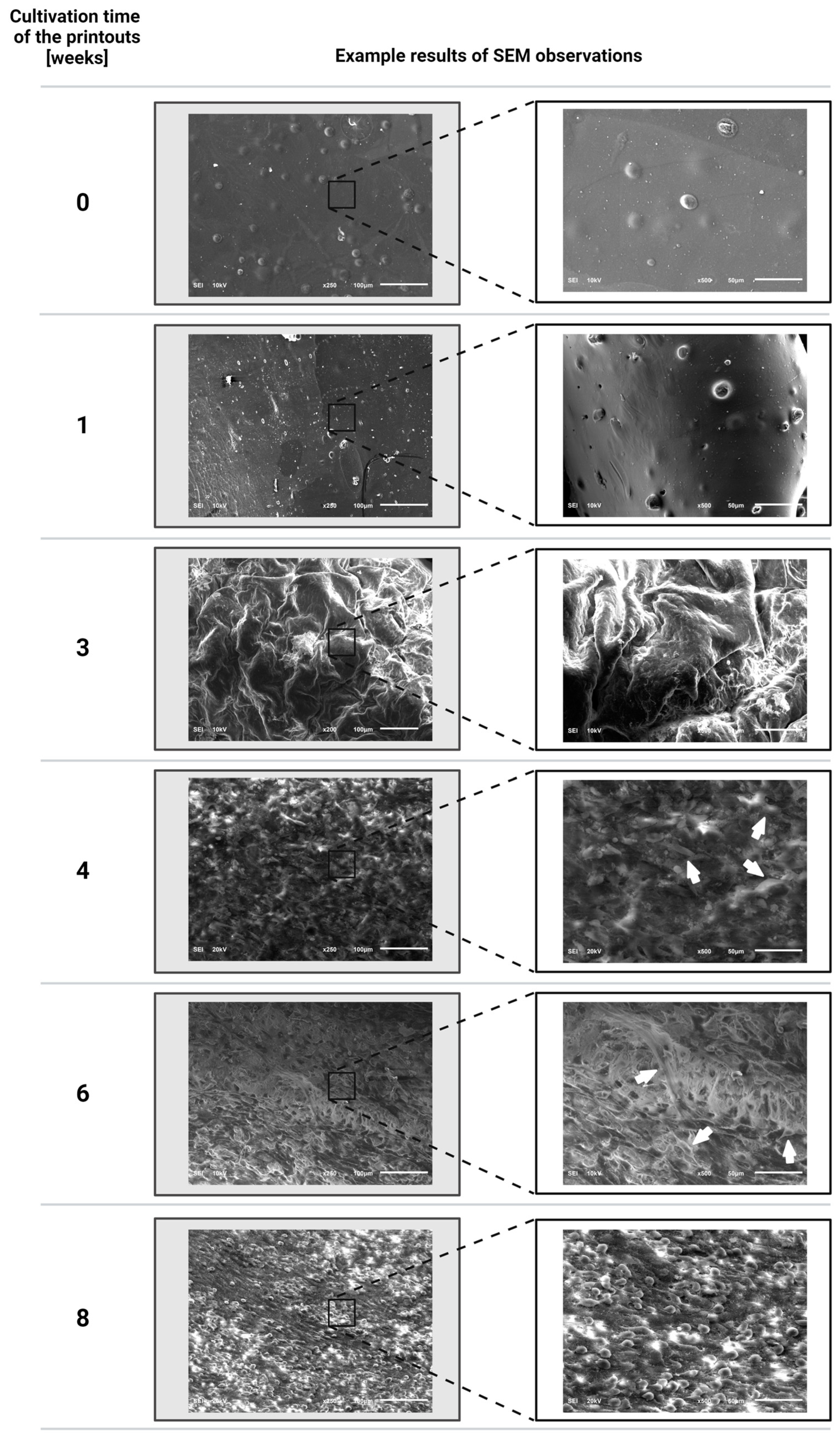
3.3. Evaluation Results of the Proliferation of Fibroblast Cells of the NIH/3T3 Line Incorporated into the Printouts after Individual Periods of Cultivation of the Structures Using a Scanning Electron Microscope

The scanning electron microscopy technique allows observations of the surface of hydrogel printouts at high resolution. The SEM observations provide information on the morphology, topography, and surface roughness, as well as the porosity and microstructure of the hydrogel, which allows analysis of the gel’s transport properties, which directly affect the diffusion of oxygen and nutrients. In bioprinted hydrogel structures, the SEM technique also makes it possible to visualize the morphology, degree of growth, spreading, migration, and proliferation of cells contained in the material [62,63]. The results obtained from the evaluation of changes in the morphology of the printouts after individual cultivation periods presented in Figure 7 showed that right after the 3D bioprinting process, the surface of the hydrogel printouts is smooth. The fibroblast cells are evenly distributed inside the polymer matrix structure, and they adopt a spheroidal shape, which is a result of the rigidity of the undegraded initial polymer matrix [64,65]. During the first week of bioprinted structure cultivation, the formation of protrusions in the hydrogel structure by individual cells is observed. After three weeks of cultivation of bioprinted structures, small clusters of spreading cells are observed to appear. The SEM results confirm the data obtained using the live/dead assay, and these results indicate that after 4 weeks of incubation, a significant aggregation of biological material takes place in the bioprinted structure towards the formation of an evenly distributed cellular network in the polymer matrix in place of single spheroidal agglomerates. As the cultivation time of the bioprinted structures increases, increasingly dense, clearly spreading network structures of fibroblast cells with an elongated, spindle-shaped morphology are observed. After 6 weeks of cultivation, they form a homogeneous tissue structure over the entire volume of the hydrogel. The results of observation of bioprinted structures after 8 weeks of cultivation indicate the presence of a growing amount of biological material in the produced cell network but a change in cell morphology from an elongated shape to densely arranged cells next to each other with a renewed spherical morphology. During the cultivation of the bioprinted structures, the surface morphology of the samples also changes, which, from being initially smooth, adopts a more developed and wavy structure. This observation may be due to the degradation of the polymer matrix occurring due to the release of gelatin from it because, with the decrease in the gelatin content in the polymer matrix, the size of the pores increases. In turn, the porous structure provides living space for cells; therefore, as the porosity increases, facilitated proliferation and spreading of cells in the polymer matrix occurs [62,66]. The research conducted by the team of M. I. Patiño Vargas et al. indicates that human adult dermal fibroblasts of NHDF-Ad need about 14 days to begin the process of spreading in a plasma–agarose hydrogel matrix prepared based on 1% agarose. Cell spreading and cell migration are hindered in materials with small and rigid pores, preventing the movement of cell nuclei [67]. Degradation of the polymer matrix also changes its viscoelastic properties, and the processes that occur in the matrix architecture affect both its viscosity and susceptibility to degradation [68]. The spreading and migration of cells in the structure of bioprints are also promoted by the increased plasticity and limited rigidity of the polymer matrix due to degradation [64,65]. This is because it has been shown that the relationship between gel rigidity and cell spreading ability is contradictory between 2D and 3D culture conditions [65]. The cells contained inside the hydrogel structures are capable of migrating in a protease-independent manner when the polymer matrices exhibit sufficient mechanical plasticity [64,68]. The work of the team of O. Chaudhuri et al. indicates that the cells contained in a three-dimensional matrix initially exert deformations, which induce stresses determined by the initial elastic modulus of the matrix. In viscoelastic matrices, these forces can undergo relaxation due to mechanical deformation and remodeling of the polymer matrix. The rate of stress relaxation determines the degree of mechanical remodeling of the matrix. In rapidly relaxing matrices, this facilitates the clustering of adhesion ligands, cell shape change, spreading, and proliferation [69].
The preparation process of bioprinted hydrogel structures necessary for SEM observations based on dehydrating the samples initially and drying at the critical point may result in the shrinkage and thickening of the material structure. For this reason, there is a risk that the observed morphology of the samples does not reflect the original state of the hydrogel, which undergoes changes in the swollen condition [67,70]. For this reason, SEM microscopic observations can provide a comparative verification technique to other techniques for visualizing bioprinted hydrogel structures. Nevertheless, a key aspect of the SEM microscopic observations of the bioprinted structures was to assess the distribution, morphology, and proliferation status of the biological material in the printouts. These parameters are not directly dependent on the hydration and preparation method of the structures and prove the complete cellular hypertrophy of the printouts, which is homogeneous in the volume of the material, during their long-term cultivation conducted within this study’s framework.

3.4. Evaluation Results of the Mechanical Properties of Bioprinted Hydrogel Structures Subjected to Individual Cultivation Periods Using a Static Tensile Test

The results of the evaluation of the tensile strength of bioprinted hydrogel structures, shown in Figure 8, indicate that the printouts are characterized by very poor mechanical properties directly after the fabrication process of tubular structures. The average maximum printout breaking force equal to 0.19 ± 0.07 N is characterized by samples containing biological material in their structure. A slightly lower tensile strength of about 0.14 ± 0.12 N was registered for printouts that did not contain fibroblast cells. Thus, we can see that the presence of biological material in the polymer matrix allows for improving the mechanical properties of hydrogel printouts. For hydrogel structures, the rigidity of the material plays a key role in having a direct impact on the cellular activity and remodeling of the polymer matrix. Mechanical interactions between cells and the extracellular matrix are considered symbiotic, as there is a mutual dynamic relationship between the hydrogel, which regulates intracellular processes, and the biological material, which enables its remodeling. The viscoelastic properties of bioprinted hydrogel structures with cultivation time may vary due to a combination of shrinkage, degradation, and extracellular matrix accumulation, which can affect the water content of the material, as well as the kinetics of oxygen and nutrient diffusion [53]. This thesis is confirmed by the results obtained within the framework of this study, which indicate the occurrence of dynamic changes in the mechanical properties of bioprinted structures during the different periods of their culture.
During the initial periods of cultivation of the bioprinted structures, a decrease in the tensile strength of tubular structures is observed with respect to samples not subjected to incubation. The registered average values of maximum printout breaking force are, in fact, 0.07 ± 0.02 N, 0.12 ± 0.07 N, and 0.10 ± 0.04 N for cultivation times equal to 1, 3, and 4 weeks, respectively. The observed relationship may be related to the gradual degradation of the polymer matrix due to the release of gelatin from it. The results of SEM observations indicate that during the 4-week cultivation, the process of significant growth and spreading of fibroblast cells contained in tubular printouts starts to occur. However, their number during this period is not yet sufficient to improve the mechanical properties of bioprinted structures undergoing degradation. The initial phase of the interaction of the biological material with the hydrogel is mediated by proteins present on the cell surface, such as integrins, syndecans, and cell adhesion molecules. Integrin plays a key role here by binding to cell-adhesin motifs of the hydrogel, which regulates the downstream mechanotransduction pathway and affects the mechanical properties of the hydrogel. The team of N. Majumdera et al. describes that typically, in three-dimensional hydrogel systems, cells with a diameter of about 1 μm are embedded in a hydrogel with a pore size of 10–100 nm. This proportion indicates that degradation of the hydrogel matrix is necessary to allow the migration and even spreading of cells in the 3D matrix. This process directly affects the mechanical stability of the hydrogel network [47].
The highest, equal to 1.78 ± 0.87 N, average value of tensile strength of the bioprinted structures was registered after a cultivation time of 6 weeks. The dynamic increase in the samples’ maximum breaking force is the result of an intense homogeneous overgrowth of the printout structures by the biological material contained in them after a prolonged time of incubation. SEM observations and live/dead assay results confirm that fibroblast cells during this period form a durable, homogeneous, highly branched network within the material structure, mimicking the tissue structure. In 3D cultivation, released proteins accumulate near the cells instead of diffusing freely into the culture medium. Furthermore, numerous growth factors are associated with ECM proteins, which direct proliferation and morphogenesis in three-dimensional cultures [71]. The research conducted by the team of F. Marg et al. confirms the thesis that in the case of tubular vascular printouts, their tensile strength increases with the cultivation time of the structures, during which the production and organization of ECM by biological material takes place [72].
The performed analysis showed that after 8 weeks of cultivation of the samples, their tensile strength was significantly reduced to 0.18 ± 0.03 N. Obtaining tensile properties close to the initial cultivation times of bioprinted samples is most likely the result of a very strong cellular overgrowth of tubular structures, which can be characterized by local inhomogeneities in the thickness of the cellular network with complete degradation of the polymer matrix at the same time. In addition, during the 8-week incubation, the results of the SEM observations confirm a change in cell morphology from the spindle shape characteristic of fibroblasts spanning the polymeric structure to a spherical morphology. This phenomenon may be a result of the dense packing of biological material in the printout structure, leading to limited space for further cell growth and spreading. The high density of biological material also leads to more cell-to-cell interactions and, thus, increased metabolic activity and the intense expression of collagen, the accumulation of which can act to increase the rigidity of the constructs and, thus, result in their loss of elasticity [73]. These observations may result in the high brittleness of hydrogel samples that have been completely overgrown with biological material, in the structure of which degradation of the polymer matrix has occurred initially due to the thermal dissociation of gelatin and subsequent gradual decross-linking of the alginate network as a result of the ion exchange that is occurring. This process can also be accelerated due to the need for regular changes in the culture medium of the bioprinted structures, which intensifies the exchange rate of calcium ions cross-linking the hydrogel with sodium ions present in the culture medium environment. The observed decrease in tensile strength of printouts subjected to two months of incubation may also be a result of a stress gradient caused by the presence of cellular contraction forces, which can cause mechanical instability and deformation of the structure. Indeed, there is a correlation between cell density, the concentration of hydrogel material, its degradation, and changes occurring in the structure of the printouts caused by forces generated by growing cell networks, which can induce buckling, axial contraction, failure, or total static stability. The biological material dynamically interacts with the surrounding polymer matrix and generates traction forces that influence the macromolecules of the degrading hydrogel. The cells form focal adhesions with the ECM, and transmembrane integrins transmit mechanical force [52,74,75]. The literature data confirm that the mechanical properties of such structures can change over time together with changes in the density and distribution of biological material [52]. After 8 weeks of bioprint culturing, there can also be a complete imbalance in the system in terms of the calcium metabolism. This phenomenon occurs due to the constant supply of new growth medium, which causes a succession of ion exchanges, resulting in decalcification of the cells and leading to disintegration of their cell membrane, which results in the observed morphological changes and the disappearance of the mechanical properties provided by a coherent network of cell matrices. In addition to signaling pathways, ion channels such as Ca2+ are an important part of membrane polarity. The flow of Ca2+ ions through ion channels and transporters induces changes in the cytoskeleton [76]. The regulation of calcium ion concentrations is crucial for the proper functioning of cells, because calcium performs a role in almost all cellular processes [77]. In addition, the changes in extracellular osmolarity that occur as a result of continuous exchange of growth media can lead to changes in cell volume and, therefore, in the intracellular concentrations of macromolecules, which is a key physical parameter affecting the spatial organization of cells [78]. The hypothesis also exists that during the 8-week cultivation of the bioprinted structures, as a result of complete degradation of the polymer matrix and the significant concentration of the biological material, the internal spatial structure of the tubular bioprinted structures changes from the initial three-dimensional arrangement provided by the hydrogel matrix to a two-dimensional culture in which the cells begin layered linear growth.
The analysis of changes in the tensile strength of hydrogel bioprinted structures carried out proves that the 6-week cultivation period represents the moment when the tubular 3D structure, evenly overgrown with branched networks of biological material, can take over the function of native tissues of the body. Bioprinted structures subjected to this cultivation period can be prospective constructs ready for implantation in the physiological environment of the body so as to reduce the risk of damage to their structure during the procedure and to allow further cellular growth, inducing integration of the implant into the surrounding tissues.

4. Conclusions

A number of biomedical applications require significant mechanical strength in the manufactured implants. In the case of bioprinted structures, it poses a significant challenge. Currently proposed solutions rely on polymers or metals to achieve such mechanical strength. In soft tissue engineering, the preferred solutions are those that do not introduce rigid elements and simultaneously provide a high level of flexibility and strength. This effect can be achieved by relying on hydrogel bioinks and the growth of cells in their volume, where the required shape is obtained through the bioprinting process.
The present work represents an innovative approach to improve the mechanical properties of printed hydrogel structures as an effect of their cellular overgrowth. Directly after the 3D printing process, the tubular bioprinted hydrogel structures are characterized by an extremely low tensile strength. The cultivation of fibroblast cells of the NIH/3T3 lineage in 2A9G bioprinted 3D tubular structures for 6 weeks resulted in an almost ninefold increase in tensile strength. A prolonged incubation period of up to 2 months led to a reduction in the mechanical properties of hydrogel printouts. This reduction in mechanical properties may result from unbalanced matrix degradation processes, cell growth, and perforation of their cell membrane, leading to the disappearance of the mechanical properties provided by a coherent network of cell matrices.
The research results presented in this paper are preliminary findings for further analysis of the mutual advanced intercellular interactions in 3D bioprinted hydrogel structures.

Author Contributions

Conceptualization, D.B.; methodology, A.W., M.B., J.G. and D.B.; software, M.A.; validation, A.W., M.B., J.G. and D.B.; formal analysis, D.B.; investigation, A.W., M.B., J.G., N.B., M.A., T.W. and D.B.; data curation, A.W., M.B., J.G., N.B., M.A., T.W. and D.B.; writing—original draft preparation, A.W., N.B., M.A., T.W. and D.B.; writing—review and editing, A.W. and D.B.; visualization, A.W., N.B., M.A., T.W. and D.B.; supervision, D.B.; project administration, D.B.; funding acquisition, D.B. All authors have read and agreed to the published version of the manuscript.

Funding

This research was funded by the National Centre for Research and Development under grant no. TECHMATSTRATEG2/407770/2/NCBR/2020.

Institutional Review Board Statement

Not applicable.

Informed Consent Statement

Not applicable.

Data Availability Statement

The data that support the findings of the current study are listed within the article.

Acknowledgments

This work was completed while the first author was a Doctoral Candidate in the Interdisciplinary Doctoral School at the Lodz University of Technology, Poland.

Conflicts of Interest

The authors declare no conflicts of interest.

References

  1. Gu, Z.; Fu, J.; Lin, H.; He, Y. Development of 3D bioprinting: From printing methods to biomedical applications. Asian J. Pharm. Sci. 2020, 15, 529–557. [Google Scholar] [CrossRef] [PubMed]
  2. Kačarević, Ž.P.; Rider, P.M.; Alkildani, S.; Retnasingh, S.; Smeets, R.; Jung, O.; Ivanišević, Z.; Barbeck, M. An introduction to 3D bioprinting: Possibilities, challenges and future aspects. Materials 2018, 11, 2199. [Google Scholar] [CrossRef] [PubMed]
  3. Ke, D.; Niu, C.; Yang, X. Evolution of 3D bioprinting-from the perspectives of bioprinting companies. Bioprinting 2022, 25, e00193. [Google Scholar] [CrossRef]
  4. Ma, Y.; Deng, B.; He, R.; Huang, P. Advancements of 3D bioprinting in regenerative medicine: Exploring cell sources for organ fabrication. Heliyon 2024, 10, e24593. [Google Scholar] [CrossRef]
  5. Gillispie, G.; Prim, P.; Copus, J.; Fisher, J.; Mikos, A.G.; Yoo, J.J.; Atala, A.; Lee, S.J. Assessment methodologies for extrusion-based bioink printability. Biofabrication 2020, 12, 022003. [Google Scholar] [CrossRef]
  6. Moroni, L.; Boland, T.; Burdick, J.A.; De Maria, C.; Derby, B.; Forgacs, G.; Groll, J.; Li, Q.; Malda, J.; Mironov, V.A.; et al. Biofabrication: A Guide to Technology and Terminology. Trends Biotechnol. 2018, 36, 384–402. [Google Scholar] [CrossRef]
  7. Panwar, A.; Tan, L.P. Current status of bioinks for micro-extrusion-based 3D bioprinting. Molecules 2016, 21, 685. [Google Scholar] [CrossRef]
  8. Placone, J.K.; Engler, A.J. Recent Advances in Extrusion-Based 3D Printing for Biomedical Applications. Adv. Healthc. Mater. 2018, 7, e1701161. [Google Scholar] [CrossRef]
  9. Łabowska, M.B.; Cierluk, K.; Jankowska, A.M.; Kulbacka, J.; Detyna, J.; Michalak, I. A review on the adaption of alginate-gelatin hydrogels for 3D cultures and bioprinting. Materials 2021, 14, 858. [Google Scholar] [CrossRef]
  10. Kaliaraj, G.S.; Shanmugam, D.K.; Dasan, A.; Mosas, K.K.A. Hydrogels—A Promising Materials for 3D Printing Technology. Gels 2023, 9, 260. [Google Scholar] [CrossRef]
  11. Dell, A.C.; Wagner, G.; Own, J.; Geibel, J.P. 3D Bioprinting Using Hydrogels: Cell Inks and Tissue Engineering Applications. Pharmaceutics 2022, 14, 2596. [Google Scholar] [CrossRef] [PubMed]
  12. Yan, W.C.; Davoodi, P.; Vijayavenkataraman, S.; Tian, Y.; Ng, W.C.; Fuh, J.Y.H.; Robinson, K.S.; Wang, C.H. 3D bioprinting of skin tissue: From pre-processing to final product evaluation. Adv. Drug Deliv. Rev. 2018, 132, 270–295. [Google Scholar] [CrossRef] [PubMed]
  13. Agrawal, A.; Hussain, C.M. 3D-Printed Hydrogel for Diverse Applications: A Review. Gels 2023, 9, 960. [Google Scholar] [CrossRef] [PubMed]
  14. Gopinathan, J.; Noh, I. Recent trends in bioinks for 3D printing. Biomater. Res. 2018, 22, 11. [Google Scholar] [CrossRef] [PubMed]
  15. Bom, S.; Ribeiro, R.; Ribeiro, H.M.; Santos, C.; Marto, J. On the progress of hydrogel-based 3D printing: Correlating rheological properties with printing behaviour. Int. J. Pharm. 2022, 615, 121506. [Google Scholar] [CrossRef]
  16. Li, J.; Wu, C.; Chu, P.K.; Gelinsky, M. 3D printing of hydrogels: Rational design strategies and emerging biomedical applications. Mater. Sci. Eng. R Rep. 2020, 140, 100543. [Google Scholar] [CrossRef]
  17. Axpe, E.; Oyen, M.L. Applications of alginate-based bioinks in 3D bioprinting. Int. J. Mol. Sci. 2016, 17, 1976. [Google Scholar] [CrossRef]
  18. Leonardo, M.; Prajatelistia, E.; Judawisastra, H. Alginate-based bioink for organoid 3D bioprinting: A review. Bioprinting 2022, 28, e00246. [Google Scholar] [CrossRef]
  19. Pan, T.; Song, W.; Cao, X.; Wang, Y. 3D Bioplotting of Gelatin/Alginate Scaffolds for Tissue Engineering: Influence of Crosslinking Degree and Pore Architecture on Physicochemical Properties. J. Mater. Sci. Technol. 2016, 32, 889–900. [Google Scholar] [CrossRef]
  20. Jia, J.; Richards, D.J.; Pollard, S.; Tan, Y.; Rodriguez, J.; Visconti, R.P.; Trusk, T.C.; Yost, M.J.; Yao, H.; Markwald, R.R.; et al. Engineering alginate as bioink for bioprinting. Acta Biomater. 2014, 10, 4323–4331. [Google Scholar] [CrossRef]
  21. Liu, F.; Chen, Q.; Liu, C.; Ao, Q.; Tian, X.; Fan, J.; Tong, H.; Wang, X. Natural polymers for organ 3D bioprinting. Polymers 2018, 10, 1278. [Google Scholar] [CrossRef]
  22. Freeman, F.E.; Kelly, D.J. Tuning alginate bioink stiffness and composition for controlled growth factor delivery and to spatially direct MSC Fate within bioprinted tissues. Sci. Rep. 2017, 7, 17042. [Google Scholar] [CrossRef] [PubMed]
  23. Li, Q.; Xu, S.; Feng, Q.; Dai, Q.; Yao, L.; Zhang, Y.; Gao, H.; Dong, H.; Chen, D.; Cao, X. 3D printed silk-gelatin hydrogel scaffold with different porous structure and cell seeding strategy for cartilage regeneration. Bioact. Mater. 2021, 6, 3396–3410. [Google Scholar] [CrossRef]
  24. Sawyer, S.W.; Takeda, K.; Alayoubi, A.; Mirdamadi, E.; Zidan, A.; Bauer, S.R.; Degheidy, H. 3D bioprinting optimization of human mesenchymal stromal cell laden gelatin-alginate-collagen bioink. Biomed. Mater. 2023, 18, 015016. [Google Scholar] [CrossRef] [PubMed]
  25. Shah, P.P.; Shah, H.B.; Maniar, K.K.; Özel, T. Extrusion-based 3D bioprinting of alginate-based tissue constructs. Proc. CIRP 2020, 95, 143–148. [Google Scholar] [CrossRef]
  26. Ramdhan, T.; Ching, S.H.; Prakash, S.; Bhandari, B. Time dependent gelling properties of cuboid alginate gels made by external gelation method: Effects of alginate-CaCl2 solution ratios and pH. Food Hydrocoll. 2019, 90, 232–240. [Google Scholar] [CrossRef]
  27. Savić Gajić, I.M.; Savić, I.M.; Svirčev, Z. Preparation and Characterization of Alginate Hydrogels with High Water-Retaining Capacity. Polymers 2023, 15, 2592. [Google Scholar] [CrossRef]
  28. Solberg, A.; Draget, K.I.; Schatz, C.; Christensen, B.E. Alginate Blocks and Block Polysaccharides: A Review. Macromol. Symp. 2023, 408, 2200072. [Google Scholar] [CrossRef]
  29. Sun, J.; Tan, H. Alginate-based biomaterials for regenerative medicine applications. Materials 2013, 6, 1285–1309. [Google Scholar] [CrossRef]
  30. Gorroñogoitia, I.; Urtaza, U.; Zubiarrain-Laserna, A.; Alonso-Varona, A.; Zaldua, A.M. A Study of the Printability of Alginate-Based Bioinks by 3D Bioprinting for Articular Cartilage Tissue Engineering. Polymers 2022, 14, 354. [Google Scholar] [CrossRef]
  31. Williams, P.A.; Campbell, K.T.; Silva, E.A. Alginate hydrogels of varied molecular weight distribution enable sustained release of sphingosine-1-phosphate and promote angiogenesis. J. Biomed. Mater. Res. A 2018, 106, 138–146. [Google Scholar] [CrossRef] [PubMed]
  32. Boontheekul, T.; Kong, H.-J.; Mooney, D.J. Controlling alginate gel degradation utilizing partial oxidation and bimodal molecular weight distribution. Biomaterials 2005, 26, 2455–2465. [Google Scholar] [CrossRef]
  33. Lee, K.Y.; Mooney, D.J. Alginate: Properties and biomedical applications. Prog. Polym. Sci. 2012, 37, 106–126. [Google Scholar] [CrossRef]
  34. Pawar, S.N.; Edgar, K.J. Alginate derivatization: A review of chemistry, properties and applications. Biomaterials 2012, 33, 3279–3305. [Google Scholar] [CrossRef]
  35. Cascone, S.; Lamberti, G. Hydrogel-based commercial products for biomedical applications: A review. Int. J. Pharm. 2020, 573, 118803. [Google Scholar] [CrossRef]
  36. Heid, S.; Boccaccini, A.R. Advancing bioinks for 3D bioprinting using reactive fillers: A review. Acta Biomater. 2020, 113, 1–22. [Google Scholar] [CrossRef] [PubMed]
  37. Liu, S.; Yang, H.; Chen, D.; Xie, Y.; Tai, C.; Wang, L.; Wang, P.; Wang, B. Three-dimensional bioprinting sodium alginate/gelatin scaffold combined with neural stem cells and oligodendrocytes markedly promoting nerve regeneration after spinal cord injury. Regen. Biomater. 2022, 9, rbac038. [Google Scholar] [CrossRef] [PubMed]
  38. Wang, X.; Ao, Q.; Tian, X.; Fan, J.; Tong, H.; Hou, W.; Bai, S. Gelatin-based hydrogels for organ 3D bioprinting. Polymers 2017, 9, 401. [Google Scholar] [CrossRef]
  39. Kaliampakou, C.; Lagopati, N.; Pavlatou, E.A.; Charitidis, C.A. Alginate–Gelatin Hydrogel Scaffolds; An Optimization of Post-Printing Treatment for Enhanced Degradation and Swelling Behavior. Gels 2023, 9, 857. [Google Scholar] [CrossRef]
  40. Sonaye, S.Y.; Ertugral, E.G.; Kothapalli, C.R.; Sikder, P. Extrusion 3D (Bio)Printing of Alginate-Gelatin-Based Composite Scaffolds for Skeletal Muscle Tissue Engineering. Materials 2022, 15, 7945. [Google Scholar] [CrossRef]
  41. Kang, D.; Liu, Z.; Qian, C.; Huang, J.; Zhou, Y.; Mao, X.; Qu, Q.; Liu, B.; Wang, J.; Hu, Z.; et al. 3D bioprinting of a gelatin-alginate hydrogel for tissue-engineered hair follicle regeneration. Acta Biomater. 2023, 165, 19–30. [Google Scholar] [CrossRef]
  42. Mohammadrezaei, D.; Moghimi, N.; Vandvajdi, S.; Powathil, G.; Hamis, S.; Kohandel, M. Predicting and elucidating the post-printing behavior of 3D printed cancer cells in hydrogel structures by integrating in-vitro and in-silico experiments. Sci. Rep. 2023, 13, 1211. [Google Scholar] [CrossRef]
  43. Zhang, B.; Gao, L.; Gu, L.; Yang, H.; Luo, Y.; Ma, L. High-resolution 3D Bioprinting System for Fabricating Cell-laden Hydrogel Scaffolds with High Cellular Activities. Proc. CIRP 2017, 65, 219–224. [Google Scholar] [CrossRef]
  44. Rosińska, K.; Bartniak, M.; Wierzbicka, A.; Sobczyk-Guzenda, A.; Bociaga, D. Solvent types used for the preparation of hydrogels determine their mechanical properties and influence cell viability through gelatine and calcium ions release. J. Biomed. Mater. Res. B Appl. Biomater. 2023, 111, 314–330. [Google Scholar] [CrossRef] [PubMed]
  45. Wierzbicka, A.; Bartniak, M.; Rosińska, K.; Bociaga, D. Optimization of the preparation process stages of the bioink compositions based on sodium alginate and gelatin to improve the viability of biological material contained in hydrogel 3D printouts. Eng. Biomater. 2022, 25, 165. [Google Scholar] [CrossRef]
  46. Scientific Image and Illustration Software BioRender. Available online: https://www.biorender.com/ (accessed on 12 July 2024).
  47. Majumder, N.; Mishra, A.; Ghosh, S. Effect of varying cell densities on the rheological properties of the bioink. Bioprinting 2022, 28, e00241. [Google Scholar] [CrossRef]
  48. Gregory, T.; Benhal, P.; Scutte, A.; Quashie, D.; Harrison, K., Jr.; Cargill, C.; Grandison, S.; Savitsky, M.J.; Ramakrishnan, S.; Ali, J. Rheological characterization of cell-laden alginate-gelatin hydrogels for 3D biofabrication. J. Mech. Behav. Biomed. Mater. 2022, 136, 105474. [Google Scholar] [CrossRef]
  49. Gao, T.; Gillispie, G.J.; Copus, J.S.; Pr, A.K.; Seol, Y.J.; Atala, A.; Yoo, J.J.; Lee, S.J. Optimization of gelatin-alginate composite bioink printability using rheological parameters: A systematic approach. Biofabrication 2018, 10, 034106. [Google Scholar] [CrossRef]
  50. Sánchez-Sánchez, R.; Rodríguez-Rego, J.M.; Macías-García, A.; Mendoza-Cerezo, L.; Díaz-Parralejo, A. Relationship between shear-thinning rheological properties of bioinks and bioprinting parameters. Int. J. Bioprint 2023, 9, 422–431. [Google Scholar] [CrossRef]
  51. Gerdes, S.; Ramesh, S.; Mostafavi, A.; Tamayol, A.; Rivero, I.V.; Rao, P. Extrusion-based 3D (Bio)Printed Tissue Engineering Scaffolds: Process-Structure-Quality Relationships. ACS Biomater. Sci. Eng. 2021, 7, 4694–4717. [Google Scholar] [CrossRef]
  52. Hull, S.M.; Brunel, L.G.; Heilshorn, S.C. 3D Bioprinting of Cell-Laden Hydrogels for Improved Biological Functionality. Adv. Mater. 2022, 34, e2103691. [Google Scholar] [CrossRef] [PubMed]
  53. Ahearne, M. Introduction to cell-hydrogel mechanosensing. Interface Focus. 2014, 4, 20130038. [Google Scholar] [CrossRef] [PubMed]
  54. Haugh, M.G.; Vaughan, T.J.; Madl, C.M.; Raftery, R.M.; McNamara, L.M.; O’Brien, F.J.; Heilshorn, S.C. Investigating the interplay between substrate stiffness and ligand chemistry in directing mesenchymal stem cell differentiation within 3D macro-porous substrates. Biomaterials 2018, 171, 23–33. [Google Scholar] [CrossRef]
  55. Liu, J.; Zheng, H.; Poh, P.S.; Machens, H.G.; Schilling, A.F. Hydrogels for engineering of perfusable vascular networks. Int. J. Mol. Sci. 2015, 16, 15997–16016. [Google Scholar] [CrossRef]
  56. Choi, Y.J.; Jun, Y.J.; Kim, D.Y.; Yi, H.G.; Chae, S.H.; Kang, J.; Lee, J.; Gao, G.; Kong, J.S.; Jang, J.; et al. A 3D cell printed muscle construct with tissue-derived bioink for the treatment of volumetric muscle loss. Biomaterials 2019, 206, 160–169. [Google Scholar] [CrossRef]
  57. Geevarghese, R.; Somasekharan, L.T.; Bhatt, A.; Kasoju, N.; Nair, R.P. Development and evaluation of a multicomponent bioink consisting of alginate, gelatin, diethylaminoethyl cellulose and collagen peptide for 3D bioprinting of tissue construct for drug screening application. Int. J. Biol. Macromol. 2022, 207, 278–288. [Google Scholar] [CrossRef] [PubMed]
  58. Boucard, E.; Vidal, L.; Coulon, F.; Mota, C.; Hascoët, J.Y.; Halary, F. The degradation of gelatin/alginate/fibrin hydrogels is cell type dependent and can be modulated by targeting fibrinolysis. Front. Bioeng. Biotechnol. 2022, 10, 920929. [Google Scholar] [CrossRef] [PubMed]
  59. Andreazza, R.; Morales, A.; Pieniz, S.; Labidi, J. Gelatin-Based Hydrogels: Potential Biomaterials for Remediation. Polymers 2023, 15, 1026. [Google Scholar] [CrossRef]
  60. Giuseppe, M.D.; Law, N.; Webb, B.; A Macrae, R.; Liew, L.J.; Sercombe, T.B.; Dilley, R.J.; Doyle, B.J. Mechanical behaviour of alginate-gelatin hydrogels for 3D bioprinting. J. Mech. Behav. Biomed. Mater. 2018, 79, 150–157. [Google Scholar] [CrossRef]
  61. Bajpai, S.K.; Sharma, S. Investigation of swelling/degradation behaviour of alginate beads crosslinked with Ca2+ and Ba2+ ions. React. Funct. Polym. 2004, 59, 129–140. [Google Scholar] [CrossRef]
  62. Khatun, M.R.; Bhattacharyya, A.; Gunbayar, M.; Jung, M.; Noh, I. Study on Bioresponsive Gelatin-Hyaluronic Acid-Genipin Hydrogel for High Cell-Density 3D Bioprinting. Gels 2023, 9, 601. [Google Scholar] [CrossRef]
  63. Martinez-Garcia, F.D.; Fischer, T.; Hayn, A.; Mierke, C.T.; Burgess, J.K.; Harmsen, M.C. A Beginner’s Guide to the Characterization of Hydrogel Microarchitecture for Cellular Applications. Gels 2022, 8, 535. [Google Scholar] [CrossRef]
  64. Wisdom, K.M.; Adebowale, K.; Chang, J.; Lee, J.Y.; Nam, S.; Desai, R.; Rossen, N.S.; Rafat, M.; West, R.B.; Hodgson, L.; et al. Matrix mechanical plasticity regulates cancer cell migration through confining microenvironments. Nat. Commun. 2018, 9, 4144. [Google Scholar] [CrossRef]
  65. Stowers, R.S.; Allen, S.C.; Suggs, L.J. Dynamic phototuning of 3D hydrogel stiffness. Proc. Natl. Acad. Sci. USA 2015, 112, 1953–1958. [Google Scholar] [CrossRef]
  66. Gong, C.; Kong, Z.; Wang, X. The effect of agarose on 3d bioprinting. Polymers 2021, 13, 4028. [Google Scholar] [CrossRef]
  67. Patiño Vargas, M.I.; Martinez-Garcia, F.D.; Offens, F.; Becerra, N.Y.; Restrepo, L.M.; van der Mei, H.C.; Harmsen, M.C.; van Kooten, T.G.; Sharma, P.K. Viscoelastic properties of plasma-agarose hydrogels dictate favorable fibroblast responses for skin tissue engineering applications. Biomater. Adv. 2022, 139, 212967. [Google Scholar] [CrossRef]
  68. Chaudhuri, O.; Cooper-White, J.; Janmey, P.A.; Mooney, D.J.; Shenoy, V.B. Effects of extracellular matrix viscoelasticity on cellular behaviour. Nature 2020, 584, 535–546. [Google Scholar] [CrossRef]
  69. Chaudhuri, O.; Gu, L.; Klumpers, D.; Darnell, M.; Bencherif, S.A.; Weaver, J.C.; Huebsch, N.; Lee, H.; Lippens, E.; Duda, G.N.; et al. Hydrogels with tunable stress relaxation regulate stem cell fate and activity. Nat. Mater. 2016, 15, 326–334. [Google Scholar] [CrossRef]
  70. Koch, M.; Włodarczyk-Biegun, M.K. Faithful scanning electron microscopic (SEM) visualization of 3D printed alginate-based scaffolds. Bioprinting 2020, 20, e00098. [Google Scholar] [CrossRef]
  71. 3D Cell Culture vs. Traditional 2D Cell Culture Explained. Available online: https://www.mimetas.com/en/blogs/345/3d-cell-culture-vs--traditional-2d-cell-culture-explained.html (accessed on 12 July 2024).
  72. Marga, F.; Jakab, K.; Khatiwala, C.; Shepherd, B.; Dorfman, S.; Hubbard, B.; Colbert, S.; Gabor, F. Toward engineering functional organ modules by additive manufacturing. Biofabrication 2012, 4, 022001. [Google Scholar] [CrossRef]
  73. Du Plessis, L.H.; Gouws, C.; Nieto, D. The influence of viscosity of hydrogels on the spreading and migration of cells in 3D bioprinted skin cancer models. Front. Cell Dev. Biol. 2024, 12, 1391259. [Google Scholar] [CrossRef]
  74. Balaban, N.Q.; Schwarz, U.S.; Riveline, D.; Goichberg, P.; Tzur, G.; Sabanay, I.; Mahalu, D.; Safran, S.; Bershadsky, A.; Addadi, L.; et al. Force and focal adhesion assembly: A close relationship studied using elastic micropatterned substrates. Nat. Cell Biol. 2001, 3, 466–472. [Google Scholar] [CrossRef]
  75. Morley, C.D.; Ellison, S.T.; Bhattacharjee, T.; O’Bryan, C.S.; Zhang, Y.; Smith, K.F.; Kabb, C.P.; Sebastian, M.; Moore, G.L.; Schulze, K.D.; et al. Quantitative characterization of 3D bioprinted structural elements under cell generated forces. Nat. Commun. 2019, 10, 3029. [Google Scholar] [CrossRef]
  76. Chen, C.; Bai, X.; Ding, Y.; Lee, I.S. Electrical stimulation as a novel tool for regulating cell behavior in tissue engineering. Biomater. Res. 2019, 23, 25. [Google Scholar] [CrossRef]
  77. Cooper, D.; Dimri, M. Biochemistry, Calcium Channels. In StatPearls [Internet]; StatPearls Publishing: Treasure Island, FL, USA, 2023. [Google Scholar] [PubMed]
  78. Finan, J.D.; Guilak, F. The effects of osmotic stress on the structure and function of the cell nucleus. J. Cell Biochem. 2010, 109, 460–467. [Google Scholar] [CrossRef]
Figure 1. (A) Schematic of a 3D bioprinted structure. (B) Image of 3D bioprinted structure directly after the cross-linking processes.
Applsci 14 08977 g001
Figure 2. Schematic of the preparation of hydrogel bioinks and 3D bioprinted structures. Source: elaborated by authors using BioRender.com [46].
Applsci 14 08977 g002
Figure 3. (A) Schematic of a dedicated holder for performing static tensile tests of tubular bioprinted structures developed in the Autodesk Fusion 360 2.0.19440 x86 software (Autodesk Inc., San Francisco, CA, USA). (B) Photo of a tubular bioprinted structure mounted in a designed holder just before the tensile test procedure.
Applsci 14 08977 g003
Figure 4. Results of the evaluation of the rheological properties of polymer solutions containing no biological material and hydrogel compositions supplemented with fibroblast cells of the NIH/3T3 line.
Applsci 14 08977 g004
Figure 5. Results of the assessment of changes in cell viability of the immortalized fibroblasts of the NIH/3T3 line contained in the hydrogel printouts during different incubation periods of the structures.
Applsci 14 08977 g005
Figure 6. Summary of the results of assessing the viability and proliferation of cells contained in hydrogel printouts obtained using the live/dead assay. Arrows indicate the presence of spheroidal agglomerates. Elaborated by the authors using BioRender.com [46].
Applsci 14 08977 g006
Figure 7. Summary of the analysis of changes in cell morphology contained in hydrogel printouts using SEM microscopic observations. Arrows indicate the presence of spindle-shaped fibroblasts. Elaborated by authors using BioRender.com [46].
Applsci 14 08977 g007
Figure 8. Results of the evaluation of the changes in the mechanical properties of bioprinted hydrogel structures after their individual cultivation periods.
Applsci 14 08977 g008
Disclaimer/Publisher’s Note: The statements, opinions and data contained in all publications are solely those of the individual author(s) and contributor(s) and not of MDPI and/or the editor(s). MDPI and/or the editor(s) disclaim responsibility for any injury to people or property resulting from any ideas, methods, instructions or products referred to in the content.

