Article

Antimicrobial Action of Lactobacillus spp. Isolated from Yoghurt against Escherichia coli, Salmonella Enteritidis and Listeria monocytogenes: A Pilot Study

by
Natalia Wiktorczyk-Kapischke
,
Katarzyna Grudlewska-Buda
,
Anna Budzyńska
,
Karolina Chomacka-Gollus
,
Eugenia Gospodarek-Komkowska
and
Krzysztof Skowron
*
Department of Microbiology, Nicolaus Copernicus University in Toruń, L. Rydygier Collegium Medicum in Bydgoszcz, 9 M. Skłodowska-Curie St., 85-094 Bydgoszcz, Poland
*
Author to whom correspondence should be addressed.
Appl. Sci. 2024, 14(19), 9010; https://doi.org/10.3390/app14199010 (registering DOI)
Submission received: 22 August 2024 / Revised: 1 October 2024 / Accepted: 4 October 2024 / Published: 6 October 2024
(This article belongs to the Special Issue Role of Microbes in Agriculture and Food, 2nd Edition)

Abstract

:
Milk has been a dietary staple around the world for centuries. In recent years, consumer interest in healthy foods and organic products has increased due to their health-promoting properties. Fermented dairy products, including yoghurt, are receiving special attention for their properties and the presence of probiotic bacteria. The quantitative and qualitative (MALDI TOF MS) evaluation of lactic acid bacteria (LAB) in different types of yoghurt (with different shelf lives) was carried out. The effect of the Lactobacillus spp. strains isolated from yoghurts (with potential antimicrobial activity) against foodborne pathogenic bacteria (Escherichia coli, Salmonella Enteritidis and Listeria monocytogenes) was evaluated. The presence of Lactobacillus spp. (Lactobacillus rhamnosus and Lactobacillus paracasei) in the tested yoghurts was demonstrated. In the samples tested, not all the lactic acid bacteria (LAB) declared by the manufacturer were identified. The number of live bacteria present in the product was influenced by the type of yoghurt. The number of bacteria did not fall below the World Health Organization (WHO) recommended level by the last day of validity. It was shown that a mixed culture (L. rhamnosus and L. paracasei, isolated from tested yoghurts) had the most significant effect on changing the number of pathogenic microorganisms. The consumption of dairy products, which are a source of LAB, can reduce the risk of foodborne pathogen infections.

1. Introduction

Milk and products made from it have been a dietary staple around the world for centuries. Dairy consumption is recommended as a component of healthy eating patterns among all age groups [1]. In recent years, consumer interest in healthy and functional foods has been growing. As a result, there has been an increase in the consumption of fermented dairy beverages among consumers in different age groups. However, consumption patterns vary from country to country [1,2]. Yoghurt is currently one of the most popular dairy products and also the most versatile meal, because it provides more nutrients than milk [3]. Yoghurt is a beverage made from standardized milk, concentrated by the addition of milk powder or evaporation of part of the water, pasteurized and soured with pure starter cultures (Lactobacillus delbrueckii subsp. bulgaricus and Streptoccous thermophilus) [2]. Other species of the genus Lactobacillus, L. paracasei subsp. paracasei and subsp. tolerans, and L. rhamnosus, are increasingly used in yoghurt production [4]. In order to meet the National Yoghurt Association criteria, the final yoghurt product must contain live lactic acid bacteria (LAB) at a level of ≥108 CFU (colony forming unit)/g at the time of manufacture, and the cultures must remain viable until the end of the declared shelf life [3]. Yoghurt has many nutritional and therapeutic properties and can be a suitable carrier for probiotic bacteria (mainly from the Bifidobacterium and Lactobacillus genera) [5,6,7,8,9]. LAB are characterized by their ability to acidify the intestinal environment and produce antibacterial substances (mainly bacteriocin), which affects the elimination of pathogenic microorganisms in the human colon. Bioactive substances influence the immune system by modulating the gut microbiota [5,9,10,11]. The consumption of fermented dairy beverages, including yoghurt, is recommended to help rebuild the imbalance of the intestinal microbiota (caused, among other things, by stress or antibiotic therapy) [4], which also has a direct effect on the central nervous system by modulating the concentration of pro- and anti-inflammatory cytokines, affects the tryptophan content (a precursor of serotonin), the production of numerous neuromediators and the expression of their receptors in the brain [12]. Fermented dairy products may be used in the therapeutic management of diarrhea [13]. Cifelli et al. [7] showed that yoghurt consumption was associated with lower body weight and body mass index (BMI), helping reduce obesity among adults. In turn, Nami et al. [14] pointed to the therapeutic potential of probiotic bacterial strains isolated from dairy products in preventing oral cancer cell proliferation and improving survival rates in oral cancer patients. The consumption of yoghurt is associated with a number of health-promoting benefits for the consumer.
Food safety, and thus consumer safety, is key to maintaining proper health. According to the World Health Organization (WHO) [15], 600 million people (nearly 1 in 10 people worldwide) get sick and 420,000 die each year after consuming contaminated food. The presence of foodborne pathogens in food poses a challenge for food manufacturers and distributors to ensure the protection of public health [16,17,18]. According to a report by the European Food Safety Authority (EFSA) on trends and sources of food outbreaks in 2022, the most commonly reported gastrointestinal bacterial pathogen in the European Union was Campylobacter spp., Salmonella spp. and Listeria monocytogenes [19]. In 2022, campylobacteriosis, samonellosis and listeriosis were reported among 137,107, 65,208 and 2738 cases, respectively [19]. There has been an increase in the incidence of the disease compared to 2021, demonstrating the need to monitor these microorganisms in food [19]. Listeriosis is particularly dangerous for the elderly and pregnant women (low incidence but high mortality) [20]. The main sources of L. monocytogenes are unpasteurized dairy products, fish (raw and smoked) or ready-to-eat foods [21]. New, effective and safe methods of eliminating potentially pathogenic microorganisms from food are constantly being sought out. Among natural methods, bacteriocins may be used. Most LAB produce bacteriocins, which exhibit antimicrobial properties against other bacterial species, including those that cause food spoilage and pathogens. However, there are gaps in evidence that yoghurt consumption can prevent infections and treat gastrointestinal disease [11,22]. Therefore, the aim of this study was to qualitatively and quantitatively evaluate the LAB contained in yoghurts (with different shelf lives), with a focus on the genus Lactobacillus. Next, the effect of isolated Lactobacillus spp. from yoghurts on selected foodborne pathogenic bacteria (Escherichia coli, Salmonella Enteritidis and L. monocytogenes) was evaluated.

2. Materials and Methods

2.1. Yoghurt Samples

The material for the study (quantitative and qualitative evaluation) consisted of 20 yoghurt samples, which were divided into two groups:
  • Group I (n = 10)—products with a long shelf life (from 15 to 10 days until the end of the expiration date) (bio natural (n = 2), bio with forest fruits (n = 2), bio drinkable with strawberries (n = 2), probiotic (n = 2), natural Greek type (n = 2)), evaluation conducted immediately after purchase;
  • Group II (n = 10)—products directly before the end of shelf life (1 day before the expiration date) (bio natural (n = 2), bio with forest fruits (n = 2), bio drinkable with strawberries (n = 2), probiotic (n = 2), natural Greek type (n = 2)).
Drinking yoghurts and those with a thick consistency were included among the samples tested. The selected types of yoghurt (bio natural, bio with forest fruits, bio drinkable with strawberries, probiotic, natural Greek type) came from the same batch. All packages were undamaged and originally sealed by the manufacturer. Until testing, the yoghurt samples were stored at the manufacturer’s recommended temperature.

2.2. Qualitative and Quantitative Evaluation of Lactic Acid Bacteria (LAB)

Samples of yoghurts from groups I and II (in duplicate) were evaluated for lactic acid bacterial counts in relation to shelf life. A series of 10-fold dilutions (up to 10−6) in phosphate-buffered saline (PBS, pH~7.4) (BTL, Zakład Enzymów i Peptonów, Poland) were performed for samples of 1 g yoghurt. Each dilution was inoculated onto the Rogosa medium (Oxoid) in duplicate. After the incubation period (72 h, 35 °C), the grown colonies were counted and reported as log colony-forming unit (CFU)/g.

2.3. Species Identification

The grown colonies were subjected to species identification based on the mass spectrometry technique (MALDI-TOF MS), according to the manufacturer’s instructions. The acquisition and analysis of mass spectra were performed with a Microflex LT/SH mass spectrometer (Bruker, Bremen, Germany) using the MALDI Biotyper software package (version 4.1) with the reference Bruker Taxonomy database (Bruker) and default parameter settings, as published previously by Schulthess et al. [23]. The correctness of the identification performed in the MALDI Biotyper 4.1 system is expressed in the form of a score index. A range between 2.000 and 3.000 (reliable identification of a microorganism to a species with a high level of certainty) was considered correct results of the analysis performed. The Bruker bacterial test standard (BTS; Bruker, which is an extract from Escherichia coli strain DH5 alpha reflecting a characteristic protein profile) was used for validation, according to the manufacturer’s instructions.

2.4. Evaluation of Antimicrobial Effects of Selected Lactobacillus spp. Strains Isolated from Yoghurts

The effects of Lactobacillus spp. isolated from tested yoghurts on changes in the abundance of L. monocytogenes, E. coli and S. Enteritidis were evaluated (one strain each). The pathogenic bacterial strains were the collection of the Department of Microbiology of Collegium Medicum in Bydgoszcz Nicolaus Copernicus University in Toruń. The isolates were stored in a brain–heart infusion broth (BHI, Merck) with 15.0% glycerol (Avantor) at −80 °C until the beginning of the research.
Two species of Lactobacillus: L. rhamnosus and L. paracasei, which were previously isolated from the tested yoghurts, were selected for further study. Lactobacillus spp. strains were inoculated onto the Rogosa medium and incubated for 72 h at 35 °C. Then, the suspensions were prepared (for each strain separately) in the LAPTg medium (L—lactose, A—tryptone, P—yeast extract, T—tomato juice, g—glucose) with a volume of 5 mL each and an optical density of 0.5 on the McFarland (McF) scale.
The examined strains (from freezing) were plated on the Columbia agar with 5.0% sheep blood (BioMérieux, Marcy-l’Étoile, France). Incubation was carried out for 24 h at 37 °C. From the grown colonies, suspensions were prepared, for each strain separately, on the LAPTg medium (5 mL) and an optical density of 0.5 McF.
The effect of Lactobacillus spp. isolated from yoghurt (each strain individually and a mix of two strains) on L. monocytogenes, E. coli and S. Enteritidis was then evaluated. For this purpose, mixed cultures were prepared. Cultures were obtained by combining 5 mL of the previously prepared suspension of a given Lactobacillus spp. strain with 5 mL of the previously prepared suspension of the above-listed pathogenic strain for each tested strain separately. For the mix variant of the two Lactobacillus spp. strains, 2.5 mL of L. rhamnosus and 2.5 mL of L. paracasei suspension were combined with 5 mL of the suspension of the tested pathogenic strain, yielding a total volume of 10 mL. The negative control was 5 mL of a sterile LAPTg medium.
All prepared mixtures were incubated for 72 h at 37 °C. Changes in the numbers of Lactobacillus spp. and L. monocytogenes, E. coli and S. Enteritidis in the mixed cultures were evaluated immediately after establishment and after 24, 48 and 72 h, respectively. For this purpose, a series of 10-fold dilutions were prepared. Subsequently, 0.1 mL from each dilution was surface-inoculated (in duplicate) onto the Rogosa agar for Lactobacillus spp., Oxford agar (OXID) for L. monocytogenes, MacConkey agar for E. coli and XLD agar for S. Enteritidis. The cultures were incubated under microaerophilic conditions for 72 h at 35 °C (Lactobacillus spp.) and 48 h at 37 °C (other species), respectively. The grown colonies were counted, and their number was reported as log CFU per 1 cm3 (log CFU × cm−3).
The changes in the number of all bacteria tested, both in monocultures and mixed cultures, after 72 h, were then counted according to the following formula:
CN = logB − logA,
where
  • CN—changes in the number of bacteria [log CFU × cm−3];
  • logA—logarithm of the initial number of bacteria in suspension;
  • logB—logarithm of the number of bacteria after 72 h of incubation.

2.5. Statistical Analysis

The significant differences of bacterial number between different experimental conditions were checked with a one-way analysis of variance and a non-parametric Bonferroni post hoc test at the significance level α = 0.05. Calculations were performed using Statistica 13.1. software (TIBCO Software Inc., Palo Alto, CA, USA).

3. Results

3.1. Identification and Quantitative Changes of Bacteria Present in Yoghurts

The presence of LAB was confirmed in all the yoghurt samples tested. In products with a long shelf life, the most bacteria were shown in bio drinkable yoghurt with strawberries and the least in probiotic. In products directly before the end of shelf life, the most bacteria were shown in bio drinkable yoghurt with strawberries, and the least in bio natural (Table 1). With the passage of time, the number of bacteria decreased in bio natural and probiotic yoghurt. In contrast, in bio products with forest fruits, bio drinkable yoghurt with strawberries and natural Greek type, the number of bacteria increased over time. The largest decrease in bacterial counts was shown in bio natural yoghurt, and the largest increase in bio product with forest fruits and bio drinkable yoghurt with strawberries (Table 1). The LAB species that were isolated from the yoghurts tested were L. rhamnosus and L. paracasei (Table 1).

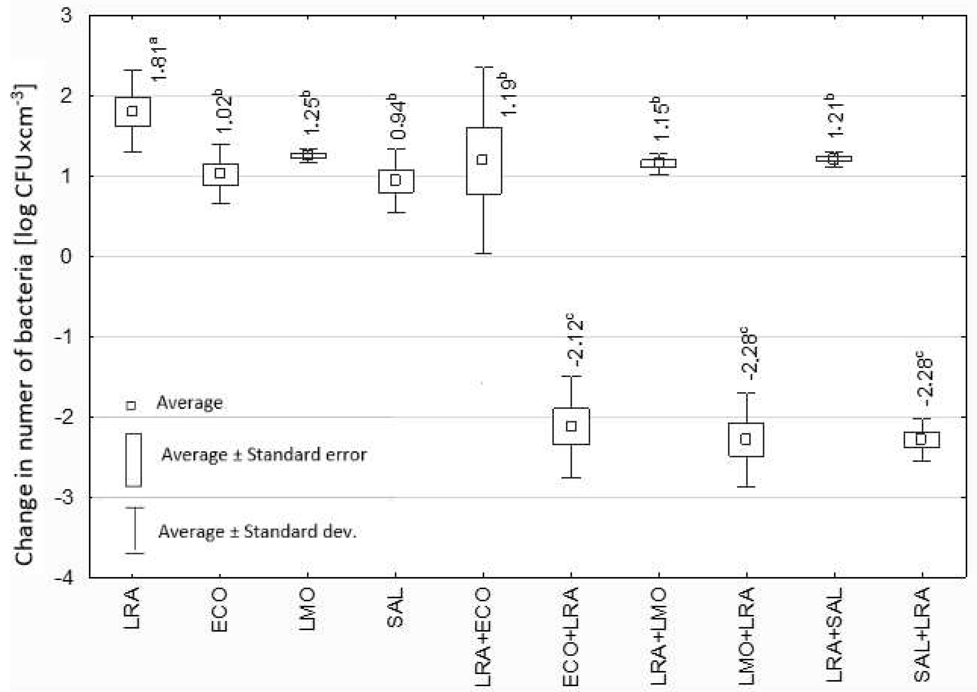
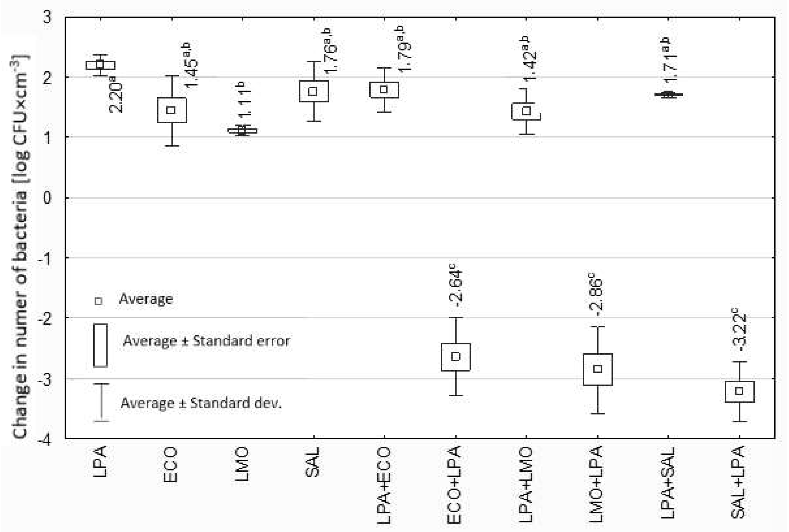
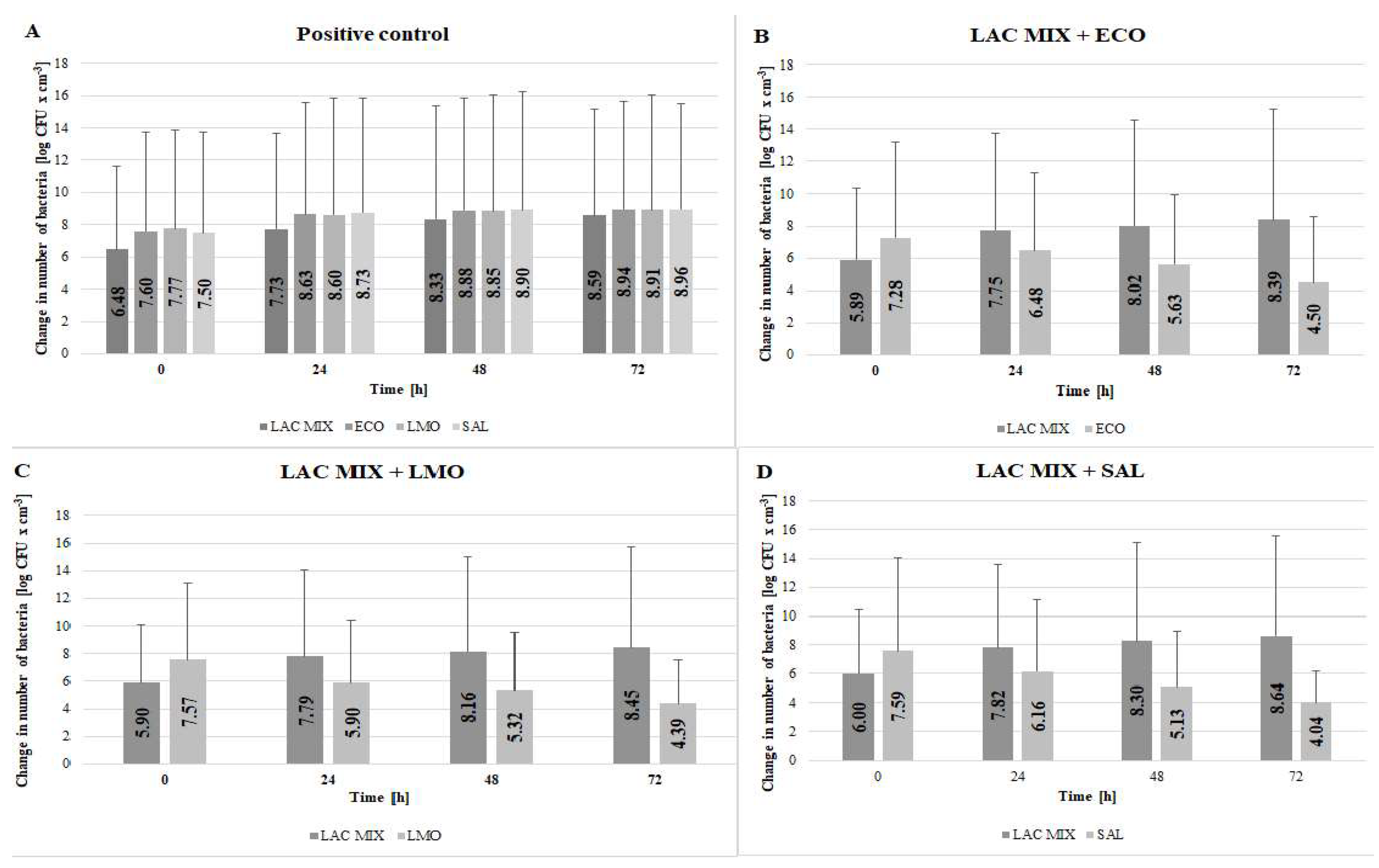
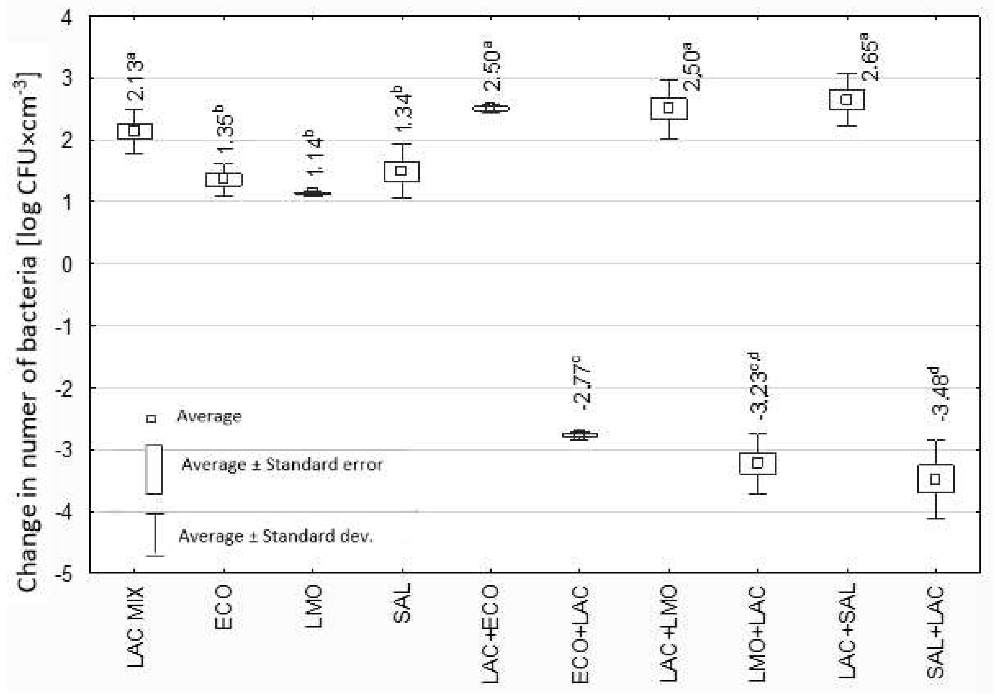
3.2. Effect of Lactobacillus spp. Isolated from Yoghurt on Selected Potentially Pathogenic Bacteria

For the variant to test the effect of L. rhamnosus on pathogenic bacteria, an increase in the number of L. rhamnosus with a decrease in the number of the other bacteria was tested (Figure 1). After 72 h of incubation, the highest number (7.67 log CFU × cm−3) of L. rhamnosus was isolated from the mixed culture with L. monocytogenes, and the lowest (7.46 log CFU × cm−3) from the culture with S. Enteritidis (Figure 1). The number was lower in each variant than in monoculture (Figure 1). Among pathogenic bacteria, the highest number of bacteria after 72 h incubation with L. rhamnosus was shown for E. coli (5.38 log CFU × cm−3) and the lowest (4.88 log CFU × cm−3) for L. monocytogenes (Figure 1).
The above trend is confirmed by the calculated changes in the number of bacteria tested after 72 h of incubation, shown in Figure 2. In monocultures, an increase in the number of all bacteria tested was observed, from 0.94 log CFU × cm−3 for S. Enteritidis to 1.81 log CFU × cm−3 for L. rhamnosus. In the case of L. rhamnosus, the calculated growth was statistically significantly higher (p ≤ 0.05) from the results for the pathogenic bacteria tested (Figure 2). In mixed cultures with pathogenic bacteria (LRA+ECO, LRA+LMO, LRA+SAL), the number of L. rhamnosus increased, but the changes in number were not statistically significant (p > 0.05). A decrease in the number of pathogenic bacteria in co-culture with L. rhamnosus was observed (Figure 2). These decreases ranged from −2.12 log CFU × cm−3 for E. coli (ECO+LRA) to −2.28 log CFU × cm−3 for L. monocytogenes (LMO+LRA) and S. Enteritidis (SAL+LRA) (Figure 2). There were no statistically significant differences between changes in the number of pathogenic bacteria in the presence of L. rhamnosus (p > 0.05).
The number of L. paracasei increased, while the number of pathogenic bacteria decreased both in single and mixed Lactobacillus spp. culture variants (Figure 3). After 72 h of incubation, the highest number (7.64 log CFU × cm−3) of L. paracasei was isolated from the mixed culture with E. coli and the lowest (7.34 log CFU × cm−3) from the culture with L. monocytogenes (Figure 3). Among pathogenic bacteria, the highest number of bacteria after 72 h incubation with L. paracasei was shown for E. coli (4.64 log CFU × cm−3) and the lowest (4.37 log CFU × cm−3) for S. Enteritidis (Figure 3).
The above trend is confirmed by the calculated changes in the number of tested bacteria after 72 h of incubation (Figure 4). In monocultures, an increase in the number of all tested bacteria was observed. For L. paracasei, the calculated increase was statistically significantly different (p ≤ 0.05) compared to the L. monocytogenes (Figure 4). In the mixed culture with all the tested pathogenic bacteria, the number of L. paracasei (LPA+ECO, LPA+LMO, LPA+SAL) increased, but the results were not statistically significant (p > 0.05). In the case of all pathogenic bacteria with L. paracasei, their numbers decreased (Figure 4). These decreases ranged from −2.64 log CFU × cm−3 for E. coli (ECO+LPA) to −3.22 log CFU × cm−3 for S. Enteritidis (SAL+LPA) (Figure 4). No statistically significant differences were found between decreases in the number of pathogenic bacteria after culturing with L. paracasei (p > 0.05).
In the mixed cultures (L. rhamnosus and L. paracasei), an increase in Lactobacillus spp. was observed, while pathogenic microorganisms decreased. After 72 h of incubation, the highest increase (8.64 log CFU × cm−3) in L. rhamnosus and L. paracasei was observed for the mixed culture with S. Enteritidis, and the lowest (8.39 log CFU × cm−3) in the presence of E. coli (Figure 5). Among pathogenic bacteria, the highest number of bacteria after 72 h incubation with the Lactobacillus spp. MIX was shown for E. coli (4.50 log CFU × cm−3) and the lowest (4.04 log CFU × cm−3) for S. Enteritidis (Figure 5).
In monocultures, an increase in the number of all bacteria tested was observed. For Lactobacillus spp. (LAC MIX), the calculated growth was statistically significantly higher (p ≤ 0.05) from the results for the pathogenic bacteria (Figure 6). In mix cultures, after 72 h of incubation, an increase in the number of Lactobacillus spp. was noted in all variants (LAC MIX+ECO, LAC MIX+LMO, LAC MIX+SAL), and these results did not differ statistically significantly (p > 0.05). In turn, for all pathogenic bacteria, after 72 h of incubation with a mix Lactobacillus species (ECO+LAC MIX, LMO+LAC MIX, SAL+LAC MIX), their numbers decreased (Figure 6). These decreases ranged from −2.77 log CFU × cm−3 for E. coli (ECO+LAC MIX) to −3.48 log CFU × cm−3 for S. Enteritidis (SAL+LAC MIX). The calculated decreases were statistically significantly different (p ≤ 0.05) for E. coli and S. Enteritidis (Figure 6).
Changes in the abundance of all tested pathogenic bacteria (average) in the presence of each variant of Lactobacillus spp. suspensions were compared (Figure 7). In each variant, there was a decrease in the number of pathogenic microorganisms tested. There was a statistically significant difference (p ≤ 0.05) between the results for the abundance of L. rhamnosus and the mixture of Lactobacillus species.

4. Discussion

There is a worldwide increase in consumer interest in organic foods and those with health-promoting properties. Awareness is growing regarding the composition and quality of food products offered on the market. Fermented dairy products, including yoghurt, are a valuable source of nutrients, as well as live LAB strains, which are characterized by antimicrobial activity, among other things, allowing for the maintenance a healthy intestinal microbiota [2]. The use of the manufacturer’s declared bacterial species and the maintenance of their counts at a level of not less than 108 CFU/g until the last day of the expiration date for consumption is a key condition from the consumer’s point of view [3]. In our study, we quantified the number of LAB in different types of yoghurts available in Poland. This study showed a decrease in LAB with shorter shelf lives in the following yoghurts: bio natural (−0.29 log CFU/g) and probiotic (−0.18 log CFU/g). The LAB count levels did not fall below the recommended value. Similar results are presented by Sady et al. [24], Nikmaram et al. [25] and Ng et al. [26]. Sady et al. [24] showed a statistically significant decrease in the levels of Bifidobacterium spp., L. acidophilus and S. thermophilus in yoghurts between the 7th and 14th days of storage. Nikmaram et al. [27] showed that the longer the storage time was, the more L. casei abundance decreased, although it did not fall below the WHO recommended values in any sample. In contrast, Xu et al. [28] showed that the total number of viable bacteria in soy yoghurt first increased and then decreased, but all of them met the standard for the number of viable bacteria in probiotic foods. In this study, the highest decrease in LAB was shown for probiotic yoghurt, which is consistent with the results obtained by Mani-López et al. [29]. According to Beal et al. [30], the decrease in the number of live bacteria during yoghurt storage is mainly related to the phenomenon of acidification during refrigerated storage. Different results regarding the change in bacterial counts during product storage time were obtained by Skryplonek et al. [31] and Li et al. [32]. In both studies, there were no major fluctuations in bacterial counts during storage [31,32]. In the study by Skrylonek et al. [31], throughout the storage period, the number of bacteria was higher than the dose (107 CFU/g) recommended by the WHO. In the present study, there was an increase in bacterial counts in Greek-type yoghurt (0.17 log CFU/g), bio drinkable with strawberries (0.45 log CFU/g) and in bio yoghurt with forest fruits (0.45 log CFU/g) during product storage. Kaptan and Kayisoglu [33] showed that the addition of forest fruit puree to probiotic yoghurt effectively promoted the growth of probiotic bacteria. Yang et al. [16] took into account the addition of blackberry juice to yoghurt and showed its positive effect on the growth of Lactobacillus spp. Different results regarding bacterial counts during the storage of fruit yoghurts were obtained by Turgut et al. [34]. During 14 days of product storage, there was a decrease in the viability of L. acidophilus, while the number of Bifidobacterium bifidum remained constant. L. rhamnosus and L. paracasei were isolated from the yoghurts tested. The obtained results differed from the composition on the packaging of the tested yoghurts. On the other hand, Schillinger [35] showed the presence of L. rhamnosus (DNA-DNA hydridation) in the tested yoghurts, despite the manufacturer’s declaration of the presence of Biogarde or Bioghurt culture (which includes L. acidophilus and/or B. bifidum in addition to S. thermophilus). According to Schillinger [35], strains of Lactobacillus spp. other than Biogarde or Bioghurt cultures were used in the production of the tested yoghurts, showing that the correct labeling of the yoghurts was not used in all cases. In our study, the MALDI TOF MS method was used to identify LAB. Different results regarding the manufacturer’s declared composition may be due to the use of other starter cultures.
Fermented dairy products are characterized by health-promoting properties due to the presence of LAB, mainly of the genus Lactobacillus. Bacteria of the genus Lactobacillus are characterized by the production of compounds with antimicrobial activity (lactic acid, organic acids, hydrogen peroxide (H2O2) or bacteriocins). LAB, through antagonistic interactions with pathogenic bacteria, maintain the gastrointestinal ecosystem in a healthy state [36]. Another aspect studied was the antimicrobial effect of Lactobacillus spp. isolated from the tested yoghurt on foodborne pathogenic bacteria. Our results showed that in the presence of Lactobacillus spp., there is a decrease in the number of pathogenic microorganisms (E. coli, L. monocytogenes and S. Enteritidis). Similar results were obtained by Yolmeh et al. [37], Kamal et al. [38], Ołdak et al. [39] and Kumar et al. [40]. We have shown that different species of the genus Lactobacillus caused a different decrease in pathogen abundance. In the single variant (one strain of Lactobacillus spp.), a greater decrease in pathogenic microorganisms occurred in L. paracasei culture than in L. rhamnosus. We showed the highest reduction in the number of L. monocytogenes after 72 h of culture with L. rhamnosus. Iglesias et al. [41] confirmed the antagonistic effect of L. rhamnosus on L. monocytogenes. The researchers [41] also showed reduced survival, adhesion and invasiveness of the L. monocytogenes strains tested, but they did not explain the basis of this mechanism. In turn, Prezzi et al. [42] confirmed the effect of L. rhamnosus on reducing the number of L. monocytogenes cheese surface, with no effect of this strain on Staphylococcus aureus. In contrast, a study by Echresh et al. [43] showed that L. rhamnosus supernatant inhibited biofilm formation by L. monocytogenes. Prezzi et al. [42] and Echresh et al. [43] also failed to explain the differences in the antagonistic effects of L. rhamnosus. We showed that L. rhamnosus had the least effect on reducing E. coli and S. Enteritidis. In contrast, Gutiérrez et al. [44] demonstrated that L. rhamnosus had the effect of reducing the number of E. coli, Listeria innocua and Staphylococcus epidermidis. In turn, Aryantini et al. [45] tested three strains of L. rhamnosus (isolated from a mare’s milk). The researchers confirmed the activity of LAB against S. Typhimurium, Shigella sonnei, L. monocytogenes and E. coli [45]. In contrast, Shi et al. [46] confirmed the antagonistic effect of L. rhamnosus SQ511 on S. Enteritidis. Researchers [46] showed that antimicrobial compounds produced by L. rhamnosus SQ511 caused a significant inhibition of growth, biofilm-forming ability, mobility, changes in the expression of the selected virulence genes and disruption of the integrity of S. Enteritidis. We showed that after 72 h of culture with L. paracasei, the highest decrease was observed for S. Enteritidis. The inhibitory effect of LAB (including L. bulgaricus, L. rhamnosus and L. paracasei) on Salmonella spp. was demonstrated by Muyyarikkandy and Amalaradjou [47]. The researchers [47] showed that LAB affect the reduction in Salmonella spp. (including multidrug-resistant strains) by modulating their gene expression. However, in our study, the greatest reduction in pathogenic bacteria counts occurred in the mixed culture (L. rhamnosus + L. paracasei). The mixed culture reduced the number of S. Enteritidis. Similar studies were conducted by Gutiérrez et al. [44], Zhang et al. [48] and Aryantini et al. [45]. Ołdak et al. [39] reported the highest antimicrobial activity of LAB against L. monocytogenes and E. coli. In a study by Kumar et al. [40], all lactic bacteria tested inhibited the growth of S. aureus, and one strain additionally reduced the growth of E. coli and S. Typhimurium. Gutiérrez et al. [44] demonstrated that L. lactis inhibited the growth of E. coli, Pseudomonas fluorescens and S. epidermidis. On the other hand, L. casei decreased the abundance of P. fluorescens, S. epidermidis and S. aureus [44]. Zhang et al. [48] showed high antimicrobial activity for L. paracasei sub. Paracasei, L. casei and two strains of L. rhamnosus against S. sonnei, E. coli and S. Typhimurium. High antimicrobial activity against 12 selected pathogenic bacteria and Aspergillus flavus strains of the genus Lactobacillus isolated from conventional curd was demonstrated by Haghshenas et al. [49]. In turn, Mohd-Zubri et al. [50] showed that L. brevis and L. plantarum species isolated from local Malaysian fermented foods showed antimicrobial activity against Porphyromonas gingivalis (a periodontal pathogen). Different results, compared to our on the decrease in the number of pathogenic microorganisms in the presence of LAB were presented by Sharma et al. [51]. This may be due to the fact that the study used a cell-free supernatant (L. casei, L. delbrueckii, L. fermentum, L. plantarum and L. pentosus). In a trial using S. aureus, L. monocytogenes, E. coli and Klebsiella pneumoniae, the supernatant showed no inhibitory activity against the listed pathogens. Weak activity was noted against Pseudomonas aeruginosa and Proteus mirabilis, while moderate activity was noted against Bacillus cereus, S. Typhi and Shigella flexneri [51].
The action of LAB is related to antimicrobial peptides that penetrate the outer membranes of Gram-negative bacteria and the cell walls of Gram-positive bacteria, and produce cavities, causing the efflux of intracellular contents [52]. LAB activity may also result from organic acid production [2]. The evaluation of the mechanisms of LAB action on pathogenic bacteria requires further research. The presence of LAB in dairy products can benefit human health but also contributes to the reduction or elimination of foodborne bacteria.
The limitation of this study was the small number of yoghurt samples and Lactobacillus spp. strains isolated from the yoghurt samples, which could be related to the use of one type of medium (the Rogosa agar, which is a medium designed to culture Lactobacillus spp., among other things, from dairy product samples) and incubation conditions. Future studies should consider a wider range of isolation methods, including molecular methods. Additionally, it would be valuable to assess the mechanisms influencing the reduction in pathogenic bacteria by Lactobacillus bacteria.

5. Conclusions

In our pilot study, we showed different abundances of LAB in different products, also depending on the shelf life, especially for L. rhamnosus and L. paracasei. Despite the observed decreases, the abundance did not fall below the required values for these products. This highlights that yoghurts are characterized by the presence of live LAB cultures throughout their shelf life. The results of studies on the decrease in the number of pathogenic bacterial species tested in the presence of Lactobacillus spp. strains constitute the basis for formulating new probiotics as biotherapeutic agents or dietary supplements or additives to fermented milk beverages. Lactobacillus spp. strains isolated from yoghurts in mixed cultures caused a decrease in the abundance of selected pathogenic bacterial species (E. coli, L. monocytogenes and S. Enteritidis). It was shown that the greatest decrease in the number of pathogenic bacteria occurred in the mixed culture including L. rhamnosus and L. paracasei. The presence of LAB in yoghurts may, through an antagonistic effect, contribute to the elimination of bacteria, causing food spoilage, and the reduction in foodborne bacterial infections. However, further studies are necessary, taking into account a larger number of strains and an animal model, and it is also necessary to understand the mechanisms underlying the antagonistic effects of LAB on selected pathogenic bacteria.

Author Contributions

N.W.-K.: conceptualization, formal analysis, writing—review and editing, supervision. K.G.-B.: methodology, validation, investigation, writing—original draft preparation, project administration. A.B.: validation, data curation, visualization. K.C.-G.: investigation, data curation, writing—original draft preparation, visualization. E.G.-K.: formal analysis, resources, funding acquisition. K.S.: conceptualization, methodology, resources, writing—review and editing, supervision, project administration, funding acquisition. All authors have read and agreed to the published version of the manuscript.

Funding

This research was funded by the Nicolaus Copernicus University with funds from the maintenance of the research potential of the Department of Microbiology PDB WF.

Institutional Review Board Statement

Not applicable.

Data Availability Statement

The data presented in this study are available upon request to the corresponding author.

Conflicts of Interest

The authors declare no conflicts of interest.

References

  1. U.S. Department of Agriculture; U.S. Department of Health and Human Services. Dietary Guidelines for Americans, 2020–2025, 9th ed; December 2020. Available online: http://www.dietaryguidelines.gov/resources/2020-2025-dietary-guidelines-online-materials (accessed on 10 July 2024).
  2. Fisberg, M.; Machado, R. History of yogurt and current patterns of consumption. Nutr. Rev. 2015, 73 (Suppl. S1), 4–7. [Google Scholar] [CrossRef] [PubMed]
  3. Chandan, R.C.; Gandhi, A.; Shah, N.P. Yogurt: Historical background, health benefits, and global trade. In Yogurt in Health and Disease Prevention; Academic Press: London, UK, 2017; pp. 3–29. [Google Scholar] [CrossRef]
  4. Ouweh, G.J.; Vest, A.C. Anti-microbial components from lactic acid bacteria. In Lactic Acid Bacteria: Microbiology and Functional Aspects, 2nd ed.; Salminen, S., Von, A., Eds.; Marcel Dekker Inc.: New York, NY, USA, 2004; pp. 139–160. [Google Scholar]
  5. Molaee Parvarei, M.; Fazeli, M.R.; Mortazavian, A.M.; Sarem Nezhad, S.; Mortazavi, S.A.; Golabchifar, A.A.; Khorshidian, N. Comparative effects of probiotic and paraprobiotic addition on microbiological, biochemical and physical properties of yogurt. Food Res. Int. 2021, 140, 110030. [Google Scholar] [CrossRef]
  6. Kapka-Skrzypczak, L.; Niedźwiecka, J.; Wojtyła, A.; Kruszewski, M. Probiotyki i prebiotyki jako aktywny składnik żywności funkcjonalnej. Pediatr. Endocrinol. Diabetes Metab. 2012, 18, 79–83. [Google Scholar] [PubMed]
  7. Presti, I.; D’Orazio, G.; Labra, M.; La Ferla, B.; Mezzasalma, V.; Bizzaro, G.; Giardina, S.; Michelotti, A.; Tursi, F.; Vassallo, M.; et al. Evaluation of the probiotic properties of new Lactobacillus and Bifidobacterium strains and their in vitro effect. App. Microbiol. Biotech. 2015, 99, 613–5626. [Google Scholar] [CrossRef]
  8. Abdi-Moghadam, Z.; Darroudi, M.; Mahmoudzadeh, M.; Mohtashami, M.; Jamal, A.M.; Shamloo, E.; Rezaei, Z. Functional yogurt, enriched and probiotic: A focus on human health. Clin. Nutr. ESPEN 2023, 57, 575–586. [Google Scholar] [CrossRef]
  9. Cifelli, C.J.; Agarwal, S.; Fulgoni, V.L. Association of Yogurt Consumption with Nutrient Intakes, Nutrient Adequacy, and Diet Quality in American Children and Adults. Nutrients 2020, 12, 3435. [Google Scholar] [CrossRef] [PubMed]
  10. Zhu, Y.; Jain, N.; Holschuh, N.; Smith, J. Associations between frequency of yogurt consumption and nutrient intake and diet quality in the United Kingdom. J. Nutr. Sci. 2021, 10, e85. [Google Scholar] [CrossRef] [PubMed]
  11. Hasegawa, Y.; Bolling, B.W. Yogurt consumption for improving immune health. Curr. Opin. Food Sci. 2023, 51, 101017. [Google Scholar] [CrossRef]
  12. Wyka, J.; Tajner-Czopek, A.; Rytel, E.; Habanova, M.; Malczyk, E.; Misiarz, M. Żywność fermentowana—Znaczenie dla zdrowia człowieka. Acta Sci. Pol. Biotechnol. 2017, 16, 101–106. [Google Scholar]
  13. Świderski, F.; Waszkiewicz-Robak, B. Towaroznawstwo Żywności Przetworzonej z Elementami Technologii; Wydawnictwo SGGW: Warszawa, Poland, 2010. [Google Scholar]
  14. Nami, Y.; Tavallaei, O.; Kiani, A.; Moazami, N.; Samari, M.; Derakhshankhah, H.; Jaymand, M.; Haghshenas, B. Anti-oral cancer properties of potential probiotic lactobacilli isolated from traditional milk, cheese, and yogurt. Sci. Rep. 2024, 14, 6398. [Google Scholar] [CrossRef] [PubMed]
  15. World Health Organization (WHO). Food Safety. Available online: https://www.who.int/news-room/fact-sheets/detail/food-safety (accessed on 15 September 2024).
  16. Yang, H.; Hewes, D.; Salaheen, S.; Federman, C.; Biswas, D. Effects of blackberry juice on growth inhibition of foodborne pathogens and growth promotion of Lactobacillus. Food Control 2014, 37, 15–20. [Google Scholar] [CrossRef]
  17. Alvarez-Sieiro, P.; Montalbán-López, M.; Mu, D.; Kuipers, O.P. Bacteriocins of lactic acid bacteria: Extending the family. Appl. Microbiol. Biotechnol. 2016, 100, 2939–2951. [Google Scholar] [CrossRef]
  18. Darbandi, A.; Asadi, A.; Mahdizade Ari, M.; Ohadi, E.; Talebi, M.; Halaj Zadeh, M.; Darb Emamie, A.; Ghanavati, R.; Kakanj, M. Bacteriocins: Properties and potential use as antimicrobials. J. Clin. Lab. Anal. 2022, 36, e24093. [Google Scholar] [CrossRef]
  19. EFSA and ECDC (European Food Safety Authority and European Centre for Disease Prevention and Control): The European Union One Health 2021 Zoonoses Report. EFSA J. 2022, 20, 7666. [CrossRef]
  20. World Health Organization (WHO). Listeriosis. 2018. Available online: https://www.who.int/news-room/fact-sheets/detail/listeriosis (accessed on 15 September 2024).
  21. Wiktorczyk-Kapischke, N.; Skowron, K.; Wałecka-Zacharska, E. Genomic and pathogenicity islands of Listeria monocytogenes-overview of selected aspects. Front. Mol. Biosci. 2023, 10, 1161486. [Google Scholar] [CrossRef]
  22. Khaneghah, A.M.; Abhari, K.; Eş, I.; Soares, M.B.; Oliveira, R.B.A.; Hasseini, H.; Rezai, M.; Balthazar, C.F.; Silva, R.; Cruz, A.G.; et al. Interactions between probiotics and pathogenic microorganisms in hosts and foods: A review. Trends Food Sci. Technol. 2020, 95, 205–218. [Google Scholar] [CrossRef]
  23. Schulthess, B.; Brodner, K.; Bloemberg, G.V.; Zbinden, R.; Böttger, E.C.; Hombach, M. Identification of Gram-positive cocci by use of matrix-assisted laser desorption ionization-time of flight mass spectrometry: Comparison of different preparation methods and implementation of a practical algorithm for routine diagnostics. J. Clin. Microbiol. 2013, 51, 1834–1840. [Google Scholar] [CrossRef] [PubMed]
  24. Sady, M.; Domagała, J.; Grega, T.; Kalicka, D. Wpływ czasu przechowywania na mikroflorę jogurtów z dodatkiem nasion amarantusa i ziaren owsa. Żywność Nuka Technol. Jakość 2007, 6, 242–250. [Google Scholar]
  25. Nikmaram, P.; Mousavi, S.M.; Kiani, H.; Emamdjomeh, Z.; Razavi, S.H.; Mousavi, Z. Modeling the effect of inulin, pH and storage time on the viability of selected Lactobacillus in a probiotic fruity yogurt drink using the Monte Carlo simulation. J. Food Qual. 2016, 39, 362–369. [Google Scholar] [CrossRef]
  26. Ng, E.W.; Yeung, M.; Tong, P.S. Effects of starter cultures on the survival of Lactobacillus acidophilus. Int. J. Food Microbiol. 2011, 45, 169–175. [Google Scholar] [CrossRef] [PubMed]
  27. Nikmaram, P.; Mousavi, S.M.; Emam-Djomeh, Z.; Kiani, H.; Razavi, S.H. Evaluation and prediction of metabolite production, antioxidant activities, and survival of Lactobacillus casei 431 in a pomegranate juice supplemented yogurt drink using support vector regression. Food Sci. Biotechnol. 2015, 24, 2105–2112. [Google Scholar] [CrossRef]
  28. Xu, X.; Cui, H.; Xu, J.; Yuan, Z.; Li, J.; Yang, L.; Wang, S.; Liu, H.; Zhu, D. Effects of cold storage time on the quality and active probiotics of yogurt fermented by Bifidobacterium lactis and commercial bacteria Danisco. J. Food Sci. 2023, 88, 2796–2806. [Google Scholar] [CrossRef] [PubMed]
  29. Mani-López, E.; Palou, E.; López-Malo, A. Probiotic viability and storage stability of yogurts and fermented milks prepared with several mixtures of lactic acid bacteria. J. Dairy Sci. 2014, 97, 2578–2590. [Google Scholar] [CrossRef]
  30. Beal, C.; Skokanova, J.; Latrille, E.; Martin, N.; Corrieu, G. Combined effects of culture conditions and storage time on acidification and viscosity of stirred yogurt. J. Dairy Sci. 1999, 82, 673–681. [Google Scholar] [CrossRef]
  31. Skryplonek, K.; Jasińska, M. Jakość fermentowanych napojów probiotycznych otrzymanych z mrożonej serwatki kwasowej i mleka w czasie chłodniczego przechowywania. Żywność Nauka Technol. Jakość 2016, 1, 32–44. [Google Scholar]
  32. Li, C.; Song, J.; Kwok, L.; Wang, J.; Dong, Y.; Yu, H.; Hou, Q.; Zhang, H.; Chen, Y. Influence of Lactobacillus plantarum on yogurt fermentation properties and subsequent changes during postfermentation storage. Am. Dairy Sci. Assoc. 2017, 1, 2512–2525. [Google Scholar] [CrossRef]
  33. Kaptan, B.; Kayisoglu, S. Influence of forest fruit purees (strawberry or blackberry) on the biochemical, microbiological, sensory properties, and consumer preferences of standard and probiotic cultured yogurts. J. Food Saf. Food Qual. 2024, 75, 77–85. [Google Scholar] [CrossRef]
  34. Turgut, T.; Cakmakci, S. Probiotic Strawberry Yogurts: Microbiological, Chemical and Sensory Properties. Probiotics Antimicrob. Prot. 2018, 10, 64–70. [Google Scholar] [CrossRef]
  35. Schillinger, U. Isolation and identification of lactobacilli from novel-type probiotic and mild yoghurts and their stability during refrigerated storage. Int. J. Food Microbiol. 1999, 47, 79–87. [Google Scholar] [CrossRef] [PubMed]
  36. Pyar, H.; Peh, K.K. Characterization and identification of Lactobacillus acidophilus using biolog rapid identification system. Int. J. Pharm. Pharm. Sci. 2014, 6, 189–193. [Google Scholar]
  37. Yolmeh, M.; Khomeiri, M.; Ahmadi, Z. Application of mixture design to introduce an optimum cell-free supernatant of multi-strain mixture (MSM) for Lactobacillus against food-borne pathogens. LWT 2017, 83, 298–304. [Google Scholar] [CrossRef]
  38. Kamal, R.M.; Alnakap, M.E.; Abal El Aal, S.F.; Bayoumi, M.A. Bio-controlling capability of probiotic strain Lactobacillus rhamnosus against some common foodborne pathogens in yoghurt. Int. Dairy J. 2018, 85, 1–7. [Google Scholar] [CrossRef]
  39. Ołdak, A.; Zielińska, D.; Rzepkowska, A.; Kołożyn-Krajewska, D. Comparison of Antibacterial Activity of Lactobacillus plantarum Strains Isolated from Two Different Kinds of Regional Cheeses from Poland: Oscypek and Korycinski Cheese. BioMed Res. Int. 2017, 2017, 6820369. [Google Scholar] [CrossRef]
  40. Kumar, A.; Kumar, D. Characterization of Lactobacillus isolated from dairy samples for probiotic properties. Clin. Microbiol. 2015, 33, 117–123. [Google Scholar] [CrossRef]
  41. Iglesias, M.N.; Viñas, I.; Colás-Medà, P.; Collazo, C.; Serrano, J.C.E.; Abadias, M. Adhesion and invasion of Listeria monocytogenes and interaction with Lactobacillus rhamnosus GG after habituation on fresh-cut pear. J. Func. Foods 2017, 34, 453–460. [Google Scholar] [CrossRef]
  42. Prezzi, L.E.; Lee, S.H.I.; Nunes, V.M.R.; Corassin, C.H.; Pimentel, T.C.; Rocha, R.S.; Ramos, G.L.P.A.; Guimarães, J.T.; Balthazar, C.F.; Durate, M.C.K.H.; et al. Effect of Lactobacillus rhamnosus on growth of Listeria monocytogenes and Staphylococcus aureus in a probiotic Minas Frescal cheese. Food Microbiol. 2020, 92, 103557. [Google Scholar] [CrossRef]
  43. Echresh, S.; Behbahani, B.A.; Falah, F.; Noshad, M.; Ibrahim, S.A. Assessment of the probiotic, anti-bacterial, and anti-biofilm characteristics of Lacticaseibacillus rhamnosus CWKu-12, along with its potential impact on the expression of virulence genes in Listeria monocytogenes ATCC 19115. LWT. 2024, 203, 116391. [Google Scholar] [CrossRef]
  44. Gutiérrez, S.; Martínez-Blanco, H.; Rodríguez-Aparicio, L.B.; Ferrero, M.A. Effect of fermented broth from lactic acid bacteria on pathogenic bacteria proliferation. J. Dairy Sci. 2016, 9, 2654–2665. [Google Scholar] [CrossRef] [PubMed]
  45. Aryantini, N.P.; Yamasaki, E.; Kurozono, H.; Sujaya, N.; Urshima, T.; Fukuda, K. In vitro safety assessments and antimicrobial activities of Lactobacillus rhamnosus strains isolated from a fermented mare’s milk. Anim. Sci. J. 2017, 88, 517–525. [Google Scholar] [CrossRef]
  46. Shi, S.; Gong, L.; Yu, H.; He, G.; Zhang, J.; Han, Y.; Liu, Y.; Hu, J.; Dong, J.; Liu, J.; et al. Antagonistic activity and mechanism of Lactobacillus rhamnosus SQ511 against Salmonella enteritidis. 3 Biotech 2022, 12, 126. [Google Scholar] [CrossRef] [PubMed]
  47. Muyyarikkandy, M.S.; Amalaradjou, M.A. Lactobacillus bulgaricus, Lactobacillus rhamnosus and Lactobacillus paracasei Attenuate Salmonella Enteritidis, Salmonella Heidelberg and Salmonella Typhimurium Colonization and Virulence Gene Expression In Vitro. Int. J. Mol. Sci. 2017, 18, 2381. [Google Scholar] [CrossRef]
  48. Zhang, Y.; Zhang, L.; Du, M.; Yi, H.; Guo, C.; Tuo, Y.; Han, X.; Li, J.; Zhang, L.; Yang, L. Antimicrobial activity against Shigella sonnei and probiotic properties of wild Lactobacillus from fermented food. Microbiol. Res. 2011, 167, 27–31. [Google Scholar] [CrossRef] [PubMed]
  49. Haghshenas, B.; Kiani, A.; Mansoori, S.; Mohammadi-Noori, E.; Nami, Y. Probiotic properties and antimicrobial evaluation of silymarin-enriched Lactobacillus bacteria isolated from traditional curd. Sci. Rep. 2023, 13, 10916. [Google Scholar] [CrossRef] [PubMed]
  50. Mohd-Zubri, N.S.; Ramasamy, K.; Abdul-Rahman, N.Z. Characterization and potential oral probiotic properties of Lactobacillus plantarum FT 12 and Lactobacillus brevis FT 6 isolated from Malaysian fermented food. Arch. Oral Biol. 2022, 143, 105515. [Google Scholar] [CrossRef] [PubMed]
  51. Sharma, C.; Singh, B.P.; Thakur, N.; Gulati, S.; Gupta, S.; Mishra, S.K.; Panwar, H. Antibacterial effects of Lactobacillus isolates of curd and human milk origin against food-borne and human pathogens. 3 Biotech 2017, 7, 31. [Google Scholar] [CrossRef]
  52. Çakmakoğlu, S.K.; Dere, S.; Bekiroğlu, H.; Bozkurt, F.; Karasu, S.; Dertli, E.; Türker, M.; Sagdic, O. Production of bioactive peptides during yogurt fermentation, their extraction and functional characterization. Food Biosci. 2024, 61, 104805. [Google Scholar] [CrossRef]
Figure 1. Changes in the number of bacteria in relation to culture time. (A) Positive control. (B) Changes in the number of bacteria in a culture of L. rhamnosus with E. coli. (C) Changes in the number of bacteria in a culture of L. rhamnosus with L. monocytogenes. (D) Changes in the number of bacteria in cultures of L. rhamnosus and S. Enteritidis. LRA—L. rhamnosus; ECO—E. coli; LMO—L. monocytogenes; SAL—S. Enteritidis; CFU—colony-forming unit.
Applsci 14 09010 g001
Figure 2. Changes in the number of L. rhamnosus and foodborne bacteria after 72 h of incubation in individual cultures (LRA—L. rhamnosus; ECO—E. coli; LMO—L. monocytogenes; SAL—S. Enteritidis) and in mixed cultures (LRA+ECO—changess in the number of L. rhamnosus in a mixed culture of L. rhamnosus and E. coli; ECO+LRA—changes in the number of E.coli in a mixed culture of L. rhamnosus and E. coli; LRA+LMO—changes in the number of L. rhamnosus in a mixed culture of L. rhamnosus and L. monocytogenes; LMO+LRA—changes in the number of L. monocytogenes in a mixed culture of L. rhamnosus and L. monocytogenes; LRA+SAL—changes in the number of L. rhamnosus in a mixed culture of L. rhamnosus and S. Enteritidis; SAL+LRA—changes in the number of S. Enteritidis in a mixed culture of L. rhamnosus and S. Enteritidis). a–c—values marked with different letters differ in a statistically significant way (p ≤ 0.05).
Applsci 14 09010 g002
Figure 3. Changes in the number of bacteria in relation to culture time. (A) Positive control. (B) Changes in the number of bacteria in a culture of L. paracasei with E. coli. (C) Changes in the number of bacteria in a culture of L. paracasei with L. monocytogenes. (D) Changes in the number of bacteria in cultures of L. paracasei and S. Enteritidis. LPA—L. paracasei; ECO—E. coli; LMO—L. monocytogenes; SAL—S. Enteritidis; CFU—colony-forming unit.
Applsci 14 09010 g003
Figure 4. Changes in the number of L. paracasei and foodborne bacteria after 72 h of incubation in individual cultures (LPA—L. paracasei; ECO—E. coli; LMO—L. monocytogenes; SAL—S. Enteritidis) and in mixed cultures (LPA+ECO—changes in the number of L. paracasei in a mixed culture of L. paracasei and E. coli; ECO+LPA—changes in the number of E.coli in a mixed culture of L. paracasei and E. coli; LPA+LMO—changes in the number of L. paracasei in a mixed culture of L. paracasei and L. monocytogenes; LMO+LPA—changes in the number of L. monocytogenes in a mixed culture of L. paracasei and L. monocytogenes; LPA+SAL—changes in the number of L. paracasei in a mixed culture of L. paracasei and S. Enteritidis; SAL+LPA—changes in the number of S. Enteritidis in a mixed culture of L. paracasei and S. Enteritidis). a–c—values marked with different letters differ in a statistically significant way (p ≤ 0.05).
Applsci 14 09010 g004
Figure 5. Changes in the number of bacteria in relation to culture time. (A) Positive control. (B) Changes in the number of bacteria in a culture of L. paracasei+L. rhamnosus (LAC MIX) with E. coli. (C) Changes in the number of bacteria in a culture of L. paracasei+L. rhamnosus (LAC MIX) with L. monocytogenes. (D) Changes in the number of bacteria in cultures of L. paracasei+L. rhamnosus (LAC MIX) and S. Enteritidis. LAC MIX—L. paracasei+L. rhamnosus; ECO—E. coli; LMO—L. monocytogenes; SAL—S. Enteritidis; CFU—colony-forming unit.
Applsci 14 09010 g005
Figure 6. Changes in the number of L. paracasei + L. rhamnosus and foodborne bacteria after 72 h of incubation in individual cultures (LAC MIX—L. paracasei + L. rhamnosus; ECO—E. coli; LMO—L. monocytogenes; SAL—S. Enteritidis) and mixed cultures (LAC MIX+ECO—changes in the number of L. paracasei + L. rhamnosus in a mixed culture of L. paracasei + L. rhamnosus and E. coli; ECO+LAC MIX—changes in the number of E. coli in a mixed culture of L. paracasei + L. rhamnosus and E. coli; LAC MIX+LMO—changes in the number of L. paracasei + L. rhamnosus in a mixed culture of L. paracasei + L. rhamnosus and L. monocytogenes; LMO+LAC MIX—changes in the number of L. monocytogenes in a mixed culture of L. paracasei + L. rhamnosus and L. monocytogenes; LAC MIX+SAL—changes in the number of L. paracasei + L. rhamnosus in a mixed culture of L. paracasei + L. rhamnosus and S. Enteritidis; SAL+LAC MIX—changes in the number of S. Enteritidis in a mixed culture of L. paracasei+ L. rhamnosus and S. Enteritidis). a–c—values marked with different letters differ in a statistically significant way (p ≤ 0.05).
Applsci 14 09010 g006
Figure 7. Changes in the number of tested foodborne bacteria in the presence of individual Lactobacillus species (LRA—Lactobacillus rhamnosus; LPA—Lactobacillus paracasei; LAC MIX—L. rhamnosus and L. paracasei). a–c—values marked with different letters differ in a statistically significant way (p ≤ 0.05).
Applsci 14 09010 g007
Table 1. Comparison of the occurrence of bacterial species declared by the manufacturer with the identified species, and their change in abundance depending on the expiry date.
Type of YoghurtBio Natural
(n = 2)
Bio with Forest Fruits
(n = 2)
Bio Drinkable with Strawberries (n = 2)Probiotic
(n = 2)
Natural Greek Type
(n = 2)
Bacterial species declared by the manufacturer: “yoghurt bacteria cultures” and the listed speciesL. acidophilus, B. lactisL. acidophilus, B. lactisL. acidophilus, B. lactisL. casei, L. bulgaricus, L. acidophilus, S. thermophilus, Bifidobacterium spp.S. thermophilus, L. delbrueckii, subsp. bulgaricus, L. acidophilus, B. lactis
Long shelf life
(n = 10)
Bacterial species isolated from the productL. rhamnosusL. rhamnosusL. rhamnosusL. paracaseiL. rhamnosus
Average number of bacteria [log CFU/g]8.33 (6.33)8.62 (6.71)8.66 (7.05)8.27 (6.87)8.46 (6.15)
Directly before the end of shelf life
(n = 10)
Bacterial species isolated from the productL. rhamnosusL. rhamnosusL. rhamnosusL. paracaseiL. rhamnosus
Average number of bacteria [log CFU/g]8.04 (7.10)9.06 (7.87)9.11 (6.55)8.09 (6.50)8.63 (7.00)
Changes in the number of bacteria in stored yoghurt [log CFU/g]−0.290.450.45−0.180.17
CFU—colony-forming unit; (…)—standard deviation.
Disclaimer/Publisher’s Note: The statements, opinions and data contained in all publications are solely those of the individual author(s) and contributor(s) and not of MDPI and/or the editor(s). MDPI and/or the editor(s) disclaim responsibility for any injury to people or property resulting from any ideas, methods, instructions or products referred to in the content.

