Article

Exploring the Potential Protective Effect of Probiotics in Obesity-Induced Colorectal Cancer: What Insights Can In Vitro Models Provide?

by
Rejane Viana
1,2,†,
Ana C. Rocha
2,3,4,5,†,
André P. Sousa
2,3,4,5,*,†,
Diogo Ferreira
2,6,7,
Rúben Fernandes
2,3,5,8,
Cátia Almeida
2,4,
Patrick J. Pais
2,8,
Pilar Baylina
1,2,5 and
Ana Cláudia Pereira
2,3,5,8
1
ESS—School of Health, IPP—Polytechnic Institute of Porto, 4200-072 Porto, Portugal
2
CECLIN—Center of Clinical Studies, HE-FP—Hospital Fernando Pessoa, 4420-096 Gondomar, Portugal
3
FCS—Faculty of Health Sciences, Fernando Pessoa University, 4200-253 Porto, Portugal
4
FMUP—Faculty of Medicine of the University of Porto, 4200-319 Porto, Portugal
5
I3S—Instituto de Investigação e Inovação em Saúde, 4200-135 Porto, Portugal
6
Department of Functional Biology and Health Sciences, Faculty of Biology, University of Vigo, 36310 Vigo, Spain
7
TBIO—Center for Translational Health and Medical Biotechnology Research, Escola Superior de Saúde do Porto, 4200-072 Porto, Portugal
8
FP-BHS—Biomedical and Health Sciences Research Unit, FP-I3ID—Instituto de Investigação, Inovação e Desenvolvimento, FFP—Fundação Fernando Pessoa, 4249-004 Porto, Portugal
*
Author to whom correspondence should be addressed.
These authors contributed equally to this work.
Appl. Sci. 2024, 14(2), 951; https://doi.org/10.3390/app14020951
Submission received: 29 November 2023 / Revised: 5 January 2024 / Accepted: 19 January 2024 / Published: 22 January 2024
(This article belongs to the Section Biomedical Engineering)

Abstract

:
Colorectal cancer (CRC) is the third most common cancer diagnosed today and the third leading cause of death among cancer types. CRC is one of the gastrointestinal tumors with obesity as the main extrinsic risk factor, since, according to authors, the meta-inflammation sustained by the excess adipose tissue can provide abundant circulating lipids, as well as hormones and metabolites crucial to tumor development and aggressiveness. The gut microbiota can protect the colon from meta-inflammation and endocrine changes caused by obesity. The present study aimed to investigate the antitumor activity of a commercial probiotic in intestinal tumor cells under two adiposity conditions. Experimental assays were performed on the Caco2 cell line (colon adenocarcinoma) supplemented with differentiated adipocyte’s secretomes of the 3T3-L1 cell line (mouse pre-adipocytes) in two adiposity conditions: (i) differentiation without the use of Pioglitazone (noPGZ) and (ii) differentiation using Pioglitazone (PGZ). The Caco2 cells were first exposed to both secretomes for 24 h and evaluated and subsequently exposed to probiotic extract followed by secretome and evaluated. The effects of these treatments were evaluated using cytotoxicity assays by MTT, cell migration by injury, and antioxidant activity by glutathione assay. The use of secretomes showed a statistically significant increase in cell viability in Caco2 cells, either in noPGZ (p < 0.01) or PGZ (p < 0.05), and the probiotic was not able to reduce this effect. In the injury assay, secretome increased cell migration by more than 199% in both adiposity conditions (p < 0.001 in noPGZ and p < 0.01 in PGZ). In the probiotic treatment, there was a reduction in cell migration compared to the control in adiposity conditions. The antioxidant response of Caco2 cells was increased in both adiposity conditions previously exposed to the probiotic supernatant. This pilot work brings to light some findings that may answer why the modulation of the intestinal microbiota using probiotics is an alternative strategy leading to improvements in the condition and stage of the colon tumor. Additional studies are needed to clarify the role of Pioglitazone in this type of tumor and the metabolites of obesity that are attenuated by the use of probiotics.

1. Introduction

According to the WHO (2021), obesity is a global epidemic that constitutes an acquired disease. In 2016, about 650 million adults (13% of the world population) were obese, and 1.9 billion were overweight [1,2]. According to the WHO European Region (2022), almost 60% of adults and nearly one in three children are overweight or obese (29% of boys and 27% of girls) [3].
Obesity is a risk factor for several diseases, including cancer [4,5]. Recent findings indicate that over 40% of individuals diagnosed with gastrointestinal cancer, such as colorectal cancer, are overweight, with obesity being the primary external risk factor for the development of this cancer [4,5]. This risk factor is linked to the growth of intra-abdominal tumors, which thrive in the omentum majus environment. This region is rich in white adipocytes that function as an energy reservoir, where lipolysis and β-oxidation in tumor cells facilitate rapid proliferation through energy metabolism [6]. One of the four emerging markers proposed by Hanahan and Weinberg is unregulated energy metabolism. Thus, generalized obesity and increased energy supply in the omentum majus potentiate important risk factors produced by excess white adipose tissue (WAT) [6,7]. Currently, the intestinal microbiota has been associated with vital physiological functions and could have a beneficial or detrimental impact on the metabolism of adipose tissue and cancer [8].
White adipose tissue (WAT), an endocrine organ that may store energy in the form of triglycerides for the maintenance of local and systemic energetic homeostasis, is the source of changes that lead to obesity, modulating different biological processes and consequently influencing the metabolism of various organs, such as the liver, muscle, pancreas, and brain, via endocrine mechanisms [2,9]. Under normal conditions, WAT is deposited in subcutaneous locations, where it carries out thermoregulatory functions and facilitates the easy mobilization of triglycerides in response to need [10]. However, in the context of obesity, WAT exceeds its capacity for energy storage in subcutaneous regions, leading to a compensatory response characterized by hyperplasia (an increase in the number of adipocytes) and hypertrophy (an increase in adipocyte size). This excess adipose tissue is deposited in other areas of the body, such as the visceral region [2,10].
The growth of WAT involves tissue remodeling characterized by increased extracellular matrix production, higher immune cells infiltration, and a stronger pro-inflammatory response [2,11]. This condition defines meta-inflammation, supported by WAT, as a medical issue associated with obesity.
Excess fat, especially visceral fat, being metabolically active and continuously releasing free fatty acids (FFAs) into portal circulation, can lead to insulin resistance and several other health problems, including systemic inflammation, dyslipidemia, atherosclerosis, and metabolic syndrome [2,11].
Insulin resistance stands as the primary mechanism derived from the remodeled white adipose tissue deposited in the visceral region. This resistance is closely associated with various metabolic dysfunctions, including type 2 diabetes mellitus (T2DM), cancer, and cardiometabolic risk. Traditional treatment for diabetic patients involves antidiabetic drugs or insulin therapy, such as Pioglitazone (PGZ). PGZ is an insulin-sensitizing drug involved in the mediation of nuclear peroxisome proliferator-activated gamma receptor (PPARy), a transcriptional regulator of adipocyte differentiation and lipid storage that is abundant in adipose tissue [12]. Paradoxically, treatment with PGZ can result in weight gain by the formation of new adipocytes, despite promoting improvement of lipid profile, insulin sensitivity, and glycemic control [12]. The expansion of adipose tissue is accompanied by inflammatory changes that contribute to the development of low-grade systemic inflammation, characterized by high infiltration of class M1 macrophages and pro-inflammatory cytokines, chemokines, and acute phase proteins [2] The accumulation of macrophages is notably more pronounced in visceral adipose tissue compared to subcutaneous adipose tissue [2,13].
Cytokines released by metabolically active WAT are involved in tumor progression and risk because they promote chronic inflammation, stimulating a favorable microenvironment [14]. It is established that abnormal lipid metabolism induced by obesity, adipokines, hormonal imbalance, chronic inflammation, intestinal microbiota changes, and homeostasis of bile acid can significantly influence the metabolic regulation of colorectal cancer tumorigenesis (CRC) [5]. However, the molecular mechanisms that correlate body weight and gastrointestinal cancer, especially in CRC, are still poorly understood [5].
A unique aspect of CRC is its intimate association with the gut microbiota, which forms an essential part of the tumor microenvironment. The gut microbiota contributes to carcinogenesis, either increasing or decreasing the risks, from three categories: (i) altering the balance of proliferation and death of host cells, (ii) guiding the function of the immune system, and (iii) influencing the metabolism of factors produced by the host, dietary intake, and pharmaceuticals [15]. The positive correlation between limited microbiota diversity in obese patients favors increased caloric absorption, while obesity seems to be responsible for changes in the gut microbiota, which include metabolic disorders, insulin resistance, and chronic inflammation [16]. Figure 1 presents a summary of the previously mentioned.
Thus, the significance of modulating the gut microbiota has been steadily growing as a fundamental aspect of human health, with evidence of its complex role in intestinal physiology, weight regulation, and energy metabolism. The objective of this study was to investigate the antitumor activity of a commercial probiotic in intestinal tumor cells under two adiposity conditions.

2. Materials and Methods

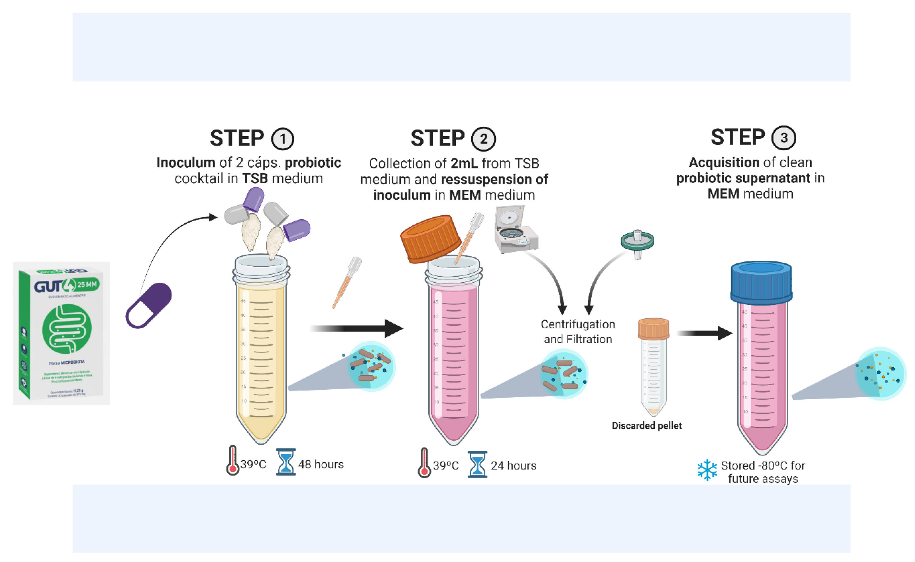
2.1. Growth of Probiotic Bacteria and Collection of the Supernatant

A commercial probiotic cocktail (GUT4 25MM™, Faes Farma, Lisbon, Portugal) consisting of 2 strains of Lactobacillus spp. (L. acidophilus CUL60 and L. acidophilus CUL21) was used. Initially, the bacterium was inoculated in bacterial culture medium Tryptic Soy Broth (TSB) in a 15 mL Falcon containing 13 mL of TSB + 2 capsules of probiotic cocktail and incubated at 39 °C for 48 h. Subsequently, the probiotic cocktail was resuspended in minimum essential medium (MEM), a medium of animal origin and specific to cell lines (A4192202 BenchStable™, Gibco, Waltham, MA, USA).
Next, 2 mL of the inoculum grown in TSB was added to 13 mL of MEM and incubated at 39 °C for 24 h. After this period, the supernatant was centrifuged and filtered. This constituted the probiotic supernatant used in the following assays (this process is described in Figure 2). The presence of Lactobacillus spp. strains was confirmed by Sanger sequencing, following amplification with the primers described in Table 1.
The specifications for the preparation of all solutions and chemicals utilized in this study are consolidated in Table 1.

2.2. Adipocyte Differentiation and Secretome Collect

Two methods of differentiation were applied to the 3T3-L1 cell line (mouse pre-adipocytes), following previously published works [17]. Briefly, the 3T3-L1 cell line was subjected to a 15-day differentiation process using three distinct differentiation mediums (Medium of differentiation I—MD I; Medium of differentiation II—MD II; Medium of differentiation III—MD III). The 3T3-L1 cells were maintained in culture until they reached a 75% confluence. Upon reaching confluence (day 0), DM I, which contained 0.25 µM IBMX, 2 µM insulin, and 1 µM DEX supplemented with 10% non-calf bovine serum (NCBS) and 1% antibiotics, was added. After five days (day 5) in contact with MDI, it was replaced with MD II, consisting of DMEM supplemented with 10% FBS and 2 µΜ of insulin. After another five days (day 10), the cell culture was washed twice with phosphate-buffered saline (PBS), and MD III (a basal medium without serum or supplementation) was added for five days. One group was without the use of Pioglitazone (noPGZ) [18], and the other was treated with a concentration of 10 µM Pioglitazone (PGZ) in DMI [17]. The secretome collected from the mature adipocytes was used in the Caco2 cell line of colon adenocarcinoma, which served as an in vitro model of CRC.

2.3. Viability Assay in Caco2

The Caco2 cells (passage 13) were initially seeded at a final concentration of 1 × 10⁵ cells/mL and placed in a 96-well plate. Cells were cultured until they reached 80% confluence in a final volume of 100 μL over a 24 h period.
The culture medium, MEM, was aspirated and discarded, and 100μL of the different treatments was tested. These treatments included: (I) secretome of adipocyte differentiation, with or without PGZ, and (II) incubation with the probiotic’s supernatant for 24 h, followed by adiposity secretomes, without PGZ (noPGZ) or with PGZ (PGZ). The cells were maintained with these treatments for an additional 24 h period of incubation. After the 24 h incubation period with treatments I and II, the supernatants were collected, and 100 μL of MTT solution (3-(4,5-dimethylthiazol-2-yl)-2,5-diphenyltetrazolium bromide) was added, representing 10% of the solution in MEM.
For the cytotoxicity assay, a colorimetric reagent CellTiter 96® Non-Radioactive Cell Proliferation Assay (Promega, Madison, WI, USA) was used according to the manufacturer’s instructions. Briefly, after the treatments were discarded, the Caco2 cell line was incubated in MTT solution at 37 °C with 5% CO2 for one hour. After this period, the MTT reagent was removed, and 100 μL of DMSO (dimethyl sulfoxide) was added. The DMSO solution containing the cell extracts was transferred to another 96-well plate for absorbance reading at 550 and 650 nm using a microplate reader with Thermo Lab System Multiskan Ascent 354™ filters (Thermofisher Scientifics, Waltham, MA, USA). The results were performed in triplicate and standardized for the control.

2.4. Injury Assay in Caco2

The Caco2 cell line, at a final concentration of 5 × 10⁵ cells/well, was seeded and incubated at 37 °C with 5% CO2. For the injury assay, the cells were seeded in 6-well plates, with each well containing 1 mL of medium, and cultured for 24 h until reaching a confluence of approximately 80%. After this period of incubation, the medium was removed, and a vertical mechanical scratch was performed. The scratched area was then washed with a 1% PBS buffer solution. Afterwards, 1 mL of treatments I and II, as previously described, was added. Photographic records were taken at time 0 and after 24 h of adding each treatment to assess the reintegration area of the wound closure. The migration rate was calculated by measuring the area of the wound using Image J software 1.53e (U.S. National Institutes of Health, Bethesda, Rockville, MD, USA).

2.5. Antioxidant Assay in Caco2

The GSH/GSSG ratio test was performed using the Biovision test kit (Biovision Incorporated at Abcam Company, Milpitas, CA, USA) to assess the response in the oxidative stress protection that probiotics can provide to the Caco2 cell line under treatment conditions I and II. The protocol was performed according to the manufacturer’s protocol. Briefly, Caco2 cells (5 × 10⁴ cells/mL) were sown in 96-well plates and placed to grow until they reached 75% confluence with final volume of 100 µL/well. Then, the cells were incubated with treatments I and II for a 24 h period. GSH and GSSG analysis was performed by dilution of whole-cell lysates, as well as serial dilutions of GSH and GSSG standards. The samples were incubated at room temperature for 45 min in a one-step fluorometric reaction using an appropriate assay buffer and Monochlorobimane fluorophores. The assay uses a dye that reacts with glutathione, either in oxidized or reduced form, in a reaction catalyzed by the enzyme glutathione S-transferase. Fluorescence intensity was measured using an excitation/emission (EX/EM) reader of 490/520 nm after cleavage. GSH and total GSH were determined using the standard curve, while GSSG was determined by the formula = (total GSH − GSH)/2.

2.6. Inflammatory Genes Quantified by RT-qPCR

The total RNA was extracted from homogenized Caco2 cell line (5 × 10⁵ cells/mL) cultivated in 6-well plates until they reached confluence and after receiving treatments for 24 h. A total of 250 ng/µL of RNA was initially reverse-transcribed to complementary DNA (cDNA) with a high-capacity cDNA kit. The multiplex PCR kit Master Mix from Meridian was used in real time to carry out PCR according to the manufacturer’s instructions.
Test primers were used to evaluate the expression of metabolic and inflammatory pathways that Caco2 cells were exposed to. The primers are consolidated in Table 2.
PCR was performed on Quant Gene 9600™ (Biovision, Changsha, China), under the following thermal cycler conditions presented in Table 3.
The 18S expression control gene was chosen to normalize the results. The log-normalized and classified data were then used in Minitab using the ANOVA test of 2 samples.

2.7. Statistical Analyses

The statistical analysis was performed comparing the results of the control group to treatment groups using one-way analysis of variance (ANOVA), normality of data was assessed using the Shapiro–Wilk test, and then the Dunnet test was applied. GraphPad Prism 8.0.1 software (GraphPad Software, San Diego, CA, USA) was used. A 95% confidence interval (95% CI) was selected, and a significance level of p < 0.05 was applied to all statistical analyses.

3. Results

3.1. Gene Expression

First, the probiotic extract (supernatant) used in these assays was sequenced, and the result confirmed the presence of probiotic strains of interest. It was possible to confirm the presence of Lactobacillus acidophilus (with 99% matching) (see Figure 3).
After, to evaluate the impact of mimicking adiposity conditions in vitro through the differentiation of 3T3-L1 pre-adipocytes, two different secretome conditions were produced and tested in the Caco2 cell line: one with PGZ and one without the drug. Initially, we evaluated the impact of the secreted products of adipocytes on the viability of the Caco2 tumoral cell line. Our findings consistently demonstrated that, across all conditions, exposure to conditions resembling adiposity led to a notable increase in the viability of the tumoral cells. Subsequently, we analyzed the effect of a probiotic supernatant on the viability of tumoral cells when added prior to the adiposity condition, which is presented below.

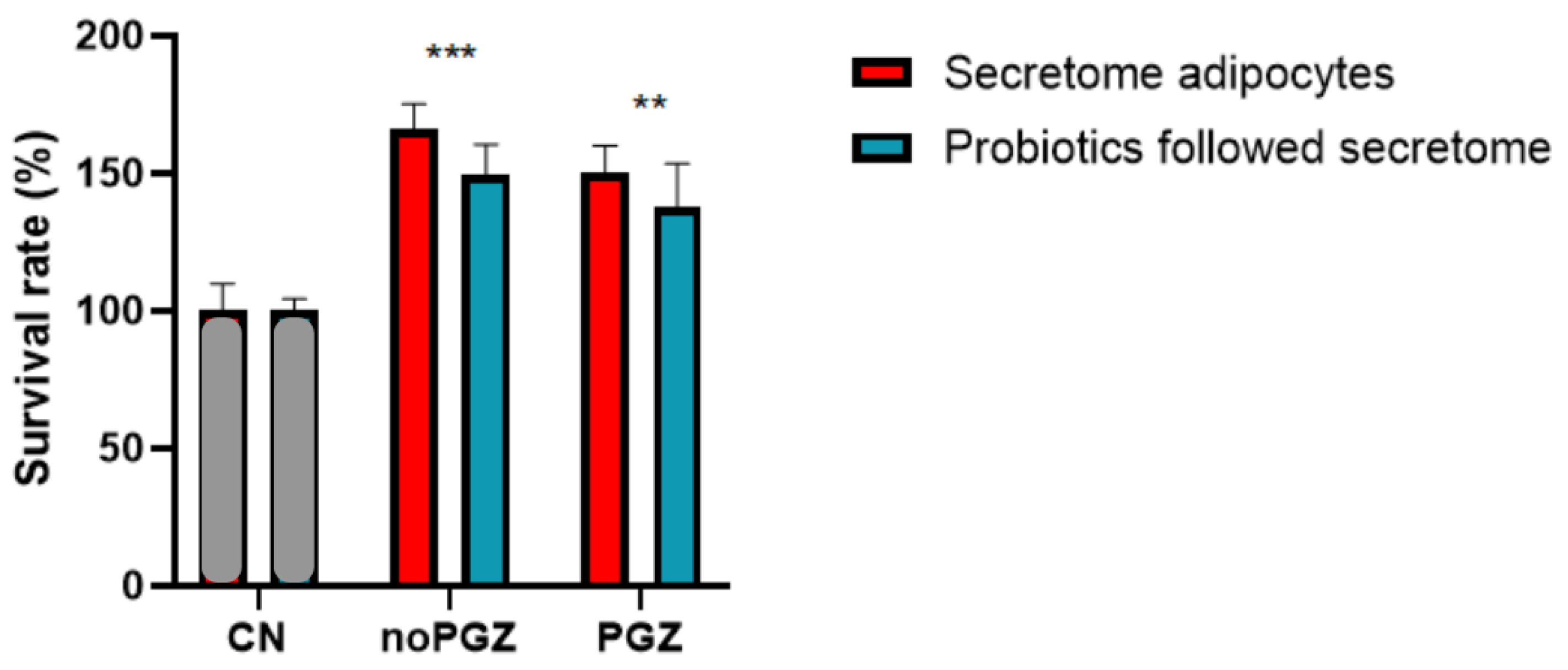
3.2. Adiposity Increases Colon Tumor

The use of both adiposity secretome conditions demonstrated a statistically significant increase in Caco2 cell viability compared to the control (cells without treatment). Notably, in the condition without PGZ (*** p < 0.001), the increase was higher than in the condition with PGZ (** p < 0.01), with cell viability increasing by 66% and 50%, respectively (Figure 4).
To evaluate the potential impact of probiotics on tumor progression, we implemented a two-step approach, initially exposing cells to probiotic supernatant and subsequently supplementing the same cells with the secretome of adipocyte differentiation. The results showed an increase in Caco2 cell viability, regardless of the use of probiotics. Statistical significance results were observed in the condition with and without PGZ. An increase of 56% in cell viability was observed without PGZ (noPGZ), and a 48% increase in the condition was observed with PGZ (PGZ) (Figure 4).
These results demonstrate that adiposity alone can increase the viability of colon adenocarcinoma, and the probiotic was not effective in reversing this effect. Nonetheless, the effect of obesity under the influence of PGZ on the colon adenocarcinoma cells seems to be mitigated by the probiotic supernatant.

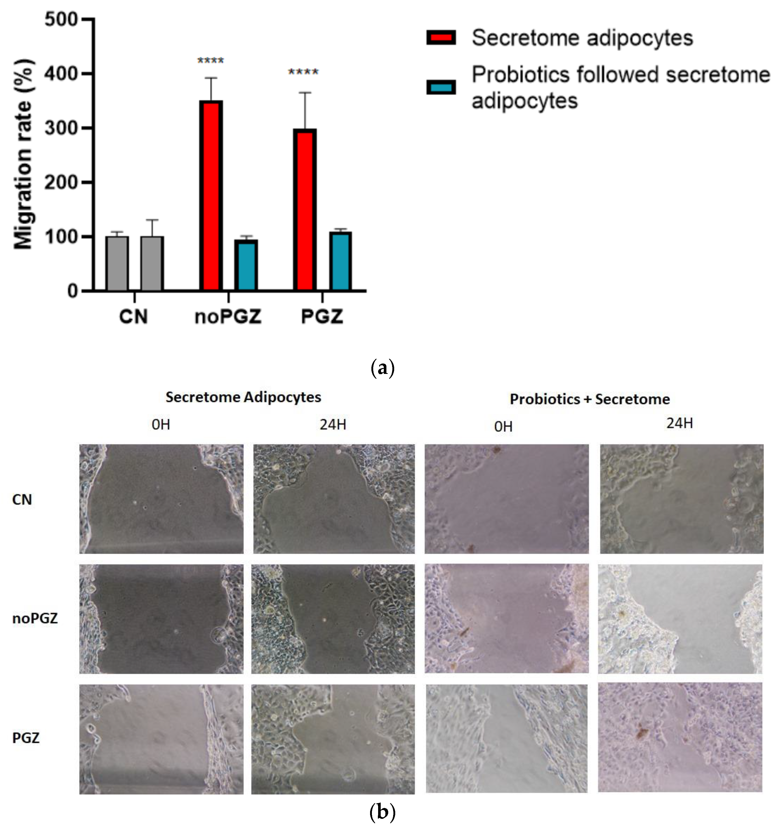
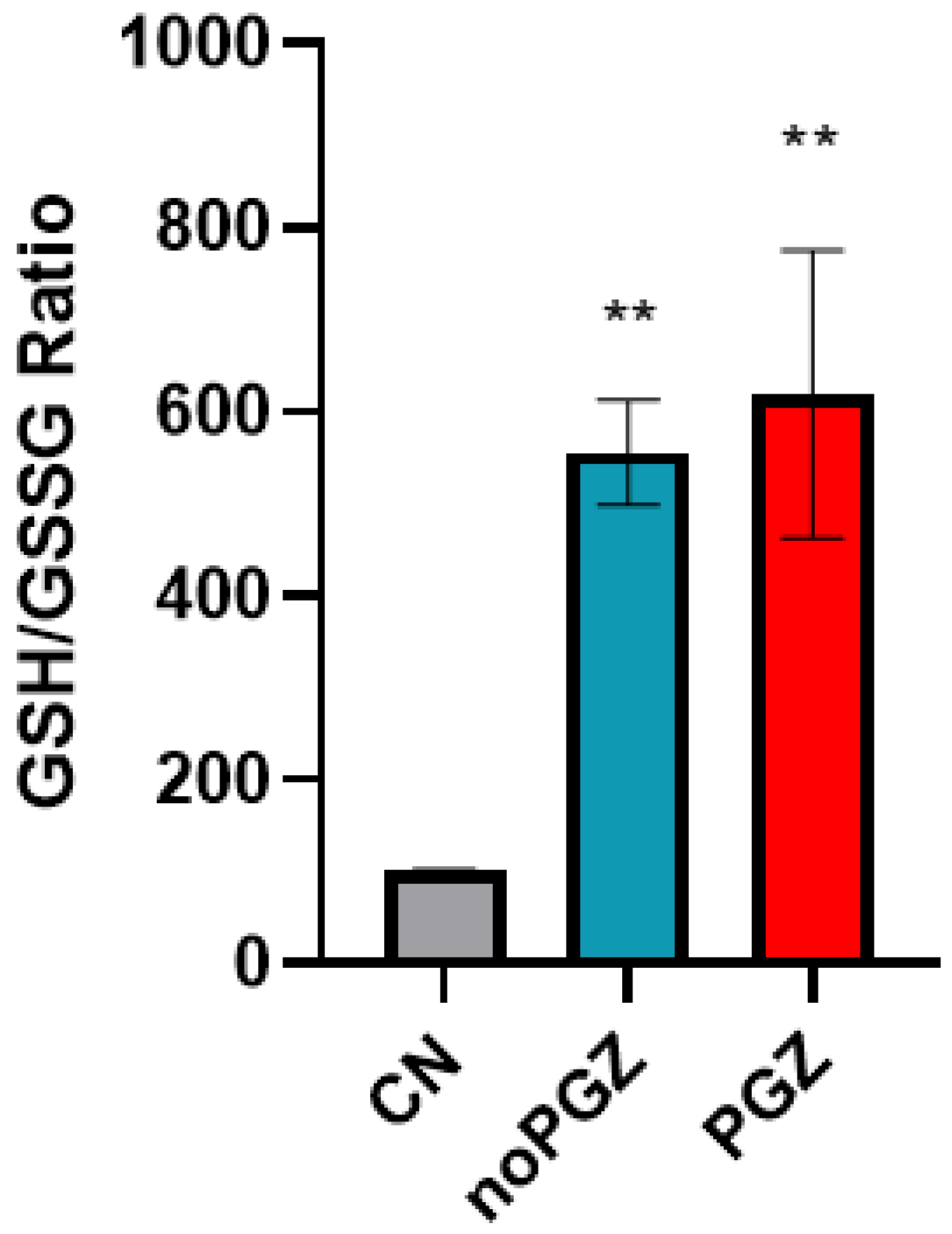
3.3. Invasion Activity and Antioxidant

The injury assay aimed to evaluate the migration capacity of colon adenocarcinoma cells (Figure 5). The results showed that a greater cell migration capacity was associated with an increased risk of aggressiveness of the tumor following treatment.
The results from the cell migration assay in treatment I (Figure 5a) showed an increased ability to reintegrate the wound when exposed to adiposity conditions. In all conditions, statistical significance was observed, especially without PGZ (noPGZ), which showed an increase of 252% (p < 0.001) compared to the control. The condition with PGZ (PGZ) showed an increase of 199% (p < 0.01) in the closure reintegration rate.
In treatment II, where probiotics were added before adiposity secretome supplementation (Figure 5a), the result demonstrated a reduction in the migration capacity of colon adenocarcinoma cells, compared to treatment I. Although not statistically significant, the use of probiotics resulted in an attenuation of the reintegration rate of closure. The condition without PGZ (noPGZ) decreased by 7% and with PGZ (PGZ) decreased by 8% in the rate of cell reintegration.
To evaluate the impact of adiposity with and without Pioglitazone on the antioxidant response in colon adenocarcinoma cells previously exposed to a probiotic supernatant, the ratio of reduced to oxidized glutathione was assessed.
The results obtained (Figure 6) showed that both adipose conditions increased antioxidant activity, with an increase in reduced glutathione or a decrease in oxidized glutathione compared to the control. The ratio increases gave statistical significance in both conditions, with or without PGZ, with an increase of 519% and 456%, respectively.

3.4. Inflammatory Genes Responsive to Adiposity

Gene expressions of pro-inflammatory and anti-inflammatory factors were evaluated to evaluate how different adiposity conditions, exposed to previous supplementation with probiotics, could interfere with the inflammatory response of colon tumor cells. The real-time PCR (RT-PCR) results for the expression of inflammatory response genes showed no statistical significance in any of the conditions tested compared to the control (Figure 7).
However, we noticed the trend of an increase in IL10 in the condition without Pioglitazone (noPGZ) compared to the control.
It was not possible to observe in isolation how the use of a probiotic supernatant interferes in the gene expression of cytokines involved with the inflammatory cascade, but the condition of adiposity treated pharmacologically (PGZ) showed fewer fluctuations when compared to untreated adiposity with Pioglitazone (noPGZ).

4. Discussion

Colorectal cancer (CRC) has multiple origins, with emphasis on hereditary risk factors, age, inflammatory diseases, obesity, diet, and dysbiosis of the intestinal microbiota [8,19]. Among these factors, regulation of the intestinal microbiota can prevent the poor prognosis of CRC [15,20,21].
The use of probiotics has been gaining importance due to its potential to induce an antitumor effect in healthy individuals and a protective effect in patients already affected by the disease [21]. According to various authors, the use of probiotics promotes physicochemical changes in the gut microbiota, either by increased production of short-chain fatty acids (SCFAs) or increased antitumor and antimutagenic components. These alterations have a positive influence on the immune modulation within the gastrointestinal tract and contribute to a reduction in both intestinal and systemic mucosal inflammation [22,23,24]. Then, bacterial strains that contribute to the degradation of lipid molecules may potentially interrupt the conditioning factors of the tumor microenvironment, positioning probiotics as a supplementary option in both treatment and cancer prevention.
Our results suggest that an obesity-associated tumor microenvironment promotes tumor growth and aggressiveness by increasing tumor cell viability and migration capability. These results support what is described in the literature, which indicates that host lipids and cytokines released by adipose tissue are associated with the development of several types of cancer, including CRC [2,5,25,26,27].
On the other hand, our results showed that probiotic supplementation had a mitigating effect on the impact of adiposity environment on colorectal tumoral cells. Furthermore, although we did not observe a reduction in the viability of the Caco2 cell line supplemented with probiotics, we acknowledge the inverse relationship between probiotics consumption and its benefits in colon cancer. We hypothesized that (a) because cell culture does not accurately replicate the conditions in the colon, there may have been interferences, and (b) a longer treatment duration and future assessments involving the co-culture of different probiotic strains may be required.
The fact that probiotic bacteria have better growth conditions in environments with a low oxygen supply supports the established role of probiotics in the complex gastrointestinal ecosystem in vivo. Probiotics operate in closed environments with low oxygen supply and with a reduction in intestinal lumen pH through the production of organic acids (acetic, butyric, propionic, and lactic acids). These organic acids contribute to maintaining an anaerobic environment and inhibiting the growth of pathogens [25]. However, replicating this specific condition in our study proved unfeasible and may have hindered the attainment of more promising results.
The literature currently lacks comprehensive data on the optimal culture medium for probiotics. Nevertheless, there is evidence suggesting that the inclusion of micronutrients and minerals such as fructo-oligosaccharide (FOS) and inulin, which are non-digestible carbohydrates serving as substrates of bacteria, appear to enhance the growth and viability of probiotics [28].
The balanced composition of the intestinal microbiota plays an important role in innate and adaptive gastrointestinal immunity. In the context of oncology, it works as a protective mechanism against tumor aggressiveness and exhibits antitumor activity [29]. However, in cases of imbalance of the microbiota, such as in cases of marked adiposity, intestinal dysbiosis will develop. This dysbiosis compromises intestinal bioactivity and increases the abundance of inducing species (pathogenic bacteria) which can cause inflammatory bowel diseases and cancer [26].
Conversely, specific probiotic bacterial strains have the capacity to generate butyrate and lactate, two metabolites derived from the production of short-chain fatty acids (SCFAs) important for metabolic protection against cancer [30]. These SCFA metabolites are fermented acids produced by Gram-positive bacteria of the intestinal microbiota and are recognized as microbiota-derived metabolites capable of improving intestinal barrier immunity, promoting the production of adhesion molecules, deterring invaders, and reducing host intestinal inflammation [31].
SCFAs are essential regulators for the recruitment, activation, and differentiation of various immune cells such as dendritic cells (CDs), neutrophils, macrophages, and lymphocytes. Additionally, they also play a pivotal role in regulating the expression of (i) pro-inflammatory cytokines, such as IL-12 and IL-6, and (ii) anti-cytokines’ association between SCFAs and receptors coupled to G proteins, for example, GPR109a associated with CD, which results in an increased expression of IL-10 and a reduction in IL-6 [32]. Consequently, this leads to the expansion of T-regulatory cells, which, in turn, inhibit the proliferation of Th17 cells, indicating their crucial role in anti-inflammatory pathways such as apoptosis, especially in inflammation-induced colorectal cancer [29,32,33].
Probiotics have the potential to modulate the immune system, either at the gut level or systematically, and offer other therapeutic benefits such as improving lactose tolerance, reducing inflammation, and exhibiting antioxidant properties [34].
The imbalance between the production of pro-oxidative molecules (reactive oxygen species (ROS) and reactive nitrogen species (ERN)) and the efficiency of antioxidation defenses is one of the factors associated with CCR [30].
The oxidative stress produced by the dysbiotic intestinal microbiota promotes the stimulation of chronic inflammation through the production of ROS that induce DNA damage, activation of oncogenes, and inactivation of tumor suppressor genes [30,35].
Within mammalian cells, glutathione is the main low-molecular-weight intracellular thiol that plays a critical role in defending against oxidative and nitrosative stress. Reduced glutathione levels have been detected in the early stages of apoptosis, and the identification of these values by simple in vitro assays allows for the detection of changes in total glutathione in apoptosis and other contexts [36].
Our research showed a heterogeneous trend in the conditions with the use of the PPARy agonist used for optimizing adipogenesis. Recent studies have supported the fact that this class of antidiabetic drug, specifically thiazolidinediones, may be associated to a modest 9% reduction in the risk of developing CRC in patients with T2DM treated with Pioglitazone [37].
Despite Pioglitazone being directed to reduce insulin resistance, its use can still increase the accumulation of lipid droplets in mature adipocytes and activate tumor suppression pathways by apoptosis, such as the mTOR pathway, for example [38]. Our results evidenced that using Pioglitazone promotes protection activity in colorectal tumor cells exposed to the adipose microenvironment (Figure 5).
A study by Ninomiya et al. (2014) evaluated the effect of Pioglitazone on five tumor cell lines (Capan-1, Aspc-1, BxPC-3, PANC-1, and MIApaCa-2), and the results confirmed an inhibitory effect on the proliferation of all five examined cell lines when the concentration of Pioglitazone exceeded 10 µM [39,40].
In summary, high levels of PPARy expression have been associated with cell proliferation. According to various authors, treatment with thiazolidinedione can act by inhibiting cell proliferation and inducing cell differentiation [41].
Although this is only a preliminary and pilot study, it presented some limitations. The lack of knowledge of the composition of pre-adipocyte composition, the use of limited cell lines and probiotic models, and low understanding of the immunomodulation process caused by both treatments need to be addressed in future studies to allow a better understanding of the real impact of probiotics use against colorectal cancer.

5. Conclusions

In this study, we observed promising results using probiotics to modulate the gut microbiota in an in vitro model of colon adenocarcinoma. Although preliminary, these results represent a starting point in how the use of probiotics can potentially mitigate risk factors associated with gastrointestinal cancers, including colorectal cancer. Probiotic therapy may offer an alternative strategy leading to improvements in the meta-inflammatory and metabolic conditions present in patients with obesity and/or diabetics using Pioglitazone therapy.
The present study suggests that the administration of probiotics as a preventive measure can lessen the aggressiveness and progression of colon adenocarcinoma due to the intricate relationship between the tumor and adipose tissue.
In general, there is an attractive research route for cancer prevention or treatment therapy for cancer, intervening in the modulation of the intestinal microbiota. Nevertheless, further studies are needed to investigate and understand the underlying mechanism between the gut microbiota and the immune system in the context of cancer.

6. Future Perspectives

Although this is a preliminary study, the probiotics seemed to produce an impact against colorectal cancer cell lines. In order to understand this process deeply, some complementary studies should be addressed, such as the evaluation of different probiotic formulations (e.g., with the Akkermansia muciniphila strain, which showed great results in regulating the microbiome and reducing obese characteristics), the evaluation of pre-adipocyte secretome with metabolomic and/or proteomic analysis, and the evaluation of immune response in the presence of both the probiotics and the pre-adipocyte secretome, as well as the testing of other colorectal healthy and cancer cell lines. Additionally, evaluation of the same processes should also be performed with in vivo models (animal and human).

Author Contributions

R.V.: research, software, original draft preparation, and editing; A.C.R.: investigation, software, original draft preparation, and review; A.P.S.: investigation, original draft preparation, and review; D.F.: investigation, data analysis, and draft preparation; R.F.: review and editing, funding and resources, and project administration; C.A.: investigation, draft preparation, and software; P.J.P.: investigation, review, and draft preparation; P.B.: conceptualization and editing; A.C.P.: conceptualization, investigation, review and editing, and project administration. All authors have read and agreed to the published version of the manuscript.

Funding

Pilar Baylina (P.B.) acknowledges on the behalf of the authors the support of Fundação para a Ciência e Tecnologia (FCT), Portuguese Government, under the Strategic Project Reference: UID/BIM/04293/2013. P.B. was also supported by FEDER/02/SAICT/2020/072560. Patrick J. Pais was funded by FCT, grant number 2021.09498.BD. Ana Catarina Rocha was funded by FCT, grant number 2021.06521.BD. André. P. Sousa was funded by FCT, grant number 2022.12441.BD.

Institutional Review Board Statement

Not applicable.

Informed Consent Statement

Not applicable.

Data Availability Statement

Publicly available datasets were analyzed in this study. This data can be found here: https://pubmed.ncbi.nlm.nih.gov/(accessed on 28 November 2023).

Conflicts of Interest

The authors declare no conflicts of interest.

References

  1. World Health Organization. Obesity and Overweight. 2021. Available online: https://www.who.int/news-room/fact-sheets/detail/obesity-and-overweight (accessed on 12 July 2023).
  2. Chait, A.; den Hartigh, L.J. Adipose Tissue Distribution, Inflammation and Its Metabolic Consequences, Including Diabetes and Cardiovascular Disease. Front. Cardiovasc. Med. 2020, 7, 22. [Google Scholar] [CrossRef] [PubMed]
  3. World Health Organization. Regional Office for Europe. WHO European Regional Obesity Report. 2022. Available online: https://iris.who.int/handle/10665/353747 (accessed on 3 October 2023).
  4. Kamal, N.; Ilowefah, M.A.; Hilles, A.R.; Anua, N.A.; Awin, T.; Alshwyeh, H.A.; Aldosary, S.K.; Jambocus, N.G.S.; Alosaimi, A.A.; Rahman, A.; et al. Genesis and Mechanism of Some Cancer Types and an Overview on the Role of Diet and Nutrition in Cancer Prevention. Molecules 2022, 27, 1794. [Google Scholar] [CrossRef] [PubMed]
  5. Ye, P.; Xi, Y.; Huang, Z.; Xu, P. Linking Obesity with Colorectal Cancer: Epidemiology and Mechanistic Insights. Cancers 2020, 12, 1408. [Google Scholar] [CrossRef] [PubMed]
  6. Nieman, K.M.; Kenny, H.A.; Penicka, C.V.; Ladanyi, A.; Buell-Gutbrod, R.; Zillhardt, M.R.; Romero, I.L.; Carey, M.S.; Mills, G.B.; Hotamisligil, G.S.; et al. Adipocytes promote ovarian cancer metastasis and provide energy for rapid tumor growth. Nat. Med. 2011, 17, 1498–1503. [Google Scholar] [CrossRef] [PubMed]
  7. Hanahan, D.; Weinberg, R.A. Hallmarks of cancer: The next generation. Cell 2011, 144, 646–674. [Google Scholar] [CrossRef] [PubMed]
  8. Singh, S.; Sharma, P.; Sarma, D.K.; Kumawat, M.; Tiwari, R.; Verma, V.; Nagpal, R.; Kumar, M. Implication of Obesity and Gut Microbiome Dysbiosis in the Etiology of Colorectal Cancer. Cancers 2023, 15, 1913. [Google Scholar] [CrossRef]
  9. Luo, L.; Liu, M. Adipose tissue in control of metabolism. J. Endocrinol. 2016, 231, R77–R99. [Google Scholar] [CrossRef]
  10. Machado, S.A.; Pasquarelli-do-Nascimento, G.; da Silva, D.; Farias, G.R.; de Oliveira Santos, I.; Baptista, L.B. Browning of the white adipose tissue regulation: New insights into nutritional and metabolic relevance in health and diseases. Nutr. Metab. 2022, 19, 61. [Google Scholar] [CrossRef]
  11. Endalifer, M.L.; Diress, G. Epidemiology, Predisposing Factors, Biomarkers, and Prevention Mechanism of Obesity: A Systematic Review. J. Obes. 2020, 2020, 6134362. [Google Scholar] [CrossRef]
  12. Palavicini, J.P.; Chavez-Velazquez, A.; Fourcaudot, M.; Tripathy, D.; Pan, M.; Norton, L.; DeFronzo, R.A.; Shannon, C.E. The Insulin-Sensitizer Pioglitazone Remodels Adipose Tissue Phospholipids in Humans. Front. Physiol. 2021, 12, 784391. [Google Scholar] [CrossRef]
  13. Cancello, R.; Tordjman, J.; Poitou, C.; Guilhem, G.; Bouillot, J.L.; Hugol, D.; Coussieu, C.; Basdevant, A.; Bar Hen, A.; Bedossa, P.; et al. Increased infiltration of macrophages in omental adipose tissue is associated with marked hepatic lesions in morbid human obesity. Diabetes 2006, 55, 1554–1561. [Google Scholar] [CrossRef]
  14. Divella, R.; De Luca, R.; Abbate, I.; Naglieri, E.; Daniele, A. Obesity and cancer: The role of adipose tissue and adipo-cytokines-induced chronic inflammation. J. Cancer 2016, 7, 2346–2359. [Google Scholar] [CrossRef] [PubMed]
  15. Garrett, W.S. Cancer and the microbiota. Science 2015, 348, 80–86. [Google Scholar] [CrossRef]
  16. Cӑtoi, A.F.; Vodnar, D.C.; Corina, A.; Nikolic, D.; Citarrella, R.; Pérez-Martínez, P.; Rizzo, M. Gut Microbiota, Obesity and Bariatric Surgery: Current Knowledge and Future Perspectives. Curr. Pharm. Des. 2019, 25, 2038–2050. [Google Scholar] [CrossRef]
  17. Teixeira, C.; Sousa, A.P.; Santos, I.; Rocha, A.C.; Alencastre, I.; Pereira, A.C.; Martins-Mendes, D.; Barata, P.; Baylina, P.; Fernandes, R. Enhanced 3T3-L1 Differentiation into Adipocytes by Pioglitazone Pharmacological Activation of Peroxisome Proliferator Activated Receptor-Gamma (PPAR-γ). Biology 2022, 11, 806. [Google Scholar] [CrossRef] [PubMed]
  18. Coelho, P.; Almeida, J.; Prudêncio, C.; Fernandes, R.; Soares, R. Effect of Adipocyte Secretome in Melanoma Progression and Vasculogenic Mimicry. J. Cell Biochem. 2016, 117, 1697–1706. [Google Scholar] [CrossRef] [PubMed]
  19. Corrêa, R.S.; Pinto, J.R.F.E.; Santos, L.V.; Góis, M.C.; Silva, R.P.; Silva, H.M. Rectal cancer survival in a Brazilian Cancer Reference Unit. J. Coloproctol. 2015, 36, 203–207. [Google Scholar] [CrossRef]
  20. Sánchez-Alcoholado, L.; Ramos-Molina, B.; Otero, A.; Laborda-Illanes, A.; Ordóñez, R.; Medina, J.A.; Gómez-Millán, J.; Queipo-Ortuño, M.I. The Role of the Gut Microbiome in Colorectal Cancer Development and Therapy Response. Cancers 2020, 12, 1406. [Google Scholar] [CrossRef]
  21. Machado, F.F.; Lazzaretti, R.K.; Poziomyck, A.K. Uso de prebióticos, probióticos e simbióticos nos pré e pós- operatórios do câncer colorretal: Uma revisão. Rev. Bras. Cancerol. 2014, 60, 363–370. [Google Scholar] [CrossRef]
  22. Sivamaruthi, B.S.; Kesika, P.; Chaiyasut, C. Effect of probiotics in the treatment of colorretal cancer. Arch. Health Investig. 2019, 8, 394–399. [Google Scholar] [CrossRef]
  23. Drago, L. Probiotics and Colon Cancer. Microorganisms 2019, 7, 66. [Google Scholar] [CrossRef] [PubMed]
  24. Markowiak-Kopeć, P.; Śliżewska, K. The Effect of Probiotics on the Production of Short-Chain Fatty Acids by Human Intestinal Microbiome. Nutrients 2020, 12, 1107. [Google Scholar] [CrossRef] [PubMed]
  25. do Carmo, M.S.; Santos, C.I.D.; Araújo, M.C.; Girón, J.A.; Fernandes, E.S.; Monteiro-Neto, V. Probiotics, mechanisms of action, and clinical perspectives for diarrhea management in children. Food Funct. 2018, 9, 5074–5095. [Google Scholar] [CrossRef] [PubMed]
  26. Chaplin, A.; Rodriguez, R.M.; Segura-Sampedro, J.J.; Ochogavía-Seguí, A.; Romaguera, D.; Barceló-Coblijn, G. Insights behind the Relationship between Colorectal Cancer and Obesity: Is Visceral Adipose Tissue the Missing Link? Int. J. Mol. Sci. 2022, 23, 13128. [Google Scholar] [CrossRef] [PubMed]
  27. Safaei, M.; Sundararajan, E.A.; Driss, M.; Boulila, W.; Shapi’i, A. A systematic literature review on obesity: Understanding the causes & consequences of obesity and reviewing various machine learning approaches used to predict obesity. Comput. Biol. Med. 2021, 136, 104754. [Google Scholar] [CrossRef]
  28. Misra, S.; Pandey, P.; Mishra, H.N. Novel approaches for co-encapsulation of probiotic bacteria with bioactive compounds, their health benefits and functional food product development: A review. Trends Food Sci. Technol. 2021, 109, 340–351. [Google Scholar] [CrossRef]
  29. Al-Ishaq, R.K.; Koklesova, L.; Kubatka, P.; Büsselberg, D. Immunomodulation by Gut Microbiome on Gastrointestinal Cancers: Focusing on Colorectal Cancer. Cancers 2022, 14, 2140. [Google Scholar] [CrossRef] [PubMed]
  30. Tajik, N.; Frech, M.; Schulz, O.; Schälter, F.; Lucas, S.; Azizov, V.; Dürholz, K.; Steffen, F.; Omata, Y.; Rings, A.; et al. Targeting zonulin and intestinal epithelial barrier function to prevent onset of arthritis. Nat. Commun. 2020, 11, 1995. [Google Scholar] [CrossRef]
  31. Cheng, Y.; Ling, Z.; Li, L. The Intestinal Microbiota and Colorectal Cancer. Front. Immunol. 2020, 11, 615056. [Google Scholar] [CrossRef]
  32. Chambers, E.S.; Preston, T.; Frost, G.; Morrison, D.J. Role of Gut Microbiota-Generated Short-Chain Fatty Acids in Metabolic and Cardiovascular Health. Curr. Nutr. Rep. 2018, 7, 198–206. [Google Scholar] [CrossRef]
  33. Singh, N.; Gurav, A.; Sivaprakasam, S.; Brady, E.; Padia, R.; Shi, H.; Thangaraju, M.; Prasad, P.D.; Manicassamy, S.; Munn, D.H.; et al. Activation of Gpr109a, receptor for niacin and the commensal metabolite butyrate, suppresses colonic inflammation and carcinogenesis. Immunity 2014, 40, 128–139. [Google Scholar] [CrossRef]
  34. Frantz, A.L.; Rogier, E.W.; Weber, C.R.; Shen, L.; Cohen, D.A.; Fenton, L.A.; Bruno, M.E.; Kaetzel, C.S. Targeted deletion of MyD88 in intestinal epithelial cells results in compromised antibacterial immunity associated with downregulation of polymeric immunoglobulin receptor, mucin-2, and antibacterial peptides. Mucosal Immunol. 2012, 5, 501–512. [Google Scholar] [CrossRef] [PubMed]
  35. Rébé, C.; Ghiringhelli, F. Interleukin-1β and Cancer. Cancers 2020, 12, 1791. [Google Scholar] [CrossRef] [PubMed]
  36. Nagaraju, G.P.; Aliya, S.; Alese, O.B. Role of adiponectin in obesity related gastrointestinal carcinogenesis. Cytokine Growth Factor Rev. 2015, 26, 83–93. [Google Scholar] [CrossRef] [PubMed]
  37. Wang, K.; Hirschenson, J.; Moore, A.; Mailloux, R.J. Conditions Conducive to the Glutathionylation of Complex I Subunit NDUFS1 Augment ROS Production following the Oxidation of Ubiquinone Linked Substrates, Glycerol-3-Phosphate and Proline. Antioxidants 2022, 11, 2043. [Google Scholar] [CrossRef] [PubMed]
  38. Liu, Y.; Jin, P.P.; Sun, X.C.; Hu, T.T. Thiazolidinediones and risk of colorectal cancer in patients with diabetes mellitus: A meta-analysis. Saudi J. Gastroenterol. 2018, 24, 75–81. [Google Scholar] [CrossRef] [PubMed]
  39. Ninomiya, I.; Yamazaki, K.; Oyama, K.; Hayashi, H.; Tajima, H.; Kitagawa, H.; Fushida, S.; Fujimura, T.; Ohta, T. Pioglitazone inhibits the proliferation and metastasis of human pancreatic cancer cells. Oncol. Lett. 2014, 8, 2709–2714. [Google Scholar] [CrossRef]
  40. Belfiore, A.; Genua, M.; Malaguarnera, R. PPAR-γ agonists and their effects on IGF-I receptor signaling: Implications for cancer. PPAR Res. 2009, 2009, 830501. [Google Scholar] [CrossRef]
  41. Elnemr, A.; Ohta, T.; Iwata, K.; Ninomia, I.; Fushida, S.; Nishimura, G.; Kitagawa, H.; Kayahara, M.; Yamamoto, M.; Terada, T.; et al. PPARgamma ligand (thiazolidinedione) induces growth arrest and differentiation markers of human pancreatic cancer cells. Int. J. Oncol. 2000, 17, 1157–1164. [Google Scholar] [CrossRef]
Figure 1. Summary of the study.
Applsci 14 00951 g001
Figure 2. Summary of supernatant acquisition. To obtain the bacterial supernatant resuspended in minimum essential medium (MEM) for cell culture, it was first necessary to place the cocktail bacteria in a culture medium suitable for bacterial strains to grow in, the Tryptic Soy Broth (TSB), for 48 h at 39 °C. Subsequently, the content was vortexed, and 2 mL was collected to be resuspended in MEM. After this period, the content was centrifuged at 4000 rpm for 10 min to separate the bacterial part of the extract containing products released by the bacteria. The supernatant was filtered in a sterile filter of 0.2 µm and consecutively stored in an arc of −80 °C to be used in future tests.
Applsci 14 00951 g002
Figure 3. Sanger sequencing image for the Lac1 gene (specific and conserved areas) in (a) Forward direction (5′–3′) and (b) Reverse direction (3′–5′).
Applsci 14 00951 g003
Figure 4. Results of cell viability assays by MTT in Caco2 supplemented with red indicating the 3T3-L1 differentiation secretome exclusively and blue indicating probiotic supernatant followed by 3T3-L1 differentiation secretome. CN: negative control; noPGZ: without Pioglitazone; PGZ: with Pioglitazone; ** p < 0.01; *** p < 0.001.
Applsci 14 00951 g004
Figure 5. Cell line migration Caco2: (a) graphic representation and (b) photographic representation. The results express the migration rate in Caco2 cells after a mechanical tear, followed by the addition of the treatments in question, where the red is a graphic and photographic representation in which Caco2 was supplemented exclusively with the adiposity secretome produced in the differentiation of adipocytes 3T3-L1, and the blue is a graphical and photographic representation in which Caco2 was first supplemented with supernatant of bacteria from the probiotic cocktail resuspended in MEM, followed by secretome of the differentiation of adipocytes 3T3L1 under conditions without Pioglitazone (noPGZ) and with Pioglitazone (PGZ). **** p < 0.0001.
Applsci 14 00951 g005
Figure 6. Results of stress oxidative assay in cellular Caco2 supplemented with supernatant of probiotics followed by 3T3-L1 differentiation secretome. CN: negative control; noPGZ: without Pioglitazone; PGZ: with Pioglitazone. ** p < 0.01.
Applsci 14 00951 g006
Figure 7. Results of gene expression of interleukins by RT-qPCR in Caco2 cells supplemented with supernatant of probiotic cocktail bacteria, followed by secretome of differentiation of 3T3L1 adipocytes under different conditions. CN: negative control; noPGZ: without Pioglitazone; PGZ: with Pioglitazone. Interleukins IL-1b and IL-10 were evaluated.
Applsci 14 00951 g007
Table 1. Sequence set of primers for Sanger sequencing.
Target SpeciesPrimer Forward (5′-3′)Primer Reverse (3′-5′)Size (bp—Base Pairs)Annealing Temperature (°C)
Lactobacillus ssp.AGCAGTAGGGAATCTTCCACATTYCACCGCTACACATG34660
Total BacteriaCCTACGGGAGGCAGCAGATTACCGCGGCTGCTGG16960
Table 2. Sequence of primers for inflammatory genes quantified by RT-qPCR.
GenePrimer Forward (5′-3′)Primer Reverse (3′-5′)
IL 1-βACCTAGCTGTCAACGTGTGGTCAAAGCAATGTGCTGGTGC
IL-10TGAAAACAAGAGCAAGGCCGATAGAGTCGCCACCCTGATG
Table 3. Amplification conditions in the RT-qPCR thermal cycler.
Number of CyclesTemperature (°C)Time
19502:00
409500:05
6000:20
14
Disclaimer/Publisher’s Note: The statements, opinions and data contained in all publications are solely those of the individual author(s) and contributor(s) and not of MDPI and/or the editor(s). MDPI and/or the editor(s) disclaim responsibility for any injury to people or property resulting from any ideas, methods, instructions or products referred to in the content.

