Article

Biomineralization Process Inspired In Situ Growth of Calcium Carbonate Nanocrystals in Chitosan Hydrogels

by
Xinyue Zeng
,
Zheng Zhu
,
Wei Chang
,
Bin Wu
and
Wei Huang
*
State Key Laboratory of Materials Processing and Die & Mould Technology, School of Materials Science and Engineering, Huazhong University of Science and Technology, Wuhan 430074, China
*
Author to whom correspondence should be addressed.
Appl. Sci. 2024, 14(20), 9193; https://doi.org/10.3390/app14209193 (registering DOI)
Submission received: 4 September 2024 / Revised: 25 September 2024 / Accepted: 1 October 2024 / Published: 10 October 2024
(This article belongs to the Section Chemical and Molecular Sciences)

Abstract

:
Biological composites such as bone, nacre, and teeth show excellent mechanical efficiency because of the incorporation of biominerals into the organic matrix at the nanoscale, leading to hierarchical composite structures. Adding a large volume of ceramic nanoparticles into an organic molecular network uniformly has been a challenge in engineering applications. However, in natural organisms, biominerals grow inside organic fibers, such as chitin and collagen, forming perfect ceramic/polymer composites spontaneously via biomineralization processes. Inspired from these processes, the in situ growth of calcium carbonate nanoparticles inside the chitosan network to form ceramic composites was proposed in the current work. The crystal growth of CaCO3 nanoparticles in the chitosan matrix as a function of time was investigated. A weight percentage of ~35 wt% CaCO3 composite was realized, resembling the high weight percentage of mineral phase in bones. Scanning and transmission electron microscopy indicated the integration of CaCO3 nanocrystals with chitosan macromolecules. By growing CaCO3 minerals inside the chitosan matrix, the elastic modulus and tensile strength increases by ~110% and ~90%, respectively. The in situ crystal growth strategy was also demonstrated in organic frameworks prepared via 3D printing, indicating the potential of fabricating ceramic/polymer composites with complicated structures, and further applications in tissue engineering.

1. Introduction

Tissue engineering has been considered as an effective strategy for tissue repair and regeneration, which is an advanced and emerging field that originates from the intersection of biomedical science, materials science, and engineering. Its primary purpose is to repair or replace damaged tissues using biocompatible engineering material [1]. Due to advances in materials science, numerous biomaterials have been developed for use in tissue engineering. These biomaterials possess properties that closely resemble those of natural tissues, providing an optimal environment for the growth and differentiation of tissue cells [2,3,4,5]. Biological tissues such as bones and teeth are composed by organic molecules and inorganic minerals, formed via biomineralization processes, which are a common natural phenomenon that occurs in various living organisms, including bacteria, microorganisms, plants, and animals. These organisms produce minerals in their bodies, leading to crystal formation and playing different roles in different biological systems. By accurately regulating the nucleation, orientation, growth, and assembly of inorganic minerals, organisms can create organic and inorganic composite materials [6,7]. Thus, it is of great significance to fabricate tissue engineering scaffolds that have a similar organic/inorganic composite structure in biomineralized tissues.
Natural mineralized tissues in various plants and animals often exhibit extraordinary strength and toughness, mainly due to their unique hierarchical structure [8]. These structures range from microscopic anisotropic to molecular and atomic-scale crystal units, each contributing to the overall strengthening and toughening of the material [9,10]. For example, human bones (Figure 1a) exhibit a multi-level composite structure formed by the orderly arrangement of mineralized collagen, which provides essential mechanical support for the body. Through this mineralization process, the strength and toughness of collagen fibers have been significantly enhanced [11,12,13]. The joints of the bone consist of a thin, smooth layer of elastic tissue known as articular cartilage. This cartilage consists of four structural layers: the superficial zone, the transitional zone, the radial zone, and the calcified zone. Articular cartilage lubricates the surface and serves as a load-bearing medium over extended periods of time [14,15]. Similarly, the mantis shrimp (Figure 1b) is famous for its extraordinary hunting ability and incredibly tough exoskeleton, which is composed of highly mineralized chitin fibers and arranged in a spiral layer. This unique structure provides mantis shrimp with extraordinary resistance to pressure and impact, enabling it to hit its prey with great strength and high speed while remaining unscathed. These layered structures provide the mantis shrimp with exceptional compressive and impact resistance [16]. The formation of such specialized tissues relies on the biomineralization process, which involves the production and deposition of minerals within the organism. This process is very complex and involves the sophisticated regulation between cells, proteins, and minerals [17].
To replicate the structure and function of natural mineralized tissues, researchers have developed various methods for preparing ceramic/biopolymer composite materials, including hybridization, electrospinning, and 3D printing. For instance, Gong et al. [18] incorporated low-crystallinity hydroxyapatite (HAP) particles into a double network (DN) hydrogel system. Tensile testing revealed that the physical fragmentation of the amorphous phase in HAP consumed substantial energy, which reinforced and toughened the gel matrix. Liu et al. [19]. successfully fabricated a ceramic/biopolymer hybrid hydrogel biomimetic osteosynthesis membrane using electrospinning technology. This membrane induced in situ mineralization and controlled the localized, long-term release of ions, thereby promoting osteogenic differentiation and angiogenesis. Monavari et al. [20] prepared bioactive nanoparticles/gelatin composite hydrogel osteogenic materials through 3D printing. In summary, ceramic/polymer composites offer a wide range of applications and development potential due to their diverse and excellent properties, meeting the various needs of modern industry and science and technology.
Ceramic materials such as calcium carbonate (CaCO3) have received widespread attention because of their biocompatibility in the human body, which makes them very suitable for the field of biomedicine [21]. These applications mainly include using artificial bones and cartilage substitutes, promoting tissue regeneration and repair, and promoting drug delivery [22]. Chitosan is a natural biopolymer extracted from chitin. It has aroused great interest due to its biocompatibility, biodegradability, and the ability to form hydrogels. This three-dimensional hydrogel network provides excellent matrix conditions for the nucleation and growth of inorganic nanocrystals [23,24,25,26,27]. In this study, we draw inspiration from the mineralized organic matrices found in organisms to develop an innovative method for the in situ growth of CaCO3 nanocrystals in chitosan hydrogels. This approach, grounded in biomimetic biomineralization, effectively strengthens the hydrogel. The chitosan hydrogel network synthesized in the experiment mimics the organic matrix in organisms by creating an environment that restricts ionic diffusion and controls crystal formation. Compared to direct ceramic/polymer blending, this in situ growth technique produced more uniformly distributed nanoparticles, reduced agglomeration, and achieved a structure that more closely resembles natural biological tissues. This research is significant for understanding the mechanism of crystal growth in organic three-dimensional networks and exploring the potential medical applications of hydrogel materials. By combining 3D printing technology with ion diffusion mechanism, we successfully prepared a chitosan/CaCO3 hydrogel, which facilitated the formation of CaCO3 nanocrystals in the hydrogel matrix. This method allows for controlled mineral deposition within polymers, thereby forming a composite structure that mimics natural mineralized tissue. We investigated the size, morphology, and distribution of CaCO3 nanocrystals across various mineralization reactions, as well as their impact on the mechanical properties of composite hydrogels. Our findings demonstrate that the incorporation of CaCO3 nanocrystals into the chitosan hydrogel matrix significantly enhances the mechanical strength and stiffness of the hydrogel, making it more suitable for load-bearing applications in tissue engineering.

2. Experiments and Methods

2.1. Materials and Sample Preparation

The chemical reagents used in this experiment include chitosan (deacetylated ≥ 75%) and glacial acetic acid (CH3COOH), both sourced from Shanghai Aladdin Biochemical Technology Co., Ltd., Shanghai, China, primarily for preparing the chitosan solution. Calcium chloride (CaCl2, purity ≥ 96%) and sodium bicarbonate (NaHCO3, ACS grade) were also obtained from the same supplier and used as reactants in the mineralization process. Sodium hydroxide (NaOH, ACS grade, 97%, flakes) was procured from Shanghai Macklin Biochemical Technology Co., Ltd., Shanghai, China and utilized in the preparation of chitosan hydrogels. All reagents were used as received, without further purification, and deionized water was employed throughout the experiment.
The preparation of chitosan hydrogels involved dissolving chitosan powder in the CH3COOH solution to create a homogeneous solution, followed by film formation using 3D printing technology. The chitosan molecules were then coagulated in a NaOH solution, resulting in the formation of a three-dimensional network structure [28,29,30]. The detailed procedure was as follows: First, an appropriate amount of chitosan powder was weighed and gradually added to a 0.5 M CH3COOH solution to prepare an 8% chitosan solution by weight. The mixture was ultrasonically stirred at room temperature to ensure the complete dissolution of the chitosan, resulting in a uniform solution. To prevent bubbles from affecting the hydrogel’s structure during molding, the solution was placed in a degassing mixer until all bubbles were eliminated. The degassed chitosan solution was then used to create chitosan films through a 3D printing technique, utilizing a self-assembled extrusion 3D printer in the laboratory. Given the viscosity of the chitosan solution, a balance between printing speed and needle aperture size was critical. If the printing speed was too fast or the needle aperture too small, the extrusion of the solution became discontinuous or failed altogether, preventing film formation. Conversely, if the printing speed was too slow or the needle aperture too large, aggregation occurred, affecting the uniformity and size of the film. After optimization, the printing speed was set to 3 mm/s, and a 400 µm needle was chosen. Due to the mobility of the slurry, when extruded from the needle of the 3D printer, it spreads on the glass sheet, making the chitosan filament size larger than the 400 µm pinhole. If the printing pitch is too small, it causes the excessive aggregation of the chitosan solution, while too large pitch prevents the extruded chitosan filament from spreading to form a film. After testing, it was found that the optimal printing pitch for forming the film is 800 µm. The size of the films, as shown in Figure 2a,b, allows for subsequent mechanical testing and characterization (Figure S5). The dried chitosan film samples were neutralized using a 1M NaOH solution at room temperature for 6 h. After gelation, the chitosan hydrogel samples were thoroughly rinsed with deionized water to remove any residual NaOH, and then stored in deionized water for future use.
The in situ growth of CaCO3 nanocrystals in the previously prepared chitosan hydrogel was conducted using an ion diffusion technique as follows: First, the chitosan hydrogel sample was immersed in a 0.1M CaCl2 solution for 24 h. Ca2⁺ was introduced into the hydrogel network by diffusion, forming a Ca2⁺-loaded chitosan hydrogel. This was followed by a biomimetic mineralization reaction, where the Ca2⁺-loaded chitosan hydrogel samples were immersed in a 0.1 M NaHCO3 solution for reaction times ranging from 5 min to 12 h, resulting in CaCO3 nanocrystals of varying sizes and morphologies produced at different time intervals. The mineralization reactions were conducted at room temperature. After the reactions were completed, the hydrogel samples were removed and rinsed with deionized water to eliminate unreacted ions and excess CaCO3 particles from the surface. The mineralized chitosan/CaCO3 hydrogel samples are shown in Figure 2b. The samples in their wet state could be stored in closed containers with deionized water to maintain their water content, in preparation for further characterization as described below.

2.2. Composition and Structure Characterization

The chitosan/CaCO3 hydrogel samples prepared above were dried and subjected to compositional and structural characterization. This characterization primarily included observing the surface morphology and internal microstructure of the hydrogel at different mineralization times using scanning electron microscopy (SEM, Japanese electronics JSM-7600F, Tokyo, Japan) and transmission electron microscopy (TEM, Japan Electronics Corporation JEM2100, Tokyo, Japan). The effect of mineralization time on the size, distribution, and morphology of the CaCO3 nanocrystals was analyzed. Additionally, the crystalline nature and composition of the CaCO3 nanoparticles within the hydrogel were analyzed using X-ray diffraction (XRD, Shimadzu Corporation, Kyoto, Japan, XRD-7000), Raman spectroscopy (Horiba JobinYvon Company, Kyoto, Japan, Laser Confocal Raman Spectrometer LabRAM HR800), Fourier transform infrared spectrometer (FTIR, Bruker V70, Rheinstetten, Germany), and wide-angle X-ray diffraction (WAXD, Bruker lus microfocus light tube, Germany) techniques. These techniques were employed to determine whether the desired mineral crystals (CaCO3) were present in the hydrogel following biomimetic mineralization. Thermogravimetric analysis (TGA, Platinum-Elmer Instruments Shanghai Co., Pyris1 TGA, Shanghai, China) was also used to analyze the content of CaCO3 in the hydrogel. Through the compositional and structural characterizations described above, the analysis and understanding of the mineralization mechanism of CaCO3 in the chitosan polymer network would contribute to a deeper understanding of the mechanisms and processes of biomineralization in nature.

2.3. Mechanical Property Tests

The mechanical properties of the chitosan/CaCO3 hydrogel samples were tested in a wet state to prevent any internal structural changes and alterations in mechanical properties that might occur due to drying. Uniaxial tensile tests were performed to evaluate the impact of CaCO3 nanoparticles, formed at different mineralization times, on the mechanical properties of the composite hydrogels, including tensile strength, modulus, and deformability, to identify the group with the optimal mechanical properties. The tensile tests were performed on a universal material testing machine (Shenzhen Sansi Universal Tensile Strength Machine, HCS350G-TNS, Shenzhen, China).

3. Results and Discussions

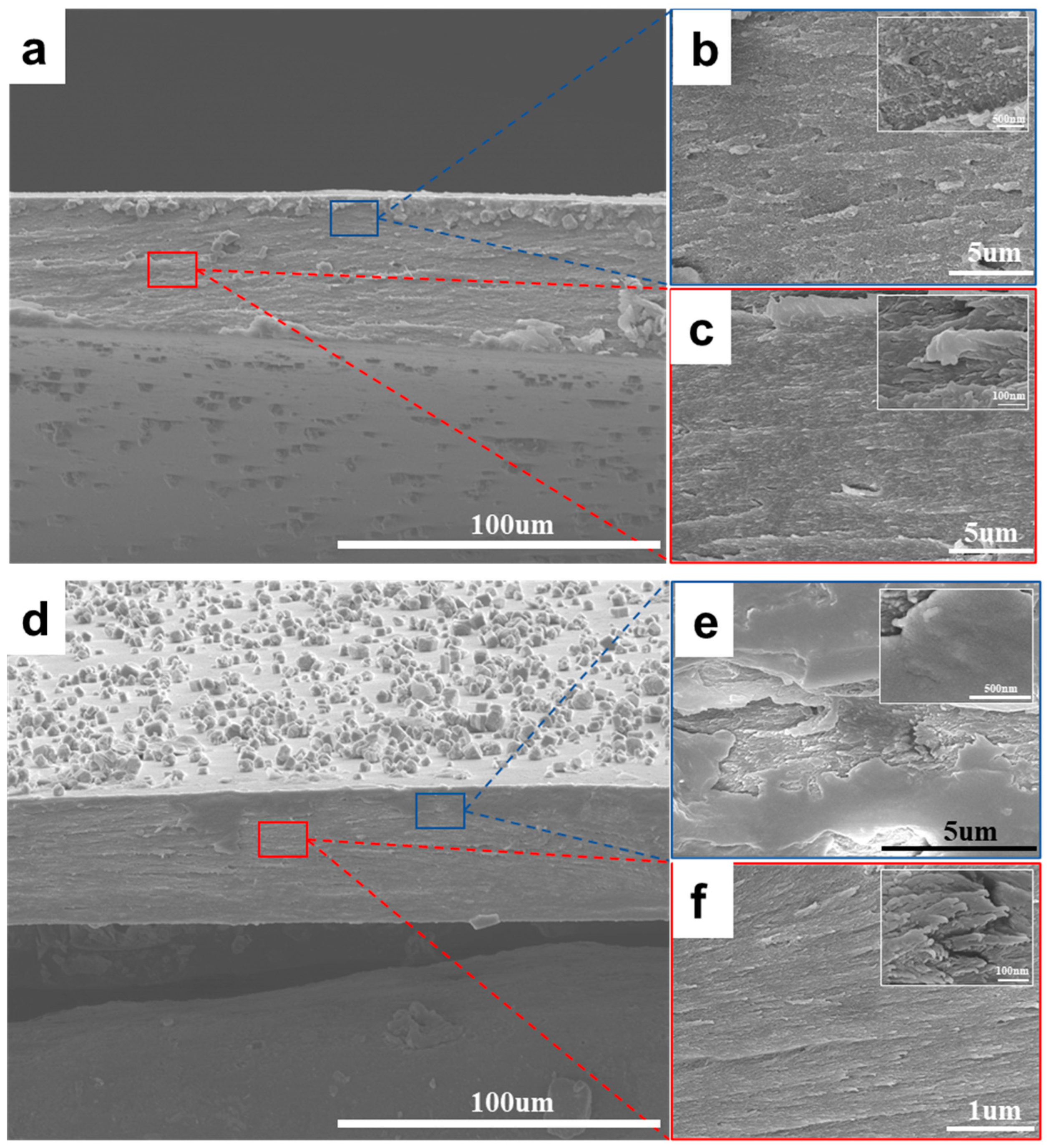
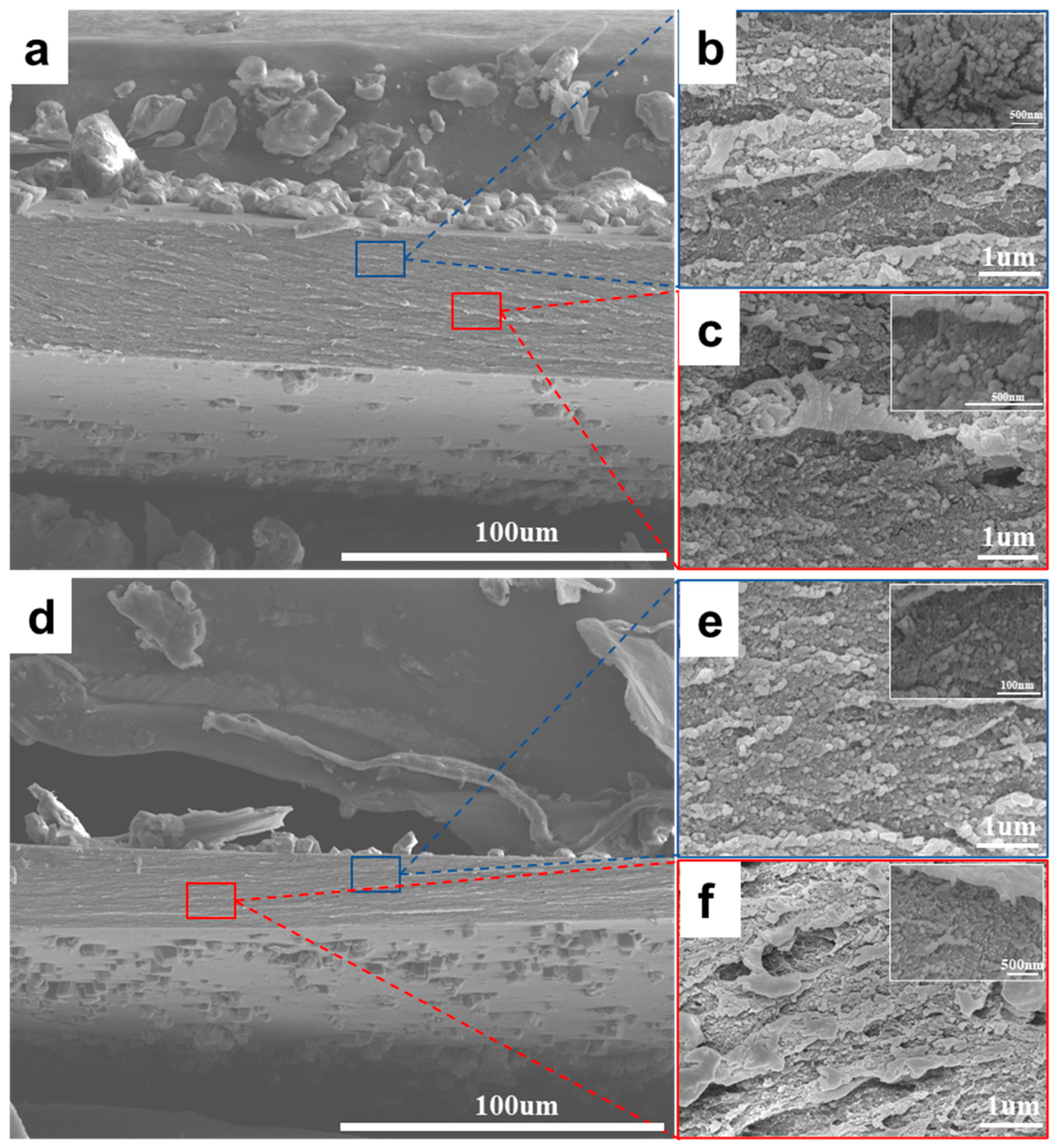
3.1. Structure and Composition of Chitosan/CaCO3 Composites

The microstructures of the 3D printed chitosan film and chitosan/CaCO3 composite hydrogel prepared via the in situ growth of CaCO3 were investigated via scanning electron microscopy (SEM). The 3D-printed chitosan films changes from transparent to white color after the reaction (Figure 2a,b). Micrographs of the surfaces and cross-sections of the hydrogel samples before and after 12 h of mineralization are shown in Figure 2c–f. The average thicknesses of the 3D-printed films are around 50~60 µm. Figure 2c,d show the surfaces of the chitosan hydrogel before and after mineralization. Large microscale crystals with an average size of 5~6 µm are clearly observed on the samples after CaCO3 growth. While in the cross-sections of the samples, microscale crystals are no longer existing even after 12 h mineralization. Nanoparticles with an average size of ~100 nm are noticed (Figure 2f). The results clearly indicate that the crystals grown on the surfaces and cross-sections of the chitosan hydrogel exhibit different morphologies and sizes. Thus, the chitosan molecule network inside the films can indeed affect the crystal growth of CaCO3. The presence of nanoparticles inside the organic network might be caused by the heterogeneous nucleation and confinement of the chitosan molecules. The detailed mechanisms will be discussed later.
Since SEM alone could not fully determine the crystal phases grown in the sample, XRD, Raman spectroscopy, and FTIR were employed in the subsequent analysis. The XRD results corresponded to the calcite standard pattern, aside from the characteristic peak of the chitosan polymer at approximately 20° (Figure 3a). Additionally, the Raman spectrum displayed a carbonate characteristic peak at around 1084 cm⁻1 (Figure 3c). The FTIR spectra of pure chitosan showed a broad, strong absorption peak near 3353 cm−1, which is attributed to the telescopic vibration peak resulting from the overlap of O-H and N-H stretching vibrations (Figure 3d). Absorption peaks near 2869 cm−1 represent the -CH2-and -CH3 structures, and characteristic absorption peaks near 1656 cm−1 and 1579 cm−1 correspond to the stretching vibration of C=O in the amide I band and the bending vibration of N-H in the amide II band. Peaks near 1022 cm−1 are attributed to the stretching vibrations of C-O-C (ether bond) or C-OH (hydroxyl group), which represent the glycan ring structure of chitosan. In addition to these characteristic chitosan peaks, the FTIR spectrum of chitosan/CaCO3 revealed a strong, asymmetric C=O stretching vibration peak near 1373 cm−1, a C-O bending vibration peak near 870 cm−1, and a smaller absorption peak near 710 cm−1, corresponding to the O-C-O bending vibration, indicating the CO32− structure of CaCO3. The results indicate that CaCO3 nanoparticles were successfully encapsulated within the cross-linked chitosan layer, forming a chitosan/CaCO3 composite hydrogel structure. Furthermore, wide-angle X-ray diffraction (WAXD) analysis confirmed that, compared to the diffraction pattern of chitosan (Figure 3e), the mineralized samples exhibited diffraction rings consistent with CaCO3 (Figure 3f). In the context of tissue engineering, the mineral content within ceramic/polymer composites significantly influences their mechanical properties, in addition to their structural composition [31]. Therefore, after determining the microstructure and phase composition of the CaCO3 crystals, TGA was conducted on the sample mineralized for 12 h (Figure 3b). Based on the TGA of the curves, as the temperature increased, chitosan decomposed due to its poor thermal stability, resulting in a final CaCO3 mass fraction of approximately 13%.

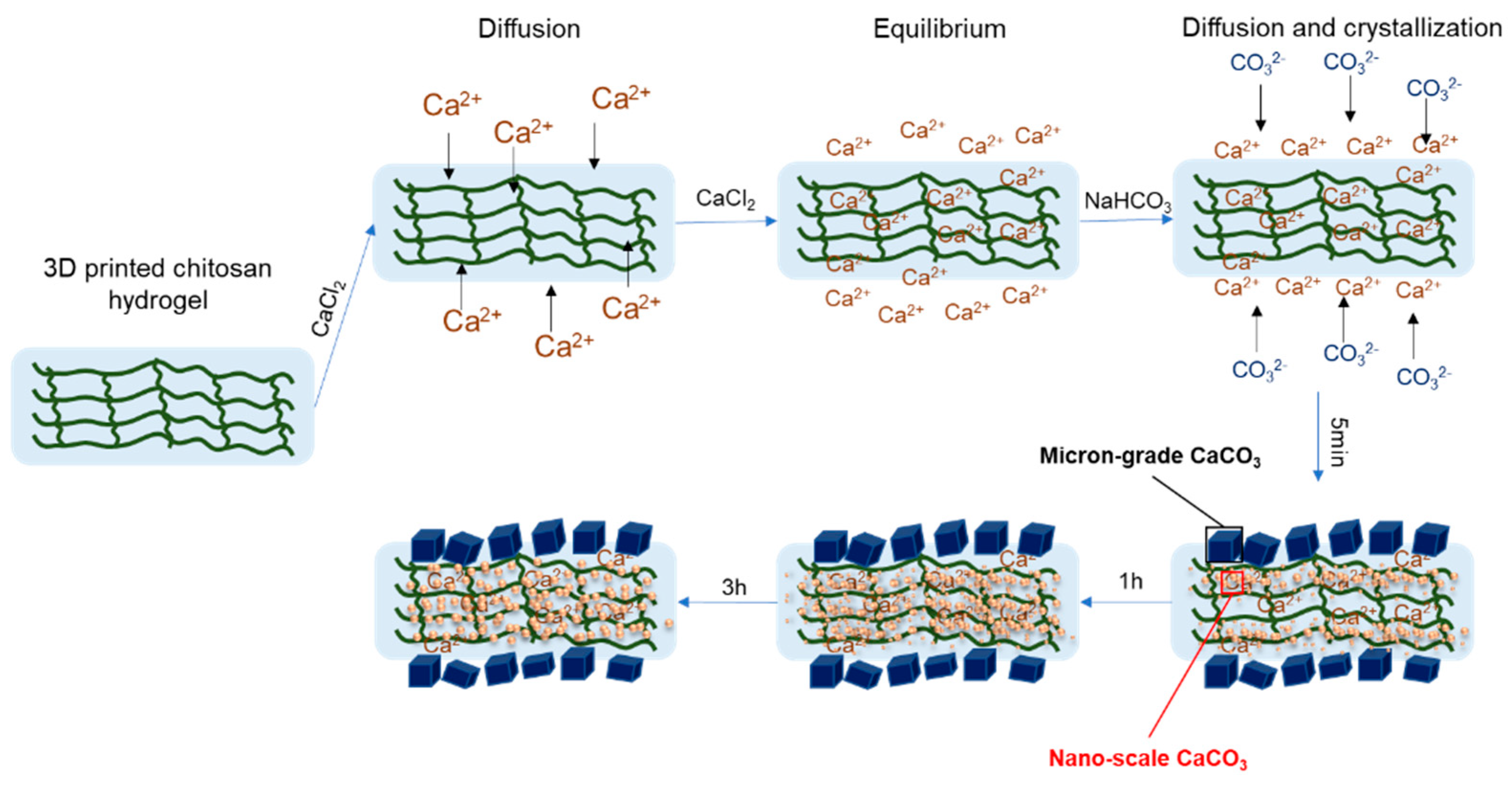
3.2. Crystal Growth Mechanisms of CaCO3 Nanoparticles in Chitosan Hydrogels

In this study, chitosan/CaCO3 composite hydrogels were successfully prepared using a simple in situ mineralization method. To better understand the growth mechanism of CaCO3 nanocrystals within the polymer network, samples at various stages of mineralization were further studied via SEM (Figure 4 and Figure 5). Micrographs of samples mineralized for 5 min, 1 h, 3 h, and 6 h were acquired. The results revealed that in the cross-section of samples mineralized for 5 min, the CaCO3 nanocrystals inside the chitosan matrix, as well as microparticles on the surfaces, already start forming. Figure 4a shows the overall distribution of calcite nanoparticles. In areas near the surfaces, nanoparticles with a size around ~100nm are observed, while in the center, almost no calcite nanoparticles are noticed. After 1 h of mineralization, the distribution of CaCO3 nanocrystals became more uniform. Nanoparticles start forming in the center areas (Figure 4d–f). As the mineralization time increases to 3 h (Figure 5), the CaCO3 crystals within the sample became more uniform and stable in both size and distribution. Most of the nanoparticles are in the range of 80~100 nm, even in the center areas. The result of mineralization for 6 h is similar to that of 3 h, indicating that the overall crystallization process is no longer limited by the diffusion process.
Unlike the direct blending method of ceramics and polymers, the in situ growth of CaCO3 nanoparticles using chitosan hydrogel as a template more closely replicates the microenvironment of biomineralization found in nature. To verify whether the ceramic materials and polymers in the chitosan/CaCO3 composite hydrogel produced through this biomimetic mineralization process resemble natural mineralized tissues, TEM was conducted to further analyze the nanostructure and interfaces of chitosan molecules and calcite crystals (Figure 6). The calcite nanoparticles are embedded in the chitosan matrix during the mineralization process, forming a composite nanostructure (Figure 6a–d). It is clearly shown in Figure 6a that the large nanoparticle is composed of many small particles of ~100 nm, which is very much like the particle attachment crystallization process. Figure 6c–e show the crystal lattice of calcite nanoparticles, indicating a single crystal-like diffraction pattern. Due to the existence of chitosan molecules, the CaCO3 nanoparticles in the mineralized hydrogel are observed bounding to the chitosan molecules (Figure 6f). Amorphous areas are noticed both inside the particle and at the edges. This combination of amorphous and crystalline regions is very similar to the chitin/hydroxyapatite composite nanocoating of mantis shrimp dactyl clubs (Figure 6g,h).
Natural mineralized tissues are typically subject to a variety of stringent regulatory processes in the organism, ensuring that the desired minerals are mineralized and precipitated in a specific manner, resulting in functional biological tissues [32,33]. Before discussing the growth mechanism of CaCO3 crystals in a chitosan hydrogel medium, it is essential to understand the mineralization process in nature and the crystal growth process in solution. According to the literature, the pearl layer structure of mollusk shells, teeth, bones, and similar tissues are ceramic/polymer composite structures [34,35,36,37]. A common feature of these structures is that the organic matrix regulates and directs the growth and orientation of mineral crystals in the organism. Organisms obtain ions from food or the environment and transport them through the bloodstream to areas requiring mineralization. The organic matrix forms a reticular network that provides support for mineral generation. The mineralization process is divided into two stages: nucleation and growth. The formation of nucleation sites on the organic matrix reduces the system’s free energy, causing minerals to precipitate from the solution. Organisms can selectively promote or inhibit crystal growth to control the shape and distribution of crystals. This selective promotion or inhibition, regulated by cells and other complex mechanisms, allows organisms to control the morphology and precise arrangement of minerals. The growth of crystals in the solution is governed by crystal kinetics and thermodynamics, requiring the use of supersaturation as a driving force to control crystal nucleation and growth [38]. Both biomineralization and the process of crystal growth in the solution involve ion diffusion, which not only affects the rate of crystal growth but also determines the morphology and properties of the crystals.
The surface and cross-section of the chitosan/CaCO3 composite hydrogel observed by SEM reveal that nanoscale CaCO3 grew internally, differing from the larger micrometer-sized particles present on the surface. Damian Palin et al. [24] have previously utilized polymer networks to control the orientation and morphology of CaCO3, producing single-crystal composites with anisotropic structures. This finding suggests that the three-dimensional network within polymers can influence crystal growth. In this study, the 3D network of chitosan hydrogel, used as the crystallization substrate, potentially regulates the crystal nucleation and growth process mechanically. By comparing SEM images of CaCO3 nanoparticles and the chitosan network in the samples at different mineralization times, a mechanistic understanding of the in situ growth of CaCO3 in the hydrogel using this method can be derived (Figure 7). The crystalline microenvironment of the chitosan hydrogel is almost entirely determined by the porous structure of the hydrogel network. The dense and complex interlaced network structure within the chitosan hydrogel eliminates the space and conditions necessary for convection and laminar flow, causing the diffusion of the primary mode of transfer in this medium. This process can be analyzed according to Fick’s second law of diffusion as follows [39,40,41]:
F = A f = A D c x
c t = D 2 c x 2 + 2 c y 2 + 2 c z 2
where F is the flux, f is the flux through a unit area, A is the area over which diffusion occurs, D is the diffusion coefficient, c is the concentration of the diffusing substance, and x, y, z represent the spatial diffusion distances. Fick’s second law of diffusion describes how particles tend to move from an area of higher concentration to an area of lower concentration. Based on the diffusion experiments in this study, it can be simplified into one dimensional case. The diffusion equation can be as follows:
c t = D 2 c x 2
Since the volume of the NaHCO3 solution is much larger than the thin chitosan film, this can be considered as a semi-infinite solid scenario. By solving the above equation of (3), the final concentration of CO32− ions as a function of time and distance can be describe as follows:
c x , t = c 2 [ 1 + e r f ( x 4 D t ) ]
where c is the concentration of CO32− in the original solution, and erf is the error function. The form of the error function is expressed as follows:
erf z = 2 π 0 z e x 2 d x
z = x 4 D t
According to the above solution for diffusion equations, the concentration of CO32− ions on the surface ( x = 0 ) is independent of time, which correspond to the stable size of calcite crystals grown on the surface initially. The concentration of ions increases as a function of time, which explains why nanoparticles grow from 5 min to 6 h. Interestingly, in our experiments, as the diffusion time kept growing, no obvious changes in size and shape of the calcite crystals were observed. This indicates that diffusion is no longer the main controlling mechanism of crystal growth. Additional thermodynamic driving force is needed to overcome the energy barrier.
Given the good solubility of chitosan hydrogel, the diffusion principle is applied to initially introduce Ca2+ into the hydrogel, ensuring uniform distribution in the network. Subsequently, CO32− is introduced, creating a concentration gradient between the internal hydrogel environment and the NaHCO3 solution, establishing a double diffusion system. The hydrogel surface, being the first to contact the solution and lacking a three-dimensional network to restrict crystal growth, allows for the rapid formation and growth of CaCO3 particles to a micrometer size. Inside the hydrogel, the network pores limit crystal growth. Over time, as Ca2+ diffuses into the solution and CO32− diffuses into the hydrogel, CaCO3 nucleates heterogeneously in the fiber network, progressing from the surface to the interior. Once nucleation occurs, the nuclei continue to adsorb Ca2+ and CO32− to promote crystal growth. However, the limited space and dense distribution provided by the three-dimensional network physically restrict and locally inhibit the crystal growth process. Additionally, the presence of the fiber network may influence the solute diffusion rate governed by crystal growth kinetics, leading to changes in crystal morphology and size. As a result of these factors, the chitosan/CaCO3 composite hydrogels obtained in this study exhibit CaCO3 crystals with varying morphologies on the surface and cross-section. Interestingly, the sizes and morphology of the calcite nanoparticles are very similar to the naturally formed HAP and calcite nanoparticles, indicating that the mineralization processes are probably the same. Starting from an organic matrix, with ions diffusing and transporting inside the molecular network, heterogenous crystal growth occurs. Due to the restriction of polymer network, the sizes of the nanoparticles are limited to a nanometer scale.

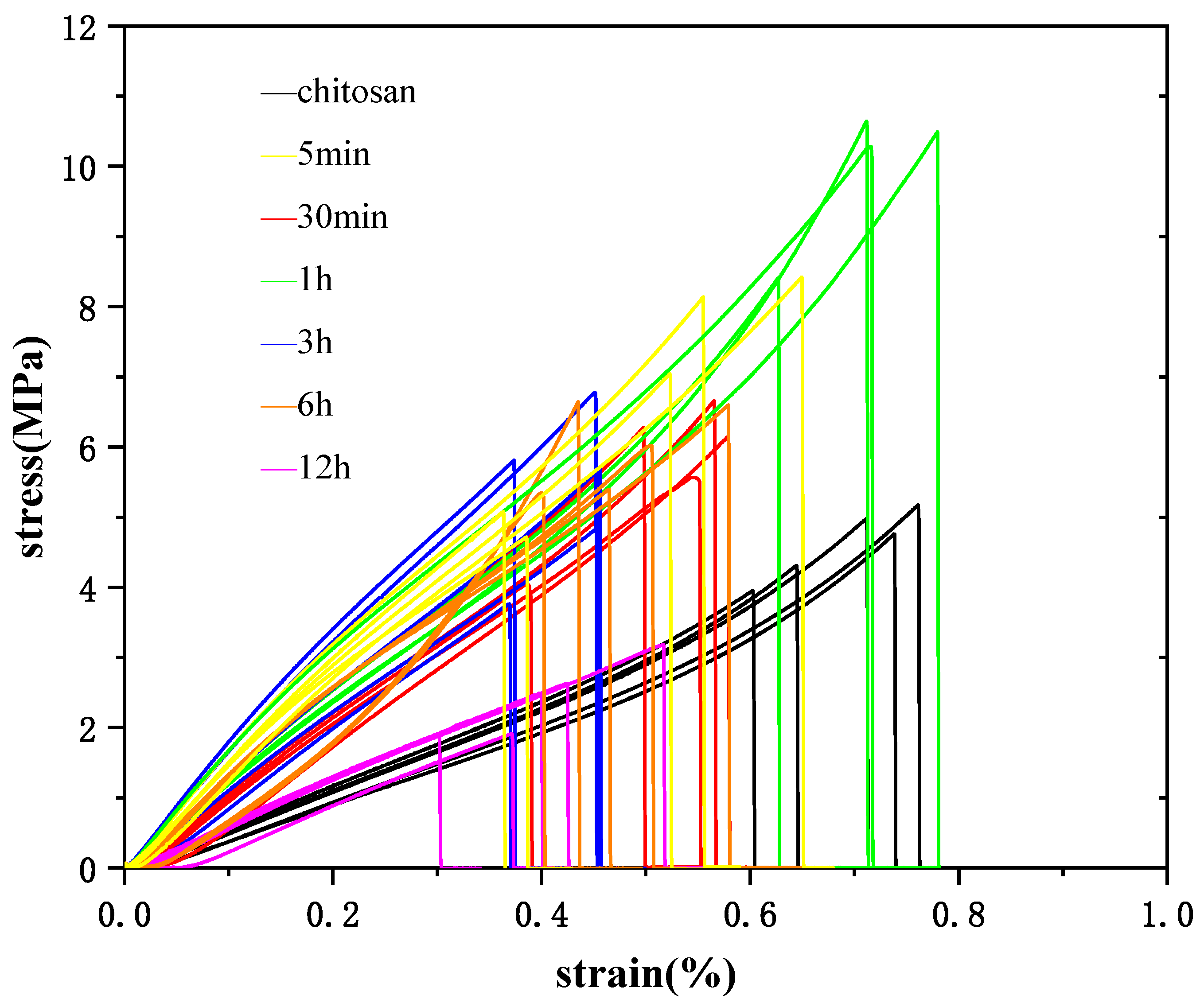
3.3. Mechanical Properties of Chitosan/CaCO3 Composites

The growth process of CaCO3 crystals in the chitosan hydrogel has been largely determined through the aforementioned experiments. However, as a promising direction in the fields of biomedicine and materials science, this ceramic/polymer composite material should possess certain mechanical properties for practical applications. Based on the previous results, it is evident that the number and size of crystals in the hydrogel vary with different mineralization times. Therefore, the next step involves studying the mechanical properties of the hydrogel at various mineralization times. Uniaxial tensile tests were performed on the different samples. The results, including stress–strain curves with computational errors, are presented in Figure 8 and Table 1. The findings indicate that as mineralization time increases, the Young’s modulus and tensile strength of the composite hydrogel initially increase and then decrease, while the ultimate strain decreases. The hydrogel sample mineralized for 1 h exhibited the best mechanical properties, with a 110% increase in Young’s modulus and a 90% increase in tensile strength compared to the pre-mineralized state. The experimental results indicate a 17% increase in tensile strength compared to the microfiber chitosan hydrogel network prepared by Wu et al. [28] using 3D printing technology. Additionally, the strength increased by 260% compared to the alginate/polyacrylamide and glass fabric composite hydrogel material prepared by Xue et al. [42]. These findings suggest that the hydrogels produced through this process and method demonstrate superior mechanical strength.
The increase in Young’s modulus and tensile strength can be attributed to the effective distribution of calcium carbonate nanocrystals within the three-dimensional fiber network of chitosan during the early stages of mineralization. This distribution forms a strong interface, and the higher stiffness of CaCO3 crystals provides structural support, thereby enhancing the overall strength of the hydrogel. However, as the crystals grow and aggregate, over-mineralization occurs, leading to the accumulation of CaCO3 crystals on the fiber network. This aggregation weakens interfacial bonding and causes stress concentration, resulting in reduced elasticity, increased brittleness, and diminished mechanical properties. The decrease in ultimate strain after mineralization is likely due to the restriction of chitosan network movement and deformation by the introduced CaCO3 during the tensile process, which reduces the ductility and toughness of the hydrogel.

4. Conclusions and Outlook

In summary, chitosan/CaCO3 composite hydrogel materials were successfully prepared in this study, providing a novel approach for the development of ceramic/polymer composites by utilizing chitosan hydrogel as a substrate for the in situ growth of CaCO3 nanoparticles via diffusion. The hydrogel network served as a medium for crystal mineralization, enabling the maintenance of solute diffusion while regulating and controlling crystal morphology. Calcite nanoparticles were formed uniformly inside the chitosan molecular networks with a strong interface between calcite crystals and chitosan chains. The nanostructure resembled that observed previously in natural tissues such as mantis shrimp dactyl club, chiton teeth, and nacre. By adjusting the mineralization time, it was found that the tensile strength of the hydrogel could be significantly enhanced in an optimal range of the mineralization period while maintaining the hydrogel’s inherent toughness. Additionally, the in situ growth of CaCO3 in the chitosan hydrogel using our method ensures good interfacial bonding between the CaCO3 and chitosan matrix, closely mimicking the biomineralization process in nature. This approach avoids the inhomogeneous agglomeration that may occur when the two components are mixed directly.
The next step will be optimizing the mineralization process based on crystallization principles, further adjusting the experimental conditions to more precisely regulate crystal growth in the polymer network. This will deepen our understanding of the diffusion process and crystal growth mechanism in the gel, ultimately leading to the development of composites with improved performance. Moreover, beyond CaCO3 and chitosan, other materials can be explored to expand the performance and application areas of ceramic/polymer composites, paving the way for future advancements in tissue engineering, materials science, and biomedicine.

Supplementary Materials

The following supporting information can be downloaded at: https://www.mdpi.com/article/10.3390/app14209193/s1, Figure S1: Images of the surface morphology of chitosan/CaCO3 composite hydrogel at different mineralization time; Figure S2: TGA of chitosan/CaCO3 composite hydrogel at different times; Figure S3: Images of the surface morphology of chitosan/CaCO3 composite hydrogel; Figure S4: SEM image of CaCO3 on the surface of chitosan/CaCO3 composite hydrogel; Figure S5: The printing route and sample photographs of chitosan/CaCO3 composite hydrogel.

Author Contributions

Conceptualization, W.H. and B.W.; methodology, W.H. and X.Z.; validation, X.Z., Z.Z. and W.C.; formal analysis, X.Z.; investigation, X.Z.; resources, W.H. and B.W.; data curation, X.Z.; writing—original draft preparation, X.Z.; writing—review and editing, X.Z., W.H. and B.W.; supervision, W.H.; project administration, W.H. and B.W.; funding acquisition, W.H. and B.W. All authors have read and agreed to the published version of the manuscript.

Funding

This work was funded by the National Natural Science Foundation Council of China (Grant No. 52305359), the Hubei Provincial Natural Science Foundation of China (No. 2023AFB141) and the startup funding from the Huazhong University of Science and Technology.

Institutional Review Board Statement

The study was not applicable to humans or animals.

Informed Consent Statement

The study was not applicable to humans or animals.

Data Availability Statement

The original contributions presented in the study are included in the article and Supplementary Material, further inquiries can be directed to the corresponding author.

Acknowledgments

This work was supported by the National Natural Science Foundation Council of China (Grant No. 52305359), the Hubei Provincial Natural Science Foundation of China (No. 2023AFB141) and the startup funding from the Huazhong University of Science and Technology. The authors would like to thank the Analytical and Testing Center of Huazhong University of Science and Technology for their support of materials characterizations.

Conflicts of Interest

The authors declare no conflict of interest.

References

  1. He, B.; Yuan, X.; Zhou, A.; Zhang, H.; Jiang, D. Designer functionalised self-assembling peptide nanofibre scaffolds for cartilage tissue engineering. Expert Rev. Mol. Med. 2014, 16, e12. [Google Scholar] [CrossRef]
  2. Farag, M.M. Recent trends on biomaterials for tissue regeneration applications: Review. J. Mater. Sci. 2023, 58, 527–558. [Google Scholar] [CrossRef]
  3. Szwed-Georgiou, A.; Płociński, P.; Kupikowska-Stobba, B.; Urbaniak, M.M.; Rusek-Wala, P.; Szustakiewicz, K.; Piszko, P.; Krupa, A.; Biernat, M.; Gazińska, M.; et al. Bioactive Materials for Bone Regeneration: Biomolecules and Delivery Systems. ACS Biomater. Sci. Eng. 2023, 9, 5222–5254. [Google Scholar] [CrossRef] [PubMed]
  4. Armengol, E.S.; Hock, N.; Saribal, S.; To, D.; Summonte, S.; Veider, F.; Kali, G.; Bernkop-Schnürch, A.; Laffleur, F. Unveiling the potential of biomaterials and their synergistic fusion in tissue engineering. Eur. J. Pharm. Sci. 2024, 196, 106761. [Google Scholar] [CrossRef] [PubMed]
  5. Asadi, N.; Del Bakhshayesh, A.R.; Davaran, S.; Akbarzadeh, A. Common biocompatible polymeric materials for tissue engineering and regenerative medicine. Mater. Chem. Phys. 2020, 242, 122528. [Google Scholar] [CrossRef]
  6. Polishchuk, I.; Bracha, A.A.; Bloch, L.; Levy, D.; Kozachkevich, S.; Etinger-Geller, Y.; Kauffmann, Y.; Burghammer, M.; Giacobbe, C.; Villanova, J.; et al. Coherently aligned nanoparticles within a biogenic single crystal: A biological prestressing strategy. Science 2017, 358, 1294–1298. [Google Scholar] [CrossRef]
  7. Kunitake, M.E.; Mangano, L.M.; Peloquin, J.M.; Baker, S.P.; Estroff, L.A. Evaluation of strengthening mechanisms in calcite single crystals from mollusk shells. Acta Biomater. 2013, 9, 5353–5359. [Google Scholar] [CrossRef]
  8. Fratzl, P.; Weinkamer, R. Nature’s hierarchical materials. Prog. Mater. Sci. 2007, 52, 1263–1334. [Google Scholar] [CrossRef]
  9. Jia, Y.; Liu, K.; Zhang, X.S. Modulate stress distribution with bio-inspired irregular architected materials towards optimal tissue support. Nat. Commun. 2024, 15, 4072. [Google Scholar] [CrossRef]
  10. Xia, Y.; Gu, Y.; Zhou, X.; Xu, H.; Zhao, X.; Yaseen, M.; Lu, J.R. Controllable Stabilization of Poly (N-isopropylacrylamide)-Based Microgel Films through Biomimetic Mineralization of Calcium Carbonate. Biomacromolecules 2012, 13, 2299–2308. [Google Scholar] [CrossRef]
  11. McNally, E.A.; Schwarcz, H.P.; Botton, G.A.; Arsenault, A.L. A model for the ultrastructure of bone based on electron microscopy of ion-milled sections. PLoS ONE 2012, 7, e29258. [Google Scholar] [CrossRef] [PubMed]
  12. Shah, F.A.; Zanghellini, E.; Matic, A.; Thomsen, P.; Palmquist, A. The Orientation of Nanoscale Apatite Platelets in Relation to Osteoblastic–Osteocyte Lacunae on Trabecular Bone Surface. Calcif. Tissue Int. 2016, 98, 193–205. [Google Scholar] [CrossRef] [PubMed]
  13. Rho, J.-Y.; Kuhn-Spearing, L.; Zioupos, P. Mechanical properties and the hierarchical structure of bone. Med. Eng. Phys. 1998, 20, 92–102. [Google Scholar] [CrossRef]
  14. Arash, A.; Dehgan, F.; Benisi, S.Z.; Jafari-Nodoushan, M.; Pezeshki-Modaress, M. Polysaccharide base electrospun nanofibrous scaffolds for cartilage tissue engineering: Challenges and opportunities. Int. J. Biol. Macromol. 2024, 277, 134054. [Google Scholar] [CrossRef]
  15. Hoemann, C.D.; Lafantaisie-Favreau, C.-H.; Lascau-Coman, V.; Chen, G.; Guzmán-Morales, J. The Cartilage-Bone Interface. J. Knee Surg. 2012, 25, 85–98. [Google Scholar] [CrossRef]
  16. Huang, W.; Shishehbor, M.; Guarín-Zapata, N.; Kirchhofer, N.D.; Li, J.; Cruz, L.; Wang, T.; Bhowmick, S.; Stauffer, D.; Manimunda, P.; et al. A natural impact-resistant bicontinuous composite nanoparticle coating. Nat. Mater. 2020, 19, 1236–1243. [Google Scholar] [CrossRef] [PubMed]
  17. Zimmermann, E.A.; Fiedler, I.A.; Busse, B. Breaking new ground in mineralized tissue: Assessing tissue quality in clinical and laboratory studies. J. Mech. Behav. Biomed. Mater. 2021, 113, 104138. [Google Scholar] [CrossRef]
  18. Fukao, K.; Tanaka, K.; Kiyama, R.; Nonoyama, T.; Gong, J.P. Hydrogels toughened by biominerals providing energy-dissipative sacrificial bonds. J. Mater. Chem. B 2020, 8, 5184–5188. [Google Scholar] [CrossRef] [PubMed]
  19. Liu, W.; Bi, W.; Sun, Y.; Wang, L.; Yu, X.; Cheng, R.; Yu, Y.; Cui, W. Biomimetic or-ganic-inorganic hybrid hydrogel electrospinning periosteum for accelerating bone regeneration. Mater. Sci. Eng. C 2020, 110, 110670. [Google Scholar] [CrossRef]
  20. Monavari, M.; Homaeigohar, S.; Fuentes-Chandía, M.; Nawaz, Q.; Monavari, M.; Venkatraman, A.; Boccaccini, A.R. 3D printing of alginate dialdehyde-gelatin (ADA-GEL) hydrogels incorporating phytotherapeutic icariin loaded mesoporous SiO2-CaO nanoparticles for bone tissue engineering. Mater. Sci. Eng. C 2021, 131, 112470. [Google Scholar] [CrossRef]
  21. Zhu, L.; Wang, G.; Shi, W.; Ma, X.; Yang, X.; Yang, H.; Guo, Y.; Yang, L. In situ generation of biocompatible amorphous calcium carbonate onto cell membrane to block membrane transport protein—A new strategy for cancer therapy via mimicking abnormal mineralization. J. Colloid Interface Sci. 2019, 541, 339–347. [Google Scholar] [CrossRef] [PubMed]
  22. Liu, X.; Wang, Z. Chitosan-calcium carbonate scaffold with high mineral content and hierarchical structure for bone regeneration. Smart Mater. Med. 2023, 4, 552–561. [Google Scholar] [CrossRef]
  23. Fu, J.; Leo, C.P.; Show, P.L. Recent advances in the synthesis and applications of pH-responsive CaCO3. Biochem. Eng. J. 2022, 187, 108446. [Google Scholar] [CrossRef]
  24. Asenath-Smith, E.; Li, H.; Keene, E.C.; Seh, Z.W.; Estroff, L.A. Crystal Growth of Calcium Carbonate in Hydrogels as a Model of Biomineralization. Adv. Funct. Mater. 2012, 22, 2891–2914. [Google Scholar] [CrossRef]
  25. Damian, P.; Style, R.W.; Zlopaša, J.; Petrozzini, J.J.; Pfeifer, M.A.; Jonkers, H.M.; Dufresne, E.R.; Estroff, L.A. Forming Anisotropic Crystal Composites: Assessing the Mechanical Translation of Gel Network An-isotropy to Calcite Crystal Form. J. Am. Chem. Soc. 2021, 143, 3439–3447. [Google Scholar]
  26. Di Martino, A.; Sittinger, M.; Risbud, M.V. Chitosan: A versatile biopolymer for orthopaedic tis-sue-engineering. Biomaterials 2005, 26, 5983–5990. [Google Scholar] [CrossRef] [PubMed]
  27. Deepthi, S.; Venkatesan, J.; Kim, S.-K.; Bumgardner, J.D.; Jayakumar, R. An overview of chitin or chitosan/nano ceramic composite scaffolds for bone tissue engineering. Int. J. Biol. Macromol. 2016, 93, 1338–1353. [Google Scholar] [CrossRef] [PubMed]
  28. Wu, Q.; Maire, M.; Lerouge, S.; Therriault, D.; Heuzey, M.C. 3D Printing of Microstructured and Stretchable Chitosan Hydrogel for Guided Cell Growth. Adv. Biosyst. 2017, 1, 1700058. [Google Scholar] [CrossRef]
  29. Rivas-Araiza, R.; Alcouffe, P.; Rochas, C.; Montembault, A.; David, L. Micron range mor-phology of physical chitosan hydrogels. Langmuir ACS J. Surf. Colloids 2010, 26, 17495–17504. [Google Scholar] [CrossRef]
  30. Sereni, N.; Enache, A.; Sudre, G.; Montembault, A.; Rochas, C.; Durand, P.; Perrard, M.-H.; Bozga, G.; Puaux, J.-P.; Delair, T.; et al. Dynamic Structuration of Physical Chitosan Hydrogels. Langmuir 2017, 33, 12697–12707. [Google Scholar] [CrossRef]
  31. Cai, X.; Chen, L.; Jiang, T.; Shen, X.; Hu, J.; Tong, H. Facile synthesis of anisotropic porous chitosan/hydroxyapatite scaffolds for bone tissue engineering. J. Mater. Chem. 2011, 21, 12015–12025. [Google Scholar] [CrossRef]
  32. Marsi, T.C.O.; Santos, T.G.; Pacheco-Soares, C.; Corat, E.J.; Marciano, F.R.; Lobo, A.O. Biomineralization of superhydrophilic vertically aligned carbon nanotubes. Langmuir ACS J. Surf. Colloids 2012, 28, 4413–4424. [Google Scholar] [CrossRef] [PubMed]
  33. Mao, L.-B.; Gao, H.-L.; Yao, H.-B.; Liu, L.; Cölfen, H.; Liu, G.; Chen, S.-M.; Li, S.-K.; Yan, Y.-X.; Liu, Y.-Y.; et al. Synthetic nacre by predesigned matrix-directed mineralization. Science 2016, 354, 107–110. [Google Scholar] [CrossRef]
  34. Sugawara-Narutaki, A. Bio-inspired synthesis of polymer–inorganic nanocomposite materials in mild aqueous systems. Polym. J. 2013, 45, 269–276. [Google Scholar] [CrossRef]
  35. Weiner, S.; Wagner, H.D. The Material Bone: Structure-Mechanical Function Relations. Annu. Rev. Mater. Sci. 1998, 28, 271–298. [Google Scholar] [CrossRef]
  36. Baht, G.S.; Hunter, G.K.; Goldberg, H.A. Bone sialoprotein–collagen interaction promotes hydroxyapatite nucleation. Matrix Biol. 2008, 27, 600–608. [Google Scholar] [CrossRef]
  37. McKee, M.D.; Buss, D.J.; Reznikov, N. Mineral tessellation in bone and the Stenciling Principle for extracellular matrix mineralization. J. Struct. Biol. 2021, 241, 107823. [Google Scholar] [CrossRef]
  38. Gebauer, D.; Gale, J.D.; Cölfen, H. Crystal Nucleation and Growth of Inorganic Ionic Materials from Aqueous Solution: Selected Recent Developments, and Implications. Small 2022, 18, e2107735. [Google Scholar] [CrossRef]
  39. Nicolin, D.J.; Rossoni, D.F.; Mario, L.; Jorge, M. Study of uncertainty in the fitting of diffusivity of Fick’s Second Law of Diffusion with the use of Bootstrap Method. J. Food Eng. 2016, 184, 63–68. [Google Scholar] [CrossRef]
  40. Hashim, F.A.; Mostafa, R.R.; Hussien, A.G.; Mirjalili, S.; Sallam, K.M. Fick’s Law Al-gorithm: A physical law-based algorithm for numerical optimization. Knowl. Based Syst. 2023, 260, 110146. [Google Scholar] [CrossRef]
  41. Mauro, J.C. Fick’s Laws of Diffusion. In Materials Kinetics: Transport and Rate Phenomena; Mauro, J.C., Ed.; Elsevier: Amsterdam, The Netherlands, 2021; Chapter 3; pp. 39–58. [Google Scholar]
  42. Xue, Q.; He, Y.; Zhang, X.; Zhang, X.; Cai, M.; Guo, C.F.; Yang, C. Strong Interfaces Enable Efficient Load Transfer for Strong, Tough, and Impact-Resistant Hydrogel Composites. ACS Appl. Mater. Interfaces 2022, 14, 33797–33805. [Google Scholar] [CrossRef] [PubMed]
Figure 1. The design strategy, preparation process, and microstructural characteristics of chitosan/CaCO3 composites. (a) Schematic diagram illustrating the hierarchical structure of the natural skeleton. (b) Schematic representation of the compositional components of the mantis shrimp’s dactyl club. (c) Preparation method of the chitosan/CaCO3 composite hydrogel and a schematic diagram of its structure.
Applsci 14 09193 g001
Figure 2. Microstructures of chitosan hydrogel before and after mineralization. (a) Images of 3D printed pure chitosan hydrogel samples at their unmineralized state. (b) Images of chitosan/CaCO3 composite hydrogel samples after 12 h of mineralization. (c) SEM images of the surface of pure chitosan hydrogel at its unmineralized state. (d) SEM image of the chitosan/CaCO3 composite hydrogel surface after 12 h of mineralization. (e) SEM images of the cross-section of unmineralized chitosan hydrogel. (f) SEM image of the cross-section of the chitosan/CaCO3 composite hydrogel after 12 h of mineralization. Nanoparticles are noticed.
Applsci 14 09193 g002
Figure 3. Testing and analysis of chitosan/CaCO3 composite hydrogels. (a) XRD test curve of the chitosan/CaCO3 composite hydrogel. (b) TGA of chitosan/CaCO3 composite hydrogel. (c) Raman spectral profile of chitosan/CaCO3 composite hydrogels. (d) FTIR analysis of hydrogel before and after mineralization. (e) WAXD diffractogram of unmineralized pure chitosan hydrogel. (f) WAXD diffractogram of chitosan/CaCO3 composite hydrogel.
Applsci 14 09193 g003
Figure 4. SEM images of chitosan/CaCO3 composite hydrogels after 5 min and 1 h mineralization. (ac) show SEM images of the chitosan/CaCO3 composite hydrogel cross-sections after 5 min of mineralization, with (b) representing the area near the surface and (c) representing the area near the center. (df) show SEM images of the chitosan/CaCO3 composite hydrogel cross-sections after 1 h of mineralization, with (e) representing the area near the surface and (f) representing the area near the center.
Applsci 14 09193 g004
Figure 5. SEM images of chitosan/CaCO3 composite hydrogel after 3 and 6 h mineralization. (ac) depict the cross-sections after 3 h of mineralization, where (b) is near the surface and (c) is near the center. (df) present the cross-sections after 6 h of mineralization, with (e) representing the area near the surface and (f) representing the area near the center.
Applsci 14 09193 g005
Figure 6. TEM images of chitosan/CaCO3 composite hydrogel. (a,b) TEM image of nanoparticles formed via in situ growth. (cf) TEM images and diffraction patterns of chitosan/CaCO3 composite hydrogel. Nanoparticles shows single crystal-like diffraction pattern. Amorphous regions are noticed inside and on the edge of the calcite nanoparticles. (g,h) TEM images of the nanocomposite coating in mantis shrimp dactyl clubs, showing the combination of chitin macromolecules and hydroxyapatite nanocrystals. Images are taken from [16] with permission.
Applsci 14 09193 g006
Figure 7. Schematic of in situ growth of CaCO3 nanocrystals inside chitosan hydrogels via the double diffusion method. Calcium ions are diffused into the 3D-printed chitosan hydrogel first, until an equilibrium is reached. Samples are then immersed in NaHCO3 solution. As the CO32− is introduced, calcite crystals start growing at the surface first. Microcrystals are formed on the surface, while crystal sizes are limited to ~100 nm inside the hydrogel due to the confinement of the chitosan molecular network.
Applsci 14 09193 g007
Figure 8. Stress–strain curve of chitosan/CaCO3 composite hydrogel at different mineralization times. Compared to the unmineralized chitosan films, mineralized films show an increase in both stiffness and tensile strength.
Applsci 14 09193 g008
Table 1. Calculated results and error analysis of tensile tests for chitosan/CaCO3 composite hydrogels at different mineralization times.
Mineralization TimeYoung’s Moduli
E [MPa]
Tensile Strength
σm [MPa]
Ultimate Strain
εb [%]
0 min5.59 ± 0.344.64 ± 0.5073.64 ± 6.68
5 min13.84 ± 2.156.68 ± 1.7154.87 ± 9.67
30 min10.91 ± 0.865.74 ± 1.0357.68 ± 7.21
1 h11.68 ± 1.388.72 ± 2.9068.05 ± 19.15
3 h13.88 ± 3.115.37 ± 1.1346.34 ± 3.41
6 h12.15 ± 1.576.00 ± 0.6352.12 ± 8.04
12 h6.96 ± 0.212.42 ± 0.5444.47 ± 8.06
Disclaimer/Publisher’s Note: The statements, opinions and data contained in all publications are solely those of the individual author(s) and contributor(s) and not of MDPI and/or the editor(s). MDPI and/or the editor(s) disclaim responsibility for any injury to people or property resulting from any ideas, methods, instructions or products referred to in the content.

