Article

Differential Relapse of Proximal and Distal Segments after Mandibular Setback Surgery

1
Department of Orthodontics, College of Dentistry, Chosun University, Gwangju 61453, Republic of Korea
2
Postgraduate Orthodontic Program, Arizona School of Dentistry & Oral Health, A.T. Still University, Mesa, AZ 85206, USA
3
Graduate School of Dentistry, Kyung Hee University, Seoul 54913, Republic of Korea
*
Author to whom correspondence should be addressed.
Appl. Sci. 2024, 14(20), 9299; https://doi.org/10.3390/app14209299
Submission received: 11 September 2024 / Revised: 3 October 2024 / Accepted: 9 October 2024 / Published: 12 October 2024

Abstract

:
This study aimed to evaluate the differential positional changes of the proximal and distal segments during mandibular setback surgery relapse. Thirty patients with mandibular prognathism who underwent bilateral sagittal split ramus osteotomy were included. Skull models from pretreatment, postsurgery, and post-treatment cone-beam computed tomography were superimposed to evaluate condylar displacement and rotational changes, and to determine the center of rotation (CRot) in the proximal and distal segments. During postsurgical relapse, the proximal segment rotated 2.4 ± 2.1° counterclockwise, with the CRot located near the mandibular condyle. The distal segment rotated 2.0 ± 2.3° counterclockwise, resulting in forward and upward movement of the chin. The displaced condyle partially returned to its original position during postsurgical orthodontic treatment. A vertical bony step (VBS) between the proximal and distal segments commonly occurs during mandibular setback surgery. As the VBS increases, the CRot of the distal segment shifts posterosuperiorly, following a predictable pattern outlined by regression equations. These findings suggest that relapse after mandibular setback surgery consistently occurs in an anterosuperior direction, with translatory movement becoming more prominent as the VBS increases. Including these regression equations in presurgical planning can enhance the precision of relapse simulations and help clinicians more accurately anticipate postsurgical relapse.

1. Introduction

Mandibular prognathism is a malocclusion in which the lower jaw is abnormally large compared to the upper jaw. It occurs when the lower jaw overgrows or the upper jaw undergrows. Mandibular setback surgery is used to correct mandibular prognathism. Bilateral mandibular ramus sagittal split osteotomy (BSSRO) is one of the most commonly used orthognathic procedures to improve mandibular prognathism. BSSRO allows mandibular setback or advancement by splitting the mandible through two incisions: a lateral incision extending from the second molar to the inferior border, and a medial incision running posteriorly along the ramus, avoiding the mandibular nerve canal. The anteroposterior relapse rate of mandibular setback surgery varies from 14 to 20% [1,2,3], while the pattern of relapse varies depending on the surgical technique [4]. After mandibular setback surgery with BSSRO, relapse occurs in the form of rotational relapse, where the mandible rotates counterclockwise [5]. Forward movement of the chin is observed in the postoperative period due to the counterclockwise rotation of the mandible [1,4,6,7]. Specifically, posterior displacement of the proximal segments during surgery, which appears as clockwise rotation, induces stretching of the pterygomasseteric sling, which causes the mandible to relapse with counterclockwise rotation [8,9,10]. This counterclockwise rotation increases the prominence of the chin point and mandibular border. In addition, factors that contribute to stability after mandibular setback surgery have been reported to include the amount of setback, tension of the soft tissues surrounding the mandible, and the method of bone fixation [9,11,12,13]. It is known that condylar displacement during orthognathic surgery significantly impacts early postsurgical relapse and temporomandibular joint disorders [14,15,16]. As the distal segment setbacks along the occlusal plane, a vertical height difference between the lower borders of the proximal and distal segments of the mandible, known as the vertical bony step (VBS), occurs inevitably due to the divergence between the occlusal plane and mandibular plane. This VBS has been reported to be associated with relapse after mandibular BSSRO surgery [17,18].
Using a computer-based three-dimensional simulation technique, it is possible to measure and analyze the anatomical structures of the maxillofacial region in three dimensions prior to surgery and predict the position of the bone segments after osteotomy in three dimensions. At this time, it is necessary to determine the amount of simulation surgery by considering the postoperative relapse of the bone segments. To our knowledge, the relapse of the proximal and distal segments has rarely been investigated separately. Previous studies have suggested that postsurgical relapse patterns differ between them [17,19]. Since chin point prominence is often the chief complaint of patients undergoing mandibular setback surgery, it is crucial to account for rotational relapse in advance. Therefore, incorporating proximal and distal segment relapse simulations into the three-dimensional simulation of mandibular setback surgery is desirable. However, this requires information about the center of rotation (CRot) of proximal and distal segments and the amount of rotation during the relapse of mandibular setback surgery. This study aimed to evaluate the condylar displacement pattern and the CRot of the rotational relapse of the proximal and distal segments of the mandible.

2. Materials and Methods

2.1. Subjects

This retrospective study was reviewed and approved by the Institutional Review Board (IRB) of Chosun University Dental Hospital (IRB approval number CUDH IRB 2301 003). Among the patients who visited the Department of Orthodontics at Chosun University Dental Hospital from 2013 to 2020, 211 patients were diagnosed with mandibular prognathism and underwent orthognathic surgery at the Department of Oral and Maxillofacial Surgery at our hospital. Thirty patients met the inclusion criteria. The inclusion criteria were as follows:
(1) Skeletal Class III malocclusion with mandibular prognathism; (2) single jaw surgery with bilateral sagittal split ramus osteotomy (BSSRO) mandibular setback or double jaw surgery including BSSRO and Le Fort I osteotomy; (3) availability of cone-beam computed tomography (CBCT) scans at the time points before treatment (T0), within one month of surgery (T1), and after orthodontic treatment (T2).
The exclusion criteria were as follows:
(1) Presence of congenital anomalies, including cleft lip and palate; (2) degenerative disease of the temporomandibular joint; (3) an anterior open bite of 1.2 mm or more at pretreatment; (4) an open bite of a second molar of 1.2 mm or more at postsurgery CBCT; (5) a history of previous maxillofacial surgery, trauma, or infection in the maxillofacial region; (6) severe mandibular chin deviation greater than 5 mm from the facial midline.
According to the above criteria, 181 patients were excluded.
A splint was used to ensure a stable mandibular position during surgery, and semi-rigid fixation of the bone segments was performed using mini-plates and screws after orthognathic surgery. To ensure the adequate strength of the surgical splint, the surgical bite can be opened to accommodate the necessary thickness of the surgical splint. However, this may induce autorotation of the mandible upon splint removal, which must be distinguished from mandibular rotation caused by relapse. In the present study, the postoperative occlusion of the maxillary second molars ranged from complete contact (0 mm) to an open occlusion of up to 1.2 mm, with a mean value of 0.3 ± 0.4 mm.
The 30 patients selected had a mean age of 21.9 ± 4.3 years. The duration of presurgical orthodontic treatment was 10.7 ± 7.4 months, and the duration of postsurgical orthodontic treatment was 18.3 ± 10.9 months. There were 15 patients each who underwent isolated single mandibular setback surgery and bimaxillary surgery. The mean amount of mandibular setback, measured from the pogonion (Pog), was 13.6 ± 5.3 mm.

2.2. Data Acquisition and Generation of 3D Skull Model

CBCT images obtained at three different time points—before treatment (T0), within one month of surgery (T1), and after the completion of postsurgical orthodontic treatment (T2)—were used. To evaluate the position of the mandible at different time points, stereolithography (STL) files were generated from CBCT scans at T0, T1, and T2 using InVivoDental 5.4 software (Anatomage, San Jose, CA, USA).
The generated STL files were imported into the reverse engineering software Geomagic Design X 2014 (3D Systems, Rock Hill, SC, USA). At the T0 stage, the 3D skull model was oriented by defining the midsagittal plane passing through the nasion, basion, and ANS, and it was aligned parallel to the default sagittal plane of the software. The Frankfort horizontal plane passing through the right orbitale, left porion, and right porion was aligned parallel to the default horizontal plane of the software. A right-handed coordinate system was used for measurements (Figure 1). The T1 and T2 models were superimposed on the T0 model using an iterative closest point algorithm using regions unaffected during surgery.

2.3. Measurements of Variables

2.3.1. Landmarks and Measurement of Changes to Mandible

Measurement points were located on the 3D skull model (Figure S1 and Table 1) [18,20,21]. In the case of bilateral measurement points, those on the right side were used. For points other than the medial and lateral poles of the mandible, only vertical (y-axis) and anteroposterior (z-axis) displacements were measured by projecting the landmarks onto the midsagittal plane. Because Pog and Me (menton) change according to distal segment rotation [17], only the anteroposterior change of Pog (Pog-Z) and vertical changes of Me (Me-Y) were measured to evaluate distal segment movement. When VBS occurs, the lower border of the distal segment moves downward relative to the lower border of the proximal segment and the pretreatment mandibular plane. When VBS occurred at the time of surgery, it was measured as a negative value because it always occurred as a downward movement of the distal segment of the mandible. When the VBS was resolved during postsurgical orthodontic treatment (T2–T1), it was measured as a positive value because the upward movement of the mandible resolved it.

2.3.2. Measurement of Condylar Displacement

To assess surgical condylar displacement and postsurgical condylar movement, the movements of the medial and lateral poles of the right condyle were measured in the mediolateral (x-axis), vertical (y-axis), and anteroposterior (z-axis) directions.

2.3.3. Measurement of the Center of Rotation of the Proximal and Distal Segments

To determine the centers of rotation of the segments, two arbitrary measurement points (P1, P2, and D1, D2) were set on each proximal and distal segment of the T1 models. The T1 model was superimposed onto the T2 model based on unaffected areas during surgery. The points that were located on T1 were displaced on T2, then designated P1′, P2′, D1′, and D2′. The location where the vertical bisector of two lines connecting the same measurement points (P1–P1′, P2–P2′, and D1–D1′, D2–D2′) met was marked as the center of rotation (CRot) and the angle of rotation was measured. The medial pole of the right condyle was set as the origin point (0, 0). Only the y-axis and z-axis coordinates of the condyles were measured.

2.4. Validation of Variable Measurement

The first author (KJD) performed all of the above measurements. To assess intraexaminer reproducibility, ten random samples were selected and remeasured at 2-month intervals. Measurement errors were calculated using the Dahlberg formula (Table 2).

2.5. Statistical Analysis

Statistical analysis was performed using IBM SPSS Statistics for Windows 27.0 (IBM, Armonk, NY, USA). The Shapiro–Wilk normality test was performed to assess the normality of the data. Wilcoxon signed-rank tests, one-sample t-tests, and paired t-tests were used to evaluate the significance of the surgical movements and relapse patterns of the mandibular segments, depending on the normality of the data. The independent samples t-test was used to assess differences in means between types of surgery. Additionally, Pearson’s correlation analysis or Spearman’s correlation analysis was used to analyze correlations between measurements, depending on the normality of the data. Linear regression analysis was used to predict the relationship between (1) the coordinates of the CRot of the distal segment and (2) the CRot of the distal segment and VBS that occurred during surgery.

3. Results

3.1. Condylar Displacement

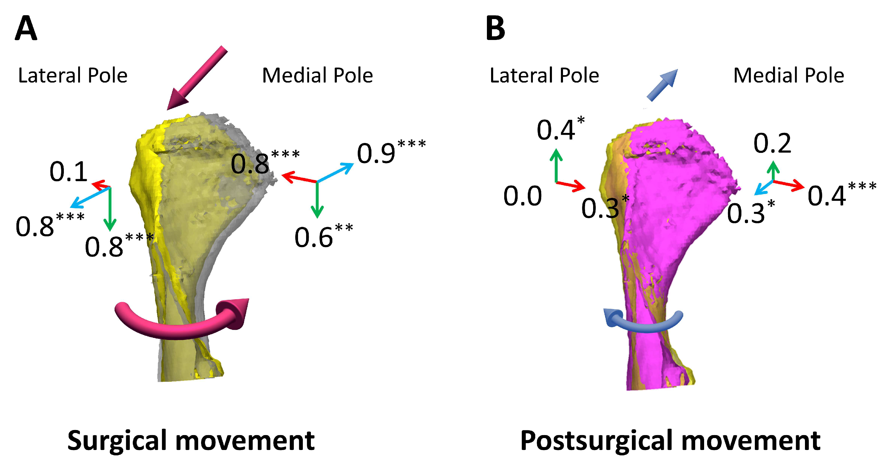
The inferior displacement of the condyle by more than 2 mm was clinically considered as sagging [22], but sagging was not observed in all cases. The mean vertical displacement of the medial pole and lateral pole during surgery was −0.5 (minimum: −1.9; maximum: 1.4) and −0.8 (minimum: −1.9; maximum: 0.3), respectively (Table 3). During mandibular setback surgery, the medial pole of the condyle was displaced laterally, inferiorly, and posteriorly (Figure 2A and Table 3). The lateral pole was displaced inferiorly and anteriorly (p < 0.001). During T2-T1, the medial pole partially returned medially and anteriorly, and the lateral pole partially returned medially and superiorly (p < 0.01) (Figure 2B and Table 3). As a result, at the end of the treatment (T2), the medial pole showed displacement in the lateral direction (p < 0.05) and posterior direction (p < 0.001) compared to the pretreatment, while the lateral pole showed displacement in the inferior direction (p < 0.01) and anterior direction (p < 0.05) (Table 3). In other words, the vertical position of the medial pole returned to its original position and the mediolateral change in the lateral pole was reverted (p > 0.05).

3.2. Relapse of Proximal and Distal Segments and Their Center of Rotation

During T2–T1, the proximal segments rotated 2.4 ± 2.1°, and all proximal segments rotated counterclockwise. The CRots of the proximal segments were distributed around the condyle, and the mean CRot of the proximal segments was 12.3 ± 26.1 mm superior and 16.6 ± 48.0 mm posterior to the medial pole (Figure 3A and Figure 4A and Table 4). During T2–T1, the distal segments rotated 2.0 ± 2.3° counterclockwise, and the mean CRot was 3.0 ± 92.3 mm superior and 13.5 ± 59.3 mm anterior to the medial pole of the condyle (Figure 3B and Figure 4B and Table 4). The CRots of the distal segments were distributed along the line passing the condyle posterosuperiorly and anteroinferiorly (Figure 3B). Only five samples with the CRot located inferior to the menton showed a clockwise rotation of the distal segment, and other samples showed a counterclockwise rotation. Both rotations resulted in an anterosuperior movement of the distal segments. This indicates that the relapse of the BSSRO mandibular setback always occurs in the anterosuperior direction, not in the anteroinferior direction. The difference was that a larger anterior movement of dentition, rather than the chin, occurs with clockwise rotation, and the opposite occurs with counterclockwise rotation. The regression equation between the z- and y-coordinates of the CRot of the distal segment obtained through linear regression analysis was as follows:
y = 1.45 × z + 22.7                     ( 95 %   C I :   ( 1.668 , 1.236 ) )  
The model’s goodness of fit was represented by an R2 value of 0.871. There was 6.2 ± 2.9 mm of VBS, which developed during surgery, but this was resolved by 4.4 ± 1.8 mm during T2–T1, stabilizing at 1.9 ± 2.3 mm (Table 4).

3.3. Associations between VBS Development and Center of Rotation

Figure 5A shows that the CRot of the distal segment tended to be located more posteriorly and superiorly as the amount of VBS (T1–T0) increased (p < 0.05). The regression equations between VBS (T1–T0) and the z- and y-coordinates of the CRot of the distal segment were as follows:
z = 8.74 × V B S + 68.1             ( 95 %   C I :   ( 1.694 , 15.788 ) )
y = 14.6 × V B S 88.2             ( 95 %   C I :   ( 25.371 , 3.835 ) )
(Figure 5A and Table 5).

3.4. Associations between VBS Development and Postsurgical Changes

Among the correlations tested, the correlation coefficient between the developed VBS (T1–T0) and Pog-Z (T2–T1), which is the anterior relapse of mandibular setback, was the largest (r = 0.623, p < 0.001), and developed VBS was also correlated with Me-Y (T2–T1), which is the superior movement of menton (r = 0.389, p = 0.034) (Table 5). Figure 5B shows the scatter plot of developed VBS and postsurgical changes (Pog-Z, Me-Y).

3.5. Comparison of Postsurgical Changes between Single-Jaw and Double-Jaw Surgeries

Postoperative relapse did not show a significant difference depending on single-jaw or double-jaw surgery (Table 6).

4. Discussion

In the present study, the positional changes in the proximal and distal segments after BSSRO setback surgery were evaluated. During surgery, the condyles rotated medially with inferior and lateral movement, a result which is consistent with the previous study of [23]. During mandibular setback surgery, the proximal segments are routinely rotated medially to reduce the transverse gap between the proximal and setback distal segments, which inevitably causes the condyle’s inferior and lateral movement out of the mandibular fossa. During postsurgical orthodontic treatment, condyles returned partially to the pretreatment position via lateral rotation with superior and medial movement [24]. In another study, it was explained that the medial rotation of the condyle during surgery was due to screw insertion during fixation [25].
The present study evaluated only the right condyle in patients with less than 5 mm of chin asymmetry. Another study showed that although the condylar heads moved downward after BSSRO surgery in both the asymmetry and symmetry groups, there were almost no changes in the condylar head between pretreatment and post-treatment in the axial and sagittal plane [26]. In the present study, although the condylar position changes during surgery were reduced during postsurgical orthodontic treatment, significant changes in the anteroposterior position of the medial and lateral poles remained. In the present study, the amount of mandibular setback, the developed VBS, and the resolved VBS did not correlate with condylar movements (Table S1), highlighting the difficulty in predicting these movements. This may be due to the complex proximal segment movements influenced by the bony interferences and gaps between the distal and two proximal segments that arise during mandibular setback.
According to a previous study [27], gender does not significantly affect postsurgical mandibular relapse, so this study did not examine gender differences. Additionally, no significant difference in relapse was reported between single- and double-jaw surgeries [28], which is consistent with the present study results.
Regarding relapse according to the surgical methods used for mandibular setback, counterclockwise rotation of the mandible has been reported with BSSRO, whereas clockwise rotation has been reported with intraoral vertical ramus osteotomy [4]. When the distal segment is set back along the presurgical occlusal plane, VBS occurs, which can extend the pterygomasseteric sling and cause postsurgical counterclockwise rotation of the mandible [10,17]. Also, during postsurgical orthodontic treatment, the proximal and distal segments of the mandible showed different movements, resulting in a significant resolution of the VBS [17]. It has been reported that the proximal segments rotate counterclockwise due to the contraction of the masticatory muscles, while the distal segment rotates clockwise during the postoperative period with monocortical fixation [29]. They explained that the reason for such movements was that the proximal segment was pulled upwards by the masseter muscle but fixed at the TMJ, while the distal segment’s upward movement was restricted by the maxillary molars. This caused the mandible to bend at the mini-plate [29]. When a fixation fails, the masticatory muscles pull the mandibular ramus anterosuperiorly, causing the proximal segments to rotate counterclockwise, while the symphysis area is displaced posteroinferiorly by the suprahyoid muscles and masticatory force [30].
In the present study, the bite opening of the second molar was 0.3 ± 0.4 mm immediately after surgery (T1). When the bite opening of the second molar is 0.3 mm, the mandible will autorotate counterclockwise 0.5° after the removal of the surgical splint. Therefore, this autorotation can explain about 0.5° of the total 2.4° counterclockwise rotation of the proximal segment, and the remaining 1.9° can be considered as pure rotational relapse.
The relapse of the distal segment investigated in the present study is the same as the relapse of the chin, and is the result of the relapse of the proximal segment and the change in the relationship between the proximal and distal segments. In the present study, the proximal segments rotated counterclockwise by a mean of 2.4°, and the distal segment also rotated counterclockwise, but the amount of rotation was less than that of the proximal segment, rotating by a mean of 2.0°. This suggests that the counterclockwise rotation of the proximal segments was partially counteracted by the clockwise rotation of the distal segment. Because the proximal and distal segments of the mandible were connected with semi-rigid fixation using mini-plates (Figure S2), the movement of the distal segment is greatly influenced by the proximal segment. Even if the distal segment rotates clockwise relative to the proximal segment, it may appear that the distal segment also rotates counterclockwise when the proximal segments rotate more counterclockwise.
The clockwise rotation of the distal segment was observed only in five patients, all of whom had the CRot of the distal segment positioned anteroinferior to the menton. The clockwise rotation occurred because the clockwise rotation of the distal segment was sufficient to counteract the counterclockwise rotation of the proximal segment. In the present study, an anterior relapse of Pog-Z and a superior relapse of Me-Y were observed after surgery in all patients. So, it is thought that the relapse after BSSRO setback surgery always manifests as an anterosuperior movement. This explains why the CRots of the distal segments were distributed along the line passing through the condyle in a posterosuperior-to-anteroinferior direction (Figure 3B).
Previous studies have reported that the relapse of mandibular setback is related to the amount of mandibular setback and the amount of VBS [17,18]. If the amount of mandibular setback or the development of VBS is large, then the relapse should be incorporated into the surgery simulation. During this process, the relapse can be simulated by rotating the distal segment, with the CRot of the distal segment at 3.0 mm superior and 13.5 mm anterior to the medial pole of the condyle. At this time, the distal segment can be rotated counterclockwise until 1–2 mm of VBS remains because the mean remaining amount of VBS at post-treatment was 1.9 mm. For a more precise prediction of the counterclockwise rotation of the distal segment, the amount of VBS measured during the surgery simulation can be used to calculate the exact CRot coordinate using the regression equations shown in the present study (Figure 6). When considering the postsurgical counterclockwise rotation of the distal segment, an intentional Class II open bite should be created when the amount of VBS or mandibular setback is significant (Figure 6). In the present study, only VBS showed a correlation with the coordinates of the CRot for relapse; thus, the regression equations were made using solely VBS measurements. Further research on various surgical factors influencing the position of CRot is necessary to enhance the predictability of relapse.
While the regression model demonstrates the potential for predicting relapse, it was validated only within the tested cohort. Further validation through additional cohorts or external datasets is necessary to establish the model’s robustness and applicability in broader clinical practice.

5. Conclusions

  • The mandibular condyles rotated medially with lateral and downward movement during mandibular setback surgery. They gradually recovered during postsurgical orthodontic treatment but never fully returned to the pretreatment position.
  • During the postoperative healing period, the mesial and distal fragments of the mandible each have different centers of rotation (CRot) and move differently.
  • Increased VBS during surgery was correlated with greater relapse of the mandibular setback and a corresponding posterosuperior shift of the CRot of the distal segment.
  • Two regression equations were formulated to predict the coordinates of the CRot of the distal segment for the postsurgical relapse. These can be used to simulate relapse during the surgery simulation, although they need external validation.

Supplementary Materials

The following supporting information can be downloaded at: https://www.mdpi.com/article/10.3390/app14209299/s1, Figure S1: Landmarks; Figure S2: A miniplate and 4 screws for semi-rigid fixation; Table S1: Correlation between the VBS and condyle movement.

Author Contributions

Conceptualization, S.-H.L. and J.-D.K.; methodology, S.-H.L.; software, J.-D.K.; validation, J.-D.K.; formal analysis, J.-D.K.; investigation, S.-H.L.; resources, S.-H.L.; data curation, J.-D.K.; writing—original draft preparation, S.-H.L. and J.-D.K.; writing—review and editing, J.-D.K. and J.H.P.; visualization, S.-H.L. and J.-D.K.; supervision, S.-H.L. and J.H.P.; project administration, S.-H.L.; funding acquisition, S.-H.L. All authors have read and agreed to the published version of the manuscript.

Funding

This research was funded by Chosun University, 2021 (Chosun-2021). The funders had no role in the design of the study; in the collection, analyses, or interpretation of data; in the writing of the manuscript; or in the decision to publish the results.

Institutional Review Board Statement

The study was conducted in accordance with the Declaration of Helsinki, and the protocol was approved by the Institutional Review Board (IRB) of Chosun University Dental Hospital (IRB approval number CUDH IRB 2301 003, 26 January 2023).

Informed Consent Statement

Patient consent was waived due to the retrospective design of this study.

Data Availability Statement

The data presented in this study are available on request from the corresponding author due to privacy or ethical restrictions.

Conflicts of Interest

The authors declare no conflict of interest.

References

  1. Kim, J.-W.; Lee, N.-K.; Yun, P.-Y.; Moon, S.-W.; Kim, Y.-K. Postsurgical Stability After Mandibular Setback Surgery with Minimal Orthodontic Preparation Following Upper Premolar Extraction. J. Oral Maxillofac. Surg. 2013, 71, 1968.e1–1968.e11. [Google Scholar] [CrossRef] [PubMed]
  2. Kim, C.-S.; Lee, S.-C.; Kyung, H.-M.; Park, H.-S.; Kwon, T.-G. Stability of Mandibular Setback Surgery with and Without Presurgical Orthodontics. J. Oral Maxillofac. Surg. 2014, 72, 779–787. [Google Scholar] [CrossRef] [PubMed]
  3. Ko, E.W.-C.; Hsu, S.S.-P.; Hsieh, H.-Y.; Wang, Y.-C.; Huang, C.S.; Chen, Y.R. Comparison of Progressive Cephalometric Changes and Postsurgical Stability of Skeletal Class III Correction with and without Presurgical Orthodontic Treatment. J. Oral Maxillofac. Surg. Off. J. Am. Assoc. Oral Maxillofac. Surg. 2011, 69, 1469–1477. [Google Scholar] [CrossRef] [PubMed]
  4. Al-Delayme, R.M.A.; Alsagban, A.A.R.; Ahmed, F.T.; Farag, A.; Al-Allaq, T.; Virdee, P.; Alnajjar, Z.; Alsamaraie, S.A.I.; Elnashef, S.; Arab, L.; et al. The Direction of Double-Jaw Surgery Relapse for Correction of Skeletal Class III Deformity: Bilateral Sagittal Split Versus Intraoral Vertical Ramus Setback Osteotomies. J. Maxillofac. Oral Surg. 2019, 18, 280–287. [Google Scholar] [CrossRef] [PubMed]
  5. Lee, N.-K.; Kim, Y.-K.; Yun, P.-Y.; Kim, J.-W. Evaluation of Post-Surgical Relapse after Mandibular Setback Surgery with Minimal Orthodontic Preparation. J. Cranio-Maxillofac. Surg. Off. Publ. Eur. Assoc. Cranio-Maxillofac. Surg. 2013, 41, 47–51. [Google Scholar] [CrossRef]
  6. Park, H.-M.; Yang, I.-H.; Choi, J.-Y.; Lee, J.-H.; Kim, M.-J.; Baek, S.-H. Postsurgical Relapse in Class III Patients Treated with Two-Jaw Surgery: Conventional Three-Stage Method Versus Surgery-First Approach. J. Craniofac. Surg. 2015, 26, 2357–2363. [Google Scholar] [CrossRef]
  7. Proffit, W.R.; Phillips, C.; Turvey, T.A. Stability After Mandibular Setback: Mandible-Only Versus 2-Jaw Surgery. J. Oral Maxillofac. Surg. 2012, 70, e408–e414. [Google Scholar] [CrossRef]
  8. Cho, H.J. Long-Term Stability of Surgical Mandibular Setback. Angle Orthod. 2007, 77, 851–856. [Google Scholar] [CrossRef]
  9. Proffit, W.R.; Turvey, T.A.; Phillips, C. The Hierarchy of Stability and Predictability in Orthognathic Surgery with Rigid Fixation: An Update and Extension. Head Face Med. 2007, 3, 21. [Google Scholar] [CrossRef]
  10. Komori, E.; Aigase, K.; Sugisaki, M.; Tanabe, H. Cause of Early Skeletal Relapse after Mandibular Setback. Am. J. Orthod. Dentofac. Orthop. 1989, 95, 29–36. [Google Scholar] [CrossRef]
  11. Chou, J.I.-C.; Fong, H.-J.; Kuang, S.-H.; Gi, L.-Y.; Hwang, F.-Y.; Lai, Y.-C.; Chang, R.C.-S.; Kao, S.-Y. A Retrospective Analysis of the Stability and Relapse of Soft and Hard Tissue Change after Bilateral Sagittal Split Osteotomy for Mandibular Setback of 64 Taiwanese Patients. J. Oral Maxillofac. Surg. 2005, 63, 355–361. [Google Scholar] [CrossRef] [PubMed]
  12. Kim, C.-H.; Lee, J.-H.; Cho, J.-Y.; Lee, J.-H.; Kim, K.-W. Skeletal Stability After Simultaneous Mandibular Angle Resection and Sagittal Split Ramus Osteotomy for Correction of Mandible Prognathism. J. Oral Maxillofac. Surg. 2007, 65, 192–197. [Google Scholar] [CrossRef]
  13. Paeng, J.-Y.; Hong, J.; Kim, C.-S.; Kim, M.-J. Comparative Study of Skeletal Stability between Bicortical Resorbable and Titanium Screw Fixation after Sagittal Split Ramus Osteotomy for Mandibular Prognathism. J. Cranio-Maxillofac. Surg. Off. Publ. Eur. Assoc. Cranio-Maxillofac. Surg. 2012, 40, 660–664. [Google Scholar] [CrossRef] [PubMed]
  14. Yang, H.J.; Hwang, S.J. Change in Condylar Position in Posterior Bending Osteotomy Minimizing Condylar Torque in BSSRO for Facial Asymmetry. J. Cranio-Maxillofac. Surg. 2014, 42, 325–332. [Google Scholar] [CrossRef] [PubMed]
  15. Costa, F.; Robiony, M.; Toro, C.; Sembronio, S.; Polini, F.; Politi, M. Condylar Positioning Devices for Orthognathic Surgery: A Literature Review. Oral Surg. Oral Med. Oral Pathol. Oral Radiol. Endodontol. 2008, 106, 179–190. [Google Scholar] [CrossRef]
  16. Wolford, L.M.; Reiche-Fischel, O.; Mehra, P. Changes in Temporomandibular Joint Dysfunction after Orthognathic Surgery. J. Oral Maxillofac. Surg. 2003, 61, 655–660. [Google Scholar] [CrossRef] [PubMed]
  17. Batbold, M.; Lim, S.-H.; Jeong, S.-R.; Oh, J.-S.; Kim, S.-J.; Kim, Y.-J.; Cho, J.-H.; Kang, K.-H.; Kim, M.; Hong, M.; et al. Vertical Bony Step between Proximal and Distal Segments after Mandibular Setback Is Related with Relapse: A Cone-Beam Computed Tomographic Study. Am. J. Orthod. Dentofac. Orthop. 2022, 161, e524–e533. [Google Scholar] [CrossRef]
  18. Yang, H.J.; Hwang, S.J. Contributing Factors to Intraoperative Clockwise Rotation of the Proximal Segment as a Relapse Factor after Mandibular Setback with Sagittal Split Ramus Osteotomy. J. Cranio-Maxillofac. Surg. 2014, 42, e57–e63. [Google Scholar] [CrossRef]
  19. Kim, H.-M.; Lim, S.-H.; Oh, J.-S.; Jeong, S.-R.; Kang, S.-G. Orthodontic Treatment with Isolated Mandibular Setback Surgery: Use of 3D Surgical Simulation including Postsurgical Relapse of Mandibular Setback. Clin. J. Korean Assoc. Orthod. 2022, 12, 36–52. [Google Scholar] [CrossRef]
  20. Olbrisch, C.; Santander, P.; Moser, N.; Klenke, D.; Meyer-Marcotty, P.; Quast, A. Three-Dimensional Mandibular Characteristics in Skeletal Malocclusion: A Cross-Sectional Study. J. Orofac. Orthop. Fortschritte Kieferorthopadie Organ Off. J. Dtsch. Ges. Kieferorthopadie 2024, 85, 134–145. [Google Scholar] [CrossRef]
  21. You, K.H.; Lee, K.J.; Lee, S.H.; Baik, H.S. Three-Dimensional Computed Tomography Analysis of Mandibular Morphology in Patients with Facial Asymmetry and Mandibular Prognathism. Am. J. Orthod. Dentofac. Orthop. Off. Publ. Am. Assoc. Orthod. Its Const. Soc. Am. Board Orthod. 2010, 138, 540.e1–e8; discussion 540–541. [Google Scholar] [CrossRef] [PubMed]
  22. Ehardt, L.; Ruellas, A.; Edwards, S.; Benavides, E.; Ames, M.; Cevidanes, L. Long-Term Stability and Condylar Remodeling after Mandibular Advancement: A 5-Year Follow-Up. Am. J. Orthod. Dentofac. Orthop. Off. Publ. Am. Assoc. Orthod. Its Const. Soc. Am. Board Orthod. 2021, 159, 613–626. [Google Scholar] [CrossRef] [PubMed]
  23. Lee, W.; Park, J.U. Three-Dimensional Evaluation of Positional Change of the Condyle after Mandibular Setback by Means of Bilateral Sagittal Split Ramus Osteotomy. Oral Surg. Oral Med. Oral Pathol. Oral Radiol. Endod. 2002, 94, 305–309. [Google Scholar] [CrossRef] [PubMed]
  24. Hwang, H.-S.; Jiang, T.; Sun, L.; Lee, K.-M.; Oh, M.-H.; Biao, Y.; Oh, H.-K.; Bechtold, T.E. Condylar Head Remodeling Compensating for Condylar Head Displacement by Orthognathic Surgery. J. Cranio-Maxillofac. Surg. 2019, 47, 406–413. [Google Scholar] [CrossRef]
  25. Küçükçakır, O.; Ersan, N.; Arslan, Y.Z.; Cansız, E. Evaluation of Mandibular Condyle Position in Class III Patients after Bimaxillary Orthognathic Surgery: A Cone-Beam Computed Tomography Study. Korean J. Orthod. 2024, 54, 247–256. [Google Scholar] [CrossRef]
  26. Choi, B.-J.; Kim, B.-S.; Lim, J.-M.; Jung, J.; Lee, J.-W.; Ohe, J.-Y. Positional Change in Mandibular Condyle in Facial Asymmetric Patients after Orthognathic Surgery: Cone-Beam Computed Tomography Study. Maxillofac. Plast. Reconstr. Surg. 2018, 40, 13. [Google Scholar] [CrossRef]
  27. Shujaat, S.; Shaheen, E.; Politis, C.; Jacobs, R. Three-Dimensional Evaluation of Distal and Proximal Segment Skeletal Relapse Following Isolated Mandibular Advancement Surgery in 100 Consecutive Patients: A One-Year Follow-up Study. Int. J. Oral Maxillofac. Surg. 2022, 51, 113–121. [Google Scholar] [CrossRef] [PubMed]
  28. Rizk, M.Z.; Torgersbråten, N.; Mohammed, H.; Franzen, T.J.; Vandevska-Radunovic, V. Stability of Single-Jaw vs Two-Jaw Surgery Following the Correction of Skeletal Class III Malocclusion: A Systematic Review and Meta-Analysis. Orthod. Craniofac. Res. 2021, 24, 314–327. [Google Scholar] [CrossRef]
  29. Fujioka, M.; Fujii, T.; Hirano, A. Comparative Study of Mandibular Stability after Sagittal Split Osteotomies: Biocortical versus Monocortical Osteosynthesis. Cleft Palate-Craniofacial J. Off. Publ. Am. Cleft Palate-Craniofacial Assoc. 2000, 37, 551–555. [Google Scholar] [CrossRef]
  30. Ellis, E.; Esmail, N. Malocclusions Resulting from Loss of Fixation after Sagittal Split Ramus Osteotomies. J. Oral Maxillofac. Surg. Off. J. Am. Assoc. Oral Maxillofac. Surg. 2009, 67, 2528–2533. [Google Scholar] [CrossRef]
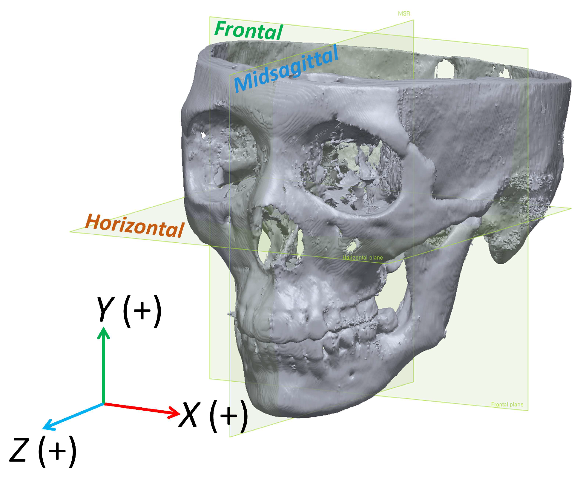
Figure 1. A 3-dimensional coordinate system (x, y, z) for measurements. x-axis, medial (+) and lateral (−); y-axis, superior (+) and inferior (−); z-axis, anterior (+) and posterior (−) directions.
Applsci 14 09299 g001
Figure 2. Movements of the right condyle during treatment. (A) Surgical movement; (B) postsurgical movement. Gray shows pretreatment, yellow shows postsurgery, and pink shows post-treatment condyles. During surgery, the right condyle rotated medially with lateral–inferior movement (3-dimensional red arrows) and then partially returned to its original position during postsurgical orthodontic treatment (3-dimensional blue arrows). Statistically significant, * p < 0.05; ** p < 0.01; *** p < 0.001.
Applsci 14 09299 g002
Figure 3. Scatter plot for z- and y-coordinates of the mandibular rotation’s center of rotation (CRot) during postsurgical orthodontic treatment. (A) Proximal segment; (B) distal segment. The medial pole of the right condyle was set as the origin point (0, 0). The five red dots located anteroinferior to the chin indicate the clockwise rotation of the distal segment following surgery. All other dots indicate counterclockwise rotation. The regression equation between the z- and y-coordinates of CRot of the distal segment was y = −1.45 × z + 22.7.
Applsci 14 09299 g003
Figure 4. Illustration of the average positions of CRot and the amount of rotational relapse of the proximal and distal segments of the mandible during postsurgical orthodontic treatment. (A) Proximal segment; (B) distal segment. The relapse of the distal segment is the same as the relapse of the chin, and this is the result of the relapse of the proximal segment and the change in the relationship between the proximal and distal segments.
Applsci 14 09299 g004
Figure 5. Scatter plot and regression line between the vertical bony step (VBS) developed during surgery and various postsurgical changes. (A) VBS (T1–T0) and the y- and z-coordinates of CRot of the distal segment rotation during postsurgical orthodontic treatment; (B) VBS (T1–T0) and the postsurgical changes of Pog-Z and Me-Y.
Applsci 14 09299 g005
Figure 6. Illustration of surgical simulation considering relapse. (A) Postmandibular osteotomy with the mandible’s proximal (green) and distal (blue) segments split. (B) To account for the expected relapse, the surgical occlusion was adjusted to create a Class II open bite. A 16.3 mm mandibular setback (red arrow) created 9 mm of VBS (yellow arrow). The overlap between the proximal and distal segments (dark green) indicates the area to be removed during surgery. (C) Simulation of the relapse during postsurgical orthodontic treatment. The center of rotation (CRot) of the distal segment was calculated based on the VBS, located 43.2 mm above and 10.6 mm behind the medial pole of the right condyle (red dot). The distal segment of the mandible (yellow) was then rotated counterclockwise until the VBS measurement reached 2 mm (yellow), a position that is typically maintained after treatment. The proximal segment (purple) was rotated around the medial pole of the condyle. (D) Superimposition of the post-treatment skull (pink) with the relapse simulation model (yellow and purple) demonstrates that the relapse simulation was accurate in this case. Both maxillary and mandibular incisors were retracted during postsurgical orthodontic treatment.
Applsci 14 09299 g006
Table 1. Reference planes, landmarks, and measurements.
VariablesDescription
Reference planes
 Frankfort horizontal (FH) planeConstructed by orbitale on the right side and porion on both sides.
 Horizontal planeConstructed by medial pole of right condyle and parallel to FH plane.
 Frontal planeConstructed by porion on both sides and perpendicular to horizontal plane.
 Midsagittal planeConstructed by nasion, basion, and ANS, and perpendicular to horizontal and frontal plane.
Landmarks and measurements
 Menton (Me)The most inferior midpoint on the symphysis.
 Me-YMeasured the vertical change in menton along the y-axis.
 Pogonion (Pog)The most anterior midpoint on the symphysis.
 Pog-ZMeasured the anteroposterior change in pogonion along the z-axis.
 Vertical bony step (VBS)The vertical distance between the proximal segment and distal segment along a line between the first and second molars and 95° to the molar occlusal plane which is constructed by the mesiobuccal cusp tip of the mandibular first molar and the distobuccal cusp tip of the mandibular second molar.
 Center of rotation (CRot)A specific point or axis around which the jaw or certain bony segments are rotating during surgical or orthodontic procedures.
Table 2. Analysis of measurement error 1.
VariablesT1–T0T2–T1
Proximal segment (PS)
     PS rotation (°)0.30.4
Distal segment (DS)
     DS rotation (°) 0.5
     VBS (mm)0.20.3
     Pog-Z (mm)0.20.1
     Me-Y (mm)0.10.1
T1, within one month of surgery; T0, before treatment; T2, after completion of postsurgical orthodontic treatment; PS, proximal segment; DS, distal segment; VBS, vertical bony step; Pog-Z, change of z-coordinate of pogonion; Me-Y, change of y-coordinate of menton. 1 Dahlberg formula was used: S = √(∑d^2/2n) (d, the difference between remeasured values; n, the number of double measurements).
Table 3. Positional changes of condyle with regard to surgical change (T1–T0), postsurgical change (T2–T1), and total change (T2–T0) 1−3,†,*.
Movement DirectionSurgical
Change
p ValuePostsurgical
Change
p ValueTotal Changep Value
T1–T0T2–T1T2–T0
Medial poleMediolateral (X) 1−0.8 ± 0.9<0.001 * +0.4 ± 0.60.001 *−0.4 ± 0.90.027 *
Superoinferior (Y) 2−0.5 ± 0.80.003 *,†+0.2 ± 0.90.279 −0.3 ± 1.00.067
Anterorposterior (Z) 3−0.9 ± 0.7<0.001 * +0.3 ± 0.80.041 *−0.6 ± 0.8<0.001 *
Lateral poleMediolateral (X) 1−0.1 ± 0.70.582 +0.3 ± 0.60.028 *+0.2 ± 0.70.175
Superoinferior (Y) 2−0.8 ± 0.6<0.001 * +0.4 ± 0.80.013 *−0.4 ± 0.70.004 *
Anterorposterior (Z) 3+0.8 ± 0.9<0.001 * −0.0 ± 1.10.941 +0.8 ± 1.1<0.001 *,†
Values are presented as mean ± standard deviation. Significance was determined using paired t-test. The Wilcoxon signed-rank test results are presented when normality was not satisfied (). 1 X indicates medial–lateral direction; +, denotes medial displacement; −, denotes lateral displacement. 2 Y indicates superoinferior direction; +, denotes upward displacement; −, denotes downward displacement. 3 Z indicates anteroposterior direction; +, denotes anterior displacement; −, denotes posterior displacement. * Statistically significant, p < 0.05.
Table 4. Positional changes in proximal and distal segments of mandible †,*.
VariablesT1–T0p ValueT2–T1p Value
Proximal Segment (PS)PS rotation (°)2.1 ± 2.9<0.001 *−2.4 ± 2.1<0.001 *,†
Distal Segment (DS)DS rotation (°) −2.0 ± 2.3<0.001 *
VBS (mm)−6.2 ± 2.9<0.001 * 4.4 ± 1.8<0.001 *
Pog-Z (mm)13.6 ± 5.3<0.001 * 4.0 ± 1.9<0.001 *
Me-Y (mm)−0.8 ± 1.90.015 *,† 2.3 ± 1.2<0.001 *
Values are mean ± standard deviation. Significance was determined using paired t-test. The Wilcoxon signed-rank test results are presented when normality was not satisfied (). * Statistically significant, p < 0.05.
Table 5. Correlations between VBS development and postsurgical changes †,*.
VBS (T1–T0)
Variables (T2–T1)r (ρ)p Value
Proximal segment
PS rotation (°)0.0240.897
Distal segment
DS rotation (°)0.2430.181
Crot-Y−0.465 0.010 *
Crot-Z0.4330.017 *
Pog-Z (mm)−0.623 0.000 *
Me-Y (mm)−0.389 0.034 *
r (ρ), Pearson correlation coefficient. Significance was determined using Pearson’s correlation test. Spearman’s correlation results are presented when normality was not satisfied (). * Statistically significant, p < 0.05.
Table 6. Comparison of postsurgical changes between single-jaw and double-jaw surgeries *.
Type of Surgery
1-Jaw Surgery2-Jaw Surgery
Variables (T2–T1)MeanSDMeanSDp Value
PS rotation (°)−2.591.84−2.312.520.731
DS rotation (°)−2.152.10−1.922.610.792
Pog-Z (mm)4.231.563.692.200.445
Me-Y (mm)2.310.782.201.520.799
VBS (mm)4.411.544.312.190.886
Independent t-test was performed. * Statistically significant, p < 0.05.
Disclaimer/Publisher’s Note: The statements, opinions and data contained in all publications are solely those of the individual author(s) and contributor(s) and not of MDPI and/or the editor(s). MDPI and/or the editor(s) disclaim responsibility for any injury to people or property resulting from any ideas, methods, instructions or products referred to in the content.