Review

Balance of Antioxidants vs. Oxidants in Perinatal Asphyxia

by
Dimitrios Rallis
1,
Niki Dermitzaki
1,
Maria Baltogianni
1,
Konstantina Kapetaniou
2 and
Vasileios Giapros
1,*
1
Neonatal Intensive Care Unit, School of Medicine, University of Ioannina, 45110 Ioannina, Greece
2
Department of Pediatrics, School of Medicine, University of Ioannina, 45110 Ioannina, Greece
*
Author to whom correspondence should be addressed.
Appl. Sci. 2024, 14(21), 9651; https://doi.org/10.3390/app14219651
Submission received: 19 September 2024 / Revised: 16 October 2024 / Accepted: 21 October 2024 / Published: 22 October 2024

Abstract

:
Perinatal asphyxia refers to an acute event of cerebral ischemia and hypoxia during the perinatal period, leading to various degrees of brain injury. The mechanisms involved in perinatal asphyxia include the production of reactive oxygen species (ROS), accumulation of intracellular calcium, lipid peroxidation, excitatory amino acid receptor overactivation, energy failure, and caspase-mediated cell death. Both primary and secondary neuronal damage are caused by the overproduction of ROS following a hypoxic/ischemic event. ROS can react with nearly any type of molecule, including lipids, proteins, polysaccharides, and DNA. Neonates who suffer from perinatal asphyxia are prone to oxidative stress, which is characterized by a disruption in the oxidant/antioxidant balance, favoring oxidants over the intracellular and extracellular antioxidant scavenging mechanisms. Current research has focused on developing treatment strategies that potentially improve the endogenous antioxidant neuroprotective mechanisms or minimize injury resulting from hypoxia/ischemia. In this narrative review, we aim to present evidence regarding the contribution of oxidant/antioxidant balance to the pathogenesis and progression of perinatal asphyxia. Also, we aim to explore the role of potential antioxidant therapies as promising treatment strategies for perinatal asphyxia, especially as an adjunct to therapeutic hypothermia in infants with perinatal asphyxia. The current literature on antioxidant treatments in newborns is limited; however, allopurinol, melatonin, and erythropoietin have shown some positive effects in clinical trials. Inhibitors of nitric oxide synthase, N-acetylcysteine, and docosahexaenoic acid have shown promising neuroprotective effects in preclinical studies. Finally, nanotherapeutics could potentially modulate oxidative stress in hypoxemic/ischemic brain injury by targeted medication delivery. Future research on neuroprotectants and their processes is warranted to develop innovative treatments for hypoxia/ischemia in clinical practice.

1. Introduction

Neonatal encephalopathy (NE) predominantly occurs during the perinatal period as a result of a sentient event that affects cerebral blood flow and oxygen supply and leads to impaired brain function. The prevalence of NE varies significantly; in developed countries, the prevalence is estimated to 1 to 8 per 1000 live births, while in underdeveloped regions it is much higher, at 26 per 1000 live births [1]. Although therapeutic hypothermia has been established as a standard therapy, NE continues to be a significant contributor to neonatal mortality and developmental disorders among term infants globally [2].
The transition from fetal to neonatal life introduces physiological oxidative stress, which stimulates the body’s antioxidant defense mechanisms. Birth represents a significant challenge to newborns, marked by a rapid adjustment from the relatively low-oxygen intrauterine environment to the more oxygen-rich external environment. This transition exposes newborns to reactive free radicals (RFRs), produced more intensely in response to the newborn’s less efficient natural antioxidant systems [3]. Evidence suggests that an imbalance between antioxidant and oxidant systems can result in oxidative damage, further highlighting the complexity of oxidative metabolism. Newborns, especially those born prematurely, have an immature antioxidant defense system (ADS) and thus, they are particularly vulnerable to oxidative stress and oxidative damage from RFRs [4]. Perinatal hypoxic/ischemic damage initiates a series of detrimental biochemical processes, including elevated neurotransmitter levels, overproduction of oxygen free radicals, heightened intracellular calcium concentrations, and activation of inflammatory mediators and signaling pathways, all of which contribute to tissue damage [5].
Previous reviews of the antioxidant/oxidant balance in asphyxiated neonates have focused on the biochemical basis of oxidative metabolism and the pathogenesis of NE [6,7,8], while several biomarkers have been identified with a potential role in the diagnosis and prognosis of injury in NE [8,9,10]. Moreover, the currently available treatments and adjuvant neuroprotective approaches based on antioxidant therapies have not been extensively discussed [6,7,8,9,11]. As research in NE is still evolving, clinical evidence and preclinical experimental data emerge, offering potential novel adjuvant therapeutic options that may improve the outcomes of hypothermia. Furthermore, limited data have been presented about novel techniques that could potentially modulate the oxidative stress in hypoxemic/ischemic brain injury by targeted delivery of medication, such as nanotherapeutics.
Hence, this narrative review seeks to offer a comprehensive elucidation of the pathophysiology of antioxidant/oxidant balance and the significance of free radicals and oxidative biomarkers in the context of NE, as well as to explore potential novel adjuvant therapies that could mitigate RFR production and enhance the outcomes of therapeutic hypothermia.

2. Materials and Methods

The Literature Search Strategy
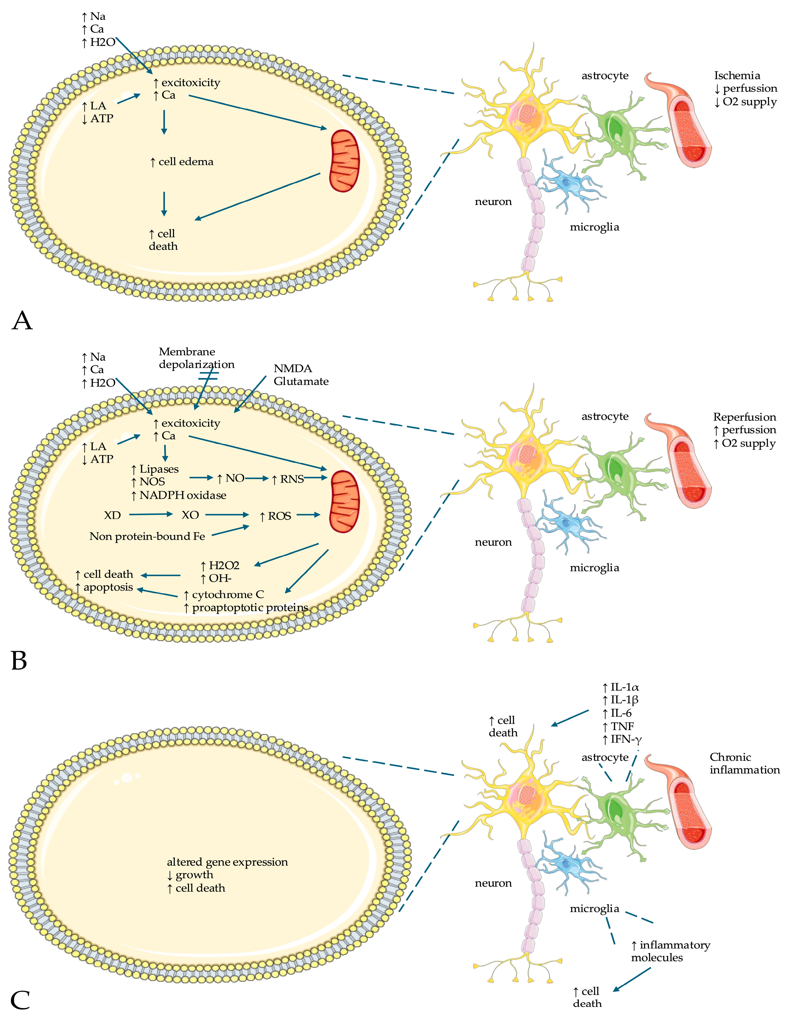
A literature search was conducted in August 2024 in PubMed. Only English-language articles with full text available were considered. The terms ‘oxidate stress’, ‘oxidants’, ‘antioxidants’, ‘free radicals’ and ‘perinatal asphyxia’, ‘hypoxic-ischemic encephalopathy’, ‘neonatal encephalopathy’ were utilized. The search strategy is presented in Table 1.
Our review is organized as follows: (1) the basics of the pathophysiology of oxidative stress in NE, (2) evidence from animal models of hypoxia/ischemia, (3) evidence of free radical biomarkers in NE, (4) a clinical assessment of neonates with encephalopathy, and (5) a summary of the latest research in the treatment options in NE (Figure 1).

3. Pathophysiology

3.1. Aerobic Metabolism and Oxidative Phosphorylation

Human beings utilize aerobic metabolism and oxidative phosphorylation to generate energy. These processes are executed within the mitochondria and require oxygen. This metabolic pathway releases electrons, which are transferred to the electron transport chain via nicotinamide adenine dinucleotide (NADH) and flavin adenine dinucleotide (FADH2). The electron transport chain consists of the enzyme complexes I, III, and IV, which create membrane potential by transferring protons (H+) from the inner to the outer membrane. Consequently, protons flow back into the matrix due to the membrane potential, thus releasing energy that is stored in the form of adenosine triphosphate (ATP). Oxygen is crucial in this pathway for accepting these electrons, thereby protecting mitochondria from damage [12,13].
The central nervous system is known as the “oxygen regulator” because it requires substantial energy to sustain membrane potentials. Consequently, the central nervous system relies on aerobic metabolism which, compared to anaerobic metabolism, provides more ATP molecules [14]. Within the central nervous system, ion pumps that facilitate action potentials are ATP-dependent and thus require a significant amount of energy. Nevertheless, the brain is unable to store energy in forms like glycogen or phosphocreatine that could be quickly released. Therefore, in circumstances of low oxygen, glucose energy reserves are depleted rapidly and cells die within minutes [15,16].

3.2. Biological Antioxidant Systems

Oxidative stress represents a metabolic imbalance caused by an excess of free radical production or a reduced capacity of the antioxidant defense system (ADS) [3]. While free radicals are necessary for cell communication and the proper operation of biochemical pathways, an extreme imbalance of the ADS and free radicals can damage tissues and lead to acute or chronic illnesses [17]. The ADS is developed later in pregnancy to prepare the fetus for oxygenation after birth and encompasses both enzymatic and non-enzymatic components. Antioxidant enzymes neutralize reactive species by undergoing chemical reactions. Non-enzymatic antioxidant systems consist of proteins that sequester metals (including ferritin, transferrin, and ceruloplasmin), vitamins that prevent the oxidation of lipids (like A, C, D, and E), and small molecules that diminish reactive species.
Sufficient levels of vitamin A in the tissues could provide a defense against infections and the harmful effects of oxygen free radicals [18]. A previous study found significantly lower levels of vitamin A in neonates with reduced antioxidant defenses, as indicated by their increased need for delivery room resuscitation compared to controls [18]. In another study, low vitamin A levels during the first month of birth were found to greatly increase the likelihood of developing long-term respiratory impairment [19]. The antioxidant characteristics of vitamin C include the production of ascorbyl radicals, a terminal reactive oxygen species (ROS) with a relatively minimal harmful effect on biological systems. Although vitamin C is mainly considered an antioxidant, under specific circumstances, vitamin C may show prooxidant qualities in the presence of free (unbound) redox metals, such as iron or copper [20]. Numerous antioxidant processes are linked to vitamin D; when the condition of vitamin D and oxidation was assessed in neonates with NE, vitamin D levels were found to be lower than normal [21]. Additionally, the administration of vitamin D significantly reduced the malondialdehyde (MDA) content generated by hypoxemia/ischemia and elevated glutathione (GSH) and SOD (superoxide dismutase) activity, both of which were suppressed by hypoxemia/ischemia. Among numerous mechanisms of cellular injury that have been linked to the creation of ROS [22], it has been shown that calcitriol, an active form of vitamin D, suppresses ROS formation by interfering with the hydrogen peroxide pathway [23]. Vitamin E protects cellular membranes from ROS-caused lipid peroxidation, with the hydroxyl group on its six-membered ring situated outside of the membrane and scavenging ROS in the aqueous environment.
Furthermore, there has been great interest in the content of trace elements and their potential role as key regulators of antioxidant defense and oxidative stress. Copper is a cofactor in numerous detoxification enzymes and proteins, including SODs, ceruloplasmin, and copper–thioneine [24]. In preterm newborns, a decrease in SOD activity was linked to low copper levels [25]. Regarding ferrum, studies have shown that children with ferrum-deficiency anemia exhibit a decrease in antioxidant activities, such as that of catalase [26]. Zinc is essential for cell differentiation, metabolism, and growth, while promoting antioxidant enzyme activities like Zinc–SOD and Zinc–thioneine, hormone structures, and brain development [24]. Moreover, the importance of selenium for the best possible operation of the body’s natural antioxidant defense mechanisms is becoming more widely recognized [27]. The “selenoenzymes” glutathione peroxidases (GPx), thioredoxin reductases, and selenoprotein P are produced only when selenium is present [28,29]. Through selenoproteins, selenium is linked to the regulation of ROS levels and redox balance in nearly every tissue. Due to its significant antioxidant effect, low selenium levels have been linked to impaired oxidative system defenses [30]. Evidence suggests that supplementing with selenium may lessen pregnancy-related oxidative stress [31]. Overall, adequate selenium levels potentially strengthen endogenous antioxidant defenses to minimize the consequences of oxidant stress, even if a lack of selenium is unlikely to entirely account for free radical injury in neonates [32]. Mercury exists in three different forms, with a unique toxicological profile of each: elemental mercury can damage the nervous system and kidneys; organic mercury compounds, such as methylmercury, can cross the blood–brain barrier; and inorganic mercury salts can damage the kidneys and nervous system [33].
Moreover, GSH can combine with another GSH molecule to form oxidized glutathione (GSSG), which then donates two electrons to free radicals, aiding in their neutralization. The enzyme GSH reductase facilitates the conversion of GSSG back to GSH. SODs are important antioxidant enzymes that convert superoxides to hydrogen peroxide. Catalases and GPx catalyze the conversion of hydrogen peroxide to water and oxygen. The haem complex is broken down and converted to biliverdin by haem oxygenases, glutaredoxins, thioredoxins, peroxiredoxins, and other components of the ADS in red blood cells [13,17].

3.3. Hypoxia/Ischemia Reperfusion Injury

In conditions of ischemia and decreased oxygen supply followed by reperfusion, free radicals containing oxygen (ROS) or nitrogen (reactive nitrogen species, RNS) can be generated. Free radicals are extremely reactive and unstable due to an unpaired electron that they contain in their external membrane [34]. When the number of RFRs surpasses the capacity of the body’s endogenous ADS to neutralize them, it can lead to oxidative damage. This damage may affect DNA, polysaccharides, proteins, and lipids, compromising cellular function and potentially causing cell death through either apoptosis or necrosis [35]. The developing brain is characterized by a high oxygen requirement, a low antioxidant defense of polyunsaturated fatty acids which are the main component of phospholipids, a high concentration of metals that catalyze ROS production, and a large number of immature cells; thus, it is particularly vulnerable to ROS/RNS-induced injury [36].
The progression of NE can be segmented into three stages, though in actuality, these stages overlap, with events often occurring simultaneously (Figure 2). The initial stage lasts 6 h and is marked by a decrease in blood flow to the fetus, resulting in systemic and cerebral hypoperfusion. Cerebral hypoperfusion leads to brain ischemia, hypoxia, acidosis, and cellular damage due to energy depletion. During hypoxia/ischemia, the brain areas with the highest metabolic activity and numerous excitatory glutamatergic synapses are primarily affected [37]. A key factor in hypoxic/ischemic-related brain damage is the overstimulation of postsynaptic receptors due to the excitotoxicity of glutamate neurotransmission, leading to cell death [38]. During prolonged hypoxia/ischemia, neurons may face a depletion of high-energy metabolites, an increase in cytotoxic edema, and an extracellular accumulation of excitatory amino acids [39]. Some neurons may die immediately or shortly after the injury, while others might initially recover at different stages, eventually leading to cell death [40]. Cerebral hypoxia triggers anaerobic metabolism, which results in an intracellular increase in lactic acid, sodium, calcium, and water; a decrease in ATP levels; and the reuptake of neurotransmitters, leading to secondary excitotoxicity. Intracellular calcium triggers the release of lipases, nitric oxide synthase (NOS), and substances that promote cell death; enhances the production of free radicals; and causes direct damage to the mitochondria.
The second stage, which can last from 6 to 48 h, is characterized by ongoing excitotoxicity, a decrease in mitochondrial function and oxidative stress due to alterations in the membrane potential, a decrease in ATP production, and an alkaline environment despite sufficient oxygen supply. Immediately after a severe injury, there is a recovery of blood flow to the brain, which is vital for survival but paradoxically leads to reperfusion injury due to an overproduction of radicals [41]. This causes not just an initial burst of oxidative stress but also a gradual breakdown of the mitochondrial electron transport chain, a depletion in ATP levels, and further damage to brain cells [8]. During reperfusion, reactive molecules are mainly generated by the mitochondrial respiratory chain complex and the xanthine oxidase (XO) system. After reperfusion/reoxygenation, hypoxanthine is gradually increased, leading to the conversion of xanthine dehydrogenase (XD) to XO; in turn, XO is the main source of ROS by producing superoxide radicals using oxygen [8]. Additionally, the NADPH oxidase system and NOS uncoupling processes are also engaged, though their contributions are comparatively minor [12]. NOS and mitochondrial electron leak are identified as key factors in the production of ROS and RNS [42,43]. Moreover, N-methyl-D-aspartate (NMDA) and glutamate receptors trigger the production of nitric oxide (NO) through the activation of NOS. Peroxynitrite (ONOO−) and hydroxyl radicals (OH−) can be created when NO combines with superoxide. This leads to further cell damage through protein oxidation, lipid peroxidation, DNA damage, and mitochondrial inactivation [6]. Additionally, damage from overactive calcium channels can also result in the production of oxygen through NADPH oxidase system activity [44]. Additionally, non-protein-bound iron from hemoglobin increases the formation of hydroxyl radicals through the Fenton reaction, which significantly increases ROS levels [45]. These radicals and RNS exacerbate the damage to the mitochondria that started with the initial decrease in ATP levels and the overstimulation of glutamate receptors, leading to a further breakdown of oxidative phosphorylation, which results in a worsening of the energy crisis and the death of neurons [46]. Several studies have indicated that an increase in ROS can promote the release of cytochrome C from the mitochondria, which catalyzes the oxidation of cardiolipin [47,48]. Subsequently, pro-apoptotic proteins increase in the cytoplasm and then bind to Apaf-1, forming the Apaf-1/caspase-9/Sitka complex, which in turn inactivates caspase-3, resulting in apoptosis and the death of neurons [49].
The third stage, which can continue for several days or even weeks, is marked by inflammation and altered gene expression that result in defects in the formation of new neurons, the development of synapses, and the growth of axons [50,51]. Oxidative stress interacts with the immune system, creating a “vulnerability period” for NE. Initially, oxidative stress is triggered by immune cells at the site of residence, such as microglia. These microglia are highly responsive to hypoxia/ischemia and are activated by an increase in NO, glutamate, and ROS, leading to the production of a large number of inflammatory molecules [52]. Astrocytes are also activated, producing interleukins (IL-1α, IL-1β, IL-6), tumor necrosis factor-alpha, and interferon-gamma, which hinder the growth of new neurons, directly damage immune cells, and cause neuronal cell death [53].

4. Animal Models of Neonatal Hypoxia/Ischemia

The most commonly employed model for neonatal hypoxic/ischemic injury in animals is the model established by Rice-Vannucci [54]. Although initially developed for rats [54], this model has also been applied to other rodents [55,56,57]. In this model, after blocking one carotid artery, the piglets are exposed to a period of hypoxia, reproducing the injury of stroke of the middle cerebral artery, which is rare in preterm neonates [58]. Other methods for inducing brain injury from hypoxia/ischemia in piglets include the combination of hypoxia and hypotension-induced ischemia [59], cardiac arrest followed by cardiopulmonary resuscitation [60], and bilateral common carotid artery occlusion in conjunction with hypoxia [61].
In previous studies, it was shown that six hours after the asphyxic event, post-asphyxic hypothermia significantly reduced the formation of protein carbonyls in the parietal cortex and the striatum of newborn piglets [62,63]. On the other hand, when comparing hypothermic versus normothermic piglets 48 h after the hypoxic insult, no significant changes in cerebral protein carbonyl levels were found [64]. This evidence indicated that the role of oxidative stress in brain damage was crucial during the initial hours after the hypoxic event, which then normalized over time. Nonetheless, newborn piglets exposed to hypoxemia followed by 29 h of hypothermia had higher levels of carbonylated proteins compared to those that remained normothermic. Similar findings were also recorded in the cerebral white matter of newborn piglets subjected to cardiac arrest followed by hypothermia of limited duration [65]. Additionally, significantly lower levels of antioxidant enzymes like SOD, GPx, or catalase activities were found in the cerebral tissue of newborn rats exposed to asphyxia [66]. Of note, there was a reduction in antioxidant enzyme activities in newborn rats that were asphyxiated, both in normoxic and hypoxic conditions; however, the activities of these enzymes were lower in asphyxiated rats compared to controls [66].
Following an ischemic event, mild hypothermia delayed the utilization of SOD, GPx, and GSH in the brain tissue [67]. In newborn rats exposed to hypoxemia/ischemia followed by hypothermia, a notable increase in antioxidant protection was noted, characterized by higher levels of SOD and GPx expression [68]. Hypothermia resulted in an increase in the expression of SOD-1 in the neocortex and the caudate putamen, although the expression of GPx-1 increased only in the caudate putamen but not in the neocortex [69].
Finally, the expression of DNA-repair genes (DNA glycosylases OGG1, NEIL1, and NEIL3) was reduced in hypothermic pigs compared to controls or resuscitated pigs [70]. Decreased expression of these genes was primarily detected in the hippocampus but also included the cortex, cerebellum, and liver [70].

5. Free Radical Biomarkers in NE

The effectiveness of a biomarker in NE is affected by the timing of the brain insult; however, the period following birth does not necessarily equate to the period following the insult [71]. Regrettably, the timing of the insult, whether it occurs before or during birth, is often not known. Even the time between fetal death and delivery can vary significantly, with cell death occurring at different times after the insult [72]. This lack of precise timing complicates the categorization of cerebral injuries as primary (acute energy failure), secondary (following a second energy failure and subsequent cell death), or tertiary (increased sensitivity to subsequent cell death) [73]. Currently, with more advanced techniques for detecting ROS and RNS, it may be possible to enhance free radical biomarkers and more accurately detect the timing of the insult (Table 2).

5.1. Enzymes Against Free Radicals

XO is an enzyme containing molybdenum that produces free radicals in the presence of purines hypoxanthine and xanthine. XO increases with hypoxia/ischemia, leading to the breakdown of adenine monophosphate into these purines [10]. The main source of XO is likely from endothelial cells rather than the brain tissue itself, and the term XO refers to two forms, xanthine oxidoreductase, which is present in healthy cells, and xanthine dehydrogenase, which is converted to an oxygen radical-producing form during ischemia. Although XO is barely detectable in the adult human brain [74], studies have shown that newborns have low levels of both total xanthine dehydrogenase and XO [75].
SOD, GPx, and catalase are the initial line of defense against oxidative stress. After experiencing hypoxia/ischemia, their activity sharply increases to counteract the rise in ROS and their detrimental effects. Research has shown that asphyxiated newborns have significantly higher levels of SOD, catalase, and GPx in their cord blood compared to non-asphyxiated newborns [76]. However, by 24 h, only catalase and SOD levels remain elevated in the blood of infants who have experienced NE compared to those who have not [21]. Studies have also found a correlation between the levels of catalase and SOD in the cord blood and the severity of neonatal asphyxia [76]. Therefore, the measurement of these antioxidant enzymes in the cord blood could provide valuable insights into the timing and severity of neonatal asphyxia [76]. Of note, these findings are based on small groups of participants, so further research is needed to confirm these results in larger studies. Regarding the levels of these antioxidant enzymes in other body fluids, a previous study has shown that the activity of SOD was significantly higher in the cerebrospinal fluid up to 72 h after birth in term newborns who have experienced neonatal asphyxia, whereas the activities of GPx and catalase increased only in the most severely affected cases [77]. Most of the research on these topics was conducted before the standard use of therapeutic hypothermia, which raises questions about how hypothermia might affect the levels of these antioxidant enzymes.

5.2. Nitric Oxide

The upregulation of NOS during ischemia leads to increased production of NO and RNS. Studies have shown that newborns with neonatal asphyxia have higher levels of NO in their blood and cerebrospinal fluid, in addition to a higher ratio of nitrate to nitrite in their blood, which is a measure of NO levels [78,79]. Evidence suggested that higher NO levels were found in infants with early signs of cerebral injury compared to those without brain damage; thus, the increase in NO was related to the severity of neonatal asphyxia [78].

5.3. Perfusion-Assisted Breathing

The perfusion-assisted breathing (PAB) assay has been utilized in adults to evaluate the oxidative status in various conditions associated with oxidative stress, including diabetes mellitus, acute coronary syndrome, and stroke [80,81,82]. In neonates, it was shown that PAB, when used together with the severity of hypoxic/ischemic encephalopathy, could more accurately predict the outcome and the risk of developing neurological issues in term newborns who have suffered from asphyxia [83]. The study indicated that PAB, the fifth-minute Apgar score, and the pH level in the first hour after birth were crucial factors in determining the outcome for neonates who had experienced asphyxia. Additionally, the measurement of PAB in the blood could be a useful tool in assessing the prognosis of asphyxia during birth [83].

5.4. Markers of Lipid Damage (Malondialdehyde and 4-Hydroxynonenal)

The amount of damage to lipids during periods of hypoxemia/ischemia can exceed the body’s ability to self-repair, leading to negative effects on the structure and function of cell membranes, loss of function, and even cell death. Once this damage begins, several products of lipid damage are released, many of which can be detected in the brain due to its high fat content.
Malondialdehyde (MDA), which is a sign of damage to n-3 and n-6 fatty acids, has been extensively studied because it is easy to measure using common spectrophotometry. Evidence is consistent that serum MDA levels are higher in severely asphyxiated compared to non-asphyxiated newborns [21,79,81,83,84]. As for predicting outcomes, higher MDA levels in the blood at 24 h are linked to more severe NE [21], with the highest levels found in infants who died or had long-term neurological problems [85,86]. A potential issue with using MDA in the blood is that it might not be specific to the brain, as it could also reflect damage to other tissues due to widespread organ damage. Using MDA in the cerebrospinal fluid might provide a better idea of the damage to the brain in asphyxiated infants. Studies have shown that MDA levels in the cerebrospinal fluid are higher in asphyxiated compared to non-asphyxiated term infants, with a stronger link to the severity of NE [76]. This is also seen in infants who have died or have long-term neurological issues compared to those who have recovered normally [86]. However, more research is needed to confirm that MDA in the cerebrospinal fluid is a reliable indicator of brain damage in asphyxiated infants. Regarding 4-hydroxynonenal (4-HNE), a byproduct of damage to n-6 fatty acids, it was found that 4-HNE levels in the blood increased following a period of hypoxia [87]. However, evidence in this biomarker is limited because 4-HNE is difficult to measure and requires advanced liquid chromatography techniques as it is unstable [88].

5.5. Peroxidation-like Compounds

Prostaglandin-like compounds, including isoprostanes, neuroprostanes, and neurofurans, are formed from the free radical-induced breakdown of arachidonic acid and docosahexaenoic acid (DHA), which are types of polyunsaturated fatty acids. F2-isoprostanes appear to be the most reliable indicators of the extent of oxidative stress following hypoxia–reperfusion events [89], and they can be measured accurately using ultra-high-performance liquid chromatography–mass spectrometry techniques, even in newborns. Elevated levels of 8-iso-15(R)-PGF2α and total isoprostanes were found in infants with acidosis and depression compared to those without these conditions; 8-iso-15(R)-PGF2α levels were correlated with the severity of asphyxia [90]. However, a study that longitudinally measured serum F2-isoprostanes within the first 5 days after birth in neonates who experienced severe, moderate, or mild asphyxia found no differences between the groups and no correlation with brain damage detected through neuroimaging [91]. This limited and conflicting evidence suggests that more research is needed to determine if prostaglandin-like compounds can serve as biomarkers following perinatal asphyxia.

5.6. Protein Oxidation Markers

Protein carbonyls and products of advanced protein oxidation are formed through protein carbonylation, nitration, crosslinking, or the loss of thiol groups by free radicals. Elevated levels of protein carbonyls were found in asphyxiated term newborns at birth and 48 h after birth [85]. Higher levels of protein carbonyls were seen in neonates who developed seizures; however, there was no difference in the Sarnat stage or neurodevelopmental outcome at 9 months of age [85]. Moreover, higher levels of advanced-oxidation protein products were found in asphyxiated compared to non-asphyxiated preterm infants, with a positive correlation between this marker and plasma hydroperoxides [92]. It has been reported that higher plasma advanced-oxidation protein product levels at 4–6 days after birth have been seen in term infants with severe NE, who required therapeutic hypothermia, compared to those with milder or moderate NE [91]. However, subsequent measurements in hypothermic asphyxiated infants and controls showed no difference in advanced-oxidation protein product levels between the two groups [21]. Given this lack of difference in advanced-oxidation protein product levels during and after hypothermia, a possible role for hypothermia in regulating protein oxidation, which warrants further investigation, is suggested. Additionally, a significant independent association between magnetic resonance imaging (MRI) scores indicating brain injury and blood levels of advanced-oxidation protein products over the first 5 days of life in neonates with NE has also been reported, with a stronger correlation seen in male infants, indicating a potential role for advanced-oxidation protein products as a biomarker for cerebral oxidative injury [91].

5.7. Uric Acid

Uric acid is crucial in the development of oxidative damage in the brain during reperfusion/reoxygenation. Therefore, the amount of uric acid excreted through urine has been suggested as a simple and non-invasive way to measure the production of ROS related to oxygen deprivation in newborns. Several studies have consistently found that the ratio of uric acid to creatinine in the urine increases within 48–72 h of birth in both term and preterm newborns who have experienced neonatal asphyxia [84,93]. Additionally, there is a positive correlation between this ratio and the severity of neonatal asphyxia, as indicated by the Sarnat stage [84].

5.8. Iron Not Bound by Proteins

After experiencing hypoxemia followed by reoxygenation, the iron that was previously bound to hemoglobin interacts with O2− and H2O2, forming highly reactive OH. Prior research showed that as the severity of asphyxia increased, so did the levels of iron not bound by proteins in the blood [94]. However, it was also noted that even infants who were moderately or severely asphyxiated had low or undetectable levels of this iron. Nonetheless, the presence of low or undetectable levels of this iron was not linked to any neurological problems at one year of age, even though the study only included a small number of infants [94].

6. Clinical Assessment of Neonates with Encephalopathy

Several scoring methods can be used to evaluate NE and predict the outcomes with clinical examination. There is a clear correlation between neuro-developmental outcomes and the Sarnat staging of encephalopathy at less than six hours of life [95]. However, a multicenter prospective analysis of newborns with hypothermia and NE revealed that a clinical neurological exam performed before six hours of life and showing moderate to severe encephalopathy was insufficient to predict unfavorable outcomes [96]. Studies on hypoxemic/ischemic newborns treated with hypothermia have also demonstrated that the Thompson score is a useful prognostic tool for predicting outcomes at 4 to 5 years of age [97]. To appropriately predict outcomes in infants with NE, clinical indicators alone are not sufficient unless clinical examination is combined with multiorgan dysfunction and Apgar score.
Regarding laboratory findings, it has been previously demonstrated that poor outcomes were linked to low initial newborn arterial pH (umbilical or arterial, within the first hour after birth). According to a meta-analysis, poor newborn outcomes were significantly correlated with low initial arterial pH in encephalopathic neonates prior to the hypothermia era [98]. Also, a lower initial pH was linked to more severe brain injury on MRI in a cohort of hypoxic/ischemic infants treated with therapeutic hypothermia [99]. Moreover, in a follow-up to the NICHD randomized trial of therapeutic hypothermia, early hypocarbia was linked to death or worse outcomes at 18 to 22 months of age [100]. Also, it was shown that worse 2-year outcomes were related to substantial variability in PCO2 during hypothermia [101]. Additionally, a longer time for serum lactate to normalize was correlated with the severity of encephalopathy and the manifestation of seizures in neonates with NE [102]. Finally, hypo- and hyperglycemia in the early postnatal period have also been correlated with neurologic adverse outcomes [103].
Neuroimaging remains the cornerstone in the evaluation of asphyxiated newborns. In at-risk groups, electroencephalography (EEG) and amplitude-integrated EEG (aEEG) enable real-time evaluation of brain development and function in addition to seizure detection. The predictive accuracy of aEEG is time-dependent, such that a burst-suppression pattern persisting beyond 36 to 48 h suggests a significant risk of an aberrant neurodevelopmental outcome [104]. Also, suppressed aEEG tracings longer than 48 h increase the likelihood of severe impairments in survivors, albeit they are not a valid early single predictor in hypothermic neonates [105]. When combined with an aberrant early MRI, a persistently abnormal 48 h EEG background pattern was highly indicative of an unfavorable prognosis [106]. Evoked potentials, including visual, brain-stem auditory, and somatosensory, are electrical reactions to sensory input. In term newborns with NE, delayed or absent responses on the somatosensory-evoked potentials during the first few weeks of life were associated with poorer neurological outcomes at 18 to 24 months of age [107]. Although evoked potentials are effective supplemental prognostic tools in severe cases, the lack of rigorous studies and consensus regarding their prognostic efficacy has limited their utilization in clinical practice. Near-infrared spectroscopy can be utilized for the noninvasive assessment and continuous bedside monitoring of cerebral hemodynamics and oxygenation. Previous evidence has shown that at six and twenty-four hours postpartum, cerebral blood volume and cerebral oxygenation levels were higher in neonates with aberrant MRI findings [108]. Also, persistent higher levels of cerebral oxygenation in hypothermic neonates with NE were correlated with poor neurological outcomes [109]. Finally, brain MRI is a standard procedure used for the diagnosis and prognosis of newborns with NE. MRI is also becoming a common procedure for assessing the type and extent of brain damage in newborns with NE. According to a previous meta-analysis, brain MRI during the newborn era has a high sensitivity but moderate specificity for predicting unfavorable neurodevelopmental outcomes at one year [110], whereas a normal brain MRI is correlated with normal neurological results and little chance of significant neurodevelopmental impairment [111].

7. Therapeutic Strategies

7.1. Hypothermia

The effectiveness of hypothermia in reducing brain damage [112] and lessening severe neurological effects [113] in newborns with moderate to severe NE has been well-documented and is now the standard treatment for term and early preterm neonates with NE [50]. The main advantages of hypothermia arise from decreasing brain metabolism, which lessens the damage from secondary brain energy failure [114]. Furthermore, hypothermia prevents brain cell death, reduces harmful effects from overstimulation, controls the activity of glial cells [115], and stimulates certain RNA molecules [116]. Additionally, hypothermia has been shown to directly decrease the levels of oxygen-based free radicals after hypoxia/ischemia and reperfusion, as seen in both in vivo [42] and in vitro studies [117] (Table 3).

7.2. Mitochondrial Therapy

There has been a growing interest in the application of small molecule inhibitors and activators aimed at mitochondria [118], such as tetra- and triphenylphosphonium, as well as the combination of various methods for delivering mitochondria-penetrating peptides [119]. Bioactive therapeutics and antioxidants, as an alternative to targeting mitochondria, are slowly becoming a viable approach for developing supplementary therapies. Metformin has been shown to limit mitochondrial respiration by directly suppressing mitochondrial complex I in the respiratory chain [120]. Metformin can also affect inflammatory T cells by blocking the utilization of 2-deoxyglucose in glycolysis, as well as reducing the production of IL-1β [121]. In animal studies with metabolic syndrome, treatment of the mother with metformin was effective in preventing fetal inflammation, which is a risk factor for neonatal hypoxic/ischemic encephalopathy [122]. Coenzyme Q10 (CoQ10) can decrease the production of ROS through the mitochondrial complex and research has shown that CoQ10 acts as a neuroprotective agent in various animal models, including rodent models of Alzheimer’s disease [123], middle cerebral artery occlusion [124], and traumatic brain injury [125]. However, the potential role of CoQ10 in neonatal hypoxia/ischemia has not yet been fully explored. Finally, mitoquinone, a ubiquinone derivative conjugated to triphenylphosphonium cation, is efficient in reducing superoxide production, lipid peroxidation, and subsequent ROS generation, and thus, mitoquinone could be used to treat structural mitochondrial damage [126].

7.3. Antioxidant Methods for Protecting the Brain from Injury Due to Low Oxygen Levels

The involvement of free radicals in the onset of hypoxia/ischemia brain damage, followed by reperfusion, has led to the investigation of antioxidant treatment methods for NE, with the goal of either enhancing the body’s natural ADS or reducing the production of ROS and RNS at various stages. Although the use of atmospheric air for the resuscitation of newborns has become a standard procedure in neonatal care, the application of other antioxidant substances is primarily confined to research environments, with only limited evidence from preclinical and/or clinical studies. Antioxidant treatments are possible approaches to treating perinatal asphyxia, in combination with therapeutic hypothermia for infants with moderate to severe NE [127].

7.4. Reduction in Free Radical Production Initiators and Scavengers

Allopurinol is a specific inhibitor of XO, competing with this enzyme to reduce the production of ROS during reperfusion [128,129]. Allopurinol, by blocking the activity of XO, also reduces the production of NO from nitrites, while acting as a chelator for non-protein-bound iron and a direct scavenger for free radicals [128,129]. These characteristics position it as a potential candidate in the treatment of hypoxic/ischemic encephalopathy. Maternal administration of allopurinol to fetuses experiencing hypoxia resulted in a decrease in the levels of the S-100B protein, an indicator of brain injury, in the infant’s cord blood [130]. Previous evidence has shown that allopurinol reduced oxidative stress and improved blood flow and brain activity in newborns without causing toxicity [131]. However, a later study found no benefits in newborns given allopurinol within the first four hours after birth compared to those given a placebo, likely due to the delay in its administration [132].
Erythropoietin, essential for brain development, is a promising agent in treating NE by increasing the expression of genes that prevent cell death, reducing inflammation, lowering levels of oxygen free radicals, decreasing the activation of caspases, lessening the effects of glutamate excitotoxicity, and enhancing signaling pathways [133,134]. Under conditions of hypoxia, the expression of erythropoietin receptors and the secretion of erythropoietin increase, making it a particularly effective option for treatment [135].
Melatonin, a powerful natural compound, has multiple beneficial effects including antioxidant, anti-inflammatory, and anti-apoptotic properties. It can easily cross the blood–brain barrier, making it an attractive option for treating hypoxic/ischemic encephalopathy [136,137].
N-acetylcysteine (NAC), a precursor of cysteine, possesses antioxidant effects, including the neutralization of ROS and the replenishment of glutathione in cells [138]. These attributes, along with its ability to dissolve in fat and its low toxicity [139], have positioned NAC as a promising candidate for neuroprotection in NE. NAC reduces the severity of hypoxic/ischemic damage in newborn rats [140] and can be used in conjunction with hypothermia to lessen brain damage [141,142]. Administering NAC to rats alongside hypothermia has been shown to enhance myelin production [141], improve brain function [141], and lead to better long-term motor outcomes [142].
DHA is known for its ability to neutralize free radicals and its role in reducing glutamate excitotoxicity and decreasing NO levels. Evidence of enhanced antioxidant enzyme activities in cultures of neurons enriched with DHA has suggested a potential neuroprotective function for DHA [143].
Edaravone acts as a neutralizer of free radicals and has been investigated for its multiple antioxidant effects across various experimental models, including those involving animal models of NE [144].
Phenobarbital administered within the first two hours of life in term newborns with perinatal asphyxia has been associated with lower levels of lipid peroxides and antioxidant enzymes in the cerebrospinal fluid [145].
Molecular hydrogen, a compound recently recognized for its antioxidant properties, has shown neuroprotective effects and maintains the reactivity of blood vessels in newborn pigs exposed to asphyxia [146].
Deferoxamine is an iron-binding antioxidant that crosses the blood–brain barrier and helps remove excess iron due to its capacity to bind free iron in the brain. Deferoxamine has been shown to lessen brain edema and injury in animal models of hypoxia/ischemia, lower the levels of excitatory amino acids, and improve the histological condition of the hippocampus in newborn rats following hypoxia/ischemia [127,147]. Following birth asphyxia, iron is released from ferritin, hemosiderin, or transferrin [148,149], leading to the production of free radicals [150]. Ferrous ions can also trigger the formation of MDA, which is a marker of lipid oxidation and oxidative damage to tissues [151]. By binding to iron, deferoxamine stops the creation of the harmful peroxynitrite anion and hydroxyl radicals and thus reduces the risk of iron-dependent free radical-induced damage, especially to polyunsaturated lipids [11].
SOD, an enzyme that neutralizes free radicals, could be beneficial in reducing brain damage; however, its use is restricted as SOD has limited ability to cross the blood–brain barrier. Encapsulation of SOD with biodegradable poly nanoparticles has been shown to effectively lower the volume of brain infarcts, edema, and the formation of ROS in an animal model of cerebral ischemia/reperfusion injury [152].

7.5. Nitric Oxide Inhibitors

NO can be both protective and harmful for the brain during periods of hypoxia/ischemia. The epithelial cells of blood vessels release NO that helps maintain the blood flow to the brain and protects the brain from ischemic injury. However, increased levels of NO can lead to overstimulation, cell edema, and cell death, in addition to injury in the blood vessel epithelial cells themselves [153]. The production of NO is controlled by neuronal, endothelial, and inducible NOS. These types of NOS are more active under conditions of hypoxia, leading to the suggestion that blocking NO production could be a way to protect the brain from injury [154].

7.6. Magnesium

Magnesium is a promising therapeutic agent for brain injury due to its ability to block NMDA receptors, which helps prevent harmful effects from excessive calcium or glutamate. Magnesium also has a significant role in helping reduce inflammation and damage from oxidative stress [46].

7.7. Stem Cells

Recent research indicates that bone marrow and umbilical cord blood stem cells might be beneficial in treating brain injury. Specifically, stem cells from umbilical cord blood offer protection against inflammation, cell death, and oxidative damage and control immune regulation. Certain types of cells, like endothelial progenitor cells and mesenchymal stem cells found in umbilical cord blood and tissue, are effective in reducing inflammation caused by brain damage. Mesenchymal stem cells have a strong ability to change the immune response, protecting against harmful immune reactions triggered by brain injury [155,156]. Administration of stem cells from umbilical cord blood can also lessen the damage to brain white matter by combining with other treatments for brain injury [49].

7.8. Nanomaterials

The application of nanotechnology or techniques based on nanocarriers is rapidly growing as a promising tool for the detection and treatment of cerebral illnesses [157,158]. Nanoparticles are carriers or delivery tools that enhance the bioavailability of therapeutics because of their enhanced solubility, reduced degradation by enzymes, decreased protein binding, and extended and targeted delivery at the injury site [159,160]. Therapeutic medicines can be more effectively created to counteract excitotoxicity by utilizing the advantages of nanoparticle drug delivery and comprehending the biomolecular mechanisms behind neurological disease processes. Polymeric nanoparticles, liposomes, hydrogels, and dendrimers are just a few of the many nanoparticle platforms that are utilized to deliver medications to the brain [161]. Polymeric nanoparticles have demonstrated the ability to traverse both an intact and compromised blood–brain barrier, and they can offer controlled drug release, targeting capabilities, and sustained drug action by protecting against proteases [162]. To avoid steric clearance and non-specific binding, nanoparticles can be modified in terms of size, shape, flexibility, and surface charge to change pharmacokinetics and enhance brain accumulation [163,164]. Densely polyethylene glycol-coated nanoparticles can move through the brain parenchyma by diffusion [165]. Among the drug-incorporating techniques are covalent conjugation to surface end groups and loading into lipophilic bilayers, cores, or nanoparticle matrices [166]. Nanoparticles can also be adorned with surface ligands or surfactants to enhance pharmacokinetics or cellular absorption [162].
According to current evidence, the endogenous antioxidant enzyme SOD, which breaks down SOX into H2O2 and water, is one of the most promising enzyme therapy possibilities. Reducing SOX levels at the expense of increasing H2O2 levels has a clear therapeutic effect by lowering mitochondrial oxidative stress and fragmentation, even if SOD transforms SOX into H2O2 [167]. Several of the following processes leading to neuronal death are mitigated by antioxidant intervention through the scavenging of ROS and injury-associated SOX. Numerous investigations have already demonstrated the therapeutic potential of different antioxidants as well as SOD-loaded nanoparticles or SOD-mimetic platforms. Though the suppression of excitotoxic damage appears promising, more research is necessary to determine the optimal therapeutic intervention point [157,161].

8. Discussion

The neonatal brain, due to its unique properties, is more vulnerable to oxidative stress injury. Furthermore, as the pathophysiology of neonatal brain injury is exceedingly complex, oxidative stress is becoming a major concern in neonatal brain damage, especially since a precise and efficient treatment is lacking. Only modest progress has been made in clinical practice despite a growing understanding of the pathophysiology of hypoxic/ischemic encephalopathy. One of the biggest obstacles is identifying this crucial situation as soon as possible and initiating treatment promptly. Before being implemented in clinical settings, more research is necessary to explore the mechanisms underlying the neuroprotective impact of treatment alternatives and how they can enhance antioxidant defense under hypoxic/ischemic situations.
The optimization of protocols in the utilization of antioxidant treatment is needed to achieve optimal and long-term protection, including timing and proper dose, taking into consideration the amount of injury, age, gender of the newborn, and comorbidities. Additionally, it is feasible that a combination of pharmaceutical treatment and therapeutic hypothermia would be better for hypoxic/ischemic injury than a single regimen. Melatonin and erythropoietin have shown some positive effects in clinical trials but need to be confirmed in larger studies before they can be used routinely. The effectiveness of allopurinol in treating neonatal encephalopathy is still uncertain. Promising neuroprotective effects of inhibitors of NOS, NAC, and DHA have been observed in preclinical studies, suggesting they should be tested in clinical trials. However, the current literature on antioxidant treatments in newborns is limited by small and varied sample sizes, often from samples taken before cooling, and by differences in the dosages used and the outcomes measured. Research in this field could advance through integrated analyses that combine insights from animal and human studies with those from in vitro investigations. Future research on neuroprotectants and their processes would lead to the development of innovative treatments for illnesses caused by hypoxia/ischemia, as a result of new technical advancements.

9. Conclusions

Free radicals are a key factor in brain damage following a period of hypoxia, ischemia, and reperfusion in newborns with asphyxia. Over the last few decades, various markers of oxidative and nitrosative damage from different body fluids have been suggested to measure the level of free radical damage after birth asphyxia. Among these markers, the antioxidant enzymes, the rate at which uric acid is excreted, the levels of NO, MDA, and the neuroprotective biomarkers have been more thoroughly studied both in vitro and in vivo, showing potential for predicting the severity and outcome of NE. Meanwhile, other markers have recently gained attention and need further evaluation. Various approaches to using antioxidants as protective measures for neonatal encephalopathy have been explored. Although it is standard practice to resuscitate newborns with asphyxia using room air and therapeutic hypothermia is now the preferred treatment, there is ongoing research into other molecules with antioxidant properties.
Based on the current research, it is evident that the body’s protective mechanisms play a role in determining the severity of brain damage caused by hypoxia/ischemia. From a treatment standpoint, it is important to develop strategies that either enhance the brain’s protective mechanisms or reduce the damage caused by hypoxia/ischemia. Antioxidants appear to be a promising target for both experimental and clinical therapy. Since free radicals are involved in many processes leading to cell death, by altering the level of oxidative stress, it is possible to influence the survival of cells. Further research is therefore warranted, focusing on how oxidative stress could have beneficial effects on how cells adapt to stress and their survival.

Author Contributions

Conceptualization, D.R. and V.G.; methodology, D.R.; validation, D.R. and V.G.; formal analysis, D.R.; investigation, D.R.; writing—original draft preparation, D.R.; writing—review and editing, N.D., M.B., K.K. and V.G.; supervision, V.G. All authors have read and agreed to the published version of the manuscript.

Funding

This research received no external funding.

Data Availability Statement

Not applicable.

Conflicts of Interest

The authors declare no conflicts of interest.

References

  1. Kurinczuk, J.J.; White-Koning, M.; Badawi, N. Epidemiology of neonatal encephalopathy and hypoxic-ischaemic encephalopathy. Early Hum. Dev. 2010, 86, 329–338. [Google Scholar] [CrossRef] [PubMed]
  2. Natarajan, G.; Pappas, A.; Shankaran, S. Outcomes in childhood following therapeutic hypothermia for neonatal hypoxic-ischemic encephalopathy (HIE). Semin. Perinatol. 2016, 40, 549–555. [Google Scholar] [CrossRef] [PubMed]
  3. Marseglia, L.; D’Angelo, G.; Manti, S.; Arrigo, T.; Barberi, I.; Reiter, R.J.; Gitto, E. Oxidative Stress-Mediated Aging during the Fetal and Perinatal Periods. Oxidative Med. Cell. Longev. 2014, 2014, 358375. [Google Scholar] [CrossRef] [PubMed]
  4. Ozsurekci, Y.; Aykac, K.; Perrone, S. Oxidative Stress Related Diseases in Newborns. Oxidative Med. Cell. Longev. 2016, 2016, 2768365. [Google Scholar] [CrossRef]
  5. Berger, R.; Garnier, Y. Perinatal brain injury. J. Perinat. Med. 2000, 28, 261–285. [Google Scholar] [CrossRef]
  6. Qin, X.; Cheng, J.; Zhong, Y.; Mahgoub, O.K.; Akter, F.; Fan, Y.; Aldughaim, M.; Xie, Q.; Qin, L.; Gu, L.; et al. Mechanism and Treatment Related to Oxidative Stress in Neonatal Hypoxic-Ischemic Encephalopathy. Front. Mol. Neurosci. 2019, 12, 88. [Google Scholar] [CrossRef]
  7. Nuñez, A.; Benavente, I.; Blanco, D.; Boix, H.; Cabañas, F.; Chaffanel, M.; Fernández-Colomer, B.; Fernández-Lorenzo, J.R.; Loureiro, B.; Moral, M.T.; et al. Oxidative stress in perinatal asphyxia and hypoxic-ischaemic encephalopathy. An. Pediatría 2018, 88, 228.e1–228.e9. [Google Scholar] [CrossRef]
  8. Martini, S.; Austin, T.; Aceti, A.; Faldella, G.; Corvaglia, L. Free radicals and neonatal encephalopathy: Mechanisms of injury, biomarkers, and antioxidant treatment perspectives. Pediatr. Res. 2019, 87, 823–833. [Google Scholar] [CrossRef]
  9. Riljak, V.; Kraf, J.; Daryanani, A.; Jiruška, P.; Otáhal, J. Pathophysiology of Perinatal Hypoxic-Ischemic Encephalopathy—Biomarkers, Animal Models and Treatment Perspectives. Physiol. Res. 2016, 65, S533–S545. [Google Scholar] [CrossRef]
  10. Shi, Z.; Luo, K.; Deol, S.; Tan, S. A systematic review of noninflammatory cerebrospinal fluid biomarkers for clinical outcome in neonates with perinatal hypoxic brain injury that could be biologically significant. J. Neurosci. Res. 2021, 100, 2154–2173. [Google Scholar] [CrossRef]
  11. Kletkiewicz, H.; Klimiuk, M.; Woźniak, A.; Mila-Kierzenkowska, C.; Dokladny, K.; Rogalska, J. How to Improve the Antioxidant Defense in Asphyxiated Newborns—Lessons from Animal Models. Antioxidants 2020, 9, 898. [Google Scholar] [CrossRef] [PubMed]
  12. Granger, D.N.; Kvietys, P.R. Reperfusion injury and reactive oxygen species: The evolution of a concept. Redox Biol. 2015, 6, 524–551. [Google Scholar] [CrossRef] [PubMed]
  13. Kalyanaraman, B. Teaching the basics of redox biology to medical and graduate students: Oxidants, antioxidants and disease mechanisms. Redox Biol. 2013, 1, 244–257. [Google Scholar] [CrossRef] [PubMed]
  14. Stamati, K.; Mudera, V.; Cheema, U. Evolution of oxygen utilization in multicellular organisms and implications for cell signalling in tissue engineering. J. Tissue Eng. 2011, 2, 2041731411432365. [Google Scholar] [CrossRef]
  15. Johnston, M.V.; Fatemi, A.; Wilson, M.A.; Northington, F. Treatment advances in neonatal neuroprotection and neurointensive care. Lancet Neurol. 2011, 10, 372–382. [Google Scholar] [CrossRef]
  16. Wassink, G.; Gunn, E.R.; Drury, P.P.; Bennet, L.; Gunn, A.J. The mechanisms and treatment of asphyxial encephalopathy. Front. Neurosci. 2014, 8, 40. [Google Scholar] [CrossRef]
  17. Torres-Cuevas, I.; Parra-Llorca, A.; Sánchez-Illana, A.; Nuñez-Ramiro, A.; Kuligowski, J.; Cháfer-Pericás, C.; Cernada, M.; Escobar, J.; Vento, M. Oxygen and oxidative stress in the perinatal period. Redox Biol. 2017, 12, 674–681. [Google Scholar] [CrossRef]
  18. Sonowal, R.; Jain, A.; Bhargava, V.; Khanna, H.D.; Kumar, A. Antioxidant Levels in Cord Blood of Term Low Birth Weight Neonates Requiring Delivery Room Resuscitation. J. Neonatol. 2021, 35, 20–23. [Google Scholar] [CrossRef]
  19. Spears, K.; Cheney, C.; Zerzan, J. Low plasma retinol concentrations increase the risk of developing bronchopulmonary dysplasia and long-term respiratory disability in very-low-birth-weight infants. Am. J. Clin. Nutr. 2004, 80, 1589–1594. [Google Scholar] [CrossRef]
  20. Jomova, K.; Raptova, R.; Alomar, S.Y.; Alwasel, S.H.; Nepovimova, E.; Kuca, K.; Valko, M. Reactive oxygen species, toxicity, oxidative stress, and antioxidants: Chronic diseases and aging. Arch. Toxicol. 2023, 97, 2499–2574. [Google Scholar] [CrossRef]
  21. Mutlu, M.; Sariaydin, M.; Aslan, Y.; Kader, S.; Dereci, S.; Kart, C.; Yaman, S.O.; Kural, B. Status of vitamin D, antioxidant enzymes, and antioxidant substances in neonates with neonatal hypoxic-ischemic encephalopathy. J. Matern. Fetal Neonatal Med. 2016, 29, 2259–2263. [Google Scholar] [CrossRef] [PubMed]
  22. Stessman, L.E.; Peeples, E.S. Vitamin D and Its Role in Neonatal Hypoxic-Ischemic Brain Injury. Neonatology 2018, 113, 305–312. [Google Scholar] [CrossRef] [PubMed]
  23. Ibi, M.; Sawada, H.; Nakanishi, M.; Kume, T.; Katsuki, H.; Kaneko, S.; Shimohama, S.; Akaike, A. Protective effects of 1α,25-(OH)2D3 against the neurotoxicity of glutamate and reactive oxygen species in mesencephalic culture. Neuropharmacology 2001, 40, 761–771. [Google Scholar] [CrossRef]
  24. Marriott, L.D.; Foote, K.D.; Kimber, A.C.; Delves, H.T.; Morgan, J.B. Zinc, copper, selenium and manganese blood levels in preterm infants. Arch. Dis. Child. Fetal Neonatal Ed. 2007, 92, F494–F497. [Google Scholar] [CrossRef]
  25. Nassi, N.; Ponziani, V.; Becatti, M.; Galvan, P.; Donzelli, G. Anti-oxidant enzymes and related elements in term and preterm newborns. Pediatr. Int. 2009, 51, 183–187. [Google Scholar] [CrossRef]
  26. Altun, D.; Kurekci, A.E.; Gursel, O.; Hacıhamdioglu, D.O.; Kurt, I.; Aydın, A.; Ozcan, O. Malondialdehyde, Antioxidant Enzymes, and Renal Tubular Functions in Children with Iron Deficiency or Iron-Deficiency Anemia. Biol. Trace Elem. Res. 2014, 161, 48–56. [Google Scholar] [CrossRef]
  27. Boskabadi, H.; Navaee Boroujeni, A.; Mostafavi-Toroghi, H.; Hosseini, G.; Ghayour-Mobarhan, M.; Hamidi Alamdari, D.; Biranvandi, M.; Saber, H.; Ferns, G.A. Prooxidant-antioxidant balance in perinatal asphyxia. Indian J. Pediatr. 2014, 81, 248–253. [Google Scholar] [CrossRef]
  28. Leite, H.P.; Nogueira, P.C.K.; de Oliveira Iglesias, S.B.; de Oliveira, S.V.; Sarni, R.O.S. Increased plasma selenium is associated with better outcomes in children with systemic inflammation. Nutrition 2015, 31, 485–490. [Google Scholar] [CrossRef]
  29. Freitas, R.G.B.O.N.; Nogueira, R.J.N.; Antonio, M.A.R.G.M.; Barros-Filho, A.d.A.; Hessel, G. Selenium deficiency and the effects of supplementation on preterm infants. Rev. Paul. Pediatr. 2014, 32, 126–135. [Google Scholar] [CrossRef]
  30. Bizerea, T.; Dezsi, S.; Marginean, O.; Stroescu, R.; Rogobete, A.; Bizerea-Spiridon, O.; Ilie, C. The Link Between Selenium, Oxidative Stress and Pregnancy Induced Hypertensive Disorders. Clin. Lab. 2018, 64, 1593–1610. [Google Scholar] [CrossRef]
  31. Tara, F.; Rayman, M.P.; Boskabadi, H.; Ghayour-Mobarhan, M.; Sahebkar, A.; Alamdari, D.H.; Razavi, B.S.; Tavallaie, S.; Azimi-Nezhad, M.; Shakeri, M.T.; et al. Prooxidant-antioxidant balance in pregnancy: A randomized double-blind placebo-controlled trial of selenium supplementation. J. Perinat. Med. 2010, 38, 473–478. [Google Scholar] [CrossRef] [PubMed]
  32. Tindell, R.; Tipple, T. Selenium: Implications for outcomes in extremely preterm infants. J. Perinatol. 2018, 38, 197–202. [Google Scholar] [CrossRef] [PubMed]
  33. Park, J.-D.; Zheng, W. Human Exposure and Health Effects of Inorganic and Elemental Mercury. J. Prev. Med. Public Health 2012, 45, 344–352. [Google Scholar] [CrossRef] [PubMed]
  34. Boskabadi, H.; Ghayour-Mobarhan, M.; Saeidinia, A. Serum pro-oxidant/antioxidant balance in term versus preterm neonates. Medicine 2022, 101, e31381. [Google Scholar] [CrossRef]
  35. Keihanian, F.; Basirjafari, S.; Darbandi, B.; Saeidinia, A.; Jafroodi, M.; Sharafi, R.; Shakiba, M. Comparison of quantitative and qualitative tests for glucose-6-phosphate dehydrogenase deficiency in the neonatal period. Int. J. Lab. Hematol. 2017, 39, 251–260. [Google Scholar] [CrossRef]
  36. Ikonomidou, C.; Kaindl, A.M. Neuronal death and oxidative stress in the developing brain. Antioxid. Redox Signal 2011, 14, 1535–1550. [Google Scholar] [CrossRef]
  37. Rennie, J.M.; Hagmann, C.F.; Robertson, N.J. Outcome after intrapartum hypoxic ischaemia at term. Semin. Fetal Neonatal Med. 2007, 12, 398–407. [Google Scholar] [CrossRef]
  38. Millar, L.J.; Shi, L.; Hoerder-Suabedissen, A.; Molnar, Z. Neonatal Hypoxia Ischaemia: Mechanisms, Models, and Therapeutic Challenges. Front. Cell Neurosci. 2017, 11, 78. [Google Scholar] [CrossRef]
  39. Gunn, A.J.; Gunn, T.R.; de Haan, H.H.; Williams, C.E.; Gluckman, P.D. Dramatic neuronal rescue with prolonged selective head cooling after ischemia in fetal lambs. J. Clin. Investig. 1997, 99, 248–256. [Google Scholar] [CrossRef]
  40. Cotten, C.M.; Shankaran, S. Hypothermia for hypoxic-ischemic encephalopathy. Expert. Rev. Obstet. Gynecol. 2010, 5, 227–239. [Google Scholar] [CrossRef]
  41. Hope, P.L.; Cady, E.B.; Chu, A.; Delpy, D.T.; Gardiner, R.M.; Reynolds, E.O.R. Brain Metabolism and Intracellular pH During Ischaemia and Hypoxia: An In Vivo 31P and 1H Nuclear Magnetic Resonance Study in the Lamb. J. Neurochem. 2006, 49, 75–82. [Google Scholar] [CrossRef] [PubMed]
  42. Zhao, M.; Zhu, P.; Fujino, M.; Zhuang, J.; Guo, H.; Sheikh, I.; Zhao, L.; Li, X.-K. Oxidative Stress in Hypoxic-Ischemic Encephalopathy: Molecular Mechanisms and Therapeutic Strategies. Int. J. Mol. Sci. 2016, 17, 2078. [Google Scholar] [CrossRef] [PubMed]
  43. Mayurasakorn, K.; Niatsetskaya, Z.V.; Sosunov, S.A.; Williams, J.J.; Zirpoli, H.; Vlasakov, I.; Deckelbaum, R.J.; Ten, V.S. DHA but Not EPA Emulsions Preserve Neurological and Mitochondrial Function after Brain Hypoxia-Ischemia in Neonatal Mice. PLoS ONE 2016, 11, e0160870. [Google Scholar] [CrossRef] [PubMed]
  44. Weidinger, A.; Kozlov, A. Biological Activities of Reactive Oxygen and Nitrogen Species: Oxidative Stress versus Signal Transduction. Biomolecules 2015, 5, 472–484. [Google Scholar] [CrossRef] [PubMed]
  45. Signorini, C.; Perrone, S.; Sgherri, C.; Ciccoli, L.; Buonocore, G.; Leoncini, S.; Rossi, V.; Vecchio, D.; Comporti, M. Plasma esterified F2-isoprostanes and oxidative stress in newborns: Role of nonprotein-bound iron. Pediatr. Res. 2008, 63, 287–291. [Google Scholar] [CrossRef]
  46. McCord, J.M. Oxygen-derived free radicals in postischemic tissue injury. N. Engl. J. Med. 1985, 312, 159–163. [Google Scholar]
  47. Petrosillo, G.; Ruggiero, F.M.; Paradies, G. Role of reactive oxygen species and cardiolipin in the release of cytochrome c from mitochondria. FASEB J. 2003, 17, 2202–2208. [Google Scholar] [CrossRef]
  48. Turrens, J.F. Mitochondrial formation of reactive oxygen species. J. Physiol. 2003, 552, 335–344. [Google Scholar] [CrossRef]
  49. Nakka, V.P.; Gusain, A.; Mehta, S.L.; Raghubir, R. Molecular Mechanisms of Apoptosis in Cerebral Ischemia: Multiple Neuroprotective Opportunities. Mol. Neurobiol. 2007, 37, 7–38. [Google Scholar] [CrossRef]
  50. Douglas-Escobar, M.; Weiss, M.D. Hypoxic-ischemic encephalopathy: A review for the clinician. JAMA Pediatr. 2015, 169, 397–403. [Google Scholar] [CrossRef]
  51. Gunn, A.J.; Bennet, L. Timing still key to treating hypoxic ischaemic brain injury. Lancet Neurol. 2016, 15, 126–127. [Google Scholar] [CrossRef] [PubMed]
  52. Kaur, C.; Rathnasamy, G.; Ling, E.-A. Roles of Activated Microglia in Hypoxia Induced Neuroinflammation in the Developing Brain and the Retina. J. Neuroimmune Pharmacol. 2012, 8, 66–78. [Google Scholar] [CrossRef] [PubMed]
  53. Jellema, R.K.; Lima Passos, V.; Ophelders, D.R.M.G.; Wolfs, T.G.A.M.; Zwanenburg, A.; De Munter, S.; Nikiforou, M.; Collins, J.J.P.; Kuypers, E.; Bos, G.M.J.; et al. Systemic G-CSF attenuates cerebral inflammation and hypomyelination but does not reduce seizure burden in preterm sheep exposed to global hypoxia–ischemia. Exp. Neurol. 2013, 250, 293–303. [Google Scholar] [CrossRef]
  54. Rice, J.E., 3rd; Vannucci, R.C.; Brierley, J.B. The influence of immaturity on hypoxic-ischemic brain damage in the rat. Ann. Neurol. 1981, 9, 131–141. [Google Scholar] [CrossRef]
  55. Alexander, M.; Garbus, H.; Smith, A.L.; Rosenkrantz, T.S.; Fitch, R.H. Behavioral and histological outcomes following neonatal HI injury in a preterm (P3) and term (P7) rodent model. Behav. Brain Res. 2014, 259, 85–96. [Google Scholar] [CrossRef]
  56. Chen, W.; Jadhav, V.; Tang, J.; Zhang, J.H. HIF-1alpha inhibition ameliorates neonatal brain injury in a rat pup hypoxic-ischemic model. Neurobiol. Dis. 2008, 31, 433–441. [Google Scholar] [CrossRef]
  57. Vannucci, R.C.; Vannucci, S.J. Perinatal hypoxic-ischemic brain damage: Evolution of an animal model. Dev. Neurosci. 2005, 27, 81–86. [Google Scholar] [CrossRef]
  58. Jantzie, L.L.; Robinson, S. Preclinical Models of Encephalopathy of Prematurity. Dev. Neurosci. 2015, 37, 277–288. [Google Scholar] [CrossRef]
  59. Kyng, K.J.; Skajaa, T.; Kerrn-Jespersen, S.; Andreassen, C.S.; Bennedsgaard, K.; Henriksen, T.B. A Piglet Model of Neonatal Hypoxic-Ischemic Encephalopathy. J. Vis. Exp. 2015, 99, e52454. [Google Scholar] [CrossRef]
  60. Guerguerian, A.M.; Brambrink, A.M.; Traystman, R.J.; Huganir, R.L.; Martin, L.J. Altered expression and phosphorylation of N-methyl-D-aspartate receptors in piglet striatum after hypoxia-ischemia. Brain Res. Mol. Brain Res. 2002, 104, 66–80. [Google Scholar] [CrossRef]
  61. Broad, K.D.; Fierens, I.; Fleiss, B.; Rocha-Ferreira, E.; Ezzati, M.; Hassell, J.; Alonso-Alconada, D.; Bainbridge, A.; Kawano, G.; Ma, D.; et al. Inhaled 45-50% argon augments hypothermic brain protection in a piglet model of perinatal asphyxia. Neurobiol. Dis. 2016, 87, 29–38. [Google Scholar] [CrossRef] [PubMed]
  62. Rogalska, J.; Danielisova, V.; Caputa, M. Effect of neonatal body temperature on postanoxic, potentially neurotoxic iron accumulation in the rat brain. Neurosci. Lett. 2006, 393, 249–254. [Google Scholar] [CrossRef] [PubMed]
  63. Suzuki, Y.J.; Carini, M.; Butterfield, D.A. Protein carbonylation. Antioxid. Redox Signal 2010, 12, 323–325. [Google Scholar] [CrossRef] [PubMed]
  64. Barata, L.; Arruza, L.; Rodriguez, M.J.; Aleo, E.; Vierge, E.; Criado, E.; Sobrino, E.; Vargas, C.; Ceprian, M.; Gutierrez-Rodriguez, A.; et al. Neuroprotection by cannabidiol and hypothermia in a piglet model of newborn hypoxic-ischemic brain damage. Neuropharmacology 2019, 146, 1–11. [Google Scholar] [CrossRef]
  65. Santos, P.T.; O’Brien, C.E.; Chen, M.W.; Hopkins, C.D.; Adams, S.; Kulikowicz, E.; Singh, R.; Koehler, R.C.; Martin, L.J.; Lee, J.K. Proteasome Biology Is Compromised in White Matter After Asphyxic Cardiac Arrest in Neonatal Piglets. J. Am. Heart Assoc. 2018, 7, e009415. [Google Scholar] [CrossRef]
  66. Kletkiewicz, H.; Nowakowska, A.; Siejka, A.; Mila-Kierzenkowska, C.; Wozniak, A.; Caputa, M.; Rogalska, J. Deferoxamine prevents cerebral glutathione and vitamin E depletions in asphyxiated neonatal rats: Role of body temperature. Int. J. Hyperth. 2016, 32, 211–220. [Google Scholar] [CrossRef]
  67. Zhang, H.; Zhang, J.J.; Mei, Y.W.; Sun, S.G.; Tong, E.T. Effects of immediate and delayed mild hypothermia on endogenous antioxidant enzymes and energy metabolites following global cerebral ischemia. Chin. Med. J. 2011, 124, 2764–2766. [Google Scholar]
  68. Toader, A.M.; Filip, A.; Decea, N.; Muresan, A. Neuroprotective strategy in an experimental newborn rat model of brain ischemia and hypoxia: Effects of Resveratrol and hypothermia. Clujul Med. 2013, 86, 203–207. [Google Scholar]
  69. Chevin, M.; Guiraut, C.; Sebire, G. Effect of hypothermia on interleukin-1 receptor antagonist pharmacodynamics in inflammatory-sensitized hypoxic-ischemic encephalopathy of term newborns. J. Neuroinflammation 2018, 15, 214. [Google Scholar] [CrossRef]
  70. Dalen, M.L.; Alme, T.N.; Bjoras, M.; Munkeby, B.H.; Rootwelt, T.; Saugstad, O.D. Reduced expression of DNA glycosylases in post-hypoxic newborn pigs undergoing therapeutic hypothermia. Brain Res. 2010, 1363, 198–205. [Google Scholar] [CrossRef]
  71. Tan, S. Fault and blame, insults to the perinatal brain may be remote from time of birth. Clin. Perinatol. 2014, 41, 105–117. [Google Scholar] [CrossRef] [PubMed]
  72. Derrick, M.; Englof, I.; Drobyshevsky, A.; Luo, K.; Yu, L.; Tan, S. Intrauterine fetal demise can be remote from the inciting insult in an animal model of hypoxia-ischemia. Pediatr. Res. 2012, 72, 154–160. [Google Scholar] [CrossRef] [PubMed]
  73. Thornton, C.; Rousset, C.I.; Kichev, A.; Miyakuni, Y.; Vontell, R.; Baburamani, A.A.; Fleiss, B.; Gressens, P.; Hagberg, H. Molecular mechanisms of neonatal brain injury. Neurol. Res. Int. 2012, 2012, 506320. [Google Scholar] [CrossRef]
  74. Michel, T.M.; Camara, S.; Tatschner, T.; Frangou, S.; Sheldrick, A.J.; Riederer, P.; Grunblatt, E. Increased xanthine oxidase in the thalamus and putamen in depression. World J. Biol. Psychiatry 2010, 11, 314–320. [Google Scholar] [CrossRef]
  75. Tan, S.; Radi, R.; Gaudier, F.; Evans, R.A.; Rivera, A.; Kirk, K.A.; Parks, D.A. Physiologic levels of uric acid inhibit xanthine oxidase in human plasma. Pediatr. Res. 1993, 34, 303–307. [Google Scholar] [CrossRef]
  76. Kumar, A.; Ramakrishna, S.V.; Basu, S.; Rao, G.R. Oxidative stress in perinatal asphyxia. Pediatr. Neurol. 2008, 38, 181–185. [Google Scholar] [CrossRef]
  77. Gulcan, H.; Ozturk, I.C.; Arslan, S. Alterations in antioxidant enzyme activities in cerebrospinal fluid related with severity of hypoxic ischemic encephalopathy in newborns. Biol. Neonate 2005, 88, 87–91. [Google Scholar] [CrossRef]
  78. Gunes, T.; Ozturk, M.A.; Koklu, E.; Kose, K.; Gunes, I. Effect of allopurinol supplementation on nitric oxide levels in asphyxiated newborns. Pediatr. Neurol. 2007, 36, 17–24. [Google Scholar] [CrossRef]
  79. Kumar, A.; Mittal, R.; Khanna, H.D.; Basu, S. Free radical injury and blood-brain barrier permeability in hypoxic-ischemic encephalopathy. Pediatrics 2008, 122, e722–e727. [Google Scholar] [CrossRef]
  80. Boskabadi, H.; Moeini, M.; Tara, F.; Tavallaie, S.; Saber, H.; Nejati, R.; Hosseini, G.; Mostafavi-Toroghi, H.; Ferns, G.A.A.; Ghayour-Mobarhan, M. Determination of Prooxidant–Antioxidant Balance during Uncomplicated Pregnancy Using a Rapid Assay. J. Med. Biochem. 2013, 32, 227–232. [Google Scholar] [CrossRef]
  81. Ghayour-Mobarhan, M.; Alamdari, D.H.; Moohebati, M.; Sahebkar, A.; Nematy, M.; Safarian, M.; Azimi-Nezhad, M.; Reza Parizadeh, S.M.; Tavallaie, S.; Koliakos, G.; et al. Determination of Prooxidant—Antioxidant Balance After Acute Coronary Syndrome Using a Rapid Assay: A Pilot Study. Angiology 2009, 60, 657–662. [Google Scholar] [CrossRef] [PubMed]
  82. Parizadeh, M.R.; Azarpazhooh, M.R.; Mobarra, N.; Nematy, M.; Alamdari, D.H.; Tavalaie, S.; Sahebkar, A.; Hassankhani, B.; Ferns, G.; Ghayour-Mobarhan, M. Prooxidant-antioxidant balance in stroke patients and 6-month prognosis. Clin. Lab. 2011, 57, 183–191. [Google Scholar]
  83. Boskabadi, H.; Zakerihamidi, M.; Heidarzadeh, M.; Avan, A.; Ghayour-Mobarhan, M.; Ferns, G.A. The value of serum pro-oxidant/antioxidant balance in the assessment of asphyxia in term neonates. J. Matern. Fetal Neonatal Med. 2017, 30, 1556–1561. [Google Scholar] [CrossRef]
  84. Banupriya, C.; Ratnakar; Doureradjou, P.; Mondal, N.; Vishnu, B.; Koner, B.C. Can urinary excretion rate of malondialdehyde, uric acid and protein predict the severity and impending death in perinatal asphyxia? Clin. Biochem. 2008, 41, 968–973. [Google Scholar] [CrossRef]
  85. Mondal, N.; Bhat, B.V.; Banupriya, C.; Koner, B.C. Oxidative stress in perinatal asphyxia in relation to outcome. Indian. J. Pediatr. 2010, 77, 515–517. [Google Scholar] [CrossRef]
  86. Shouman, B.O.; Mesbah, A.; Aly, H. Iron metabolism and lipid peroxidation products in infants with hypoxic ischemic encephalopathy. J. Perinatol. 2008, 28, 487–491. [Google Scholar] [CrossRef]
  87. Schmidt, H.; Grune, T.; Muller, R.; Siems, W.G.; Wauer, R.R. Increased levels of lipid peroxidation products malondialdehyde and 4-hydroxynonenal after perinatal hypoxia. Pediatr. Res. 1996, 40, 15–20. [Google Scholar] [CrossRef]
  88. Zelzer, S.; Mangge, H.; Oberreither, R.; Bernecker, C.; Gruber, H.J.; Pruller, F.; Fauler, G. Oxidative stress: Determination of 4-hydroxy-2-nonenal by gas chromatography/mass spectrometry in human and rat plasma. Free Radic. Res. 2015, 49, 1233–1238. [Google Scholar] [CrossRef]
  89. Sakamoto, H.; Corcoran, T.B.; Laffey, J.G.; Shorten, G.D. Isoprostanes--markers of ischaemia reperfusion injury. Eur. J. Anaesthesiol. 2002, 19, 550–559. [Google Scholar]
  90. Chafer-Pericas, C.; Cernada, M.; Rahkonen, L.; Stefanovic, V.; Andersson, S.; Vento, M. Preliminary case control study to establish the correlation between novel peroxidation biomarkers in cord serum and the severity of hypoxic ischemic encephalopathy. Free Radic. Biol. Med. 2016, 97, 244–249. [Google Scholar] [CrossRef]
  91. Negro, S.; Benders, M.; Tataranno, M.L.; Coviello, C.; de Vries, L.S.; van Bel, F.; Groenendaal, F.; Longini, M.; Proietti, F.; Belvisi, E.; et al. Early Prediction of Hypoxic-Ischemic Brain Injury by a New Panel of Biomarkers in a Population of Term Newborns. Oxid. Med. Cell Longev. 2018, 2018, 7608108. [Google Scholar] [CrossRef] [PubMed]
  92. Buonocore, G.; Perrone, S.; Longini, M.; Terzuoli, L.; Bracci, R. Total hydroperoxide and advanced oxidation protein products in preterm hypoxic babies. Pediatr. Res. 2000, 47, 221–224. [Google Scholar] [CrossRef] [PubMed]
  93. Patel, K.P.; Makadia, M.G.; Patel, V.I.; Nilayangode, H.N.; Nimbalkar, S.M. Urinary Uric Acid/Creatinine Ratio—A Marker For Perinatal Asphyxia. J. Clin. Diagn. Res. 2017, 11, SC08–SC10. [Google Scholar] [CrossRef]
  94. Dorrepaal, C.A.; Berger, H.M.; Benders, M.J.; van Zoeren-Grobben, D.; Van de Bor, M.; Van Bel, F. Nonprotein-bound iron in postasphyxial reperfusion injury of the newborn. Pediatrics 1996, 98, 883–889. [Google Scholar] [CrossRef]
  95. Lally, P.J.; Montaldo, P.; Oliveira, V.; Soe, A.; Swamy, R.; Bassett, P.; Mendoza, J.; Atreja, G.; Kariholu, U.; Pattnayak, S.; et al. Magnetic resonance spectroscopy assessment of brain injury after moderate hypothermia in neonatal encephalopathy: A prospective multicentre cohort study. Lancet Neurol. 2019, 18, 35–45. [Google Scholar] [CrossRef]
  96. Gunn, A.J.; Wyatt, J.S.; Whitelaw, A.; Barks, J.; Azzopardi, D.; Ballard, R.; Edwards, A.D.; Ferriero, D.M.; Gluckman, P.D.; Polin, R.A.; et al. Therapeutic Hypothermia Changes the Prognostic Value of Clinical Evaluation of Neonatal Encephalopathy. J. Pediatr. 2008, 152, 55–58.e51. [Google Scholar] [CrossRef]
  97. Mendler, M.R.; Mendler, I.; Hassan, M.A.; Mayer, B.; Bode, H.; Hummler, H.D. Predictive Value of Thompson-Score for Long-Term Neurological and Cognitive Outcome in Term Newborns with Perinatal Asphyxia and Hypoxic-Ischemic Encephalopathy Undergoing Controlled Hypothermia Treatment. Neonatology 2018, 114, 341–347. [Google Scholar] [CrossRef]
  98. Malin, G.L.; Morris, R.K.; Khan, K.S. Strength of association between umbilical cord pH and perinatal and long term outcomes: Systematic review and meta-analysis. BMJ 2010, 340, c1471. [Google Scholar] [CrossRef]
  99. Wayock, C.P.; Meserole, R.L.; Saria, S.; Jennings, J.M.; Huisman, T.A.G.M.; Northington, F.J.; Graham, E.M. Perinatal risk factors for severe injury in neonates treated with whole-body hypothermia for encephalopathy. Am. J. Obstet. Gynecol. 2014, 211, 41.e1–41.e8. [Google Scholar] [CrossRef]
  100. Pappas, A.; Shankaran, S.; Laptook, A.R.; Langer, J.C.; Bara, R.; Ehrenkranz, R.A.; Goldberg, R.N.; Das, A.; Higgins, R.D.; Tyson, J.E.; et al. Hypocarbia and Adverse Outcome in Neonatal Hypoxic-Ischemic Encephalopathy. J. Pediatr. 2011, 158, 752–758.e751. [Google Scholar] [CrossRef]
  101. Hansen, G.; Al Shafouri, N.; Narvey, M.; Vallance, J.K.; Srinivasan, G. High blood carbon dioxide variability and adverse outcomes in neonatal hypoxic ischemic encephalopathy. J. Matern. Fetal Neonatal Med. 2015, 29, 680–683. [Google Scholar] [CrossRef] [PubMed]
  102. Murray, D.M.; Boylan, G.B.; Fitzgerald, A.P.; Ryan, C.A.; Murphy, B.P.; Connolly, S. Persistent lactic acidosis in neonatal hypoxic-ischaemic encephalopathy correlates with EEG grade and electrographic seizure burden. Arch. Dis. Child. Fetal Neonatal Ed. 2008, 93, F183–F186. [Google Scholar] [CrossRef] [PubMed]
  103. Basu, S.K.; Kaiser, J.R.; Guffey, D.; Minard, C.G.; Guillet, R.; Gunn, A.J. Hypoglycaemia and hyperglycaemia are associated with unfavourable outcome in infants with hypoxic ischaemic encephalopathy: A post hoc analysis of the CoolCap Study. Arch. Dis. Child. Fetal Neonatal Ed. 2016, 101, F149–F155. [Google Scholar] [CrossRef] [PubMed]
  104. Thoresen, M.; Hellström-Westas, L.; Liu, X.; de Vries, L.S. Effect of Hypothermia on Amplitude-Integrated Electroencephalogram in Infants With Asphyxia. Pediatrics 2010, 126, e131–e139. [Google Scholar] [CrossRef]
  105. Azzopardi, D. Clinical applications of cerebral function monitoring in neonates. Semin. Fetal Neonatal Med. 2015, 20, 154–163. [Google Scholar] [CrossRef]
  106. De Wispelaere, L.A.T.T.; Ouwehand, S.; Olsthoorn, M.; Govaert, P.; Smit, L.S.; de Jonge, R.C.J.; Lequin, M.H.; Reiss, I.K.; Dudink, J. Electroencephalography and brain magnetic resonance imaging in asphyxia comparing cooled and non-cooled infants. Eur. J. Paediatr. Neurol. 2019, 23, 181–190. [Google Scholar] [CrossRef]
  107. Taylor, M.J.; Murphy, W.J.; Whyte, H.E. Prognostic Reliability of Somatosensory and Visual Evoked Potentials of Asphyxiated Term Infants. Dev. Med. Child Neurol. 2008, 34, 507–515. [Google Scholar] [CrossRef]
  108. Nakamura, S.; Koyano, K.; Jinnai, W.; Hamano, S.; Yasuda, S.; Konishi, Y.; Kuboi, T.; Kanenishi, K.; Nishida, T.; Kusaka, T. Simultaneous measurement of cerebral hemoglobin oxygen saturation and blood volume in asphyxiated neonates by near-infrared time-resolved spectroscopy. Brain Dev. 2015, 37, 925–932. [Google Scholar] [CrossRef]
  109. Szakmar, E.; Smith, J.; Yang, E.; Volpe, J.J.; Inder, T.; El-Dib, M. Association between cerebral oxygen saturation and brain injury in neonates receiving therapeutic hypothermia for neonatal encephalopathy. J. Perinatol. 2021, 41, 269–277. [Google Scholar] [CrossRef]
  110. Thayyil, S.; Chandrasekaran, M.; Taylor, A.; Bainbridge, A.; Cady, E.B.; Chong, W.K.K.; Murad, S.; Omar, R.Z.; Robertson, N.J. Cerebral Magnetic Resonance Biomarkers in Neonatal Encephalopathy: A Meta-analysis. Pediatrics 2010, 125, e382–e395. [Google Scholar] [CrossRef]
  111. Rollins, N.; Booth, T.; Morriss, M.C.; Sanchez, P.; Heyne, R.; Chalak, L. Predictive Value of Neonatal MRI Showing No or Minor Degrees of Brain Injury After Hypothermia. Pediatr. Neurol. 2014, 50, 447–451. [Google Scholar] [CrossRef]
  112. Rutherford, M.; Ramenghi, L.A.; Edwards, A.D.; Brocklehurst, P.; Halliday, H.; Levene, M.; Strohm, B.; Thoresen, M.; Whitelaw, A.; Azzopardi, D. Assessment of brain tissue injury after moderate hypothermia in neonates with hypoxic-ischaemic encephalopathy: A nested substudy of a randomised controlled trial. Lancet Neurol. 2010, 9, 39–45. [Google Scholar] [CrossRef] [PubMed]
  113. Azzopardi, D.; Strohm, B.; Marlow, N.; Brocklehurst, P.; Deierl, A.; Eddama, O.; Goodwin, J.; Halliday, H.L.; Juszczak, E.; Kapellou, O.; et al. Effects of hypothermia for perinatal asphyxia on childhood outcomes. N. Engl. J. Med. 2014, 371, 140–149. [Google Scholar] [CrossRef] [PubMed]
  114. Thoresen, M.; Penrice, J.; Lorek, A.; Cady, E.B.; Wylezinska, M.; Kirkbride, V.; Cooper, C.E.; Brown, G.C.; Edwards, A.D.; Wyatt, J.S.; et al. Mild hypothermia after severe transient hypoxia-ischemia ameliorates delayed cerebral energy failure in the newborn piglet. Pediatr. Res. 1995, 37, 667–670. [Google Scholar] [CrossRef] [PubMed]
  115. Drury, P.P.; Gunn, E.R.; Bennet, L.; Gunn, A.J. Mechanisms of hypothermic neuroprotection. Clin. Perinatol. 2014, 41, 161–175. [Google Scholar] [CrossRef]
  116. Ponnusamy, V.; Yip, P.K. The role of microRNAs in newborn brain development and hypoxic ischaemic encephalopathy. Neuropharmacology 2019, 149, 55–65. [Google Scholar] [CrossRef]
  117. Tissier, R.; Chenoune, M.; Pons, S.; Zini, R.; Darbera, L.; Lidouren, F.; Ghaleh, B.; Berdeaux, A.; Morin, D. Mild hypothermia reduces per-ischemic reactive oxygen species production and preserves mitochondrial respiratory complexes. Resuscitation 2013, 84, 249–255. [Google Scholar] [CrossRef]
  118. Leaw, B.; Nair, S.; Lim, R.; Thornton, C.; Mallard, C.; Hagberg, H. Mitochondria, Bioenergetics and Excitotoxicity: New Therapeutic Targets in Perinatal Brain Injury. Front. Cell. Neurosci. 2017, 11, 199. [Google Scholar] [CrossRef]
  119. Silachev, D.; Plotnikov, E.; Zorova, L.; Pevzner, I.; Sumbatyan, N.; Korshunova, G.; Gulyaev, M.; Pirogov, Y.; Skulachev, V.; Zorov, D. Neuroprotective Effects of Mitochondria-Targeted Plastoquinone and Thymoquinone in a Rat Model of Brain Ischemia/Reperfusion Injury. Molecules 2015, 20, 14487–14503. [Google Scholar] [CrossRef]
  120. Andrzejewski, S.; Gravel, S.-P.; Pollak, M.; St-Pierre, J. Metformin directly acts on mitochondria to alter cellular bioenergetics. Cancer Metab. 2014, 2, 12. [Google Scholar] [CrossRef]
  121. Carey, B.W.; Finley, L.W.S.; Cross, J.R.; Allis, C.D.; Thompson, C.B. Intracellular α-ketoglutarate maintains the pluripotency of embryonic stem cells. Nature 2014, 518, 413–416. [Google Scholar] [CrossRef] [PubMed]
  122. Desai, N.; Roman, A.; Rochelson, B.; Gupta, M.; Xue, X.; Chatterjee, P.K.; Tam Tam, H.; Metz, C.N. Maternal metformin treatment decreases fetal inflammation in a rat model of obesity and metabolic syndrome. Am. J. Obstet. Gynecol. 2013, 209, e131–e136. [Google Scholar] [CrossRef] [PubMed]
  123. Li, G.; Zou, L.; Jack, C.R.; Yang, Y.; Yang, E.S. Neuroprotective effect of Coenzyme Q10 on ischemic hemisphere in aged mice with mutations in the amyloid precursor protein. Neurobiol. Aging 2007, 28, 877–882. [Google Scholar] [CrossRef] [PubMed]
  124. Belousova, M.; Tokareva, O.G.; Gorodetskaya, E.; Kalenikova, E.I.; Medvedev, O.S. Intravenous Treatment with Coenzyme Q10 Improves Neurological Outcome and Reduces Infarct Volume After Transient Focal Brain Ischemia in Rats. J. Cardiovasc. Pharmacol. 2016, 67, 103–109. [Google Scholar] [CrossRef] [PubMed]
  125. Kalayci, M.; Unal, M.M.; Gul, S.; Acikgoz, S.; Kandemir, N.; Hanci, V.; Edebali, N.; Acikgoz, B. Effect of Coenzyme Q10 on ischemia and neuronal damage in an experimental traumatic brain-injury model in rats. BMC Neurosci. 2011, 12, 75. [Google Scholar] [CrossRef]
  126. Reddy, P.H. Mitochondrial Oxidative Damage in Aging and Alzheimer′s Disease: Implications for Mitochondrially Targeted Antioxidant Therapeutics. BioMed Res. Int. 2006, 2006, 31372. [Google Scholar] [CrossRef]
  127. Papazisis, G.; Pourzitaki, C.; Sardeli, C.; Lallas, A.; Amaniti, E.; Kouvelas, D. Deferoxamine decreases the excitatory amino acid levels and improves the histological outcome in the hippocampus of neonatal rats after hypoxia-ischemia. Pharmacol. Res. 2008, 57, 73–78. [Google Scholar] [CrossRef]
  128. Battelli, M.G.; Polito, L.; Bortolotti, M.; Bolognesi, A. Xanthine Oxidoreductase-Derived Reactive Species: Physiological and Pathological Effects. Oxid. Med. Cell Longev. 2016, 2016, 3527579. [Google Scholar] [CrossRef]
  129. Kelley, E.E. A new paradigm for XOR-catalyzed reactive species generation in the endothelium. Pharmacol. Rep. 2015, 67, 669–674. [Google Scholar] [CrossRef]
  130. Casetta, B.; Longini, M.; Proietti, F.; Perrone, S.; Buonocore, G. Development of a fast and simple LC-MS/MS method for measuring the F2-isoprostanes in newborns. J. Matern. Fetal Neonatal Med. 2012, 25, 114–118. [Google Scholar] [CrossRef]
  131. Van Bel, F.; Shadid, M.; Moison, R.M.; Dorrepaal, C.A.; Fontijn, J.; Monteiro, L.; Van De Bor, M.; Berger, H.M. Effect of allopurinol on postasphyxial free radical formation, cerebral hemodynamics, and electrical brain activity. Pediatrics 1998, 101, 185–193. [Google Scholar] [CrossRef] [PubMed]
  132. Benders, M.J.; Bos, A.F.; Rademaker, C.M.; Rijken, M.; Torrance, H.L.; Groenendaal, F.; van Bel, F. Early postnatal allopurinol does not improve short term outcome after severe birth asphyxia. Arch. Dis. Child. Fetal Neonatal Ed. 2006, 91, F163–F165. [Google Scholar] [CrossRef] [PubMed]
  133. Garg, B.; Sharma, D.; Bansal, A. Systematic review seeking erythropoietin role for neuroprotection in neonates with hypoxic ischemic encephalopathy: Presently where do we stand. J. Matern. Fetal Neonatal Med. 2018, 31, 3214–3224. [Google Scholar] [CrossRef] [PubMed]
  134. Robertson, N.J.; Tan, S.; Groenendaal, F.; van Bel, F.; Juul, S.E.; Bennet, L.; Derrick, M.; Back, S.A.; Chavez Valdez, R.; Northington, F.; et al. Which Neuroprotective Agents are Ready for Bench to Bedside Translation in the Newborn Infant? J. Pediatr. 2012, 160, 544–552.e544. [Google Scholar] [CrossRef] [PubMed]
  135. McAdams, R.M.; Juul, S.E. Neonatal Encephalopathy: Update on Therapeutic Hypothermia and Other Novel Therapeutics. Clin. Perinatol. 2016, 43, 485–500. [Google Scholar] [CrossRef]
  136. Alonso-Alconada, D.; Álvarez, A.; Arteaga, O.; Martínez-Ibargüen, A.; Hilario, E. Neuroprotective Effect of Melatonin: A Novel Therapy against Perinatal Hypoxia-Ischemia. Int. J. Mol. Sci. 2013, 14, 9379–9395. [Google Scholar] [CrossRef]
  137. Carloni, S.; Perrone, S.; Buonocore, G.; Longini, M.; Proietti, F.; Balduini, W. Melatonin protects from the long-term consequences of a neonatal hypoxic-ischemic brain injury in rats. J. Pineal Res. 2007, 44, 157–164. [Google Scholar] [CrossRef]
  138. Elbini Dhouib, I.; Jallouli, M.; Annabi, A.; Gharbi, N.; Elfazaa, S.; Lasram, M.M. A minireview on N-acetylcysteine: An old drug with new approaches. Life Sci. 2016, 151, 359–363. [Google Scholar] [CrossRef]
  139. Jenkins, D.D.; Wiest, D.B.; Mulvihill, D.M.; Hlavacek, A.M.; Majstoravich, S.J.; Brown, T.R.; Taylor, J.J.; Buckley, J.R.; Turner, R.P.; Rollins, L.G.; et al. Fetal and Neonatal Effects of N-Acetylcysteine When Used for Neuroprotection in Maternal Chorioamnionitis. J. Pediatr. 2016, 168, 67–76.e66. [Google Scholar] [CrossRef]
  140. Sekhon, B.; Sekhon, C.; Khan, M.; Patel, S.J.; Singh, I.; Singh, A.K. N-Acetyl cysteine protects against injury in a rat model of focal cerebral ischemia. Brain Res. 2003, 971, 1–8. [Google Scholar] [CrossRef]
  141. Nie, X.; Lowe, D.W.; Rollins, L.G.; Bentzley, J.; Fraser, J.L.; Martin, R.; Singh, I.; Jenkins, D. Sex-specific effects of N-acetylcysteine in neonatal rats treated with hypothermia after severe hypoxia-ischemia. Neurosci. Res. 2016, 108, 24–33. [Google Scholar] [CrossRef] [PubMed]
  142. Park, D.; Shin, K.; Choi, E.-K.; Choi, Y.; Jang, J.-Y.; Kim, J.; Jeong, H.-S.; Lee, W.; Lee, Y.-B.; Kim, S.U.; et al. Protective Effects ofN-Acetyl-L-Cysteine in Human Oligodendrocyte Progenitor Cells and Restoration of Motor Function in Neonatal Rats with Hypoxic-Ischemic Encephalopathy. Evid.-Based Complement. Altern. Med. 2015, 2015, 764251. [Google Scholar] [CrossRef] [PubMed]
  143. Wang, X.; Zhao, X.; Mao, Z.Y.; Wang, X.M.; Liu, Z.L. Neuroprotective effect of docosahexaenoic acid on glutamate-induced cytotoxicity in rat hippocampal cultures. Neuroreport 2003, 14, 2457–2461. [Google Scholar] [CrossRef] [PubMed]
  144. Yoshida, H.; Yanai, H.; Namiki, Y.; Fukatsu-Sasaki, K.; Furutani, N.; Tada, N. Neuroprotective effects of edaravone: A novel free radical scavenger in cerebrovascular injury. CNS Drug Rev. 2006, 12, 9–20. [Google Scholar] [CrossRef]
  145. Gathwala, G.; Marwah, A.; Gahlaut, V.; Marwah, P. Effect of high-dose phenobarbital on oxidative stress in perinatal asphyxia: An open label randomized controlled trial. Indian. Pediatr. 2011, 48, 613–617. [Google Scholar] [CrossRef]
  146. Domoki, F.; Olah, O.; Zimmermann, A.; Nemeth, I.; Toth-Szuki, V.; Hugyecz, M.; Temesvari, P.; Bari, F. Hydrogen is neuroprotective and preserves cerebrovascular reactivity in asphyxiated newborn pigs. Pediatr. Res. 2010, 68, 387–392. [Google Scholar] [CrossRef]
  147. Peeters-Scholte, C.; Braun, K.; Koster, J.; Kops, N.; Blomgren, K.; Buonocore, G.; van Buul-Offers, S.; Hagberg, H.; Nicolay, K.; van Bel, F.; et al. Effects of Allopurinol and Deferoxamine on Reperfusion Injury of the Brain in Newborn Piglets after Neonatal Hypoxia-Ischemia. Pediatr. Res. 2003, 54, 516–522. [Google Scholar] [CrossRef]
  148. Laptook, A.R. Birth Asphyxia and Hypoxic-Ischemic Brain Injury in the Preterm Infant. Clin. Perinatol. 2016, 43, 529–545. [Google Scholar] [CrossRef]
  149. Wang, Y.; Wu, Y.; Li, T.; Wang, X.; Zhu, C. Iron Metabolism and Brain Development in Premature Infants. Front. Physiol. 2019, 10, 463. [Google Scholar] [CrossRef]
  150. Rathnasamy, G.; Ling, E.A.; Kaur, C. Iron and iron regulatory proteins in amoeboid microglial cells are linked to oligodendrocyte death in hypoxic neonatal rat periventricular white matter through production of proinflammatory cytokines and reactive oxygen/nitrogen species. J. Neurosci. 2011, 31, 17982–17995. [Google Scholar] [CrossRef]
  151. Emerit, J.; Beaumont, C.; Trivin, F. Iron metabolism, free radicals, and oxidative injury. Biomed. Pharmacother. 2001, 55, 333–339. [Google Scholar] [CrossRef] [PubMed]
  152. Reddy, M.K.; Labhasetwar, V. Nanoparticle-mediated delivery of superoxide dismutase to the brain: An effective strategy to reduce ischemia-reperfusion injury. FASEB J. 2009, 23, 1384–1395. [Google Scholar] [CrossRef] [PubMed]
  153. Liu, H.; Li, J.; Zhao, F.; Wang, H.; Qu, Y.; Mu, D. Nitric oxide synthase in hypoxic or ischemic brain injury. Rev. Neurosci. 2015, 26, 105–117. [Google Scholar] [CrossRef] [PubMed]
  154. Favie, L.M.A.; Cox, A.R.; van den Hoogen, A.; Nijboer, C.H.A.; Peeters-Scholte, C.; van Bel, F.; Egberts, T.C.G.; Rademaker, C.M.A.; Groenendaal, F. Nitric Oxide Synthase Inhibition as a Neuroprotective Strategy Following Hypoxic-Ischemic Encephalopathy: Evidence From Animal Studies. Front. Neurol. 2018, 9, 258. [Google Scholar] [CrossRef]
  155. Li, J.; Yawno, T.; Sutherland, A.; Loose, J.; Nitsos, I.; Bischof, R.; Castillo-Melendez, M.; McDonald, C.A.; Wong, F.Y.; Jenkin, G.; et al. Preterm white matter brain injury is prevented by early administration of umbilical cord blood cells. Exp. Neurol. 2016, 283, 179–187. [Google Scholar] [CrossRef]
  156. Paton, M.C.B.; McDonald, C.A.; Allison, B.J.; Fahey, M.C.; Jenkin, G.; Miller, S.L. Perinatal Brain Injury As a Consequence of Preterm Birth and Intrauterine Inflammation: Designing Targeted Stem Cell Therapies. Front. Neurosci. 2017, 11, 200. [Google Scholar] [CrossRef]
  157. Zhou, W.; Fu, Y.; Zhang, M.; Buabeid, M.A.; Ijaz, M.; Murtaza, G. Nanoparticle-mediated therapy of neuronal damage in the neonatal brain. J. Drug Deliv. Sci. Technol. 2021, 61, 102208. [Google Scholar] [CrossRef]
  158. Khan, I.; Saeed, K.; Khan, I. Nanoparticles: Properties, applications and toxicities. Arab. J. Chem. 2019, 12, 908–931. [Google Scholar] [CrossRef]
  159. Khan, A.R.; Yang, X.; Fu, M.; Zhai, G. Recent progress of drug nanoformulations targeting to brain. J. Control. Release 2018, 291, 37–64. [Google Scholar] [CrossRef]
  160. Kim, J.; Mirando, A.C.; Popel, A.S.; Green, J.J. Gene delivery nanoparticles to modulate angiogenesis. Adv. Drug Deliv. Rev. 2017, 119, 20–43. [Google Scholar] [CrossRef]
  161. Liao, R.; Wood, T.R.; Nance, E. Nanotherapeutic modulation of excitotoxicity and oxidative stress in acute brain injury. Nanobiomedicine 2020, 7, 1–18. [Google Scholar] [CrossRef] [PubMed]
  162. Kreuter, J.; Shamenkov, D.; Petrov, V.; Ramge, P.; Cychutek, K.; Koch-Brandt, C.; Alyautdin, R. Apolipoprotein-mediated Transport of Nanoparticle-bound Drugs Across the Blood-Brain Barrier. J. Drug Target. 2008, 10, 317–325. [Google Scholar] [CrossRef] [PubMed]
  163. Yellepeddi, V.K.; Joseph, A.; Nance, E. Pharmacokinetics of nanotechnology-based formulations in pediatric populations. Adv. Drug Deliv. Rev. 2019, 151–152, 44–55. [Google Scholar] [CrossRef]
  164. Nowak, M.; Brown, T.D.; Graham, A.; Helgeson, M.E.; Mitragotri, S. Size, shape, and flexibility influence nanoparticle transport across brain endothelium under flow. Bioeng. Transl. Med. 2019, 5, e10153. [Google Scholar] [CrossRef]
  165. Zhang, C.; Nance, E.A.; Mastorakos, P.; Chisholm, J.; Berry, S.; Eberhart, C.; Tyler, B.; Brem, H.; Suk, J.S.; Hanes, J. Convection enhanced delivery of cisplatin-loaded brain penetrating nanoparticles cures malignant glioma in rats. J. Control. Release 2017, 263, 112–119. [Google Scholar] [CrossRef]
  166. Curtis, C.; Zhang, M.; Liao, R.; Wood, T.; Nance, E. Systems-level thinking for nanoparticle-mediated therapeutic delivery to neurological diseases. WIREs Nanomed. Nanobiotechnol. 2016, 9, e1422. [Google Scholar] [CrossRef]
  167. Liao, R.; Wood, T.R.; Nance, E. Superoxide dismutase reduces monosodium glutamate-induced injury in an organotypic whole hemisphere brain slice model of excitotoxicity. J. Biol. Eng. 2020, 14, 3. [Google Scholar] [CrossRef]
Figure 1. Overview of the study organization.
Applsci 14 09651 g001
Figure 2. The progression of hypoxemic/ischemic/reperfusion injury. (A). Phase of hypoxia/ischemia. (B). Phase of reperfusion. (C). Phase of chronic inflammation. ATP, adenosine triphosphate; NMDA, N-methyl-D-aspartate; NOS, nitric oxide synthase; NADPH, nicotinamide adenine dinucleotide; RNS, reactive nitrogen species; ROS, reactive oxygen species; XD, xanthine dehydrogenase; XO, xanthine oxidase, IL, interleukin; TNF, tumor necrosis factor, IF-γ, interferon-γ. The Figures of the graphical abstract were partly generated using Servier Medical Art, provided by Servier (Creative Commons, Mountain View, CA, USA), licensed under a Creative Commons Attribution 3.0 unported license.
Applsci 14 09651 g002
Table 1. The search strategy of the study.
ItemDescription
Search date31 August 2024
Time frame From initiation to 31 August 2024
Search terms ‘Oxidate stress’, ‘oxidants’, ‘antioxidants’, ‘free radicals’ and ‘perinatal asphyxia’, ‘hypoxic-ischemic encephalopathy’, ‘neonatal encephalopathy’
Database Pubmed
Inclusion and exclusion criteriaEnglish, full-text articles
Table 2. Free radical biomarkers in neonatal encephalopathy.
BiomarkerMechanism of ActionFluid to MeasurePhase of Hypoxemia/Ischemia
Xanthine oxidaseConverts hypoxanthine and xanthine to an oxygen radical-producing form of xanthine dehydrogenaseBloodHypoxia/ischemia
Superoxide dismutaseIncreases acutely to defend against ROSBlood, CSFHypoxia/ischemia
Glutathione peroxidaseIncreases acutely to defend against ROSBlood, CSFHypoxia/ischemia
CatalaseIncreases acutely to defend against ROSBlood, CSFHypoxia/ischemia
Nitric oxideIncreases due to the upregulation of NOS during ischemiaBlood, CSFHypoxia/ischemia
Perfusion-assisted breathingAssay to evaluate oxidative stressBlood Hypoxia/ischemia
MalondialdehydeBiomarker of damage to n-3 and n-6 fatty acidsBlood, CSFHypoxia/ischemia followed by reperfusion/reoxygenation
4-hydroxynonenalByproduct of damage to n-6 fatty acidsBloodHypoxia/ischemia followed by reperfusion/reoxygenation
Prostaglandin-like compounds (isoprostanes, neuroprostanes, neurofurans)Formed from the free radical-induced breakdown of arachidonic acid and docosahexaenoic acidBloodHypoxia/ischemia followed by reperfusion/reoxygenation
Protein oxidation markers (protein carbonyls)Formed through protein carbonylation, nitration, crosslinking, or the loss of thiol groups by free radicalsBlood Hypoxia/ischemia followed by reperfusion/reoxygenation
Uric acidIncreased lately during the development of oxidative damageUrineReperfusion/reoxygenation
Iron not-bound by proteins Iron interacts with O2− and H2O2 forming highly reactive OHBlood Reperfusion/reoxygenation
CSF, cerebrospinal fluid.
Table 3. Treatment options for neonatal encephalopathy.
OptionMechanism of Action Phase of Hypoxemia/Ischemia
HypothermiaDecreases ROS
Decreases brain metabolism
Controls activity of glial cells
Prevents cell death
Within six hours of hypoxia/ischemia
Reperfusion phase
Mitochondrial therapy:
1. Metformin.Suppresses mitochondrial complex I in the respiratory chain
Affects inflammatory T cells
Hypoxia/ischemia phase
Reperfusion phase
2. Coenzyme Q10.Decreases the production of ROSHypoxia/ischemia phase
Reperfusion phase
3. Mitoquinone.Reduces superoxide production, lipid peroxidation, and ROS generationHypoxia/ischemia phase
Reperfusion phase
Antioxidants
1. Free radical initiators and scavengers:
 I. Allopurinol.Inhibits xanthine oxidase
Chelator for non-protein-bound iron
Reperfusion phase
 II. Erythropoietin.Reduces inflammation
Decreases ROS
Decreases caspase activation
Prevents cell death
Hypoxia/ischemia phase
Reperfusion phase
 III. Melatonin.Antioxidant, anti-inflammatory, and anti-apoptotic action Hypoxia/ischemia phase
Reperfusion phase
 IV. N-acetylcysteine.Antioxidant (neutralizes ROS)
Reduces the severity of hypoxic–ischemic damage
Reduces the extent of demyelination in the corpus callosum
Enhances myelin production
Hypoxia/ischemia phase
Reperfusion phase
 V. Docosahexaenoic acid.Antioxidant (neutralizes ROS)
Reduces glutamate excitotoxicity
Decreases nitric oxide levels
Enhances antioxidant enzyme activities
Hypoxia/ischemia phase
Reperfusion phase
 VI. Edaravone.Antioxidant (neutralizes ROS)Reperfusion phase
 VII. Phenobarbital.Reduces the levels of lipid peroxides and antioxidant enzymesWithin two hours of hypoxia/ischemia
Reperfusion phase
 VIII. Molecular hydrogen.Antioxidant
Maintains the reactivity of blood vessels
Hypoxia/ischemia phase
Reperfusion phase
 IX. Deferoxamine.Removes iron excess
Decrease excitatory amino acids
Increases VEGF and erythropoietin
Hypoxia/ischemia phase
Reperfusion phase
 X. Superoxide dismutase.Antioxidant (neutralizes ROS)Reperfusion phase
2. Nitric oxide inhibitors.Maintains cerebral blood flowHypoxia/ischemia phase
3. Magnesium.Blocks NMDA receptors
Reduces excessive calcium or glutamate
Reduces inflammation
Reduces oxidative stress
Hypoxia/ischemia phase
Reperfusion phase
Stem cells Reduce inflammationHypoxia/ischemia phase
Reperfusion phase
NanomaterialsTargeted therapeutic material Hypoxia/ischemia phase
Reperfusion phase
ROS, reactive oxygen species; VEGF, vascular endothelial growth factor; NMDA, N-methyl-D-aspartate.
Disclaimer/Publisher’s Note: The statements, opinions and data contained in all publications are solely those of the individual author(s) and contributor(s) and not of MDPI and/or the editor(s). MDPI and/or the editor(s) disclaim responsibility for any injury to people or property resulting from any ideas, methods, instructions or products referred to in the content.

