Systematic Review

Do Vibrational Forces Induce an Anabolic Effect in the Alveolar Bone of Animal Models? A Systematic Review

by
Julio César Villegas Aguilar
1,†,
María Fernanda García Vega
1,†,
Marco Felipe Salas Orozco
2,*,
Rosa Margarita Aguilar Madrigal
3,
Eric Reyes Cervantes
4,
Julia Flores-Tochihuitl
5,
Jesús Eduardo Soto Sainz
6 and
Miguel Angel Casillas Santana
1,*
1
Master’s Degree in Stomatology with Terminal Option in Orthodontics, School of Stomatology, Meritorious Autonomous University of Puebla, Puebla 72410, Mexico
2
Faculty of Stomatology, Autonomous University of San Luis Potosí, San Luis Potosí 78290, Mexico
3
Secretary of Research and Graduate Studies, School of Stomatology, Autonomous University of Chihuahua, Chihuahua 31110, Mexico
4
Management of Innovation and Knowledge Transfer, Meritorious Autonomous University of Puebla, Puebla 72410, Mexico
5
Multidisciplinary Laboratory, School of Stomatology, Meritorious Autonomous University of Puebla, Puebla 72410, Mexico
6
Master’s Degree in Advanced Oral Rehabilitation, Faculty of Dentistry, Autonomous University of Sinaloa, Culiacán Rosales 80040, Mexico
*
Authors to whom correspondence should be addressed.
These authors contributed equally to this work.
Appl. Sci. 2024, 14(3), 1118; https://doi.org/10.3390/app14031118
Submission received: 3 November 2023 / Revised: 10 January 2024 / Accepted: 22 January 2024 / Published: 29 January 2024
(This article belongs to the Special Issue Current Updates of Orthodontics: New Techniques, Materials and Trends)

Abstract

:
Mechanical vibrations have a biphasic effect depending on the context in which they are applied; their anabolic action has been used in medicine to increase bone density. In dental specialties such as orthodontics, their catabolic effect during mechanical compression has been widely studied, but the anabolic effect of vibrations is less investigated, so it is important to carry out research to clarify the effect of vibrations on the alveolar bone, explore a new approach to its use in orthodontics, and the increase of post-treatment bone density to prevent relapse. Hence, this work aims to systematically review the literature to evaluate the evidence regarding vibratory stimulation and its anabolic effects on alveolar bone in animal models. Methodology: A systematic review followed the PRISMA guidelines in PubMed, Scopus, and Web of Science databases. With the PICO strategy, we formulate the subsequent research question: Does the application of vibrational force induce an anabolic effect in the alveolar bone of animal models? Due to the lack of human studies, the population of interest was animal models; only articles where mechanical vibrations were the intervention method and the alveolar bone density or osteogenesis were evaluated and included. The selected studies underwent quality and risk of bias assessment through ARRIVE and SYCRLE instruments, respectively. This protocol was registered in INPLASY, under ID number: 202280103. Results: All eight articles included in this work demonstrate that applying low and high frequency vibrations increases the osteogenic effect by increasing the density and volume of bone tissue and increasing the expression of osteogenic markers. The included studies present a medium quality and risk of bias. Conclusion: It is important to highlight that, regardless of the protocol used, low or high frequency vibrations increase bone density, particularly in the alveolar bone, since this is the bone of interest in orthodontics. These promising results set an important precedent for the design of experimental protocols but now in the context of post-orthodontic treatment in humans.

1. Introduction

Vibrations are oscillations that take place around an equilibrium point and travel through a certain medium (such as air, water, or soil) [1]. Vibrations have three crucial features: (1) frequency (how many times it entirely goes up and down in a cycle of one second, measured in Hertz (Hz); (2) amplitude (refers to the distance an oscillatory motion travels, measured in millimeters (mm); and (3) direction (refers to the direction in which an oscillatory motion is directed) [2].
These vibrations have been successfully used in general medicine; their biphasic behavior explains that, in the absence of mechanical compression or a healing process, they promote an increase in the anabolism of bone tissue, which improves its formation and density, this effect is known as the anabolic effect [2]. In conditions with compression or injury (use of brackets), their effect will be manifested as increased bone remodeling, known as a catabolism effect [3].
Vibration stimuli in general medicine are a non-invasive therapy whose main application has been the increase of anabolism, which allows for counteracting the effects of osteoporosis and reducing the risk of fracture [4,5].
However, for use in dentistry, vibrations have, in the first instance, been classified into low frequency vibrations (LFV), which are ≤45 Hz, and high frequency vibrations (HFV) which represent frequencies ≥90 Hz [6] but, independently, the frequency used and its use in orthodontics has focused on taking advantage of catabolism to accelerate tooth movement [3,7,8]. Although they have also been used to reduce post-treatment pain, in this work, we have concentrated on analyzing the possible use of their anabolic effect to prevent post-treatment relapse [9,10]. To this end, it is important to mention that the literature describes its anabolic effect because of the presence of Ruffini type I receptors that convert the bone tissue into a mechanosensitive medium, easily adaptable to this type of stimuli [11,12]. In addition, it has been determined that the magnitude of the effect is dependent on the frequency of the vibrations and the state of health of the tissue at the time of applying the stimulus [13]. Finally, it should be noted that the osteogenic effect is a consequence of the activation of the canonical Wnt pathway, overexpression of alkaline phosphatase (ALP), and osteoblast differentiation and proliferation, as well as regulation of the RANK/RANKL/OPG axis [14,15,16].
There is ample evidence of the anabolic effect of vibrations on long bones or the skeletal system in general [4,5,16,17]. Nevertheless, there is a lack of sufficient evidence to assess this anabolic effect on human alveolar bone. A recent study conducted by Shipley et al. demonstrated that 120 Hz vibrations improved alveolar bone density, but this is the only clinical study in humans that demonstrated such an effect [11]. However, there are multiple investigations in animal models where the main objective has been to evaluate the effect of vibrations on the alveolar bone and in the context of stomatology. This would allow us to indirectly establish the implications of using vibrations during orthodontic retention.
Furthermore, in the past ten years, much research has been done about the effects of vibrational stimuli to help move teeth during orthodontic treatment. Still, the anabolic effects of vibrational force on the alveolar bone have been researched less than the catabolic effects. Therefore, there is a real scientific need to elucidate the anabolic effects of vibration on the alveolar bone.
The systematic review aim is to evaluate the existing evidence regarding mechanical vibrations and their effect on alveolar bone density and/or osteogenic activity in animal models.

2. Materials and Methods

This systematic review was developed following the recommendations of PRISMA (Preferred Reporting Items for Systematic Reviews and Meta-Analyses) [18].

2.1. Search Methods for the Identification of Studies

A precise question was developed in congruence with the P.I.C.O. principle (population, intervention, control, and outcomes). The specific research question was: does the application of vibrational force induce an anabolic effect in the alveolar bone of animal models?
(P) Population: animals;
(I) Intervention: application of vibrations of any frequency on the alveolar bone without restriction in the application period;
(C) Comparison: animal models that did not apply mechanical vibratory stimulation;
(O) Outcomes: quantification of alveolar bone density or osteogenesis in alveolar bone, applying HFV or LFV.
This protocol was registered with the International Platform for Registered Protocols for Systematic Reviews and Meta-Analyses (INPLASY) on 28 August 2022, under ID number: 202280103.

2.2. Sources of Information

An electronic search was conducted from 1 August 2022 to 31 June 2023 in the following databases: PubMed, Scopus, and Web of Science. In addition, a manual search was also performed in the reference list of the included articles; only articles published in English were included, and articles were published in the period from 1980 to 2023. The search strategy’s sequence and combination of terms were: (1) high-frequency vibrations, low-frequency vibrations, cyclical forces, mechanical stimuli, mechanical signals; (2) alveolar bone, craniofacial skeleton, and maxilla bone; (3) Bone density, osteogenesis, orthodontic relapse, bone-remodeling; (4) 1 AND 2 AND 3.
After this first review, the studies deemed to meet the inclusion criteria were selected for further analysis. Finally, eight articles were included, as shown in Figure 1.

2.3. Eligibility Criteria

Inclusion Criteria

-
All published studies with animal models;
-
animal studies in which vibrations were applied;
-
articles that evaluated density or osteogenesis and their respective control group;
-
only articles published in English.

2.4. Exclusion Criteria

Inclusion Criteria

-
studies in which the frequency of vibrations is not mentioned;
-
models in which drugs or other treatments have been applied to the control group;
-
studies in which the results are unclear or incomplete.

2.5. Data Selection and Extraction Methods

The selection of the articles was carried out independently by three reviewers with expertise in the field. In the first phase, the electronic search yielded 65 articles; duplicates were discarded, titles and abstracts were reviewed, and irrelevant articles and those that did not meet the inclusion criteria were eliminated. In the second phase, potentially suitable registry articles were reviewed, and those not meeting the eligibility criteria were eliminated. It should be highlighted that the three reviewers worked independently, ignoring the decision-making of the others; if there was a disagreement in any of the phases, it was referred to a fourth reviewer, and a decision was made by a consensus. The study identification, selection, and exclusion process is shown in a flow chart according to the PRISMA statement (Figure 1).

2.5.1. Data Collection Process

The data collected were extracted by two authors (J.C.V.A. and M.A.C.S.) using a homogenization table: authors, year of publication, origin, study design, species, sex, age, sample, vibration regime (frequency, acceleration, magnitude, and duration), bone density quantification, and osteogenesis quantification. The data were collected individually by each of the authors without knowledge of each other. The results were summarized in the homogenized table to be analyzed as a whole; if there was any inconsistency, a third author was consulted to homogenize the information. The data were checked for validity, and any discrepancies were determined for re-examination of the study.

2.5.2. Evaluation of Risk of Bias and Quality

Each risk of bias and quality analysis was performed by three reviewers who worked independently of each other (J.C.V.A, M.A.C.S., and M.F.S.O.). In the case of non-coincidence, a fourth reviewer (M.F.G.V.) was consulted to reach a consensus. The risk of bias was performed according to the Systematic Review Center for Laboratory Animal Experimentation (SYRCLE) tool [21]. This tool consists of ten items related to selection bias, performance bias, detection bias, frictional bias, reporting bias, and others. The RoB attributed for each domain could be high, unclear, or low. In addition, a quality assessment was performed according to Animal Research: Reporting of Experiments In Vivo (ARRIVE) [22].

2.6. Synthesis of Results

A systematic and qualitative search was carried out with the information collected in the articles, and it was presented in narrative form and summarized in tables, correlating the results between the articles.

3. Results

3.1. Selection of Studies

Eight articles were included in which the effect of vibrations on alveolar bone was evaluated; one study used a white rabbit [23], and six studies were performed in Sprague Dawley rats [3,6,9,13,24,25] and one study in mice [26].

3.2. General Characteristics of the Studies

All the included studies used mechanical vibrations; four applied LFVs (≤45 Hz) [6,9,23,26], and four involved HFVs (≥90 Hz) [3,13,24,25] in their protocols; nevertheless, one article combined high and low frequency [24]. Regardless of the study, all of them had as their main objective to evaluate the effect of mechanical vibrations on alveolar bone formation, measuring for this purpose craniofacial length [23], osteogenic effect, trabecular thickness, bone density, bone volume, and osteogenic markers using different techniques [3,6,9,13,24,25,26]. The number of animals used in the study experiments ranged from 13 to 206. In the six studies with rats, the age of the rats ranged from 1.5 to 4 months [3,6,9,13,24,25]. In the studies in rabbits and mice, the ages ranged from 1.5 months to 3 months, respectively [23,26]. The main results of the included articles are summarized in Table 1.

3.3. Main Outcome Variables of the Study

It is important to note that two main treatment regimens, LFV or HFV vibrations, were used in the studies, all with the same objective: to evaluate the osteogenic effects of the vibratory stimulus.

3.3.1. Anabolic Effect Associated with Low Frequency Vibrations

LFV has been shown to increase bone density and osteogenic activity, as shown by Mao et al. The results showed that the craniofacial length was significantly greater when LFV was applied (p < 0.01) [23]. For their part, Kalajzic et al. showed that the LFV group obtained a higher tissue density (p = 0.0127) and bone volume (p = 0.0252) is similar to the control group and orthodontic force together with vibrations; on the other hand, tissue density (p = 0.0127) and bone volume (p = 0.0252) were lower in the group that only had an orthodontic force involved, this is because it suffered a process of bone remodeling by the applied force which caused bone resorption; that we can infer is that the use of vibrations inhibits the process of bone resorption even though there is an external load such as orthodontic force that can induce bone remodeling [6].
To continue with the investigation of LFV on the alveolar bone and their possible application to reduce the recurrence of dental movement after receiving orthodontic force, the authors Yadav et al. and Öztürk et al. carried out experiments to measure the rate of recurrence in teeth that were subjected to movement by orthodontic force, in addition to quantifying osteogenic parameters, such as bone volume and tissue density. The findings of Yadav et al. showed that tissue density was significantly higher when using LFV. However, bone volume did not differ significantly [26]. More recently, Öztürk et al. evaluated and compared the effects of LFV on orthodontic retention and the osteogenic effect. In their results, they reported that the vibration group presented a greater osteogenic effect, the number of trabeculae increased (p < 0.05), there was no statistically significant difference in the values of OPN (p > 0.05), the levels of cox-2 and RANKL were lower (p < 0.05) [9].

3.3.2. Anabolic Effect Associated with High Frequency Vibrations

This work included three studies that performed a vibratory stimulus above 90 Hz [3,13,25] and only one study that combined high and low frequency [24]; nevertheless, it will be described in this section.
Alikhani et al. reported that the two frequencies that obtained the greatest increase were 60 Hz and 100 Hz (p < 0.05); 60 Hz vibrations caused an increase in trabecular thickness (p < 0.05), a decrease in trabecular space (p < 0.05), an increase in tissue density (p < 0.05), and a greater expression of osteogenic markers (p < 0.05) [24].
It is important to highlight that Alikhani et al. published three studies in which they used a similar vibratory stimulus protocol, which consisted of applying 120 Hz for 5 min daily for a minimum of 14 days and a maximum of 56 days. It is worth mentioning that in each of the articles, they used a different experimental design to analyze the behavior of the vibrations, simulating different clinical scenarios. In their first work, they reported that the HFV group exhibited greater trabecular thickness, smaller intra-trabecular spaces, and increased tissue density (p < 0.05). Also, the quantitative recovery of bone tissue was faster than in the groups without vibrations (p < 0.05). Likewise, there was a greater activity of Runx2, OPN, OCN, and ALP (p < 0.05) [25]. Later, in their second article, they observed that both tissue density and bone volume decreased (p < 0.05) in the group that was stimulated with vibrations and orthodontic force, indicating a catabolic effect [3]. In the last article, they observed an anabolic effect of HFV on the alveolar bone with induced osteoporosis; the group stimulated with HFV showed restored bone volume and tissue density. They observed that recovery occurred through an increase in trabecular thickness (p < 0.02) and a decrease in intra-trabecular space (p < 0.015). On the other hand, vibrations also helped to restore the levels of osteogenic markers since no statistically significant differences were found between the “osteoporosis + HFV” group and the placebo group [13]. In this sense, the anabolic effect of HFV coincides with the results obtained by Shipley et al. They evaluated bone density in 30 patients before and after orthodontic treatment with aligners, applying vibrations of 120 Hz for 5 min a day and comparing it with a group that was not stimulated with vibrations. They report that the alveolar density increased compared to the control group (p = 0.001) [11].

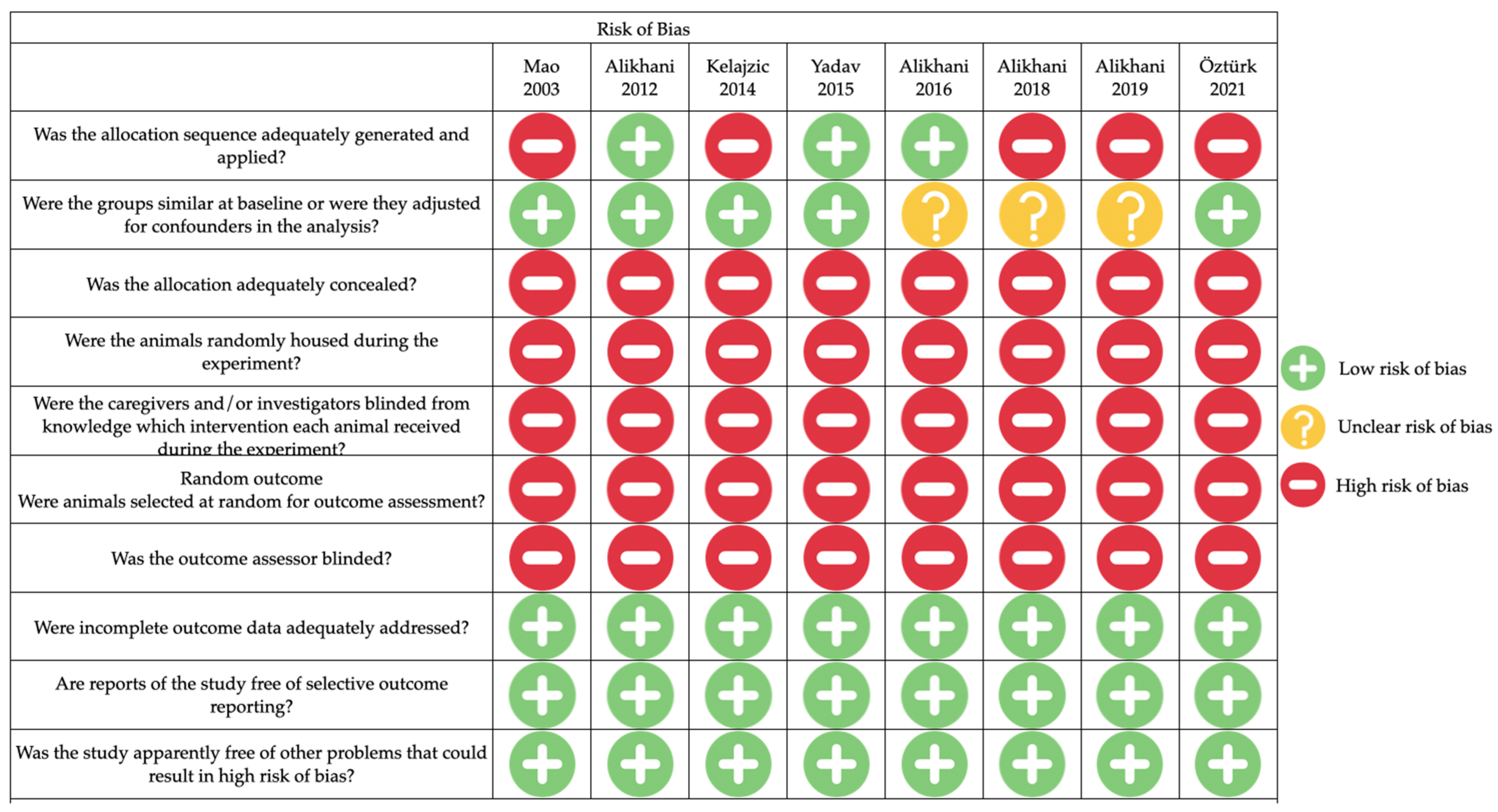
3.4. Risk of Bias Assessment

The risk of bias assessment was conducted following the principles of the Systematic Review Center for Laboratory Animal Experimentation (SYRCLE) [27] This instrument is based on the Cochrane Collaboration’s risk of bias tool adapted to the aspects of bias that play a role in animal experiments [21]. Eight articles accurately described all their outcome data, contained no selective information within their results, and declared no conflicts of interest [3,6,9,13,23,24,25,26]. However, none of the articles adequately reported on allocation concealment, randomized housing of animals, blinding during processing, randomized assessment of results, and detection of bias. Five articles stated that the intervention and control groups were similar at the start of the experiment [6,9,23,24,26]. Three articles reported an adequate allocation of the animals into the different experimental and control groups [24,25,26]. In five [6,9,23,24,26] of the eight articles evaluated, a high risk of bias rating was obtained for 60% of the criteria evaluated. The other three articles [3,13,25] received a high risk of bias rating for 50% of the criteria evaluated. The summary of the results of the risk of bias assessment can be seen in Figure 2.

3.5. Evaluation of the Quality of the Included Studies

The quality assessment was carried out following the ARRIVE 2.0 guidelines [22]. Where each of the included articles was evaluated individually using the recommended criteria for good reporting practice. To obtain a percentage of 100%, the following requirements had to be met: (1) study design, (2) sample size, (3) inclusion and exclusion criteria, (4) randomization, (5) blinding, (6) outcome measures, (7) statistical methods, (8) experimental animals, (9) experimental procedures, (10) results, (11) summary, (12) background, (13) objectives, (14) an ethical statement, (15) housing and husbandry, (16) animal care and monitoring, (17) scientific interpretation/implication, (18) generalizability/translation, (19) protocol registration, (20) access to data, and (21) declaration of interests. Based on the above, the criteria reported in 100% of the articles were 1, 6, 7, 8, 9, 10, 17, and 18, followed by criteria 11, 13, and 16 in 88%; criterion 21 (declaration of interests) was met in 50% of the articles, while the lowest percentages were for criterion 13 with 13%, and with 38% criteria: 2, 4, 15, 19 and 20. It should be noted that none of the articles met the blinding criteria (number 5); see Figure 3.

4. Discussion

Vibrations are cyclic forces performing a physical stimulus that has been shown to promote bone formation through osteoblast differentiation and proliferation [28]. This phenomenon may be possible because the bone is a mechanosensitive tissue, and bone cells react to a biomechanical environment by activating molecular signaling pathways that regulate matrix production, differentiation, and proliferation [16]. Two types of frequencies are used to stimulate the alveolar bone: the LFV (≤45 Hz) and the HFV (≥90 Hz) [29]. This systematic review contrasted the different publications related to LFV [6,9,23,26] and HFV [3,13,24,25] and their osteogenic effect on the alveolar bone. The results of this work showed that LFV and HFV produce a mechanical stimulus that leads to an osteogenic effect on the alveolar bone, improves healing after dental extractions, increases bone density, and helps counteract the effects of induced osteoporosis [3,6,9,13,23,24,25,26].
The studies analyzed in this review showed that LFV stimulates growth and osteogenesis in craniofacial bones, as demonstrated by Mao et al., where the application of vibrations of 0.2 Hz together with an orthopedic force was able to increase the length of the premaxilla compared to the control [23]. This is because vibrations affect the sutures; histological evidence shows that the premaxillary-maxillary suture treated with cyclic loading showed a significant sutural separation with islands of new bone formation [23]. This result is consistent with that reported by Mehmet et al., who reported that the application of LFV for a short period induces cranial growth through enlargement of the sagittal and parietotemporal sutures and an increase in cranial width; on the other hand, the same vibratory stimulus for a longer period stimulates the formation of cranial bones, thereby increasing their width [30]. These results suggest that the use of vibrations combined with orthopedic forces during interceptive orthodontics to achieve optimal therapeutic effects.
At the same time, the application of LFV has been suggested as an alternative to prevent relapse after orthodontic treatment [9]. Öztürk et al. reported that the application of LFV in two retention periods, one short (7 days) and one long (15 days) post orthodontic treatment, decreased relapse, with the 15-day period showing the lowest amount of relapse, which was caused by a decrease in RANKL and an increase in the number of bony trabeculae in the alveolar bone [9]. This is consistent with the findings by García-López et al., where 30 Hz stimulation of osteoblasts decreases RANKL expression and promotes an increase in osteoblast activity, which may lead to an increase in bone mineral density [31]. This may lead to an increase in trabecular bone, cortical thickness, and tissue volume, which may contribute to a decrease in the amount of recurrence; it also coincides with the report by Kalajzic et al., who observed inhibition in tooth movement when using LFV [6]. On the other hand, the results found by Yadav et al. are contrary to those mentioned above since they report that the use of LFV in a retention period does not make a difference in the amount of relapse. However, the bone density was higher in the vibration group [26].
The studies analyzed in which HFV stimulation was used were those carried out by Alikhani et al. They reported that the use of HFV presented a paradoxical effect on the alveolar bone, creating different reactions that depend on the context in which the vibrations were applied [3]. On the one hand, when there is an orthodontic force inducing a dental movement, the effect achieved is catabolic; this was consistent with the in vitro studies of Benjakul et al., where periodontal ligament cells compressed by an acrylic disc and mechanically stimulated with HFV overexpressed the genes related to bone resorption RANKL and PGE2 [32]. However, in healthy alveolar bone, in the process of post-extraction healing, and even with osteoporosis, HFV stimulates growth and increases tissue density and bone volume, promoting a process of osteogenesis [3,13,24,25]. This is possible because the alveolar bone is a mechanosensitive tissue that reacts to different external mechanical stimuli, adapting the bone mass and structure in a continuous remodeling process [15,16]. In addition, in the oral environment, the periodontal ligament has mechanoreceptors that respond to external mechanical stimuli known as Ruffini type I, which is characterized by having two types of response, both a fast adaptation response, located near the center of resistance of the tooth, and a slow adaptation, located near the apex. The fast adaptation is activated only when there is a mechanical load of pressure, while the slow adaptation depends on the frequency and magnitude of load for the transmission of the impulse [12]. At the same time, the vibratory stimulus propagates through the tissues, demonstrating that the anabolic effects are not only localized to the area of application but also affect the areas adjacent to it. However, the effect decreases the further away the application site is [13,24].
It is important to note that the anabolic effect on the alveolar bone of LFV and HFV results from activation of the canonical Wnt signaling pathway [14,15,16]. This signaling pathway acts through Wnt ligands such as Wnt10B. Initially, Wnt proteins bind to the Frizzled receptor and Lrp5/6 co-receptor found on the cell membrane, which causes a concentration of β-catenin within osteoblasts, inhibiting the degradation of phosphorylated β-catenin through the ubiquitin-proteosome pathway; the concentration of β-catenin within the cytoplasm of osteoblasts results in the translocation of this enzyme within the nucleus, where it binds to TCF/LEF transcription factors and regulates the expression of RUNX2 [33]. Alikhani et al. reported an increase in the Wnt gene expression upon application of 120 Hz vibrations [13]. This coincides with the results of Pravitharangul et al., who report that osteoblasts of mandibular origin had an overexpression of the Wnt10b gene after being subjected to vibrations of 120 Hz [14].
There are other factors that favor the anabolic effect when vibratory stimuli are used, such as the reduction of the activity and quantity of osteoclasts [6,25,26]. This is consistent with the results obtained by Kalajzic et al., who observed that the amount of recurrence was lower in rats that received vibrations of 30 Hz during a retention period [6]. Furthermore, a decrease in the number of osteoclasts was shown, thus preventing tooth displacement. These results are similar to those reported by Yadav et al., who showed that the use of LFV decreased sclerostin, which caused a decrease in the number of osteoclasts [26]. These results are consistent with the downregulation of the SOST gene in osteoblasts subjected to vibration reported by Gao et al. in their in vitro study [15]. The SOST gene encodes the protein sclerostin and is mainly secreted by osteocytes and cells of the periodontal ligament, whose function is to inhibit bone formation by binding competitively to the Wnt co-receptor, LRP5/6; sclerostin causes phosphorylation and degradation of beta-catenin and hinders the activation of osteoblasts [34,35]. In this sense, a decrease in the SOST gene represents an inhibition of the bone resorption process. Therefore, a decrease in the SOST gene helps to reduce tooth movement by decreasing the activity of the osteoclasts, which is the objective in a post-orthodontic retention stage or when a bone resorption process is to be halted [6,26].
Only two studies have an experimental design that simulated a clinical situation of orthodontic retention [9,26]. Yadav et al. reported that there was no significant difference in using or not using vibrations in the retention period [26]. On the other hand, Öztürk et al. showed that the amount of relapse in the groups that followed the vibration scheme was lower than in the groups that followed a conventional retention protocol, additionally obtaining a lower percentage of relapse in cases where the retention period was 15 days [9]. This may be due to multiple factors, including that Öztürk et al. applied the stimulus over a more extended period and gradually increased their frequencies from 10 Hz to 30 Hz over a period of 9 days and that their maximum retention period was 15 days, while Yadav et al. applied vibrations in their retention period which lasted seven days [9,26].
The anabolic effect on the alveolar bone achieved with vibrations has several clinical implications, such as bone regeneration as part of the treatment of periodontitis, or in this case, to increase tissue density to prevent post-orthodontic treatment relapse; however, there is a need to generate experimental protocols that simulate this clinical situation.

Strengths and Limitations of This Systematic Review

This study has some limitations: first, the systematic review results are based on animal studies, so it is important to consider the physiological differences between the human alveolar bone and PDL [36]. Secondly, in the studies included in the systematic review, there was a wide variation in both the frequency and the clinical situation in which the vibratory stimulus was applied [3,6,9,13,23,24,25,26]. Finally, the number of studies included was limited. Nevertheless, all studies in which the anabolic effect of vibrations on alveolar bone was evaluated in animal models were included, being the first systematic review to do so. Although the ideal scenario is to evaluate this effect in humans, only one published article has done so [11], making it impossible to conduct a systematic review in humans.

5. Conclusions

The results of the studies included in this article suggest that LFV and HFV have an anabolic effect on the alveolar bone when it is healthy and healing, even with osteoporosis. Nevertheless, the greatest anabolic activity was observed when using HFV. This anabolic effect is achieved through increasing bone density and volume, increasing osteoblastic activity, increasing the thickness of bone trabeculae, overexpressing osteogenic biomarkers, activation of the canonical Wnt pathway, and decreasing osteoclast activity. It is indispensable to understand that the conclusions were made based on a few animal studies, and the evidence is not as strong as the results obtained from randomized controlled trials in humans, which highlights the need for more future studies following this line of research in humans.

Author Contributions

Conceptualization, J.C.V.A., M.F.G.V. and M.A.C.S.; methodology, M.A.C.S.; software, J.F.-T.; validation, M.F.S.O., R.M.A.M. and E.R.C.; formal analysis, J.E.S.S.; investigation, J.C.V.A. and M.F.G.V.; resources, R.M.A.M., M.A.C.S. and J.E.S.S.; writing—original draft preparation, J.C.V.A., M.F.G.V. and M.A.C.S.; writing—review and editing, M.F.S.O., R.M.A.M. and J.F.-T.; visualization, E.R.C. and J.E.S.S.; supervision, M.F.S.O.; project administration, M.A.C.S.; funding acquisition, M.A.C.S. All authors have read and agreed to the published version of the manuscript.

Funding

This research was funded by institutional resources of the Benemérita Universidad Autónoma de Puebla.

Institutional Review Board Statement

Not applicable.

Informed Consent Statement

Not applicable.

Data Availability Statement

Not applicable.

Conflicts of Interest

The authors declare no conflicts of interest.

References

  1. Van der Zee, E.A.; Oroszi, T.; van Heuvelen, M.J.G.; Nyakas, C. Vibration detection: Its function and recent advances in medical applications. F1000Research 2020, 9. [Google Scholar] [CrossRef]
  2. Rubin, C.; Turner, A.S.; Bain, S.; Mallinckrodt, C.; McLeod, K. Low mechanical signals strengthen long bones. Nature 2001, 412, 603–604. [Google Scholar] [CrossRef]
  3. Alikhani, M.; Alansari, S.; Hamidaddin, M.A.; Sangsuwon, C.; Alyami, B.; Thirumoorthy, S.N.; Oliveira, S.M.; Nervina, J.M.; Teixeira, C.C. Vibration paradox in orthodontics: Anabolic and catabolic effects. PLoS ONE 2018, 13, e0196540. [Google Scholar] [CrossRef]
  4. ElDeeb, A.M.; Abdel-Aziem, A.A. Effect of Whole-Body Vibration Exercise on Power Profile and Bone Mineral Density in Postmenopausal Women with Osteoporosis: A Randomized Controlled Trial. J. Manip. Physiol. Ther. 2020, 43, 384–393. [Google Scholar] [CrossRef]
  5. DadeMatthews, O.O.; Agostinelli, P.J.; Neal, F.K.; Oladipupo, S.O.; Hirschhorn, R.M.; Wilson, A.E.; Sefton, J.M. Systematic review and meta-analyses on the effects of whole-body vibration on bone health. Complement. Ther. Med. 2022, 65, 102811. [Google Scholar] [CrossRef]
  6. Kalajzic, Z.; Peluso, E.B.; Utreja, A.; Dyment, N.; Nihara, J.; Xu, M.; Chen, J.; Uribe, F.; Wadhwa, S. Effect of cyclical forces on the periodontal ligament and alveolar bone remodeling during orthodontic tooth movement. Angle Orthod. 2014, 84, 297–303. [Google Scholar] [CrossRef]
  7. García Vega, M.F.; López Pérez-Franco, L.M.; Dib Kanán, A.; Román Méndez, C.D.; Soto Sainz, J.E.; Reyes Cervantes, E.; Cerda-Cristerna, B.I.; Salas Orozco, M.F.; Casillas Santana, M.A. Are mechanical vibrations an effective alternative to accelerate orthodontic tooth movement in humans? A systematic review. Appl. Sci. 2021, 11, 10699. [Google Scholar] [CrossRef]
  8. Keerthana, P.; Diddige, R.; Chitra, P. Performance comparison of vibration devices on orthodontic tooth movement—A systematic review and meta-analysis. J. Oral. Biol. Craniofacial Res. 2020, 10, 814–823. [Google Scholar] [CrossRef] [PubMed]
  9. Öztürk, T.; Gül Amuk, N. Three-dimensional imaging and molecular analysis of the effects of photobiomodulation and mechanical vibration on orthodontic retention treatment in rats: Effects of photobiomodulation and mechanical vibration on orthodontic retention treatment. J. Orofac. Orthop. 2022, 83 (Suppl. 1), 24–41. [Google Scholar] [CrossRef] [PubMed]
  10. Thammanichanon, P.; Kaewpitak, A.; Binlateh, T.; Leethanakul, C. Interval vibration reduces orthodontic pain via a mechanism involving down-regulation of TRPV1 and CGRP. In Vivo 2020, 34, 2389–2399. [Google Scholar] [CrossRef] [PubMed]
  11. Shipley, T.; Farouk, K.; El-Bialy, T. Effect of high-frequency vibration on orthodontic tooth movement and bone density. J. Orthod. Sci. 2019, 8, 15. [Google Scholar]
  12. Piancino, M.G.; Isola, G.; Cannavale, R.; Cutroneo, G.; Vermiglio, G.; Bracco, P.; Anastasi, G.P. From periodontal mechanoreceptors to chewing motor control: A systematic review. Arch. Oral. Biol. 2017, 78, 109–121. [Google Scholar] [CrossRef] [PubMed]
  13. Alikhani, M.; Alikhani, M.; Alansari, S.; Almansour, A.; Hamidaddin, M.A.; Khoo, E.; Lopez, J.A.; Nervina, J.M.; Nho, J.Y.; Oliveira, S.M.; et al. Therapeutic effect of localized vibration on alveolar bone of osteoporotic rats. PLoS ONE. 2019, 14, e0211004. [Google Scholar] [CrossRef] [PubMed]
  14. Pravitharangul, A.; Suttapreyasri, S.; Leethanakul, C. Mandible and iliac osteoblasts exhibit different Wnt signaling responses to LMHF vibration. J. Oral Biol. Craniofacial Res. 2019, 9, 355–359. [Google Scholar] [CrossRef] [PubMed]
  15. Gao, H.; Zhai, M.; Wang, P.; Zhang, X.; Cai, J.; Chen, X.; Shen, G.; Luo, E.; Jing, D. Low-level mechanical vibration enhances osteoblastogenesis via a canonical Wnt signaling-associated mechanism. Mol. Med. Rep. 2017, 16, 317–324. [Google Scholar] [CrossRef] [PubMed]
  16. Steppe, L.; Liedert, A.; Ignatius, A.; Haffner-Luntzer, M. Influence of Low-Magnitude High-Frequency Vibration on Bone Cells and Bone Regeneration. Front. Bioeng. Biotechnol. 2020, 8, 595139. [Google Scholar] [CrossRef] [PubMed]
  17. Bonanni, R.; Cariati, I.; Romagnoli, C.; D’Arcangelo, G.; Annino, G.; Tancredi, V. Whole Body Vibration: A Valid Alternative Strategy to Exercise? J. Funct. Morphol. Kinesiol. 2022, 7, 99. [Google Scholar] [CrossRef]
  18. Page, M.J.; McKenzie, J.E.; Bossuyt, P.M.; Boutron, I.; Hoffmann, T.C.; Mulrow, C.D.; Shamseer, L.; Tetzlaff, J.M.; A Akl, E.; Brennan, S.E.; et al. The PRISMA 2020 statement: An updated guideline for reporting systematic reviews. Int. J. Surg. 2021, 88, 105906. [Google Scholar] [CrossRef]
  19. El-Bialy, T. The Effect of High-Frequency Vibration on Tooth Movement and Alveolar Bone in Non-Growing Skeletal Class II High Angle Orthodontic Patients: Case Series. Dent. J. 2020, 8, 110. [Google Scholar] [CrossRef]
  20. Takano-Yamamoto, T.; Sasaki, K.; Fatemeh, G.; Fukunaga, T.; Seiryu, M.; Daimaruya, T.; Takeshita, N.; Kamioka, H.; Adachi, T.; Ida, H.; et al. Synergistic acceleration of experimental tooth movement by supplementary high-frequency vibration applied with a static force in rats. Sci. Rep. 2017, 7, 13969. [Google Scholar] [CrossRef]
  21. Sandercock, P.; Roberts, I. Systematic reviews of animal experiments. Lancet 2002, 360, 586. [Google Scholar] [CrossRef] [PubMed]
  22. du Sert, N.P.; Hurst, V.; Ahluwalia, A.; Alam, S.; Avey, M.T.; Baker, M.; Browne, W.J.; Clark, A.; Cuthill, I.C.; Dirnagl, U.; et al. The arrive guidelines 2.0: Updated guidelines for reporting animal research. PLoS Biol. 2020, 18, e3000410. [Google Scholar]
  23. Mao, J.J.; Wang, X.; Mooney, M.P.; Kopher, R.A.N.J. Strain Induced Osteogenesis of the Craniofacial Suture Upon Controlled Delivery of Low-Frequency Cyclic Forces. Front. Biosci. 2003, 8, a10–a17. [Google Scholar] [CrossRef] [PubMed]
  24. Alikhani, M.; Khoo, E.; Alyami, B.; Raptis, M.; Salgueiro, J.; Oliveira, S.; Boskey, A.; Teixeira, C. Osteogenic effect of high-frequency acceleration on alveolar bone. J. Dent. Res. 2012, 91, 413–419. [Google Scholar] [CrossRef] [PubMed]
  25. Alikhani, M.; Lopez, J.A.; Alabdullah, H.; Vongthongleur, T.; Sangsuwon, C.; Alikhani, M.; Alansari, S.; Oliveira, S.M.; Nervina, J.M.; Teixeira, C.C. High-Frequency Acceleration: Therapeutic Tool to Preserve Bone following Tooth Extractions. J. Dent. Res. 2016, 95, 311–318. [Google Scholar] [CrossRef]
  26. Yadav, S.; Assefnia, A.; Gupta, H.; Vishwanath, M.; Kalajzic, Z.; Allareddy, V.; Nanda, R. The effect of low-frequency mechanical vibration on retention in an orthodontic relapse model. Eur. J. Orthod. 2015, 38, 44–50. [Google Scholar] [CrossRef]
  27. Hooijmans, C.R.; Rovers, M.M.; de Vries, R.B.; Leenaars, M.; Ritskes-Hoitinga, M.; Langendam, M.W. SYRCLE’s risk of bias tool for animal studies. BMC Med. Res. Methodol. 2014, 14, 43. [Google Scholar] [CrossRef]
  28. Fu, Z.; Huang, X.; Zhou, P.; Wu, B.; Cheng, L.; Wang, X.; Zhu, D. Protective effects of low-magnitude high-frequency vibration on high glucose-induced osteoblast dysfunction and bone loss in diabetic rats. J. Orthop. Surg. Res. 2021, 16, 650. [Google Scholar] [CrossRef]
  29. Kanzaki, H.; Nakamura, Y. Orthodontic tooth movement and bone density. J. Oral. Biosci. 2018, 60, 49–53. [Google Scholar] [CrossRef]
  30. Amuk, M.; Amuk, N.G.; Hacıoğlu, Z. Effects of orofacial applications of low-magnitude, high-frequency mechanical vibration on cranial sutures and calvarial bones—A micro-computed tomography study in rats. Am. J. Orthod. Dentofac. Orthop. 2022, 162, 459–468. [Google Scholar] [CrossRef]
  31. García-López, S.; Villanueva, R.E.; Massó-Rojas, F.; Páez-Arenas, A.; Meikle, M.C. Micro-vibrations at 30 Hz on bone cells cultivated in vitro produce soluble factors for osteoclast inhibition and osteoblast activity. Arch. Oral. Biol. 2020, 110, 104594. [Google Scholar] [CrossRef] [PubMed]
  32. Benjakul, S.; Jitpukdeebodintra, S.; Leethanakul, C. Effects of low magnitude high frequency mechanical vibration combined with compressive force on human periodontal ligament cells in vitro. Eur. J. Orthod. 2018, 40, 356–363. [Google Scholar] [CrossRef] [PubMed]
  33. Houschyar, K.S.; Tapking, C.; Borrelli, M.R.; Popp, D.; Duscher, D.; Maan, Z.N.; Chelliah, M.P.; Li, J.; Harati, K.; Wallner, C.; et al. Wnt Pathway in Bone Repair and Regeneration—What Do We Know So Far. Front. Cell Dev. Biol. 2019, 6, 170. [Google Scholar] [CrossRef] [PubMed]
  34. Ueda, M.; Kuroishi, K.N.; Gunjigake, K.K.; Ikeda, E.; Kawamoto, T. Expression of SOST/sclerostin in compressed periodontal ligament cells. J. Dent. Sci. 2016, 11, 272–278. [Google Scholar] [CrossRef]
  35. Shu, R.; Bai, D.; Sheu, T.; He, Y.; Yang, X.; Xue, C.; He, Y.; Zhao, M.; Han, X. Sclerostin promotes bone remodeling in the process of tooth movement. PLoS ONE. 2017, 12, e0167312. [Google Scholar] [CrossRef]
  36. Akhtar, A. The flaws and human harms of animal experimentation. Camb. Q. Healthc. Ethics. 2015, 24, 407–419. [Google Scholar] [CrossRef]
Figure 1. The PRISMA flow diagram of record processing and elimination [1,9,11,16,19,20].
Applsci 14 01118 g001
Figure 2. Risk of bias summary for each of the included studies (n = 8). The green color indicates a low risk of bias; the yellow color indicates an unclear risk of bias; the red color indicates a high risk of bias [3,6,9,13,23,24,25,26].
Applsci 14 01118 g002
Figure 3. Quality assessments of the studies according to ARRIVE 2.0. Values are expressed by %.
Applsci 14 01118 g003
Table 1. Summary of characteristics of the included studies.
Author/Year/CountryDesign of StudySampleGroupsVibration ProtocolMeasurementResultsConclusion
Mao
(2003)
USA
[23]
In vivon = 21 M,
Age: 6-weeks-old,
White rabbits
EG1: n = 4, tensile cycled forces of 2 N with 0.1 Hz;
EG2: n = 5, tensile cycled forces of 2 N with 0.2 Hz;
EG3: n = 2 N of static forces over premaxilla suture;
CG: n = 4, Not stimulations.
0.2 Hz
for 10 min/d
over 12 days.
Craniofacial length.Craniofacial length was significantly higher when cyclic forces at 0.2 Hz were applied.Cyclic forces involve activation of stress-sensitive genes, leading to more craniofacial growth and osteogenesis.
Alikhani
(2012)
USA
[24]
In vivon = 85 M,
Age: 120 days,
Sprague
Dawley rats
EG: n = 43, different HFV (30–200 Hz);
SC: n = 23, 4 µε of static load over the maxilla;
CG: n = 19, not stimulations.
30, 60, 100, and 200 Hz for 5 min/d
over 28 days.
BV/TV;
Osteogenic effect;
Trabecular thickness;
Trabecular space;
Alveolar bone density;
Growth factors;
Transcription factors;
Extracellular matrix;
Mineralization.
The highest BV/TV was with 60 and 100 Hz rate.
60 Hz demonstrated a higher level compared with the static group in osteogenic effect, density, growth factors, transcription factors, extracellular matrix, mineralization, trabecular thickness, and the greatest decrease in trabecular space.
HFV vibration increase osteoblast activity, plays a key role during the mineralization process
The higher response to vibration was in trabecular bone.
Kalajzic
(2014)
USA
[6]
In vivon = 26 F,
Age: 7 weeks old,
Sprague
Dawley rats
EG1: n = 4, received occlusal vibratory stimulus twice a week without orthodontic force;
EG2: n = 9, received the orthodontic force only;
EG3: n = 9, received the orthodontic force and additional vibratory stimulus twice a week
CG: n = 4, not stimulations.
30 Hz
for 10 min/d,
Twice per week with five
Applications.
BV/TV;
Alveolar bone density.
No significant difference between the control group and groups using vibrations in BV/TV and alveolar bone density.LFV may cause different effects depending on force magnitude, frequency, or point of application.
Yadav
(2015)
USA
[26]
In vivon = 30 M,
Age: 12 weeks old,
CD1 mice
EG1: n = 10, applied orthodontic force for 7 days, then, the orthodontic force was removed, and molar was allowed to relapse;
EG2: n = 10, applied orthodontic force for 7 days, then exposed it to vibrational stimuli without orthodontic forces;
CG: n = 10, applied orthodontic force for 7 days.
30 Hz
for 15 min/d
over 7 days.
Bone volume;
Alveolar bone density.
No significant differences in bone volume and density when comparing the CG and vibration group.
Density was significantly more in the vibration group when compared to EG1.
LFV vibrations increase bone density despite inducing a process of bone resorption with orthodontic forces.
Alikhani
(2016)
USA
[25]
In vivon = 85 M,
Age: 4 months,
Sprague Dawley rats
The maxillary right third molar was extracted in all rats;
EG: received 120 Hz;
SC: received 4 µε of static load;
CG: Not stimulations.
120 Hz
for 5 min/d
over 56 days.
BV/TV;
Level of bone formation;
Trabecular thickness;
Trabecular number;
Trabecular spacing;
Tissue mineral density;
Height;
Osteogenic markers: ALP, Runx 2, OPN, OCN.
EG had significantly greater, BV/TV, level of bone formation, trabecular thickness, trabecular number, tissue mineral density and height.
EG: obtained the least trabecular spacing.
EG: exhibited significantly higher ALP, Runx 2, osteopontin and osteocalcin activity.
HFV promotes alveolar bone formation and decrease RANKL expression and osteoclast activation.
Alikhani
(2018)
USA
[3]
In vivon = 206 M,
Age: 120 days,
Sprague Dawley rats
EG: received orthodontic force + HFV;
OTM: received orthodontic force;
CG: not received orthodontic force.
120 Hz
for 5 min/d
over 14 days.
Alveolar bone density;
Bone volume.
Alveolar bone density decreased more during tooth movement in experimental group.HFV treatment during orthodontic tooth movement
produces a catabolic effect
HFV increased the number of osteoclasts.
Alikhani
(2019)
USA
[13]
In vivon = 14 F,
Age: 16 weeks old,
Spices: Sprague Dawley rats
EG1: received bilateral ovariectomy;
EG2: received ovariectomy and HFV;
EG3: received ovariectomy and 10 με of static load;
SG: received ovariectomy surgery without having the ovaries removed;
CG: Not stimulations.
120 Hz
for 5 min/d
over 28 and
56 days.
Alveolar bone density;
BV/TV;
BMD;
Osteogenic markers:
OPG, Runx2, Osterix, ALP, OCN, ColIα1, BMP2, Wnt.
HFV group reestablished the alveolar bone density the levels of the Sham group.
BV/TV and BMD was significantly higher with HFV, compared to ovariectomy group.
HFV restored osteogenic markers to levels similar to the Sham group.
HFV-induced bone remodeling depends on the inflammatory condition of the tissue.
In absence of inflammatory condition, HFV is anabolic.
The highest osteogenic effect of HFV was observed closest to the HFV application site.
Öztürk
(2021)
Turkey
[9]
In vivon = 40,
Age: not specified,
Rats
CG: n = 6, not stimulations;
CG2: n = 6, orthodontic tooth movement (10 days) + relapse (only relapse);
PC1: n = 6, orthodontic tooth movement (10 days) + long-term retention (15 days);
PC2: n = 6, orthodontic tooth movement (10 days) + short-term retention (7 days);
EG1: n = 8, orthodontic tooth movement (10 days) + long-term retention (15 days) + 10–20–30 Hz cumulative increased frequencies mechanical vibration;
EG2: n = 8, orthodontic tooth movement (10 days) + short-term retention (7 days) + 10–20–30 Hz cumulative increased frequencies mechanical vibration.
10–20–30 Hz cumulative increased
for 10 min/d
first 3 days
10 Hz.
Next 3 days
20 Hz
Final 3 days
30 Hz.
Trabecular thickness;
Trabecular number;
OPG, RANKL, COX-2.
Trabecular thickness was lower in vibration Group than control groups.
Trabecular number in vibration group was higher than CG and PC.
No statistically significant intergroup difference in the OPG level.
RANKL and COX-2 was significantly lower in EG.
HFV yielded a better bone structure by increasing trabecular number and levels of RANKL and COX-2.
Abbreviations: EG: experimental group, CG: control group, SG: sham group, PC: positive control; M: male, F: female; BV/TV: bone volume/tissue volume, BMD: bone mineral density, OTM: orthodontic tooth movement. µε: microstrain, HFV: high frequency vibration, LFV: low frequency vibrations, OPG: osteoprotegerin, OPN: osteopontin, OCN: osteocalcin, ALP: alkaline phosphatase, RANKL: receptor activator for nuclear factor B ligand, Cox-2: cyclooxygenase-2.
Disclaimer/Publisher’s Note: The statements, opinions and data contained in all publications are solely those of the individual author(s) and contributor(s) and not of MDPI and/or the editor(s). MDPI and/or the editor(s) disclaim responsibility for any injury to people or property resulting from any ideas, methods, instructions or products referred to in the content.

