Article

Evaluation of Tooth Movement Accuracy with the F22 Aligner System: A Retrospective Study

by
Palone Mario
1,
Silvia Squeo de Villagomez
2,
Pellitteri Federica
1,
Francesca Cremonini
1,
Renato Salvatore
3 and
Luca Lombardo
1,*
1
Postgraduate School of Orthodontics, University of Ferrara, Via Luigi Borsari 46, 44121 Ferrara, Italy
2
Private Practice in Bisceglie, Via Ottavio Tupputi 4, 76011 Bisceglie, Italy
3
Department of Economics and Law, University of Cassino and Southern Lazio, Viale dell’Università, 03043 Cassino, Italy
*
Author to whom correspondence should be addressed.
Appl. Sci. 2024, 14(4), 1641; https://doi.org/10.3390/app14041641
Submission received: 11 January 2024 / Revised: 8 February 2024 / Accepted: 13 February 2024 / Published: 18 February 2024

Abstract

:
Background: To investigate the accuracy of an F22 Aligner system, considering the amount of prescribed movement, tooth type, grip points, sex and age. Methods: Digital models of 120 patients (64 females and 56 males; mean age 35.2 years ± 7.4) affected by mild-to-moderate Class I malocclusion and treated via F22 Aligners, retrospectively selected from the University of Ferrara Orthodontics Clinic’s electronic database; post-treatment models were generated, and three angular values per tooth and four linear intra-arch measurements per arch were acquired. For angular measurements, planned (T1) and achieved (T2) values were obtained thorough digital model superimpositions. Linear measurements were acquired from pre-treatment, reference and post-treatment models. Statistical comparisons were performed to assess accuracy among tooth types and prescribed movements, tooth type, grip points, sex and age were investigated via chi-squared automatic interaction detection regression trees. Results: Mean accuracy for inclination and angulation were 86.76% and 88.01%, respectively, whereas rotation was less accurate (61.59%), especially for rounded teeth. All variables investigated influenced accuracy, with the exception of inclination, which was only influenced by age. Regarding linear measurements, good expansive capacity was shown, except for the distance between mandibular second premolars. Conclusions: F22 aligners are a viable solution for the treatment of Class I malocclusion of mild-to-moderate complexity, although clinicians should bear in mind the lower predictability of rotation, as well as the influence of the variables investigated.

1. Introduction

The undisputed global trend that orthodontics is experiencing today is the growing use of clear aligners (CAs) for the resolution of malocclusions of various types and severity, not only in adults, but also in growing patients [1]. This is due to the fact that CAs are aesthetically inobtrusive [2], facilitate daily oral hygiene procedures [3], do not require urgent intervention, and offer both considerable comfort for the patient and convenience for the clinician [4].
The remarkable initial success of the clear aligner treatment (CAT) has led manufacturers to develop increasingly sophisticated operating protocols [5], with the latest generation materials that, according to the manufacturers, provide superior performance [6]. This has purportedly made it possible to extend the indications for CAT to even the most severe malocclusions. However, these improvements were not validated by scientific research before they were put on the market; in fact, the most famous study to exhaustively investigate the effectiveness of CAs was conducted about 11 years after their launch, by Align Technology in 1998 (Santa Clara, CA, USA) [7].
In that study, Kravitz et al. showed that the mean accuracy was 41%, with the most accurate movement being lingual crown tipping (47.1%), and the least accurate movements both derotation of the mandibular canines (28%) and extrusion of the maxillary incisors (18%) [7].
Recently, the same authors replicated their initial study, given that in the interim improvements had been introduced regarding aligner materials (introduction of the Smart Track® material, Align Technology Santa Clara, CA, USA), clinical protocols (G protocols) and digital planning [8]. However, they found only a minimal improvement in Invisalign aligner accuracy, which they calculated as at around 50% on average, with the weaknesses of the system remaining the same. In light of these results, they therefore confirmed that generally CAT should preferably be used to treat cases of mild-to-moderate difficulty, in which they were even more effective than fixed appliances (FAs) [8,9,10].
As for complex treatments involving extraction, ref. [11], root torque, ref. [12], overbite modifications > 1.5 mm [13] or severe rotations of the rounded teeth [14], these would be difficult to resolve satisfactorily with CAT; in such cases, it would be appropriate to opt for FAs [15,16,17]. Another alternative is the so-called hybrid approach, which involves combining CAs with both other orthodontic appliances (rapid palatal expander, intraoral distalizers, etc.), with or without the use of skeletal anchorage [18], auxiliaries such as fixed vestibular or lingual sectional appliances and derotation or extrusion chains [19].
As a matter of fact, Houle et al. reported that about 80% of patients treated only with the Invisalign system usually need mid-course corrections or at least one (and often multiple) additional finishing clear aligner phase, which increases both costs and treatment time [20]. In fact, the recourse to additional CAs is so common that Bilello et al. consider refinement/additional aligner stage as a normal phase of CAT, comparing it to the finishing phase with FAs. They reported a high percentage of accuracy of the movements investigated when additional CAs are taken into account, but acknowledged that this causes treatment times to increase significantly, with an average of 21.2 additional aligners required for each treated arch [21].
Despite the evidence in the literature, complex treatments are still undertaken with CAT and, for this reason, the results may differ greatly from those provided for in the virtual setup [7,8,22,23], leading to an increase in treatment time, and in the need for multiple refinements and mid-course corrections [20]. However, the intrinsic efficacy of CAT is further hampered by other factors, such as fitting [24,25] and patient compliance, and these factors should also be taken into account.
The F22 aligner system was introduced in 2015 in order to overcome the issues described above and it is actually spread in twelve European countries and traded by Sweden & Martina Spa (Due Carrare, Padova). This system was developed and diffused only after the execution of different studies regarding aligner fitting [24,26], aesthetic properties [27,28], mechanical properties [29,30] and the amount of overcorrections needed to be included in the first phase of CAT [31]. Moreover, another feature is that each virtual set-up of the F22 Aligner system has to be controlled and supervised by a specialized orthodontist expert in CAT treatment, in order to avoid unpredictable movements, thus reducing additional aligners and refinement stages.
Therefore, the aim of this study was to evaluate the clinical efficacy of the F22 Aligner system with respect to digitally planned movements in a large sample with mild-to-moderate Class I malocclusion, treated according to the clinical indications in the literature.
Specifically, only crown tipping movements were prescribed—no translation or root torque—and the treatment with F22 was carried out without resorting to any auxiliary treatment.
The secondary objective was to investigate whether the effectiveness of CAT depends on the following factors: amount of prescribed movement, tooth type, the presence or absence of grip points and/or demographic aspects such as age and gender.

2. Materials and Methods

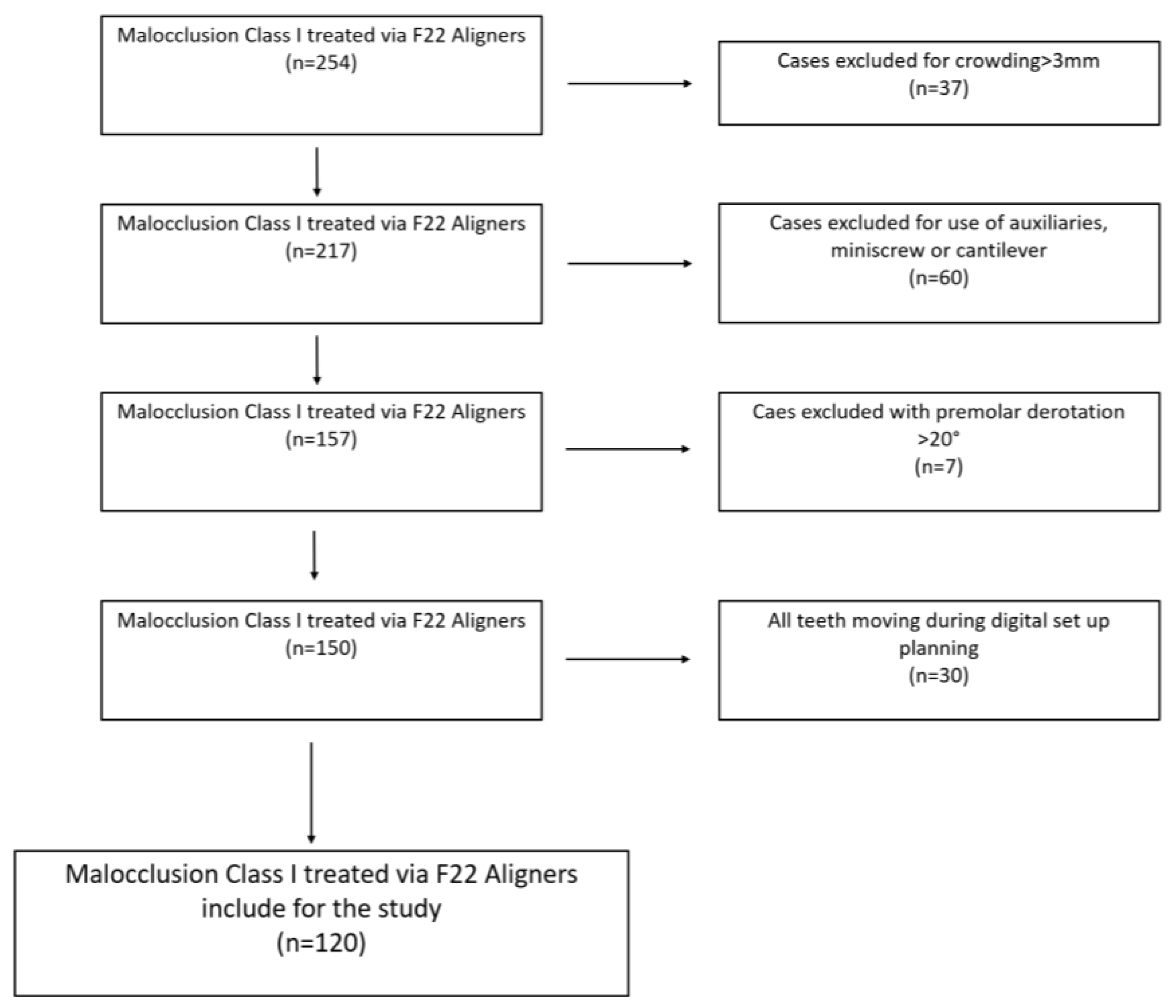
2.1. Sample Selection

The study group, retrospectively selected from the University of Ferrara Orthodontics Clinic’s electronic database, consisted of adult patients with complete permanent dentition treated via CAT between 2016 and 2020. The study design was approved by the University of Ferrara Postgraduate School Ethics Committee (registration number 9/2020), and the research was conducted in conformity to the Declaration of Helsinki.
The inclusion criteria used for the retrospective selection of patients were:
  • Class I dental malocclusion with minimal crowding (≤3 mm) in both arches [32], treated with a series of 12–20 aligners per arch.
  • No use of auxiliaries (derotation elastomeric chains, inter-arch elastics or extrusion elastics), miniscrews, cantilevers or vestibular or lingual fixed partial appliances.
  • Grip points and IPR (interproximal reduction) were permitted.
  • Planned derotation of rounded teeth ≤ 20° (premolars and canines) is included.
  • The use of grip points positioned for derotation ≥ 10° of rounded teeth and >20° for mandibular incisors and maxillary lateral incisors, which have a mesiodistally narrow clinical crown. The triangular grip points were applied with the long side in the direction of force application.
  • No previous or active periodontal disease at the beginning of orthodontic therapy.
  • Presence of at least one posterior tooth (first or second molar) per quadrant that had not been planned to move.
  • F22 Aligners were worn for 2 weeks prior to passing to the following step.
  • F22 Aligner therapy completed with adequate patient compliance (20–22 h per day), as documented in a personal diary.
This retrospective selection resulted in a final sample of 120 patients (64 female and 56 male; mean age 35.2 years ± 7.4), starting from 254 cases affected by dental Class I Malocclusion treated via F22 Aligners. A flow chart of retrospective sample selection is graphically illustrated in Figure 1.
Each patient expressed written consent for the use of their records for research; the setup for each case was performed by two specialists in orthodontics, experts in CAT and certified by the Italian Board of Orthodontic Aligners.
Digital planning involved digitally positioning the center of rotation between the apical one-third and middle-root one-third in single-rooted teeth, and 2 mm apical to the furcation in multirooted teeth, and the tooth movements planned were mainly coronals. Pure radicular movements, such as root torque and mesial-distal root tipping, were thus avoided [33].
The clinical staging used involved a step every 2° for the following investigated movements: inclination (buccal-lingual crown tipping), angulation (mesial-distal crown tipping) and rotation.
Each patient enrolled retrospectively in the study was treated with polyurethane F22 Aligners with vestibular grip points created using GRADIA DIRECT LoFlo flow composite (GC Orthodontics Europe GmbH, Harkortstraße, Germany).
Grip points were used are for both stabilization (rectangular shaped) and derotation (triangular shaped). The former were usually used in posterior teeth when anterior intrusion was planned; the regular dimension was 3 mm in length and 1 mm in height, and usually positioned in the center of the clinical crown. The latter were triangular-shaped and applied on teeth that had to be rotated, especially rounded teeth (canines and premolars); regular dimension was 1 mm in height, with a longer surface oriented towards the opposite side of rotation, spanning about 3 mm in length.
Considering the whole sample mean number of aligners, the mean IPR values and mean number of grip points for both arches used in the first phase of CAT with F22 aligners have been reported.

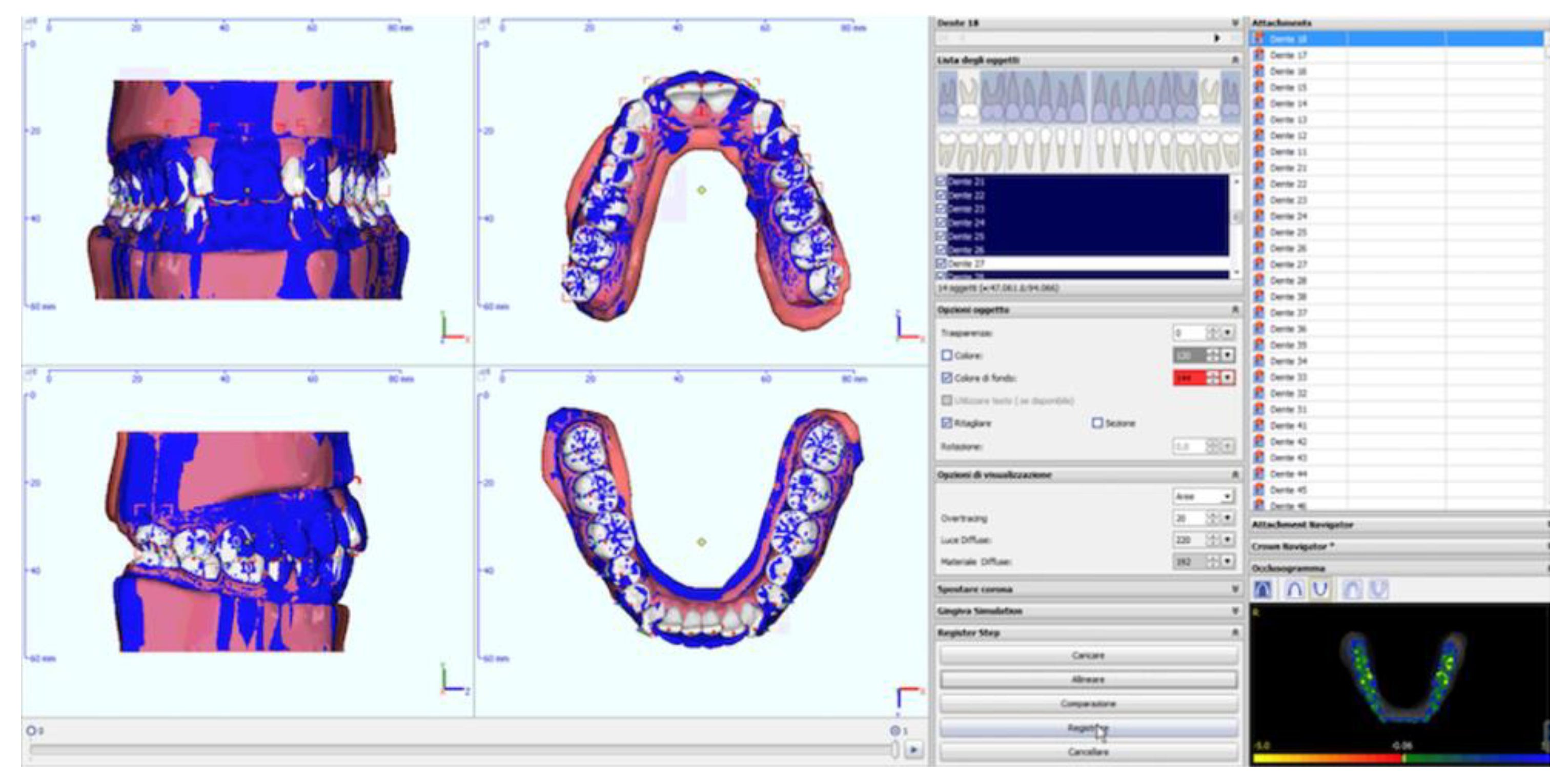
2.2. Analysis of Digital Models

For each patient, maxillary and mandibular models of the initial malocclusion (pre-treatment model), of the final step of digital planning (reference model) and of the end of the initial prescribed series of aligners (pre-finishing model) were acquired in STL format using the appropriate setup software. Thus, a total of 720 models were investigated. Each model was imported into Onyxceph 3TM software (Firma Image Instruments GmbH, Chemnitz, Germany) in order to perform the various digital superimpositions at the reference unmoved first and/or second molars in each quadrant (Figure 2) [14,34].
After the segmentation of the teeth and digital model superimposition, using best-fitting at the posterior teeth with no movement planned, a specific algorithm made it possible to acquire the following data:
  • Prescribed movements (T1): the difference between the pre-treatment and reference model;
  • Achieved movements (T2): the difference between the pre-treatment and pre-finishing model (Figure 3) for each movement investigated (inclination, angulation and rotation) and for each tooth (where present, third molars were excluded).
Prescribed movements (T1) ≤ 2° and the respective achieved movements (T2) were omitted in order to take into account the imprecision of the measurement of the method used due to the very small size of the prescription; after the application of this cut-off, in no case were there T1 − T2 pairs that had opposite directions of movement.
The same models were imported into Orthoanalyzer 2017 software (3Shape, Copenhagen, Denmark) in order to measure the following intra-arch linear distances in pre-treatment (T0), reference (T1) and pre-finishing (T2) models:
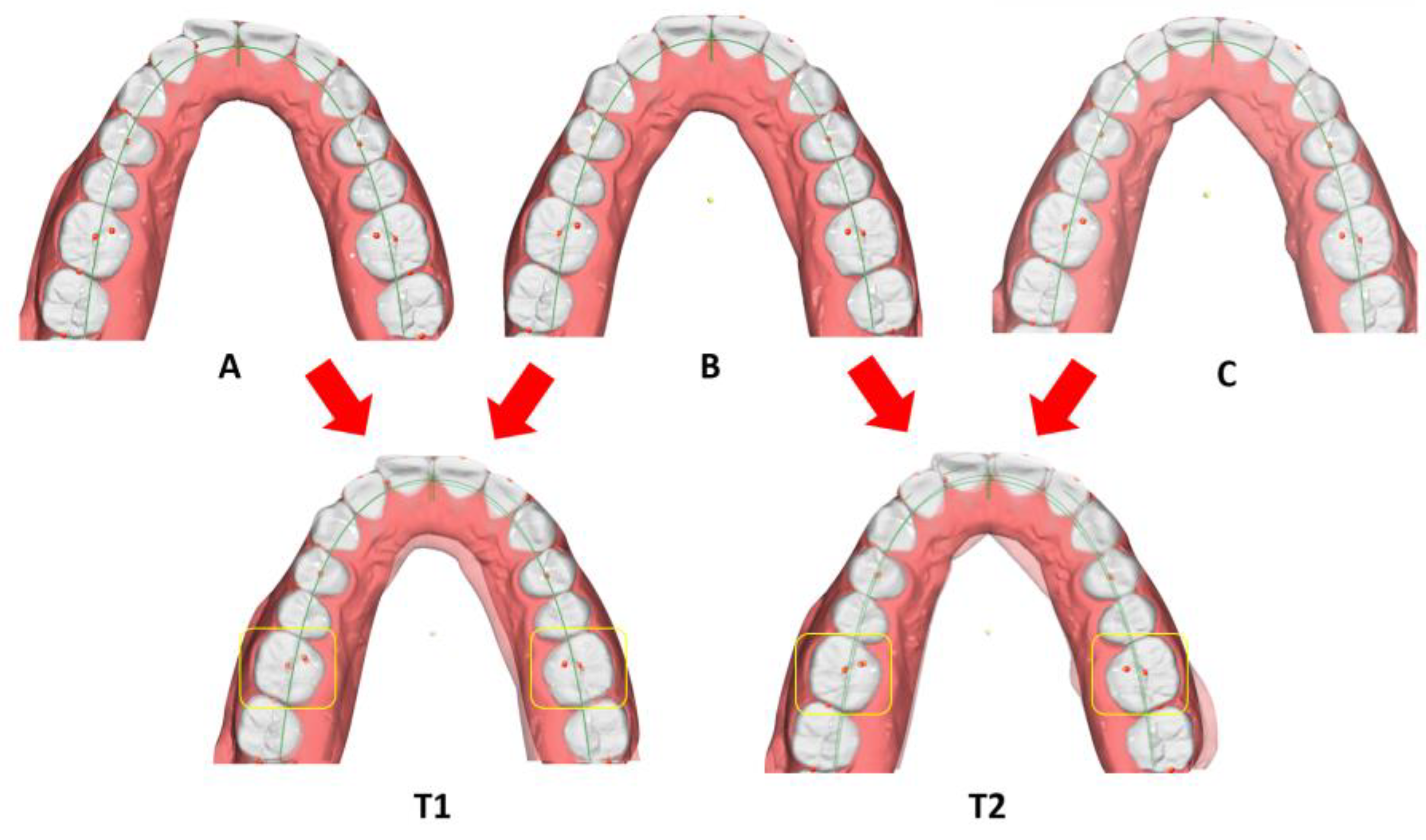
  • Inter-canine distance (Ic): measured between the tips of the canine cusps;
  • Inter-premolar distance (Ip1): measured between the vestibular cusps on the first premolars;
  • Inter-premolar distance (Ip2): measured between the vestibular cusps on the second premolars;
  • Inter-molar distance (Im): measured between the mesiovestibular cusps on the first molars (Figure 4).
Both angular and linear measurements were imported into Excel (Microsoft, Redmond, USA) for further analysis.

2.3. Analysis of Measurements

After obtaining the various angular values at T1 and T2, the following data were obtained for each movement analyzed and for each tooth:
  • Imprecision = the difference between the prescribed movement (T1) and that clinically achieved (T2), expressed as absolute values.
Imprecision = |T1 − T2|
  • Accuracy = the difference between the movement achieved (T2) and that prescribed (T1), expressed as absolute values and percentages (%).
Accuracy = [|T2/T1|] × 100
The imprecision of CAT in terms of linear intra-arch measurements (T0, T1 and T2), i.e., the difference between the planned (T1) and obtained (T2) measurements was calculated via the following formula, and absolute values expressed:
  • Imprecision = the difference between the prescribed movement (T1) and that clinically achieved (T2).
Imprecision = |T1 − T2|

2.4. Statistical Analysis

All the data obtained were synthesized and expressed as means and standard deviation (SD) for each movement analyzed and for each tooth type (incisors, canines, premolars and molars), per arch (maxilla and mandible) and for both (total). The following data are reported considering the absolute values of the movements prescribed (|T1|) and those achieved (|T2|). The average values of imprecision (|T1 − T2|) and accuracy [|T2/T1|] × 100 were also subjected to descriptive analysis.
After verifying the normal distribution of the data obtained through the Anderson–Darling test for each single angular movement, the accuracy of each individual movement was investigated by comparing the various tooth types in the two arches using the Kruskal–Wallis test. Any relationship between accuracy (dependent variable) and independent variables was investigated using chi-squared automatic interaction detection (CHAID) regression trees [35], as a function of recursive partitions (splits) of the data. The independent variables investigated were the amount of prescribed movement (T1) not expressed as an absolute value, tooth type (incisors, canines, premolars and molars for both arches), presence or absence of grip points and age and sex of the subjects examined.
As regards linear intra-arch distances, the mean initial (T0), prescribed (T1) and achieved (T2) measurements were reported as means and SD. ANOVA was carried out to verify the presence or absence of statistically significant differences between the linear measurements (T0, T1 and T2). Significantly different measurements were directly compared (T0 − T1; T0 − T2; T1 − T2) via the Siegel–Tukey test.
After 4 weeks, intra-arch linear distance measurements were re-performed on 50% of the total sample to verify the repeatability of linear measurements. The method error was calculated via the Dahlberg test, and the systematic error using Student’s dependent t-test. Dahlberg values were in the range between 0.071 mm and 0.294 mm, with an average value of 0.205 mm. The average p value was 0.43, and in no case was there a statistically significant difference between the various measurements, evidencing the good repeatability of the same.
All statistical analyses were performed with a significance level set at 0.05 (p ≤ 0.05 considered significant).
On the basis of a sample of 120 cases, it is possible to affirm that the probability of rejecting the hypothesis of equality between averages of inclination between the study sample and the control sample is greater than π = (1 − β) = 0.9999, when the averages between them actually differ by 1. The probability of rejecting the hypothesis of equality when instead the averages are equal is 1% (α = 0.01) [36].

3. Results

Starting from a total of 254 cases affected by dental Class I malocclusion, after the use of inclusion and exclusion criteria, a final sample composed of 120 patients was investigated (Figure 1).
The first phase of CAT with F22 aligners was accomplished with a mean number of aligners of 14.61 and 14.50, mean IPR values of 0.15 mm and 0.18 mm, and a mean number of grip points of 3.8 and 4 for maxillary and mandibular arch, respectively. Almost one posterior tooth (first or second molars) was not moved in the virtual set-up planning and thus it was used as tooth reference for the superimposition phase.
In the descriptive analysis, means and SD were calculated for prescribed (|T1|), achieved (|T2|) and imprecision values (|T2 − T1|) of investigated movements (inclination, angle and rotation). In all cases, a reasonably appropriate observation number was obtained, with the exception of the tooth type molars (n = 19 and n = 21 for inclination, n = 2 and n = 3 for angulation and n = 27 and n = 18 for rotation, considering the maxillary and mandibular molars, respectively) (Table 1).
Regarding the accuracy [(|T2/T1|) × 100], a total mean value of approximately 86.76 ± 0.44.89% (n = 975) was recorded for inclination. This was highest for the maxillary incisors, at 91.25 ± 53.54% (n = 192), and lowest for the mandibular incisors, at 82.29 ± 34.19% (n = 238). Mean angulation accuracy was 88.01 ± 55.68% (n = 719), with a high of 98.29 ± 73.42% (n = 42) at the maxillary premolars and a low of 72.73 ± 41.45% (n = 97) at the maxillary canines. Rotation accuracy was on average 61.59 ± 32.54% (n = 1224), and highest at the mandibular incisors, 72.64 ± 32.06% (n = 307), and lowest at the maxillary canines, 49.59 ± 29.43% (n = 128). Considering the average values of the two arches separately, there were no obvious differences in inclination or angulation accuracy, but there was a slight difference in rotation accuracy, which was 59.45 ± 31.31% (n = 600) in the maxillary arch and 63.67 ± 33.56% (n = 624) in the mandibular (Table 2).
Table 3 shows pairwise statistical comparisons of the accuracy at the various tooth types in the two arches. While no statistically significant differences were found in inclination, there were statistically significant differences in angulation accuracy in two pairwise comparisons, namely maxillary incisors (91.16 ± 58.31%) and maxillary canines (72.3 ± 41.45%), with p = 0.007, and maxillary incisors (91.16 ± 58.31%) and mandibular premolars (79.51 ± 69.65%), with p = 0.006.
However, eight pairs displayed statistically significant differences in rotation accuracy. Specifically, rotation was significantly more accurate at the mandibular incisors (=72.64 ± 32.06%) than at the mandibular canines (57.70 ± 28.90%, p < 0.001), mandibular premolars (54.19 ± 35.39%, p < 0.001), maxillary canines (49.59 ± 29.43%, p < 0.001) and maxillary incisors (61.28 ± 28.28%, p < 0.001), but significantly less accurate at the maxillary canines (46.59 ± 29.43%) than at the maxillary incisors (61.28 ± 28.28%, p < 0.001) and maxillary premolars (62.86 ± 35.03%, p < 0.001), and at the mandibular premolars (54.19 ± 35.79%) than at both the maxillary premolars (62.86 ± 35.03%, p = 0.044) and maxillary incisors (61.28 ± 28.28%, p < 0.001) (Table 3).
The analysis of the CHAID regression trees reveals that mean inclination accuracy is not influenced by any of the variables taken into consideration, i.e., amount of prescribed movement (T1), tooth type and presence or absence of grip points.
As for the angulation, distal angulation in the T1 range −20.1° to −0.2°was more accurate (accuracy 67.9–76.3%, n = 422) than mesial angulation in the T1 range −0.2° to 19.6° (accuracy 59.4–67.9%, n = 297). Distal tipping accuracy was of between 51% and 59.4% at the mandibular premolars (n = 42), 59.4% and 67.9% at the maxillary incisors and mandibular canines (n = 179) and 84.8% and 93.2% at the maxillary and mandibular incisors (n = 201). Paradoxically, grip points seemed to lower the effectiveness of T1 between −0.2° and 19.6° (n = 52), as they yielded an accuracy value of between 42.5% and 51%, as compared to the range 59.4% to 67.9% when they were absent (n = 245) (Figure 5).
As for rotation, both maxillary and mandibular canines and the mandibular premolars displayed an average accuracy of between 40.6% and 45.1% (n = 445), the mandibular incisors of between 58.4% and 62.8% (n = 307), and the maxillary incisors and premolars of between 49.5% and 53.9% (n = 472). Grip points increased the rotation accuracy at both the mandibular incisors and the maxillary incisors and premolars. Indeed, at the mandibular incisors, the presence of grip points raised the accuracy to between 80.6% and 85% (n = 15), as compared to the range 53.9% to 58.4% (n = 292) recorded for the group without grip points. Furthermore, at the maxillary incisors and premolars, grip points increased the accuracy to 58.4–62.8% (n = 88), as compared to 49.5–53.9% (n = 384) in the group without grip points (Figure 5).
Figure 6 and Figure 7 show the CHAID regression trees investigating the mean total accuracy as influenced by the patient variables age and sex. Inclination and angulation were affected by age, but not sex. Specifically, inclination accuracy was 67.5–69.3% in the age range 13–23.5 years (n = 216), 83.7–85.5% between 23.5 and 33.5 years (n = 490), and 67.5–69.3% between 33.5 and 64 years (n = 526). As for angulation, for the age range 13–23.5 years the accuracy was 58.7–61.2% (n = 200), between 23.5 and 53.5 years it was 71.1–73.6% (n = 637), and between 53.5 and 64 years it was 48.8–51.2% (n = 81). Rotation accuracy showed a similar pattern, being 39.8–44.7% for the age range 13 to 23.5 years (n = 333), rising to 54.5–59.4% in the age range 23.5–33.5 years (n = 612), and falling to 44.7–49.6% in the age range 33.5–64 years (n = 599). In the older group (33.5–64 years of age), there was a significant sex-related difference in rotation effectiveness, as clinical accuracy was on average higher in females (44.7–49.6%, n = 462) than in males (34.9–39.8%, n = 137). In the female group, the accuracy was lower in those aged between 33.5 and 53.5 years (n = 361), between 44.7% and 49.6%, than in those aged 53.5 to 64 years (n = 101), which was between 54.5% and 59.4%. An inverse trend was seen in the male group, with an average rotation accuracy of 49.6–54.5% in those aged between 33.5 and 43 years (n = 86), but 20.1–25% in those aged between 43 and 51 years (n = 27), and 10.3–15.2% in the 51–64-year age group (n = 24) (Figure 6 and Figure 7).
The descriptive analysis of the intra-arch distances is shown in Table 4, which reveals that the average imprecision (|T1 − T2|) was less than 1 mm for all distances investigated in both arches (Table 4).
However, statistical analysis via ANOVA revealed statistically significant differences between T0, T1 and T2 for all the distances investigated, with the exception of the maxillary measure Ip2 (F = 2.66, p = 0.072) and both maxillary (F = 0.71, p = 0.491) and mandibular Im (F = 1.06, p = 0.346). However, the subsequent post hoc comparative analysis revealed no statistically significant differences between prescribed (T1) and achieved (T2) intra-arch measurements, with the exception of the mandibular measure Ip2 (p = 0.002), although this difference was minimal (0.24 mm) (Table 5).

4. Discussion

The aim of the study was to evaluate the accuracy of tooth movement via the F22 Aligner system, comparing clinical results with respect to those digitally planned, in a large sample affected with mild–moderate Class I malocclusion, without resorting to auxiliaries and adhering to literature recommendations during the virtual setup procedure [10,22,37]. The F22 alignment system was created after various research efforts and clinical testing in the University, before its distribution, and has some characteristics that distinguish it from other clear aligner systems present in the global market and described in the literature [19,24,26,27,28,29,30,31,38]. The treatment of the sample involved planned movements that were purely coronal, while movements considered unpredictable, such as radicular and translation movements, were avoided [12,15]. The study was designed both to consider angular movements (inclination, angulation and rotation) and to investigate the expansive capacity of the F22 Aligner system, quantifying the change in intra-arch transverse measurements in both arches. The analyses were performed taking into account the different tooth types, whose responsiveness to orthodontic forces are conditioned by their different crown and root morphology [38], as well as their positions in different sections of the arch, with their different bone densities [39].
Despite this system not being the most widespread in the literature and on the market, findings could be generalized to other CAT systems, if attention is paid to what the literature has stated.
The prescribed (T1) and achieved movements (T2) were measured via superimposing digital models using unmoved posterior teeth as reference landmarks. This explains why the number of molar measurements was very low, and the molar tooth type was therefore omitted from the subsequent comparative analyses. The principle used for superimposition was “automatic best-fit registration”, which as Adel et al. have reported yields excellent agreement for rotation movements (>0.90), is good for tip (0.890) and moderate for torque (0.740) [34].
This analysis showed that the F22 Aligner is most accurate for angulation (88.01% ± 55.68%, n = 719), closely followed by inclination (86.76% ± 44.89%, n = 975). This is in line with findings by Papadimitriou et al. [37], Haouili et al. [8], Lombardo et al. [38] and Bilello et al. [21], who all agree that crown tipping movements are the most readily achieved movements generally in CAT. Furthermore, a comparison between the various tooth types showed no differences in inclination accuracy, but in angulation there were significant differences in accuracy when comparing the maxillary incisors (91.16% ± 58.31%, n = 229) with both the maxillary canines (72.73% ± 41.45%, n = 97) and the mandibular premolars (79.51% ± 69.65%, n = 174). This is likely due both to the coronal morphology (the premolars, especially mandibular, have a short clinical crown) and radicular morphology (canines have a very long root). Although our results are slightly different from those of Lombardo et al. in this regard, the discrepancy could be due to the different recording method and the larger sample analyzed [38].
The lowest average accuracy was found for rotation (61.59% ± 32.54%, n = 1224). This has also been found in other studies in the literature, which report an average accuracy ranging from 40% to 66.8% [7,9,10,34]; Bilello et al. found greater accuracy, about 86%, also considering the finishing stage in the Invisalign system [21].
As reported in the literature, with the Invisalign system, the teeth whose rotation is least accurately corrected are the rounded teeth (canines and premolars) [10,23,37], as their coronal morphology prevents the transmission of adequate and effective force couples at the crown [36]. Moreover, the roots are relatively large, which makes the movement even more difficult to achieve. In this regard, our data are perfectly in line with those reported by other authors with the Invisalign system, with a rotation accuracy that ranged from 49.59% ± 29.43% (n = 128) for maxillary canines to 62.86% ± 35.03% (n = 152) for maxillary premolars, and 57.70% ± 29.90% (n = 143) for mandibular canines and 54.19% ± 35.79% (n = 174) for mandibular premolars. On the whole, the imprecision of the rotation of the rounded teeth was between 2.57° ± 2.16° and 4.38° ± 3.23°, in line with results reported by Charalampakis et al., who recorded a 3.05° discrepancy between the planned and achieved rotation of the mandibular canines [23].
Kravitz et al. found that neither the use of ellipsoidal brackets nor IPR can increase the rotation accuracy substantially in the Invisalign system, although a slight improvement was noted [8]. The same authors highlighted that the accuracy of maxillary canine rotation decreased considerably if >15° was planned, falling from 35.8% to 18.8%. They suggest overcorrections be incorporated in the attempt to overcome this problem [7].
Similarly, Simon et al. reported a decrease in accuracy from 43.3% to 23.6% when programming derotations > 15° for premolars, and recommended reducing the derotation speed by <1.5° per step to increase predictability [14]. Overall, however, it is evident that generally CAT struggles to effectively rotate the rounded teeth, and despite the introduction of purportedly more effective clinical protocols and new materials, the situation has not improved much over time.
The teeth that F22 Aligner system derotated with greatest efficiency were the mandibular incisors (72.64% ± 32.06%, n = 307), especially when compared to the mandibular canines, mandibular premolars, maxillary canines and maxillary incisors. This could be explained by the “paddle-shaped” coronal morphology of the mandibular incisors [40] and the smaller root surface to be moved.
Analysis of the variable amounts of prescribed movement, tooth type and presence or absence of grip points revealed that these only have an effect on angulation and rotation movements. As for the former, it seems that mesial coronal tipping (accuracy from 59.4% to 67.9%, n = 297) is less predictable than the distal coronal tipping (from 67.9% to 76.3%, n = 422); this could be due to the distal thrust surface generally being smaller than the mesial one [41]. The effectiveness of distal tipping also appears to depend on the different crown heights of the various tooth types, as reflected by the different accuracy figures for the various tooth groups, with the lowest being recorded for the mandibular premolars (from 51% to 59.4%, n = 42), intermediate for the maxillary incisors and mandibular canines (from 59.4% to 67.9%, n = 179), and highest for the maxillary canines, mandibular premolars and mandibular incisors (from 84.8% to 93.2%, n = 201). However, this pattern was not repeated when mesial tipping was planned, perhaps because the distal coronal thrust surface is more homogeneous across the various tooth types [41].
It was also interesting to note that the presence of grip points tends to lower the percentage movement obtained through mesial tipping (from 59.4 to 67.9%, n = 245 to 42.5 to 51%, n = 52). Although grip points were not positioned to increase the angulation accuracy, but rather to facilitate rotation (for rotations > 10° of rounded teeth, and rotations > 20° of maxillary and mandibular lateral incisors), it can be assumed that major derotation reduces the predictability of tipping angulation movement prescribed for the same teeth. Alternatively, if CAs fit poorly in the later stages of CAT, grip points may even generate opposing forces, as previously reported by Simon et al. [14].
As for rotation, lower predictability was found for the canines, both maxillary and mandibular, and the mandibular premolars (range 40.6–45.1%, n = 445), whereas the rotation of the mandibular incisors (range 58.4–62.8%, n = 307), maxillary incisors and maxillary premolars (range 49.5–53.9%, n = 472) was more predictable. These results seem to be due to the coronal morphology of the rounded teeth, which do not lend themselves to derotation with CAs, as reported by Boyd [42]. The application of grip points does not seem to be detrimental in this regard, as reported by Kravitz et al. [7], and in fact would seem to increase the predictability of mandibular incisor rotation > 20°, although the number of observations was limited (n = 20). Nevertheless, the same benefit was seen for the >20° rotation of the maxillary incisors and premolars (from 49.5 to 53.9% to 58.4 to 62.8%, n = 88), so clinicians should consider their application in these cases.
A further objective of the study was to evaluate the influence of age and gender on F22 Aligner system accuracy. Age did seem to have an influence on either inclination or angulation accuracy. For both, there was a relatively lower predictability in the 13–23.5-year group than in the intermediate age group (23.5–33.5 years for inclination and 23.5–53.5 years for angulation), in which predictability was also greater than the older age group (33.5–64 years for inclination and 53.5–64 years for angulation). The same trend was found for rotation, with the F22 Aligner system being more predictable in the intermediate age group than in both the younger and older groups. This trend contrasts with that reported by Chisari et al. with the Invisalign system, as they found an S-shaped cubic relationship between age and the percentage movement obtained, which becomes quadratic and U-shaped in women and more linear in men [43]. Those authors recorded a lower predictability of movements in intermediate age, the opposite trend to that which we, like both Harris et al. and Dudic et al., found [44,45]. A potential explanation for the latter case could be the joint effect of biological response, which is more favorable in younger patients [46]., and compliance, which is usually greater in adults than in adolescents and pre-adolescents [47]. That being said, Chisari et al. investigated a sample that was four times lesser than that with respect to the current study, and they investigated the Invisalign system [43].
Another interesting observation was the gender difference found in the older group with regard to rotation. Specifically, a greater percentage of prescribed rotation was achieved in females (44.7–49.6%, n = 366) than in males (34.9–39.8%, n = 109), likely due to greater compliance by females [48]. Moreover, in females the predictability of movements increased with advancing age, while in males the trend found was precisely the opposite. This effect could be due to elderly females having a lower mineralized bone component, with a decrease in bone quality (osteoporosis) and consequently reduced resistance to orthodontic movement. In males, on the other hand, the opposite condition would be more frequent, i.e., a reduction in the clastic component (osteopetrosis) [43].
As for intra-arch widths, F22 aligners demonstrated adequate expansive capacity, with average imprecision that was less than 1 mm in all cases. The only statistically significant difference between prescribed (T1) and achieved movement (T2) in this regard was the inter-premolar distance at the mandibular first premolars. This may be due to the morphology of the mandibular first premolars, which have a reduced crown height on the lingual thrust side [41], but in any case, the difference may be considered clinically minimal (0.24 mm).
These conclusions are comparable with those by both Houle et al. and Lione et al. for the Invisalign system, who reported that CAT produces highly accurate changes in transversal dimensions, although those authors mainly prescribed bodily expansion, while in this study only crown tipping movements were planned, in accordance with the literature evidence [20,49]. In general, however, it would appear that CAs manage to widen the arches satisfactorily, albeit, as reported by Galan-Lopez et al., to a lesser extent than self-ligating appliances [50].
Overall, the above data indicate that the F22 Aligner system is a valid method for malocclusion cases of mild-to-moderate complexity, although rotation, particularly of rounded teeth, remains problematic, and accuracy is not significantly improved by the application of rotation grip points except for incisors and maxillary premolars. Clinicians should therefore consider planning overcorrections or using various auxiliaries, such as derotation chains or partial fixed appliances to improve rotation accuracy, or indeed correcting such rotations before using CAs. In addition, the clinician should take into account demographic factors, namely age and sex, as these were found to influence the predictability of the movements achieved. In particular, a greater number of finishing steps, overcorrections or attenuated movements should be planned for both younger (from 13 to 23.5 years of age) and older patients (>33.5 years for inclination and rotation and >53.5 years for angulation).

Limitations of the Study

Although the following study has the merit of investigating a large number of patients, it has limitations that must be taken into account. Firstly, the design was retrospective, which could increase sampling bias and affect sample homogeneity, although in this case the size of the sample likely minimized these potential shortcomings. Another possible drawback was the method used for digital superimposition, which was based on teeth that had no prescribed CAT movement, although small counter-reactions on these teeth cannot be excluded. Superimpositions would be better made on stable portions of the arch, such as the gum tissue, although this is often deficient, especially in the mandibular arch. In addition, this study considered the position of the dental crowns, so root movements cannot be considered objectively. Further studies involving cone-beam computed tomography would be desirable in this regard.

5. Conclusions

From the results of the following study, it is possible to conclude that:
  • In mild-to-moderate cases of Class I malocclusion, the F22 Aligner system displays excellent accuracy for inclination (86.76% ± 44.89%) and angulation movements (88.01% ± 55.68%), but only moderate for rotation (61.59% ± 32.54%).
  • It is least accurate at the derotation of rounded teeth, in particular maxillary (49.59% ± 29.43%) and mandibular canines (57.70% ± 28.90%) and mandibular premolars (54.19% ± 35.79%). The derotation of maxillary premolars is more accurate (62.86% ± 35.03%), presumably due to their squarer crowns, and they were positively affected by the application of grip points.
  • The percentage of prescribed angulation achieved depends on the amount of prescribed movement, the direction of movement (difference between distal and mesial tipping) and the tooth type (greater for maxillary premolars and canines, and mandibular incisors). Moreover, the presence or absence of grip points in mesial crown tipping presents a paradoxical effect.
  • The percentage of rotation achieved depends on the tooth type, and is lowest for the rounded teeth, which is not improved by the application of grip points, with the exception of the maxillary premolars. For the remaining tooth types investigated, the accuracy is greater, and improves with the use of grip points.
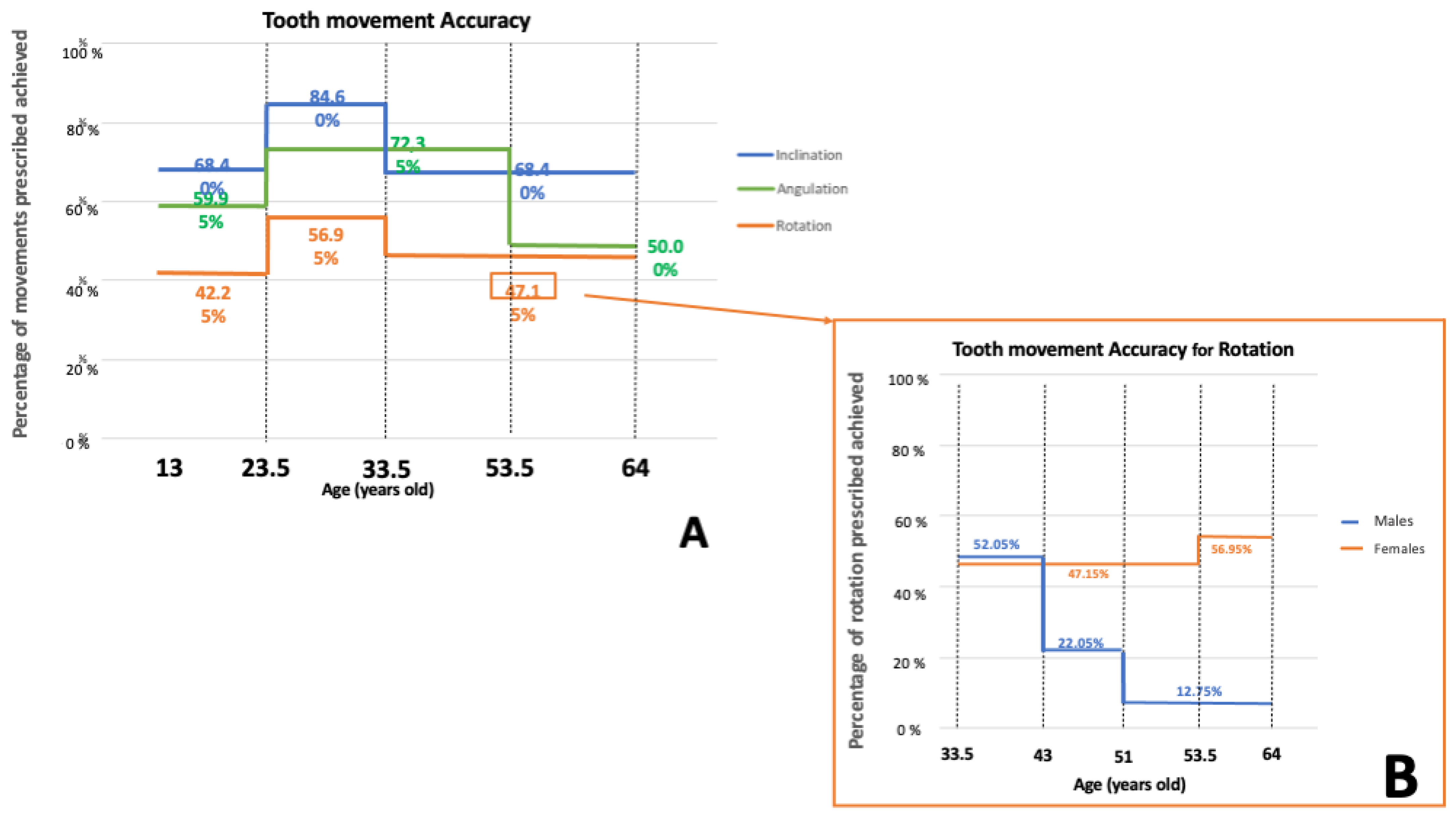
  • For all the movements investigated, the variables sex and age influence the percentage of movement achieved, which is greater in the intermediate age group (young adult patients). As for rotation, in the older group the trend seen in females (inverse relationship between accuracy and age) was reversed in males (direct relationship between accuracy and age).
  • The F22 Aligner system seems to have a good expansive capacity, with differences between prescription and result always less than 1 mm. The only region where there is a significant difference between prescribed and achieved expansion was the distance between the mandibular first premolars, although the discrepancy was clinically minimal (0.24 mm).

Author Contributions

Conceptualization, L.L., P.M. and S.S.d.V.; methodology, L.L., P.M. and S.S.d.V.; software, P.M. and F.C.; validation, L.L., P.M. and S.S.d.V.; formal analysis, P.M., R.S. and P.F.; investigation, S.S.d.V.; resources, S.S.d.V.; data curation, P.M., S.S.d.V. and P.F.; writing—original draft preparation, S.S.d.V., P.M. and P.F.; writing—review and editing, P.M. and P.F.; visualization, L.L., P.M. and S.S.d.V.; supervision, L.L. and P.M.; project administration, L.L. and P.M. All authors have read and agreed to the published version of the manuscript.

Funding

This research received no external funding.

Institutional Review Board Statement

The study was conducted in accordance with the Declaration of Helsinki, and approved by the Institutional Review Board (or Ethics Committee) of the University of Ferrara (protocol code 9/2020, 7 January 2020).

Informed Consent Statement

Informed consent was obtained from all subjects involved in the study.

Data Availability Statement

All authors assured that all data and materials as well as software application or custom code support their published claims and comply with field standards. The raw data supporting the conclusions of this article will be made available by the corresponding author on request.

Acknowledgments

Authors give special thanks to Teresa Oliverio, Angela Arreghini, Niki Arveda, Maria La Rosa, Carlucci Antonella and Giuseppe Siciliani, who contributed to the development of the F22 Aligner system.

Conflicts of Interest

The authors declare no conflicts of interest.

References

  1. Gierie, W.V. Clear aligner therapy: An overview. J. Clin. Orthod. 2018, 52, 665–674. [Google Scholar] [PubMed]
  2. Jeremiah, H.G.; Bister, D.; Newton, J.T. Social perceptions of adults wearing orthodontic appliances: A cross-sectional study. Eur. J. Orthod. 2011, 33, 476–482. [Google Scholar] [CrossRef] [PubMed]
  3. Rossini, G.; Parrini, S.; Castroflorio, T.; Deregibus, A.; Debernardi, C.L. Periodontal health during clear aligners treatment: A systematic review. Eur. J. Orthod. 2015, 37, 539–543. [Google Scholar] [CrossRef] [PubMed]
  4. Miller, K.B.; McGorray, S.P.; Womack, R.; Quintero, J.C.; Perelmuter, M.; Gibson, J.; Dolan, T.A.; Wheeler, T.T. A comparison of treatment impacts between Invisalign aligner and fixed appliance therapy during the first week of treatment. Am. J. Orthod. Dentofac. Orthop. 2007, 131, 302.e1–302.e9. [Google Scholar] [CrossRef] [PubMed]
  5. Henick, D.; Dayan, W.; Dunford, R.; Warunek, S.; Al-Jewair, T. Effects of Invisalign (G5) with virtual bite ramps for skeletal deep overbite malocclusion correction in adults. Angle Orthod. 2021, 91, 164–170. [Google Scholar] [CrossRef]
  6. Vidal Bernárdez, M.L.; Vilches Arenas, Á.; Sonnemberg, B.; Solano Reina, E.; Solano Mendoza, B. EX30 vs. SmartTrack materials in maxillary expansion with the Invisalign system. J. Clin. Orthod. 2022, 56, 343–350. [Google Scholar]
  7. Kravitz, N.D.; Kusnoto, B.; BeGole, E.; Obrez, A.; Agran, B. How well does Invisalign work? A prospective clinical study evaluating the efficacy of tooth movement with Invisalign. Am. J. Orthod. Dentofac. Orthop. 2009, 135, 27–35. [Google Scholar] [CrossRef]
  8. Haouili, N.; Kravitz, N.D.; Vaid, N.R.; Ferguson, D.J.; Makki, L. Has Invisalign improved? A prospective follow-up study on the efficacy of tooth movement with Invisalign. Am. J. Orthod. Dentofac. Orthop. 2020, 158, 420–425. [Google Scholar] [CrossRef] [PubMed]
  9. Zheng, M.; Liu, R.; Ni, Z.; Yu, Z. Efficiency, effectiveness and treatment stability of clear aligners: A systematic review and meta-analysis. Orthod. Craniofac. Res. 2017, 20, 127–133. [Google Scholar] [CrossRef]
  10. Rossini, G.; Parrini, S.; Castroflorio, T.; Deregibus, A.; Debernardi, C.L. Efficacy of clear aligners in controlling orthodontic tooth movement: A systematic review. Angle Orthod. 2015, 85, 881–889. [Google Scholar] [CrossRef]
  11. Dai, F.F.; Xu, T.M.; Shu, G. Comparison of achieved and predicted crown movement in adults after 4 first premolar extraction treatment with Invisalign. Am. J. Orthod. Dentofac. Orthop. 2021, 160, 805–813. [Google Scholar] [CrossRef]
  12. Jiang, T.; Jiang, Y.N.; Chu, F.T.; Lu, P.J.; Tang, G.H. A cone-beam computed tomographic study evaluating the efficacy of incisor movement with clear aligners: Assessment of incisor pure tipping, controlled tipping, translation, and torque. Am. J. Orthod. Dentofac. Orthop. 2021, 159, 635–643. [Google Scholar] [CrossRef]
  13. Khosravi, R.; Cohanim, B.; Hujoel, P.; Daher, S.; Neal, M.; Liu, W.; Huag, G. Management of overbite with the Invisalign appliance. Am. J. Orthod. Dentofac. Orthop. 2017, 151, 691.e2–699.e2. [Google Scholar] [CrossRef]
  14. Simon, M.; Keilig, L.; Schwarze, J.; Jung, B.A.; Bourauel, C. Treatment outcome and efficacy of an aligner technique—Regarding incisor torque, premolar derotation and molar distalization. BMC Oral Health 2014, 14, 68. [Google Scholar] [CrossRef]
  15. Zhang, X.J.; He, L.; Guo, H.M.; Tian, J.; Bai, Y.X.; Li, S. Integrated three-dimensional digital assessment of accuracy of anterior tooth movement using clear aligners. Korean J. Orthod. 2015, 45, 275–281. [Google Scholar] [CrossRef] [PubMed]
  16. Papageorgiou, S.N.; Koletsi, D.; Iliadi, A.; Peltomaki, T.; Eliades, T. Treatment outcome with orthodontic aligners and fixed appliances: A systematic review with meta-analyses. Eur. J. Orthod. 2020, 42, 331–343. [Google Scholar] [CrossRef] [PubMed]
  17. Yassir, Y.A.; Nabbat, S.A.; McIntyre, G.T.; Bearn, D.R. Clinical effectiveness of clear aligner treatment compared to fixed appliance treatment: An overview of systematic reviews. Clin. Oral Investig. 2022, 26, 2353–2370. [Google Scholar] [CrossRef] [PubMed]
  18. Palone, M.; Baciliero, M.; Cervinara, F.; Maino, G.B.; Paoletto, E.; Cremonini, F.; Lombardo, L. Class II treatment of transverse maxillary deficiency with a single bone-borne appliance and hybrid clear aligner approach in an adult patient: A case report. J. World Fed. Orthod. 2022, 11, 80–94. [Google Scholar] [CrossRef] [PubMed]
  19. Lombardo, L.; Palone, M.; Carlucci, A.; Siciliani, G. Clear aligner hybrid approach: A case report. J. World Fed. Orthod. 2020, 9, 32–43. [Google Scholar] [CrossRef] [PubMed]
  20. Houle, J.P.; Piedade, L.; Todescan, R.J.; Pinheiro, F.H. The predictability of transverse changes with Invisalign. Angle Orthod. 2017, 87, 19–24. [Google Scholar] [CrossRef] [PubMed]
  21. Bilello, G.; Fazio, M.; Amato, E.; Crivello, L.; Galvano, A.; Currò, G. Accuracy evaluation of orthodontic movements with aligners: A prospective observational study. Prog. Orthod. 2022, 23, 12. [Google Scholar] [CrossRef]
  22. Buschang, P.H.; Ross, M.; Shaw, S.G.; Crosby, D.; Campbell, P.M. Predicted and actual end-of-treatment occlusion produced with aligner therapy. Angle Orthod. 2015, 85, 723–727. [Google Scholar] [CrossRef]
  23. Charalampakis, O.; Iliadi, A.; Ueno, H.; Oliver, D.R.; Kim, K.B. Accuracy of clear aligners: A retrospective study of patients who needed refinement. Am. J. Orthod. Dentofac. Orthop. 2018, 154, 47–54. [Google Scholar] [CrossRef] [PubMed]
  24. Lombardo, L.; Palone, M.; Longo, M.; Arveda, N.; Nacucchi, M.; De Pascalis, F.; Spedicato, G.A.; Siciliani, G. MicroCT X-ray comparison of aligner gap and thickness of six brands of aligners: An in-vitro study. Prog. Orthod. 2020, 21, 12. [Google Scholar] [CrossRef] [PubMed]
  25. Mantovani, E.; Castroflorio, E.; Rossini, G.; Garino, F.; Cugliari, G.; Deregibus, A.; Castroflorio, T. Scanning electron microscopy evaluation of aligner fit on teeth. Angle Orthod. 2018, 88, 596–601. [Google Scholar] [CrossRef] [PubMed]
  26. Palone, M.; Longo, M.; Arveda, N.; Nacucchi, M.; Pascalis, F.; Spedicato, G.A.; Siciliani, G.; Lombardo, L. Micro-computed tomography evaluation of general trends in aligner thickness and gap width after thermoforming procedures involving six commercial clear aligners: An in vitro study. Korean J. Orthod. 2021, 51, 135–141. [Google Scholar] [CrossRef] [PubMed]
  27. Cremonini, F.; Zabini, F.; Oliverio, T.; Bianchi, A.; Scalia, S.; Siciliani, G.; Lombardo, L. Optical properties of seven types of clear aligners before and after in vitro aging. J. Clin. Orthod. 2022, 56, 149–157. [Google Scholar]
  28. Lombardo, L.; Arreghini, A.; Maccarrone, R.; Bianchi, A.; Scalia, S.; Siciliani, G. Optical properties of orthodontic alignersߞSpectrophotometry analysis of three types before and after aging. Prog. Orthod. 2015, 16, 41. [Google Scholar] [CrossRef] [PubMed]
  29. Albertini, P.; Mazzanti, V.; Mollica, F.; Pellitteri, F.; Palone, M.; Lombardo, L. Stress Relaxation Properties of Five Orthodontic Aligner Materials: A 14-Day In-Vitro Study. Bioengineering 2022, 9, 349. [Google Scholar] [CrossRef] [PubMed]
  30. Lombardo, L.; Martines, E.; Mazzanti, V.; Arreghini, A.; Mollica, F.; Siciliani, G. Stress relaxation properties of four orthodontic aligner materials: A 24-hour in vitro study. Angle Orthod. 2017, 87, 11–18. [Google Scholar] [CrossRef]
  31. Palone, M.; Pignotti, A.; Morin, E.; Pancari, C.; Spedicato, G.A.; Cremonini, F.; Lombardo, L. Analysis of overcorrection to be included for planning clear aligner therapy: A retrospective study. Angle Orthod. 2023, 93, 11–18. [Google Scholar] [CrossRef]
  32. Bernabé, E.; Flores-Mir, C. Estimating arch length discrepancy through Little’s Irregularity Index for epidemiological use. Eur. J. Orthod. 2006, 28, 269–273. [Google Scholar] [CrossRef]
  33. Proffit, W.; Fields, H.; Larson, B.; Sarver, D. Contemporary Orthodontics, 6th ed.; Mosby: Maryland Heights, MO, USA, 2018. [Google Scholar]
  34. Adel, S.M.; Vaid, N.R.; El-Harouni, N.; Kassem, H.; Zaher, A.R. TIP, TORQUE & ROTATIONS: How accurately do digital superimposition software packages quantify tooth movement? Prog. Orthod. 2022, 23, 8. [Google Scholar]
  35. Zeileis, A.; Torsten, H.; Kurt, H. Model-Based Recursive Partitioning. J. Comput. Graph. Stat. 2008, 17, 492–514. [Google Scholar] [CrossRef]
  36. Pandis, N.; Polychronopoulou, A.; Eliades, T. Sample size estimation: An overview with applications to orthodontic clinical trial designs. Am. J. Orthod. Dentofac. Orthop. 2011, 140, e141–e146. [Google Scholar] [CrossRef]
  37. Papadimitriou, A.; Mousoulea, S.; Gkantidis, N.; Kloukos, D. Clinical effectiveness of Invisalign® orthodontic treatment: A systematic review. Prog. Orthod. 2018, 19, 37. [Google Scholar] [CrossRef]
  38. Lombardo, L.; Arreghini, A.; Ramina, F.; Ghislanzoni, L.T.H.; Siciliani, G. Predictability of orthodontic movement with orthodontic aligners: A retrospective study. Prog. Orthod. 2017, 18, 35. [Google Scholar] [CrossRef] [PubMed]
  39. Misch, C.E. Contemporary Implant Dentistry, 4th ed.; Mosby: Maryland Heights, MO, USA, 2008. [Google Scholar]
  40. Elkholy, F.; Mikhaiel, B.; Schmidt, F.; Lapatki, B.G. Mechanical load exerted by PET-G aligners during mesial and distal derotation of a mandibular canine: An in vitro study. J. Orofac. Orthop. 2017, 78, 361–370. [Google Scholar] [CrossRef]
  41. Nelson, S.J. Wheeler’s Dental Anatomy, Physiology and Occlusion, 11th ed.; Elsevier: Amsterdam, The Netherlands, 2020. [Google Scholar]
  42. Boyd, R.L. Predictability of Successful Orthodontic Treatment Using Invisalign. The Greater Philadelphia Society of Orthodontists Page. Available online: http://www.gpso.org/events/2003_outline.pdf. (accessed on 1 September 2023).
  43. Chisari, J.R.; McGorray, S.P.; Nair, M.; Wheeler, T.T. Variables affecting orthodontic tooth movement with clear aligners. Am. J. Orthod. Dentofac. Orthop. 2014, 145 (Suppl. S4), S82–S91. [Google Scholar] [CrossRef] [PubMed]
  44. Harris, E.F. Effects of patient age and sex on treatment: Correction of Class II malocclusion with the Begg technique. Angle Orthod. 2001, 71, 433–441. [Google Scholar] [PubMed]
  45. Dudic, A.; Giannopoulou, C.; Kiliaridis, S. Factors related to the rate of orthodontically induced tooth movement. Am. J. Orthod. Dentofac. Orthop. 2013, 143, 616–621. [Google Scholar] [CrossRef]
  46. Schubert, A.; Jäger, F.; Maltha, J.C.; Bartzela, T.N. Age effect on orthodontic tooth movement rate and the composition of gingival crevicular fluid: A literature review. J. Orofac. Orthop. 2020, 81, 113–125. [Google Scholar] [CrossRef] [PubMed]
  47. Torsello, F.; D’Amico, G.; Staderini, E.; Marigo, L.; Cordaro, M.; Castagnola, R. Factors Influencing Appliance Wearing Time during Orthodontic Treatments: A Literature Review. Appl. Sci. 2022, 12, 7807. [Google Scholar] [CrossRef]
  48. Weiss, J.; Eiser, H.M. Psychological timing of orthodontic treatment. Am. J. Orthod. Dentofac. Orthop. 1977, 72, 198–204. [Google Scholar] [CrossRef]
  49. Lione, R.; Paoloni, V.; Bartolommei, L.; Gazzani, F.; Meuli, S.; Pavoni, C.; Cozza, P. Maxillary arch development with Invisalign system. Angle Orthod. 2021, 91, 433–440. [Google Scholar] [CrossRef]
  50. Galan-Lopez, L.; Barcia-Gonzalez, J.; Plasencia, E. A systematic review of the accuracy and efficiency of dental movements with Invisalign®. Korean J. Orthod. 2019, 49, 140–149. [Google Scholar] [CrossRef]
Figure 1. Flow chart regarding the retrospective selection of the final sample investigated.
Applsci 14 01641 g001
Figure 2. Process of digital matching between the pre-treatment model, reference and post-treatment model, respectively, using Onyxceph 3TM software (Firma Image Instruments GmbH, Chemnitz, Germany). The matching was achieved using two posterior teeth, which were not moved, as reference landmarks after the segmentation.
Applsci 14 01641 g002
Figure 3. Synthesis of the digital matching process between pre-treatment (A), reference (B) and post-treatment model (C), and use of a dedicated algorithm to quantify planned movements (T1) and those achieved (T2).
Applsci 14 01641 g003
Figure 4. Summary of intra-arch linear measurements in the pre-treatment (A), reference (B) and post-treatment model (C). Ic: inter-canine width; Ip1: inter-first-premolar width; Ip2: inter-second-premolar width; Im: inter-molar width.
Applsci 14 01641 g004
Figure 5. Graphical representation of CHAID regression trees investigating the accuracy of tooth movement achieved for angulation (A) and rotation (B) with respect to the amount of prescribed movements, tooth type (incisors, canines, premolars and molars) and presence/absence of grip points (yes or no). All splits were identified with significance thresholds set at 0.05. n = number of observations; Y = average accuracy.
Applsci 14 01641 g005
Figure 6. Graphical representation of CHAID regression trees investigating the accuracy of tooth movement achieved for inclination (A), angulation (B) and rotation (C) movements by sex and age. All splits were identified with significance thresholds set at 0.05. n = number of observations; Y = average accuracy.
Applsci 14 01641 g006
Figure 7. Graphical representation of percentage of movements prescribed achieved for inclination, angulation and rotation, considering age variable (A,B). (B) Represents the percentage of rotation movements achieved between 33.5 and 64 years of age where both sex and age have an influence.
Applsci 14 01641 g007
Table 1. Descriptive analysis of prescribed (T1) and achieved movements (T2) and imprecision (|T1 − T2|) thereof by tooth type, arch (maxilla and mandible) and for both arches (total).
Inclination Angulation Rotation
Prescribed Movements (T1)Achieved Movements (T2)Imprecision (|T1 − T2|)Prescribed Movements (T1)Achieved Movements (T2)Imprecision (|T1 − T2|)Prescribed Movements (T1)Obtained Movements (T2)Imprecision (|T1 − T2|)
ArchTooth TypeN. ObservationsMean (°)SD (°)Mean (°)SD (°)Mean (°)SD (°)N. ObservationsMean (°)SD (°)Mean (°)SD (°)Mean (°)SD (°)N. ObservationsMean (°)SD (°)Mean (°)SD (°)Mean (°)SD (°)
MaxillaIncisor1924.311.773.852.361.701.262295.923.545.043.501.971.823208.666.125.334.613.492.68
Canine924.112.013.271.981.561.34974.311.863.072.001.911.601288.464.994.103.204.383.23
Premolar1594.511.853.772.081.321.11423.531.703.422.421.981.321525.663.403.562.622.572.16
Molar193.381.112.511.581.460.8622.900.603.952.051.451.05274.642.603.382.461.941.30
MandibleIncisor2385.022.734.252.841.441.112164.582.474.092.721.671.293078.186.145.954.942.922.37
Canine1234.511.874.072.441.601.47793.661.943.171.901.601.331439.795.535.503.944.483.00
Premolar1714.322.074.032.571.521.23563.571.442.942.412.041.411746.954.703.752.663.903.49
Molar213.591.333.522.341.820.9332.800.593.801.002.131.43183.701.271.920.982.061.14
Maxilla4624.301.843.652.191.531.233705.213.144.343.191.961.716277.725.414.573.943.382.73
Mandible5534.642.354.112.661.511.233544.202.263.702.551.721.336428.085.695.164.283.522.92
Total10154.532.243.792.581.811.937244.682.863.843.092.393.9412698.305.514.814.204.183.96
SD: standard deviation; N: number.
Table 2. Descriptive analysis of accuracy of each movement investigated. Accuracy is expressed as a percentage according to the formula [|T2/T1|] × 100.
InclinationAngulationRotation
ArchTooth TypeN. ObservationsMean (%)SD (%)N. ObservationsMean (%)SD (%)N. ObservationsMean (%)SD (%)
MaxillaIncisor19291.2553.5422991.1658.3132061.2828.28
Canine9282.4350.869772.7341.4512849.5929.43
Premolar15984.0035.874298.2973.4215262.8635.03
MandibleIncisor23882.2934.1921690.0648.5830772.6432.06
Canine12391.1549.017990.7953.3214357.7028.90
Premolar17190.9045.165679.5169.6517454.1935.79
Maxilla44386.2647.3936887.3357.1460059.4531.31
Mandible53287.1742.6835188.7154.1162463.6733.56
Total97586.7644.8971988.0155.68122461.5932.54
SD: standard deviation; N: number.
Table 3. Statistical comparison of accuracy [|T2/T1|] × 100 of each movement investigated by tooth type for both maxilla and mandible (p < 0.05 *).
Tooth Type/ArchInclinationAngulationRotation
p-ValueSignificancep-ValueSignificancep-ValueSignificance
Incisor–MaxillaCanine–Maxilla1.000NS0.007*<0.001*
Premolar–Maxilla1.000NS0.421NS1.000NS
Incisor–Mandible1.000NS0.931NS0.021*
Canine–Mandible0.939NS0.995NS0.228NS
Premolar–Mandible0.797NS0.006*0.001*
Canine–MaxillaPremolar–Maxilla0.997NS1.000NS0.038*
Incisor–Mandible0.997NS0.155NS<0.001*
Canine–Mandible0.706NS0.475NS0.609NS
Premolar–Mandible0.602NS0.996NS1.000NS
Premolar–MaxillaIncisor–Mandible1.000NS0.800NS0.212NS
Canine–Mandible0.838NS0.920NS0.664NS
Premolar–Mandible0.708NS1.000NS0.044*
Incisor–MandibleCanine–Mandible0.801NS1.000NS<0.001*
Premolar–Mandible0.601NS0.083NS<0.001*
Canine–MandiblePremolar–Mandible1.000NS0.204NS0.789NS
NS: not significant.
Table 4. Descriptive analysis of initial (T0), prescribed (T1) and achieved (T2) inter-arch distances and imprecision of the same (T2 − T1) for each arch.
Initial
(T0)
Prescribed
(T1)
Achieved
(T2)
Imprecision |(T2 − T1)|
ArchLinear MeasurementsN. of ObservationsMin (mm)Max (mm)Mean (mm)SD (mm)Min (mm)Max (mm)Mean (mm)SD (mm)Min (mm)Max (mm)Mean (mm)SD (mm)Min (mm)Max (mm)Mean (mm)SD (mm)
MaxillaIc11728.6240.3933.502.5429.7240.6334.382.0930.0940.5134.072.080.007.040.730.86
Ip111828.2650.3840.243.4533.8550.3841.512.7833.3850.7141.182.900.005.930.570.71
Ip211534.0356.7645.643.6334.4657.1246.643.3534.2157.1146.433.320.002.140.340.36
Im11837.7458.8648.553.7638.0658.9549.103.6237.9058.8948.953.590.001.730.320.38
MandibularIc12021.2131.7125.562.0622.3629.8826.792.0021.2430.7026.391.740.0010.720.771.18
Ip111827.0744.0933.212.7728.9442.0734.292.2128.7742.9134.052.210.004.260.640.61
Ip211730.3951.2938.773.1533.2652.9739.862.7733.1951.4539.552.780.002.130.480.44
Im11336.5254.1742.233.1137.4654.4142.823.0237.0553.6142.592.970.001.840.370.41
Ic: inter-canine width; Ip1: first inter-premolar width; Ip2: second inter-premolar width; Im: inter-molar width. SD: standard deviation; N: number.
Table 5. Descriptive analysis of initial (T0), prescribed (T1) and achieved (T2) inter-arch distances and imprecision of the same (T2 − T1) for each arch.
T0 − T1T0 − T2T1 − T2
ArchLinear MeasurementsNo. of ObservationsDifference (mm)p-ValueSignificanceDifference (mm)p-ValueSignificanceDifference (mm)p-ValueSignificance
MaxillaIc1170.880.01*0.570.13NS0.310.54NS
Ip11181.270.004*0.940.05*0.330.68NS
Ip21150.990.07NS0.790.19NS0.200.89NS
Im1180.550.48NS0.150.95NS0.410.67NS
MandibularIc1201.31<0.001*0.830.003*0.480.14NS
Ip11181.080.002*0.840.02*0.240.002*
Ip21171.090.01*0.780.10NS0.310.70NS
Im1130.590.32NS0.230.84NS0.360.65NS
N: number; NS: not significant; *: p > 0.05.
Disclaimer/Publisher’s Note: The statements, opinions and data contained in all publications are solely those of the individual author(s) and contributor(s) and not of MDPI and/or the editor(s). MDPI and/or the editor(s) disclaim responsibility for any injury to people or property resulting from any ideas, methods, instructions or products referred to in the content.

