Article

Enhanced Enzymatic Degradability of Livestock Blood Pretreated with Ultrasonic Technique

Environmental Technology Division, Korea Testing Laboratory, Seoul 08389, Republic of Korea
*
Author to whom correspondence should be addressed.
Appl. Sci. 2024, 14(4), 1676; https://doi.org/10.3390/app14041676
Submission received: 20 January 2024 / Revised: 13 February 2024 / Accepted: 17 February 2024 / Published: 19 February 2024
(This article belongs to the Section Materials Science and Engineering)

Abstract

:
Livestock blood, a major organic waste generated by the livestock industry, poses a risk of pollution due to its rapid decomposition. However, it is a potential protein source for agricultural purposes due to its protein-rich organic matter. In this study, we conducted studies on an eco-friendly, scalable, and effective protein degradation process using livestock blood to reduce waste and produce an amino acid liquid fertilizer that can be recycled for agricultural use. Ultrasonic technology was applied as a pretreatment method to improve the enzymatic hydrolysis efficiency of proteins in livestock blood, and the optimal conditions that led to 95.91% solubilization rate of hemoglobin were ultrasound duration for 30 min at an ultrasound density of 0.5 W/mL. As a result of hydrolyzing ultrasonically pretreated blood by mixing exo- and endo-type proteolytic enzymes, the optimal combination was a mixture of Savinase® 1% and Flavourzyme® 1%. After 4 h of reaction, the protein concentration was 27.8 mg/mL and the amino acid concentration was confirmed to be 54.6 mg/mL. This is about 4.2 times higher than the amino acid concentration of blood without ultrasound pretreatment, 13.1 mg/mL, and it was confirmed that sonication has a significant effect on improving protein degradation efficiency. As protein degradation increased, the viscosity of blood gradually decreased, suggesting that the physical force applied to the agitator torque diminished during the enzyme reaction; a significant correlation between protein and amino acid concentrations (biological factors) and torque (mechanical factor) was observed. Measuring torque during an enzyme reaction can confirm the extent of the enzyme reaction, so it can be used as an indicator of reaction progress when scaling up the process in the future.

1. Introduction

Livestock blood, which is produced during the bloodletting of animals in slaughterhouses, is among the major organic wastes generated by the livestock industry. With recent changes in human dietary habits due to economic growth, there has been a growing demand for livestock products, leading to an increase in the quantity of livestock blood generated. In particular, pigs are among the most commonly used livestock for meat production worldwide. Approximately 1.5–3 L of blood is produced from a slaughtered pig, which accounts for approximately 13% of the pig slaughter byproducts [1]. Pig livestock blood contains high-quality proteins (34–38% w/w), making it a valuable resource for potential reuse. However, the utilization of livestock blood as a valuable recycled resource has been extremely limited due to the appearance and odor of oxidized discarded blood and its susceptibility to spoilage and instability [2,3,4,5].
Livestock blood has two components: plasma and solid components, including red blood cells (RBCs). Plasma contains proteins such as albumin, globulin, and fibrinogen, while RBCs contain hemoglobin. When broken down or hydrolyzed, these components can serve as sources of high-quality amino acids and peptides [2,6]. For instance, amino acids and peptides derived from hemoglobin exhibit antioxidant, growth-promoting, and antimicrobial properties [7,8,9]. Therefore, the hydrolysis of livestock blood to produce high-quality amino acids and eco-friendly fertilizer contributes to waste reduction in the livestock industry and the transition to environmentally friendly agriculture.
High-quality amino acids can be produced under milder conditions through the utilization of proteolytic enzymes to create amino acid liquid fertilizer from livestock blood, which is more efficient than the chemical decomposition methods using acids or alkalis. Liquid amino acid fertilizers produced through enzymatic hydrolysis are more advantageous than the chemically produced ones; they are chloride-free, contain a wider range of amino acid components, promote plant growth, and activate beneficial microorganisms in the soil. Furthermore, liquid amino acid fertilizers are eco-friendly and cost-effective because they reduce the environmental pollution caused by chemical fertilizers and wastewater treatment costs [10]. Peptidase, protein-hydrolyzing enzymes, can be divided into exopeptidases and endo-peptidases depending on their cleavage patterns. Exopeptidases act on the ends of peptide chains and sequentially release amino acids, while endopeptidases act internally by cleaving various peptides. Due to the specificity of these two types of proteases, they can influence the formation of lysates with various peptide profiles. Accordingly, research on the enzymatic hydrolysis of livestock blood has primarily focused on its application as a raw material in food additives and pharmaceutical products [11,12,13,14]. While research has been consistently conducted on various types of proteases to produce amino acid fertilizers from different organic waste materials, such as fishery [15] and food waste [16], studies on the production of amino acid fertilizers using livestock blood as the raw material have not yet been published.
Low efficiency and high cost have been pointed out as major disadvantages of protein degradation reactions using enzymes [17,18]. One of the recently proposed approaches to improve enzyme degradation is the use of ultrasound as a technology that can accelerate enzymatic reactions [19,20]. Since blood cells contain more proteins than plasma, access to these proteins within the blood cells should be facilitated. The blood cell structures can hinder the hydrolysis of blood cell proteins. This problem can be addressed through ultrasonic treatment, which weakens the resistance of blood cell structures, to release the proteins within the cells into the plasma, thereby facilitating the degradation of livestock blood proteins into amino acids. Ultrasound induces a cavitation phenomenon, and the frequency of ultrasound has a significant impact on the formation of cavitation bubbles. In particular, destructive cavitation effects are more pronounced in the lower-frequency range compared with the higher-frequency range. Furthermore, intense ultrasound applied at lower frequencies (below 40 kHz) can effectively destroy biological cells and facilitate the elution of soluble organic materials [21,22]. A study on the ultrasonic treatment of sludge demonstrated that among the intracellular components, proteins exhibit particularly high solubility [23]. Similarly, high levels of depolymerization have been observed when treating high molecular weight substances with ultrasound [24,25]. Based on these research findings, it can be inferred that the ultrasonic treatment of blood aids in the solubilization of blood cell proteins and destroys various bacteria that may be present in the blood.
The aim of this study was to determine the feasibility of utilizing livestock blood as a high-quality amino acid fertilizer for agricultural purposes. Ultrasonic pretreatment was employed to enhance the solubilization of livestock blood proteins and improve the efficiency of enzymatic hydrolysis. Moreover, the optimal conditions for protein enzyme hydrolysis, both when applied individually and in combination based on enzyme type, were determined to achieve the highest possible amino acid conversion efficiency.

2. Materials and Methods

2.1. Livestock Blood Samples Used in the Study

2.1.1. Preparation of Livestock Blood Samples

The livestock blood samples used in the experiment were collected during bloodletting to prevent them from being contaminated with foreign substances; the bloodletting was conducted in a slaughterhouse in Korea. The collected samples were placed in a cool box (4 °C) and transferred to the laboratory within 30 min since the time of collection to maintain their freshness. In the laboratory, the samples were homogenized, and 1 L aliquots were obtained and refrigerated. Experiments were conducted within one week.
Since livestock blood coagulates in the air when discharged outside the living body, a pretreatment process was required to grind and homogenize the coagulated blood in order to check the effectiveness of the ultrasound treatment and enzyme degradation using livestock blood. The blood samples that had clotted prior to the experiment were ground using a cutter-type lab-scale grinder. In order to derive optimal grinding conditions, the hemoglobin solubilization rate was confirmed according to grinding speed and grinding time. The grinding time was maintained at 1 min and the grinding speed was increased from 3000 rpm to 15,000 rpm to confirm the hemoglobin solubilization rate. As the grinding speed increased, the hemoglobin concentration increased, showing a solubilization rate of 50.12% at about 10,000 rpm, and then did not increase any further above 10,000 ppm. To determine the optimal grinding time, the grinding speed was fixed at 10,000 rpm, and the hemoglobin solubilization rate was confirmed at 30 s intervals depending on the grinding time. As a result of the hemoglobin solubilization rate according to grinding time, the solubilization rate increased rapidly until 3 min, then showed a gradual increase, and did not increase further after 10 min. Therefore, the optimal grinding speed and time were set at 10,000 rpm and 10 min, and these conditions were applied and fixed as the grinding conditions before the experiments for the ultrasound treatment and enzyme degradation.

2.1.2. Physical Properties of Livestock Blood Samples

The basic characteristics of the livestock blood samples analyzed in this study are outlined in Table 1. The analysis revealed that the blood samples consisted of 79.18% moisture, 1.29% inorganic matter, and 19.53% organic matter, which was comprised mostly of proteins (accounting for 93.5% of organic matter). The protein content was 18.26%; hemoglobin accounted for 14.08% of the total proteins, constituting the largest proportion of the protein content. This result is consistent with the report of Bah et al. [26].
Since this study is preliminary research exploring the resource utilization of waste livestock blood to produce amino acid liquid fertilizer applicable to soil, the salt content in livestock blood is also an important assessment item. Salt cannot be easily eliminated once introduced into soil and may lead to the loss of soil nutrients by interfering with the nutrient and moisture absorption of plants; at high concentrations, it causes plasmolysis and destruction of plant tissue. Therefore, it is crucial to determine the salt concentration of livestock blood when utilizing it as a fertilizer. The average salt content in livestock blood was 0.95%, which is below the salt content limit (1.0%) specified in the fertilizer manufacturing standards in Korea. Therefore, additional preprocessing is not required to remove salts from livestock blood.

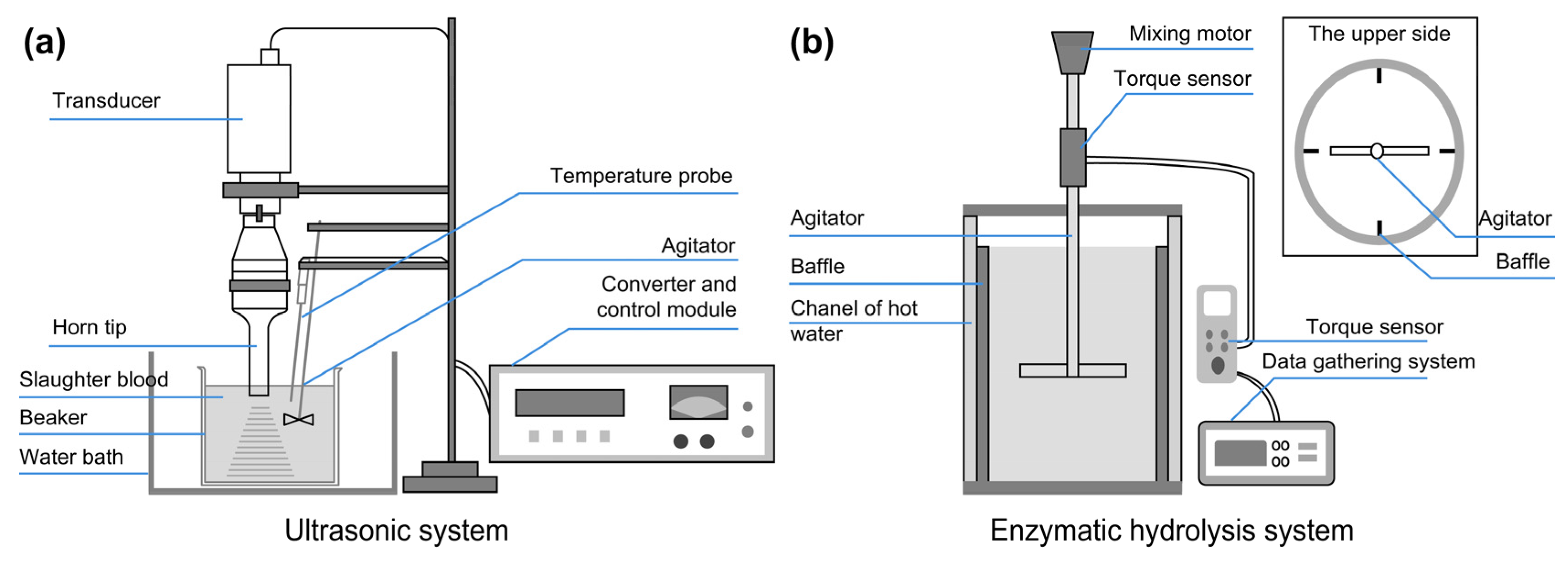
2.2. Method for Ultrasonic Treatment of Livestock Blood

Figure 1a illustrates the architecture of the ultrasonic treatment device for blood solubilization, which consists of an ultrasound generator and a reactor. The ultrasound generator consists of a converter, control module, transducer, and horn tip. The probe-shaped horn tip had a frequency of 20 kHz and a maximum power of 2000 W. The frequency of the horn tip was set to 20 kHz because previous studies have indicated that the low-frequency range (below 40 kHz) enables the effective destruction of cells [21,22], and subjecting cells to a 30 min treatment at 20 kHz resulted in high protein solubilization efficiency, while higher frequencies were less efficient for the ultrasonic treatment of proteins [27]. The reactor consisted of a 2 L beaker, a water bath for temperature control, a temperature sensor, and an agitator with a maximum speed of 1000 rpm.
The ultrasonic treatment experiments were conducted in a batch mode using 1 L of livestock blood as the starting material, with agitation at 200 rpm to ensure proper ultrasound dispersion. Experiments were performed to assess the solubilization efficiency of blood, with the ultrasonic irradiation density set between 0.1 and 2.0 W/mL and irradiation time from 10 to 60 min at 20 kHz frequency.

2.3. Method for Enzymatic Hydrolysis of Ultrasonic-Pretreated Livestock Blood

Figure 1b illustrates the architecture of the reactor for enzymatic hydrolysis as a double-jacketed vessel that uses a water bath to enable hot water supply and circulation and maintains a constant temperature within the reactor during enzymatic reactions. To ensure that the samples are evenly mixed during enzymatic reactions, four baffles were installed on the inner walls of the reactor.
We used commercial enzymes (Novozymes, Vagsbaerd, Denmark); their properties are presented in Table 2 [28]. Three different enzymes were selected: Savinase® and Alcalase® (serine-type endopeptidases) and Flavourzyme® (exopeptidase). Flavourzyme® is a fungal protease/peptidase complex produced by the submerged fermentation of a selected strain of Aspergillus oryzae. Alcalase® is produced by Bacillus licheniformis, while Savinase® is produced by submerged fermentation of a genetically modified microorganism. Each enzyme, utilized either individually or in combination, was added at a concentration of 2% based on the volume of the 3 L blood samples. The reaction temperature was maintained at 55 °C, which is the optimal temperature for enzyme activity. The samples were allowed to react for 4 h; samples were obtained every hour and analyzed to monitor protein and amino acid changes.
The enzymatic reaction decreases the protein content of blood and increases its amino acid content, which, in turn, gradually reduces the viscosity of blood. To monitor the progress of the enzymatic reaction and determine its completion, the physical force applied to the mixing torque during the reaction was measured by installing a torque sensor (EUROSTAR Power Control-Visc P4; IKA Works, Staufen, Germany) and a data gathering system (UtoEgineering, Namyangju, Republic of Korea). A 3 L sample of ultrasonically treated blood was mixed and left to react with the enzymes at 100 rpm and 55 °C for 4 h. Torque was automatically measured every 2 s during the 4 h reaction period, and the protein and amino acid concentrations were measured at 10 min intervals.

2.4. Analysis of Physicochemical Properties of Samples

The characteristics of the livestock blood used in the experiment were analyzed using standard methods to determine their moisture, organic matter, and inorganic matter content [29]. Sulfo-phospho-vanillin analysis was performed to quantify the total lipid content of the blood samples. The concentrations of various proteins in the blood, including total proteins, albumin, fibrinogen, platelets, and hemoglobin count, were estimated using an automatic hematology analyzer (Coulter Ac•T; Beckman Coulter, Carlsbad, CA, USA).
To determine the solubilization rate of the blood proteins after undergoing ultrasonic treatment, we performed hemoglobin quantification analysis using the cyanmethemoglobin method in the following order: hemoglobin within the blood proteins was reacted with potassium ferricyanide (K4[Fe(CN)6]·3H2O) to generate methemoglobin; the generated methemoglobin was reacted with potassium cyanide to form cyanmethemoglobin; and the concentration of the final product was determined using a UV-Vis Spectrophotometer (Cary 4000; Varian Medical Systems, Belrose, New South Wales, Australia) at a wavelength of 540 nm, utilizing hemoglobin standard reagents. The protein solubilization rate was calculated using the measured hemoglobin concentration according to the following formula [30]:
S R % = H t H u H w H u × 100 ( % )
where SR is the solubilization rate; Ht is hemoglobin in treated plasma; Hu is hemoglobin in untreated plasma; and Hw is hemoglobin in whole blood.
To evaluate the morphological changes in the blood samples subjected to ultrasonic treatment, the external appearances of the blood samples before and after treatment were compared using a phase contrast microscope (TS100F; NIKON, Tokyo, Japan). Changes in particle size distribution before and after treatment were assessed using a particle size analyzer (1090LD; CILAS, Orleans, France). Additionally, to determine whether any potential pathogens present in the pulmonary blood were eliminated, the dry rehydratable film method (3M™ Petrifilm, NEOGEN, Lansing, MI, USA) was applied to analyze the general bacteria and pathogenic Escherichia coli, while Salmonella sp. was analyzed using the standard plate culture method [31].
To assess the extent of protein degradation resulting from enzymatic hydrolysis, the concentrations of proteins and amino acids in the blood samples were analyzed.

3. Results and Discussion

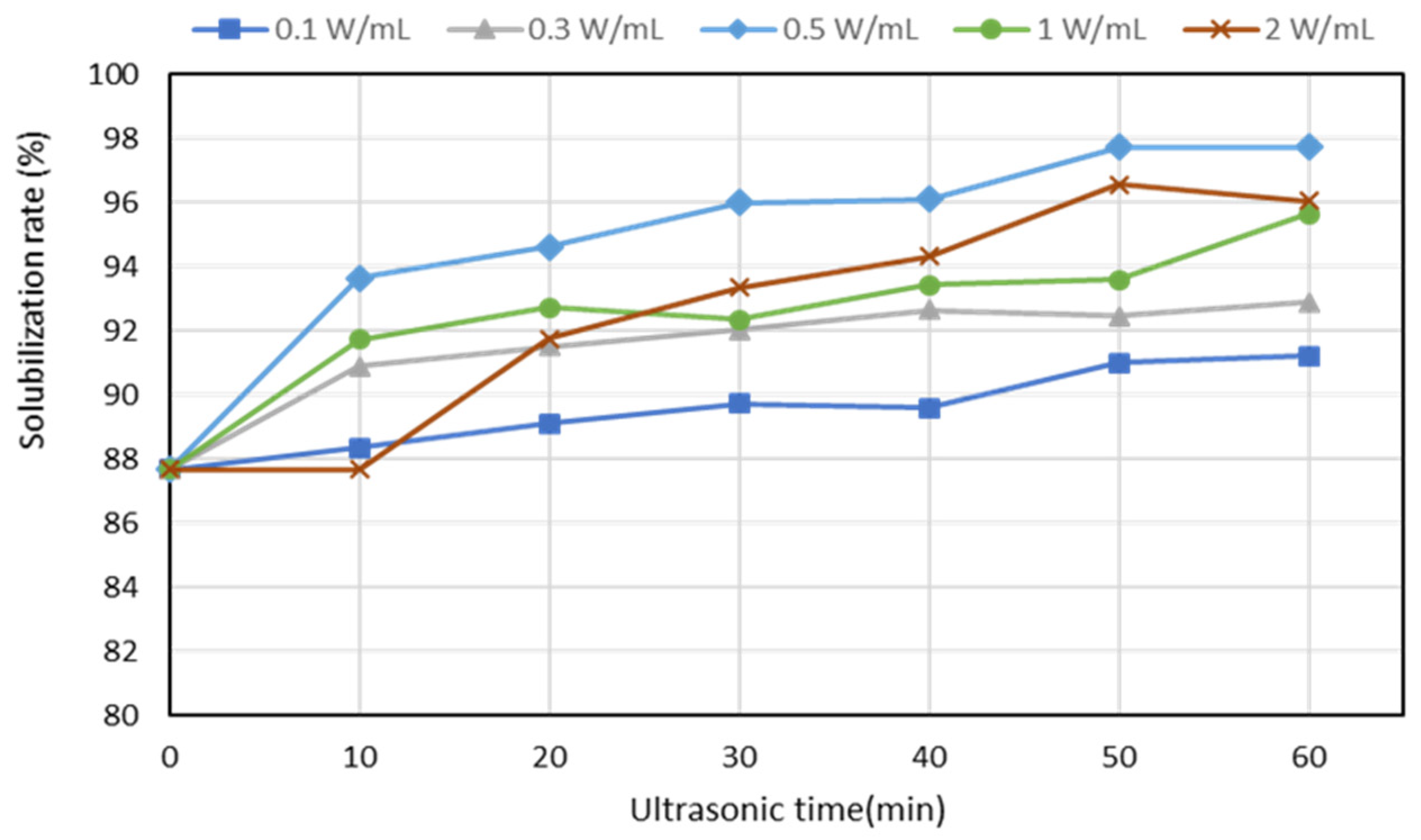
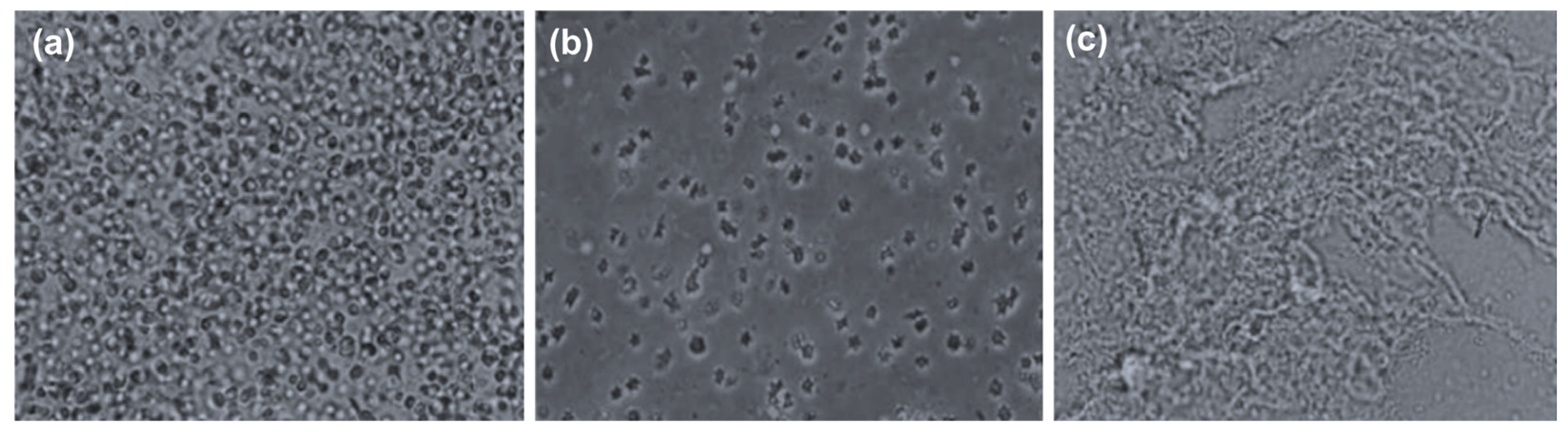
3.1. Protein Solubilization by Ultrasonic Pretreatment

The results of ultrasonic treatment for the solubilization of proteins in livestock blood are shown in Figure 2. At the irradiation densities of 0.1 and 0.3 W/mL, the maximum solubilization rate was approximately 91–93%. At the irradiation density of ≥0.5 W/mL, a maximum solubilization rate of 96–97% was achieved, although the density-dependent solubilization effect did not vary significantly. While higher ultrasonic irradiation densities are generally known to yield better ultrasound efficiency [32], our experiments did not yield any substantial difference in ultrasound efficiency with varying densities. Instead, we noticed that the solubilization rate increased as the ultrasonic duration increased. This difference may be attributed to the specific characteristics of the samples or the ultrasonic treatment methodology. In particular, blood can be easily hemolyzed even with minimal physical force, indicating a limited impact of ultrasonic density [33]. Our results revealed that an ultrasonic density of 0.5 W/mL and a duration of 30 min are the optimal ultrasonic treatment conditions for the solubilization of livestock blood.
Table 3 presents the changes in the characteristics of livestock blood before and after ultrasonic treatment. The untreated samples were not subjected to any processing, while the ultrasonic-treated samples were subjected to ultrasonic treatment at the optimal density and duration conditions of 0.5 W/mL for 30 min. The parameters analyzed were pH, electrolytes, chemical components, plasma hemoglobin content, protein solubilization rate, particle size, and microorganisms. The analysis revealed that the hemoglobin concentration in the plasma of the grinding treated samples was 5.59%, while that of the ultrasonic-treated samples was 14.63%, which corresponds to a solubilization rate of 95.91%. This result indicates that during the ultrasonic treatment, most of the hemoglobin RBCs were hemolyzed into plasma, resulting in an increased protein content in the plasma. Therefore, blood proteins can be solubilized without additional chemical treatments, making it conducive to efficient enzymatic hydrolysis in future biological processes.
The analysis of particle size distribution in the blood samples before and after ultrasound treatment revealed that the average particle size of the untreated samples was 131.16 μm, while that of the post-treated samples was 44.32 μm. This result indicates that both grinding and ultrasonic treatments contributed to the destruction of RBCs and breakdown of high molecular weight substances in the blood. This is further supported by the protein content in the untreated and post-treated blood samples, which were 387.0 g/L and 319.2 g/L, respectively. As mentioned earlier, this decrease in protein content is attributed to the disruption of protein chains caused by ultrasonic treatment. The protein content in the plasma of the post-treated samples was 2.5 times higher than that of the untreated samples. These results confirm that ultrasonic treatment disrupts RBCs, thereby enhancing the solubilization of proteins. The reduced particle size in the post-treated blood samples can be attributed to the decrease in protein size, which, in turn, enhances enzymatic degradability.
The morphological changes in RBCs before and after the grinding and ultrasonic treatments were observed using a phase-contrast microscope in three phases (Figure 3): (a) at the baseline (no treatment), RBCs exhibited a distinct spherical shape; (b) after grinding, hemoglobin within RBCs hemolyzed into the plasma due to RBC damage; and (c) after grinding and ultrasonic treatments, RBCs were completely destroyed and the boundaries between plasma and blood cells were indistinguishable, resulting from the complete destruction of RBCs due to two-stage solubilization of grinding followed by ultrasonic treatment. In summary, enzymes can easily access proteins without hindrance from blood cells, facilitating the cleavage of peptide bonds. This phenomenon is likely to enhance enzymatic hydrolysis reactions.
Pigs may carry various pathogens in their blood or may be exposed to them when they are slaughtered in a contaminated environment. Considering that pathogens in the blood can act as inhibitors of enzymatic hydrolysis [34], we analyzed these pathogens in the blood samples (Table 3). The analysis revealed a general bacterial count of 1.5 × 10⁵ CFU/mL in the untreated blood samples and 1.3 × 10² CFU/mL in the post-treated blood samples, indicating a 99.93% reduction in bacterial count. Total coliforms were detected at 2.0 × 10³ CFU/mL in the untreated samples; however, they were not detected in the post-treated samples, indicating a 100% elimination rate. Salmonella was not detected in both the untreated and post-treated samples. Microbiological contamination of livestock blood may lead to the production of foul odor and disease during the utilization of livestock blood as a resource. It can also lead to a decrease in amino acid conversion rates when using proteins from livestock blood in enzymatic degradation processes. Furthermore, if pathogens survive enzymatic hydrolysis, they can have various negative effects, including gas generation. The eradication of pathogens is most significantly influenced by the cavitation effect of ultrasound, as confirmed in a previous study [35]. Additionally, ultrasound treatment results in a slight increase in temperature, which can contribute to the inactivation and eradication of microorganisms. Thus, ultrasonic treatment is advantageous for the solubilization of proteins within RBCs and eradication of pathogens in livestock blood and is an efficient process for the utilization of livestock blood as a resource.

3.2. Enzymatic Degradation of Untreated Livestock Blood

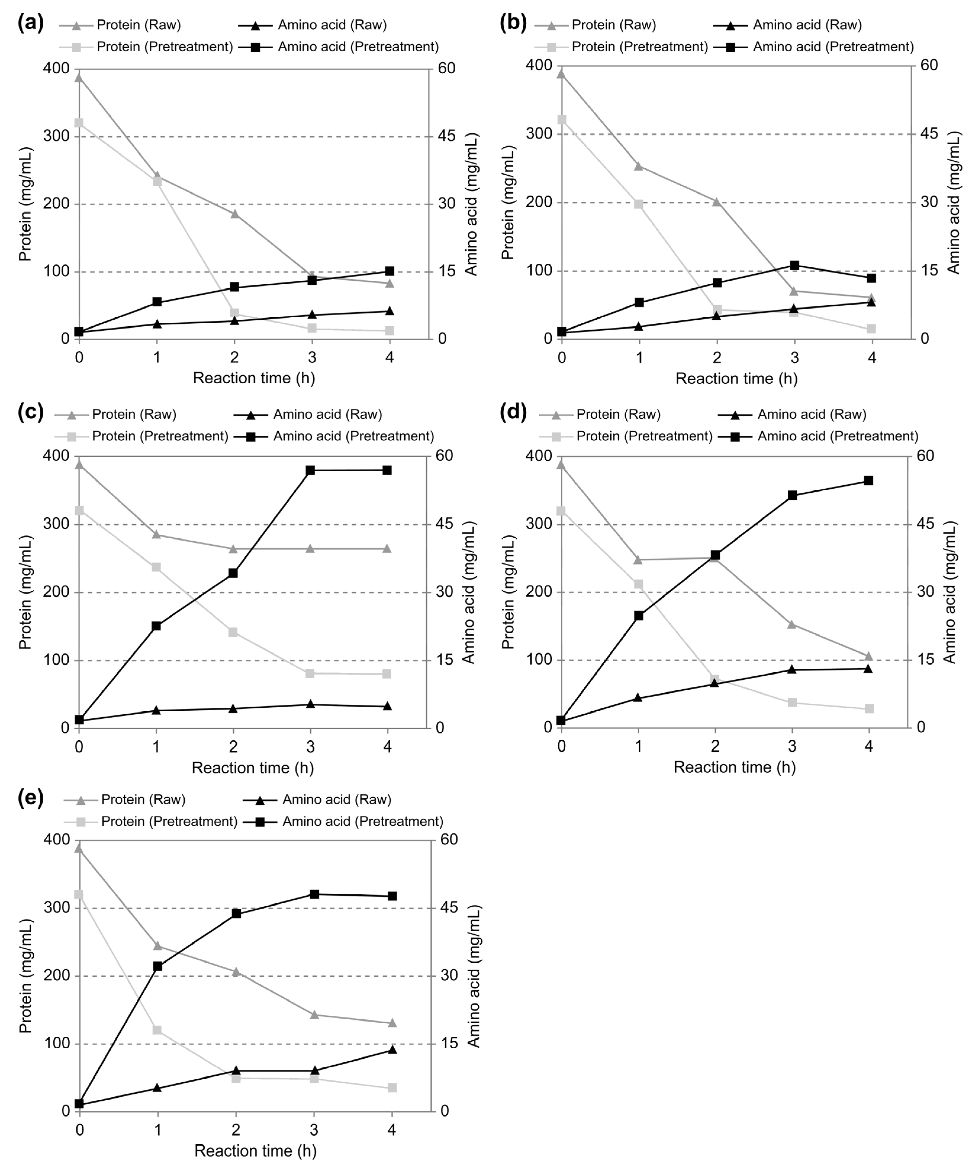
3.2.1. Effects on Protein Reduction and Amino Acid Conversion for Livestock Blood Depending on Enzyme Type

In previous studies, various chemical methods, including the use of acids or alkalis, have been utilized to convert proteins into amino acids [4,36]. However, employing such chemical methods for protein breakdown necessitates additional washing steps; hence, using chemical reagents in the utilization of livestock blood as a resource may incur additional costs [37]. Therefore, in this study, we aimed to streamline the process and ensure cost-effectiveness by opting for a biological approach that relies on enzymes derived from microorganisms to circumvent chemical treatment.
Proteases, which are protein-degrading enzymes, can be classified into endopeptidases and exopeptidases according to the different hydrolysis sites, such as non-terminal and amino-terminal or carboxyl-terminal hydrolysis sites. Exopeptidases catalyze the hydrolysis of the peptide bonds near the N- or C-terminal ends of the substrate. Aminopeptidases can liberate single amino acids, dipeptides (dipeptidyl peptidases), or tripeptides (tripeptidyl peptidases) from the N-terminal end of their substrates. Single amino acids can be released from dipeptide substrates by dipeptidases or from polypeptides by carboxypeptidases, while peptidyl dipeptidases liberate dipeptides from the C-terminal end of a polypeptide chain. Endopeptidases cleave peptide bonds both within and distant from the ends of a polypeptide chain [38]. Savinase® and Alcalase® are endopeptidases which break peptide bonds from the C-terminal amino acids, whereas flavourzyme® is an exopeptidase that breaks the N-terminal of peptide chains. Compared to individual proteases, the combination of endopeptidase and exopeptidase can simultaneously hydrolyze both the non-terminal and amino-terminal sites of protein to obtain single amino acids with a low molecular weight [39,40].
The typically used enzyme concentration for protein degradation is approximately 2% (w/v). We also applied a 2% (w/v) concentration for each enzyme. We used two commercial endo-type enzymes, Savinase® and Alcalase®, as well as the exo-commercial type enzyme, Flavourzyme®. These enzymes were used individually or in combination in five variants: (1) 2% (w/v) Savinase®; (2) 2% (w/v) Alcalase®; (3) 2% (w/v) Flavourzyme®; (4) 1% (w/v) Savinase® + 1% (w/v) Flavourzyme®; and (5) 1% (w/v) Alcalase® + 1% (w/v) Flavourzyme®. The extent of protein degradation and amino acid generation depending on the type and composition of enzymes is presented in Table 4 and Figure 4. As shown in Figure 4, no significant differences in characteristics were observed between Savinase® and Alcalase®. While most of the proteins appeared to be degraded within 2 h of treatment, the increase in amino acids did not necessarily correlate with the decrease in proteins. This suggests that the reduction in protein concentration does not necessarily lead to an increased amino acid concentration, and the protein molecules may exist in a precipitate form or may not have been fully broken down into amino acids. The use of the Flavourzyme® resulted in a significantly lower protein degradation rate compared with the endo-type enzymes because exo-type enzymes primarily cleave peptide bonds on the outer regions of proteins. Consequently, when considering the total protein content, a relatively smaller amount of degraded proteins was measured. The combination of endo- and exo-type enzymes led to the observation that, with time, proteins steadily decreased, and amino acid conversion occurred more effectively compared with the individual use of each enzyme. This suggests that when endo- and exo-type enzymes are used in combination, the endo-type enzyme initially breaks down proteins to the peptide level, upon which the exo-type enzyme cleaves peptide bonds from the exterior, resulting in the efficient generation of low molecular weight peptides and amino acids.
A comparison of the changes in protein concentration in the ultrasound-treated blood samples, based on each enzyme treatment (Table 4), revealed that the single use of Savinase® resulted in the highest protein reduction rate, with the concentration of the remaining proteins after 4 h of reaction reduced to 12.5 mg/mL. In contrast, the single use of Flavourzyme® resulted in the lowest protein reduction rate; the protein concentration decreased to 78.4 mg/mL. However, the single use of Flavourzyme® increased the amino acid concentration to 56.9 mg/mL, indicating the highest amino acid generation. Comparing the protein reduction rate relative to the initial protein concentration yielded the following order: Savinase® > Alcalase® > Savinase® + Flavourzyme® > Alcalase® + Flavourzyme® > Flavourzyme®. The highest amino acid conversion rate relative to the initial protein concentration was exhibited by Flavourzyme® and Savinase® + Flavourzyme® at very similar levels, followed by Alcalase® + Flavourzyme®, Savinase®, and Alcalase®. Flavourzyme® exhibited the highest overall amino acid conversion rate compared to other enzymes. However, exo-type enzymes are economically more expensive than endo-type enzymes, making them less cost-effective for large-scale industrial processes. On the other hand, when used individually, endo-type enzymes have lower amino acid conversion rates and are not suitable for amino acid production. In the combined use of exo-type and endo-type enzymes, the mixture was found to have an amino acid conversion rate comparable to that of a single use of exo-type enzymes. Additionally, considering the cost-saving effect of the combined use of enzymes, it is considered most desirable to use a combination of both enzyme types for this process.
The comparison of the results before and after ultrasound treatment revealed that the untreated blood samples exhibited a maximum amino acid conversion rate of only 13.7 mg/mL in all enzymatic reactions, while that of the ultrasound-treated blood samples increased to 56.9 mg/mL, which is more than four times higher than the amino acid conversion rate of the untreated blood. The comparison of the concentration changes over reaction time revealed that the ultrasound-treated samples had significantly higher protein reduction and amino acid conversion rates than the untreated samples. This finding suggests that ultrasonic treatment induces hemolysis in RBCs, facilitating enzymatic reactions and altering the protein structure, thus accelerating the enzyme hydrolysis reaction rate. This result is consistent with a report of Koirala et al. [20], who investigated a study on ultrasonic pretreatment process before the enzymatic hydrolysis of caprine milk samples, and reported that the concentration of water-soluble proteins in goat milk increased through ultrasonic pretreatment. They reported that ultrasonication enhanced the production of protein hydrolysates, which may be due to the evolution of complex cellular structures into simpler soluble matrices due to the cavitation effect of sonication.
Considering the protein reduction efficacy and cost-effectiveness of the enzymes used, we identified that the optimal enzyme combination is the combined use of Savinase® and Flavourzyme®, mixed at 1% each. Table 5 shows the changes in the properties of the livestock blood samples after enzyme hydrolysis. Chemical characteristics such as pH, salinity, and 3-components remained very similar to the values before treatment, probably because proteolytic enzymes do not significantly affect the chemical characteristics of blood. In contrast, protein, amino acid, and particle size exhibited significant differences before and after enzyme treatment due to the morphological changes that occurred as proteins are broken down and converted into peptides and amino acids. This finding suggests that chemical reactions did not take place during the processing of livestock blood, and the proteolytic enzymes specifically cleaved proteins. Particularly, the reduction in particle size from 44.32 μm to 8.84 μm, along with an increase in amino acid content, indicated that efficient enzymatic hydrolysis took place as blood components were broken down into smaller molecules.

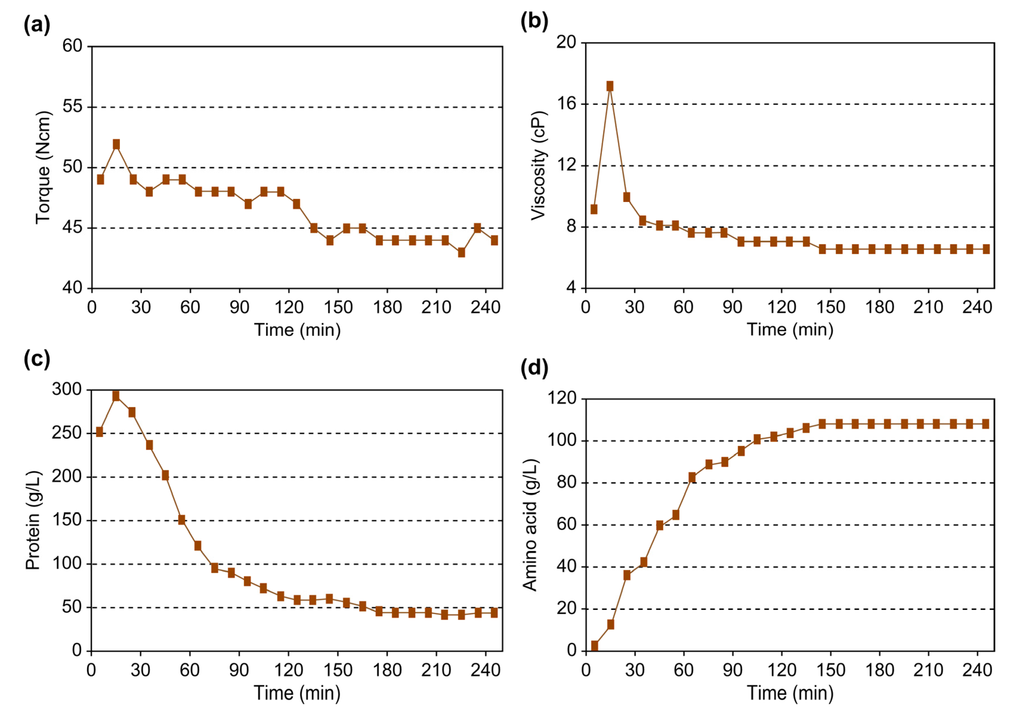
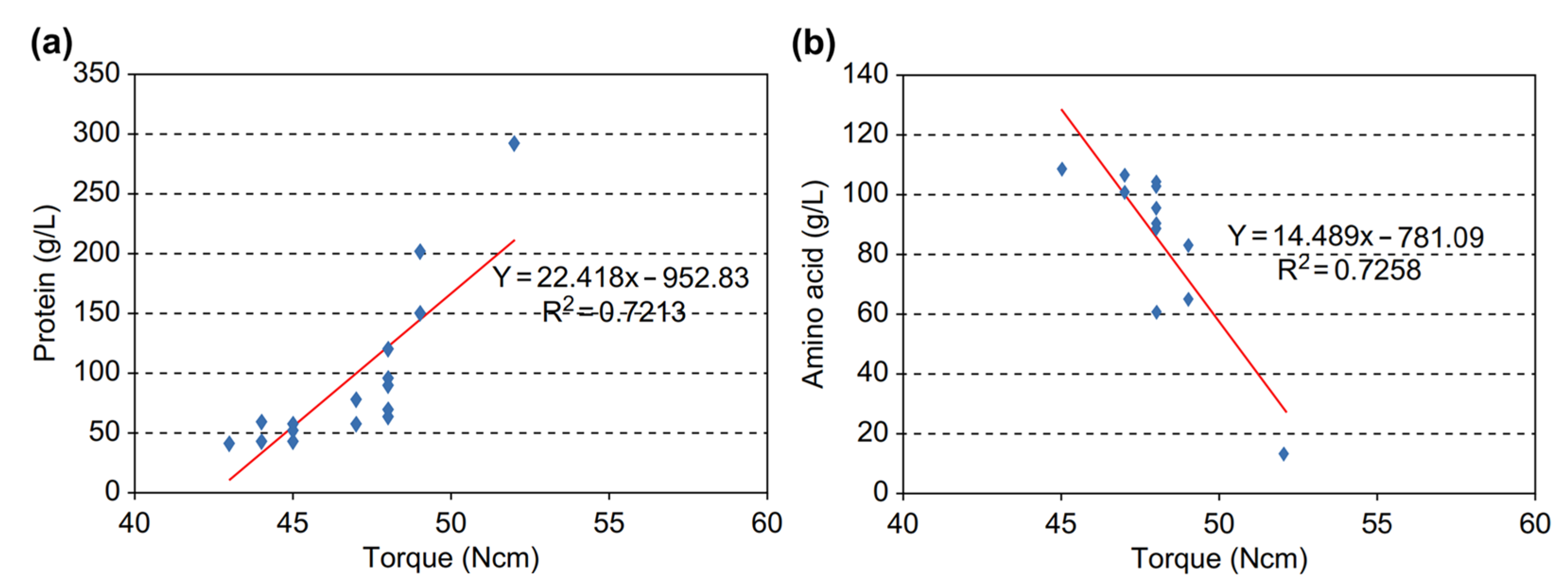
3.2.2. Analysis of the Torque of Reaction Stirrer According to Enzyme Reaction

The livestock blood underwent a gradual reduction in viscosity as it was enzymatically hydrolyzed into amino acids. Our objective in this study was to analyze the correlation between torque (a mechanical factor) and the changes in viscosity, protein content, and amino acid content of the target substance during enzymatic hydrolysis and to determine whether torque, as a mechanical factor, can be used to control the biological process of enzymatic reactions.
The changes in torque, viscosity, and protein and amino acid concentrations over time are depicted in Figure 5. Over the 4 h reaction period, torque decreased from 52 to 44 Ncm, and viscosity dropped from 17.17 to 6.57 cP. Upon introducing the enzymes to the blood samples, the viscosity of the blood samples initially increased for approximately 10 min, then decreased as protein hydrolysis continued; after 130 min, both the torque and viscosity remained relatively constant, indicating near completion of enzymatic reactions. Considering the inherently low viscosity of blood, the comparison of protein and amino acid concentrations revealed that the protein concentration decreased from 239.85 to 56.31 g/L after 4 h. The protein concentration rapidly declined for up to 2 h, then gradually decreased. In contrast, the initial amino acid concentration (2.07 g/L) significantly increased over time, peaking at 107.89 g/L around 140 min into the enzyme reaction, after which it plateaued. This distinct pattern compared to protein reduction suggests that amino acid conversion might have reached a plateau due to enzyme activity inhibition after a certain reaction time. These findings lead to the assumption that the conversion of proteins into amino acids under the given experimental conditions is completed in around 140 min.
The correlation between torque and each of the analyzed parameters is shown in Figure 6. The correlation equation y = 22.418x 952.83 (R2 = 0.7213) revealed a significant correlation between torque and protein concentration changes. Torque and amino acid concentration exhibited a similar level of correlation, with an equation of y = 14.489x + 781.09 (R2 = 0.7205). This suggests that there is a significant relationship between biological factors, such as protein and amino acid concentrations, and the mechanical factor, torque. However, torque did not exhibit a strong correlation with protein and amino acid concentrations, likely due to the naturally low viscosity of livestock blood. Typically, the correlation between torque and viscosity is more prominent in high-viscosity conditions. Furthermore, the correlation coefficient between protein and amino acid concentrations was calculated at an R2 value of 0.949, indicating a very high correlation. Consequently, the torque value was calculated at 44 Ncm, which corresponds to around 140 min, when the amino acid content peaked. This result confirms that the torque value measured during enzymatic reactions can be employed to assess the progress of enzymatic reactions and serve as an indicator of reaction efficiency.

4. Conclusions

The aim of this study is to determine the feasibility of converting livestock blood, a major livestock organic waste whose treatment is highly challenging due to its high pollution potential and rapid decomposition, into an environmentally safe and high-quality liquid amino acid fertilizer for agricultural use. Doing so would also reduce waste accumulation and pollution.
(1)
Ultrasound pretreatment resulted in the improved enzymatic hydrolysis of the proteins in livestock blood, as evidenced by the high hemoglobin solubilization rate (97.6%) of the ultrasound-treated blood samples; it also resulted in particle size reduction and pathogen elimination.
(2)
Protein hydrolysis experiments using different types of enzymes showed that Flavourzyme®, an exo-type enzyme, exhibited the highest amino acid conversion efficiency. However, considering the relatively lower cost-effectiveness of exo-type enzymes, especially for large-scale processes, we determined the optimal condition to be the combination of the endo-type enzyme Savinase® and the exo-type enzyme Flavourzyme®. This combination yielded an amino acid conversion rate similar to that achieved with the single use of Flavourzyme®. Subsequent enzymatic hydrolysis experiments conducted on ultrasound-treated and untreated blood under the optimal conditions revealed that the amino acid concentration in ultrasound-treated blood reached 54.6 mg/mL, a nearly 4.2-fold increase compared to 13.1 mg/mL in untreated blood. This finding demonstrates that the ultrasonic treatment of livestock blood enhances proteolytic hydrolysis efficiency, particularly when using a combination of endo- and exo-type enzymes, resulting in a substantial increase in amino acid conversion.
(3)
As enzymatic hydrolysis progressed, the viscosity of livestock blood gradually decreased. Monitoring the agitator torque to gauge the reduction in applied physical force revealed a significant correlation between biological factors, such as protein and amino acid concentrations, and the mechanical factor, torque. Thus, it was confirmed that measuring torque during enzymatic reactions can be utilized to evaluate the extent of enzymatic reactions and thus serve as an indicator of reaction progress.

Author Contributions

Conceptualization, Y.-W.J.; methodology, M.-J.J.; validation, Y.-W.J. and M.-J.J.; formal analysis, M.-J.J.; investigation, M.-J.J. and Y.-W.J.; writing—original draft preparation, Y.-W.J.; writing—review and editing, Y.-W.J.; supervision, Y.-W.J.; project administration, Y.-W.J.; funding acquisition, Y.-W.J. All authors have read and agreed to the published version of the manuscript.

Funding

This research was supported by the Korea Environment Industry & Technology Institute (KEITI) through the Development of Demonstration Technology for Converting Unconventional Waste Biomass to Energy project funded by the Korea Ministry of Environment (MOE) (RE202201941). And this research was also supported by the Korea Institute of Energy Technology Evaluation and Planning (KETEP) and the Korea Ministry of Trade, Industry & Energy (MOTIE) (20213030040070).

Institutional Review Board Statement

Not applicable.

Informed Consent Statement

Not applicable.

Data Availability Statement

The original contributions presented in the study are included in the article, further inquiries can be directed to the corresponding author.

Conflicts of Interest

The authors declare no conflicts of interest.

References

  1. Nowak, B.; von Mueffling, T. Porcine blood cell concentrates for food products: Hygiene, composition, and preservation. J. Food Prot. 2006, 69, 2183–2192. [Google Scholar] [CrossRef]
  2. Lynch, S.A.; Mullen, A.M.; O’Neill, E.E.; García, C.A. Harnessing the potential of blood proteins as functional ingredients: A review of the state of the art in blood processing. Compr. Rev. Food Sci. 2017, 16, 330–344. [Google Scholar] [CrossRef]
  3. Lee, D.M.; Choi, M.S.; Woo, G.I.; Shin, Y.M.; Lee, K.H.; Chun, Y.P.; Chun, T.; Choi, I. Effect of gender-specific bovine serum supplemented medium on cell culture. J. Anim. Sci. Technol. 2009, 51, 413–420. [Google Scholar] [CrossRef]
  4. Hyun, C.K.; Shin, H.K. Production of angiotensin I converting enzyme inhibitory peptides from bovine blood plasma proteins. Korean J. Biotechnol. Bioeng. 1999, 14, 600–605. [Google Scholar]
  5. Aziz, A.; Basheer, F.; Sengar, A.; Irfanullah; Khan, S.U.; Farooqi, I.H. Biological wastewater treatment (anaerobic-aerobic) technologies for safe discharge of treated slaughterhouse and meat processing wastewater. Sci. Total Environ. 2019, 686, 681–708. [Google Scholar] [CrossRef]
  6. Renata, T.D.; Miriam, C.C.; Valdemiro, C.S. Bovine blood components: Fractionation, composition, and nutritive value. J. Agric. Food. Chem. 1999, 47, 231–236. [Google Scholar]
  7. Bah, C.S.F.; Bekhit, A.E.-D.A.; Carne, A.; McConnell, M.A. Production of bioactive peptide hydrolysates from deer, sheep and pig plasma using plant and fungal protease preparations. Food Chem. 2015, 176, 54–63. [Google Scholar] [CrossRef]
  8. Moreto, M.; Miro, L.; Amat, C.; Polo, J.; Manichanh, C.; Perez-Bosque, A. Dietary supplementation with spray-dried porcine plasma has prebiotic effects on gut microbiota in mice. Sci. Rep. 2020, 10, 2926. [Google Scholar] [CrossRef]
  9. Nikhita, R.; Sachindra, N.M. Optimization of chemical and enzymatic hydrolysis for production of chicken blood protein hydrolysate rich in angiotensin-I converting enzyme inhibitory and antioxidant activity. Poultry Sci. 2021, 100, 101047. [Google Scholar] [CrossRef]
  10. Ann, S.W.; Kim, Y.C.; Hwang, I.S.; Cho, J.K.; Kim, M.S.; Lee, J.K.; Eum, W.Y. Effect of seafood amino acid fertilizer and Korean effective microorganisms on the fruit quality of fuji apple. J. Environ. Sci. 2010, 19, 1293–1299. [Google Scholar]
  11. Bah, C.S.F.; Carne, A.; McConnell, M.A.; Mros, S.; Bekhit, A.E.-D.A. Production of bioactive peptide hydrolysates from deer, sheep, pig and cattle red blood cell fractions using plant and fungal protease preparations. Food Chem. 2016, 202, 458–466. [Google Scholar] [CrossRef]
  12. Zheng, Y.; Chen, Q.; Shan, A.; Zhang, H. Optimization of enzymatic hydrolysis of blood cells with a neutral protease. BioMed. Res. Int. 2013, 2013, 278927. [Google Scholar] [CrossRef]
  13. Fu, Y.; Bak, K.H.; Liu, J.; De Gobba, C.; Tøstesen, M.; Hansen, E.T.; Petersen, M.A.; Ruiz Carrascal, J.; Bredie, W.L.P.; Lametsch, R. Protein hydrolysates of porcine hemoglobin and blood: Peptide characteristics in relation to taste attributes and formation of volatile compounds. Food Res. Int. 2019, 121, 28–38. [Google Scholar] [CrossRef]
  14. Fu, Y.; Liu, J.; Zhang, W.; Wæhrens, S.S.; Tøstesen, M.; Hansen, E.T.; Bredie, W.L.P.; Lametsch, R. Exopeptidase treatment combined with Maillard reaction modification of protein hydrolysates derived from porcine muscle and plasma: Structure-taste relationship. Food Chem. 2020, 306, 125613. [Google Scholar] [CrossRef]
  15. Fahlivi, M.; Campus, F.I.; Jonsson, A. Physicochemical Characteristics of Liquid Fertilizer from Fish Viscera. United Nations University Fisheries Training Programme, 2018, Iceland [Final Project]. Available online: http://www.unuftp.is/static/fellows/document/rizal15prf.pdf (accessed on 1 January 2023).
  16. Sun, T.; Xiao, W.; Jiang, C.; Wang, J.; Liu, Z. Producing amino acid fertilizer by hydrolysis of the fermented mash of food waste with the synergy of three proteases expressed by engineered Candida utilis. Bioresource Technol. Rep. 2019, 7, 100268. [Google Scholar] [CrossRef]
  17. Wang, Z.; Lin, X.; Li, P.; Zhang, J.; Wang, S.; Ma, H. Effects of low intensity ultrasound on cellulase pretreatment. Bioresour. Technol. 2012, 117, 222–227. [Google Scholar] [CrossRef]
  18. de Carvalho Silvello, M.A.; Martínez, J.; Goldbeck, R. Low-frequency Ultrasound with Short Application Time Improves Cellulase Activity and Reducing Sugars Release. Appl. Biochem. Biotechnol. 2020, 191, 1042–1055. [Google Scholar] [CrossRef] [PubMed]
  19. Kabawa, B.; Sampers, I.; Raes, K. Effect of ultrasonic treatment on enzymes: Decoupling the relation between the ultrasonic driven conformational change and enzyme activity. Ultrason. Sonochem. 2023, 101, 106720. [Google Scholar] [CrossRef] [PubMed]
  20. Koirala, S.; Prathumpai, W.; An, A.K. Effect of ultrasonication pretreatment followed by enzymatic hydrolysis of caprine milk proteins and on antioxidant and angiotensin converting enzyme (ACE) inhibitory activity of peptides thus produced. Int. Dairy J. 2021, 118, 105026. [Google Scholar] [CrossRef]
  21. Bougrier, C.; Carrere, H.; Delgenes, J.P. Solubilization of waste-activated sludge by ultrasonic treatment. Chem. Eng. J. 2005, 106, 163–169. [Google Scholar] [CrossRef]
  22. Timothy, J.M.; John, P.L. Applied Sonochemistry: Uses of Power Ultrasound in Chemistry and Processing; Wiley-VCH: Weinheim, Germany, 2002. [Google Scholar]
  23. Wang, F.; Lu, S.; Ji, M. Components of released liquid from ultrasonic waste activated sludge disintegration. Ultrason. Sonochem. 2006, 13, 334–338. [Google Scholar] [CrossRef]
  24. Lee, D.H.; Shin, H.J. Study on the process to decrease the molecular weight of β-(1,6)-branchedβ-(1-3)-D-Glucans. Korean J. Biotechnol. Bioeng. 2003, 18, 352–355. [Google Scholar]
  25. Portenlanger, G. Mechanical and radical effects of ultrasound: Ultrasound inwate water and sludge treatment. TU Hambg. Harbg. Rep. Sanit. Eng. 1999, 25, 139–151. [Google Scholar]
  26. Bah, C.S.F.; Bekhit, A.E.-D.A.; Carne, A.; McConnell, M.A. Slaughterhouse blood: An emerging source of bioactive compounds. Compr. Rev. Food Sci. Food Saf. 2013, 12, 314–331. [Google Scholar] [CrossRef]
  27. Jambrak, A.R.; Mason, T.J.; Lelas, V.; Krešić, G. Ultrasonic effect on physicochemical and functional properties of α-lactalbumin. LWT-Food Sci. Technol. 2010, 43, 254–262. [Google Scholar] [CrossRef]
  28. Santos, S.D.; Martins, V.G.; Salas-Mellado, M.; Prentice, C. Evaluation of functional properties in protein hydrolysates from bluewing searobin (Prionotus punctatus) obtained with different microbial enzymes. Food Bioprocess Technol. 2011, 4, 1399–1406. [Google Scholar] [CrossRef]
  29. Eaton, A.D.; Clesceri, L.S.; Greenberg, A.E.; Franson, M.A.H. Standard Methods for the Examination of Water and Wastewater, 19th ed.; American Public Health Association (APHA): Washington, DC, USA, 1995. [Google Scholar]
  30. Zwart, A.; Assendelft, O.W.; Bull, B.S.; England, J.M.; Lewis, S.M.; Zijlstra, W.G. Recommendations for reference method for haemoglobinometry in human blood (ICSH standard 1995) and specifications for international haemiglobincyanide standard (4th ed). J. Clin. Pathol. 1996, 49, 271–274. [Google Scholar] [CrossRef]
  31. Schönenbrücher, V.; Mallinson, E.T.; Bülte, M. A comparison of standard cultural methods for the detection of foodborne Salmonella species including three new chromogenic plating media. Int. J. Food Microbiol. 2008, 123, 61–66. [Google Scholar] [CrossRef]
  32. Tiehm, A.; Nickel, K.; Zellhorn, M.; Neis, U. Ultrasonic waste activated sludge disintegration for improving anaerobic stabilization. Wat. Res. 2001, 35, 2003–2009. [Google Scholar] [CrossRef]
  33. Aliger, H. Ultrasonic Disruption. Am. Lab. 1975, 10, 75–85. [Google Scholar]
  34. Yarullina, L.G.; Akhatova, A.R.; Kasimova, R.I. Hydrolytic enzymes and their proteinaceous inhibitors in regulation of plant-pathogen interactions. Russ. J. Plant Physiol. 2016, 63, 193–203. [Google Scholar] [CrossRef]
  35. Broekman, S.; Pohlmann, O.; Beardwood, E.S.; Meulenaer, E.C. Ultrasonic treatment for microbiological control of water systems. Ultrason. Sonochem. 2010, 17, 1041–1048. [Google Scholar] [CrossRef] [PubMed]
  36. Sheen, S.J.; Sheen, V.L. Characteristics of fraction-1-protein degradation by chemical and enzymic treatments. J. Agric. Food Chem. 1987, 35, 948–952. [Google Scholar] [CrossRef]
  37. Seeley, E.H.; Oppenheimer, S.R.; Mi, D.; Chaurand, P.; Caprioli, R.M. Enhancement of protein sensitivity for MALDI imaging mass spectrometry after chemical treatment of tissue sections. J. Am. Soc. Mass. Spectrom. 2008, 19, 1069–1077. [Google Scholar] [CrossRef] [PubMed]
  38. Mótyán, J.A.; Tóth, F.; Tőzsér, J. Research applications of proteolytic enzymes in molecular Biology. Biomolecules 2013, 3, 923–942. [Google Scholar] [CrossRef]
  39. Ambigaipalan, P.; Al-Khalifa, A.S.; Shahidi, F. Antioxidant and angiotensin I converting enzyme (ACE) inhibitory activities of date seed protein hydrolysates prepared using Alcalase, Flavourzyme and Thermolysin. J. Funct. Foods 2015, 18, 1125–1137. [Google Scholar] [CrossRef]
  40. Zhu, H.; Chen, J.; Zhang, Y.; Goh, K.L.; Wan, C.; Zheng, D.; Zheng, M. Preparation and investigation of novel endopeptidase-exopeptidase co-immobilized nanoflowers with improved cascade hydrolysis. Int. J. Biol. Macromol. 2023, 246, 125622. [Google Scholar] [CrossRef]
Figure 1. Schematic diagram of experimental equipment: (a) Ultrasonic system, (b) Enzymatic hydrolysis system.
Applsci 14 01676 g001
Figure 2. Changes in solubilization rate with different ultrasonic density and ultrasonic time (sample—1 L/batch, homogenization at 10,000 rpm for 10 min, ultrasonic frequency at 20 kHz, agitation at 200 rpm).
Applsci 14 01676 g002
Figure 3. Morphological changes in red blood cell (magnification ×40); (a) before treatment, (b) only grinding (grinding speed: 10,000 rpm, grinding duration: 10 min), (c) after treatment (capacity: 1 L/batch, grinding speed: 10,000 rpm; grinding duration: 10 min; ultrasonic frequency: 20 kHz; ultrasonic density: 0.5 W/mL; ultrasonic duration: 30 min; agitation: 200 rpm).
Applsci 14 01676 g003
Figure 4. Change in enzymatic degradability of raw and ultrasonic-treated livestock blood according to reaction time; (a) Case 1: 2% Savinase®; (b) case 2: 2% Alcalase®; (c) Case 3: 2% Flavourzyme®; (d) Case 4: 1% Savinase® + 1% Flavourzyme®; (e) Case 5: 1% Alcalase® + 1% Flavourzyme® (untreated sample conditions—grinding speed: 10,000 rpm; grinding duration: 10 min; treated sample conditions—sample: 1 L; ultrasonic frequency: 20 kHz; ultrasonic density: 0.5 W/mL; ultrasonic duration: 30 min; agitation: 200 rpm).
Applsci 14 01676 g004
Figure 5. Changes in (a) torque, (b) viscosity, (c) protein concentration, and (d) amino acid concentration according to enzymatic reaction time (untreated sample—grinding speed: 10,000 rpm; grinding duration: 10 min; pretreatment conditions—ultrasonic frequency: 20 kHz; ultrasonic density: 0.5 W/mL; ultrasonic duration: 30 min; agitation: 200 rpm; enzymatic degradation treatment condition—sample: 3 L; temperature: 55 °C; reaction time: 4 h; enzymes: 1% (w/v) Savinase® + 1% (w/v) Flavourzyme®).
Applsci 14 01676 g005
Figure 6. Correlation between stirrer torque and (a) protein concentration and (b) amino acid concentration.
Applsci 14 01676 g006
Table 1. Characteristics of livestock blood used in this study.
ParametersConcentration (wt.%)
Moisture content79.18 ± 0.14
Inorganic content1.29 ± 0.14
Organic content19.53 ± 0.15
    Protein18.26 ± 0.03
      Albumin2.03 ± 0.08
      Globulin2.05 ± 0.04
      Fibrinogen0.44 ± 0.46
      Hemoglobin14.08 ± 0.07
    Lipid0.17 ± 0.01
Salt 0.95 ± 0.01
Table 2. Characteristic of commercial enzymes used.
EnzymeCharacteristicSourceActivity
pH RangeTemperature Range (°C)
Savinase®Endopeptidase-8–12 (10)20–60 (55)
Alcalase®EndopeptidaseBacillus licheniformis6.5–8.545–65 (60)
Flavourzyme®ExopeptidaseAspergillus oryzae5–750
Activity: indicates the range of conditions under which the enzyme shown by the manufacturer is active. The optimal conditions are given in parentheses.
Table 3. Changes in the properties of livestock blood after ultrasonic treatment.
PropertiesGrinding TreatedUltrasonic Treated
Hemoglobin in whole blood (%)15.25
Hemoglobin in plasma (%)5.5914.63
Solubilization rate (%)36.3295.91
pH7.487.14
Salinity (%)0.960.92
Particle size (um)131.1644.32
3-componentMoisture content (%)81.6981.07
Organic content (%)17.6318.21
Inorganic content (%)0.690.72
Chemical componentCarbon (%)45.0046.03
Hydrogen (%)6.216.31
Oxygen (%)17.6016.73
Nitrogen (%)12.6513.11
Sulfur (%)0.140.15
Chlorine (%)1.071.09
BacteriaGeneral Bacteria (CFU/mL)1.5 × 1051.3 × 102
Total Coliforms (CFU/mL)2.0 × 103N.D.
Salmonella (CFU/mL)N.D.N.D.
N.D.: not detectable; capacity: 1 L/batch; grinding speed: 10,000 rpm; grinding time: 10 min; ultrasonic frequency: 20 kHz; ultrasonic density: 0.5 W/mL; ultrasonic duration: 30 min; agitation: 200 rpm.
Table 4. Enzymatic degradability of untreated and ultrasonic-treated livestock blood.
ParametersCase 1 cCase 2 cCase 3 cCase 4 cCase 5 c
Protein (mg/mL)Raw a83.361.3263.0104.9130.4
Ultrasonic-treated b12.513.878.427.833.8
Amino acid (mg/mL)Raw a6.38.14.613.113.7
Ultrasonic-treated b15.113.456.954.647.5
Protein Reduction Rate (%) dRaw a78.584.232.172.966.3
Ultrasonic-treated b96.195.775.691.389.5
Amino acid Conversion Rate (%) eRaw a1.62.11.193.343.5
Ultrasonic-treated b4.74.217.717.014.8
a Livestock blood after grinding treatment (grinding speed: 10,000 rpm; grinding duration: 10 min); b livestock blood after grinding and ultrasonic treatment (sample: 1 L; ultrasonic frequency: 20 kHz; ultrasonic density: 0.5 W/mL; ultrasonic duration: 30 min; agitation: 200 rpm); c treatment conditions: sample: 100 mL; temperature: 55 °C; reaction time: 4 h; d ratio of reduced protein compared to supplied protein concentration: ((initial protein concentration final protein concentration)/initial protein concentration) × 100; e amino acid conversion rate compared to supplied protein concentration: (initial amino acid concentration final amino acid concentration)/initial protein concentration) × 100.
Table 5. Change in the blood property by enzymatic degradation treatment of livestock blood.
PropertiesEnzymatic Degradation
BeforeAfter
pH7.207.08
Salinity (%)0.910.91
Particle size (µm)48.248.84
3-componentMoisture content (%)79.2279.41
Organic content (%)19.4919.37
Inorganic content (%)1.291.22
Chemical componentCarbon (%)46.4547.20
Hydrogen (%)6.396.52
Oxygen (%)17.9619.01
Nitrogen (%)12.8913.41
Sulfur (%)0.120.11
Chlorine (%)1.111.20
Substrate concentrationProtein (mg/mL)319.227.8
Amino acid (mg/mL)1.554.6
Disclaimer/Publisher’s Note: The statements, opinions and data contained in all publications are solely those of the individual author(s) and contributor(s) and not of MDPI and/or the editor(s). MDPI and/or the editor(s) disclaim responsibility for any injury to people or property resulting from any ideas, methods, instructions or products referred to in the content.

