Abstract
The results of using nonstationary high-resolution THz spectroscopy for detecting a characteristic set of metabolites of thermal decomposition products of urine from prostate cancer patients, aimed at identifying specific markers, are presented. Studies of the composition of biological fluid in prostate cancer were carried out using 12 urine samples from cancer patients and 4 samples from conditionally healthy volunteers. Differences in the composition and content of substances in the urine samples of cancer patients compared to the urine of conditionally healthy volunteers were identified, which allows preliminary conclusions to be drawn about substances promising for use as markers of prostate cancer in urine. The presented approach is promising for developing a method for noninvasive study of the composition of biological fluids, which makes it possible to identify metabolite markers of various pathologies and diseases.
1. Introduction
A modern trend in medical diagnostics and therapy monitoring is the development of an approach based on identifying the metabolic profile of a disease. A metabolic profile is a characteristic set of metabolites—final or intermediate human metabolites synthesized by human normal or pathologic tissues in the case of a specific pathology or disease. Exhaled breath, biological tissues, and liquids can be considered as biological media for identifying biomarkers. Biological liquids (blood, urine, etc.) are the fastest to respond to changes in a person’s condition; hence, they are used for clinical tests (the primary tests for making a diagnosis and therapy monitoring are general urine and blood tests). In the metabolic approach, biological liquids are the most promising objects for identifying biomarkers, for two reasons. First, the concentration of many substances in them is higher than in gaseous secretions of a living organism (exhaled breath) and second, sampling, of urine, for example, is a simple and cheap manipulation. Physicochemical methods, such as gas or liquid chromatography, including in combination with mass spectrometry, or spectroscopic methods are used for analysis of metabolites.
The chromatography–mass spectrometry method is as follows: Passing through a chromatograph, the sample is separated into components, and the mass spectrometer is responsible for their identification and analysis. Depending on the characteristics of the composition under study and the requirements for the accuracy of the result, one of two methods is used, either high-precision liquid chromatography or gas chromatography with mass spectrometric detection. The disadvantages of chromatographic methods include the impossibility of simultaneous study of both polar and non-polar substances. This depends on the choice of chromatographic column, as well as the difficulty of sample preparation for several samples. The main disadvantage of mass spectrometry is that the method is destructive, i.e., it is not the substance itself that is studied, but the products of its transformation. Spectroscopic methods (e.g., nuclear magnetic resonance (NMR), spectroscopies of IR or THz ranges, etc.) have more advantages over chromatography–mass spectrometry methods. NMR is a non-destructive method, which makes it possible to carry out repeated experiments with the same sample. NMR requires minimal sample preparation and can detect even previously unknown compounds. Disadvantages of NMR include the difficulty of detecting compounds with high molecular weight (for example, long chains of fats) and the overlap of peaks corresponding to different compounds on each other. Combining NMR with other methods, for example, mass spectroscopy, will improve the efficiency of metabolite identification. Nuclear magnetic resonance will make it possible to find unknown compounds, which can then be targeted in other samples using mass spectroscopy.
Analysis of data in the literature allows the state of the art in the area of analysis of metabolites in urine to be highlighted. Urine should be analyzed, first of all, from a human body in a healthy state, as well as from a body in a diseased state, in particular having prostate cancer, using various physical methods. The study of the urine of living organisms (humans, animals) aimed at identifying the sets of metabolites characterizing a healthy organism [1], the influence of physical activity [2], and the sets characterizing pathologies, is carried out primarily by chromatographic and some spectroscopic methods.
Using high-resolution 1H nuclear magnetic resonance spectroscopy, variations in the composition of urine samples of two groups were investigated: a group of 60 healthy men and women for 30 days and a group of 25 guinea pigs of similar genetics, diet, and living environment [3]. This study identified important factors such as differences in metabolite excretion due to sampling time, gender, and inter- and intra-individual variability, each of which influences the increased use of urine in metabolic and future clinical diagnostic studies. A total of 23 urinary metabolites were quantitatively variable in normal guinea pigs. In total, 24 urinary metabolites were quantitatively variable in healthy subjects. For example, 12 metabolites were found to be statistically different in men and women: 2-hydroxyisobutyrate, 4-aminohippurate, aspartate, citrate, creatine, formate, hippurate, histidine, lactate, threonine, transaconitate, and N-methylhistidine.
The first database of specific urine volatiles (urine volatolome), consisting of 841 compounds of 80 different chemical classes, including cancer-associated substances, was presented in the review [4].
Prostate cancer (PC) is one of the most common male diseases worldwide. Consequently, the proportion of prostate cancer diagnosed at a distant stage increased from 3.9% to 8.2% over the past decade [5]. According to data from World Cancer Research Fund International, prostate cancer is the second most common cancer in men worldwide and the fourth most common cancer overall. There were more than 1.4 million new cases of prostate cancer in 2020 [6]. Zimbabwe had the highest rate of prostate cancer mortality in 2020, followed by Barbados. Haiti, Zambia, Jamaica, Trinidad and Tobago, The Bahamas, Dominican Republic, Saint Lucia, and Côte d’Ivoire are also among the 10 countries with high prostate cancer mortality.
The most commonly used serum marker of cancer is prostate-specific antigen (PSA), which is an androgen-controlled serine protease produced by both prostate epithelial cells and prostate cancer (PC). PSA is secreted into the prostate ducts as an inactive 244 amino acid proenzyme (proPSA), which is activated by cleavage of seven N-terminal amino acids. The PSA that enters blood circulation unchanged is rapidly bound by protease inhibitors, primarily alpha1-antchymotrypsin, although some fraction is inactivated in lumen by proteolysis and circulates as free PSA [7]. Currently, the main methods of PC detection are the determination of PSA levels in blood, digital rectal examination of the prostate, ultrasound and also magnetic resonance imaging, and, if cancer is suspected, biopsy of the prostate. Unfortunately, none of the methods provides a 100% guarantee of diagnosing the presence or absence of a cancerous tumor. In addition to the blood, the PSA secreted from the prostate gland, enters the urine. In [8], where more than 400 samples for serum and urinary PSA levels were analyzed, urinary PSA was found to have a higher predictive ability in differentiating aggressive prostate cancer and may serve as a better surrogate tumor biomarker for detecting aggressive prostate cancer. Also, the combination of PSA ratios of serum and urine enhanced the efficacy of both biomarkers in predicting aggressive prostate diseases.
In addition to PSA tests in urine, other urine metabolites were studied to identify PC markers that allow detection of the disease at earlier stages.
In [9], based on the statistical analysis of the data obtained by 1H-nuclear-magnetic resonance (1H-NMR) studies of urine samples of 50 patients with PC and 50 volunteers from a control group, 20 detected metabolites were revealed as candidate biomarkers for prostate cancer. Sets of 7–9 metabolites, where a set of 7 metabolites (guanidinoacetate, betaine, phenylacetylglycine, taurine, dimethylglycine, L-alanine, and L-lactate), 8 metabolites (guanidinoacetate, L-alanine, phenylacetylglycine, L-lactate, glycine, acetate, dimethylglycine, and formate), and 9 metabolites (guanidinoacetate, L-alanine, phenylacetylglycine, acetate, L-lactate, glycine, dimethylglycine, formate, and trimethylamine) were identified by three types of statistical analysis. It is proposed to consider guanidinoacetate, phenylacetylglycine, and glycine as markers for PC.
Metabolic characteristics of semen (n = 7), urine (n = 7), and plasma (n = 7) samples were compared in Ref. [10]. Three types of human fluid samples were analyzed using two complementary analytical methods: liquid chromatography coupled with mass spectrometry (HPLC-ESI-TOF/MS) and gas chromatography coupled with mass spectrometry (GCMS). The analysis of seminal fluid identified the following classes of compounds: fatty acids, sphingolipids, phospholipids, sugars and their derivatives, as well as amino acids. Compared with plasma, semen samples have a similar profile; however, detectable levels of metabolites were lower in the case of ejaculate. In urine samples, the levels of detected metabolites were lower than in semen metabolic profiles. However, despite the lower concentrations of specific metabolites in urine, ease of sample collection is the advantage of urine for diagnostics.
Profiling of urine metabolites using mass spectrometry and a multivariate statistical analysis to predict the outcome of prostate cancer treatment were conducted in [11]. This study revealed that the appearance of fragments of a monoacylglycerol skeleton from glycerides may statistically correlate with disease progression.
However, analysis of data in the literature shows that work contributing to the search for a characteristic set of metabolites in PC for diagnostic purposes is still ongoing. The reported spectral responses of substances in the THz frequency range, which are regarded as metabolic biomarkers appearing in urine, are presented in [12], where the results of a relatively small number of studies that have already been carried out on urine samples by various THz methods used for urine analysis are summarized.
The purpose of the present work is to use nonstationary high-resolution THz spectroscopy for studying the characteristic set of metabolites of thermal decomposition products of the urine of conditionally healthy volunteers and cancer patients to identify markers typical of PC. The earlier pioneer studies of the composition of thermal decomposition products of urine of cancer patients receiving platinum-containing therapy showed that the patient’s body reacts to nephrotoxic therapy and that the compositions of thermal decomposition products of urine of the same patient before and after chemotherapy are different. Furthermore, the composition of these samples differs from the composition of the urine sample of conditionally healthy volunteers [13]. The substances (nitriles) appeared in urine in higher concentrations after chemotherapy than before. It may be that traces of amino acids from proteins appeared in urine due to kidney distortion influenced by platinum-containing chemotherapy.
2. Materials and Methods
The THz spectroscopy technique based on the effect of freely decaying polarization involves periodic induction and decay of macroscopic polarization in a sample of a gas mixture. Two types of instruments can be used: a phase manipulation spectrometer and a fast frequency sweep spectrometer [14]. The approach based on fast frequency sweep through the absorption line [15] is implemented in a THz spectrometer with a radiation source based on a backward wave tube (0.118–0.178 THz). The fast frequency sweep mode allows simultaneous measurement of all absorption lines and detection of several substances. The urine was studied with the fast frequency sweep spectrometer. The line shape obtained with such a spectrometer typically has a form [16] of beats between a freely decaying polarization signal and modulated radiation from the source.
The advantage of terahertz gas spectroscopy is its high resolution because the absorption lines at working pressure are narrow and overlapped rarely. The line of the rotational spectrum is the fingerprint of a specific substance because absorption line parameters, such as central frequency and line strength, are determined by molecular structure. It has benefits over THz time-domain spectroscopy and IR spectroscopy (where frequency resolution is lower). Moreover, the detection of only one absorption line is, in principle, enough for revealing the substance in a multicomponent gas mixture, which is of greater benefit than the spectroscopic methods where spectral lines corresponding to the molecular parts or molecular bonds are detected. Some substances of urine samples are volatile and appear in a gas mixture over the sample without heating. The heating of samples in liquid or solid state allows the gaseous state of the sample to be obtained. Some large biological molecules, such as proteins, sugars, and fats, are decomposed on heating, but there are specific features in the content of the resulting gas mixture of the products of thermal decomposition that characterize the patient’s state.
Urine samples from 12 patients with PC were studied (patient information is available in Table 1). This study is approved by the Institutional Review Board of Nizhny Novgorod Regional Oncology Hospital protocol code 5, from 18 May 2015.

Table 1.
Information about patients.
The following abbreviations are used in this table. The TNM Staging System of Cancer includes the extent of the tumor (T), the spread to the lymph nodes (N), and the presence of metastasis (M). The ISUP system grades the cancer between 1 and 5 depending on the Gleason score. The lower the grade, the less likely the cancer is to spread. WBC—white blood cells; RBC—red blood cells; PSA—prostate-specific antigen; UAm—urinalysis; HGB—hemoglobin.
In addition, the composition of thermal decomposition products of samples of cancer patients’ urine was compared with the composition of urine samples of 4 conditionally healthy volunteers (male, 25–34 years old, without chronic diseases).
The samples were prepared as follows: A 1–2 mL sample was poured into a flask where it was dehydrated to ensure accurate spectroscopic measurements aimed at identifying substances present in the sample in trace concentrations. A cryogenic trap with liquid nitrogen was used to dehydrate the liquid sample. The sample was frozen, most of the water was removed, and a thin film or crystallized residue remained in the flask. Volatile substances from the resulting residue were released, first without heating and then with heating to 200 °C, into the measuring cell, which had been evacuated to a pressure of 10−3 mbar. The operating pressure in the cell was 5 × 10⁻²−1 × 10⁻¹ mbar. Before letting the mixture of thermal decomposition products into the cell, the entire operating range of the spectrometer was registered, which was subsequently used as a background record for identifying absorption lines.
A block diagram of the experiment is presented in Figure 1.
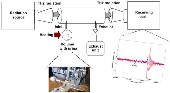
Figure 1.
Block diagram of the experiment measuring spectra of urine thermal decomposition products using the fast frequency sweep spectrometer. The example of spectra presented here includes the red (sample of prostate cancer patient) with absorption lines of formic acid (HCOOH) and carbonyl sulfide (OCS) and blue (sample of conditionally healthy volunteer) curves with an absorption line of carbonyl sulfide (OCS).
The absorption spectra of thermal decomposition products of urine taken from cancer patients were measured. From the spectroscopic parameters of the detected absorption lines (central frequencies, estimated absorption coefficients) substances present in the studied gas mixture were identified using electronic catalogs [17,18]. A comparative analysis of the chemical composition of vapors and thermal decomposition products was then carried out to identify differences in the urine samples from prostate cancer patients and healthy volunteers. Each chemical substance has its own unique spectrum. The spectroscopic characteristics of the absorption lines of a substance are determined by the geometry of the molecule, masses of its constituent atoms, and the total dipole moment. Therefore, the absorption lines are unambiguous signs of the presence of a given substance in the studied multicomponent gas mixture. For qualitative analysis of changes in the composition of different samples, it is possible to count the number of lines recorded in the measured spectrum for each individual substance in the mixture of thermal decomposition products of urine. It is important to take measurements under the same conditions (the same amount of liquid sample in the flask before dehydration, the operating pressure in the cell maintained at a certain level, the same stage of dehydrated sample heating). With a higher relative concentration of a specific substance in the mixture, the number of its absorption lines will increase due to the increase in the concentration of the substance. The amplitude of the detector signal of the stronger lines will also increase. This is true for the case when both strong and weak lines of the substance lie in this range. Consequently, an increase or a decrease in the number of absorption lines enables a qualitative conclusion that the concentration of a given substance increases or decreases from sample to sample. Some substances are always present in the thermal decomposition products of urine, regardless of the volunteer’s health condition.
3. Results
The results of the analysis of the composition of urine thermal decomposition products for all samples of cancer patients and conditionally healthy volunteers are presented in Table 2. Substances that are absent in conditionally healthy volunteers are highlighted in grey.
Table 2.
Substances identified in samples of urine from cancer patients and conditionally healthy volunteers (substances that are absent in conditionally healthy volunteers are highlighted in grey).
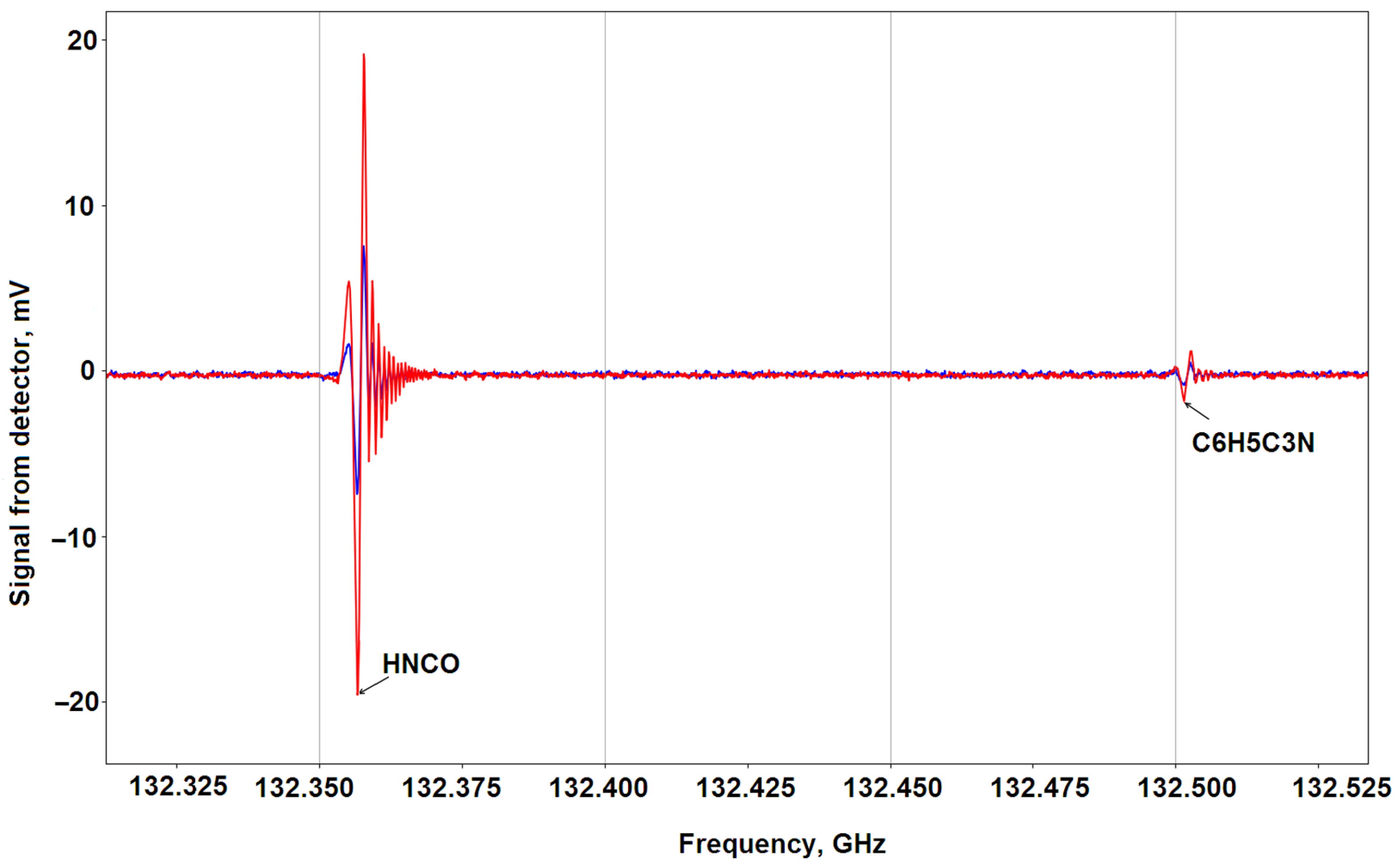
The following features can be noted: Some substances are encountered in all samples. These are, for example, isocyanic acid (stably present), propanediol (its relative concentration is higher in healthy people), phenol, methylphenyl ether, methyl formate, propanal, benzaldehyde, glycol aldehyde (its relative concentration in sick patients is significantly higher than in healthy people), methylbutyronitrile, ethynylbenzonitrile (its relative concentration is higher in sick patients), methylmercaptan (its relative concentration is higher in sick patients), alanine, ethylene glycol, urea (its relative concentration is significantly higher in healthy people), methyl carbamate, cyanoethynylbenzene, and carbonyl sulfide. An example of a spectral region recorded for a sample from a prostate cancer patient (sample 16) compared to the same region from a conditionally healthy volunteer (sample 1.1) with isocyanic acid and cyanoethynylbenzene lines present in both samples is shown in Figure 2.
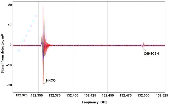
Figure 2.
Recording of a section of a spectrum with absorption lines of isocyanic acid (HNCO) fexp = 132.3567 GHz (fcat1 = 132.3561165 GHz lg I1 = −5.2957 6 1 5 5 ← 5 1 4 5, fcat2 = 132.3567380 GHz lg I2 = −3.6772 6 1 5 7 ← 5 1 4 6, fcat3 = 132.3567521 GHz lg I3 = −3.8265 6 1 5 5 ← 5 1 4 4, fcat4 = 132.3567638 GHz lg I4 = −3.7516 6 1 5 6 ← 5 1 4 5, fcat5 = 132.3572919 GHz lg I3 = −5.2957 6 1 5 6 ← 5 1 4 6 [17]) and cyanoethynylbenzene (C6H5C3N) fexp = 132.5015 ГГц (fcat1,2 = 132.5014714 GHz lg I1,2 = −3.9968 122 32 90 ← 121 32 89 and 122 32 91 ← 121 32 90 [18]. Measurements were made for sample 16 of a prostate cancer patient (red) in comparison with the same section of a healthy volunteer (sample 1.1) (blue).
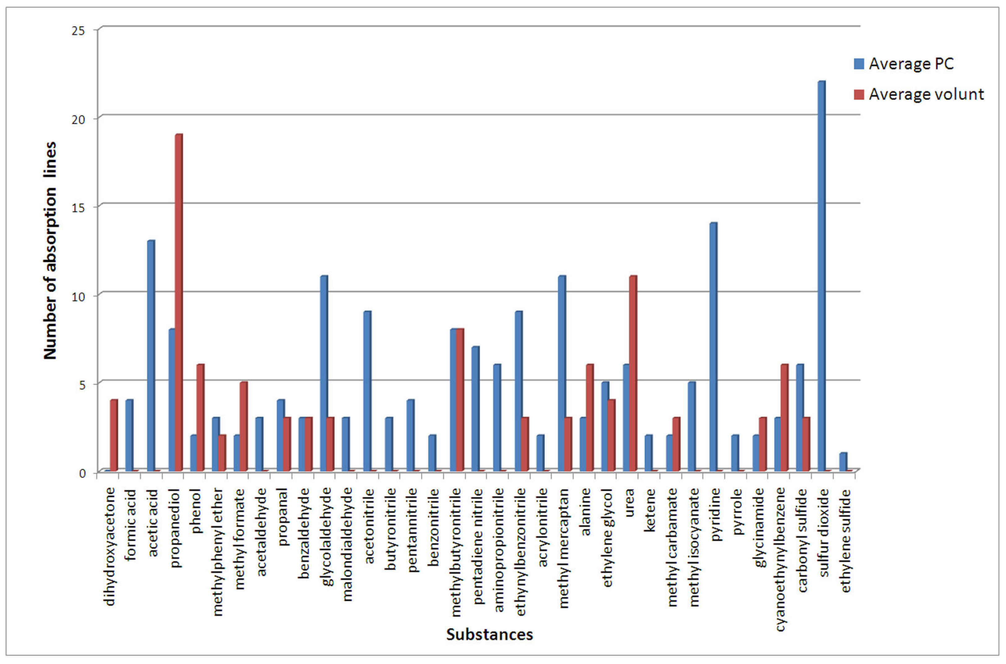
A diagram demonstrating the average number of lines (for substances) for patients with prostate cancer and for conditionally healthy volunteers is presented in Figure 3.
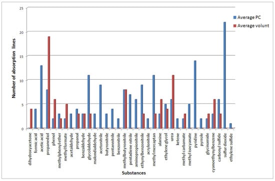
Figure 3.
Average number of lines of substances for patients with prostate cancer and for conditionally healthy volunteers.
A number of nitriles (acetonitrile, butyronitrile, pentannitrile, pentanedienenitrile, benzonitrile, aminopropionitrile) appear in the urine of prostate cancer patients. The amount of sulfur-containing compounds also increases—it appears that sulfur dioxide and the content of methyl mercaptan increase. In addition, acids (formic and acetic) as well as aromatic heterocycles (pyridine and pyrrole) appear in the urine. Another compound noted in a number of prostate cancer patients having a non-zero average value is methyl isocyanate.
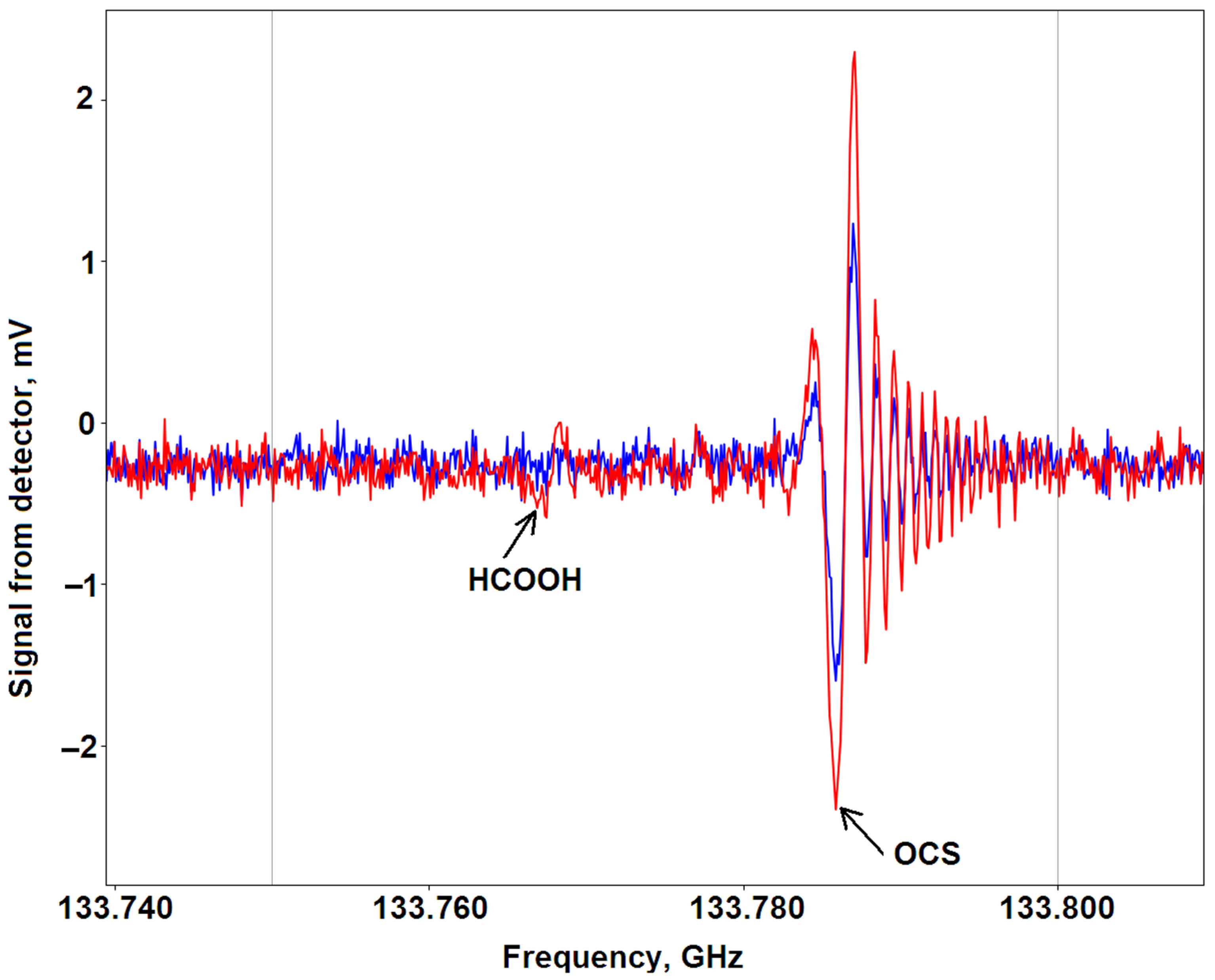
An example of a recording of a section of the spectrum obtained in the study of sample 16 of a prostate cancer patient is shown in Figure 4 in comparison with the same section of a conditionally healthy volunteer (sample 1.1). The figure shows the absorption lines of formic acid present in the thermal decomposition products of urine from a sample from a prostate cancer patient and the lines of carbonyl sulfide in the thermal decomposition products of urine in both samples.
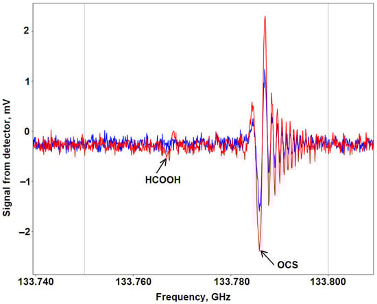
Figure 4.
Recording of a section of a spectrum with the absorption line of formic acid (HCOOH) fexp = 133.7675 GHz (fcat = 133.76707 GHz lg I = −3.8331 6 0 6 ← 5 0 5 [17]) in urine thermal decomposition products from a sample from a prostate cancer patient and the carbonyl sulfide line fexp = 133.7859 GHz (fcat = 133.7859 GHz lg I = −3.2369 11 ← 10 [18]) in thermal decomposition products of urine in sample 16 from a prostate cancer patient (red) and in sample 1.1 from a conditionally healthy volunteer (blue).
4. Discussion
It is known from the literature [19] that the destruction of some amino acids when heated to temperatures above 200 °C is characterized by decarboxylation and deamination reactions. As a result of heating, unsaturated hydrocarbons are formed, the structure of which corresponds to that of amino acids. For example, the thermal decomposition of α-Alanine, β-Alanine, α-Valine, α-Leucine, and α-Isoleucine is accompanied by the appearance of ammonia, carbon dioxide, and carbon monoxide in the thermal decomposition products of all these acids. The other compounds recorded for these five amino acids on such heating are different. In addition to the above-mentioned compounds, for example, α-Alanine decomposes into ethane, ethene, propene, butene-2, acetaldehyde, ethylamine, N-ethylideneethylamine, 2-methyl-5-ethylpyridine, and N-ethylpropionamide, whereas β-Alanine decomposes into ethene, propene, acetonitrile, acetone, pyridine, 3-picoline, 2,4-Puthidine, 3,5-dimethylpyridine, and 2,3,5-trimethylpyridine [19]. Some compounds that can appear during amino acid decomposition (for example, ethane, ethene, propane, and some others) are non-polar (i.e., have a zero dipole moment); therefore, they cannot be registered by nonstationary THz gas spectroscopy. The chemical compounds with a non-zero dipole moment can be detected by their spectra. Based on the analysis of data in the literature and the comparison of substances obtained during the study of urine samples, it can be assumed that heating of some amino acids that enter the composition of proteins, possibly including PSA, may be accompanied by the appearance of nitriles in thermal decomposition products. As has been noted above, the appearance of nitriles (acetonitrile, butyronitrile, pentannitrile, pentanedienenitrile, benzonitrile, aminopropionitrile) in the urine of prostate cancer patients can be caused by thermal decomposition of amino acids including PSA amino acids. The emergence of sulfur-containing compounds, such as sulfur dioxide, hydrogen sulfide, and methyl mercaptan, may be due to the thermal decomposition of sulfur-containing amino acids (methionine, cysteine, cystine), which may also be associated with the thermal decomposition of proteins in urine. In addition, when denatured, PSA consisting of a large number of amino acids (with disulfide bridges) can also give rise to sulfur-containing compounds. At the same time, some light amino acids (for example, alanine) appear in the products of thermal decomposition of urine.
The resulting list of substances and the differences in the composition of substances identified for patients with prostate cancer and conditionally healthy volunteers, given the accumulation of data, allow conclusions to be drawn about significant substances appearing in urine heated to obtain a gas mixture with respect to prostate cancer. In future, a comparison can be made with the products of thermal decomposition of amino acids, which make up PSA, which increases in urine in prostate cancer patients. This will make it possible to use the identified markers among the thermal decomposition products of urine to create chemical sensors for specific substances, which can be used to diagnose prostate cancer.
5. Conclusions
The study analyzes the current state of the art in the field of urine examination using physical methods and identifies the methods most often used in such analysis. The composition of thermal decomposition products of urine from prostate cancer patients has been analyzed in comparison with the urine of conditionally healthy volunteers using the THz gas spectroscopy method. The differences in spectral data, i.e., presence of absorption lines of some substances (methyl mercaptan, nitriles, formic and acetic acids, pyridine, and pyrrole) in data obtained in investigations of urine samples of prostate cancer patients in comparison with healthy volunteers were found for the first time in the world. Differences in the composition and content of substances in urine samples of cancer patients and of conditionally healthy volunteers have been identified, which enables preliminary conclusions about substances that are promising for use as markers of prostate cancer in urine. The appearance of nitriles (acetonitrile, butyronitrile, pentannitrile, pentanedienenitrile, benzonitrile, aminopropionitrile) in the urine of prostate cancer patients can be caused by thermal decomposition of amino acids including ones in PSA. The presented approach is novel for urine analysis and is promising for developing a method for noninvasive study of the composition of biological liquids, which makes it possible to identify metabolite markers of various pathologies and diseases.
Author Contributions
Conceptualization, V.V., A.M. and V.A. (Vagif Atduev); methodology, A.M., V.A. (Vagif Atduev), E.D. and V.A. (Vladimir Anfertev); validation, V.V., A.M. and V.A. (Vagif Atduev); formal analysis, E.D. and M.C.; investigation, E.D., V.V., V.A. (Vladimir Anfertev), K.A. and M.R.; resources, V.A. (Vagif Atduev) and K.A.; data curation, E.D., M.C. and A.M.; writing—original draft preparation, M.C., E.D., V.A. (Vagif Atduev), A.M. and K.A.; writing—review and editing, V.V., M.C., E.D., V.A. (Vagif Atduev) and A.M.; visualization, E.D. and M.C.; supervision, V.V., A.M. and V.A. (Vagif Atduev); project administration, E.D. and A.M.; funding acquisition, V.V. All authors have read and agreed to the published version of the manuscript.
Funding
The studies of samples of biological liquids aimed at identifying disease markers were carried out under the State Assignment FFUF-2024-0024.
Institutional Review Board Statement
The study was conducted according to the guidelines of the Declaration of Helsinki, and approved by the Institutional Review Board of Nizhny Novgorod Regional Oncology Hospital (protocol code 5, from 18 May 2015).
Informed Consent Statement
Informed consent was obtained from all subjects involved in the study.
Data Availability Statement
The data are contained within the article and are partially available in a publicly accessible repository.
Acknowledgments
The authors acknowledge Nadezhda Krivatkina for assistance in editing the manuscript.
Conflicts of Interest
The authors declare no conflicts of interest.
References
- Bouatra, S.; Aziat, F.; Mandal, R.; Guo, A.C.; Wilson, M.R.; Knox, C.; Bjorndahl, T.C.; Krishnamurthy, R.; Saleem, F.; Liu, P.; et al. The Human Urine Metabolome. PLoS ONE 2013, 8, e73076. [Google Scholar] [CrossRef] [PubMed]
- Deda, O.; Gika, H.G.; Taitzoglou, I.; Raikos, N.; Theodoridis, G. Impact of Exercise and Aging on Rat Urine and Blood Metabolome. An LC-MS Based Metabolomics Longitudinal Study. Metabolites 2017, 7, 10. [Google Scholar] [CrossRef]
- Saude, E.J.; Adamko, D.; Rowe, B.H.; Marrie, T.; Sykes, B.D. Variation of metabolites in normal human urine. Metabolomics 2007, 3, 439–451. [Google Scholar] [CrossRef]
- Llambrich, M.; Brezmes, J.; Cumeras, R. The untargeted urine volatilome for biomedical applications: Methodology and volatilome database. Biol. Proced. Online 2022, 24, 20. [Google Scholar] [CrossRef] [PubMed]
- Siegel, R.L.; Miller, K.D.; Fuchs, H.E.; Jemal, A. Cancer statistics. CA Cancer J. Clin. 2022, 72, 7–33. [Google Scholar] [CrossRef] [PubMed]
- World Cancer Research Fund International. Available online: https://www.wcrf.org/cancer-trends/prostate-cancer-statistics/ (accessed on 15 October 2023).
- Balk, S.P.; Ko, Y.-J.; Bubley, G.J. Biology of Prostate-Specific Antigen. J. Clin. Oncol. 2003, 21, 383–391. [Google Scholar] [CrossRef] [PubMed]
- Hoti, N.; Lih, T.-S.; Dong, M.; Zhang, Z.; Mangold, L.; Partin, A.W.; Sokoll, L.J.; Kay Li, Q.; Zhang, H. Urinary PSA and Serum PSA for Aggressive Prostate Cancer Detection. Cancers 2023, 15, 960. [Google Scholar] [CrossRef] [PubMed]
- Yang, B.; Zhang, C.; Cheng, S.; Li, G.; Griebel, J.; Neuhaus, J. Novel Metabolic Signatures of Prostate Cancer Revealed by 1H-NMR Metabolomics of Urine. Diagnostics 2021, 11, 149. [Google Scholar] [CrossRef] [PubMed]
- Buszewska-Forajta, M.; Raczak-Gutknecht, J.; Struck-Lewicka, W.; Nizioł, M.; Artymowicz, M.; Markuszewski, M.; Kordalewska, M.; Markuszewski, M.J. Untargeted Metabolomics Study of Three Matrices: Seminal Fluid, Urine, and Serum to Search the Potential Indicators of Prostate Cancer. Front. Mol. Biosci. 2022, 9, 849966. [Google Scholar] [CrossRef] [PubMed]
- Ma, Y.; Zheng, Z.; Xu, S.; Attygalle, A.; Kim, I.Y.; Du, H. Untargeted urine metabolite profiling by mass spectrometry aided by multivariate statistical analysis to predict prostate cancer treatment outcome. Analyst 2022, 13, 3043–3054. [Google Scholar] [CrossRef] [PubMed]
- Abina, A.; Korošec, T.; Puc, U.; Jazbinšek, M.; Zidanšek, A. Urinary Metabolic Biomarker Profiling for Cancer Diagnosis by Terahertz Spectroscopy: Review and Perspective. Photonics 2023, 10, 1051. [Google Scholar] [CrossRef]
- Vaks, V.; Anfertev, V.; Chernyaeva, M.; Domracheva, E.; Yablokov, A.; Maslennikova, A.; Zhelesnyak, A.; Baranov, A.; Schevchenko, Y.; Pereira, M.F. Sensing nitriles with THz spectroscopy of urine vapours from cancers patients subject to chemotherapy. Sci. Rep. 2022, 12, 18117. [Google Scholar] [CrossRef] [PubMed]
- Vaks, V.L.; Anfertev, V.A.; Balakirev, V.Y.; Basov, S.A.; Domracheva, E.G.; Illyuk, A.V.; Kupriyanov, P.V.; Pripolzin, S.I.; Chernyaeva, M.B. High resolution terahertz spectroscopy for analytical applications. Phys. Uspekhi 2020, 63, 708–720. [Google Scholar] [CrossRef]
- Vaks, V.L.; Anfertev, V.A.; Chernyaeva, M.B.; Domracheva, E.G.; Pripolzin, S.I.; Baranov, A.N.; Teissier, R.; Ayzenshtadt, A.A.; Gavrilova, K.A. On the Possibility of Advancement of the Non-Stationary Gas Spectroscopy Method Realized by Using Fast Frequency Sweep Mode up the Terahertz Frequency Range. Radiophys. Quantum Electron. 2023, 65, 760–774. [Google Scholar] [CrossRef]
- Schmalz, T.G.; Flygare, W.H. Coherent Transient Microwave Spectroscopy and Fourier Transform Methods. In Laser and Coherence Spectroscopy; Steinfeld, J., Ed.; Plenum Press: New York, NY, USA, 1978; pp. 125–196. [Google Scholar] [CrossRef]
- Pickett, H.M.; Cohen, E.A.; Drouin, B.J.; Pearson, J.C. Submillimeter, Millimeter, and Microwave Spectral Line Catalog. JPL Molecular Spectroscopy. California Institute of Tech-nology. Available online: http://spec.jpl.nasa.gov/ftp/pub/catalog/catform.html (accessed on 13 November 2023).
- Endres, C.P.; Schlemmer, S.; Schilke, P.; Stutzki, J.; Müller, H.S.P. The Cologne Database for Molecular Spectroscopy, CDMS, in the Virtual Atomic and Molecular Data Centre, VAMDC. Mol. Spectrosc. 2016, 327, 95–104. [Google Scholar] [CrossRef]
- Gren’, A.I.; Vysotskaya, L.E.; Mikhailova, T.V. Chemistry of the Taste and Odor of Meat Foods; Naukova Dumka: Kiev, Ukraine, 1985; pp. 28–31. (In Russian) [Google Scholar]

Disclaimer/Publisher’s Note: The statements, opinions and data contained in all publications are solely those of the individual author(s) and contributor(s) and not of MDPI and/or the editor(s). MDPI and/or the editor(s) disclaim responsibility for any injury to people or property resulting from any ideas, methods, instructions or products referred to in the content. |
© 2024 by the authors. Licensee MDPI, Basel, Switzerland. This article is an open access article distributed under the terms and conditions of the Creative Commons Attribution (CC BY) license (https://creativecommons.org/licenses/by/4.0/).