Article

Microplastics in Mediterranean Mussel Mytilus galloprovincialis: Comparison between Cultured and WildType Mussels from the Northern Adriatic

1
Faculty of Educational Sciences, Juraj Dobrila University of Pula, Zagrebačka 30, 52100 Pula, Croatia
2
Faculty of Science, University of Zagreb, Rooseveltov trg 6, 10000 Zagreb, Croatia
3
METRIS Research Centre, Istrian University of Applied Sciences, Preradovićeva 9D, 52100 Pula, Croatia
4
Faculty of Natural Sciences, Juraj Dobrila University of Pula, Zagrebačka 30, 52100 Pula, Croatia
*
Author to whom correspondence should be addressed.
Appl. Sci. 2024, 14(5), 2056; https://doi.org/10.3390/app14052056
Submission received: 12 January 2024 / Revised: 19 February 2024 / Accepted: 26 February 2024 / Published: 29 February 2024
(This article belongs to the Section Marine Science and Engineering)

Abstract

:
This study aimed to assess microplastics (MPs) in the digestive glands of Mytilus galloprovincialis mussels. Mussels were collected from pristine, aquaculture, and port areas of the northern Adriatic Sea (Slovenia and Croatia coastline). MPs were detected using scanning electron microscopy (SEM) and energy dispersive spectrometry (EDS). The characterized MPs were further categorized based on their shape, size, and color. The highest number of 32.85 ± 27.98 MPs per mussel digestive gland was found in the mussels sampled from the ACI marina Rovinj (Croatia). The predominant MPs found in the mussel digestive glands at all investigated locations belonged to the smallest analyzed category (5–10 μm in size) and exhibited spherical and fragment shapes. The most abundant MPs found in mussels by color were white particles. MPs were found in both wild and farmed mussels and, hence, the results point out that the abundance of microplastic pollution is omnipresent along the coastal waters of the northern Adriatic Sea.

1. Introduction

Small pieces of plastic with sizes from 1 μm up to 5 mm are categorized as microplastics (MPs) and constitute the most numerous debris reported in the marine environment [1,2,3,4,5]. Consequently, the contamination by these particulates poses a major risk to many aquatic organisms [6,7,8,9]. Included as Descriptor 10 in the European Marine Strategy Framework Directive (MSFD), this anthropogenic contaminant directive has the goal of safeguarding the marine environment across Europe, with the main goal of achieving a Good Environmental Status (GES) by 2020 [10,11]. MPs are generally categorized into primary or secondary types; however, it is worth noting that these definitions lack standardization [12]. Primary MPs are intentionally manufactured and include small spherical particles derived from their application in cosmetic and personal care products, sandblasting media, and pre-production pellets [13]. Secondary MPs, such as fibers, fragments, and pellets, are formed indirectly from the breakdown of larger plastic units due to environmental degradation by UV light, heat, wind, and waves [14,15]. The origins of secondary MPs can come from the improper disposal of plastic litter, the shedding of fibers during regular usage, the washing of synthetic textiles, and the degradation of tires, road markings, and paints [16,17,18,19]. MPs can also be classified based on other criteria such as color, stage of erosion, shape (fiber, film, spheres, or fragment), or polymer type (polyethylene, polystyrene, nylon, etc.) [20]. Once MPs enter the marine environment, they become available in the water column for ingestion by marine organisms. Scientific research into the presence of MPs in the marine environment is growing rapidly [21].
The presence of MPs in the marine biota and abiotic compartments of the sea presents a great threat to the marine environment on a global scale. Very few studies have documented the amount of MPs present in the waters and sediment on the coastline of the Adriatic Sea. A study has documented an average concentration of 406 × 103 MPs/km2 while monitoring the Trieste Bay in the northern Adriatic Sea [22]. Furthermore, the mean density of plastic debris isolated from the sediment of five beaches on the northern Adriatic coast was recorded to be 12.1 items/kg [23]. Among the detected MPs, fragments have turned out to be the most dominant form of MPs found in the sediment, while MPs of <5 mm size amounted to the 61% of the recorded plastic items [23]. In the northern Adriatic Sea in the Gulf of Venice, another study screened the MP pollution in the sediment and the results pointed out a density of 28.4 to 2250 items/kg of dry sediment, with fragments being the most found form of MPs [24]. On the other hand, Korez et al. [25] have documented from 0.5 up to 1.0 MPs/kg of dry sediment along the coast of Slovenia northern Adriatic Sea, which was unexpectedly low. This could be due to the differences in the used methods or the years in which the surveys were conducted (taking into account also the different sampled areas). In the south of the Adriatic Sea along the beaches of Montenegro, 609 MPs/kg of dry sediment was recorded, and the most documented shape of MPs was filaments, followed by fragments and films [26]. Even though the sediment was mostly screened for the presence of MPs in the Adriatic Sea, some studies have searched for MPs in the marine biota, mostly in fishes [27,28,29] and crustaceans [30].
Bivalves serve as sentinel organisms utilized to evaluate environmental conditions, quality, and pollution levels in various regions, including the Mediterranean Sea, the North Sea, and the Baltic Sea [31,32]. Among bivalve species, mussels thrive in both intertidal and subtidal zones, enduring conditions ranging from heavily polluted areas to pristine sites [33]. They have been widely used in marine monitoring initiatives, primarily due to their sessile lifestyle. Additionally, mussels are benthic organisms with a selective mechanism of suspension feeding and the ability to easily excrete non-essential particulate matter [34,35]. They filter substantial amounts of water during feeding, averaging 7.5 L of seawater per hour, thereby maximizing their exposure to any adverse materials present in the aquatic environment [36]. This can lead to the accumulation of various chemical pollutants, microorganisms, and MPs in their body tissue [14,37,38,39,40,41]. Consequently, mussels offer an accurate reflection of the local environmental conditions [28]. Despite their ecological importance, they are globally harvested and cultured for human consumption [42]. The Mediterranean mussel, Mytilus galloprovincialis Lamarck, 1819, holds significant commercial value in Slovenia and Croatia, particularly in the northern Adriatic Sea, leading to extensive mariculture practices [43].
To date, numerous studies worldwide have highlighted the widespread occurrence of MPs in mussels, proposing them as a sentinel species for detecting MP pollution in coastal areas [21,32,44,45,46,47,48,49,50]. Patterson et al. have gone even further and suggested the use of mussel species for the bioremediation of plastic contamination [51]. All the aforementioned studies have found a close relationship between MP abundance in field-collected mussels and human activity, providing evidence for a positive and quantitative correlation between MPs in mussels and the surrounding waters. M. galloprovincialis was used previously in a study as the organism of interest for screening the occurrence of MPs in the northern Adriatic Sea. In the aforementioned study, 1.11 MPs/g wet weight of mussel tissue was recorded, fibers were identified as the mostly occurring MPs, and the color black (50–70%) was the most dominant color found inside the mussel tissue [51]. Moreover, it has been demonstrated that mussels are prone to the ingestion of MPs and smaller nanoplastics in their gut. This may lead to the accumulation of plastic items smaller than 10 mm in the digestive gland or in the hemolymph [7,52,53,54,55]. In a study conducted by Kolandhasamy et al. [56], microfibers were identified in the foot and mantle of mussels, with adherence accounting for approximately 50% of the MP uptake in mussels. Consequently, the study concluded that mussels may potentially absorb MPs through both adherence and ingestion, thus establishing new pathways for bioavailability, accumulation, and toxicity of MPs to aquatic animals. Microbeads (spherical MPs) were recorded on mussel gills after MP exposure, showing that plastic beads can be taken and ingested from the organism’s surrounding environment [57,58,59]. Furthermore, several studies have observed a higher accumulation of MPs in the digestive gland of mussels [60,61,62,63]. The detection of plastic in mussel tissues and organs indicates the potential for MPs to accumulate at higher trophic levels. This observation aligns with the findings on littoral crabs after they had been fed on mussels pre-exposed to plastic pollution [64]. A lot of research to date has stated a range of different adverse effects on mussels exposed to MPs leading to the disruption of mussel homeostasis. In particular, alterations in immunological responses, mean clearance rate alterations, decreased filtration rates, increased abnormality in gills (e.g., deciliation and hypertrophy) and digestive glands (e.g., atrophy and necrosis), an increase in oxidative stress (stress proteins elevation, peroxisomal proliferation; antioxidant system turns on), and genotoxicity (DNA damage and changes in gene expression) have been observed [21,27,63,64,65,66,67]. Furthermore, mussels are able to excrete foreign particles like MPs without them passing through their digestive system [6,68,69,70]. The movement of small MPs from the gut through epithelial membranes into tissues seems limited, but more research is needed to understand the potential absorption and accumulation of nanoplastics (particles < 1 μm) in mussels [71,72,73]. Zhao et al. have found the same type of MPs inside the mussel tissue and in the water column, but this was different from the particle type noted in the feces and pseudofeces. These findings led them to the conclusion that the ingestion of particles by mussels could depend upon the MPs’ size [74]. In the mentioned study, over 40% of the MPs were rejected in pseudofeces or egested in feces. Von Moos et al. have demonstrated that industrial high-density polyethylene (HDPE) particles are taken from the gills and further transported the stomach and the digestive gland, where they were observed in the lysosomes 3 h post exposure [57]. The mentioned study also noted histological changes and a very high inflammatory response, demonstrated by the subsequent formation of granulocytomas and destabilization of the lysosomal membrane. In laboratory conditions, the ingestion and retention of MPs within the mussel gut have also been observed [21,57,62,75]. The enumeration of particles can thus reflect an integrated exposure over time due to the retention of MPs either within the lumen of their digestive tract, within internal tissues, or even adherent to tissue surfaces. There is also supporting evidence suggesting that MPs can act as carriers for different chemical pollutants such as polycyclic aromatic hydrocarbons (PAHs) and heavy metals (As, Ni, Fe, Zn, Cd, and Hg), whether these pollutants are added during polymer synthesis or absorbed directly from seawater [49,76,77,78].
Despite the numerous concerns regarding the potential negative effects of plastic particles, the observation and monitoring of MPs in the tissue of marine organisms are still in the early days. The methods used for the detection and quantification of MPs in mussels vary enormously, thus making the obtained data quite incomparable between studies, and no comparison and/or inter-calibration of the used techniques has been performed [32,79,80]. The comparison between studies investigating the basic level of MPs inside wild mussels is problematic. As a result, there is an absence of a standardized research approach that include sampling, sample digestion, separation, and visual assessment of MPs in the aquatic environments, leading to diminished comparability across various surveys [18,81]. Most studies that have investigated the ingestion of MPs by mussels did so in controlled laboratory conditions where the mussels were fed with commercial MPs (mostly polyethylene or polystyrene, sizes ranging from 0 to100 µm) [34,57,80,81,82]. The mussel tissues used in this experimental exposure were gills and digestive glands, and the results have pointed out that the digestive gland stores most of the MP within the mussel body tissue [34,56]; thus, in this research, the mussel digestive gland was chosen as the target organ. Furthermore, it is worth noting that most studies that have investigated the presence of MPs in mussels have used the digestion of mussel tissue with aggressive chemicals (e.g., H2O2), and the whole procedure may have resulted in the loss of some small MPs during the troublesome procedure of preparing the samples for stereomicroscopy and FTIR analysis [83]. The main advantage of using scanning electron microscopy–energy dispersive spectrum (SEM-EDS) analysis for the quick screening of the sample of interest is the possibility to determine plastic vs. non-plastic pellets as well as identify smaller fragments [84,85]. To date, the representative abundance of MP particles associated with mussel digestive gland sections is scarce and has not included additional analysis by SEM [54,57,86]. Therefore, this study aimed to compare the presence of MPs in cultured and native mussels collected from locations in the northern Adriatic. In particular, the objectives were as follows: (i) to assess the presence of MPs in cryostat tissue sections of the mussel digestive gland with light microscopy in parallel with the SEM analyses; (ii) to quantify the presence of MPs in mussels collected at pristine and aquaculture areas and compare them with mussels from ports in Croatia and Slovenia (the northern Adriatic). To the best of our knowledge, this is the first study that compares the presence of MPs in mussel tissues by cryosections followed by SEM-EDS analysis of cultured and wild-type mussels.

2. Materials and Methods

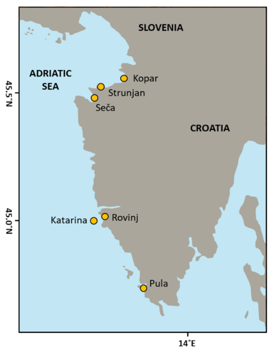
2.1. Sample Collection

Mytilus galloprovincialis mussels (N = 40 per sampling site) were collected from three sampling sites on the Croatian coastline and three sampling sites on the Slovenian coastline of the northern Adriatic (Figure 1) in February 2014. A few aspects were considered in selecting the sampling sites. One aspect was the types of mussel, which were native and farmed. Native mussels were collected at the pristine site, St. Catherine Island (Katarina, Croatia, 45°07′67″ N 13°62′96″ E), and farmed mussels were collected at 2 sites, Strunjan and Seča (Slovenia, 45°816′24″ N 13°834′57″ E and 45°807′50″ N 13°844′10″ E). The other aspect was the degree of pollution at the sampling sites. In brief, the pristine site is in the open area with less anthropic activities, and polluted sites were considered as highly, moderately, and slightly contaminated sites depending on the anthropic activities and measured data. The site defined as highly polluted was ACI marina Rovinj (Rovinj, Croatia, 44°87′55″ N; 13°84′67″ E), moderate was ACI marina Pula (Pula, Croatia, 44°887′55″ N 13°884′67″ E), and the slightly contaminated site was near the Kopar Marina (Kopar, Slovenia, site code 00TM). The polluted sites were selected according to previous monitoring data available on the water quality, the location, and the environments nearby [87]. The mussels were put in ice bags and transferred to the laboratory.

2.2. Tissue Preparation

The digestive glands were carefully excised from 40 adult mussels (N = 40 per sampling site) and immediately fixed by immersion in precooled n-hexan. Following this, half of the digestive glands were embedded in O.C.T.T M (Microm Inc. GmbH, Walldorf, Germany) and further sectioned into 10 μm thick slices on a cryotome (Zeiss Hyrax C 50, Microm GmbH, Munich, Germany). The frozen tissue sections were mounted onto slides, allowed to thaw to room temperature, and subsequently treated with hematoxylin and eosin staining. Analysis and digital image capture were performed using a light microscope (Nikon-SA (Belmont, CA, USA)) associated with a CCD Ikeagami ICD-803P digital camera (Tokyo, Japan).

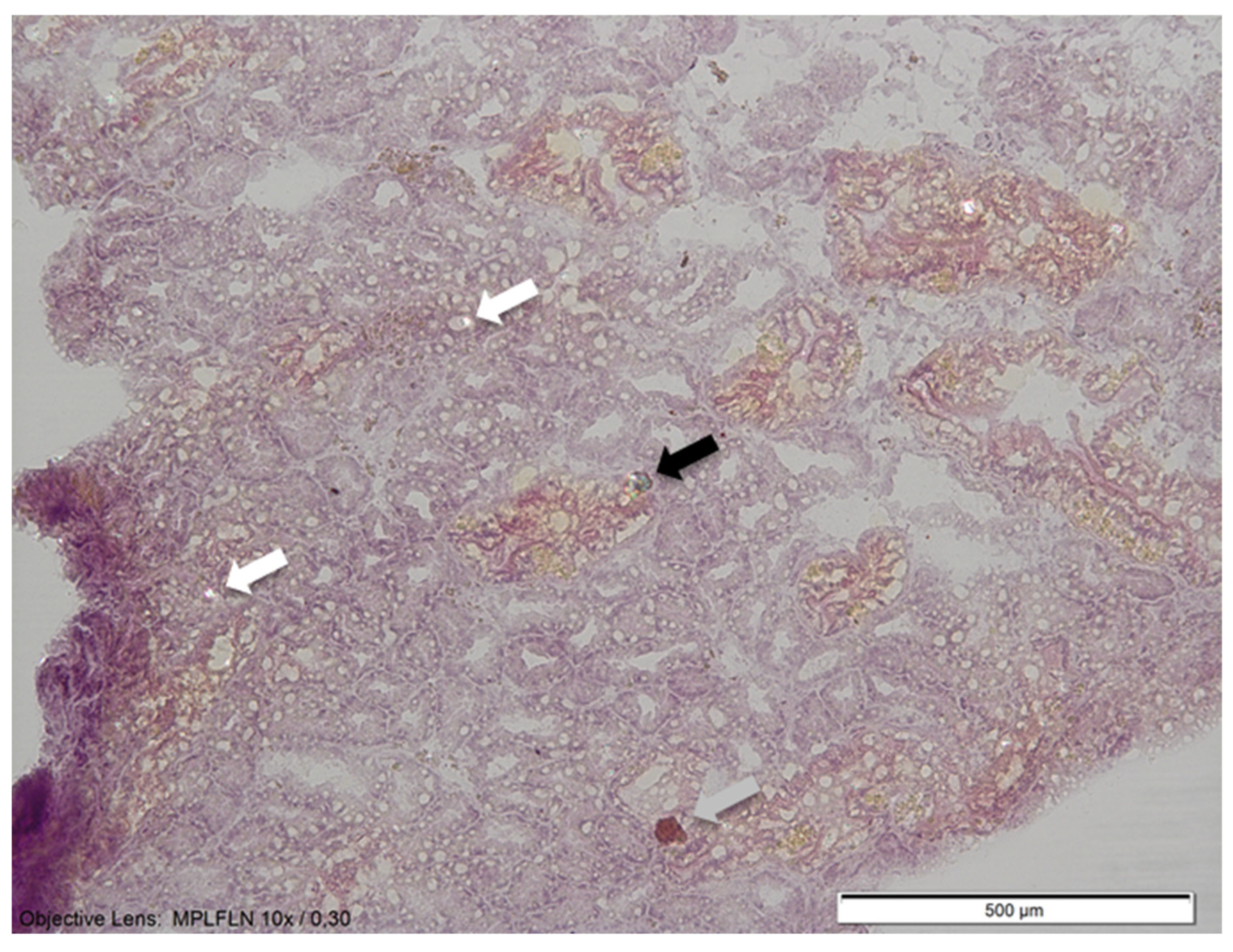
2.3. Observation and Validation of Microplastic

The incursion of airborne fibers is a recurrent phenomenon in MP research [88]; thus, rigorous precautions should be taken while processing samples. As general control rules, work surfaces were cleaned with 99% ethanol, and laboratory cotton suits and nitrile gloves with no dust were worn when working to minimize contamination from the air. Moreover, seven procedural blanks (one for each cryosection and staining per sampling site) were analyzed together with the samples to detect contamination. The procedural blanks were completely free of any form of plastic contamination, as was seen in our previous study [83]. Slides and coverslips were cleaned and checked under the microscope for contamination with airborne fibers before use. To avoid any potential airborne contamination, contamination-free slides were left exposed to air during dissection. Airborne contamination was considered to be negligible.
For the SEM, cryosection samples were left without a coverslip. The type of MP was examined under an SEM (SEM FEI FEG250QUANTA, FEI Company, Hilsboro, OR, USA equipped with OXFORD PENTAFET EDS detector, Oxford Instruments, Abingdon, Oxfordshire, UK), and the images were taken with an optimized acceleration voltage of 3 kV and a detector working distance of about 10 mm. During the SEM observation of particle morphology, shape and size, the qualitative elemental composition of MPs was used to distinguish MPs measured with an EDS detector from non-microplastic samples, as the plastics are carbon-based and other materials are expected to be non-organic [85]. According to the chemical composition of the detected items, some MPs were verified and excluded from non-MPs. The MPs counted in all size fractions were used to quantify MP abundance for each mussel cryosection by visual characterization under light microscopy, followed by chemical characterization by using SEM-EDS analysis to compare visual and chemical assignations of MPs in parallels.
MPs and non-MPs analyzed using a light microscope with a polarizer are shown in Figure 2 (marked with a black and grey arrow) and Figure 3a and b, respectively. Based on previous research, MP particles found in the mussels were categorized based on their size, color, and shape [88,89]. A visual assessment was applied to identify the morphotypes of the MPs according to the physical characteristics of the particles [27]. The MPs were classified into four morphotypes: film, line, fragment, and spheres. Film was characterized as an isolated part of large plastic debris; line was defined as an MP with a slender and elongated appearance; spheres were round MPs that looked like a ball in shape; a fragment was characterized as an incomplete part that could not be classified as film, sphere, or line. When classifying MPs, particles smaller than 5 μm were not considered (Figure 2, white arrow). Furthermore, larger particles that were not MPs (Figure 2, gray arrow, Figure 3a and Figure 4b) or were not included in the tissue section (Figure 3b) were also excluded from the count and defined as fiber contamination. Additionally, also according to the previous research conducted by Van Cauwenberghe and Janssen [89], MP particles found in the mussel digestive glands were classified into five categories according to their size: 5–10 μm, 11–15 μm, 16–20 μm, 21–25 μm, and greater than 25 µm. Further, MPs found on six different stations were classified into eight categories according to color, namely, yellow, green, transparent, pink, blue, purple, orange, and white according to Dahl et al. [90].

2.4. Data Analysis

Statistical analyses were performed using Statistica 9.0 (StatSoft InC., Tulsa, OK, USA). Statistical differences in localities were calculated using one-way ANOVA, and the statistical difference between individual two samples was calculated with the post hoc Tukey HSD test. Statistical significance was accepted at p < 0.05.

3. Results and Discussion

In the northern Adriatic, the River Po Delta contributes to a plastic flux of about 70 kg (km/day), which is subsequently dispersed along the coastlines of Italy, Slovenia, and Croatia [91,92]. The sea surface area with the highest pollution levels (>10 g/km2 of floating debris) forms an elongated band along the Italian coastline, gradually narrowing from northwest to southeast [91]. Additionally, this area includes two aquaculture sites for mussel farming, Strunja and Seča, in Slovenia. This presents an increased risk for the accumulation of MPs in humans through the food web chain.

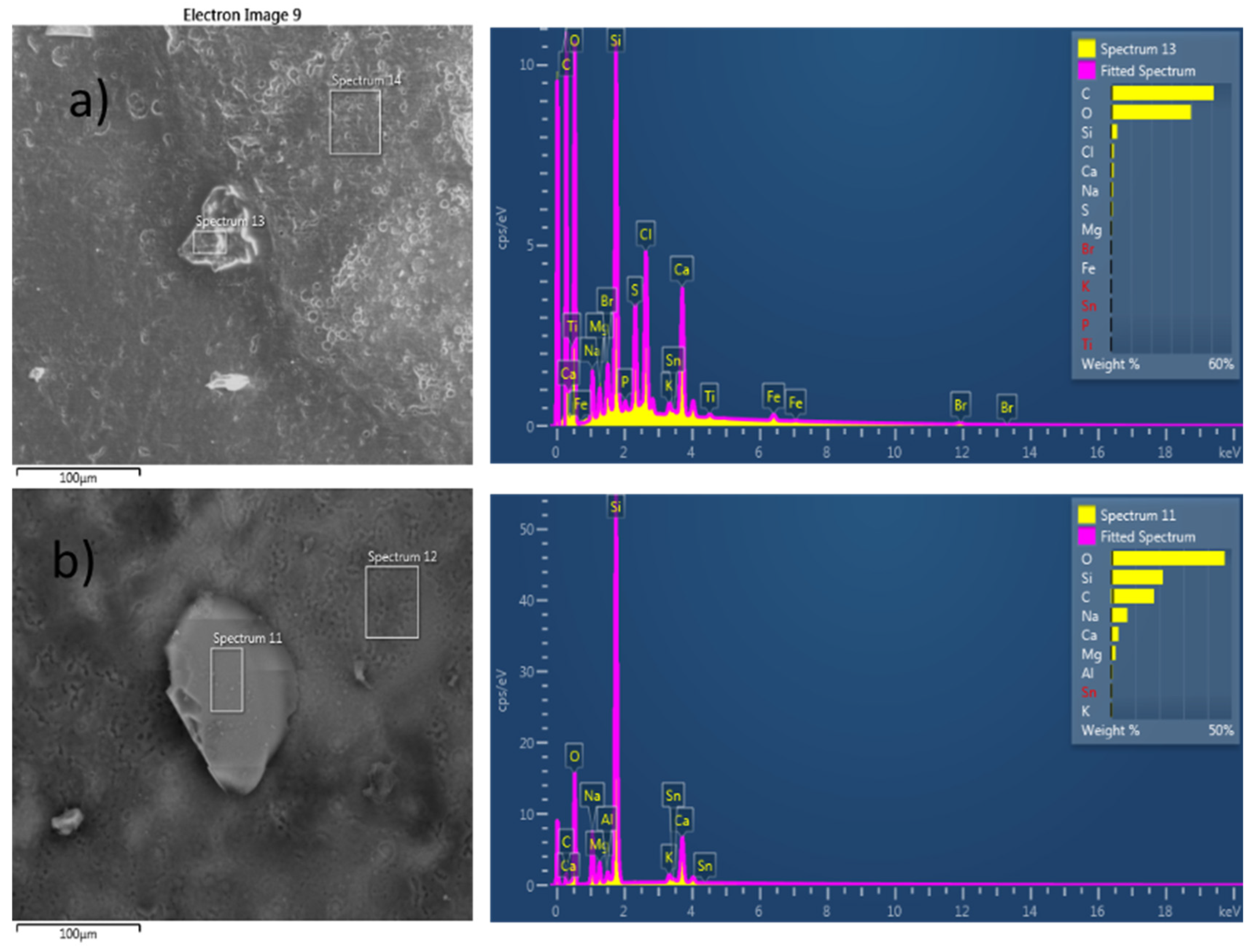
3.1. Identification of Microplastics Found in Mussel Digestive Glands Using Scanning-Electron Microscopy

In this study, MP particles were recorded by using SEM on the mussel digestive gland cryosections. Elemental chemical compositions were analyzed by using an energy dispersive spectrometry (EDS) detector. The presence of common elements used in plastic production in microplastic particles, such as carbon (C), hydrogen (H), oxygen (O), nitrogen (N), and various additional elements depending on the type of plastic, such as fluorine (F), chlorine (Cl), sulfur (S), or phosphorus (P), were detected. These elements are characteristic of different types of plastics, can be detected using methods such as SEM-EDS, were identified as MPs (Figure 4a), and were included in further data analysis. On the contrary, a particle that consisted of a larger portion of oxygen and silicon atoms was identified as a mineral particle (Figure 4b) and was not taken into account, as was found in many studies [32,51,93,94,95]. Analyzing cryosections of a mussel’s digestive gland with SEM-EDS could offer a good way to perform detection. Cryosections can be placed directly on a slide and examined using SEM-EDS, enabling the detection of MPs through the atom compositions present in the sample. An additional advancement in using SEM-EDS as a method for MP detection in bivalve tissue is the capability to differentiate diatoms from MPs in mussels, as previously outlined by Li et al. [32]. Khoironi et al. have utilized SEM-EDS analysis and shown the presence of SiO2 at 0.14% (w/w), Na2O at 24.27% (w/w), and Al2O3 at 0.27% (w/w) in MPs obtained in mussels, indicating the components which are mostly found in the plastic industries [32]. Furthermore, the deep fissures observed on the surfaces of MPs may potentially increase the surface area that could serve as a mechanism for the accumulation of pollutants on the MPs and further inside the marine organism [37].

3.2. Abundance of Microplastics Found in Mussel Digestive Glands

Statistical differences in the number, size, and shape of MPs were found between sites (Table 1). The total number of identified MPs per mussel digestive gland is shown in Figure 5.
The highest number of MP particles (32.85 ± 27.98 MPs/digestive gland) was found in the digestive gland of mussels sampled from the ACI marina Rovinj (Croatia), with a significant difference when compared to Pula (p < 0.001), Katarina (p < 0.0001), Seča (p < 0.001) and Strunjan (p < 0.0001). The location of ACI marina Rovinj was already previously noted as a highly polluted area [95,96]. Thus, this result was awaited since this location is situated within the urbanized area of the city Rovinj, on the marine coast surrounded by a sea that is influenced by water originating from the fish processing factory “Mirna”, wastewater from sewers, and ship docks. The following location, with slightly fewer MPs per digestive gland (21.30 ± 19.71 MPs/digestive gland), was Kopar (Slovenia), a place surrounded by tourist settlements and, thus, influenced by industrial wastewater and the harbor. In the mussels sampled from the ACI marina Pula, 14.10 ± 7.26 pieces of MPs were found per mussel digestive gland. The ACI marina Pula sampling site also accounted for a higher number of MPs per mussel digestive gland, indicating the influence of the immediate vicinity of a highly urbanized area, the port, and shipyards in Pula, which emit large amounts of urban and industrial waste into the water [95,96]. Nevertheless, the number of MPs found in mussels from Pula was not statistically different from the number of MP particles recorded in the farmed mussels from Seča and Strunjan (Slovenia). These data are especially concerning since these mussels are cultured for human consumption. The lowest number of particles was noted, as anticipated, in the digestive glands of mussels sampled from the pristine sea, St. Catherine Island (8.35 ± 5.59 MPs/digestive gland). The location at St. Catherine Island is located on the west coast of the Istrian peninsula and is 2 km away by airline from Rovinj. Therefore, this location was considered a control station since there were no sources of pollution close to the spot where the mussels were sampled. Nonetheless, it is still deeply concerning that even in this preserved location, acknowledged as a Natura 2000 site, MPs were identified in mussel tissue. This underscores the pervasive issue of MP pollution in the global seas.
Altogether, these data further confirm the presence of MPs to be from 8 up to 32 pieces of MPs per mussel digestive gland at all six sampled locations. This number is notably high, but direct comparisons to previously published data are challenging. Many previous studies have reported the presence of a considerable number of MPs in mussels. A higher number of MPs was also recorded in the mussel Perna perna from Guanabara Bay (southwestern Atlantic), and the number of MPs/mussels was up to 31.2 ± 17.8 (size of MPs ≥ 0.45 μm) [69]. In another study, the number of MPs from the mussel M. edulis collected along the coastline of China was also higher and varied from 1.5 to 7.6 items/individual [32]. Furthermore, it was noted that MP abundance was greater within the mussel Perna viridis (2.8–14.7 items/individual) compared to the other two species from Hong Kong: Brachidontes variabilis (0.2–0.7 items/individual) and Xenostrobus securis (0.6–3.1 items/individual) [50]. These data suggest that not all bivalves can intake the same number of MPs and, thus, only some are suitable for the identification of higher concentrations of MPs in their surroundings. MPs were found along the Hong Kong coast, with an average of 1.60–14.7 particles per mussel (P. viridis) per site [97]. Patterson et al. have recorded 1.5 to 7.6 items/individual in mussels (P. viridis and P. perna) and have further stated that the MP abundance in mussels reflected the abundance found in the water column [51]. The abundance of MPs varied among four cultured bivalve species, ranging from 0.5 to 3.3 items/individual or 0.3 to 20.1 items/g of wet weight in the digestive system. In the aforementioned study, significant differences were observed between species and regions, but no seasonal variations were detected [88].
In numerous studies, the quantity of MPs found per mussel was considerably lower. Webb et al. have recorded the MP abundance in the mussel P. perna from around New Zealand to be from 0 to 1.5 particles/mussel, and tissue MP concentrations ranged from 0 to 0.48 particles/g tissue (w/w) [98]. MPs were also recorded in mussels (M. galloprovincialis) from the Ionian Sea, and their abundance ranged from 1.7 to 2 items/individual [52]. In another study, MPs were present in M. edulis sampled from the French–Belgian–Dutch coastline, and the number of MPs found per mussel was on average 0.2 ± 0.3 MPs/g [99]. However, it is important to note that many studies employed digestion protocols, involving aggressive chemical extraction, subsequent filtration, and examination under a stereo microscope for MP quantification. During this long process, many smaller MP particles may have been lost or simply remained undetectable under a microscope. For instance, Vandeermersch et al. have found an average of 0.18 ± 0.14 items/g of wet weight for the acid mix method and 0.12 ± 0.04 total items/g of wet weight using the nitric acid method in the field-collected mussels M. galloprovincialis from three different “hotspot” locations in Europe (Po estuary, Italy; Tagus estuary, Portugal; Ebro estuary, Spain) [79]. The average amount of MPs found in mussels sold on the market in five Turkish cities was determined to be 0.6 ± 0.1 MPs/mussel using chemical digestion and, further, μ-Raman spectroscopy [94]. Furthermore, the presence of MPs recorded in two commercially grown bivalves Mytilus edulis and Crassostrea gigas was found to be 0.36 ± 0.07 particles/g w/w and 0.47 ± 0.16 particles/g w/w, respectively [89]. In a study conducted on mussels from 23 different locations along the Turkish coast, only 48% of the sampled mussels were found to have MPs. The average MP abundance was quite low and consisted of 0.69 items/individual [32]. The protocol used in the aforementioned study consisted of the digestion of the mussel tissue with H2O2, further determination of MPs under a stereomicroscope, and characterization by confirming with FTIR analyses. It is unlikely that so few MPs were found in the mussels from this location if compared to other studies from the same area. In our opinion, this could be attributed to the method employed for microplastic isolation. This further confirms the urgency for researchers to use a uniform method of MP detection in mussels as a bioindicator and monitoring of MP pollution.
Our study indicated a greater abundance of MPs in mussels from urban areas (locations Rovinj, Pula, and Kopar) in comparison with the mussels living in pristine marine waters (Strunjan, Seča, and Katarina). Berglund et al. have also noted higher concentrations of MPs (microfibers and particles) in urban areas compared to rural locations [100]. Contrary to our findings, in one study, the abundance of MPs was found to be significantly higher (2.7 items/g) in the areas with less human activities compared to the areas with intensive human activities (1.6 items/g) [32]. In another study, significantly higher concentrations of MPs were observed in farmed blue mussels compared to wild ones [101]. This finding once again states that MP pollution is of global concern, emphasizing the need for a collective effort worldwide to reduce the production of MPs.

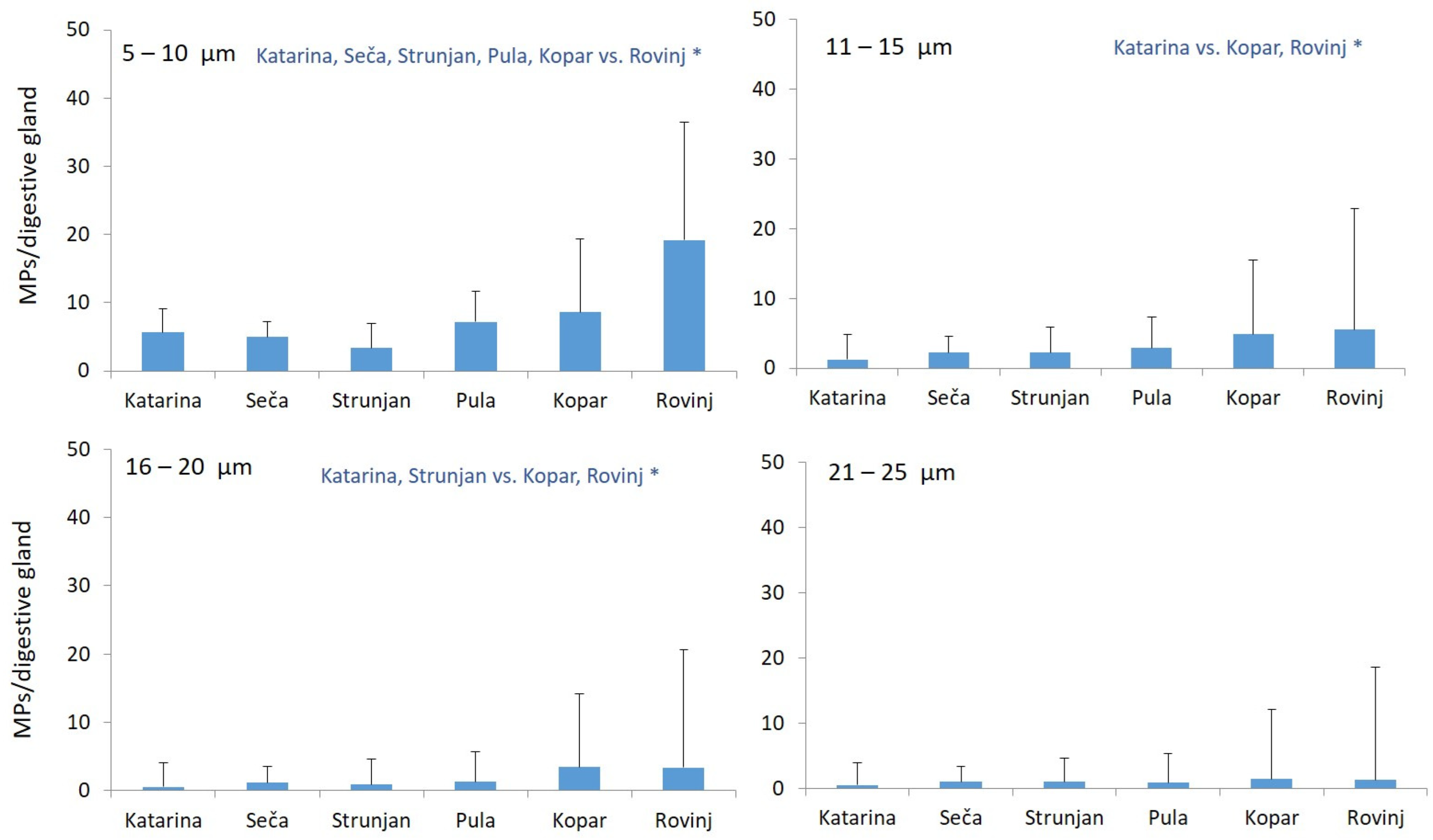
3.3. Size Distribution of Microplastics Found in Mussel Digestive Glands

Most of the MP particles found in the digestive glands of the mussel M. galloprovincialis at all investigated locations fit the smallest category analyzed herein (5–10 μm size), while the smallest number of particles were found to fit in the 21–25 μm category (Figure 6). Qu et al. have also demonstrated that mussels are more likely to ingest smaller-sized MPs (<5 μm) rather than particles with greater diameter [75], which are less efficiently cleared than the larger ones (>10 μm) [80]. This could become a problem for mussel predators, including humans, who could bioaccumulate and biomagnify MPs in their bodies through the marine food chain. Patterson et al. have proposed that the prevalence of MPs in a certain size range within mussel ingestion could be attributed to the abundance of those specific sizes in the water column [51]. Mussels are known to take up particles present in the water during their filtration process, which may explain the observed size distribution of ingested MPs. Furthermore, mussels can select particles for ingestion, and they often show a preference for smaller particles resembling their natural food items. These phenomena raise significant concerns about the environmental risks associated with smaller-sized plastic particles. Their ease of uptake by organisms (including mussels) and the increase in the bioavailability of associated chemicals amplify the potential risks and underscore the need for urgent attention and action to mitigate the impact of MPs on marine life and ecosystems. It was demonstrated in this study that mussels are prone to the ingestion of smaller-sized MPs into their digestive gland. These small size MPs can be further transported to other organs of the organism. Namely, in different studies, MPs were detected in the mussel foot, mantle, gills, and hemolymph [7,52,56]. Furthermore, several studies have observed the negative effects caused by the presence of MPs inside the mussel body. The abnormality in the mussel’s body functions included DNA damage, oxidative stress, atrophy, and necrosis of the digestive gland tissue, reductions in condition and fitness indices, hypertrophy of the gills, decreased filtration rates, and alterations in the response of the mussel immune system and filtration processes [21,63,67,76,82]. Additionally, mussels that accumulate smaller-sized MPs in their tissue could pose a threat to higher predators throughout the marine food web [64].
The results obtained in our study point out that the highest abundance of the smallest MPs (size 5–10 μm) occurred in the mussels sampled at the ACI marina Rovinj location (19.20 ± 17.30 MPs/digestive gland), while the same value was the lowest at the St. Catherine Island location (5.65 ± 3.51 MPs/digestive gland). The number of MPs with a size of 5–10 μm found in the mussels from ACI marina Rovinj was significantly different from those found in Pula (p < 0.0001), Katarina (p < 0.0001), Seča (p < 0.0001), Strunjan (p < 0.0001), and Kopar (p < 0.05). Other size categories of MPs were found in smaller numbers and were inversely proportional to the size distribution up to the last size class (particle > 25 µm), which showed no differences in sampled locations (Table 1, hence the graph is not shown herein). The location at St. Catherine Island had the lowest value of particles classified into the largest category (>25 µm), while the other five stations had approximately similar values of these particles (4–7 MPs/digestive gland).
Our described method using SEM allows for a highly specific differentiation based on size. Moreover, this method facilitates the further classification of very small MP particles, providing a comprehensive analysis of MPs in the sample. To date, mostly larger MP particles have been recorded in mussel tissue using different methods. In one study, the proportion of MPs in the blue mussel (M. edulis) was noted to be less than 250 μm in size and accounted for 17–79% of the total MPs [32]. All recovered MPs from the mussel M. galloprovincialis from different Italian stocks were the shape of filaments and ranged from 750 to 6000 μm in maximum length [102]. The size of MPs found in mussels from New Zealand was found to be in the range from 50 up to 700 µm, with a median diameter of 100 μm [98]. In another study, in both mussels (M. galloprovincialis) and fish species (Sardina pilchardus, Pagellus erythrinus, Mullus barbatus), MP sizes between 100 and 500 µm were the most abundant size class (52.6% and 67.6%, respectively) [52]. Ding et al. have found eighteen different types of polymers with a diameters slightly less than between 7 and 5000 μm by using μ-FT-IR, with polyvinyl chloride (PVC) and rayon being the most abundant ones [88].

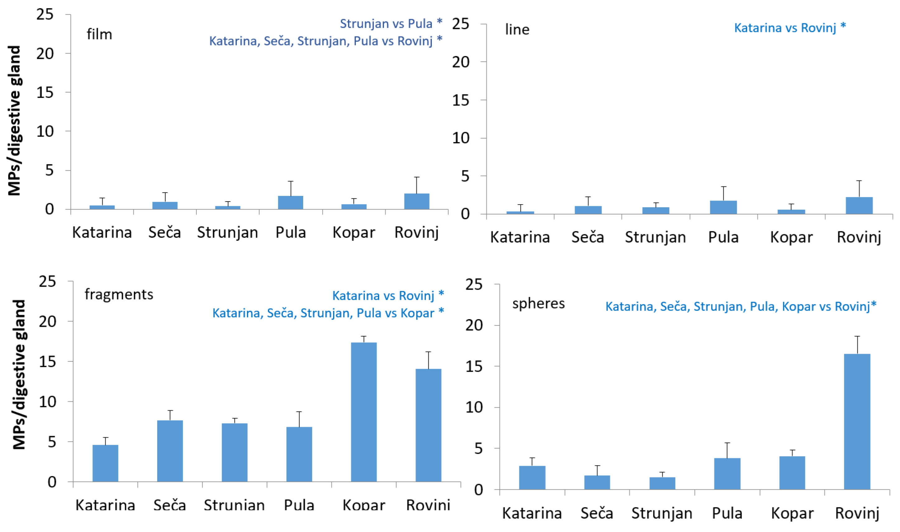
3.4. Different Shapes of Microplastics Found in Mussel Digestive Glands

The majority of MP particles found in the mussel tissue at all sampled locations were in the form of fragments, followed by spheres, lines, and film, with statistical differences found at all sampling locations (Table 1, Figure 7). The MPs in the line shape were found in a low number in the mussels sampled at St. Catherine, Seča, and Strunjan locations (0–1 MPs/digestive gland). In the mussels sampled at the ACI marina Rovinj, spheres and fragments (16.55 ± 14.69 MPs/digestive gland and 14.05 ± 11.19 MPs/digestive gland, respectively) were predominantly found, and were observed in much larger quantities compared to all other investigated sites (p < 0.01). Films were the most rarely found shape of MPs and were especially low in abundance in the mussel tissues from St. Catherine Island, Seča, and Strunjan (0–1 MPs/digestive gland) but were found in a higher number in the mussels from the ACI marina Rovinj (2.00 ± 2.15 MPs/digestive gland). Like in our study, fragments were frequently identified as the most prevalent shape of MPs in mussels in many studies. In the study by Gedik and Eryaşar [83], the morphology of MPs was ordered as follows: fragments (67.6%) > fibers (28.4%) > films (4.05%). Also, in the study by Webb et al., fragments were noted to be the most abundant shape of MPs found in the green-lipped mussel P. perna from New Zealand [98]. In another study, the majority of ingested MPs in the mussels M. galloprovincialis from the Ionian Sea were fragments (77.8%), followed by fibers (22.2%) [52]. On the contrary, Renzi et al. have noted filaments to be the most frequent MPs present in maricultured and natural mussels (M. galloprovincialis) from different Italian stocks [102]. In our study, one fiber was determined as a contaminant present in the mussel digestive gland cryosections, and this shape accounted for the less frequent type of material observed in cryosections. Fibers have been the most often found MPs in many studies until now [36,37,54,60,84], and we suppose this could be because of the cryosections analysis. In Qingdao, China, microfibers were also the most found form among all types of MPs discovered in locally cultured bivalve species (scallop Chlamys farreri, mussel Mytilus galloprovincialis, oyster Crassostrea gigas, and clam Ruditapes philippinarum) [88]. Furthermore, Li et al. have also found fibers, followed by fragments, to be the most common MPs found in the mussel M. edulis [32]. In research conducted in laboratory conditions with blue mussels (M. edulis), it was demonstrated that mussels take up MP fibers in a quantifiable and predictable manner, and most of the fiber ended up in the mussel digestive gland, with less in the gills [34]. The results from a study conducted by Vasanthni et al. have confirmed that fibers were the predominant type of MPs observed in the mussel P. viridis from Kasimedu, Chennai, India, followed by spheres, flakes, sheets, and fragments [67]. Furthermore, in a study conducted by Gündoğdu et al., the MPs found in mussels bought at the Turkish market in five different cities were identified as fibers (62.7%) and fragments (37.3%) [94]. Fragments or fibers were also mainly recorded as the dominant shape of MPs found in the edible green-lipped mussel P. viridis by using an automated mapping technique of Raman microspectroscopy [97]. According to Patterson et al., the higher prevalence of fibers in mussel bodies compared to other morphological types could be attributed to the slower egestion rate of fibers that may lead to a prolonged accumulation of fibers in the mussel’s digestive gland [51].
Additionally, fluctuations in local oceanographic conditions can hasten the gradual breakdown of plastic objects into smaller fragments and spheres [103]. Higher levels of microplastics (MPs) in the form of fragments and spheres were observed in mussels collected from the marinas Rovinj and Kopar, situated closest to the bay, compared to Pula, which is located in the offshore area relative to the other two sites. Moreover, the reduced presence of microplastics in mussels from Pula may be partly attributed to the influence of the northern Adriatic gyre, which could contribute to diminished accumulation and recirculation of seawater [104].

3.5. Color of Microplastics Found in Mussel Digestive Glands

The most abundant MPs by color found in mussels were white, while the least abundant particles were green particles (Figure 8). The majority of white particles (26.75 ± 6.94) were found in the mussel digestive glands sampled at the ACI marina Rovinj, while the fewest were found in the mussels from St. Catherine Island (6.25 ± 4.41). The second most abundant MP particles were orange and transparent in color and were found in mussel tissues from all investigated locations. Transparent MPs, along with the blue and red, were also recorded in the mussel P. perna from Guanabara Bay (southwestern Atlantic) [54]. Contrary to our findings, in another study the predominant colors of MPs recorded by FT-IR analysis in mussels were blue (54.4%) and pink (29.4%); only some were black (4.4%), yellow (4.4%), or transparent (4%); and a few were green (1.4%) [52]. Also, in our study, a greater number of green MP particles was noted, but only in the mussels sampled from the Kopar location. In mussels from Hong Kong, the MPs found were either red (67%), transparent (16%), blue (9%), or black (6%), and the proportion of the MP colors found differed among species and sites [36]. Patterson et al. have demonstrated blue-colored MPs to be the most dominant color found in mussels, and they further explained in their conclusion that the fragments are usually blue, transparent, and black [51]. The higher occurrences of blue MPs identified in mussels originate from synthetic textiles and materials used for fishing (e.g., ropes, lines, and floating buoys). Films are mostly transparent. The prevalence of white items observed in this study could result from the increased presence of white MPs in seawater, greater contamination of mussel prey with white MPs, and/or mussels exhibiting a preference for actively ingesting white particles. It must also be noted that the white and transparent MP items detected in the mussel digestive glands could have been due to a weathering process happening in the environment that might have resulted in the loss of MP color.
Altogether, the direct comparison of our data with other studies that have researched the presence of MPs inside the mussel M. galloprovincialis sampled from the northern Adriatic Sea is difficult since this was the first time that this methodology was used (FTIR analysis or different digestion protocols were used in previously published studies). Nonetheless, the previous studies all indicate the higher presence of MPs in the mussel tissue. The work conducted by [41] on the mussel M. galloprovincialis sampled from the northern Adriatic Sea indicates the presence of 1.11 MPs/g w/w of mussels’ tissue. This result is much lower if compared to our data, especially considering the highest number of MPs per mussel digestive gland was found at the ACI marina Rovinj location (32.85 MP/digestive gland). The differences could be attributed to different sampling locations and periods, but specifically also due to the differences in the used methodologies because the aforementioned study used filtration protocols where a significant number of MPs could be lost. Furthermore, in the same study, fragments consisted of 10–40% of all MP items, and this is contrary to our finding where fragments were found to be the dominant form of MPs in mussels. Fragments being the most dominant shape of MPs found in the mussels from the northern Adriatic Sea was also documented by [103]. As in our findings, Gomiero et al., ref. [103], have also noted the prevalence of smaller sizes of MPs (20–40 µm). Regarding the color found in the waters of the northern Adriatic Sea, there are very little data. In a study on MPs floating in the northwestern Adriatic Sea, transparent color appeared as the most recorded MP color, with the color white recorded in only one location as the dominant [104]. The color white was recorded as the most dominant color also in our study. Contrary to our findings, in another study, the dominant colors of MPs in sediment from the Montenegrin coast (southern Adriatic Sea) followed the order: blue > yellow > red > clear > black > green > blue-white > white. An interesting finding is present in the study of [30], which noted a significantly higher number of fragments present in the hepatopancreas of the crustacean Nephrops norvegicus. These data are in accordance with the highest number of MPs classified as fragments in our study of the mussel digestive gland.
To date, there are still only limited data concerning the presence of MPs in mussel tissue utilizing SEM-EDS as the analytical methodology for detection. During the last decade, different digestion protocols have been proposed to extract MPs from mussels. A significant part of these protocols involves organic matter digestion through the use of highly aggressive chemicals, such as hydrogen peroxide (H2O2), sodium hydroxide (NaOH), and nitric acid (HNO3), as well as further analyzing the samples under a polarized light microscope [41,44,46,83,105]. It is worth noting that these chemicals can lead to the undesirable destruction of certain MPs, as has been previously observed by Catarino et al. [105]. Furthermore, a lot of research to date has used FT-IR methods or µ-Raman spectroscopy to detect MPs present in the body of a marine organism [83,106]. But doing so, the smallest size particles cannot be detected. Hence, analyzing cryosections of a mussel’s digestive gland with SEM-EDS could offer a practical alternative. Moreover, sample processing is rapid, allowing for the simultaneous observation of multiple individuals. Up to five individuals can be compared under identical conditions due to the ability to place five digestive glands on a single slide. Furthermore, a crucial aspect of microplastic sample processing is contamination reduction, which was effectively avoided in this case.

4. Conclusions

To the best of our knowledge, this is the first time that cryosections of the mussel digestive gland have been used to detect MP particles in samples captured from native locations. The advantage of the methodology used in our study is that the diverse shapes of MPs observed on the cryosections of mussel digestive glands did not result from the breakdown of larger plastics, as could be the case in studies that used a method of MP extraction from organic tissue. Thus, the observed sizes and shapes of the MPs found within this study were probably the same as ingested and stored in the mussel digestive gland. It is imperative to establish a standardized, efficient, and cost-effective approach that can be universally adopted by researchers in this field. Moreover, the contamination during the processing of samples was considered to be negligible. Standardization will enhance the accuracy and comparability of data across studies, contributing to a comprehensive understanding of MP pollution. We propose the detection of MPs inside an organism by using SEM-EDS as a unique and quite fast method for the detection of MPs, thus making it suitable for a future large-scale monitoring program. In our research, with the combination of two unique techniques, polarized light microscopy and the valued addition of SEM, we were able to not only see the sizes and shapes of MPs present within the wild mussel digestive gland, but also to detect at the same time the elements present in the MPs noted under the microscope, thus confirming the identifications of MP materials.
A higher number of MP items was recorded in the mussels sampled from the harbors located near the urban areas. Nevertheless, it is of great concern that a very similar and statistically not different number of MPs was noted in the cultivated mussels and the ones from pristine areas. This research highlights that MP pollution is pervasive in both wild and farmed mussels found in the coastal waters of the northern Adriatic Sea. The mussel digestive gland could be used as the monitoring biomarker to detect sites polluted with MPs. To date, no data have been reported on the MPs of native mussels from the Slovenian and Croatian coastline of the northern Adriatic Sea; however, seawater monitoring will require additional research.

Author Contributions

Conceptualization, I.K. and P.B.; methodology, I.K., K.Š. and V.Š.; formal analysis, E.P. and M.B.; writing—original draft preparation, P.B. and. I.K.; writing—review and editing, M.B., E.P. and V.Š.; project administration, I.K. and P.B.; funding acquisition, I.K. All authors have read and agreed to the published version of the manuscript.

Funding

This work was supported by the Ministry of Science and Education, Republic of Croatia (grant for Ines Kovačić), the Juraj Dobrila University of Pula by project, Microplastics in marine organisms from Northern Adriatic (grant for Ines Kovačić).

Institutional Review Board Statement

Not applicable.

Informed Consent Statement

Not applicable.

Data Availability Statement

Data is contained within the article.

Acknowledgments

We highly appreciate Lorena Perić who helped in the fieldwork, as well as Nevenka Bihari who gave laboratory support at the Central for Marine Research Rovinj, Ruđer Bošković Institute.

Conflicts of Interest

The authors declare no conflicts of interest.

References

  1. Browne, M.A.; Dissanayake, A.; Galloway, T.S.; Lowe, D.M.; Thompson, R.C. Ingested microscopic plastic translocates to the circulatory system of the mussel, Mytilus edulis (L.). Environ. Sci. Technol. 2008, 42, 5026–5031. [Google Scholar] [CrossRef]
  2. Arthur, C.; Baker, J.E.; Bamford, H.A. (Eds.) Proceedings of the International Research Workshop on the Occurrence, Effects, and Fate of Microplastic Marine Debris. In Proceedings of the International Research Workshop on the Occurrence, Effects, and Fate of Microplastic Marine Debris, University of Washington Tacoma, Tacoma, WA, USA, 9–11 September 2008. [Google Scholar]
  3. Eriksen, M.; Mason, S.; Wilson, S.; Box, C.; Zellers, A.; Edwards, W.; Farley, H.; Amato, S. Microplastic pollution in the surface waters of the Laurentian Great Lakes. Mar. Pollut. Bull. 2013, 77, 177–182. [Google Scholar] [CrossRef] [PubMed]
  4. Bergmann, M.; Gutow, L.; Klages, M. (Eds.) Marine Anthropogenic Litter; Springer International Publishing: Cham, Switzerland, 2015; p. 447. [Google Scholar] [CrossRef]
  5. La Daana, K.K.; Asmath, H.; Gobin, J.F. The status of marine debris/litter and plastic pollution in the Caribbean Large Marine Ecosystem (CLME): 1980–2020. Environ. Pollut. 2022, 300, 118919. [Google Scholar] [CrossRef]
  6. Wright, S.L.; Thompson, R.C.; Galloway, T.S. The physical impacts of microplastics on marine organisms: A review. Environ. Pollut. 2013, 178, 483–492. [Google Scholar] [CrossRef]
  7. Cole, M.; Liddle, C.; Consolandi, G.; Drago, C.; Hird, C.; Lindeque, P.K.; Galloway, T.S. Microplastics, microfibres and nanoplastics cause variable sub-lethal responses in mussels (Mytilus spp.). Mar. Pollut. Bull. 2020, 160, 111552. [Google Scholar] [CrossRef]
  8. Miller, M.E.; Hamann, M.; Kroon, F.J. Bioaccumulation and biomagnification of microplastics in marine organisms: A review and meta-analysis of current data. PLoS ONE 2020, 15, e0240792. [Google Scholar] [CrossRef]
  9. Raju, P.; Santhanam, P.; Perumal, P. Impacts of microplastics on marine organisms: Present perspectives and the way forward. Egypt. J. Aquat. Res. 2022, 48, 205–209. [Google Scholar] [CrossRef]
  10. Zarfl, C.; Fleet, D.; Fries, E.; Galgani, F.; Gerdts, G.; Hanke, G.; Matthies, M. Microplastics in oceans. Mar. Pollut. Bull. 2011, 62, 1589–1591. [Google Scholar] [CrossRef]
  11. Vasilakopoulos, P.; Palialexis, A.; Boschetti, S.T.; Cardoso, A.C.; Druon, J.-N.; Konrad, C.; Kotta, M.; Magliozzi, C.; Palma, M.; Piroddi, C.; et al. Marine Strategy Framework Directive, Thresholds for MSFD Criteria: State of Play and Next Steps; EUR 31131 EN; Publications Office of the European Union: Luxembourg, 2022; ISBN 978-92-76-53689-5. JRC128344. [Google Scholar] [CrossRef]
  12. GESAMP. Sources, Fate and Effects of Microplastics in the Marine Environment: A Global Assessment; Kershaw, P.J., Ed.; Joint Group of Experts on the Scientific Aspects of Marine Environmental Protection: Paris, France, 2015; 96p. [Google Scholar]
  13. Jiang, X.; Conner, N.; Lu, K.; Tunnell, J.W.; Liu, Z. Occurrence, distribution, and associated pollutants of plastic pellets (nurdles) in coastal areas of South Texas. Sci. Total Environ. 2022, 842, 156826. [Google Scholar] [CrossRef]
  14. Rubin, A.E.; Gnaim, R.; Levi, S.; Zucker, I. Risk assessment framework for microplastic in marine environments. Sci. Total Environ. 2023, 901, 166459. [Google Scholar] [CrossRef] [PubMed]
  15. Pinlova, B.; Nowack, B. From cracks to secondary microplastics-surface characterization of polyethylene terephthalate (PET) during weathering. Chemosphere 2024, 352, 141305. [Google Scholar] [CrossRef]
  16. Boucher, J.; Friot, D. Primary Microplastics in the Oceans: A Global Evaluation of Sources; IUCN: Gland, Switzerland, 2017; Volume 10, p. 43. [Google Scholar] [CrossRef]
  17. Efimova, I.; Bagaeva, M.; Bagaev, A.; Kileso, A.; Chubarenko, I.P. Secondary microplastics generation in the sea swash zone with coarse bottom sediments: Laboratory experiments. Front. Mar. Sci. 2018, 5, 313. [Google Scholar] [CrossRef]
  18. Huber, M.; Archodoulaki, V.M.; Pomakhina, E.; Pukánszky, B.; Zinöcker, E.; Gahleitner, M. Environmental degradation and formation of secondary microplastics from packaging material: A polypropylene film case study. Polym. Degrad. Stab. 2022, 195, 109794. [Google Scholar] [CrossRef]
  19. Osman, A.I.; Hosny, M.; Eltaweil, A.S.; Omar, S.; Elgarahy, A.M.; Farghali, M.; Yap, P.-S.; Wu, Y.-S.; Nagandran, S.; Batumalaie, K.; et al. Microplastic sources, formation, toxicity and remediation: A review. Environ. Chem. Lett. 2023, 21, 2129–2169. [Google Scholar] [CrossRef]
  20. Hidalgo-Ruz, V.; Gutow, L.; Thompson, R.C.; Thiel, M. Microplastics in the marine environment: A review of the methods used for identification and quantification. Environ. Sci. Technol. 2012, 46, 3060–3075. [Google Scholar] [CrossRef]
  21. Li, J.; Wang, Z.; Rotchell, J.M.; Shen, X.; Li, Q.; Zhu, J. Where are we? Towards an understanding of the selective accumulation of microplastics in mussels. Environ. Pollut. 2021, 286, 117543. [Google Scholar] [CrossRef]
  22. Gajšt, T.; Bizjak, T.; Palatinus, A.; Liubartseva, S.; Kržan, A. Sea surface microplastics in Slovenian part of the Northern Adriatic, Mar. Pollut. Bull. 2016, 113, 392–399. [Google Scholar] [CrossRef] [PubMed]
  23. Munari, C.; Scoponi, M.; Mistri, M. Plastic debris in the Mediterranean Sea: Types, occurrence and distribution along Adriatic shorelines. Waste. Manag. 2017, 67, 385–391. [Google Scholar] [CrossRef] [PubMed]
  24. Mistri, M.; Scoponi, M.; Sfriso, A.A.; Munari, C.; Curiotto, M.; Sfriso, A.; Orlando-Bonaca, M.; Lipej, L. Microplastic Contamination in Protected Areas of the Gulf of Venice. Water Air Soil Pollut. 2021, 113, 379. Available online: https://link.springer.com/article/10.1007/s11270-021-05323-9 (accessed on 1 January 2024). [CrossRef]
  25. Korez, Š.; Gutow, L.; Saborowski, R. Microplastics at the strandlines of Slovenian beaches. Mar. Pollut. Bull. 2019, 145, 334–342. [Google Scholar] [CrossRef] [PubMed]
  26. Bošković, N.; Joksimović, D.; Peković, M.; Perošević-Bajčeta, A.; Bajt, O. Microplastics in Surface Sediments along the Montenegrin Coast, Adriatic Sea: Types, Occurrence, and Distribution. J. Mar. Sci. Eng. 2021, 9, 841. [Google Scholar] [CrossRef]
  27. Avio, C.G.; Gorbi, S.; Milan, M.; Benedetti, M.; Fattorini, D.; d’Errico, G.; Pauletto, M.; Bargelloni, L.; Regoli, F. Pollutants bioavailability and toxicological risk from microplastics to marine mussels. Environ. Pollut. 2015, 198, 211–222. [Google Scholar] [CrossRef]
  28. Pellini, G.; Gomiero, A.; Fortibuoni, T.; Ferrà, C.; Grati, F.; Tassetti, A.N.; Polidori, P.; Fabi, G.; Scarcella, G. Characterization of microplastic litter in the gastrointestinal tract of Solea solea from the Adriatic Sea. Environ. Pollut. 2018, 234, 943–952. [Google Scholar] [CrossRef]
  29. Bošković, N.; Joksimović, D.; Bajt, O. Microplastics in fish and sediments from the Montenegrin coast (Adriatic Sea): Similarities in accumulation. Sci. Total Environ. 2022, 850, 158074. [Google Scholar] [CrossRef]
  30. Martinelli, M.; Gomiero, A.; Guicciardi, S.; Frapiccini, E.; Strafella, P.; Angelini, S.; Domenichetti, F.; Belardinelli, A.; Colella, S. Preliminary results on the occurrence and anatomical distribution of microplastics in wild populations of Nephrops norvegicus from the Adriatic Sea. Environ. Pollut. 2021, 278, 116872. [Google Scholar] [CrossRef]
  31. Kanduč, T.; Šlejkovec, Z.; Falnoga, I.; Mori, N.; Budič, B.; Kovačić, I.; Pavičić-Hamer, D.; Hamer, B. Environmental status of the NE Adriatic Sea, Istria, Croatia: Insights from mussel Mytilus galloprovincialis condition indices, stable isotopes and metal (loid)s. Mar. Pollut. Bull. 2018, 126, 525–534. [Google Scholar] [CrossRef]
  32. Li, J.; Qu, X.; Su, L.; Zhang, W.; Yang, D.; Kolandhasamy, P.; Li, D.; Shi, H. Microplastics in mussels along the coastal waters of China. Environ. Pollut. 2016, 214, 177–184. [Google Scholar] [CrossRef] [PubMed]
  33. Hamer, B.; Korlević, M.; Durmiši, E.; Nerlović, V.; Bierne, N. Nuclear marker Me 15–16 analyses of Mytilus galloprovincialis populations along the eastern Adriatic coast. Cah. Biol. Mar. 2012, 53, 35. [Google Scholar]
  34. Woods, M.N.; Stack, M.E.; Fields, D.M.; Shaw, S.D.; Matrai, P.A. Microplastic fiber uptake, ingestion, and egestion rates in the blue mussel (Mytilus edulis). Mar. Pollut. Bull. 2018, 137, 638–645. [Google Scholar] [CrossRef]
  35. Gonçalves, C.; Martins, M.; Sobral, P.; Costa, P.M.; Costa, M.H. An assessment of the ability to ingest and excrete microplastics by filter-feeders: A case study with the Mediterranean mussel. Environ. Pollut. 2019, 245, 600–606. [Google Scholar] [CrossRef]
  36. Stabili, L.; Acquaviva, M.I.; Cavallo, R.A. Mytilus galloprovincialis filter feeding on the bacterial community in a Mediterranean coastal area (Northern Ionian Sea, Italy). Water Res. 2005, 39, 469–477. [Google Scholar] [CrossRef]
  37. Mubiana, V.K.; Blust, R. Effects of temperature on scope for growth and accumulation of Cd, Co, Cu and Pb by the marine bivalve Mytilus edulis. Mar. Environ. Res. 2007, 63, 219–235. [Google Scholar] [CrossRef] [PubMed]
  38. Signa, G.; Di Leonardo, R.; Vaccaro, A.; Tramati, C.D.; Mazzola, A.; Vizzini, S. Lipid and fatty acid biomarkers as proxies for environmental contamination in caged mussels Mytilus galloprovincialis. Ecol. Indic. 2015, 57, 384–394. [Google Scholar] [CrossRef]
  39. Coppola, F.; Almeida, Â.; Henriques, B.; Soares, A.M.; Figueira, E.; Pereira, E.; Freitas, R. Biochemical responses and accumulation patterns of Mytilus galloprovincialis exposed to thermal stress and Arsenic contamination. Ecotoxicol. Environ. Saf. 2018, 147, 954–962. [Google Scholar] [CrossRef]
  40. Musella, M.; Wathsala, R.; Tavella, T.; Rampelli, S.; Barone, M.; Palladino, G.; Biagi, E.; Brigidi, P.; Turroni, S.; Franzellitti, S.; et al. Tissue-scale microbiota of the Mediterranean mussel (Mytilus galloprovincialis) and its relationship with the environment. Sci. Total Environ. 2020, 717, 137209. [Google Scholar] [CrossRef]
  41. Pizzurro, F.; Recchi, S.; Nerone, E.; Salini, R.; Barile, N.B. Accumulation Evaluation of Potential Microplastic Particles in Mytilus galloprovincialis from the Goro Sacca (Adriatic Sea, Italy). Microplastics 2022, 1, 303–318. [Google Scholar] [CrossRef]
  42. De Witte, B.; Devriese, L.; Bekaert, K.; Hoffman, S.; Vandermeersch, G.; Cooreman, K.; Robbens, J. Quality assessment of the blue mussel (Mytilus edulis): Comparison between commercial and wild types. Mar. Pollut. Bull. 2014, 85, 146–155. [Google Scholar] [CrossRef]
  43. Pavičić Hamer, D.; Kovačić, I.; Koščica, L.; Hamer, B. Physiological indices of maricultured mussel Mytilus galloprovincialis Lamarck, 1819 in Istria, Croatia: Seasonal and transplantation effect. J. World Aquac. Soc. 2016, 47, 768–778. [Google Scholar] [CrossRef]
  44. Santana, M.F.M.; Ascer, L.G.; Custódio, M.R.; Moreira, F.T.; Turra, A. Microplastic contamination in natural mussel beds from a Brazilian urbanized coastal region: Rapid evaluation through bioassessment. Mar. Pollut. Bull. 2016, 106, 183–189. [Google Scholar] [CrossRef]
  45. Bråte, I.L.N.; Hurley, R.; Iversen, K.; Beyer, J.; Thomas, K.V.; Steindal, C.C.; Green, N.W.; Olsen, M.; Lusher, A. Mytilus spp. as sentinels for monitoring microplastic pollution in Norwegian coastal waters: A qualitative and quantitative study. Environ. Pollut. 2018, 243, 383–393. [Google Scholar] [CrossRef]
  46. Khoironi, A.; Anggoro, S.; Sudarno. The existence of microplastic in Asian green mussels. In IOP Conference Series: Earth and Environmental Science, Proceedings of the International Conference on Green Agro-industry and Bioeconomy (ICGAB 2017), Batu, Indonesia, 24–25 October 2017; IOP Publishing: Bristol, UK, 2018; Volume 131, p. 012050. [Google Scholar] [CrossRef]
  47. Li, J.; Lusher, A.L.; Rotchell, J.M.; Deudero, S.; Turra, A.; Bråte, I.L.N.; Sun, C.; Hossain, M.S.; Li, Q.; Kolandhasamy, P.; et al. Using mussel as a global bioindicator of coastal microplastic pollution. Environ. Pollut. 2019, 244, 522–533. [Google Scholar] [CrossRef]
  48. Ding, J.; Sun, C.; He, C.; Li, J.; Ju, P.; Li, F. Microplastics in four bivalve species and basis for using bivalves as bioindicators of microplastic pollution. Sci. Total Environ. 2021, 782, 146830. [Google Scholar] [CrossRef]
  49. Masiá, P.; Ardura, A.; Garcia-Vazquez, E. Microplastics in seafood: Relative input of Mytilus galloprovincialis and table salt in mussel dishes. Food Res. Int. 2022, 153, 110973. [Google Scholar] [CrossRef]
  50. Joyce, P.W.; Falkenberg, L.J. Microplastic abundances in co-occurring marine mussels: Species and spatial differences. Reg. Stud. Mar. Sci. 2023, 57, 102730. [Google Scholar] [CrossRef]
  51. Patterson, J.; Jeyasanta, K.I.; Laju, R.L.; Edward, J.P. Microplastic contamination in Indian edible mussels (Perna perna and Perna viridis) and their environs. Mar. Pollut. Bull. 2021, 171, 112678. [Google Scholar] [CrossRef]
  52. Digka, N.; Tsangaris, C.; Torre, M.; Anastasopoulou, A.; Zeri, C. Microplastics in mussels and fish from the Northern Ionian Sea. Mar. Pollut. Bull. 2018, 135, 30–40. [Google Scholar] [CrossRef] [PubMed]
  53. Abidli, S.; Pinheiro, M.; Lahbib, Y.; Neuparth, T.; Santos, M.M.; Trigui El Menif, N. Effects of environmentally relevant levels of polyethylene microplastic on Mytilus galloprovincialis (Mollusca: Bivalvia): Filtration rate and oxidative stress. Environ. Sci. Pollut. Res. 2021, 28, 26643–26652. [Google Scholar] [CrossRef]
  54. Calmão, M.; Blasco, N.; Benito, A.; Thoppil, R.; Torre-Fernandez, I.; Castro, K.; Izagirre, U.; Garcia-Velasco, N.; Soto, M. Time-course distribution of fluorescent microplastics in target tissues of mussels and polychaetes. Chemosphere 2023, 311, 137087. [Google Scholar] [CrossRef] [PubMed]
  55. Gonçalves, J.M.; Benedetti, M.; d’Errico, G.; Regoli, F.; Bebianno, M.J. Polystyrene nanoplastics in the marine mussel Mytilus galloprovincialis. Environ. Pollut. 2023, 333, 122104. [Google Scholar] [CrossRef] [PubMed]
  56. Kolandhasamy, P.; Su, L.; Li, J.; Qu, X.; Jabeen, K.; Shi, H. Adherence of microplastics to soft tissue of mussels: A novel way to uptake microplastics beyond ingestion. Sci. Total Environ. 2018, 610, 635–640. [Google Scholar] [CrossRef]
  57. Von Moos, N.; Burkhardt-Holm, P.; Köhler, A. Uptake and effects of microplastics on cells and tissue of the blue mussel Mytilus edulis L. after an experimental exposure. Environ. Sci. Technol. 2012, 46, 11327–11335. [Google Scholar] [CrossRef] [PubMed]
  58. Détrée, C.; Gallardo-Escárate, C. Polyethylene microbeads induce transcriptional responses with tissue-dependent patterns in the mussel Mytilus galloprovincialis. J. Molluscan Stud. 2017, 83, 220–225. [Google Scholar] [CrossRef]
  59. Masiá, P.; Ardura, A.; García-Vázquez, E. Virgin polystyrene microparticles exposure leads to changes in gills DNA and physical condition in the Mediterranean mussel Mytilus galloprovincialis. Animals 2021, 11, 2317. [Google Scholar] [CrossRef]
  60. Faggio, C.; Tsarpali, V.; Dailianis, S. Mussel digestive gland as a model tissue for assessing xenobiotics: An overview. Sci. Total Environ. 2018, 636, 220–229. [Google Scholar] [CrossRef]
  61. Kinjo, A.; Mizukawa, K.; Takada, H.; Inoue, K. Size-dependent elimination of ingested microplastics in the Mediterranean mussel Mytilus galloprovincialis. Mar. Pollut. Bull. 2019, 149, 110512. [Google Scholar] [CrossRef]
  62. Rivera-Hernández, J.R.; Fernández, B.; Santos-Echeandia, J.; Garrido, S.; Morante, M.; Santos, P.; Albentosa, M. Biodynamics of mercury in mussel tissues as a function of exposure pathway: Natural vs microplastic routes. Sci. Total Environ. 2019, 674, 412–423. [Google Scholar] [CrossRef]
  63. Pedersen, A.F.; Gopalakrishnan, K.; Boegehold, A.G.; Peraino, N.J.; Westrick, J.A.; Kashian, D.R. Microplastic ingestion by quagga mussels, Dreissena bugensis, and its effects on physiological processes. Environ. Pollut. 2020, 260, 113964. [Google Scholar] [CrossRef]
  64. Farrell, P.; Nelson, K. Trophic level transfer of microplastic: Mytilus edulis (L.) to Carcinus maenas (L.). Environ. Pollut. 2013, 177, 1–3. [Google Scholar] [CrossRef]
  65. Détrée, C.; Gallardo-Escárate, C. Single and repetitive microplastics exposures induce immune system modulation and homeostasis alteration in the edible mussel Mytilus galloprovincialis. Fish Shellfish Immunol. 2018, 83, 52–60. [Google Scholar] [CrossRef]
  66. Alnajar, N.; Jha, A.N.; Turner, A. Impacts of microplastic fibres on the marine mussel, Mytilus galloprovinciallis. Chemosphere 2021, 262, 128290. [Google Scholar] [CrossRef] [PubMed]
  67. Vasanthi, R.L.; Arulvasu, C.; Kumar, P.; Srinivasan, P. Ingestion of microplastics and its potential for causing structural alterations and oxidative stress in Indian green mussel Perna viridis—A multiple biomarker approach. Chemosphere 2021, 283, 130979. [Google Scholar] [CrossRef] [PubMed]
  68. Wegner, A.; Besseling, E.; Foekema, E.M.; Kamermans, P.; Koelmans, A.A. Effects of nanopolystyrene on the feeding behavior of the blue mussel (Mytilus edulis L.). Environ. Toxicol. Chem. 2012, 31, 2490–2497. [Google Scholar] [CrossRef] [PubMed]
  69. Birnstiel, S.; Soares-Gomes, A.; da Gama, B.A. Depuration reduces microplastic content in wild and farmed mussels. Mar. Pollut. Bull. 2019, 140, 241–247. [Google Scholar] [CrossRef]
  70. Ward, J.E.; Rosa, M.; Shumway, S.E. Capture, ingestion, and egestion of microplastics by suspension-feeding bivalves: A 40-year history. Anthr. Coasts 2019, 2, 39–49. [Google Scholar] [CrossRef]
  71. Ward, J.E.; Zhao, S.; Holohan, B.A.; Mladinich, K.M.; Griffin, T.W.; Wozniak, J.; Shumway, S.E. Selective ingestion and egestion of plastic particles by the blue mussel (Mytilus edulis) and eastern oyster (Crassostrea virginica): Implications for using bivalves as bioindicators of microplastic pollution. Environ. Sci. Technol. 2019, 53, 8776–8784. [Google Scholar] [CrossRef]
  72. Batel, A.; Linti, F.; Scherer, M.; Erdinger, L.; Braunbeck, T. Transfer of benzo [a] pyrene from microplastics to Artemia nauplii and further to zebrafish via a trophic food web experiment: CYP1A induction and visual tracking of persistent organic pollutants. Environ. Toxicol. Chem. 2016, 35, 1656–1666. [Google Scholar] [CrossRef]
  73. Catarino, A.I.; Macchia, V.; Sanderson, W.G.; Thompson, R.C.; Henry, T.B. Low levels of microplastics (MP) in wild mussels indicate that MP ingestion by humans is minimal compared to exposure via household fibres fallout during a meal. Environ. Pollut. 2018, 237, 675–684. [Google Scholar] [CrossRef]
  74. Zhao, S.; Ward, J.E.; Danley, M.; Mincer, T.J. Field-based evidence for microplastic in marine aggregates and mussels: Implications for trophic transfer. Environ. Sci. Technol. 2018, 52, 11038–11048. [Google Scholar] [CrossRef]
  75. Qu, X.; Su, L.; Li, H.; Liang, M.; Shi, H. Assessing the relationship between the abundance and properties of microplastics in water and in mussels. Sci. Total Environ. 2018, 621, 679–686. [Google Scholar] [CrossRef]
  76. Avio, C.G.; Gorbi, S.; Regoli, F. Experimental development of a new protocol for extraction and characterization of microplastics in fish tissues: First observations in commercial species from Adriatic Sea. Mar. Environ. Res. 2015, 111, 18–26. [Google Scholar] [CrossRef]
  77. Verdú, I.; González-Pleiter, M.; Leganés, F.; Rosal, R.; Fernandez-Pinas, F. Microplastics can act as vector of the biocide triclosan exerting damage to freshwater microalgae. Chemosphere 2021, 266, 129193. [Google Scholar] [CrossRef]
  78. Sun, Y.; Wang, X.; Xia, S.; Zhao, J. Cu (II) adsorption on Poly (Lactic Acid) Microplastics: Significance of microbial colonization and degradation. Chem. Eng. J. 2022, 429, 132306. [Google Scholar] [CrossRef]
  79. Vandermeersch, G.; Van Cauwenberghe, L.; Janssen, C.R.; Marques, A.; Granby, K.; Fait, G.; Kotterman, M.J.J.; Diogène, J.; Bekaert, K.; Robbens, J.; et al. A critical view on microplastic quantification in aquatic organisms. Environ. Res. 2015, 143, 46–55. [Google Scholar] [CrossRef]
  80. Fernández, B.; Albentosa, M. Insights into the uptake, elimination and accumulation of microplastics in mussel. Environ. Pollut. 2019, 249, 321–329. [Google Scholar] [CrossRef]
  81. Cutroneo, L.; Reboa, A.; Besio, G.; Borgogno, F.; Canesi, L.; Canuto, S.; Dara, M.; Enrile, F.; Forioso, I.; Greco, G.; et al. Microplastics in seawater: Sampling strategies, laboratory methodologies, and identification techniques applied to port environment. Environ. Sci. Pollut. Res. 2020, 27, 8938–8952. [Google Scholar] [CrossRef]
  82. Pavičić-Hamer, D.; Kovačić., I.; Sović, T.; Marelja, M.; Lyons, D.M. Exposure to Polymethylmethacrylate Microplastics Induces a Particle Size-Dependent Immune Response in Mediterranean Mussel Mytilus galloprovincialis. Fishes 2022, 7, 307. [Google Scholar] [CrossRef]
  83. Gedik, K.; Eryaşar, A.R. Microplastic pollution profile of Mediterranean mussels (Mytilus galloprovincialis) collected along the Turkish coasts. Chemosphere 2020, 260, 127570. [Google Scholar] [CrossRef]
  84. Wesch, C.; Bredimus, K.; Paulus, M.; Klein, R. Towards the suitable monitoring of ingestion of microplastics by marine biota: A review. Environ. Pollut. 2016, 218, 1200–1208. [Google Scholar] [CrossRef]
  85. Blair, R.M.; Waldron, S.; Phoenix, V.R.; Gauchotte-Lindsay, C. Microscopy and elemental analysis characterisation of microplastics in sediment of a freshwater urban river in Scotland, UK. Environ. Sci. Pollut. Res. 2019, 26, 12491–12504. [Google Scholar] [CrossRef]
  86. Stollberg, N.; Kröger, S.D.; Reininghaus, M.; Forberger, J.; Witt, G.; Brenner, M. Uptake and absorption of fluoranthene from spiked microplastics into the digestive gland tissues of blue mussels, Mytilus edulis L. Chemosphere 2021, 279, 130480. [Google Scholar] [CrossRef] [PubMed]
  87. CDI–Marine Data Access. Available online: http://harmonia.maris2.nl/search (accessed on 23 October 2013).
  88. Ding, L.; fan Mao, R.; Guo, X.; Yang, X.; Zhang, Q.; Yang, C. Microplastics in surface waters and sediments of the Wei River, in the northwest of China. Sci. Total Environ. 2019, 667, 427–434. [Google Scholar] [CrossRef]
  89. Van Cauwenberghe, L.; Janssen, C.R. Microplastics in bivalves cultured for human consumption. Environ. Pollut. 2014, 193, 65–70. [Google Scholar] [CrossRef]
  90. Dahl, M.; Bergman, S.; Björk, M.; Diaz-Almela, E.; Granberg, M.; Gullström, M.; Leiva-Dueñas, C.; Magnusson, K.; Marco-Méndez, C.; Piñeiro-Juncal, N.; et al. A temporal record of microplastic pollution in Mediterranean seagrass soils. Environ. Pollut. 2021, 273, 116451. [Google Scholar] [CrossRef]
  91. Liubartseva, S.; Coppini, G.; Lecci, R.; Creti, S. Regional approach to modeling the transport of floating plastic debris in the Adriatic Sea. Mar. Pollut. Bull. 2016, 103, 115–127. [Google Scholar] [CrossRef]
  92. Atwood, E.C.; Falcieri, F.M.; Piehl, S.; Bochow, M.; Matthies, M.; Franke, J.; Carniel, S.; Sclavo, M.; Laforsch, C.; Siegert, F. Coastal accumulation of microplastic particles emitted from the Po River, Northern Italy: Comparing remote sensing and hydrodynamic modelling with in situ sample collections. Mar. Pollut. Bull. 2019, 138, 561–574. [Google Scholar] [CrossRef]
  93. Foo, Y.H.; Ratnam, S.; Lim, E.V.; Abdullah, M.; Molenaar, V.J.; Hwai, A.T.S.; Zhang, S.; Li, H.; Zanuri, N.B.M. Microplastic ingestion by commercial marine fish from the seawater of Northwest Peninsular Malaysia. PeerJ 2022, 10, e13181. [Google Scholar] [CrossRef]
  94. Miserli, K.; Lykos, C.; Kalampounias, A.G.; Konstantinou, I. Screening of Microplastics in Aquaculture Systems (Fish, Mussel, and Water Samples) by FTIR, Scanning Electron Microscopy–Energy Dispersive Spectroscopy and Micro-Raman Spectroscopies. Appl. Sci. 2023, 13, 9705. [Google Scholar] [CrossRef]
  95. Bihari, N.; Mičić, M.; Fafanđel, M.; Jakšić, Ž.; Batel, R. Testing quality of sea water from the Adriatic coast of Croatia with toxicity, genotoxicity and DNA integrity tests. Acta Adriat. 2004, 45, 75–81. [Google Scholar]
  96. Jakšić, Ž.; Batel, R.; Bihari, N.; Mičić, M.; Zahn, R.K. Adriatic coast as a microcosm for global genotoxic marine contamination––A long-term field study. Mar. Pollut. Bull. 2005, 50, 1314–1327. [Google Scholar] [CrossRef]
  97. Leung, M.M.L.; Ho, Y.W.; Maboloc, E.A.; Lee, C.H.; Wang, Y.; Hu, M.; Cheung, S.G.; Fang, J.K.H. Determination of microplastics in the edible green-lipped mussel Perna viridis using an automated mapping technique of Raman microspectroscopy. J. Hazard. Mater. 2021, 420, 126541. [Google Scholar] [CrossRef]
  98. Webb, S.; Ruffell, H.; Marsden, I.; Pantos, O.; Gaw, S. Microplastics in the New Zealand green lipped mussel Perna canaliculus. Mar. Pollut. Bull. 2019, 149, 110641. [Google Scholar] [CrossRef]
  99. Van Cauwenberghe, L.; Claessens, M.; Vandegehuchte, M.B.; Janssen, C.R. Microplastics are taken up by mussels (Mytilus edulis) and lugworms (Arenicola marina) living in natural habitats. Environ. Pollut. 2015, 199, 10–17. [Google Scholar] [CrossRef]
  100. Berglund, E.; Fogelberg, V.; Nilsson, P.A.; Hollander, J. Microplastics in a freshwater mussel (Anodonta anatina) in Northern Europe. Sci. Total Environ. 2019, 697, 134192. [Google Scholar] [CrossRef]
  101. Mathalon, A.; Hill, P. Microplastic fibers in the intertidal ecosystem surrounding Halifax Harbor, Nova Scotia. Mar. Pollut. Bull. 2014, 81, 69–79. [Google Scholar] [CrossRef]
  102. Renzi, M.; Guerranti, C.; Blašković, A. Microplastic contents from maricultured and natural mussels. Mar. Pollut. Bull. 2018, 131, 248–251. [Google Scholar] [CrossRef]
  103. Gomiero, A.; Strafella, P.; Øysæd, K.B.; Fabi, G. First occurrence and composition assessment of microplastics in native mussels collected from coastal and offshore areas of the northern and central Adriatic Sea. Environ. Sci. Pollut. Res. 2019, 26, 24407–24416. [Google Scholar] [CrossRef]
  104. Vianello, A.; Da Ros, L.; Boldrin, A.; Marceta, T.; Moschino, V. First evaluation of floating microplastics in the Northwestern Adriatic Sea. Environ. Sci. Pollut. Res. 2018, 25, 28546–28561. [Google Scholar] [CrossRef]
  105. Catarino, A.I.; Thompson, R.; Sanderson, W.; Henry, T.B. Development and optimization of a standard method for extraction of microplastics in mussels by enzyme digestion of soft tissues. Environ. Toxicol. Chem. 2017, 36, 947–951. [Google Scholar] [CrossRef]
  106. Gündoğdu, S.; Çevik, C.; Ataş, N.T. Stuffed with microplastics: Microplastic occurrence in traditional stuffed mussels sold in the Turkish market. Food Biosci. 2020, 37, 100715. [Google Scholar] [CrossRef]
Figure 1. Six sampling locations of the mussel Mytilus galloprovincialis in the northern Adriatic: Katarina (native), Strunjan and Seča (cultured), and Kopar, Pula, and Rovinj (polluted—ACI marinas).
Applsci 14 02056 g001
Figure 2. Representative examples of MPs (black arrow), non–MPs (gray arrow), and particles smaller than 5 µm (white arrows, excluded from counting) found in the digestive gland tissue of mussel Mytilus galloprovincialis recorded with light microscopy using a polarizer.
Applsci 14 02056 g002
Figure 3. (a) Representative examples of non–MPs (white arrows) and (b) fiber found under the digestive gland of mussel Mytilus galloprovincialis recorded with light microscopy using a polarizer.
Applsci 14 02056 g003
Figure 4. Identified MP particles on mussel cryosection samples (a) and an example of a non-plastic samples (b) on mussel cryosections analyzed by EDS.
Applsci 14 02056 g004
Figure 5. Total number of MP particles in the digestive gland of mussels sampled at different locations: Katarina (native); Strunjan and Seča (cultured); and Kopar, Pula, and Rovinj (polluted—ACI marinas) in the northern Adriatic (N = 40 per sampling site). Data are reported as mean ± standard deviation. Statistically significant differences between locations are marked with stars (*).
Applsci 14 02056 g005
Figure 6. The number of MP particles in the digestive gland of mussels sampled at different locations: Katarina (native); Strunjan and Seča (cultured); and Kopar, Pula, and Rovinj (polluted—ACI marinas) in the northern Adriatic (N = 40 per sampling site) sorted by size. Data are reported as mean ± standard deviation. Statistically significant differences between locations are marked with stars (*).
Applsci 14 02056 g006
Figure 7. Different shapes of MP particles found in the digestive glands of mussels sampled at different locations: Katarina (native); Strunjan and Seča (cultured); and Kopar, Pula, and Rovinj (polluted—ACI marinas) in the northern Adriatic (N = 40 per sampling site). Data are reported as mean ± standard deviation. Statistically significant differences between locations are marked with stars (*).
Applsci 14 02056 g007
Figure 8. Different colors of MP particles found in the digestive glands of mussels sampled at different locations: Katarina (native); Strunjan and Seča (cultured); and Kopar, Pula, and Rovinj (polluted—ACI marinas) in the northern Adriatic (N = 40 per sampling site).
Applsci 14 02056 g008
Table 1. Results of one-way ANOVA testing differences in total number, size, and type of MPs on mussel cryosections between sampling sites. Significant ANOVAs were followed by a Tukey post hoc test and, when relevant, p values (marked * or **) are given in the tables and text.
Source of VariationSS aDf bMS cF d
Total number8570.8751714.177.76 *
Shape
Film44.3758.874.69 *
Line54.97510.993.14 *
Fragment2441.805488.366.82 *
Sphere3265.075653.015.94 *
Size (µm)
5–103524.745650.958.21 *
11–15280.66556.133.81 **
16–20163.36532.674.65 *
21–2510.9652.190.90
>25112.87522.572.13
a SS—the sum of squares due to the source, b Df—degrees of freedom, c MS—the mean sum of squares due to the source, d F—the F-statistic, * p < 0.0001, ** p < 0.01.
Disclaimer/Publisher’s Note: The statements, opinions and data contained in all publications are solely those of the individual author(s) and contributor(s) and not of MDPI and/or the editor(s). MDPI and/or the editor(s) disclaim responsibility for any injury to people or property resulting from any ideas, methods, instructions or products referred to in the content.

