Article

Deletion of Transient Receptor Channel Vanilloid 4 Aggravates CaCl2-Induced Abdominal Aortic Aneurysm and Vascular Calcification: A Histological Study

by
Isehaq Al-Huseini
1,
Maryam Al-Ismaili
1,
Ammar Boudaka
2 and
Srinivasa Rao Sirasanagandla
3,*
1
Department of Physiology, College of Medicine and Health Sciences, Sultan Qaboos University, Muscat 123, Oman
2
Department of Basic Medical Sciences, College of Medicine, QU Health, Qatar University, Doha P.O. Box 2713, Qatar
3
Department of Human and Clinical Anatomy, College of Medicine and Health Sciences, Sultan Qaboos University, Muscat 123, Oman
*
Author to whom correspondence should be addressed.
Appl. Sci. 2024, 14(6), 2566; https://doi.org/10.3390/app14062566
Submission received: 11 January 2024 / Revised: 11 March 2024 / Accepted: 13 March 2024 / Published: 19 March 2024

Abstract

:
Vascular calcification is calcium deposition occurring in the wall of blood vessels, leading to mechanical stress and rupture due to a loss of elasticity and the hardening of the vessel wall. The role of the Transient Receptor Channel Vanilloid 4 (TRPV4), a Ca2+-permeable cation channel, in the progression of vascular calcification is poorly explored. In this study, we investigated the role of TRPV4 in vascular calcification and the development of abdominal aortic aneurysm (AAA). Experimental mice were randomly divided into four groups: wild-type (WT) sham operated group, WT CaCl2-induced aortic injury, TRPV4-KO sham operated group, and TRPV4-KO CaCl2-induced aortic injury. The TRPV4-knockout (TRPV4-KO) mice and wild-type (WT) mice were subjected to the CaCl2-induced abdominal aortic injury. In histopathological analysis, the aorta of the TRPV4-KO mice showed extensive calcification in the tunica media with a significant increase in the outer diameter (p < 0.0001), luminal area (p < 0.05), and internal circumference (p < 0.05) after CaCl2 injury when compared to WT mice. Additionally, the tunica media of the TRPV4-KO mice aorta showed extensive damage with apparent elongation and disruption of the elastic lamella. These results indicate a protective function of TRPV4 against vascular calcification and the progression of AAA after CaCl2 injury.

1. Introduction

The vascular system transports oxygen, nutrients, and waste products throughout the body to facilitate their exchange between different compartments [1]. Several diseases are associated with the cardiovascular system and affect both blood vessels and the heart. AAA is a stealthy ailment carrying an 85% fatality risk upon rupture, characterized by a weakening of the abdominal aorta’s vessel walls, leading to their enlargement or dilation [2,3]. AAA involves the calcification and infiltration of inflammatory cells and the degradation of elastic lamellae, remaining asymptomatic until a potentially catastrophic rupture event [4,5]. Vascular calcification is one of the prevalent pathological conditions that affect human health in a severe manner [6]. Aortic calcification is associated with an increased risk of ruptured intracranial aneurysms [7]. Different factors increase the risk of developing vascular calcification, including aging, cardiovascular diseases, diabetes, chronic kidney diseases, etc. [8,9,10,11]. Blood vessels, in particular arteries, are composed of three layers: tunica adventitia, which is the outermost connective tissue layer; tunica media, made up of smooth muscle cells and elastin in its extracellular matrix; and tunica intima, which consists of a single thin layer of endothelial cells [10]. Recent studies have identified vascular microcalcification as a novel pathological characteristic and a potential mediator of AAA, suggesting that targeting microcalcification may be a promising strategy for both prevention and treatment [12]. Vascular calcification is characterized by mineral deposition in the tunica media or intima. Intimal calcification mainly occurs in atherosclerosis, which is associated with inflammation, lipid deposition, and the formation of microcalcifications [10,13]. Tunica media calcification is not entirely described; however, it is similar to intimal calcification as both processes resemble bone formation [14]. The primary outcome in tunica media calcification is vascular stiffness that leads to low vessel compliance, which can ultimately lead to an aortic aneurysm due to mechanical stress, highlighting the importance of understanding the underlying mechanisms of calcification in cardiovascular disease [15,16].
Indeed, the exact molecular mechanism behind the development of vascular calcification has yet to be understood, possibly due to the involvement of several overlapping mechanisms. However, studies have shown that smooth muscle cells are involved in pathogenesis, differentiating into osteoblast-like cells and producing a calcifying matrix [17,18,19]. The exact signaling pathway is still under investigation. Different factors contribute to the initiation of this process. Such factors include the absence of calcification inhibitors, a rise in smooth muscle cell oxidants, damage to DNA, apoptosis, and disturbance of calcium–phosphate hemostasis [20]. In differentiated smooth muscle cells, bone-related transcription factors, which upregulate bone proteins, are detected [20]. Bone morphogenetic proteins and inflammation mediators like TNFα are observed to activate the Msx2 transcription factor that, in turn, activates other factors like Runx2 and osterix. Moreover, bone-related proteins, including osteocalcin, sclerostin, RANKL, sialoprotein and alkaline phosphatase, are expressed and thus promote vascular calcification [20].
The induction of abdominal aortic injury through the administration of calcium chloride (CaCl2) has become a pivotal approach in vascular research, contributing significantly to our understanding of arterial pathology [21,22]. This experimental model involves the topical application of CaCl2 to the abdominal aorta, leading to localized injury and subsequent pathological changes. The injury induces a cascade of events, including inflammation, elastin degradation, and medial layer thinning, mimicking aspects of aortic aneurysm development [21,23]. CaCl2-induced abdominal aortic injury offers a controlled and reproducible method to investigate the molecular and structural alterations associated with vascular diseases, providing valuable insights for developing therapeutic strategies and interventions aimed to mitigate aortic pathologies. This model has been widely employed in preclinical studies, allowing researchers to explore the mechanisms underlying aortic injuries and their implications for cardiovascular health [23,24].
The Transient Receptor Potential (TRP) is a large superfamily of channels in mammalian that are divided into six subfamilies according to their DNA and protein sequence: TRPV (vanilloid), TRPC (canonical), TRPM (melastatin), TRPA (ankyrin), TRPP (polycystin), and TRPML (mucoliptin). Twenty-eight genes encoded for TRP cation channels are expressed on different mammalian cell types [25]. The TRPV family is subdivided into six isoforms, among them, 1–4 isoforms are clearly found to play an essential role in vascular regulation [25]. The human TRPV4 gene is located in chromosome twelve, that encodes for Ca2+-permeable non-selective cation channels. It comprises six membrane-spanning helices (S1–S6), a pore loop between S5 and S6, and intracellular N-, C-termini. TRPV4 is found to be expressed in many tissues; its messenger RNA has been detected in kidney, lung, spleen, testis, and adipose tissue using multiple tissue Northern blots. TRPV4 mRNA has been detected in the brain neurons using in situ hybridization [26]. Moreover, it is expressed in the heart, blood vessel endothelium, and some vascular smooth muscle cells [25].
The TRPV4 channel is multimodal and responds to a variety of signals, including physical stimuli like heat [27], chemical stimuli like lipid agonists [1], mechanical stimuli [27], and extracellular osmolarity changes [26]. It is found to play different biological functions due to its wide range of expression in tissues and ability to respond to multiple stimuli. TRPV4 in endothelial cells has a role in vasodilation by different independent pathways like nitric oxide, prostacyclin, and endothelium-derived hyperpolarizing factor (EDHF) [28]. Moreover, its expression in smooth muscles contributes to the activation of conductance potassium channels and hyperpolarization of smooth muscles [29]. In addition, the TRPV4 channel mediates osteoblast differentiation, which is involved in vascular calcification mechanisms, as described previously [30]. Despite previous studies, the precise role of TRPV4, a Ca2+-permeable cation channel, in the progression of vascular calcification and AAA, remains unclear. To shed light on this matter, the present study utilized TRPV4-knockout mice and subjected them to standard CaCl2-induced abdominal aortic injury to investigate the involvement of the TRPV4 channel in the progression of vascular calcification.

2. Materials and Methods

2.1. Animals

All animal experiments were performed in accordance with Sultan Qaboos University (SQU) Animal Ethics Committee regulations and SQU Guidelines for Care and Use of Laboratory Animals, and the experiments were conducted after obtaining approval from the Institutional Animal Ethics Committee of SQU (SQU/AEC/2017-2018/12).
The mice were housed in SQU’s small animal facility under carefully controlled conditions, including a 12-h light and 12-h dark cycle, a room temperature of 22 °C, and a relative humidity of 50–60%. They had been acclimatized for seven days prior to the surgery with free access to food and water. The body weight of each mouse was recorded daily after the surgery. For this in vivo experiment, 8–10-week-old male wild-type (WT) (C57BL\6NCr) (n = 30) and TRPV4-knockout (TRPV4-KO) (n = 30) mice were used. The animals were randomly divided into four equal groups, with 10 mice in each sham-operated group and 20 mice in each CaCl2-induced aortic injury groups including: WT sham-operated group (n = 10), WT CaCl2-induced aortic injury (n = 20), TRPV4-KO sham operated group (n = 10), and TRPV4-KO CaCl2-induced aortic injury (n = 20). Some mice from each group did not survive the surgery, and the remaining numbers are specified in the figure legends.

2.2. CaCl2-Induced Abdominal Aortic Injury

After seven days of acclimatization, an abdominal aortic injury was induced in two groups: WT and TRPV4-KO mice. CaCl2-induced aortic injury was performed as described previously with minor modifications [31]. Briefly, the mice were anaesthetized with an intraperitoneal injection of ketamine (75 mg/kg) and xylazine (5 mg/kg) prior to the laparotomy. An abdominal incision of approximately 2 cm was made, and the infra-renal aorta was exposed by moving the intestinal loops apart. Then, small pieces of gauze soaked with 0.6 mol/L CaCl2 were placed around the infrarenal aorta circumference for 10 min. The sham-operated mice were exposed to gauze soaked in 0.95% NaCl instead of CaCl2. After gauze removal, the aorta was rinsed with 0.95% NaCl and wiped using cotton swabs. Then, the incision was closed, and the mice were allowed to recover from anesthesia on a warm plate. Free access to food and water were provided for the mice after the surgical procedure until the sacrifice day. The surgery resulted in the mortality of some mice in each group, with the surviving numbers documented in the figure legends.

2.3. Aorta Collection and Morphometry

The mice were sacrificed two weeks postoperatively with an overdose of a ketamine and xylazine combination (ketamine, 100 mg/kg and xylazine, 20 mg/kg). Careful dissection was performed to expose the whole aorta and heart. Then, the aorta was flushed twice with a freshly prepared cold phosphate-buffered solution (PBS) through the apex of the left ventricle. Extraneous tissues were removed, and the abdominal aorta of each mouse was carefully dissected under a stereomicroscope and photographed with a known scale for morphometric analysis. Morphometric analysis was then executed using ImageJ software version 1.8 from the National Institutes of Health, Bethesda, MD, USA. To acquire the digital images, an Axioskop 2 microscope (Zeiss, Jena, Germany) equipped with a known micrometer scale was employed. The images were meticulously selected for analysis, focusing on key parameters such as circumference area, lumen area, and the medial layer. The process involved manual delineation to ensure precision in the measurements. It is important to note that all measurements were performed blindly for each group, enhancing the objectivity and reliability of the morphometric assessment. This comprehensive approach allowed for a detailed morphometric evaluation, providing valuable insights into the structural characteristics of the aortic sections under scrutiny.

2.4. Histological Analysis

The initial segments of the infrarenal aorta (the part of the aorta at the level of renal artery origin) measuring 2–3 mm in length were dissected carefully with a stereomicroscope and fixed with 4% paraformaldehyde for 24 h. Then, the tissues were embedded in paraffin wax and 4-µm-thick cross-sections were collected from each animal of all experimental groups. For the morphometric analysis, six sections from each mouse were taken at the interval of 24 µm i.e., 6 sections apart between the 2 adjacent sections onto glass slides. The sections were deparaffinized three times in xylene and dehydrated in different ethanol concentrations for 5 min in each. Next, the sections were stained with hematoxylin and eosin. The fluorescence properties of hematoxylin and eosin were harnessed to visualize elastin in the examined samples. Elastic lamellae, integral components of the tissue structure, emitted a distinct red color when exposed to fluorescent light. This allowed for a detailed examination of the elastin network within the tissue. Instances of damage to the elastic lamellae were readily identifiable as they exhibited clear signs such as cuts or elongation. The use of fluorescence not only facilitated the visualization of elastin but also provided a sensitive method for detecting structural alterations in the elastic lamellae, enhancing our ability to discern and characterize tissue integrity [32].
Alizarin red staining was performed to assess aortic calcification. Alizarin-stained sections were visualized with an Axioskop 2 plus microscope (Zeiss, Jena, Germany). A morphometric analysis of the stained sections was performed using software ImageJ (National Institutes of Health, Bethesda, MD, USA).

2.5. Statistical Analysis
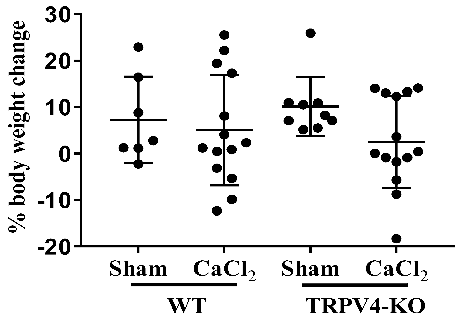
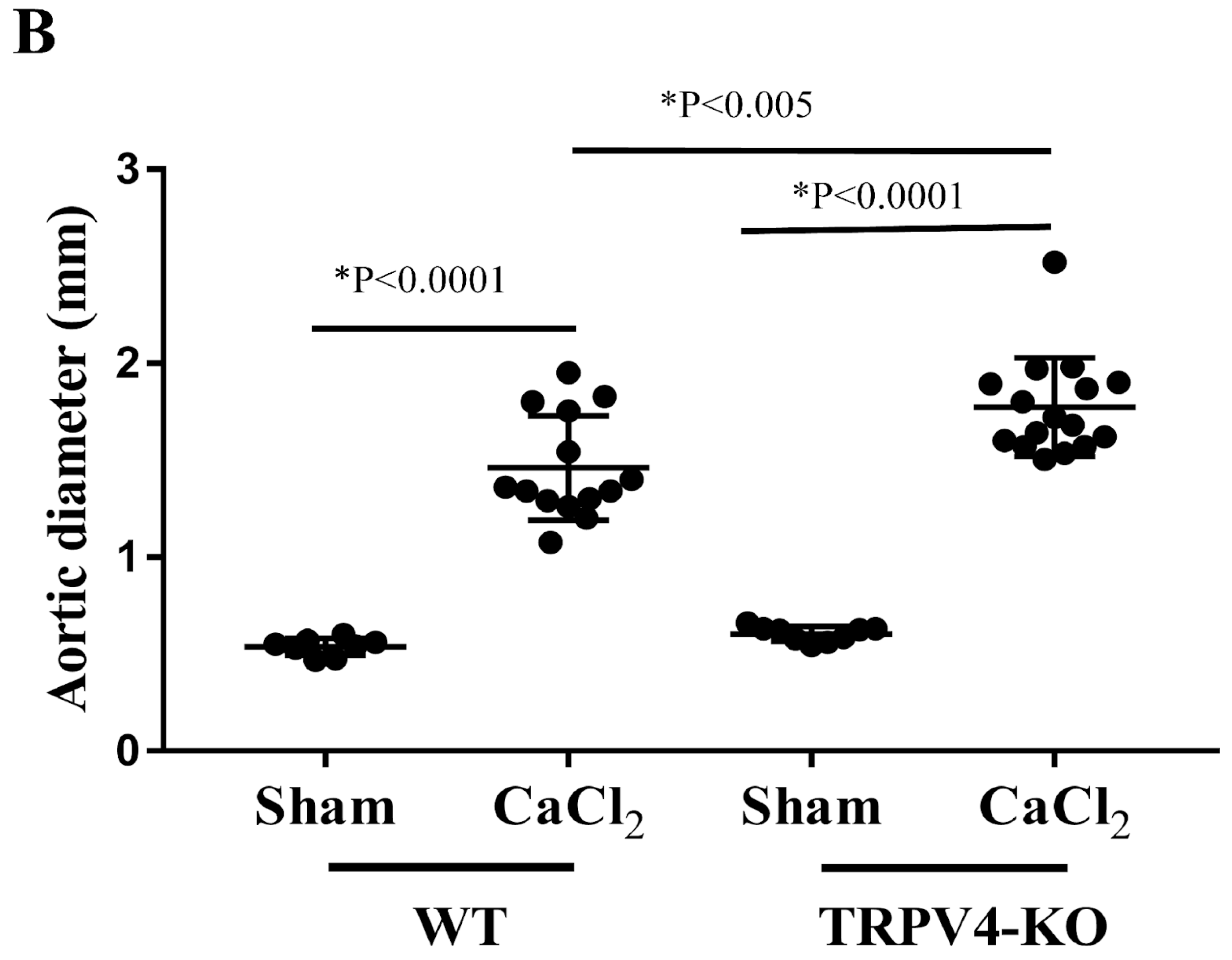
GraphPad Prism software version 7.0 (GraphPad Prism software, San Diego, CA, USA) was used to analyze the data. A one-way ANOVA followed by Tukey test to compare changes in all measured parameters between different groups were used. Experimental results were expressed as the mean ± SD. p < 0.05 was considered statistically significant.

3. Results

3.1. Deletion of TRPV4 Exacerbates Aortic Diameters in CaCl2-Induced Aortic Injury

To evaluate the role of TRPV4 in vascular calcification, we subjected WT and TRPV4-KO mice to CaCl2-induced abdominal aortic injury. The daily body weights of each mouse were meticulously documented over two weeks following the surgical procedure. The corresponding percentage changes in body weight are visually depicted in Figure 1. The CaCl2-treated groups exhibited a slight decrease in average body weights. However, no statistically significant variances were observed among the experimental groups (WT sham vs. WT CaCl2: p = 0.96, TRPV4-KO sham vs. TRPV4-KO CaCl2: p = 0.28).
Following the sacrifice of the mice, the infrarenal aortae were meticulously dissected and photographed using a predetermined scale for precise morphometric analysis. As anticipated, both wild-type (WT) and TRPV4-knockout (TRPV4-KO) mice exhibited a noteworthy augmentation in the outer diameter in response to the CaCl2-induced aorta injury model, as illustrated by representative images in Figure 2A (WT CaCl2 vs. WT sham: p < 0.0001, TRPV4-KO CaCl2 vs. TRPV4-KO sham: p < 0.0001). Interestingly, the aortae of the TRPV4-knockout (TRPV4-KO) mice demonstrated a marked increase in dilation when contrasted with those of the WT-CaCl2 mice (TRPV4-KO CaCl2 vs. WT CaCl2: p < 0.005), as morphometrically illustrated in Figure 2B. This intriguing observation suggests a potential involvement of TRPV4 in averting aortic aneurysm formation, raising the possibility that TRPV4 plays a crucial role in maintaining aortic structural integrity under conditions of CaCl2-induced injury, and indicating a possible role of TRPV4 in preventing the aortic aneurysm.

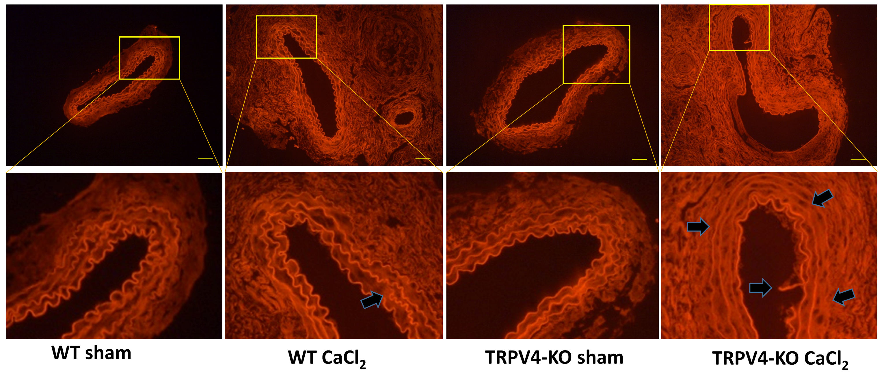
3.2. Deletion of TRPV4 Aggravates Tunica Media Damage in CaCl2-Induced Aortic Injury

Consistent with the above results, histological analysis of H&E-stained sections revealed an apparent increase in the overall aortic wall thickness of CaCl2-treated mice groups compared to sham mice groups. The adventitial layer thickness of the aorta was remarkably higher with an apparent infiltration of immune cells (Figure 3). Moreover, the tunica media of KO-CaCl2 mice was extensively damaged and detached from the adventitial layer. In addition, CaCl2-injured aortae showed more elongation and destruction of the tunica media elastic fibers in KO-CaCl2 mice compared to WT-CaCl2 mice (Figure 4).

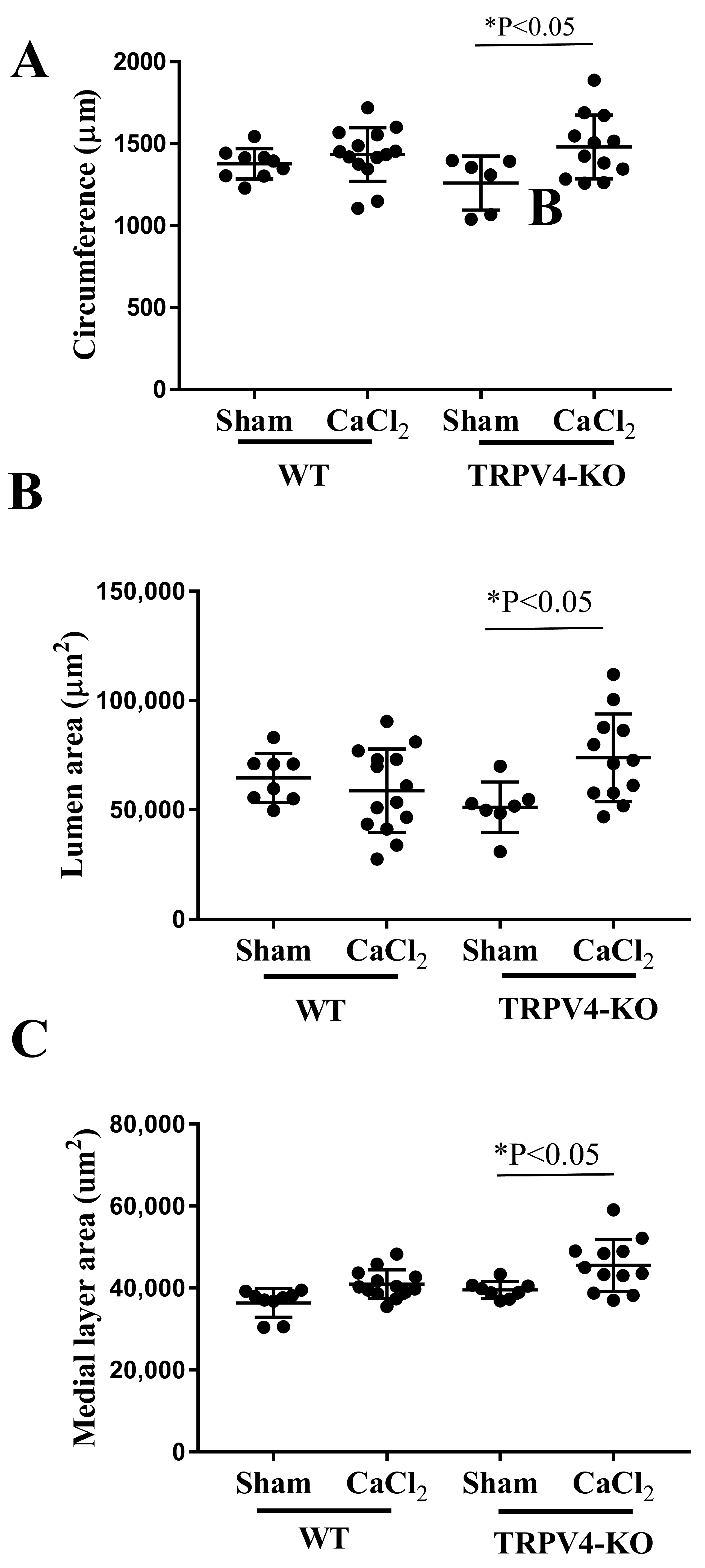
3.3. Deletion of TRPV4 Increases Tunica Media Area and Lumen Circumference in CaCl2-Induced Aortic Injury

To further investigate the morphometric changes in WT and TRPV4-KO mice, we analyzed H&E sections of the infrarenal aorta using additional parameters. The aortic circumference (µm) and lumen area (µm2) of the TRPV4-KO mice group were significantly higher in CaCl2 mice compared with sham mice (TRPV4-KO CaCl2 vs. TRPV4-KO sham: p < 0.05; Figure 5A,B). Moreover, tunica media area (µm2) was significantly increased in TRPV4-KO mice after CaCl2 injury compared to that in sham TRPV4-KO mice. (TRPV4-KO CaCl2 vs. TRPV4-KO sham: p < 0.05, Figure 5C).

3.4. Deletion of TRPV4 Enhances Aortic Calcification in CaCl2-Induced Aortic Injury

Alizarin red staining was performed on the histological sections of the infrarenal abdominal aorta to assess the intensity of calcium depositions, mainly in the medial layer. The presence of calcium deposition is indicated by orange to red pigment in the tissue. Aortic sections obtained from both WT-CaCl2 and KO-CaCl2 showed the presence of calcium depositions. However, the aortae of TRPV4-KO mice revealed more remarkable calcification after CaCl2 injury (Figure 6). In addition, the tunica media of those aortae showed n extensive damage with apparent elongation and disruption of elastic lamella in the medial layer. The medial layer area was larger in TRPV-KO-CaCl2 mice mainly due to calcium deposition and elastic fibers’ disruption and elongation.

4. Discussion

The involvement of mechanosensitive ion channels in skeletogenesis and bone formation was well studied [33,34,35]. However, their role in vascular calcification and AAA has not been adequately explored. Furthermore, to our knowledge, the role of TRPV4, a Ca2+-permeable cation channel, in the progression of vascular calcification and AAA has not been studied. In the present study, the aortae of the TRPV4-KO mice were significantly dilated compared to the WT after CaCl2-induced aortic injury. Moreover, the histological analysis and morphometric measurements revealed that the tunicae adventitia and media areas increased, leading to aortic-wall thickening. Additionally, a remarkable calcification was observed in the medial layer of TRPV4-KO mice compared to the WT mice after CaCl2-induced aorta injury.
Contrary to the present study results, in previous studies, the TRPV4 channel promoted bone formation and calcification [33,34]. Recently, it has been widely accepted that vascular calcification occurs through the trans-differentiation of vascular smooth muscle cells (VSMCs) to osteoblast-like cells [36]. The underlying mechanism of this trans-differentiation is the activation of β-catenin in VSMCs, which in turn leads to the translocation of β-catenin to the nucleus, where it can ultimately lead to the transcription of different osteoblast genes, including osteocalcin, alkaline phosphatase, and ostrex [20]. The inhibition of TRPV4 attenuated osteogenesis by reducing the expression of osteoblastic genes and Wnt3α/β-catenin in bone marrow stem cells, which conflicts with our finding [37]. However, a recent study by the Espadas–Alvarez group demonstrated that TRPV4 inhibits β-catenin transcriptional activity through the interaction with β-catenin, the main regulator of osteoblast genes [37]. This might explain the remarkable calcification seen in the aortae of TRPV4-KO mice. Moreover, the function of TRPV4 can vary depending on the specific cell type in which it is expressed. TRPV4 plays a significant role in various physiological functions, such as detecting the osmolarity in kidneys, vascular sheer-stress changes, as well as osteoclast differentiation regulation. In previous studies, a TRPV4 deficiency resulted in altered vasodilation and fibrosis in the lungs and skin. It also participates in the regulation of oxidative metabolism in adipose tissue and inflammation [38]. It is highly expressed in articular chondrocytes, and its defective function is known to cause joint arthropathy and osteoarthritis [39]. In relation to cardiovascular health, deficiency of TRPV4 in endothelial cells in the carotid artery resulted in the vasodilation function, causing the dysregulation of vascular tone and blood pressure [40]. It has been shown to possess atheroprotective effects by promoting eNOS activation and inhibiting the adhesion of monocytes and endothelial cells. This channel function depends on Ca2+ in a concentration-dependent fashion [41]. Thus, it can be hypothesized that TRPV4 works as a positive regulator for calcification in the bone and a negative regulator in the blood vessels.
Infiltration of immune cells such as macrophages and T-cells within the adventitial layer is associated with vascular calcification and AAA [42,43,44]. They also contribute to the development of vascular calcification by releasing inflammatory cytokines and promoting the differentiation of smooth muscle cells into osteoblast-like cells [45]. Consistent with these findings, we observed remarkable infiltration of various immune cells within the medial layer and adventitial layer of TRPV4-KO mice compared with that in WT, which can explain the augmented calcification in the aorta of TRPV4-KO mice after CaCl2 injury. In response to CaCl2 injury, it is known that the expression of proinflammatory cytokines will increase, leading to immune cell recruitment. The release of these cytokines recruits more immune cells for infiltration, which in turn promotes endothelial and medial layer damage where elastic fibers undergo disorganization and elongation, leading to the detachment of the media from the adventitia [46]. As a result, vessel elasticity and integrity will be lost and an aneurysm will form, as shown in the histological sections of CaCl2 groups. However, the remarkable infiltration of immune cells and medial layer damage in TRPV4-KO mice compared to WT mice should be highlighted and necessitates further investigation of the possible underlying mechanism.
Two previous studies suggested that the inhibition of TRPV4 reduces aortic inflammation and vascular remodeling, hence attenuating AAA formation [47,48]. Those results conflict with the present study’s findings showing a possible role of the TRPV4 channel in downregulating vascular calcification and AAA formation. Variations in animal models of aortic injury induction and AAA formation could be a possible reason for these conflicting results. More studies need to be conducted to explore the exact molecular mechanism behind the role of the TRPV4 channel in vascular calcification and AAA formation. The present study findings potentiate the therapeutic potential of TRPV4 activation in preventing AAA and vascular calcification.
The present study has the following limitations: Although the animal model used in the present study may closely mimic human pathophysiology, this model may lack chronicity, which is a characteristic feature of the AAA formation. Another limitation of this study is the use of global knockout of TRPV4 rather than conditional knockout in VSMCs, with a possible effect of cells other than the cells of interest (VSMCs), which can interfere with the results, leading to less accuracy.

5. Conclusions

The results of the present study indicate that deletion of TRPV4 significantly exacerbated AAA and vascular calcification after CaCl2 injury. However, the underlying molecular mechanisms investigating the precise role of the TRPV4 channel are warranted.

Author Contributions

Conceptualization, I.A.-H., A.B. and S.R.S.; methodology, I.A.-H. and M.A.-I.; formal analysis, I.A.-H. and S.R.S.; investigation, I.A.-H. and M.A.-I.; resources, A.B.; data curation, I.A.-H. and M.A.-I.; writing—original draft preparation, I.A.-H. and S.R.S.; writing—review and editing, A.B.; supervision, I.A.-H. and S.R.S.; funding acquisition, I.A.-H. All authors have read and agreed to the published version of the manuscript.

Funding

This work was supported by Deanship of Research Grant (RF/MED/PHYS/21/01), Sultan Qaboos University.

Institutional Review Board Statement

The present study received the ethical approval from the Institutional Animal Ethics Committee of Sultan Qaboos University (SQU/AEC/2017-2018/12).

Informed Consent Statement

Not applicable.

Data Availability Statement

The data presented in this study are available on request from the corresponding author.

Acknowledgments

We are grateful to Koji Shibasaki at University of Nagasaki for his generosity in providing TRPV4-KO mice.

Conflicts of Interest

The authors declare no conflicts of interest.

References

  1. Watanabe, C.; Imaizumi, T.; Kawai, H.; Suda, K.; Honma, Y.; Ichihashi, M.; Ema, M.; Mizutani, K.I. Aging of the Vascular System and Neural Diseases. Front. Aging Neurosci. 2020, 12, 557384. [Google Scholar] [CrossRef] [PubMed]
  2. Tedjawirja, V.N.; Nieuwdorp, M.; Yeung, K.K.; Balm, R.; de Waard, V. A Novel Hypothesis: A Role for Follicle Stimulating Hormone in Abdominal Aortic Aneurysm Development in Postmenopausal Women. Front. Endocrinol. 2021, 12, 726107. [Google Scholar] [CrossRef]
  3. Kent, K.C.; Zwolak, R.M.; Egorova, N.N.; Riles, T.S.; Manganaro, A.; Moskowitz, A.J.; Gelijns, A.C.; Greco, G. Analysis of risk factors for abdominal aortic aneurysm in a cohort of more than 3 million individuals. J. Vasc. Surg. 2010, 52, 539–548. [Google Scholar] [CrossRef]
  4. Sakalihasan, N.; Limet, R.; Defawe, O.D. Abdominal aortic aneurysm. Lancet 2005, 365, 1577–1589. [Google Scholar] [CrossRef]
  5. Chowdhury, M.M.; Zieliński, L.P.; Sun, J.J.; Lambracos, S.; Boyle, J.R.; Harrison, S.C.; Rudd, J.H.F.; Coughlin, P.A. Editor’s Choice—Calcification of Thoracic and Abdominal Aneurysms is Associated with Mortality and Morbidity. Eur. J. Vasc. Endovasc. Surg. Off. J. Eur. Soc. Vasc. Surg. 2018, 55, 101–108. [Google Scholar] [CrossRef]
  6. Yuan, C.; Ni, L.; Zhang, C.; Hu, X.; Wu, X. Vascular calcification: New insights into endothelial cells. Microvasc. Res. 2021, 134, 104105. [Google Scholar] [CrossRef] [PubMed]
  7. Rantasalo, V.; Gunn, J.; Kiviniemi, T.; Hirvonen, J.; Saarenpää, I.; Kivelev, J.; Rahi, M.; Lassila, E.; Rinne, J.; Laukka, D. Intracranial aneurysm is predicted by abdominal aortic calcification index: A retrospective case-control study. Atherosclerosis 2021, 334, 30–38. [Google Scholar] [CrossRef] [PubMed]
  8. Chen, N.X.; Moe, S.M. Vascular calcification: Pathophysiology and risk factors. Curr. Hypertens. Rep. 2012, 14, 228–237. [Google Scholar] [CrossRef]
  9. Disthabanchong, S. Vascular calcification in chronic kidney disease: Pathogenesis and clinical implication. World J. Nephrol. 2012, 1, 43–53. [Google Scholar] [CrossRef]
  10. Wu, M.; Rementer, C.; Giachelli, C.M. Vascular calcification: An update on mechanisms and challenges in treatment. Calcif. Tissue Int. 2013, 93, 365–373. [Google Scholar] [CrossRef]
  11. Yao, H.; Sun, Z.; Zang, G.; Zhang, L.; Hou, L.; Shao, C.; Wang, Z. Epidemiological Research Advances in Vascular Calcification in Diabetes. J. Diabetes Res. 2021, 2021, 4461311. [Google Scholar] [CrossRef]
  12. Li, Z.; Zhao, Z.; Cai, Z.; Sun, Y.; Li, L.; Yao, F.; Yang, L.; Zhou, Y.; Zhu, H.; Fu, Y.; et al. Runx2 (Runt-Related Transcription Factor 2)-Mediated Microcalcification Is a Novel Pathological Characteristic and Potential Mediator of Abdominal Aortic Aneurysm. Arterioscler. Thromb. Vasc. Biol. 2020, 40, 1352–1369. [Google Scholar] [CrossRef]
  13. Neels, J.G.; Leftheriotis, G.; Chinetti, G. Atherosclerosis Calcification: Focus on Lipoproteins. Metabolites 2023, 13, 457. [Google Scholar] [CrossRef]
  14. Lee, S.J.; Lee, I.K.; Jeon, J.H. Vascular Calcification-New Insights into Its Mechanism. Int. J. Mol. Sci. 2020, 21, 2685. [Google Scholar] [CrossRef] [PubMed]
  15. Nemcsik, J.; Kiss, I.; Tislér, A. Arterial stiffness, vascular calcification and bone metabolism in chronic kidney disease. World J. Nephrol. 2012, 1, 25–34. [Google Scholar] [CrossRef] [PubMed]
  16. Al-Huseini, I.; Ashida, N.; Kimura, T. Deletion of IκB-Kinase β in Smooth Muscle Cells Induces Vascular Calcification through β-Catenin-Runt-Related Transcription Factor 2 Signaling. J. Am. Heart Assoc. 2018, 7, e007405. [Google Scholar] [CrossRef] [PubMed]
  17. Tyson, J.; Bundy, K.; Roach, C.; Douglas, H.; Ventura, V.; Segars, M.F.; Schwartz, O.; Simpson, C.L. Mechanisms of the Osteogenic Switch of Smooth Muscle Cells in Vascular Calcification: WNT Signaling, BMPs, Mechanotransduction, and EndMT. Bioengineering 2020, 7, 88. [Google Scholar] [CrossRef] [PubMed]
  18. Alves, R.D.; Eijken, M.; van de Peppel, J.; van Leeuwen, J.P. Calcifying vascular smooth muscle cells and osteoblasts: Independent cell types exhibiting extracellular matrix and biomineralization-related mimicries. BMC Genom. 2014, 15, 965. [Google Scholar] [CrossRef] [PubMed]
  19. Durham, A.L.; Speer, M.Y.; Scatena, M.; Giachelli, C.M.; Shanahan, C.M. Role of smooth muscle cells in vascular calcification: Implications in atherosclerosis and arterial stiffness. Cardiovasc. Res. 2018, 114, 590–600. [Google Scholar] [CrossRef] [PubMed]
  20. Leopold, J.A. Vascular calcification: Mechanisms of vascular smooth muscle cell calcification. Trends Cardiovasc. Med. 2015, 25, 267–274. [Google Scholar] [CrossRef] [PubMed]
  21. Busch, A.; Bleichert, S.; Ibrahim, N.; Wortmann, M.; Eckstein, H.H.; Brostjan, C.; Wagenhäuser, M.U.; Goergen, C.J.; Maegdefessel, L. Translating mouse models of abdominal aortic aneurysm to the translational needs of vascular surgery. JVS Vasc. Sci. 2021, 2, 219–234. [Google Scholar] [CrossRef]
  22. Bumdelger, B.; Otani, M.; Karasaki, K.; Sakai, C.; Ishida, M.; Kokubo, H.; Yoshizumi, M. Disruption of Osteoprotegerin has complex effects on medial destruction and adventitial fibrosis during mouse abdominal aortic aneurysm formation. PLoS ONE 2020, 15, e0235553. [Google Scholar] [CrossRef] [PubMed]
  23. Ishida, Y.; Kuninaka, Y.; Nosaka, M.; Kimura, A.; Taruya, A.; Furuta, M.; Mukaida, N.; Kondo, T. Prevention of CaCl2-induced aortic inflammation and subsequent aneurysm formation by the CCL3–CCR5 axis. Nat. Commun. 2020, 11, 5994. [Google Scholar] [CrossRef] [PubMed]
  24. Son, B.K.; Kojima, T.; Ogawa, S.; Akishita, M. Testosterone inhibits aneurysm formation and vascular inflammation in male mice. J. Endocrinol. 2019, 241, 307–317. [Google Scholar] [CrossRef] [PubMed]
  25. Baylie, R.L.; Brayden, J.E. TRPV channels and vascular function. Acta Physiol. 2011, 203, 99–116. [Google Scholar] [CrossRef] [PubMed]
  26. Liedtke, W.; Choe, Y.; Martí-Renom, M.A.; Bell, A.M.; Denis, C.S.; Sali, A.; Hudspeth, A.J.; Friedman, J.M.; Heller, S. Vanilloid receptor-related osmotically activated channel (VR-OAC), a candidate vertebrate osmoreceptor. Cell 2000, 103, 525–535. [Google Scholar] [CrossRef]
  27. Watanabe, H.; Vriens, J.; Prenen, J.; Droogmans, G.; Voets, T.; Nilius, B. Anandamide and arachidonic acid use epoxyeicosatrienoic acids to activate TRPV4 channels. Nature 2003, 424, 434–438. [Google Scholar] [CrossRef]
  28. Köhler, R.; Heyken, W.T.; Heinau, P.; Schubert, R.; Si, H.; Kacik, M.; Busch, C.; Grgic, I.; Maier, T.; Hoyer, J. Evidence for a functional role of endothelial transient receptor potential V4 in shear stress-induced vasodilatation. Arterioscler. Thromb. Vasc. Biol. 2006, 26, 1495–1502. [Google Scholar] [CrossRef]
  29. Earley, S.; Heppner, T.J.; Nelson, M.T.; Brayden, J.E. TRPV4 forms a novel Ca2+ signaling complex with ryanodine receptors and BKCa channels. Circ. Res. 2005, 97, 1270–1279. [Google Scholar] [CrossRef]
  30. Masuyama, R.; Vriens, J.; Voets, T.; Karashima, Y.; Owsianik, G.; Vennekens, R.; Lieben, L.; Torrekens, S.; Moermans, K.; Vanden Bosch, A.; et al. TRPV4-mediated calcium influx regulates terminal differentiation of osteoclasts. Cell Metab. 2008, 8, 257–265. [Google Scholar] [CrossRef]
  31. Wang, Y.; Krishna, S.; Golledge, J. The calcium chloride-induced rodent model of abdominal aortic aneurysm. Atherosclerosis 2013, 226, 29–39. [Google Scholar] [CrossRef]
  32. de Carvalho, H.F.; Taboga, S.R. Fluorescence and confocal laser scanning microscopy imaging of elastic fibers in hematoxylin-eosin stained sections. Histochem. Cell Biol. 1996, 106, 587–592. [Google Scholar] [CrossRef]
  33. Masuyama, R. Activation of TRPV4 promotes osteoclasts differentiation. Arthritis Res. Ther. 2012, 14 (Suppl. S1), P42. [Google Scholar] [CrossRef]
  34. Hu, K.; Sun, H.; Gui, B.; Sui, C. TRPV4 functions in flow shear stress induced early osteogenic differentiation of human bone marrow mesenchymal stem cells. Biomed. Pharmacother. 2017, 91, 841–848. [Google Scholar] [CrossRef] [PubMed]
  35. Lee, H.P.; Stowers, R.; Chaudhuri, O. Volume expansion and TRPV4 activation regulate stem cell fate in three-dimensional microenvironments. Nat. Commun. 2019, 10, 529. [Google Scholar] [CrossRef] [PubMed]
  36. Jiang, W.; Zhang, Z.; Li, Y.; Chen, C.; Yang, H.; Lin, Q.; Hu, M.; Qin, X. The Cell Origin and Role of Osteoclastogenesis and Osteoblastogenesis in Vascular Calcification. Front. Cardiovasc. Med. 2021, 8, 639740. [Google Scholar] [CrossRef] [PubMed]
  37. Hou, W.; Fu, H.; Liu, X.; Duan, K.; Lu, X.; Lu, M.; Sun, T.; Guo, T.; Weng, J. Cation Channel Transient Receptor Potential Vanilloid 4 Mediates Topography-Induced Osteoblastic Differentiation of Bone Marrow Stem Cells. ACS Biomater. Sci. Eng. 2019, 5, 6520–6529. [Google Scholar] [CrossRef]
  38. Goswami, R.; Merth, M.; Sharma, S.; Alharbi, M.O.; Aranda-Espinoza, H.; Zhu, X.; Rahaman, S.O. TRPV4 calcium-permeable channel is a novel regulator of oxidized LDL-induced macrophage foam cell formation. Free Radic. Biol. Med. 2017, 110, 142–150. [Google Scholar] [CrossRef] [PubMed]
  39. O’Conor, C.J.; Leddy, H.A.; Benefield, H.C.; Liedtke, W.B.; Guilak, F. TRPV4-mediated mechanotransduction regulates the metabolic response of chondrocytes to dynamic loading. Proc. Natl. Acad. Sci. USA 2014, 111, 1316–1321. [Google Scholar] [CrossRef] [PubMed]
  40. Hartmannsgruber, V.; Heyken, W.T.; Kacik, M.; Kaistha, A.; Grgic, I.; Harteneck, C.; Liedtke, W.; Hoyer, J.; Köhler, R. Arterial response to shear stress critically depends on endothelial TRPV4 expression. PLoS ONE 2007, 2, e827. [Google Scholar] [CrossRef] [PubMed]
  41. Strotmann, R.; Harteneck, C.; Nunnenmacher, K.; Schultz, G.; Plant, T.D. OTRPC4, a nonselective cation channel that confers sensitivity to extracellular osmolarity. Nat. Cell Biol. 2000, 2, 695–702. [Google Scholar] [CrossRef] [PubMed]
  42. Márquez-Sánchez, A.C.; Koltsova, E.K. Immune and inflammatory mechanisms of abdominal aortic aneurysm. Front. Immunol. 2022, 13, 989933. [Google Scholar] [CrossRef]
  43. He, B.; Zhan, Y.; Cai, C.; Yu, D.; Wei, Q.; Quan, L.; Huang, D.; Liu, Y.; Li, Z.; Liu, L.; et al. Common molecular mechanism and immune infiltration patterns of thoracic and abdominal aortic aneurysms. Front. Immunol. 2022, 13, 1030976. [Google Scholar] [CrossRef] [PubMed]
  44. Raddatz, M.A.; Madhur, M.S.; Merryman, W.D. Adaptive immune cells in calcific aortic valve disease. Am. J. Physiol. Heart Circ. Physiol. 2019, 317, H141–H155. [Google Scholar] [CrossRef]
  45. New, S.E.; Aikawa, E. Molecular imaging insights into early inflammatory stages of arterial and aortic valve calcification. Circ. Res. 2011, 108, 1381–1391. [Google Scholar] [CrossRef] [PubMed]
  46. Weyand, C.M.; Goronzy, J.J. Immune mechanisms in medium and large-vessel vasculitis. Nat. Rev. Rheumatol. 2013, 9, 731–740. [Google Scholar] [CrossRef]
  47. Shannon, A.H.; Elder, C.T.; Lu, G.; Su, G.; Mast, A.; Salmon, M.D.; Montgomery, W.G.; Spinosa, M.D.; Upchurch, G.R., Jr.; Sharma, A.K. Pharmacologic inhibition of transient receptor channel vanilloid 4 attenuates abdominal aortic aneurysm formation. FASEB J. Off. Publ. Fed. Am. Soc. Exp. Biol. 2020, 34, 9787–9801. [Google Scholar] [CrossRef]
  48. Dutta, B.; Arya, R.K.; Goswami, R.; Alharbi, M.O.; Sharma, S.; Rahaman, S.O. Role of macrophage TRPV4 in inflammation. Lab. Investig. A J. Techol. Methods Pathol. 2020, 100, 178–185. [Google Scholar] [CrossRef]
Figure 1. Deletion of TRPV4 had no significant effect on percentage body weight after CaCl2−induced aorta injury. Graph represents percentages of body weight changes ± SD in study groups two weeks after surgery. (WT Sham n = 8, WT CaCl2 n = 14, TRPV−KO Sham n = 9, TRPV4−KO CaCl2 n = 14; One−way ANOVA followed by Tukey’s multiple comparison test). Data represent the mean ± SD. WT: wild type, TRPV4−KO: Transient Receptor Channel Vanilloid 4−Knock out.
Applsci 14 02566 g001
Figure 2. TRPV4 limits abdominal aortic aneurysm induced by CaCl2. (A) Representative photographs of infrarenal aortas of WT and TRPV4−KO mice two weeks after surgery. (B) Outer diameter of WT and TRPV4-KO infrarenal aortas two weeks after surgery (WT Sham n = 8, WT CaCl2 n = 14, TRPV−KO Sham n = 9, TRPV4−KO CaCl2 n = 15; One−way ANOVA followed by Tukey’s multiple comparison test). Data represent the mean ± SD. WT: wild type, TRPV4−KO: Transient Receptor Channel Vanilloid 4−Knock out. ‘*’ denotes statistical significance.
Applsci 14 02566 g002aApplsci 14 02566 g002b
Figure 3. Deletion of TRPV4 promotes tunica media damage in CaCl2-induced aorta injury. Hematoxylin and Eosin-stained images of cross sections of infrarenal aortas showing an enlarged adventitial layer with remarkable infiltration of immune cells after CaCl2 injury (Black arrows indicate infiltrated immune cells) (scale bar: 100 µm). WT: wild type, TRPV4−KO: Transient Receptor Channel Vanilloid 4−Knock out.
Applsci 14 02566 g003
Figure 4. Deletion of TRPV4 induces elongation and destruction of elastic lamellae in CaCl2-induced aorta injury. Representative fluorescence microscopy images showing the tunica media elastic lamellae in the cross sections of infrarenal aortas of different study groups. The elongation and destruction of elastic lamella of the tunica medial layer are clearly observed in TRPV4−KO aorta after CaCl2 injury (Black arrows indicate elongated and destructed elastic lamella) (scale bar: 100 µm). WT: wild type, TRPV4−KO: Transient Receptor Channel Vanilloid 4-Knock out.
Applsci 14 02566 g004
Figure 5. Deletion of TRPV4 increases tunica media area, lumen circumference, and lumen area in CaCl2−induced aorta injury. (A) Circumference area. (B) Lumen area. (C) Medial layer area of WT and KO infrarenal aortas from Hematoxylin and Eosin-stained images of cross sections of infrarenal aortas. TRPV4−KO showed a significant increase in tunica media area, lumen circumference, and lumen area after CaCl2 injury. One-way ANOVA followed by Tukey’s multiple comparison test (WT Sham n = 9, WT CaCl2 n = 14, TRPV−KO Sham n = 6, TRPV4−KO CaCl2 n = 13). WT: wild type, TRPV4-KO: Transient Receptor Channel Vanilloid 4−Knock out. ‘*’ denotes statistical significance.
Applsci 14 02566 g005
Figure 6. Deletion of TRPV4 promotes aortic calcification in CaCl2-induced aorta injury. Representative Alizarin red stained images of cross sections of infrarenal aortas showing a remarkable calcification in the tunica media of TRPV4−KO aorta after CaCl2 injury (scale bar: 100 µm). WT: wild type, TRPV4−KO: Transient Receptor Channel Vanilloid 4−Knock out.
Applsci 14 02566 g006
Disclaimer/Publisher’s Note: The statements, opinions and data contained in all publications are solely those of the individual author(s) and contributor(s) and not of MDPI and/or the editor(s). MDPI and/or the editor(s) disclaim responsibility for any injury to people or property resulting from any ideas, methods, instructions or products referred to in the content.

