Article

The Forces Associated with Bolus Injection and Continuous Infusion Techniques during Ultrasound-Targeted Nerve Contact: An Ex Vivo Study

1
School of Science and Engineering, University of Dundee, Dundee DD1 4HN, UK
2
School of Medicine, University of Dundee, Dundee DD2 1SY, UK
3
Ninewells Hospital, NHS Tayside, Dundee DD2 1SY, UK
*
Author to whom correspondence should be addressed.
Appl. Sci. 2024, 14(7), 2673; https://doi.org/10.3390/app14072673
Submission received: 16 February 2024 / Revised: 17 March 2024 / Accepted: 20 March 2024 / Published: 22 March 2024
(This article belongs to the Section Biomedical Engineering)

Abstract

:
Ultrasound-guided regional anaesthesia with real-time visualization of anatomical structures and needle trajectory has become the standard method for accurately achieving nerve block procedures. Nevertheless, ultrasound is particularly limited in accurately detecting the needle tip in tissues with complex echogenicity. Fat-filled circumneural fascial tissue provides a barrier to local anaesthetic diffusion. Injection during gentle needle nerve contact is more likely to spread under the circumneurium (halo sign). On the other hand, excessive force may cause hematoma or activate the piezo ion channels and intraneural calcium release. Therefore, it is vital to understand the mechanics of needle–tissue interaction for optimizing the procedural outcomes and patients’ safety. We hypothesised that continuous fluid infusion would reduce the needle force applied on the nerve compared to that of bolus injection. Thus, the primary objective of this study was to compare the forces associated with the bolus injection and continuous infusion techniques on the sciatic nerves of fresh lamb legs ex vivo. A needle combining pressure and force was inserted into six legs of lambs ex vivo using a motor stage at a constant velocity and imaged with a linear transducer. Saline injections were block randomised to bolus injection or infusion in the muscle upon gently touching and indenting of the epineurium at nine sites on six sciatic nerves at three angles (30°, 45° and 60°) in each location. The bolus was delivered over 6 s and infused for over 60 s. The result showed less force was generated during the infusion technique when gently touching the epineurium than that of the bolus technique, with p = 0.004, with significant differences observed at a 60° angle (0.49 N, p = 0.001). The injection pressure was also lower when light epineurium touches were applied (9.6 kPa, p = 0.02) and at 60° (8.9 kPa). The time to peak pressure varied across the insertion angles (p < 0.001), with the shortest time at 60° (6.53 s). This study explores future applications by emphasizing the significance of understanding needle–tissue interaction mechanics. This understanding is crucial for optimizing the procedural outcomes and enhancing patients’ safety in ultrasound-guided regional anaesthesia administration. Specifically, continuous infusion demonstrated a notable reduction in needle force compared to that of the bolus injection, especially during gentle epineurium contact.

1. Introduction

Ultrasound guiding has become the standard method for regionally delivering anaesthesia due to its ability to provide the real-time visualization of anatomical structures and the needle trajectory [1]. However, ultrasound has its limitations, particularly in accurately detecting the needle tip, especially in tissues with complex echogenicity, such as nerves [2]. This can make it challenging to achieve precise needle placement and avoid nerve damage during regional anaesthesia interventions. In order purportedly to achieve satisfactory anaesthesia delivery and prevent nerve damage, it is recommended that a local anaesthetic is injected distantly from the epineurium [3]. However, this approach fails to account for the myriad of circumneural collagen fibres and fat cells [4] that reduce local anaesthetic delivery to the axons, according to Fick’s law of diffusion [5]. Fat-filled circumneural fascial tissue is only visible via histology [4] or experimental 40 MHz microultrasound [6]. Accordingly, different authors [2,5,6] have recommended that the nerve is touched gently [6] so that the aperture of the block needle partially covers the epineurium and the injectate takes the path of least resistance away from the stiff epineurium to the elastic circumneural fat compartments [7].
Although high intraneural pressures [8] and ischemia are acknowledged causes of nerve damage, excessive forces may also cause harm. Steinfeldt et al. demonstrated that forceful needle–nerve contact is associated with intraneural haematoma [9]. Furthermore, we recently postulated that forceful nerve damage activates the piezo ion channels, the release of intracellular calcium, and the inhibition of the natural repair mechanism of the nerves [10]. Therefore, understanding the mechanics of needle–tissue interaction is helpful in practical surgical simulation, pre-operation planning, delivering therapeutic drugs, and using robot-assisted mechanical methods [11]. The pressure that generates first in a syringe–tubing needle system at the beginning of fluid injection is called a bolus. This includes the pressure inside the needle, tubing and syringe at the start of the injection. Multiple variables influence the opening injection pressure, including the injection rate, needle size, tubing length and solution viscosity [12]. Meanwhile, the pressure applied when injecting a fluid continuously into tissues with a needle is known as the continuous or inline pressure. It is affected by tissue resistance, the needle type and the injection rate [13].
A puncture force is produced when the tissue surface is penetrated, and its magnitude and duration decrease with an increasing insertion velocity. Larger-diameter needles require higher forces due to increased friction and cutting forces. Increasing the insertion velocity can reduce the puncture force by increasing the energy release rate [14]. Therefore, when monitoring a needle inserted into soft tissues, the interaction forces increase at the tip of the needle and are the same along the shaft of the needle. The needle tip force is generated due to the cutting of tissues, and the force from the needle shaft results from friction between the tissue and needle [15]. Meltsner et al. studied needle rotation upon insertion. They argue that it might decrease the frictional force, while improving accuracy. However, needle rotations have been found to increase tissue damage because of the drilling nature of insertion [11]. In contrast, highly forceful needle insertion can lead to tissue deformation, increased resistance and potential nerve injuries [16].
A study examined the use of needles by investigating the insertion of a thin bevel-tipped needle, but a thin bevel led to more force and cutting of the tissue [17]. A recent study used Fiber Bragg Gratings (FBGs) to monitor the real-time force during needle insertion. The FBGs were also incorporated into needles for epidural anaesthesia administration and loss-of-resistance detection. OCT imaging was used to visualise the tissue layers. However, optimizing FBG rubber systems and imaging adipose tissue with OCT was found to be challenging [18]. The recent advancements in technology, including modifications to needle designs and enhancements in ultrasound machine capabilities, have aimed to enhance these procedures’ precision and efficiency. These advancements include improvements in needle visibility through techniques such as dimpling and coating and adjustments in ultrasound machine algorithms to optimise the needle–beam angles. Additionally, three-dimensional ultrasound offers spatial visualization and the ability to track the local anaesthetic spread. However, despite these technological advancements, challenges persist. Needle visibility enhancements may vary in effectiveness, particularly in superficial blocks, and slower frame rates and a reduced image quality limit three-dimensional ultrasound [19]. Thus, understanding the forces and pressure involved in needle–tissue interaction and adopting techniques that minimise tissue trauma, while ensuring precise anaesthetic delivery and improving the patients’ outcomes, is essential [20].
In this study, we aim to investigate needle–tissue interaction by comparing two injection techniques, bolus injection and continuous infusion, using an ex vivo model of fresh lamb sciatic nerves. By evaluating the forces exerted on the nerve tissue during needle–nerve interaction, we aim to determine the efficacy of each technique in minimizing tissue trauma and optimizing anaesthesia delivery. Our ultimate goal is to identify the most effective approach for reducing friction between the needle and tissue, thereby enhancing patients’ safety and procedural outcomes in regional anaesthesia interventions. The findings of this study have significant implications for improving the safety and efficacy of ultrasound-guided regional anaesthesia procedures. This study’s secondary objectives are to compare the force and fluid injection pressure profiles over a range of needle insertion angles (30°, 45° and 60°) and at different tissue sites.

2. Materials and Methods

2.1. Experiment Setup

As part of this research, we utilised a B Braun 20 g needle measuring 150 mm in length with a 30° bevel (BBraun (Melsungen, Germany)) passing through an annular force sensor (LC8100-200-10) from Omega in Manchester, UK to accurately measure the force exerted during each insertion. This sensor was connected to a data acquisition system (cDAQ-9174) from National Instruments in Austin, TX, USA, which communicated real-time force data with the LabVIEW 2017 program that displayed force in real time at (Newton) 10 Hz. To administer the infusion, we used an electromechanical infusion pump (Harvard Apparatus, Holliston, MA, USA, 2001 PHD 2000) to hold a 60 mL plastic syringe attached to a pressure sensor (Pendo TECH Pressure MAT® (PMAT)) from Princeton, NJ, USA, that measures pressure at (Kpa) 10 Hz. To obtain the necessary data, we used a motor stage (TDC001, Thorlabs, Newton, NJ, USA) to drive the needle into the tissue at three different angles (30°, 45° and 60°) to ensure unbiased allocation and determine the impact of using more angles in a clinic, with a maximum velocity of 1 mm·s−1 and acceleration of 1 mm·s². We utilised an L18-5 MHz linear ultrasound transducer (Phillips EPIQ 5G, Eindhoven, The Netherlands) to provide imaging throughout the procedure as shown in Figure 1a,b.

2.2. Procedure

In this experiment, a total of six fresh legs from middle-aged lambs were used, which were placed in plastic trays and secured with a clamp. The experiment was conducted in the ultrasound laboratory of Ninewells Hospital. The experiment involved 162 insertions, which were divided into two types—simultaneous force bolus injections and simultaneous force continuous infusion experiments. Each experimental technique had 81 insertions, with 27 insertions made at each angle. The tissues targeted were muscle, the epineurium was gently touched, and epineurium indentations were made. A distance of 2.5 cm was maintained between each insertion. The motor stage targeted 9 equally spaced sites by driving the needle into the legs at a constant velocity of 1 mm·s−1.
The injections were block randomised to one of two techniques (bolus injection or infusion) at three anatomical locations (the muscle and epineurium, which was gently touched or intended). In order to perform the bolus injection, the needle tip was positioned at each anatomical location, and a 1.8 mL volume of saline (9 mg·mL−1) was delivered using an electromechanical pump (Harvard Instruments, MA) as a bolus over 6 s (18 mL·min−1). In contrast, the infusion was started at time zero before the needle had penetrated the skin and continued for the 60 s (1.8 mL·min−1), which totalled 0.03 mL/s At each site, force and pressure were measured at three angles (30°, 45° and 60°). All the needle insertions and fluid injections were guided using an L18-5 MHz linear transducer (Phillips EPIQ 5G, Eindhoven, The Netherlands). Needle insertion at extreme angles was practically impossible using our motor stage.
The force, pressure and ultrasound videos were time-synchronised, and calibrated instruments were used for each insertion to ensure accuracy and reliability. All the devices were calibrated to zero before each insertion. Time zero was regarded as when the procedure began and the motor stage started moving and the ultrasound video began recording. Then, the data were collected, synchronised and plotted in MATLAB R2023a.

2.3. Statistical Analysis

Dependent data were analysed using the paired t-test and linear mixed models. p-values < 0.05 were considered significant. Tukey–Kramer’s simultaneous confidence intervals are reported for multiple comparisons of all the pairs.

2.4. Power Analysis

As no studies were available to guide power analysis, we arbitrarily assumed an effect size of 0.3, α = 0.05, and β = 0.90. This gave a sample of 120. In order to account for any technical issues, we decided to perform 162 injections.

3. Results

Figure 2 provides visual representations of the force and pressure profiles during bolus injection and continuous infusion at three different angles when the epineurium was lightly touched, the epineurium was indented and the needle was inserted into the muscle. Both the x-axis and y-axes in the figure are standardised to provide an accurate and clear representation of the data.

3.1. Force

The results showed that the mean force was reduced when applied using the infusion technique compared to that of the bolus injection technique. The mean values are 0.76 N (95% CI: (0.65 to 0.89 N)) and 0.96 N (95% CI: (0.82 to 1.09 N)), respectively. This differentiation in mean force was statistically significant between the two techniques, with a 0.19 N difference (95% CI: (0.03 to 0.36 N)); p = 0.004.
Specifically, lighter force application was obvious when the needle delicately touched the epineurium rather than encountering muscle tissue or inducing epineural indentation. The force contrast between making gentle contact with the epineurium and muscle or epineurium indentation was measured at 0.43 N (95% CI: 0.12 to 0.73 N, p = 0.004), indicating a notably reduced force when the needle interfaced with the epineurium. Furthermore, an increased force was seen at a 60° insertion angle compared to the alternative angles, showcasing a difference of 0.49 N (95% CI: 0.23 to 0.76 N, p = 0.001), as illustrated in Figure 3 and Table 1 of this study.

3.2. Pressure

This study found that the fluid injection pressure did not significantly differ between the bolus and infusion techniques at 35.8 (31.5 to 40.2) kPa and 38.4 (36.4 to 40.4) kPa, respectively. The difference in pressure between the two techniques was only 2.6 (from 1.9 to 7.2) kPa, and the p-value was 0.254, indicating no statistically significant difference. However, the bolus technique showed a higher fluid injection pressure when it came to gentle contact with the epineurium and at a 60-degree angle. The difference in pressure when gentle contact was made with the epineurium was (95% CI) 9.6 (2.0–17.0) kPa, with a p-value of 0.02. Similarly, at a 60-degree angle, the bolus technique showed a difference in pressure of 8.9 (1.2 to 16.6) kPa, with a p-value of 0.024. These results are shown in Figure 4 and Table 2 of this study.

3.3. Time

Analysis showed significant differences in time (s) between the injection and peak pressure. The angles showed a significant effect (p < 0.001), with mean (95% CI) times of 9.44 (from 8.06 to 10.66) s at 30°, 10.66 (from 8.01 to 13.10) s at 45°, and 6.53 (from 5.92 to 9.29) s at 60°. The tissue type did not significantly affect the time (p = 0.56). Planned comparisons within the tissue types also showed no significant differences in time. The Bonferroni multiple comparison test did not indicate significant time differences between the tissue types. Similarly, the Tukey–Kramer multiple comparison test did not reveal significant time differences between the tissue types. These findings suggest that while the angles significantly influence time, the tissue types do not significantly affect time during the injection procedures, as illustrated in Figure 5.

4. Discussion

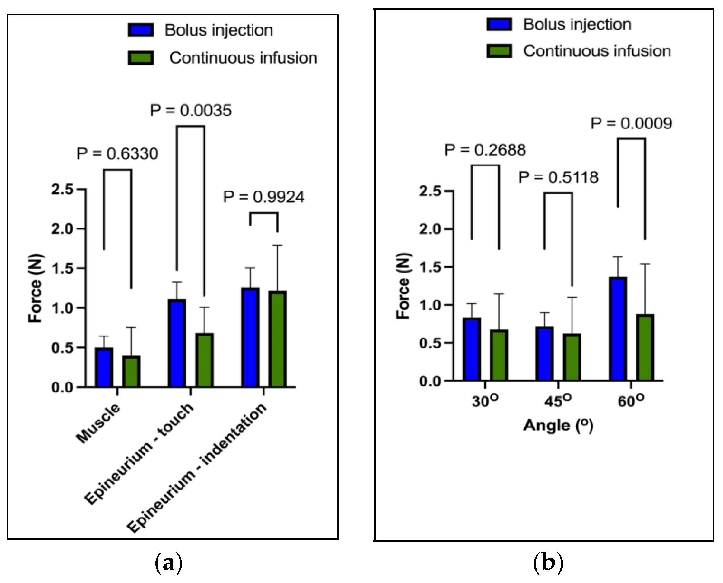
In an ex vivo model of legs of lambs, it was observed that the application of a continuous infusion resulted in less needle force generated on the epineurium compared to that of the bolus injection. The difference in the magnitude of the force applied was found to be at the maximum at a 60-degree angle. Upon analysis, it was found that continuous infusion reduced the frictional forces, thus leading to the smooth flow of the injected substance. On the other hand, the bolus injection had to overcome the resistance of the surrounding tissue in order to initiate flow, resulting in a comparatively greater force being applied on the epineurium.
The infusion technique has a principal advantage over the bolus technique in that needle insertion and fluid injection are linked together. This technique is similar to the continuous hydrolocation technique used in clinical practice to detect circumneural spaces, where sustained pressure is applied to the syringe. However, in the bolus technique, the times between needle tip positioning, the initiation of injection, and the time to peak pressure are different. Our research has shown that the mean (SD) time between the initiation of injection and the attainment of peak pressure is 2.8 (1.0) seconds. It is important to note that the delay may be such that the needle may be removed before the peak pressure response has been attained (Figure 2a,e). This pressure-monitoring facet has not been emphasised previously, but has important implications for clinical practice. If the pressure response is delayed sufficiently during injection, then an anaesthesiologist is powerless to intervene and prevent potential damage from excessively high pressures. Therefore, it is crucial to ensure that pressure monitoring is performed accurately and promptly to avoid potential harm to the patient.
During our injection experiments, we observed an interesting anomaly, which involved a sudden rise in pressure before the force of the injection at a 60-degree angle. This phenomenon occurred because the cutting edge of the needle was more perpendicular to the tissue surface at this angle, requiring a greater force to penetrate the tissue. As a general principle, we found that the greater the angle of the needle to the perpendicular fibres and the greater the stiffness of the tissues are, the greater the resistance during tissue penetration is. This increased resistance made the needles more likely to deflect and bend, and the injected liquid was more likely to flow retrograde along the path of least resistance into compliant adipose tissue.
This research has identified a wide range of forces and pressures that are measured in muscles when subjected to a gentle touch and the indentation of the epineurium. This variation in force can be attributed to the needle tip position and is consistent with the previous studies conducted on pigs [6] and soft embalmed cadavers [10,21]. However, the standard ultrasound technology used in medical procedures does not provide a sufficient resolution to accurately determine the needle tip’s precise location. When gently touching the epineurium, the needle tip may partially reside within the epineurium and partly face the circumneural fascial fibres, making it difficult to determine the exact location. This scenario makes it highly likely that any injection will take the path of least resistance and flow around the nerve. Upon indentation of the epineurium, the needle tip may partially penetrate the epineurium, making the injection intraneural, but not visible via ultrasound. In contrast, when a needle is inserted into the muscle, the tip orifice may lie partially on collagenous fibres, such as the epimysium (surrounding the muscle), or partially within the perimysium. This information highlights the complexity of needle insertion and injection techniques and emphasises the importance of considering the physical properties of different tissues when performing medical procedures.
Based on the results obtained, we have formulated a hypothesis that a continuous infusion technique is more effective in identifying the subcirumneural spaces with a less force as compared to the standard bolus technique. Our hypothesis is supported by some evidence as well. In particular, researchers using an epidural needle-based system that relies on computerised injection pump technology (Compuflo, Livingston, NJ, USA) reported higher injection accuracy with fewer dural punctures [22]. The system employs a constant infusion rate of approximately 3 mL/min and measures the changes in injection pressure at 4 Hz. Furthermore, an ex vivo study has suggested that this system is more accurate than the B-Smart (BS) in-line manometer and could be used for peripheral nerve blocking procedures [23]. In our study of isolated nerves, we followed the same fundamental principles as in previous research. However, in our study, we infused saline at a rate of 1.8 mL per minute and measured the pressure and force at a frequency of 10 Hz. Our primary focus was examining the initial interaction between the needle and the nerve and the hypothetical separation of the circumneurium. At this stage, we cannot comment on the clinical applications of this technique, as further research is needed to investigate the subsequent dosing and other aspects. In order to gain a more detailed understanding of the changes at the epineurium and the subcircumneural compartments, we plan to conduct future studies using microultrasound on cadaver models.
The research findings presented a comprehensive understanding of needle–tissue interaction dynamics, directly impacting clinical practices in regional anaesthesia. Specifically, our study explains the biomechanical factors influencing tissue trauma and injection precision. By integrating these insights into clinical protocols, practitioners can tailor their approaches to minimise tissue trauma and optimise injection precision. This involves careful consideration of needle insertion angles and infusion techniques to reduce the force exertion on tissues during needle insertion. Additionally, the findings of the result underscore the significance of real-time visualization technologies in guiding needle placement with heightened accuracy and safety. Moreover, this study assesses the stage for future research to refine the existing techniques and develop creative technologies to enhance the efficacy and safety of ultrasound-guided regional anaesthesia procedures.
This study we conducted has two main impacts that are worth highlighting. Firstly, force is a physical entity used in bioengineering to identify and measure the interaction of trocars, needles and sutures with soft tissue. However, it has yet to be widely applied to regional anaesthesia procedures [24]. Our research findings suggest that the forces generated during regional anaesthesia administration can be significant, but are rarely appreciated because they are not measured. We have performed ongoing work showing that forceful interaction with tissues is a crucial aspect of regional anaesthesia administration, which deserves further exploration. Secondly, using a liquid as a lubricant could potentially improve the ease and efficiency of medical procedures. Our study results can inform the understanding of the forces encountered during needle-based procedures in soft tissue. These findings have significant implications in the medical field as they may lead to the development of more effective techniques for carrying out procedures like anaesthesia- and other needle-based procedures.
The biomechanical factors that influence needle–tissue interaction during regional anaesthesia procedures are significant, as highlighted by this study. The study results emphasise the significance of the needle insertion angle and infusion rates in reducing tissue force exertion, which is aligned with the proposed needle–tissue interaction computation model. This model provides a comprehensive framework for predicting and planning needle insertion paths before surgery, utilising a Kriging-based friction force calculation model for accurate real-time friction estimation [25]. Our study’s recommendations for optimising the needle insertion angles and techniques can help clinicians minimise tissue damage and enhance patients’ safety. This research confirms that using shallower angles during needle insertion results in lower force generation, which is attributed to the smoother traversal of the needle through tissue with reduced resistance. Therefore, using shallower insertion angles allows for a more controlled and gradual approach, which is crucial for minimising tissue trauma during regional anaesthesia procedures. In contrast, more vertical angles resulted in a shorter time for injection to reach peak pressure and higher force. Using a more vertical angle for injection means that the needle can penetrate the tissue more directly, reducing the distance it travels to reach the target site. This direct path allows the injected fluid to encounter more resistance within the tissue, facilitating quicker pressure buildup created by the vertical angle, leading to a shorter time for the injected fluid to reach its maximum pressure. More vertical angles require a greater force to penetrate the tissue than the shallower angles, resulting in increased force exertion. The needle’s perpendicular or near-perpendicular orientation relative to the tissue surface requires greater mechanical energy to overcome tissue resistance, which is why an increased force is needed. The combination of increased force and resistance leads to a quicker escalation in pressure, resulting in the more rapid attainment of peak pressure during injection. However, it is essential to note this study’s limitations. Firstly, this study used an ex vivo lamb leg model, and the results may need to be validated using in vivo tissue dynamics. Secondly, this study did not investigate the variations in needle bevel angles, which could have influenced the force dynamics and tissue response. Lastly, future research is needed to explore the impact of different infusion rates on needle–tissue interaction, providing a deeper understanding of the needle–tissue interaction and optimised needle insertion techniques for regional anaesthesia-related procedures.

5. Conclusions

In conclusion, our study demonstrates that needle insertion and injection using a continuous infusion technique exerted a significantly lower axial force than the traditional bolus technique, particularly when gently touching the epineurium. This observation also underscores the significance of considering the needle insertion angle, as we noted significant differences in force profiles at varying angles. This finding suggests that the continuous infusion method may offer a more peaceful and controlled approach to nerve contact during regional anaesthesia procedures. By reducing the force applied to the nerve tissue, there is the potential to minimise the risk of trauma or injury, which is especially vital in sensitive anatomical regions. Moreover, the observed differences in force profiles highlight the importance of considering injection techniques in optimizing patients’ safety and procedural outcomes.
Future research and clinical trials are warranted to validate these findings and assess the practical implications of adopting continuous infusion techniques in real-world clinical settings.

Author Contributions

Conduct the study, A.A.; methodology, A.A.; validation, A.A.; collection and processing of the data, A.A.; setups, A.A., Y.Z. and J.L.; wrote the original draft, A.A. and G.M.; provided lab equipment, Z.H.; writing—review and editing, A.A., Z.H. and G.M. All authors have read and agreed to the published version of the manuscript.

Funding

This research was funded by the Cultural Affairs Department at the Libyan Embassy, London, UK. The APC was not covered by any external funding.

Institutional Review Board Statement

Not applicable.

Informed Consent Statement

Not applicable.

Data Availability Statement

The raw data supporting the conclusions of this article will be made available by the authors on request.

Acknowledgments

We would like to express our gratitude to everyone who supported us for their valuable contributions to this research.

Conflicts of Interest

The authors declare no conflicts of interest.

References

  1. Capdevila, M.; Choquet, O.; Saporito, A.; Djanikian, F.; Swisser, F.; Marques, M.; Bringuier, S.; Capdevila, X. Injection pressure monitoring during peripheral nerve blocks: From bench to operating theatre. Anaesth. Crit. Care Pain Med. 2020, 39, 603–610. [Google Scholar] [CrossRef]
  2. Server, A.; Boezaart, A.P.; Perez-Carrasco, M.; Esteves-Coelho, M.; Laredo, F.; Reina, M.A. Identification of spread after deliberated intraneural injection in five mammalian species: Not all animal models and needle types are valid for studying the effect of intraneural injections in humans. Reg. Anesth. Pain Med. 2023. [Google Scholar] [CrossRef]
  3. Barrington, M.J.; Lirk, P. Reducing the risk of neurological complications after peripheral nerve block: What is the role of pressure monitoring? Anaesthesia 2019, 74, 9–12. [Google Scholar] [CrossRef]
  4. Reina, M.A.; Boezaart, A.P.; Tubbs, R.S.; Zasimovich, Y.; Fernández-Domínguez, M.; Fernández, P.; Sala-Blanch, X. Another (Internal) Epineurium: Beyond the Anatomical Barriers of Nerves. Clin. Anat. 2020, 33, 199–206. [Google Scholar] [CrossRef]
  5. Boezaart, A.; Sala-Blanch, X.; Monzo, E.; Reina, M.A. Our best anesthetic blocks are probably related to unintentional and unnoticed intraneural injection. Reg. Anesth. Pain Med. 2019, 44, 279–280. [Google Scholar] [CrossRef] [PubMed]
  6. McLeod, G.A.; Cowie, A.; Sadler, A.; Watson, F.; Wasik, P.; Reina, M.A. Accuracy of injection pressure measurement at peripheral nerves using high-resolution 40 MHz ultrasound in an anesthetized porcine model. Reg. Anesth. Pain Med. 2023, 48, 501–507. [Google Scholar] [CrossRef]
  7. Server, A.; Reina, M.A.; Boezaart, A.P.; Prats-Galino, A.; Esteves Coelho, M.; Sala-Blanch, X. Microanatomical Nerve Architecture of 6 Mammalian Species: Is Trans-Species Translational Anatomic Extrapolation Valid? Reg. Anesth. Pain Med. 2018, 43, 496–501. [Google Scholar] [CrossRef]
  8. Hadzic, A.; Dilberovic, F.; Shah, S.; Kulenovic, A.; Kapur, E.; Zaciragic, A.; Cosovic, E.; Vuckovic, I.; Divanovic, K.-A.; Mornjakovic, Z.; et al. Combination of intraneural injection and high injection pressure leads to fascicular injury and neurologic deficits in dogs. Reg. Anesth. Pain Med. 2004, 29, 417–423. [Google Scholar] [CrossRef]
  9. Steinfeldt, T.; Poeschl, S.; Nimphius, W.; Graf, J.; Zoremba, M.; Mueller, H.-H.; Wulf, H.; Dette, F. Forced needle advancement during needle-nerve contact in a porcine model: Histological outcome. Anesth. Analg. 2011, 113, 417–420. [Google Scholar] [CrossRef] [PubMed]
  10. Zhang, S.; McLeod, G.; Sadler, A.; Xia, C.; Huang, Z. Needle insertion forces and fluid injection pressures during targeting of nerves in a soft embalmed cadaver model. J. Clin. Monit. Comput. 2022, 36, 511–519. [Google Scholar] [CrossRef]
  11. Meltsner, M.A.; Ferrier, N.J.; Thomadsen, B.R. Observations on rotating needle insertions using a brachytherapy robot. Phys. Med. Biol. 2007, 52, 6027–6037. [Google Scholar] [CrossRef]
  12. Misra, S.; Macura, K.J.; Ramesh, K.T.; Okamura, A.M. The importance of organ geometry and boundary constraints for planning of medical interventions. Med. Eng. Phys. 2009, 31, 195–206. [Google Scholar] [CrossRef]
  13. Quadri, C.; Saporito, A.; Capdevila, X. Real-time continuous monitoring of injection pressure at the needle tip for peripheral nerve blocks: Description of a new method. Anaesthesia 2018, 73, 187–194. [Google Scholar] [CrossRef]
  14. Bao, X.; Li, W.; Lu, M.; Zhou, Z.R. Experiment study on puncture force between MIS suture needle and soft tissue. Biosurface Biotribol. 2016, 2, 49–58. [Google Scholar] [CrossRef]
  15. Cheng, Z.; Chauhan, M.; Davies, B.L.; Caldwell, D.G.; Mattos, L.S. Modelling needle forces during insertion into soft tissue. In Proceedings of the 2015 37th Annual International Conference of the IEEE Engineering in Medicine and Biology Society (EMBC), Milan, Italy, 25–29 August 2015; pp. 4840–4844. [Google Scholar] [CrossRef]
  16. Gao, D.; Lei, Y.; Yao, B. Analysis of Dynamic Tissue Deformation during Needle Insertion into Soft Tissue. IFAC 2013, 46, 684–691. [Google Scholar] [CrossRef]
  17. Ko, S.Y.; Davies, B.L.; Rodriguez YBaena, F. Two-dimensional needle steering with a “programmable bevel” inspired by nature: Modeling preliminaries. In Proceedings of the 2010 IEEE/RSJ International Conference on Intelligent Robots and Systems, Taipei, Taiwan, 18–22 October 2010; pp. 2319–2324. [Google Scholar] [CrossRef]
  18. Althobaiti, M.; Ali, S.; Hariri, N.G.; Hameed, K.; Alagl, Y.; Alzahrani, N.; Alzahrani, S.; Al-Naib, I. Recent Advances in Smart Epidural Spinal Needles. Sensors 2023, 23, 6065. [Google Scholar] [CrossRef]
  19. Henderson, M.; Dolan, J. Challenges, solutions, and advances in ultrasound-guided regional anaesthesia. BJA Educ. 2016, 16, 374–380. [Google Scholar] [CrossRef]
  20. Quan, C. Perioperative nerve injury and regional anaesthesia. S. Afr. J. Anaesth. Analg. 2022, 28 (Suppl. S1), S156–S161. [Google Scholar] [CrossRef]
  21. McLeod, G.; Zihang, S.; Sadler, A.; Chandra, A.; Qiao, P.; Huang, Z.; Demore, C. Validation of the soft-embalmed Thiel cadaver as a high-fidelity simulator of pressure during targeted nerve injection. Reg. Anesth. Pain Med. 2021, 46, 540–548. [Google Scholar] [CrossRef]
  22. Gebhard, R.E.; Moeller-Bertram, T.; Dobecki, D.; Peralta, F.; Pivalizza, E.G.; Rupasinghe, M.; Ilic, S.; Hochman, M. Objective Epidural Space Identification Using Continuous Real-Time Pressure Sensing Technology: A Randomized Controlled Comparison with Fluoroscopy and Traditional Loss of Resistance. Anesth. Analg. 2019, 129, 1319–1327. [Google Scholar] [CrossRef]
  23. Weisman, R.S.; Bhavsar, N.P.; Schuster, K.A.; Gebhard, R.E. Evaluation of the B-Smart manometer and the CompuFlo computerized injection pump technology for accurate needle-tip injection pressure measurement during peripheral nerve blockade. Reg. Anesth. Pain Med. 2019, 44, 86–90. [Google Scholar] [CrossRef]
  24. Yang, C.; Xie, Y.; Liu, S.; Sun, D. Force modeling, identification, and feedback control of robot-assisted needle insertion: A survey of the literature. Sensors 2018, 18, 561. [Google Scholar] [CrossRef]
  25. Lei, Y.; Du, S.; Li, M.; Xu, T.; Hu, Y.; Wang, Z. Needle-tissue interaction model based needle path planning method. Comput. Methods Programs Biomed. 2024, 243, 107858. [Google Scholar] [CrossRef]
Figure 1. Experimental setup. Image (a) is a schematic diagram showing the leg of lamb, stage and connections between the force and pressure sensors and ultrasound machine. The data acquisition system recorded data from the force sensor, while the Pendo TECH pressure monitor recorded data from the pressure sensor. Image (b) shows the motorised stage in more detail, including its intricate workings and mechanism for holding the needle in place. It has three degrees of freedom for translation and rotation.
Applsci 14 02673 g001
Figure 2. Examples of force and pressure profiles associated with bolus injection (a,c,e) and continuous infusion (b,d,f). The x-axis and y-axes are standardised in order to make realistic comparisons between the modalities and angles. Images (a,b) show profiles during gentle needle–nerve contact at a 30° angle. Images (c,d) show profiles during nerve indentation with at a 45° angle. Images (e,f) show profiles within muscle with at a 60° angle. Compared to the bolus technique, continuous infusion is associated with a smaller force profile and a shorter gap in time between the peak force and peak in-line pressure. Note the ~10 s gap until the needle makes contact with the skin and the sensitivity of the force response as the needle passes through tissue layers. The pressure preceded force in (f) probably because the needle tip at a 60° angle was blocked due to higher tissue resistance to the flow.
Applsci 14 02673 g002
Figure 3. Peak axial force and fluid injection pressure measured during bolus injection and continuous infusion; effect of tissue location (gentle touch on epineurium, epineural indentation and muscle) and angle. Image (a) shows difference in axial force on gentle touch of epineurium, but not in muscle or on epineural indentation. Image (b) shows difference in force at 60° angle, but not 30° or 45°.
Applsci 14 02673 g003
Figure 4. Image (a) shows differences in in-line pressure at different locations, but no trend. No difference between pressures at different locations. Image (b) shows increased pressure at 60° angle.
Applsci 14 02673 g004
Figure 5. * shows a comparison of the time (s) difference from injection to peak pressure, focusing on the impact of tissue location and angle during injection. Image (a) precisely displays the effect of gentle touch on the epineurium, epineurium indentation, and muscle, while image (b) examines the difference in time involving needle insertion angle and injection techniques with notable significance at 60°.
Applsci 14 02673 g005
Table 1. Comparison of force and statistical analysis between bolus and continuous injection techniques at different angles and tissue locations.
TechniqueForce (SD)ComparisonDifferencep-Value
Bolus injection0.96 (0.61)Bolus vs. infusion0.19 (0.06–0.32)0.003
Continuous injection 0.77 (0.54)
Angle
30°0.76 (0.47)30° vs. 45°0.08 (−0.10 to 0.27)0.52
45°0.67 (0.46)30° vs. 60°0.40 (0.20 to 0.60)<0.001
60°1.16 (0.69)45° vs. 60°0.49 (0.29 to 0.69)<0.001
Tissue
Muscle0.45 (0.36)Epineurium—touch vs. muscle0.45 (0.24 to 0.65)<0.001
Epineurium—touch0.90 (0.49)Epineurium—indentation vs. muscle0.78 (0.56 to 1.02)<0.001
Epineurium—indentation1.24 (0.59)Epineurium—indentation vs. touch0.34 (0.14 to 0.54)<0.001
Table 2. Comparison of pressure and statistical analysis between bolus and continuous injection techniques at different angles and tissue locations.
TechniquePressure (SD)ComparisonDifferencep-Value
Bolus injection 38.4 (9.1) Bolus vs. infusion 2.6 (1.9–7.1) 0.25
Continuous injection35.8 (19.7)
Angle
30°32.4 (12.3)30° vs. 45°2.0 (−5.0 to 9.0)0.77
45°34.4 (17.7)30° vs. 60°12.2 (8.5 to 15.9)<0.001
60°44.6 (12.7)45° vs. 60°10.2 (3.2 to 17.2)0.02
Tissue
Muscle34.0 (14.5)Epineurium—touch vs. muscle0.6 (−4.0 to 5.2)0.94
Epineurium—touch33.4 (9.9)Epineurium—indentation vs. muscle9.9 (2.3 to 17.4)<0.007
Epineurium—indentation43.9 (18.3)Epineurium—indentation vs. touch10.5 (4.0 to 16.9)<0.001
Disclaimer/Publisher’s Note: The statements, opinions and data contained in all publications are solely those of the individual author(s) and contributor(s) and not of MDPI and/or the editor(s). MDPI and/or the editor(s) disclaim responsibility for any injury to people or property resulting from any ideas, methods, instructions or products referred to in the content.

