Article

Listeria monocytogenes from Marine Fish and the Seafood Market Environment in Northern Greece: Prevalence, Molecular Characterization, and Antibiotic Resistance

by
Pantelis Peratikos
1,
Anestis Tsitsos
1,
Alexandros Damianos
1,
Maria A. Kyritsi
2,
Christos Hadjichristodoulou
2,
Nikolaos Soultos
1 and
Vangelis Economou
1,*
1
Laboratory of Animal Food Products Hygiene—Veterinary Public Health, Aristotle University of Thessaloniki, 54124 Thessaloniki, Greece
2
Laboratory of Hygiene and Epidemiology, Faculty of Medicine, University of Thessaly, 41222 Larissa, Greece
*
Author to whom correspondence should be addressed.
Appl. Sci. 2024, 14(7), 2725; https://doi.org/10.3390/app14072725
Submission received: 28 February 2024 / Revised: 18 March 2024 / Accepted: 22 March 2024 / Published: 25 March 2024
(This article belongs to the Special Issue Detection and Control of Foodborne and Waterborne Pathogenic Bacteria)

Abstract

:
The occurrence of Listeria monocytogenes in marine fish and fish market areas was investigated. Two hundred and eighty-eight samples (123 environmental samples—siphons, knives, cutting boards, floor, sinks, water, and ice—and 165 marine fish samples) were examined. Twenty-four isolates were characterized as Listeria monocytogenes (five from environmental samples (4.0%) and 19 from fish samples (11.5%)). The strains were further characterized according to their antibiotic resistance, pathogenicity, and biofilm formation ability. They were molecularly serotyped as IIc (n = 22) and IVb (n = 2) and possessed all the virulence genes tested (inlA, inlB, inlC, inlJ, actA, hlyA, iap, plcA, and prfA), except for two strains lacking the hlyA and iap genes, respectively. All strains showed strong (41.7%) or moderate biofilm-producing ability (58.3%) and almost all showed resistance to at least one antibiotic, with the highest rates being observed against clindamycin and vancomycin. The proteomic analysis by MALDI-TOF revealed two distinct clusters that involved strains from fish only and those from both fish and the environment. The presence of Listeria monocytogenes in the fish-market environment and marine fish, along with the pathogenicity and persistence characteristics of the seafood-related strains, emphasize the need for vigilance concerning the spread of this notorious foodborne pathogen.

1. Introduction

Fish and seafood products are irreplaceable elements of the human diet. They are commodities of high nutritional value that can provide a protein source rich in essential amino acids, polyunsaturated fatty acids, and high concentrations of vitamins and trace elements, such as iodine, iron, magnesium, phosphorus, and zinc [1]. Despite its nutritional value, seafood is prone to spoilage and may carry and transmit various pathogenic microbes. The main bacteria transmitted through the seafood chain are Vibrio spp. (mainly V. cholerae, V. parahaemolyticus, and V. vulnificus), Clostridium botulinum, Yersinia spp., Salmonella spp., and Listeria monocytogenes [2,3]. Listeria monocytogenes, specifically, is a Gram-positive, non-sporulating, facultatively anaerobic bacterium [4]. It is a foodborne pathogen of major public health importance, primarily affecting humans, which causes listeriosis [5], a serious foodborne disease that can lead to meningitis, encephalitis, septicemia, and miscarriage [6]. According to the latest EFSA and ECDC One Health zoonoses report for 2022 [7], the incidence rate of the disease in the European Union is quite low (0.62 cases per 100,000 population), although it is reported to have risen in the last five years. However, it is recognized as a major threat to public health due to the high rate of hospitalization (94% of cases) and mortality (30% of cases), mainly among people in vulnerable groups [8]. Listeria monocytogenes persists in food processing facilities for long periods of time, even when appropriate hygiene measures are applied, mainly due to its biofilm formation ability on abiotic surfaces [9]. Furthermore, the contamination of food by this bacterium is quite severe since it is one of the few pathogens that can multiply at refrigeration temperatures [10]. The most suitable foods for the proliferation of Listeria monocytogenes are non-thermally processed products, food kept in refrigeration for a long time, food produced under unsanitary conditions, and cooked and frozen ready-to-eat meals [11].
In the European Union, limits are set by Commission Regulation (EC) No. 2073/2005 in ready-to-eat food for the occurrence or enumeration of L. monocytogenes. The recent European Union One Health zoonoses report states that L. monocytogenes was detected in 7.1% of ready-to-eat fish and fishery products, one of the highest among ready-to-eat foods in the European Union [7]. The prevalence of Listeria monocytogenes in seafood depends on several factors, such as the geographic area, the water quality, the seasonality, and the discharge of agricultural effluents in water bodies ending up in the sea [12,13,14]. The prevalence of the pathogen usually varies between geographic regions; furthermore, the variation in prevalence and serotypes between different regions is mainly caused by environmental factors and climatic differences between countries. These differences may affect the presence and proliferation of Listeria monocytogenes and, therefore, affect the risk posed by this foodborne pathogen. Additionally, the water quality and pollution levels prevailing in each region affect the prevalence of Listeria monocytogenes [12,14]. In cases where the fish-rearing areas receive livestock effluents, the possibility of the occurrence of this pathogen in the seawater is augmented, resulting in the contamination of the aquatic animals and seafood produced there [13]. In addition, seasonal variations can play a vital role in the appearance and diversity of pathogenic bacteria in general and specifically of Listeria monocytogenes [15,16]. A crucial factor in the interaction of Listeria monocytogenes with seafood is the species and type of fish that came in contact with the pathogen. Listeria monocytogenes is usually isolated from coastal waters and rarely from deeper waters, an observation suggesting that fish living offshore are less likely to be infected with Listeria monocytogenes compared to those living in coastal areas [16]. Also, the eating habits of marine animals affect contamination with a pathogenic bacterium or the multiplication of it. More specifically, filter-feeding organisms, such as mussels, can readily accumulate pathogens in their tissues compared to fish [12].
Listeria monocytogenes has been isolated from various places of fish processing and marketing units [15]. Fish and their products can be potentially contaminated with Listeria monocytogenes during their processing. There are several points where cross-contamination can occur; usually, these involve contaminated equipment coming in contact with the fish, such as machinery, cutting boards, knives used for cleaning the fish, floors, the hands of the staff, etc. [17,18]. Therefore, unsanitary processing practices and inadequate hygiene contribute to the contamination of fish with Listeria monocytogenes [18,19]. Contamination of the fish can occur during processing, such as filleting, cleaning, and salting [17]. Furthermore, contamination of these products can occur in the processing units as a result of the formation of biofilms by Listeria monocytogenes on abiotic surfaces [17,20]. The ability of Listeria monocytogenes to adhere to surfaces in fish processing areas and to form biofilms contributes to the persistence of the pathogenic bacterium in processing facilities for long periods of time, thus allowing the contamination of seafood [21,22].
In view of the increased rates of detection of Listeria monocytogenes in fish and fish products, this study aimed to investigate the occurrence and transmission of Listeria monocytogenes in marine fish and fish-processing areas. The Listeria monocytogenes isolates were further characterized for factors related to their persistence and pathogenicity (molecular identification and serotyping, evaluation of biofilm production, sensitivity testing to antimicrobial agents, and detection of pathogenicity genes). Finally, possible affinities between the isolates were investigated to ascertain the possible fish contamination pathways.

2. Materials and Methods

2.1. Sampling

The study was conducted among fishmongers in Thessaloniki in the winter of 2023 and in the summer of 2023. Thessaloniki is the second largest Greek city and is a coastal city and port that is situated in the northwest Aegean Sea, in the Thermaikos Gulf. The fish procured in its fish markets originate mainly from the Thermaikos Gulf and the Northern Aegean Sea.
A total of 288 samples were collected, of which 123 were environmental samples from the fish processing, preservation, and sales areas (siphons, knives, cutting boards, floors, sinks, water, and ice) and 165 fish samples, which were divided into large and small fish (Table 1). Regarding the fish samples, 6 fish species were collected, comprising 3 species of large fish (hake (Merluccius merluccius), horse mackerel (Trachurus trachurus), and chub mackerel (Scomber japonicus)) and 3 species of small fish (European pilchard (Sardina pilchardus), European anchovy (Engraulis encrasicolus), and bogue (Boops boops)). These fish are usually harvested from the pelagic–neritic area and are all oceanodromous except for hake, which is demersal, and bogue, which can be found in both areas [23]. Fish samples were collected aseptically and transported to the laboratory using sterile disposable plastic containers. Small fish (European pilchard, European anchovy, or bogue) were homogenized as a whole with the appropriate volume of Half Fraser broth (HF, Oxoid, Basingstoke, UK). From the large fish samples, 25 g were collected aseptically and homogenized in HF (the gills, skin, and digestive tract collected separately). Regarding sampling of the cutting boards, this was performed using sterile gauze or cotton swabs, depending on the type of each surface. Either 2 gauze pads or 2 cotton swabs were used for each surface, with the first being soaked in 5 mL of buffered peptone water (BPW) and the second being used dry. The surface sample was collected from an area of 100 cm2 defined by a sterile metal frame. Both the gauzes and swabs used were then placed in a sterile vial containing HF. For ice sampling, 1 kg of the ice used to cover the fish was collected in a sterile stomacher bag. For water sampling, 1 L of water was collected in a sterile bottle containing 18 mg/L of sodium thiosulfate (Merck, KGaA, Darmstadt, Germany). The attached devices and the faucet fittings were also sampled with cotton swabs, as previously described.
All samples were immediately transported aseptically in an insulated thermobox to the Laboratory of Hygienic Foods of Animal Origin—Veterinary Public Health (Department of Veterinary Medicine, School of Health Sciences, Aristotle University of Thessaloniki) for further analyses.

2.2. Microbiological Analysis

The analysis of water samples was performed after filtration of 1 L of water through membrane filters with a 0.45 μm pore size (CHMLAB GROUP, Barcelona, Spain). After filtration, the membranes and swabs used for the sampling of the faucet were placed in HF-containing Erlenmayer flasks. The analysis of samples and the detection of Listeria monocytogenes was performed in accordance with the ISO 11290-1:2017 method [24]. In brief, test tubes with HF were incubated at 30 °C for 18 h. After incubation, 0.1 mL of the HF was transferred to sterile test tubes containing 10 mL of Fraser Broth (FB, Oxoid, Basingstoke, UK) and incubated at 37 °C for 24–48 h. Following enrichment, 10 μL of FB was streaked onto agar Listeria according to Ottaviani Agosti (ALOA, Oxoid Basingstoke, UK), and then incubated at 37 °C for 24–48 h. Characteristic colonies of Listeria monocytogenes (cyan-green colonies with an opaque halo) were sub-cultured on tryptone soya yeast extract agar (TSYEA, Oxoid Ltd., Basingstoke, UK) and incubated at 37 °C for 24 h.
Identification and molecular serotyping of the Listeria monocytogenes isolates was performed by multiplex PCR. DNA extraction from the collected samples was performed according to the protocol used by Lawrence and Gilmour [25]. In brief, a colony was suspended in 50 µL of sterile Milli-Q water, heated at 100 °C for 10 min, cooled at −20 °C for 30 min, and centrifuged at 13,000 rpm for 5 min. The supernatant was removed, placed into a new sterile tube, and stored at −20 °C until further use. The extracts were subjected to a multiplex PCR for the molecular identification and serotyping of L. monocytogenes, according to the method used by Doumith et al. [26], which targets the prs, lmo0737, lmo1118, ORF2819, and ORF2110 genes (Table 2). The reaction mixture was prepared in a 25 μL volume containing 2U OneTaq™ DNA Polymerase (M0273S, NEB, Ipswich, MA, USA), 2.5 μL of 10× OneTaq standard reaction buffer (B9014S, NEB), 200 μM dNTPs (N0447S, NEB), 0.25–1.875 μL of primers, and 2 μL of DNA sample (Table 2). The Listeria monocytogenes strains CIP 105448 and ATCC 49594 were used as positive controls. The PCR was performed in a LabCycler Gradient thermal cycler (SensoQuest GmbH, Göttingen, Germany). The cycling conditions included an initial denaturation at 94 °C for 180 s, followed by 35 PCR cycles with denaturation at 94 °C for 24 s, annealing at 53 °C for 69 s, and extension at 72 °C for 69 s, followed by a final extension for 7 min at 72 °C. The DNA products were analyzed by electrophoresis using agarose (1.5%) gel with 0.5 μg/mL ethidium bromide (Sigma-Aldrich, St. Louis, MO, USA).

2.3. Proteomic Relationship of Listeria monocytogenes Isolates

Proteomic MALDI-TOF MS analysis was performed to verify the PCR characterization of the isolates and to investigate the most feasible routes of contamination accordingly. More specifically, pure colonies of the Listeria monocytogenes isolates were analyzed using a Microflex LT mass spectrometer (Bruker Daltonics, Bremen, Germany) following the manufacturer’s instructions for Listeria spp. identification. In particular, the total protein footprints of the isolates were extracted by the formic acid method, as proposed by the manufacturer. Specifically, a pure colony of the isolate was collected from TSYEA, placed in an Eppendorf tube with 300 µL of ultrapure water, and homogenized. Then, 900 µL of pure ethanol was added, followed by homogenization, centrifugation at 13,000 rpm for 2 min, and removal of the supernatant. The procedure was repeated and the Eppendorf tubes were left in the environment for 5 min for the excess ethanol to evaporate. Then, 30 µL of 70% formic acid was added, followed by homogenization. The procedure was completed by the addition of 30 µL of acetonitrile, homogenization, and centrifugation at 13,000 rpm for 2 min. Afterward, 1 µL of the protein extract was placed on the MALDI-TOF MS target plate. In each well of the plate, 1 μL of matrix solution was added (a saturated solution of cyano-4-hydroxycinnamic acid matrix (Bruker Daltonics) in 50% acetonitrile (Sigma Aldrich, St. Louis, MO, USA) with 25% trifluoroacetic acid (Sigma Aldrich, St. Louis, MO, USA)). The plate was left in the environment to dry before analysis.
Protein profiles were collected using linear positive mode analysis with a laser frequency of 20 Hz. Primary protein spectra were automatically collected using the AutoXecute control software (Flex control 3.4; Bruker Daltonics) and the spectra between 2000 and 20,000 Da were recorded. The method was calibrated using Bruker bacterial test standard (BTS), which is a protein extract of the model strain Escherichia coli DH5 inoculated with two additional proteins (RNAase A and myoglobin) to increase the maximum limit of the mass range that BTS covers. The identification of Listeria monocytogenes was achieved with the MALDI Biotyper ver. 4.0 software, using the default parameters. The obtained spectra were compared with those of the mass spectra library (v6.093 MSPs). The MBT subtyping module for Listeria spp. was used, which allows the identification of Listeria spp. isolates that are mainly isolated from environmental and food samples. The results were classified using modified score values, as suggested by the manufacturer. The Listeria monocytogenes isolates were grouped according to a main spectra dendrogram (MSP), which was prepared according to their protein profile. In addition, the resulting spectra were smoothed, and the baseline was removed and processed using the MALDI Biotyper Offline Classification 4.0 software with default parameters to create an MSP dendrogram. A distance level of 650 was set as a cut-off value to achieve the desired discrimination between the branches of the MSP dendrogram.

2.4. Assessment of Biofilm-Forming Ability of Listeria monocytogenes Strains

The biofilm formation ability of the Listeria monocytogenes strains was tested using the method of Chachlioutaki et al. [27] with modifications. Listeria monocytogenes strains were inoculated in tryptic soy broth (TSB) with 1% glucose and incubated for 24 h at 37 °C. Subsequently, the turbidity of the inoculum was adjusted to 0.5 McFarland using a nephelometer (Grand Instruments, Cambridge, UK) and diluted 1/100 in TSB with 1% glucose to achieve approximately a 106 CFU/mL inoculum. Furthermore, 100 μL of the inoculum was transferred to wells of polystyrene microtiter plates (Boettger, Bodenmais, Germany) containing 100 μL TSB with 1% glucose to achieve a final concentration of 5 × 10⁵ CFU/mL. For each Listeria monocytogenes isolate, 3 replicates were conducted, while a strip of wells served as the negative control (non-inoculated 200 μL sterile TSB with 1% glucose). The microplates were hermetically sealed with parafilm to prevent desiccation and incubated at 37 °C for 24 h. Then, the wells were washed gently with normal saline, air-dried for 1 h at 60 °C, and stained with 50 μL 0.06% crystal violet solution for 5 min at room temperature. The stained crystal violet was solubilized with 200 μL of 33% acetic acid for 10 min at room temperature. Finally, the optical density (OD) was measured at 600 nm using an automatic spectrometer (MicroDigital Co., Seongnam-si, Republic of Korea). Depending on the size of the OD of each isolate and the OD of the controls (ODc), the Listeria monocytogenes isolates were classified as no biofilm producers (OD < ODc), weak biofilm producers (ODc < OD ≤ 2*ODc), moderate biofilm producers (2*ODc < OD ≤ 4*ODc), or strong biofilm producers (OD > 4*ODc).

2.5. Antimicrobial Susceptibility Testing of Listeria monocytogenes Isolates

The antimicrobial susceptibility testing of the isolates was performed using the disc-diffusion method according to the Clinical and Laboratory Standards Institute recommendations [28]. Specifically, approximately 3 pure colonies of Listeria monocytogenes from TSYEA were suspended in 1 mL of 0.9% NaCl solution, and turbidity was adjusted to 0.5 McFarland with a nephelometer (Grand Instruments, Cambridge, UK). Subsequently, the suspension was streaked on Mueller–Hinton agar (MHA, Oxoid, Basingstoke, UK) supplemented with 5% sheep blood (Merck, KGaA, Darmstadt, Germany) using a sterile cotton swab, and the discs were then placed on the surface of each plate (5 antibiotic discs per each plate). The plates were then incubated under aerobic conditions at 37 °C for 24 h. The inhibition zones were measured using a caliper. Antibiotic discs (Oxoid, Basingstoke, UK) of 13 antibiotics that are commonly used in human and veterinary medicine were utilized (Table 3). Staphylococcus aureus ATTC 25923 was used as the positive control. The Listeria breakpoints reported by CLSI [28] were used. Since no breakpoints were available for the other antibiotics, the Staphylococcus spp. breakpoints were used, as reported elsewhere [29], except for ampicillin and vancomycin, which were derived from the CLSI breakpoints for Enterococcus spp., while for amoxicillin/clavulanic acid and meropenem, the CLSI breakpoints for Enterobacterales [30] were used (Table 3). Multidrug resistance (MDR) was defined as the non-susceptibility of an isolate to at least one antimicrobial agent in three or more categories of antimicrobial agents, according to the criteria established by Magiorakos et al. [31].

2.6. Detection of Virulence-Associated Genes in Listeria monocytogenes Isolates

Nine simplex PCR reactions were performed to identify the presence of the virulence-associated genes inlA, inlB, inlC, and inlJ (internalin genes), plcA (phospholipase C gene), prfA (regulatory gene), actA (actin gene), hlyA (haemolysin gene), and iap (p60 protein gene). The inlA, inlB, inlC, and inlJ genes were detected using the primers and the PCR conditions described by Liu et al. [32]. The reaction mixture was prepared in a 25 μL volume containing 0.8U of OneTaq™ DNA polymerase (M0273S, NEB), 2.5 μL of 10× OneTaq standard reaction buffer (Β9014S, NEB), 200 μM of dNTPs (N0447S, NEB), 1 μM of primers, and 2 μL of the DNA sample (Table 4). The PCR reaction was performed in a LabCycler Gradient thermal cycler (SensoQuest GmbH, Göttingen, Germany). The cycling program included initial denaturation at 94 °C for 120 s, 30 cycles with denaturation at 94 °C for 24 s, annealing at 55 °C for 20 s, and extension at 72 °C for 50 s, followed by a final extension for 2 min at 72 °C. The plcA, actA, hlyA, and iap genes were detected using the primers and the PCR conditions described by Rawool et al. [33] with modifications. The reaction mixture was prepared in a 25 μL volume containing 1U of OneTaq™ DNA polymerase (M0273S, NEB), 2.5 μL of 10× OneTaq standard reaction buffer (Β9014S, NEB), 200 μM of dNTPs (N0447S, NEB), 0.1 μM of primers, and 2 μL of the DNA sample (Table 4). The PCR reaction was performed in a LabCycler Gradient thermal cycler (SensoQuest GmbH, Göttingen, Germany). The cycling conditions included an initial denaturation at 95 °C for 120 s, 35 cycles with denaturation at 95 °C for 15 s, annealing at 60 °C for 30 s, and extension at 72 °C for 90 s, followed by a final extension for 10 min at 72 °C. The Listeria monocytogenes strains CIP 105448 and ATCC 49594 were used as positive controls.

2.7. Statistical Analysis

The statistical analysis of the data was performed using IBM SPSS Statistics software (v.29.0., IBM Corporation, Armonk, NY, USA). Confidence intervals for prevalence were calculated using the Clopper–Pearson method (binomial test). The significance level was set at 5% (a p-value of ≤0.05).

3. Results

3.1. Prevalence and Molecular Serogroups of Listeria monocytogenes Strains

A total of 122 Listeria spp. isolates were recovered, of which 24 were characterized as Listeria monocytogenes (19.7% of the isolates). More specifically, 5 strains of Listeria monocytogenes were recovered from the environmental samples (4.0%, 95% C.I. = 1.3–9.2%) and 19 strains from the fish samples (11.5%, 95% C.I. = 7.1–17.4%). In particular, four out of five environmental strains were isolated from the cutting boards (21.0%). One strain was recovered from a sample of ice (5.3%). All the strains recovered from fish came from the large fish (25.3%, 95% C.I. = 16–36.7%). No strains were found in the small fish (95% C.I. = 0–4%). Concerning the Listeria spp., 56 strains of Listeria spp. originated from the environmental samples and 42 strains of Listeria spp. were isolated from fish samples, of which 68 were identified as L. innocua (69.4%) and 2 as L. ivanovii (2.0%). Twenty-eight isolates were not typed to the species level. Combined with the Listeria monocytogenes-positive samples, Listeria strains were found in 61 of the 165 fish samples (37.0%, 95% C.I. = 29.6–44.8%) and in 61 of the 123 environmental samples (49.6%, 95% C.I. = 40.5–58.8%).
All Listeria monocytogenes strains belonged to the molecular serotypes IIc and IVb. More specifically, two strains were characterized as molecular serotype IVb, while the remaining strains belonged to molecular serotype IIc. The two isolates characterized as molecular serotype IVb were recovered from samples taken from cutting boards (Table 5). The majority of the IIc isolates were recovered from fish samples (19 isolates; 5 were from chub mackerel, 7 from hake, and 7 from horse mackerel), whereas 3 IIc isolates were recovered from environmental samples (2 from cutting boards and 1 from ice).

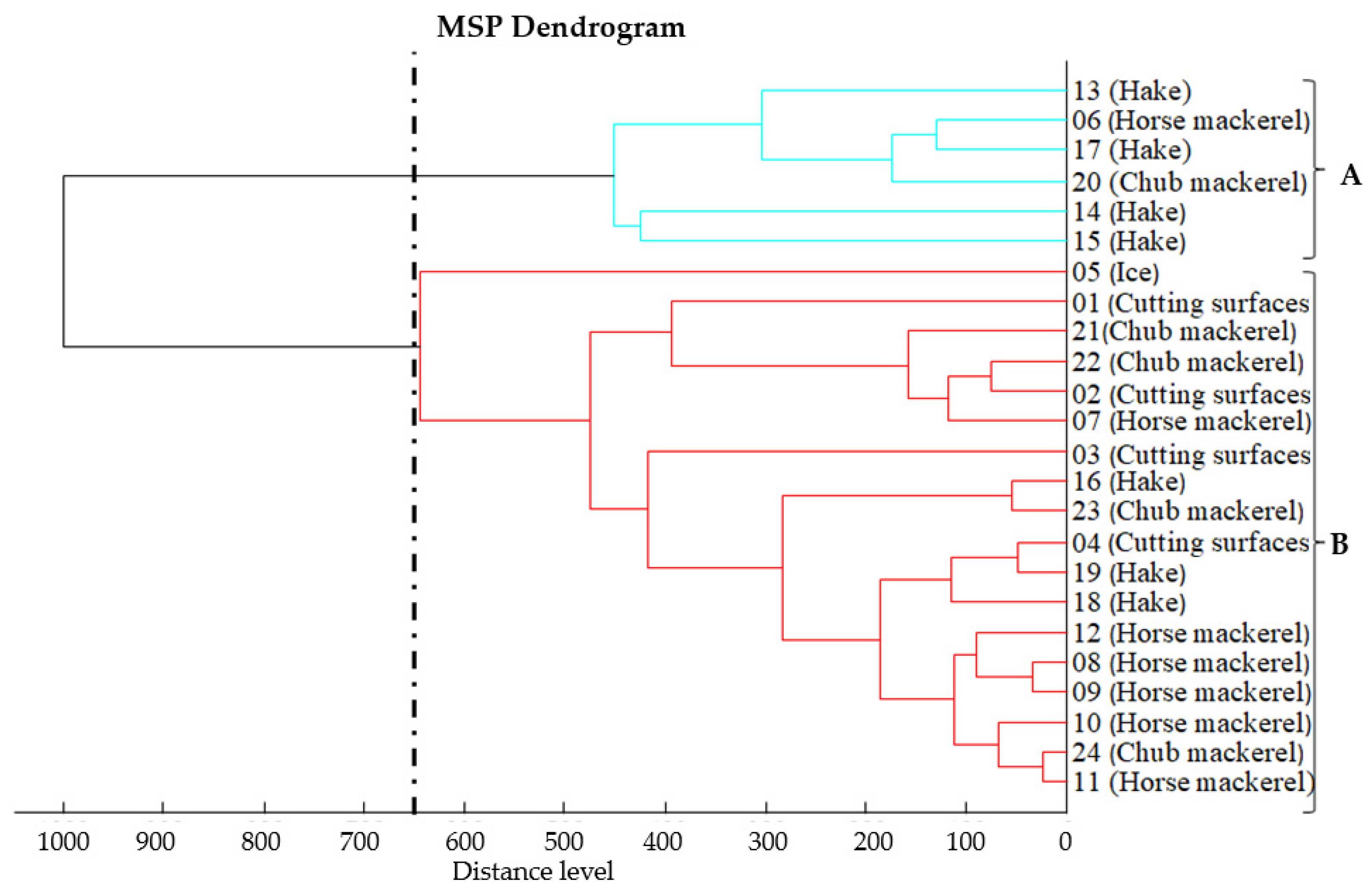
3.2. Proteomic Relatedness of Listeria monocytogenes Isolates

According to the dendrogram of the main spectra of Listeria monocytogenes strains at a 650-distance level, two clusters emerged (Figure 1). The first cluster includes 6 strains exclusively from fish, while the second cluster includes 18 strains recovered from both fish and environmental samples. In both clusters, the degree of relatedness between strains was related to the date of sampling. The highest degree of relatedness between the strains (<100-distance level) was observed in strains of cluster B.

3.3. Assessment of Biofilm-Forming Ability of Listeria monocytogenes Strains

All Listeria monocytogenes strains had the ability to produce biofilms (Table 5). Of the 24 strains, 10 showed strong biofilm-producing ability (41.7%) and 14 showed moderate biofilm-producing ability (58.3%), whereas no strain was characterized as a weak biofilm producer. The strains that showed strong biofilm production were recovered from fish samples (nine strains; two were isolated from Atlantic horse mackerel, two from chub mackerel, and five from European hake) and one from an environmental sample (cutting board). The strains that showed moderate biofilm production were recovered from fish samples (10 strains; 3 were from chub mackerel, 2 from hake, and 5 from horse mackerel) whereas 4 strains were recovered from environmental samples (3 from cutting boards and 1 from an ice sample).

3.4. Antimicrobial Susceptibility Testing of Listeria monocytogenes Strains

Most strains (n = 20, 83.3%%) of Listeria monocytogenes showed resistance to at least one antibiotic (Table 5, Supplementary Table S1). Overall, three different antimicrobial resistance profiles were observed, namely, strains resistant only to clindamycin (n = 9, 37.5%), strains resistant to clindamycin and vancomycin (n = 10, 41.7%), and one strain resistant to clindamycin, vancomycin, penicillin, and tetracycline (4.2%). The strain resistant to clindamycin, vancomycin, penicillin, and tetracycline was recovered from ice and since it showed resistance to four antibiotics, it was characterized as multiresistant. In total, resistance to clindamycin was observed in 20 of 24 strains (83.3%), while high rates of resistance were also observed for vancomycin, with 11 resistant strains (45.8%). All strains were sensitive to a combination of amoxicillin/clavulanic acid, ampicillin, ciprofloxacin, trimethoprim/sulfamethoxazole, chloramphenicol, erythromycin, gentamicin, meropenem, and rifampicin. Three of the strains were found to be sensitive to all antibiotics (12.5%).

3.5. Detection of Virulence-Associated Genes in Listeria monocytogenes Strains

All Listeria monocytogenes strains possessed all the virulence genes tested (inlA, inlB, inlC, inlJ, actA, hlyA, iap, plcA, and prfA) except in the case of two strains (Table 5). In isolate 6, the hlyA gene was not detected, and in strain 5, the iap gene was not detected. Both strains were isolated from Atlantic horse mackerel samples.

4. Discussion

An effort was made to recover the Listeria spp. present in marine fish and the seafood environment to further characterize the Listeria monocytogenes isolates and identify their possible transmission routes. In this study, the prevalence rate of Listeria monocytogenes in fish samples was 11.5% (95% C.I. = 7.1–17.4%). According to other researchers, the prevalence of Listeria monocytogenes in fish is quite variable. In general, most of the studies report a Listeria monocytogenes prevalence ranging from 5.2% to 39.0%. In a review by Ben Embarek [34] dating from 1994, it was reported that Listeria monocytogenes prevalence usually varies from 4% to 12% and is generally lower than that of other food commodities. The results of this study are also in agreement with the results reported by Miettinen and Wirtanen [15], who detected the bacterium in 15 out of 103 pooled rainbow trout samples (14.6%) that were mostly harvested from sea areas around Finland [34]. Similarly, Yücel and Balci [35] isolated the pathogen from 5 out of 48 fish samples (10.4%). Accordingly, in a survey in Poland from 2016 to 2017, Wieczorek and Osek [36] detected the bacterium in 18 of the 102 samples of marine fish (17.6%). Wang et al. [37] determined the occurrence of Listeria monocytogenes in fish markets located in China and reported that among 109 fresh fish samples, 15 (13.8%) were found to be positive for L. monocytogenes. Parihar et al. [38] collected fresh seafood samples from various fish markets in India and reported a prevalence of approximately 9%. In contrast, Lennox et al. [39] reported that 32 out of 85 fish samples (37.6%) tested positive for L. monocytogenes, a rate that is higher than that reported in the present study, possibly due to environmental differences and the market types from which the samples were collected. Jamali et al. [40] collected samples of freshwater raw fish from fish markets in northern Iran and reported a Listeria monocytogenes prevalence of 7.6%, a rate that is marginally within the 95% C.I. of this study. Momtaz and Yadollahi [41], also in Iran, reported a lower rate (7.7% of samples positive for L. monocytogenes) among 120 marine fish samples collected from supermarkets. Kuzmanović et al. [42] reported that all samples of marine fish (n = 37) tested negative for Listeria spp. and subsequently for L. monocytogenes, perhaps due to the small number of samples examined. It is interesting that in the study by Soultos et al. [43], who examined 120 samples of raw fish from fish shops in the same area as in the present study (Thessaloniki, Northern Greece), only one sample tested positive, resulting in a rate of 0.8%.
In the present study, five isolates of Listeria monocytogenes (4.0%, 95% C.I. = 1.3–9.2%) were detected from the fish-processing areas and the sales environment of the fish market. These isolates were recovered from the cutting boards and the ice in which the fish were preserved. Several studies have reported Listeria monocytogenes isolation from similar sites. Fallah et al. [44] reported a similar rate of Listeria monocytogenes in environmental samples (17.1%), with samples from the cutting boards and ice being more contaminated (cutting boards: 4/18 positive, 22.0%; ice: 5/12 positive, 28.6%) than the rest of the environmental samples. Similarly, a previous study carried out in Scandinavia by Gudbjörnsdóttir et al. [45] reported a detection rate of the pathogen in ice of 6.7%, a result that is consistent with the results of the present study. Also, Chen et al. [46] reported that the ice samples and the cutting boards collected from U.S. catfish-processing plants were contaminated with Listeria monocytogenes at a rate of 55.6% and 33.0%, respectively, possibly due to the larger prevalence of Listeria monocytogenes in the fish being observed (76.7%). In addition, Johansson et al. [47] observed that 15.4% of environmental samples from fish production plants in Finland were contaminated with L. monocytogenes. Accordingly, Akkaya et al. [48] collected samples from the fish-processing environment (floor, sinks, and siphons), equipment (knives and cutting boards), personnel (hands and clothing), and the ice used, with reported isolation rates of 20% for samples from the environment, equipment, and ice, and 5% for the personnel samples. They also reported that the knife and cutting surface samples were the most contaminated environmental samples. In a survey carried out in Thessaloniki, Soultos et al. [43] collected 100 samples from the environment, equipment, and workers of fish markets, wherein Listeria monocytogenes was isolated only from the cutting boards, the floor, and the fish storage boxes.
In addition to Listeria monocytogenes strains, 98 Listeria spp. isolates were recovered. Specifically, Listeria spp. isolates were detected in 49.6% of environmental samples and 37.0% of fish samples. Similar results were observed by Hartemink and Georgsson [49], who reported that the prevalence of Listeria spp. in samples of fresh fish was 40%. However, other studies report lower prevalence rates of Listeria spp. in the fish-processing environment, as well as in fresh fish. Jamali et al. [40] reported that Listeria spp. prevalence in the fish-processing environment and in fresh fish was 7.8% and 21.3%, respectively, with L. innocua being isolated at a higher rate (35.3%), similar to the findings of this study. Accordingly, Kuzmanović et al. [42] isolated Listeria spp. in 62 of the 470 environmental and fish samples (13.2%) they collected, with most isolated isolates being identified as L. innocua. Finally, Abdollahzadeh et al. [50] noted that in the 237 environmental and fish samples collected, the rate for Listeria spp. was 8.86%. Perhaps the complexity of the samples collected in the present study, including quite diverse sampling points, can justify the higher isolation rates.
Molecular serotyping of Listeria monocytogenes strains has revealed the most common types to belong to the molecular serotypes IIc (22/24; 91.7%) and IVb (2/24; 8.3%). Among the relevant publications, there are variations concerning the serotypes isolated from fish and fish-processing facilities. Still, the dominance of molecular serotype IIc is not reported in other studies. Fallah et al. [44] reported that most of the isolates from fish and the fish-market environment belonged to serotype 1/2a (45.7%), followed by serotypes 4b (40.3%), 1/2c (5.39%), 1/2b (4.68%), and 4c (3.96%). Also, Jamali et al. [40] reported that the 43 Listeria monocytogenes isolates were typed as 1/2a, 4b, and 1/2b, with serotype 1/2a occurring most frequently (72.1%), followed by serotypes 4b (23.3%) and 1/2b (4.7%). Momtaz and Yadollahi [41] noted that Listeria monocytogenes isolates from fresh fish samples in India belonged to serotypes 4b (66.7%), 1/2b (27.77%) and 1/2a (5.55%). In studies conducted in Israel and China, serotype 4b was the most prevalent serotype [37,51]. In contrast, Wieczorek and Osek [36] reported that among 57 positive isolates, four different serotypes were found, with the most dominant being 1/2a (70.2%), followed by serotypes 1/2b (24.6%), 1/2c (3.5%), and 4b (1.8%). Similarly, in a survey conducted in Turkey, serotype 1/2b was the most frequent in fresh seafood products [52]. In conclusion, serotype 1/2a was reported to be the most frequent serotype among Listeria monocytogenes strains of seafood origin. However, in the present study, none of the Listeria monocytogenes isolates that were collected belong to this serotype; instead, it seems that serotype IIc is persistent in the area of study.
In the present study, almost all Listeria monocytogenes strains showed resistance to at least one antimicrobial substance. The highest resistance rates were observed for clindamycin and vancomycin, followed by tetracycline and penicillin. The results of other studies show a high variability in Listeria monocytogenes resistance to antibiotics. Jamali et al. [40] reported that the Listeria monocytogenes strains were resistant to tetracycline (27.9%), ampicillin (20.9%), cephalothin, penicillin G, and streptomycin (each 16.3%), while all strains were susceptible to cefotaxime, gentamicin, kanamycin, and pefloxacin. Similarly, Fallah et al. [44] reported that Listeria monocytogenes strains from fish and environmental samples were highly resistant to penicillin (38.1%), ampicillin (38.5%), tetracycline (18.7%), and vancomycin (20.9%), with all the strains being of serotypes 1/2a and 4b, which are commonly associated with foodborne listeriosis in humans. In contrast, other researchers [53,54] reported that Listeria monocytogenes strains were sensitive to the antibiotics examined. Similarly, Wieczorek and Osek [36] conducted a study in Poland and observed that most of the Listeria monocytogenes strains from fresh and smoked fish samples were sensitive to most of the antibiotics tested, including the trimethoprim/sulfamethoxazole combination, erythromycin, and gentamycin. In the present study, resistant strains to ceftriaxone, clindamycin, and oxacillin were also identified. Two of the strains were multidrug-resistant. The reporting of high rates of resistance to vancomycin is a public health concern as this antibiotic is used for the treatment of meningitis and endocarditis due to Listeria monocytogenes infection [55]. However, all Listeria monocytogenes strains were susceptible to the trimethoprim/sulfamethoxazole combination, which is the antibiotic of choice for listeriosis treatment in patients who are allergic to penicillin [55]. Antibiotic-resistant Listeria monocytogenes strains have previously been reported to cause severe life-threatening disease, as in the case of neonatal meningitis in an infant in Greece [56]. Antibiotic resistance profiles of foodborne Listeria monocytogenes strains from the same area have been reported in different food commodities. Specifically, Andritsos and Mataragas [57] reported that 92.6% of the strains isolated from cheese showed intermediate resistance to ciprofloxacin and 7.4% showed resistance to erythromycin, ciprofloxacin, or meropenem. Similarly, Angelidis et al. [29] report that among six strains isolated from bulk milk tanks, all were resistant to penicillin and clindamycin, whereas no strain was characterized as being resistant to vancomycin. The resistance profiles of the present study are distinct from the ones reported by Andritsos and Mataragas [57] and Angelidis et al. [29], which implies that the fish production chain is more susceptible to antibiotic-resistant Listeria monocytogenes strains; still, more research is needed on this topic. Although the rates of multidrug-resistant Listeria monocytogenes foodborne strains are low, there appears to be a significant increase in the emergence of resistant Listeria monocytogenes strains from food and its processing environment, which necessitates vigilance [55].
All Listeria monocytogenes strains were able to produce biofilms. According to the literature, there is a lack of data on the production of biofilms by strains collected from fresh fish and their processing facilities. In the study by Takahashi et al. [58] concerning Listeria monocytogenes in fresh ready-to-eat seafood samples from Japan, they observed that after multiple sampling they could still isolate identical Listeria monocytogenes serotypes from the same fish-processing plant, therefore suspecting the formation of biofilms; the ability to produce biofilms was observed in all strains examined. Also, Meloni et al. [59] evaluated the biofilm production ability of 106 Listeria monocytogenes strains and reported that most of them had moderate or weak biofilm production ability. Interestingly, the strains typed as 1/2b and 4b were more potent biofilm producers than strains of other serotypes, and most of the biofilm-producing strains were isolated from environmental samples, a breaking point for possible food contamination. Conversely, Nakamura et al. [60], who collected samples from a fish processing environment in Japan, observed that strains belonging to serotypes 1/2a and 1/2c formed stronger biofilms than strains belonging to serotypes 3a and 3b.
In the present study, all Listeria monocytogenes strains possessed the infectivity genes inlA, inlC, inlB, inlJ, actA, hlyA, iap, plcA, and prfA, except for one strain lacking the hlyA gene and one strain lacking the iap gene. The results of this study agree with the results of similar studies. Jamali et al. [40] reported that all strains from fresh fish and environmental samples possessed all the infectivity genes examined (inlA, inlC, inlB. inlJ, actA, hlyA, iap, plcA, and prfA). Similarly, Momtaz and Yadollahi [41] reported that all strains collected from fresh fish samples in India had plcA, prfA, actA, hlyA, and iap genes, although no attempt was made to detect inlA, inlC, inlB, and inlJ genes. In addition, similar results were reported by Abdollahzadeh et al. [50], where all strains isolated from fresh fish, shrimp, and ready-to-eat seafood had inlA, inlC, inlJ, and hlyA genes. Wieczorek and Osek (2017) reported that all 57 strains from fresh and smoked fish samples in Poland possessed all 10 genes tested (inlA, inlB, inlC, inlJ, lmo2672, plcA, plcB, hlyA, actA, and mpl). Although certain serotypes have been involved more frequently in human listeriosis cases, it seems that most if not all Listeria monocytogenes have the genetic background to cause disease, given the opportunity.
Possible affinities between Listeria monocytogenes strains were performed by analyzing and comparing the protein profiles of the strains using MALDI-TOF. Two different clusters were identified. In cluster A, a high degree of affinity was observed between strains recovered exclusively from fish samples, although these were not from the same species of fish. The cluster A strains probably originated from points of contamination prior to the fishmonger’s shop since no strain belonging to this group was found in the shops’ environmental samples. The exact source, though, is difficult to assess and can include several points in the seafood production chain, including fishing, fishing boats, and processing [40]. In cluster B, a greater affinity was observed between the strains, which have been recovered from both fish and the environment (cutting board and ice). These strains can circulate between fish and the environment, with the source of contamination not being clear, since the surface may be contaminated by the raw material (fish) and in turn re-contaminate fish, or the other way round. It was interesting, however, that a certain strain was recovered from the ice used to chill fish. Ice was sampled directly from the fish box; therefore, it is not clear if it was contaminated during production or not. Nevertheless, ice can subsequently further contaminate both fish stored in it and the fishmonger’s environment, either directly through the melted water or indirectly through fish. Also, during fish cleaning, droplets and scales may be thrown into the fish-processing area, potentially contaminating various parts of the fish market, including the fish and the ice. In conclusion, two main routes of contamination were identified, one involving mostly fish and the second implying the cyclic contamination of both the fish and the fishmonger’s environment. In the second cluster, specific characteristics of the strains, such as their ability to form biofilms, are of importance to the persistence of the strains.
Similar affinities between the strains have been reported by Skowron et al. [17], who used MALDI-TOF to discriminate three clusters of Listeria monocytogenes strains isolated from fresh fish and from environmental samples taken from fish-processing facilities. The first cluster included strains derived from fresh fish samples and the second included strains from both fish and environmental samples, whereas the third cluster included strains derived exclusively from environmental samples. Thus, they report that fish can be contaminated with each other, either because they are stored in the same place or by human handling during processing. Similarly, Wieczorek and Osek [36] noted that the strains from fish samples showed high levels of similarity after undergoing analysis with pulsed electric field gel electrophoresis. The observations of this study imply that contaminated fishes undergoing fishing or processing are most likely to introduce Listeria monocytogenes in fishmonger premises, which will also form distinct persisting contamination sites; although the relevant evidence is indicative, further research is needed on this subject.

5. Conclusions

The presence of Listeria monocytogenes in environmental samples from fish markets and marine fish samples, and the affinities among the isolates, demonstrate the significant potential of these premises to be contaminated with this foodborne pathogen. It is quite alarming that the strains being isolated were characterized as persistent, according to their biofilm production capacity, and exhibited pathogenic potential, as witnessed by the pathogenicity genes detected and their resistance to commonly used antibiotics. Specifically, the strains exhibited increased rates of resistance to antibiotics, compared to other studies, against antibiotics that are of interest for listeriosis treatment in humans. Furthermore, there are indications that marine fish can harbor and contaminate seafood-related premises with L. monocytogenes; still, further research is needed regarding possible carriage by marine animals and the marine environment contamination routes. Future research should emphasize the genomic relatedness of Listeria monocytogenes with other reservoirs of the pathogen, possible factors influencing bacterial contamination, and decontamination procedures that do not exert resistance to antibiotics, such as specific bacteriophage or organic acid use. The presence of Listeria monocytogenes in the fish-market environment and in marine fish, along with the pathogenicity and persistence characteristic of the seafood-related strains, exert the need for vigilance concerning the spread of this notorious foodborne pathogen.

Supplementary Materials

The following supporting information can be downloaded at: https://www.mdpi.com/article/10.3390/app14072725/s1, Table S1: Inhibition zones of L. monocytogenes isolates (in mm).

Author Contributions

Conceptualization, V.E. and N.S.; methodology, P.P., M.A.K., A.T. and V.E.; validation, P.P., A.T. and A.D.; formal analysis, P.P., A.T. and A.D.; investigation, P.P., A.T. and A.D.; data curation, P.P. and M.A.K.; writing—original draft preparation, P.P. and A.T.; writing—review and editing, V.E., C.H. and N.S.; supervision, V.E. All authors have read and agreed to the published version of the manuscript.

Funding

This research received no external funding.

Institutional Review Board Statement

Not applicable.

Informed Consent Statement

Not applicable.

Data Availability Statement

The raw data supporting the conclusions of this article will be made available by the authors on request.

Conflicts of Interest

The authors declare no conflicts of interest.

References

  1. Ariño, A.; Beltrán, J.A.; Herrera, A.; Roncalés, P. Fish and Seafood: Nutritional Value. In Encyclopedia of Human Nutrition; Elsevier: Amsterdam, The Netherlands, 2013; pp. 254–261. [Google Scholar]
  2. Novoslavskij, A.; Terentjeva, M.; Eizenberga, I.; Valciņa, O.; Bartkevičs, V.; Bērziņš, A. Major Foodborne Pathogens in Fish and Fish Products: A Review. Ann. Microbiol. 2016, 66, 1–15. [Google Scholar] [CrossRef]
  3. Papadopoulou, C.; Economou, E.; Zakas, G.; Salamoura, C.; Dontorou, C.; Apostolou, J. Microbiological and Pathogenic Contaminants of Seafood in Greece. J. Food Qual. 2007, 30, 28–42. [Google Scholar] [CrossRef]
  4. Garrity, G.M.; Bell, J.A.; Lilburn, T.G. Taxonomic Outline of The Prokaryotes. In Bergey’s Manual® of Systematic Bacteriology, 2nd ed.; Springer: New York, NY, USA, 2004. [Google Scholar]
  5. Allerberger, F.; Wagner, M. Listeriosis: A Resurgent Foodborne Infection. Clin. Microbiol. Infect. 2010, 16, 16–23. [Google Scholar] [CrossRef] [PubMed]
  6. Sibanda, T.; Buys, E.M. Listeria monocytogenes Pathogenesis: The Role of Stress Adaptation. Microorganisms 2022, 10, 1522. [Google Scholar] [CrossRef] [PubMed]
  7. EFSA and ECDC (European Food Safety Authority and European Centre for Disease Prevention and Control). The European Union One Health 2022 Zoonoses Report. EFSA J. 2023, 21, e8442. [Google Scholar] [CrossRef]
  8. Bennett John, E.; Dolin, R.; Blaser, M.J. Mandell, Douglas, and Bennett’s Principles and Practice of Infectious Diseases; Elsevier: Philadelphia, PA, USA, 2020. [Google Scholar]
  9. Dzieciol, M.; Schornsteiner, E.; Muhterem-Uyar, M.; Stessl, B.; Wagner, M.; Schmitz-Esser, S. Bacterial Diversity of Floor Drain Biofilms and Drain Waters in a Listeria monocytogenes Contaminated Food Processing Environment. Int. J. Food Microbiol. 2016, 223, 33–40. [Google Scholar] [CrossRef] [PubMed]
  10. Ooi, S.T.; Lorber, B. Gastroenteritis Due to Listeria Monocytogenes. Clin. Infect. Dis. 2005, 40, 1327–1332. [Google Scholar] [CrossRef]
  11. Wu, Y.; Pang, X.; Liu, X.; Wu, Y.; Zhang, X. Functional Genomics Identified Novel Genes Involved in Growth at Low Temperatures in Listeria Monocytogenes. Microbiol. Spectr. 2022, 10, e00710-22. [Google Scholar] [CrossRef]
  12. Vrinda Menon, K.; Sunil, B.; Latha, C. Prevalence and Antibiotic Resistance Profile of Listeria Spp. Associated with Seafoods from Fish Catchment Areas in Kerala, India. Vet. World 2021, 14, 777–783. [Google Scholar] [CrossRef]
  13. Zakrzewski, A.J.; Kurpas, M.; Zadernowska, A.; Chajęcka-Wierzchowska, W.; Fraqueza, M.J. A Comprehensive Virulence and Resistance Characteristics of Listeria monocytogenes Isolated from Fish and the Fish Industry Environment. Int. J. Mol. Sci. 2023, 24, 3581. [Google Scholar] [CrossRef]
  14. Economou, V.; Gousia, P.; Kansouzidou, A.; Sakkas, H.; Karanis, P.; Papadopoulou, C. Prevalence, Antimicrobial Resistance and Relation to Indicator and Pathogenic Microorganisms of Salmonella enterica Isolated from Surface Waters within an Agricultural Landscape. Int. J. Hyg. Environ. Health 2013, 216, 435–444. [Google Scholar] [CrossRef] [PubMed]
  15. Miettinen, H.; Wirtanen, G. Prevalence and Location of Listeria monocytogenes in Farmed Rainbow Trout. Int. J. Food Microbiol. 2005, 104, 135–143. [Google Scholar] [CrossRef]
  16. Gozutok, E.; Aydin, A. Presence and Virulence Characterization of Listeria monocytogenes from Fish Samples in the Black Sea, Turkey. Ank. Üniversitesi Vet. Fakültesi Derg. 2022, 69, 387–394. [Google Scholar] [CrossRef]
  17. Skowron, K.; Wiktorczyk, N.; Grudlewska, K.; Wałecka-Zacharska, E.; Paluszak, Z.; Kruszewski, S.; Gospodarek-Komkowska, E. Phenotypic and Genotypic Evaluation of Listeria monocytogenes Strains Isolated from Fish and Fish Processing Plants. Ann. Microbiol. 2019, 69, 469–482. [Google Scholar] [CrossRef]
  18. Aalto-Araneda, M.; Korkeala, H.; Lundén, J. Strengthening the Efficacy of Official Food Control Improves Listeria monocytogenes Prevention in Fish-Processing Plants. Sci. Rep. 2018, 8, 13105. [Google Scholar] [CrossRef] [PubMed]
  19. Tarazi, Y.; Sukhon, S.; Rahbi, A.; Ismail, Z. Molecular Characterization and in Vivo Pathogenicity Study of Listeria monocytogenes Isolated from Fresh and Frozen Local and Imported Fish in Jordan. Open Vet. J. 2021, 11, 517. [Google Scholar] [CrossRef] [PubMed]
  20. Acciari, V.A.; Torresi, M.; Iannetti, L.; Scattolini, S.; Pomilio, F.; Decastelli, L.; Colmegna, S.; Muliari, R.; Bossù, T.; Proroga, Y.; et al. Listeria monocytogenes in Smoked Salmon and Other Smoked Fish at Retail in Italy: Frequency of Contamination and Strain Characterization in Products from Different Manufacturers. J. Food Prot. 2017, 80, 271–278. [Google Scholar] [CrossRef]
  21. Nguyen Trang, P.; Thi Anh Ngoc, T.; Masuda, Y.; Hohjoh, K.; Miyamoto, T. Biofilm Formation From Listeria monocytogenes Isolated From Pangasius Fish-Processing Plants. J. Food Prot. 2023, 86, 100044. [Google Scholar] [CrossRef]
  22. Papaioannou, E.; Giaouris, E.D.; Berillis, P.; Boziaris, I.S. Dynamics of Biofilm Formation by Listeria monocytogenes on Stainless Steel under Mono-Species and Mixed-Culture Simulated Fish Processing Conditions and Chemical Disinfection Challenges. Int. J. Food Microbiol. 2018, 267, 9–19. [Google Scholar] [CrossRef]
  23. Froese, R.; Pauly, D. (Eds.) FishBase. Available online: www.fishbase.org (accessed on 15 March 2024).
  24. ISO 11290-1:2017; Microbiology of the Food Chain—Horizontal Method for the Detection and Enumeration of Listeria monocytogenes and of Listeria Spp.—Part 1: Detection Method. International Organization for Standardization: Geneva, Switzerland, 2017. Available online: https://www.iso.org/standard/60313.html (accessed on 22 January 2024).
  25. Lawrence, L.M.; Gilmour, A. Incidence of Listeria Spp. and Listeria monocytogenes in a Poultry Processing Environment and in Poultry Products and Their Rapid Confirmation by Multiplex PCR. Appl. Environ. Microbiol. 1994, 60, 4600–4604. [Google Scholar] [CrossRef]
  26. Doumith, M.; Buchrieser, C.; Glaser, P.; Jacquet, C.; Martin, P. Differentiation of the Major Listeria monocytogenes Serovars by Multiplex PCR. J. Clin. Microbiol. 2004, 42, 3819–3822. [Google Scholar] [CrossRef] [PubMed]
  27. Chachlioutaki, K.; Karavasili, C.; Adamoudi, E.; Tsitsos, A.; Economou, V.; Beltes, C.; Bouropoulos, N.; Katsamenis, O.L.; Doherty, R.; Bakopoulou, A.; et al. Electrospun Nanofiber Films Suppress Inflammation in Vitro and Eradicate Endodontic Bacterial Infection in an E. Faecalis-Infected Ex Vivo Human Tooth Culture Model. ACS Biomater. Sci. Eng. 2022, 8, 2096–2110. [Google Scholar] [CrossRef] [PubMed]
  28. Clinical and Laboratory Standards Institute (CLSI). Performance Standards for Antimicrobial Susceptibility Testing, 33rd ed.; Clinical and Laboratory Standards Institute: Malvern, PA, USA, 2023; Volume CLSI. [Google Scholar]
  29. Angelidis, A.S.; Grammenou, A.S.; Kotzamanidis, C.; Giadinis, N.D.; Zdragas, A.G.; Sergelidis, D. Prevalence, Serotypes, Antimicrobial Resistance and Biofilm-Forming Ability of Listeria monocytogenes Isolated from Bulk-Tank Bovine Milk in Northern Greece. Pathogens 2023, 12, 837. [Google Scholar] [CrossRef] [PubMed]
  30. Kawacka, I.; Pietrzak, B.; Schmidt, M.; Olejnik-Schmidt, A. Listeria monocytogenes Isolates from Meat Products and Processing Environment in Poland Are Sensitive to Commonly Used Antibiotics, with Rare Cases of Reduced Sensitivity to Ciprofloxacin. Life 2023, 13, 821. [Google Scholar] [CrossRef] [PubMed]
  31. Magiorakos, A.-P.; Srinivasan, A.; Carey, R.B.; Carmeli, Y.; Falagas, M.E.; Giske, C.G.; Harbarth, S.; Hindler, J.F.; Kahlmeter, G.; Olsson-Liljequist, B.; et al. Multidrug-Resistant, Extensively Drug-Resistant and Pandrug-Resistant Bacteria: An International Expert Proposal for Interim Standard Definitions for Acquired Resistance. Clin. Microbiol. Infect. 2012, 18, 268–281. [Google Scholar] [CrossRef] [PubMed]
  32. Liu, D.; Lawrence, M.L.; Austin, F.W.; Ainsworth, A.J. A Multiplex PCR for Species- and Virulence-Specific Determination of Listeria Monocytogenes. J. Microbiol. Methods 2007, 71, 133–140. [Google Scholar] [CrossRef] [PubMed]
  33. Rawool, D.B.; Malik, S.V.S.; Shakuntala, I.; Sahare, A.M.; Barbuddhe, S.B. Detection of Multiple Virulence-Associated Genes in Listeria monocytogenes Isolated from Bovine Mastitis Cases. Int. J. Food Microbiol. 2007, 113, 201–207. [Google Scholar] [CrossRef] [PubMed]
  34. Ben Embarek, P.K. Presence, Detection and Growth of Listeria monocytogenes in Seafoods: A Review. Int. J. Food Microbiol. 1994, 23, 17–34. [Google Scholar] [CrossRef]
  35. Yücel, N.; Balci, Ş. Prevalence of Listeria, Aeromonas, and Vibrio Species in Fish Used for Human Consumption in Turkey. J. Food Prot. 2010, 73, 380–384. [Google Scholar] [CrossRef]
  36. Wieczorek, K.; Osek, J. Prevalence, Genetic Diversity and Antimicrobial Resistance of Listeria monocytogenes Isolated from Fresh and Smoked Fish in Poland. Food Microbiol. 2017, 64, 164–171. [Google Scholar] [CrossRef]
  37. Wang, X.-M.; Lü, X.-F.; Yin, L.; Liu, H.-F.; Zhang, W.-J.; Si, W.; Yu, S.-Y.; Shao, M.-L.; Liu, S.-G. Occurrence and Antimicrobial Susceptibility of Listeria monocytogenes Isolates from Retail Raw Foods. Food Control 2013, 32, 153–158. [Google Scholar] [CrossRef]
  38. Parihar, V.S.; Barbuddhe, S.B.; Danielsson-Tham, M.L.; Tham, W. Isolation and Characterization of Listeria Species from Tropical Seafoods. Food Control 2008, 19, 566–569. [Google Scholar] [CrossRef]
  39. Lennox, J.A.; Etta, P.O.; John, G.E.; Henshaw, E.E. Prevalence of Listeria monocytogenes in Fresh and Raw Fish, Chicken and Beef. J. Adv. Microbiol. 2017, 3, 1–7. [Google Scholar] [CrossRef]
  40. Jamali, H.; Paydar, M.; Ismail, S.; Looi, C.Y.; Wong, W.F.; Radmehr, B.; Abedini, A. Prevalence, Antimicrobial Susceptibility and Virulotyping of Listeria Species and Listeria monocytogenes Isolated from Open-Air Fish Markets. BMC Microbiol. 2015, 15, 144. [Google Scholar] [CrossRef] [PubMed]
  41. Momtaz, H.; Yadollahi, S. Molecular Characterization of Listeria monocytogenes Isolated from Fresh Seafood Samples in Iran. Diagn. Pathol. 2013, 8, 149. [Google Scholar] [CrossRef] [PubMed]
  42. Kuzmanovic, J.; Asanin, R.; Baltic, M.; Misic, D.; Dimitrijevic, M.; Stojanovic, M.; Asanin, N.; Kovacevic, I. Presence of Listeria Spp. in Fish Samples, Fish Products and Sea Products. Acta Vet. 2011, 61, 193–203. [Google Scholar] [CrossRef]
  43. Soultos, N.; Abrahim, A.; Papageorgiou, K.; Steris, V. Incidence of Listeria Spp. in Fish and Environment of Fish Markets in Northern Greece. Food Control 2007, 18, 554–557. [Google Scholar] [CrossRef]
  44. Fallah, A.A.; Saei-Dehkordi, S.S.; Mahzounieh, M. Occurrence and Antibiotic Resistance Profiles of Listeria monocytogenes Isolated from Seafood Products and Market and Processing Environments in Iran. Food Control 2013, 34, 630–636. [Google Scholar] [CrossRef]
  45. Gudbjörnsdóttir, B.; Suihko, M.-L.; Gustavsson, P.; Thorkelsson, G.; Salo, S.; Sjöberg, A.-M.; Niclasen, O.; Bredholt, S. The Incidence of Listeria monocytogenes in Meat, Poultry and Seafood Plants in the Nordic Countries. Food Microbiol. 2004, 21, 217–225. [Google Scholar] [CrossRef]
  46. Chen, B.-Y.; Pyla, R.; Kim, T.-J.; Silva, J.L.; Jung, Y.-S. Prevalence and Contamination Patterns of Listeria monocytogenes in Catfish Processing Environment and Fresh Fillets. Food Microbiol. 2010, 27, 645–652. [Google Scholar] [CrossRef]
  47. Johansson, T.; Rantala, L.; Palmu, L.; Honkanen-Buzalski, T. Occurrence and Typing of Listeria monocytogenes Strains in Retail Vacuum-Packed Fish Products and in a Production Plant. Int. J. Food Microbiol. 1999, 47, 111–119. [Google Scholar] [CrossRef] [PubMed]
  48. Akkaya, L.; Atabay, H.I.; Gok, V.; Kara, R. Detection of Listeria Species in Fresh Fish and Fish Market Environment by IMS Technique in Turkey. Arch. Lebensmittelhyg. 2011, 62, 16–19. [Google Scholar] [CrossRef]
  49. Hartemink, R.; Georgsson, F. Incidence of Listeria Species in Seafood and Seafood Salads. Int. J. Food Microbiol. 1991, 12, 189–195. [Google Scholar] [CrossRef] [PubMed]
  50. Abdollahzadeh, E.; Ojagh, S.M.; Hosseini, H.; Irajian, G.; Ghaemi, E.A. Prevalence and Molecular Characterization of Listeria Spp. and Listeria monocytogenes Isolated from Fish, Shrimp, and Cooked Ready-to-Eat (RTE) Aquatic Products in Iran. LWT 2016, 73, 205–211. [Google Scholar] [CrossRef]
  51. Vasilev, V.; Japheth, R.; Breuer, R.; Andorn, N.; Ben Abraham, R.; Yoni, Y.; Valinsky, L.; Agmon, V. A Survey of Listeria monocytogenes Strains, Isolated from Ready-to-Eat Foods in Israel over a Period of 10 Years, 1998–2007. Food Control 2010, 21, 1179–1181. [Google Scholar] [CrossRef]
  52. Siriken, B.; Ayaz, N.D.; Erol, I. Prevalence and Serotype Distribution of Listeria monocytogenes in Salted Anchovy, Raw Anchovy, and Raw Mussel Using IMS-Based Cultivation Technique and PCR. J. Aquat. Food Prod. Technol. 2013, 22, 77–82. [Google Scholar] [CrossRef]
  53. Davis, J.A.; Jackson, C.R. Comparative Antimicrobial Susceptibility of Listeria monocytogenes, L. Innocua, and L. Welshimeri. Microb. Drug Resist. 2009, 15, 27–32. [Google Scholar] [CrossRef]
  54. Filiousis, G.; Johansson, A.; Frey, J.; Perreten, V. Prevalence, Genetic Diversity and Antimicrobial Susceptibility of Listeria monocytogenes Isolated from Open-Air Food Markets in Greece. Food Control 2009, 20, 314–317. [Google Scholar] [CrossRef]
  55. Olaimat, A.N.; Al-Holy, M.A.; Shahbaz, H.M.; Al-Nabulsi, A.A.; Abu Ghoush, M.H.; Osaili, T.M.; Ayyash, M.M.; Holley, R.A. Emergence of Antibiotic Resistance in Listeria monocytogenes Isolated from Food Products: A Comprehensive Review. Compr. Rev. Food Sci. Food Saf. 2018, 17, 1277–1292. [Google Scholar] [CrossRef]
  56. Athanasios, T.; Anna, P.; John, D.; Antonios, A. Neonatal Meningitis Due to Multi-Resistant Listeria Monocytogenes. J. Antimicrob. Chemother. 1997, 39, 553–554. [Google Scholar]
  57. Andritsos, N.D.; Mataragas, M. Characterization and Antibiotic Resistance of Listeria monocytogenes Strains Isolated from Greek Myzithra Soft Whey Cheese and Related Food Processing Surfaces over Two-and-a-Half Years of Safety Monitoring in a Cheese Processing Facility. Foods 2023, 12, 1200. [Google Scholar] [CrossRef]
  58. Takahashi, H.; Miya, S.; Igarashi, K.; Suda, T.; Kuramoto, S.; Kimura, B. Biofilm Formation Ability of Listeria monocytogenes Isolates from Raw Ready-to-Eat Seafood. J. Food Prot. 2009, 72, 1476–1480. [Google Scholar] [CrossRef]
  59. Meloni, D.; Mazza, R.; Piras, F.; Lamon, S.; Consolati, S.G.; Mureddu, A.; Mazzette, R. The Biofilm Formation Ability of Listeria monocytogenes Isolated from Meat, Poultry, Fish and Processing Plant Environments Is Related to Serotype and Pathogenic Profile of the Strains. Vet. Sci. Dev. 2012, 2, 12. [Google Scholar] [CrossRef]
  60. Nakamura, H.; Takakura, K.-I.; Sone, Y.; Itano, Y.; Nishikawa, Y. Biofilm Formation and Resistance to Benzalkonium Chloride in Listeria monocytogenes Isolated from a Fish Processing Plant. J. Food Prot. 2013, 76, 1179–1186. [Google Scholar] [CrossRef]
Figure 1. Main spectra dendrogram of Listeria monocytogenes isolates.
Applsci 14 02725 g001
Table 1. Description of the collected samples.
SampleNumber
Small fishEuropean anchovy (Engraulis encrasicolus)30
European pilchard (Sardina pilchardus)30
Bogue (Boops boops)30
Total90
Large fish Horse mackerel (Trachurus mediterraneus)25
Chub mackerel (Scomber japonicus)25
Hake (Merlucius merlucius)25
Total75
Environmental samplesKnives19
Cutting boards19
Siphons15
Water13
Ice19
Sinks19
Floors19
Total123
Table 2. Primers for the molecular identification and serotyping of Listeria monocytogenes isolates.
GenePrimerConcentrationProduct (bp)Target
prsFor: GCTGAAGAGATTGCGAAAGAAG0.2 μΜ370Listeria spp.
Rev: CAAAGAAACCTTGGATTTGCG0.2 μΜ
ORF 2819For: AGCAAAATGCCAAAACTCGT1 μΜ471Listeria monocytogenes serotypes 1/2b, 3b, 4b, 4d, and 4e
Rev: CATCACTAAAGCCTCCCATTG1 μΜ
ORF 2110For: AGTGGACAATTGATTGGTGAA1 μΜ597Listeria monocytogenes serotypes 4b, 4d, and 4e
Rev: CATCCATCCCTTACTTTGGAC1 μΜ
lmo 0737For: AGGGCTTCAAGGACTTACCC1 μΜ691Listeria monocytogenes serotypes 1/2a, 1/2c, 3a, and 3c
Rev: ACGATTTCTGCTTGCCATTC1 μΜ
lmo 1118For: AGGGGTCTTAAATCCTGGAA1.5 μΜ906Listeria monocytogenes serotypes 1/2c and 3c
Rev: CGGCTTGTTCGGCATACTTA1.5 μΜ
Table 3. Antibiotic discs and breakpoints for Listeria monocytogenes, as used in this study.
AntibioticConcentration/DiscBreakpoint (S *) (mm)
Amoxicillin/clavulanic acid20/10 μg≥18 mm
Ampicillin10 μg≥17 mm
Ciprofloxacin5 μg≥21 mm
Chloramphenicol30 μg ≥18 mm
Clindamycin2 μg≥21 mm
Erythromycin15 μg≥23 mm
Gentamicin10 μg≥15 mm
Meropenem10 μg≥18 mm
Penicillin10 IU≥29 mm
Sulfamethoxazole/trimethoprim1.25/23.75 μg≥16 mm
Tetracycline30 μg≥19 mm
Rifampicin5 μg≥20 mm
Vancomycin30 μg≥17 mm
* Sensitive.
Table 4. Primers for the detection of virulence-associated genes in Listeria monocytogenes isolates.
GenePrimerProduct (bp)
inlAFor: ACGAGTAACGGGACAAATGC800
Rev: CCCGACAGTGGTGCTAGATT
inlBFor: TGGGAGAGTAACCCAACCAC884
Rev: GTTGACCTTCGATGGTTGCT
inlCFor: AATTCCCACAGGACACAACC517
Rev: CGGGAATGCAATTTTTCACTA
inlJFor: TGTAACCCCGCTTACACAGTT238
Rev: AGCGGCTTGGCAGTCTAATA
plcAFor: CTGCTTGAGCGTTCATGTCTCATCCCCC1484
Rev: CATGGGTTTCACTCTCCTTCTAC
prfAFor: CTGTTGGAGCTCTTCTTGGTGAAGCAATCG1060
Rev: AGCAACCTCGGTACCATATACTAACTC
hlyAFor: GCAGTTGCAAGCGCTTGGAGTGAA456
Rev: GCAACGTATCCTCCAGAGTGATCG
iapFor: ACAAGCTGCACCTGTTGCAG131
Rev: TGACAGCGTGTGTAGTAGCA
actAFor: CGCCGCGGAAATTAAAAAAAGA839
Rev: ACGAAGGAACCGGGCTGCTAG
Table 5. Origin and molecular serotype of isolation of the Listeria monocytogenes isolates.
IsolateOriginMolecular SerotypeVirulence-Associated GenesAntimicrobial Resistance ProfileBiofilm-Forming Ability 1
inlAinlBinlCinlJplcAprfAactAHlyAiap
1Cutting boardsIVb+++++++++CLI, VAN+++
2Cutting boardsIVb+++++++++CLI++
3Cutting boardsIIc+++++++++CLI, VAN++
4Cutting boardsIIc+++++++++CLI++
5IceIIc++++++++ CLI, P, TET, VAN++
6Horse mackerel IIc+++++++ +CLI++
7Horse mackerel IIc+++++++++CLI, VAN+++
8Horse mackerel IIc+++++++++CLI+++
9Horse mackerel IIc+++++++++CLI, VAN++
10Horse mackerel IIc+++++++++CLI, VAN++
11Horse mackerel IIc+++++++++-++
12Horse mackerel IIc+++++++++CLI, VAN++
13HakeIIc+++++++++CLI+++
14Hake IIc+++++++++-+++
15Hake IIc+++++++++CLI, VAN+++
16Hake IIc+++++++++-++
17Hake IIc+++++++++CLI+++
18Hake IIc+++++++++CLI++
19Hake IIc+++++++++CLI+++
20Chub mackerel IIc+++++++++CLI, VAN+++
21Chub mackerel IIc+++++++++-++
22Chub mackerel IIc+++++++++CLI, VAN+++
23Chub mackerel IIc+++++++++CLI++
24Chub mackerel IIc+++++++++CLI, VAN++
1 +: weak biofilm producing ability; ++: moderate biofilm producing ability; +++: strong biofilm producing ability.
Disclaimer/Publisher’s Note: The statements, opinions and data contained in all publications are solely those of the individual author(s) and contributor(s) and not of MDPI and/or the editor(s). MDPI and/or the editor(s) disclaim responsibility for any injury to people or property resulting from any ideas, methods, instructions or products referred to in the content.

