Review

State-of-All-the-Art and Prospective Hydrogel-Based Transdermal Drug Delivery Systems

by
Meera Alex
1,
Nour M. Alsawaftah
2 and
Ghaleb A. Husseini
1,2,3,*
1
Biosciences and Bioengineering PhD Program, American University of Sharjah, 26666 Sharjah, United Arab Emirates
2
Materials Science and Engineering PhD Program, American University of Sharjah, 26666 Sharjah, United Arab Emirates
3
Department of Chemical and Biological Engineering, American University of Sharjah, 26666 Sharjah, United Arab Emirates
*
Author to whom correspondence should be addressed.
Appl. Sci. 2024, 14(7), 2926; https://doi.org/10.3390/app14072926
Submission received: 31 January 2024 / Revised: 15 March 2024 / Accepted: 19 March 2024 / Published: 30 March 2024

Abstract

:
Over the past few decades, notable advancements have been made in the field of transdermal drug delivery systems (TDDSs), presenting a promising alternative to conventional oral drug administration. This comprehensive review aims to enhance understanding of this method by examining various transdermal techniques, the skin’s role as a barrier to TDDS, factors affecting skin diffusion, and current challenges in TDDSs. The primary focus of this analysis centers on TDDSs utilizing hydrogels. A thorough exploration of hydrogel fundamentals, encompassing structure, properties, and synthesis, is provided to underscore the importance of hydrogels as carriers in transdermal drug delivery. The concluding section delves into strategies for hydrogel-based drug delivery, addressing challenges and exploring future directions.

1. Introduction

Drug delivery systems (DDSs) are a broad area of research conducted to develop improved techniques or procedures for effective therapeutic drug administration [1]. The design of DDSs aims to improve the safety and efficacy of existing medications by controlling the rate, timing, and site of drug release within the body [2]. Various treatments employ drug administration into the body via routes including oral, parenteral, sublingual, nasal, transdermal, ocular, topical, vaginal, and rectal [3]. In recent decades, transdermal DDSs have been investigated as a preferable conventional oral drug delivery technique. Transdermal drug delivery systems (TDDSs) utilize the skin to administer drugs, serving as an alternative to oral, intravascular, subcutaneous, and transmucosal routes [2]. An ideal DDS must transport drugs to the specific target at a regulated pace over a specific time. The primary benefit of a TDDS lies in its capacity for controlled drug permeation through the skin, bypassing metabolism in the gastrointestinal (GI) tract and liver. Thus, the amount required for drug delivery is relatively lower than the rate associated with oral systems. TDDSs can sustain consistent blood levels for a duration ranging from 1 to 7 days, promoting enhanced patient adherence [1,2]. This offers great leverage for TDDSs against conventional methods which suffer from limitations like poor bioavailability, high first-pass metabolism, repeated administration, fluctuations in plasma drug level, etc. [4,5]. TDDS-based drug administration may include local penetration enhancers, application formulations (like gels), drug carriers (liposomes, nanoparticles, and hydrogels), transdermal patches, transdermal electrotransport, etc. [6]. The utilization of hydrogels as carriers has garnered significant attention in recent research. When compared to alternative carriers such as liposomes and nanoparticles, hydrogels exhibit a notable drug-loading capacity, controlled release of drugs, and minimal enzymatic degradation [7]. Additionally, they possess unique characteristics suitable for transdermal drug delivery, such as water-retaining properties, biocompatibility, control over swelling, etc. Hydrogels have a highly porous structure due to the crosslinking used in the synthesis of hydrogels. The porous nature facilitates the effortless loading of drugs into the gel and the release of drugs at a predefined rate through diffusion. In systemic drug delivery, hydrogels can elute drugs at a slow rate, thereby maintaining high drug concentration at the target and surrounding areas for a substantial time [7]. While transdermal drug delivery systems (TDDSs) offer numerous advantages, their effectiveness is constrained by the skin’s poor permeability. The application of chemical and physical methods serves to enhance transdermal drug delivery. This paper reviews the current developments, challenges, and the future of hydrogel-based TDDSs. Initially, a comprehensive overview of transdermal drug delivery is provided, followed by an exploration of the fundamental concepts of hydrogels. Then, we delve into how skin diffusion helps drug penetration, followed by an exploration of approaches for drug delivery utilizing hydrogels. Finally, the discussion encompasses present challenges and future directions in hydrogel-based transdermal drug delivery systems.

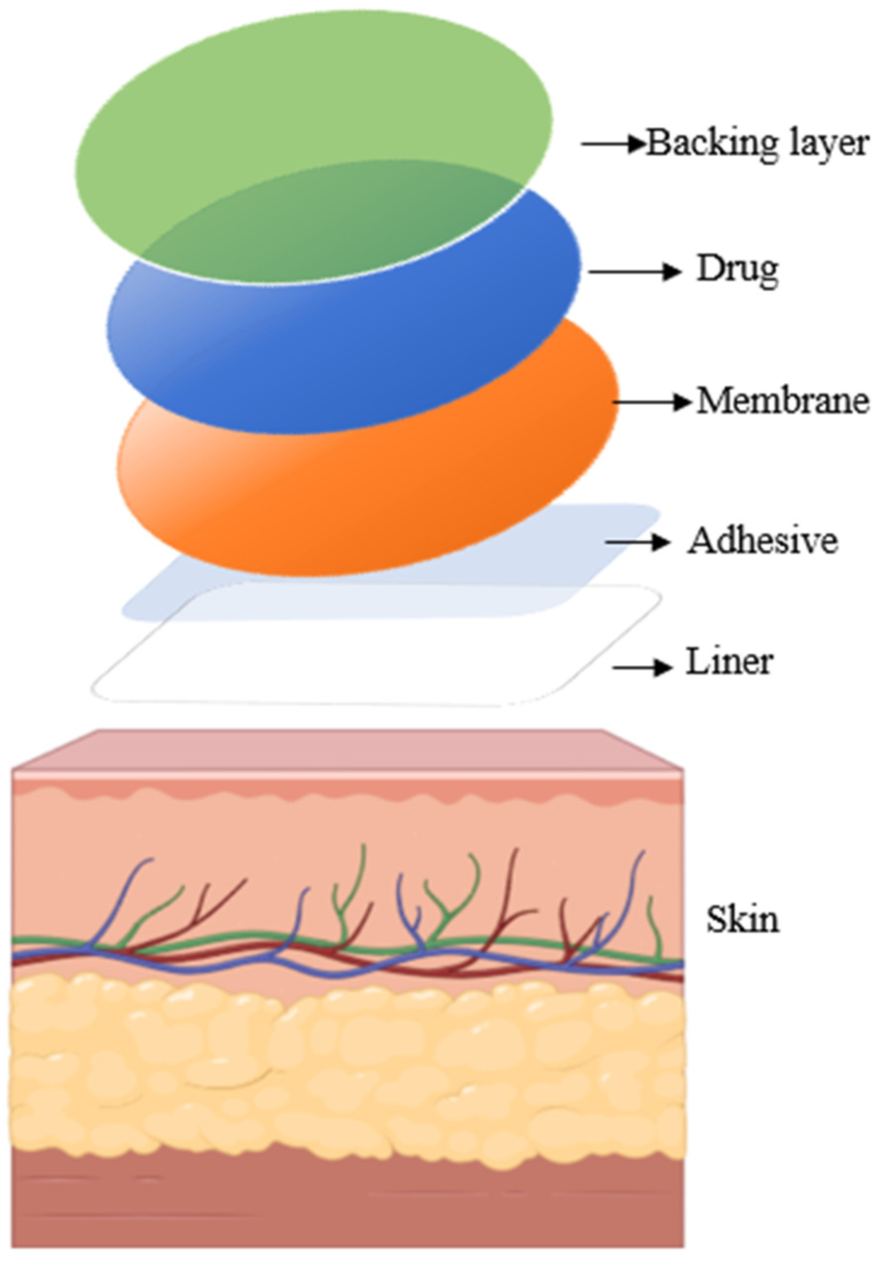
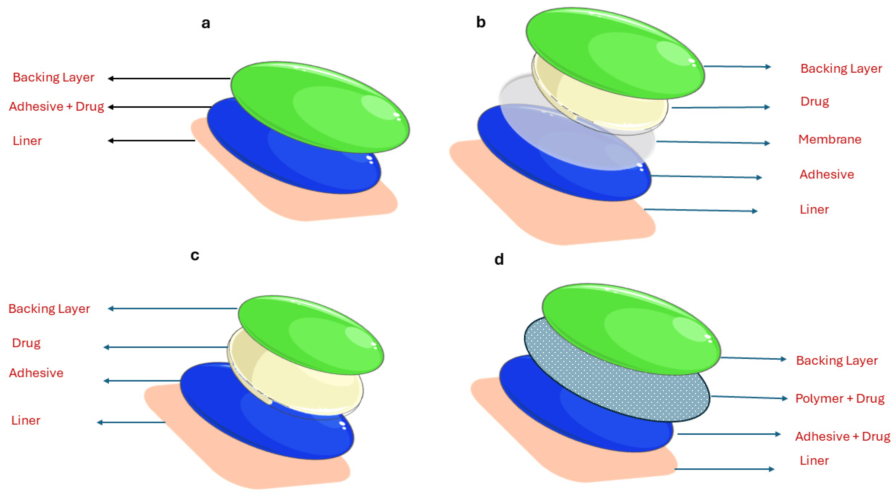
2. Overview of Transdermal Drug Delivery

Transdermal drug delivery systems (TDDSs) are drug delivery systems with controlled release mechanisms that utilize the skin as a medium for drug administration. They are formulated to perform drug delivery at a controlled and predetermined rate through systemic circulation. TDDSs focus on retaining the drug input at a sustained level for a certain period rather than on spontaneous administration [8]. Drugs permeate the skin and enter the bloodstream. The use of TDDSs dates back several years and involves applying substances or plant extracts to the skin [9]. The first systemic delivery system was designed to treat motion sickness using a 3-day scopolamine patch [9]. Additional instances involve the application of transdermal patches for administering medications like nicotine, fentanyl, nitroglycerin, and clonidine to address diverse medical conditions [10]. Like other drug delivery systems, TDDSs also have their advantages and disadvantages of being chosen as a method of drug delivery. The main advantages and disadvantages are listed in Table 1.
Generally, transdermal drug delivery is carried out via a patch that can be directly attached to the desired area. It comprises a backing layer, drug, membrane, adhesive, and liner, as portrayed in Figure 1. The topmost layer of the patch, referred to as the backing layer, protects the underlying layers from the external environment. Typically composed of pliable and waterproof materials like polyethylene or polypropylene, this layer features an adhesive coating that secures the patch to the skin, ensuring its proper placement [10]. It is usually made of a strong and mild hypoallergenic adhesive. The drug layer holds substances intended for transdermal delivery. Its formulation is designed to release drugs consistently over a specified period. A rate control membrane regulates the release pace of drugs from the patch. Typically made of semi-permeable materials, these membranes permit controlled drug passage. The liner serves as a protective layer for both the patch and adhesive. Before application to the skin surface, it is essential to remove the patch [16].
TDDSs can be broadly categorized into matrix, drug-in-adhesive, reservoir, and micro-reservoir systems according to their design [10]. However, commercially available transdermal systems are either of the reservoir or matrix types [16,17]. Figure 2 represents different types of transdermal systems. Reservoir-type patches store the drug in a solution or gel, with its distribution regulated by a rate-control membrane placed between the drug reservoir and the skin [8]. Conversely, matrix-type patches feature a straightforward design involving the incorporation of drugs into adhesive [8].

3. Fundamentals of Hydrogels

Hydrogels are known to be the first biomaterials developed for human use [18,19]. The first hydrogels were developed in 1960 using polyhydroxy ethyl-methacrylate (pHEMA) [20]. They were employed as fillers after eye enucleation [19]. Since then, hydrogels have been exploited for drug delivery and bioactive compound releases. One such important application of hydrogel polymer is modern contact lenses. In recent years, hydrogels have evolved into various forms, including smart hydrogels, shape memory hydrogels, injectable hydrogels, polymer hydrogel scaffolds, supramolecular hydrogels, self-healing hydrogels, actuators, and injectable hydrogels [7]. These find extensive applications in tissue engineering, targeted drug delivery, bone regeneration, surgical devices, gene therapy, vaccines, immunotherapy, corneal treatment, sensors for cancer therapy, absorbable bone plates, and wound healing [7].
Hydrogels are substances consisting of interconnected three-dimensional networks of hydrophilic polymers that are physically or chemically crosslinked [11]. Hydroxy groups like carboxyl (R-COOH), hydroxyl (O.H-), and amino (-NH2) groups form the hydrophilic polymers. They are interlinked in an aqueous medium. Hydrogels exhibit properties like swelling, collapse, softness, biocompatibility, and biodegradability [21]. Due to these properties, hydrogels closely resemble natural tissues. The presence of hydrophilic polymers enables this material to store and retain water content while remaining undissolved. They also possess attractive properties like sensitivity to pH, temperature, and other external stimuli [22]. These properties play a wide role in many hydrogel-based applications like wound healing, drug delivery, and tissue engineering. The broadening applications of hydrogels call for intricate control over aspects like their mechanical properties, the spatiotemporal distribution of active substances, and the refinement of their shape, structure, and overall architecture using advanced engineering techniques [23]. Advances in technology have paved the way for the development of functional hydrogels with enhanced physicochemical characteristics [23]. Key improvements in hydrogel technology involve fine-tuning their mechanical traits to make them responsive, self-repairing, and capable of shear-thinning. Furthermore, modifying the shape, structure, and architecture of hydrogels enhances their spatial precision and control [23].

3.1. Hydrogel Composition

The solid state of hydrogels takes the form of a three-dimensional network of crosslinked polymer chains. On a molecular scale, this arrangement is commonly termed the mesh, encompassing mesh size and polymer chains [24]. Hydrogels arise from the chemical or physical crosslinking of polymers. Chemical crosslinking can result from covalent bonds, hydrogen bonds, or ionic interactions [7]. Physical crosslinking may occur due to opposite charges. The product of crosslinking is a hydrogel mesh capable of retaining water. The elastic forces in the hydrogel mesh permit swelling and release of water to maintain the solidity of hydrogels. This characteristic is ascribed to hydrogel’s fluid exchange and drug release capabilities. The mesh size plays a crucial role in determining the duration of drug release and is associated with the distance between two crosslinking points [5]. It has been reported that mesh size controls the diffusion and release of the drug. The mesh size depends on the polymer and crosslinker concentrations [5]. Mesh size controls steric interactions between the drugs and the polymer network. Therefore, the mechanism of drug release is dependent on mesh size. When the mesh size is expanded, drug release primarily occurs through diffusion, while with a smaller mesh size, drugs are released via free movement [5]. Table 2 outlines different hydrogel compositions along with their respective advantages and disadvantages.

3.2. Types of Hydrogels

Hydrogels come in various types based on their origin, composition, pore size, ionic charge, physical appearance, configuration, crosslinking, responsiveness to external stimuli, and other factors [30]

3.2.1. Classification Based on Source Origin

Hydrogels can consist of natural or synthetic materials, depending on their composition. Hydrogels of natural origin are derived from non-synthetic sources, making them inherently biocompatible and bioactive [29]. Synthetic hydrogels are fabricated through chemical polymerization methods, offering enhanced durability, strength, and water absorption capacity when greater robustness is desired [29]. Hydrogels of natural origin are primarily sourced from materials such as cellulose, chitosan, hyaluronic acid, gelatin, and others [31]. They have high biocompatibility and biodegradability, low immunotoxicity, and good cytocompatibility. However, they have poor mechanical strength and low reproducibility [32]. Artificial hydrogels are created through the polymerization of monomers, such as poly-2-hydroxyethyl methacrylate (PHEMA), polyethylene glycol (PEG), polyvinyl alcohol (PVA), and polyacrylamide (PMMA) [1]. These substances exhibit stability and possess considerable mechanical strength. Additionally, in terms of composition, hydrogels can be further categorized into homopolymer hydrogels, copolymeric hydrogels, and multipolymer interpenetrating hydrogels. Homopolymeric hydrogels are derived from single monomers. Identical monomers are subjected to repeated polymerization to form a homopolymer. The crosslinks formed during polymerization are interlinked with the swelling properties of homopolymer hydrogels. Instances of homopolymer hydrogels encompass hydrogels made from polyacrylamide (PAAm), polyethylene glycol (PEG), and poly(N-isopropylacrylamide) (PNIPAAm) [30]. Copolymeric hydrogels are formed by copolymerization of two or more monomers. They can be modified to have unique properties and functionalities based on the selection of monomers. Poly (acrylic acid)-co-poly(ethylene glycol) (PAA-co-PEG) is a copolymer composed of acrylic acid and ethylene glycol. Its swelling behavior is pH-sensitive, attributed to the inclusion of acrylic acid [30]. Another significant category of hydrogels is multipolymer interpenetrating polymeric hydrogels (IPN). These hydrogels consist of two crosslinked components, either synthetic or natural polymers, interwoven into a network structure. Hydrogels of this kind exhibit distinctive properties stemming from the interpenetration of the polymer networks. With appropriate combinations of polymers, IPNs may have improved mechanical strength, stability, biocompatibility, etc. Natural hydrogels are hindered by significant drawbacks, including insufficient mechanical robustness, inconsistencies between batches, and the risk of disease transmission, particularly with animal-derived hydrogels [29]. In contrast, synthetic hydrogels often require alterations because they may not be as biocompatible and can degrade more slowly than natural hydrogels. Depending solely on natural or synthetic hydrogels can limit their use in scenarios that demand exact control over physical characteristics and smooth biological integration [29].

3.2.2. Classification of Hydrogels Based on Charge and Composition

Hydrogels can be categorized into ionic types (anionic, cationic), nonionic, and ampholytic based on their charge. Ionic hydrogels exhibit pH sensitivity, attributed to the inclusion of carboxyl, amine, or sulfonic groups. A few notable examples are PMAA, PDMAEMA, and PAA used in synthetic hydrogels. Physical or chemical crosslinking of cations and anions forms ampholytic hydrogels [33]. A well-known ampholytic hydrogel is prepared by copolymerizing two monomers, [(methacrylamido)propyl] trimethylammonium chloride (MAPTAC) and sodium styrene sulfonate (SSS), with acrylamide [34].

3.2.3. Classification Based on External Stimuli Response

Another significant categorization of hydrogels is determined by their responsiveness to external stimuli. These stimuli encompass pH, temperature, light, magnetism, electric fields, enzymes, glucose, and more. This classification is especially valuable in regulating the controlled release of drugs by adjusting external stimuli factors. pH-sensitive polymers either accept or release protons depending on the pH of the surrounding tissues [11]. This establishes the foundation for the swelling and release of drugs in a pH-sensitive environment. Thermosensitive hydrogels undergo a transition from solution to gel at a specific critical temperature. By adjusting the fluid temperature, the swelling characteristics of hydrogels can be modified [11]. Electric-responsive hydrogels are electroactive polymers that change size with electric field application. The presence of an electric field is generally found to increase drug release compared to the passive state [11]. Light-responsive hydrogels are also extensively used for many applications. The primary objective of managing drug delivery is to optimize the rate of release, ensuring that the drug remains within a therapeutic range by precisely targeting tissues and cells while minimizing potential side effects [35]. Several recent studies have focused on developing NIR-responsive hydrogel patches for transdermal drug delivery. An advantage of NIR-based hydrogel systems is their ability to shield drugs from thermal degradation, achieved through the implementation of cascade stimuli to regulate drug release. This feature proves especially beneficial for delivering temperature-sensitive proteins such as antibodies [35]. In a study by Nakielski et al., drug-loaded electrospun fibers were shown to offer sustained release of drugs, with the added capability of providing an on-demand dose when exposed to NIR-light stimulation [35]. Matai et al. designed a topical hydrogel patch for nanomaterial-assisted photothermal therapeutics, as well as for on-demand drug delivery applications. The patch exhibited photothermal stability for four cycles under cyclic photothermal heating. Additionally, the hybrid patch demonstrated NIR-stimulated drug release, as evidenced by the evaluation using methotrexate (MTX), a water-insoluble anticancer drug, and rhodamine B (RhB), a water-soluble dye. This work presents a new approach to the development of externally placed hydrogel patches for the thermal destruction of localized solid tumors and the tunable delivery of chemotherapeutic drugs at the target site [36]. One recent example of the application of NIR irradiation in hydrogels is a study conducted by Rybak et al. in 2023 [37]. Advances in the field of skin patches have led to the development of wearable and implantable bioelectronics for long-term, continuous healthcare management and targeted therapy [37]. This study presents the evolution of skin patches, from functional nanostructured materials to multifunctional and stimuli-responsive patches, including flexible substrates and emerging biomaterials for e-skin patches [37]. The study covers material selection, structure design, and promising applications. Through near-infrared (NIR) irradiation, the self-healing and drug release of the PDA-NP/PNIPAM hydrogels could be regulated. Methasone was released when the NIR laser was on, but the drug release stopped once the NIR laser was turned off [37]. Additionally, NIR irradiation successfully induced self-healable properties of the PDA-NP/PNIPAM hydrogels [37]. Thus, stimuli responses can control the drug release rate with specificity using various modes of external stimuli. Table 3 presents a compilation of various types of stimuli-responsive hydrogels along with their respective advantages and disadvantages.

3.2.4. Classification Depending on Crystallinity

Based on the degree of structural order, hydrogels can be broadly classified into amorphous (non-crystalline), crystalline, and semicrystalline. Crystallinity determines mechanical properties like strength, stiffness, etc. Amorphous polymer-based hydrogels lack long-range structural order or crystallinity in their polymer network. Crystalline hydrogels are composed of large networks of preferentially oriented regions [47]. Semicrystalline hydrogels are partly amorphous and crystalline [47]. The crystallinity properties of hydrogels are modified to achieve specific characteristics in hydrogels. This is imparted by controlling polymerization conditions like pH, temperature, concentration of polymer, and interlinking agents [30].

3.2.5. Classification Based on Appearance

This classification is independent of the polymerization method used but rather depends on physical appearance. Hydrogels may be in matrix, film, or microsphere forms. The most common forms of hydrogels are in matrix form or bulk hydrogels. They exist in gel or solid form and are large. Bulk hydrogels find application in drug delivery for controlled drug release. Film-based hydrogels are two-dimensional flat sheets used in biomedical device coating and applications in wound dressing [30]. The microsphere type of hydrogel is small, spherical, and micrometers in size. These microspheres primarily serve as scaffolds or carriers for cells, promoting their proliferation and differentiation [30]. They are found to be extensively useful in tissue engineering and regenerative medicine applications.

3.2.6. Classification Based on Crosslinking

Hydrogels may be formed by chemical or physical crosslinking. Crosslinks in physical gels are frequently reversible and dynamic due to the opposing action of the controlling stimuli, but covalent crosslinks in chemical gels are permanent and immovable [47]. Unlike chemical crosslinks that are point-like, physical crosslinks are also created over a defined range [47]. Further details of the methods used in these two types of hydrogels are detailed in the following section. Cooling a hot polymer solution is one of the most straightforward methods to create physically crosslinked gels, as seen in the production of polylactic acid-polyethylene glycol (PEG) hydrogels and polypropylene oxide-polyethylene oxide hydrogels [48]. Chemical crosslinking of hydrogels occurs when covalent bonds form between polymer chains, either during the crosslinking of polymer precursors or the polymerization of monomers [49]. A typical approach for making chemically crosslinked hydrogels involves radical polymerization using a crosslinking agent and low molecular weight monomers [50]. For example, in radical polymerization, HEMA is polymerized using ethylene glycol dimethacrylate as a crosslinker, leading to the creation of poly(2-HEMA) [50]. Additionally, polymers with hydroxyl groups, such as polyvinyl alcohol, can be chemically crosslinked using glutaraldehyde as a crosslinking agent [50].

3.3. Preparation of Hydrogels

As outlined in the preceding section, the production of hydrogels involves two types of synthesis distinguished by the crosslinking method. Hydrogels, as defined, consist of three-dimensional crosslinked polymer chains. The synthesis of these polymer chains requires linking monomers or polymers to create the desired hydrogel. The process of crosslinking can be either physical or chemical.

3.3.1. Physically Crosslinked Hydrogels

Physical crosslinking methods encompass hydrogen bonding, amphiphilic grafting, block polymer formation, and crystallization. Hydrogen bonding takes place when there are N-H, O-H, or F-H functional groups present. Hydrogen bonding with fluorine (F) in hydrogels is not common. Fluorine is highly electronegative, which makes it difficult for hydrogen bonding to occur with other atoms [51]. An example of physical crosslinking through hydrogen bonding is evident in the bond between oxygen in PEG and carboxylic acid groups in PAA [33]. In amphiphilic grafting and block polymer formation, crosslinking occurs by the self-assembly of hydrophilic or hydrophobic solvents based on their amphiphilic affinity. The crystallization crosslinking process involves the adjustment of crystallization temperature by freeze–thawing or the heating of polymers. PVAs form hydrogels by freeze–thawing [33]. Another example is the application of heat to a PEO copolymer to form a hydrogel. Physical gels have attracted attention because of their straightforward production process and the absence of crosslinkers, which reduce the risk of toxicity [52]. Nonetheless, these gels have a drawback in that their stability can be compromised by changes in factors like stress, pH, temperature, ionic strength, and the presence of solute particles) [50].

3.3.2. Chemically Crosslinked Type of Hydrogels

The formation of chemical hydrogels typically involves three methods: copolymerization of a monomer and a multifunctional crosslinker, crosslinking of polymer solutions induced by radiation, and crosslinking of polymers through chemical reactions with complementary agents. An illustration of a chemically crosslinked hydrogel is poly HEMA hydrogel, which is created through the copolymerization of HEMA with the crosslinker EGDMA [19]. PEO and PVA hydrogels can be created by employing an electron beam accelerator to irradiate their aqueous solutions [19,53].

4. Skin and Diffusion through the Skin
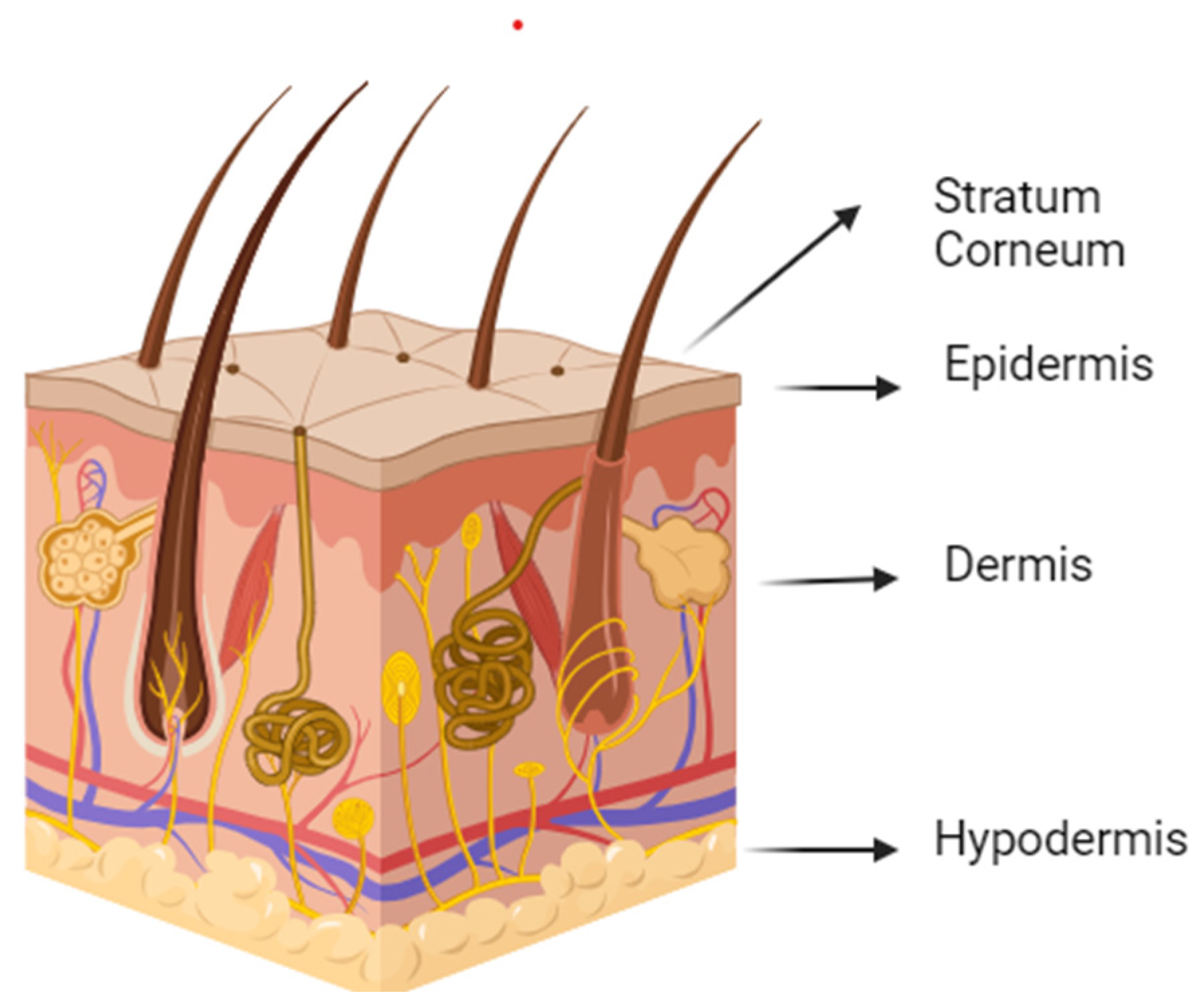
Skin poses a significant hindrance to transdermal drug delivery. Apart from serving as a physical barrier against microorganisms, the skin also functions as a hemostatic barrier, preventing water loss from the body. This is one of the most significant challenges faced by TDDSs [54]. Many researchers have studied the dissolution and kinetics of a drug delivery device together with the percutaneous absorption in the skin and modeled them as well [16]. Drug transport in skin is defined using the diffusion–reaction–deformation equations [55]. The following sections detail how skin structure influences drug diffusion and how it can be enhanced.

4.1. Skin Structure

The skin acts as the largest interface connecting the body to the external environment. Its primary function is to offer protection against various external hazards such as chemicals, heat, and physical injuries. Consisting of several layers, the skin is composed of the epidermis, dermis, and hypodermis (Figure 3). Although the dermis and hypodermis have minimal participation in drug delivery, the outermost layer, the epidermis, is essential for providing protection. The epidermis comprises two main layers: the viable epidermis and the nonviable epidermis, which includes the stratum corneum [56]. The stratum corneum, the thin outer layer of the epidermis, is accountable for the skin’s exceptional barrier properties [57]. Unlike other body tissues, the stratum corneum consists of corneocytes surrounded by an extracellular environment of lipids organized in lamellar bilayers. These lipids act as a barrier. However, they permit the passage of lipid-soluble and low molecular-weight drugs. The existence of lipids presents a notable challenge in drug administration through the skin [58]. The lipid-rich extracellular matrix of the stratum corneum has several properties that contribute to its exceptionally low permeability to water-soluble drugs. One of the exceptional features is that of the organization of the lipid-rich extracellular matrix and its organization into a highly tortuous and convoluted extracellular pathway [58].
Ref. [56] The characteristics of the lipid phase in the stratum corneum are distinguished by its lipid content, which differs from that of other tissues’ membranes. This characteristic of the epidermis impedes drug delivery through the skin [56]. The unique structural characteristic of skin makes it a strong barrier against TDDSs. Therefore, effective transdermal drug delivery requires novel designs and approaches.

4.2. Factors Influencing Drug Diffusion through the Skin

The skin acts as a substantial obstacle to drug absorption. To achieve effective drug delivery, it is crucial to understand the parameters that influence the permeability of this barrier.

4.2.1. The Physiological Factor

Factors like temperature, race, age, thickness of skin, and blood flow within the stratum corneum affect drugs’ permeability in transdermal drug delivery systems (TDDS). An increase in temperature allows subcutaneous blood vessels to dilate. This gives rise to higher permeability. Hydrated skin can enhance drug penetration by loosening the stratum corneum [59].

4.2.2. The Drug Absorption Factor

Fick’s law of absorption governs conventional drug delivery. In the case of topically applied drugs, the absorption rate is directly proportional to the concentration of the drug vehicle. The permeability coefficient, denoted as K p , serves as the parameter linking the drug and the barrier. K p comprises of K m , the partition coefficient; D, the diffusion coefficient; and L, the length of the diffusion pathway [58]. Based on this, the absorption rate (J) is directly proportional to the concentration difference across the skin barrier and the thickness of the barrier, i.e., the skin.
J = K p C v
J = D K m L C v

4.2.3. Drug Delivery Setting

Several crucial formulation factors impact the transdermal penetration of a drug, including the dosage form (a combination of drug and non-drug components), matrix (typically a polymer network dispersing the drug), pH, delivery concentration, and the area of administration. In transdermal delivery, patches are the designated dosage form for therapeutic drug delivery. The composition of these patches plays a significant role in determining how drugs permeate the skin. Optimal drug penetration occurs when the matrix pH is equal to or greater than the pKa of alkaline drugs or less than that of acidic drugs. Additionally, a larger administration area correlates with increased penetration efficacy and higher drug concentration [59].

4.3. Methods of Enhancing Drug Penetration

Numerous advanced techniques have been developed to surmount the skin barrier and improve drug penetration. These enhancement techniques are broadly categorized into active and passive methods based on the physicochemical nature of the drug [56]. The enhancement techniques are intended to increase skin permeability or provide a driving force to the drug for penetration [60]. The goal of these techniques is to increase the permeation flux [56].

4.3.1. Active Methods for Permeation Enhancement

Despite its merits, transdermal drug delivery is significantly hindered by the notable limitation that the skin’s permeability to most molecules, except for a select few that are small and hydrophobic, is remarkably low [61]. Consequently, achieving a therapeutically relevant rate of drug delivery across the skin proves challenging [61]. Employing external stimuli such as electrical, physical, or mechanical forces can enhance the skin permeability of drugs and biomolecules in transdermal drug delivery [62]. The physical methods for enhancement of skin permeation include iontophoresis, electroporation, sonophoresis, microneedles, etc.

Iontophoresis

Iontophoresis techniques improve the movement of ions across the membrane by applying a small electric potential difference [63]. It employs a minimal amount of physiologically acceptable electric current (0.5 mA/cm2 or less) to propel ionic (charged) drugs into the body, utilizing an electrode with the same polarity as the charge on the drug [64]. Consequently, electrostatic repulsion propels the drug into the skin [64]. This is recognized for enhancing the transdermal delivery of peptides and proteins by employing lower current intensity over a brief duration [64]. The effectiveness of the iontophoresis technique in permeation depends on factors such as the polarity, valency, and mobility of the drug molecule, the characteristics of the applied electrical cycle, and the formulation containing the drug [13].

Sonophoresis

Another technique that enhances drug permeation through the skin membrane involves the application of ultrasound waves [56]. Ultrasound waves are applied in two distinct frequency ranges: low-frequency (20 kHz to 100 kHz) and high-frequency (2 MHz to 16 MHz). The primary governing parameters in the sonophoresis technique are the frequency, duration, and intensity of the applied ultrasound waves [56]. Ultrasound shows promising potential for improving transdermal drug delivery by disrupting the intercellular lipid bilayer in the stratum corneum. In a specific example, a four-armed PEG hydrogel patch loaded with diclofenac utilized ultrasound to initiate drug release and act as a permeation enhancer [65]. Using a low-frequency ultrasound coupling medium in hydrogels has also resulted in increased skin permeability and localized transport [66]. Apart from the acoustic effects, ultrasound results in thermal effects by raising the temperature of the skin area [13]. Sonophoresis techniques prove valuable in drug delivery, irrespective of the drug’s solubility, ionization, dissociation constants, electrical properties, and molecular weight [13].

Electroporation

Electroporation is a method employing high-voltage (5–500 V) electric pulses with durations ranging from microseconds to milliseconds to introduce therapeutic agents into cells and tissues [56]. Brief exposure to high voltages results in the creation of small pores in the stratum corneum, enhancing permeability and facilitating drug diffusion [13]. The effectiveness of electroporation-induced transdermal permeation relies on the formulation parameters and physicochemical properties of the drug molecule [56]. Elevating electrical parameters such as pulse voltage increase and rate increases drug transport [56]. In a study [67], the in vitro permeation of buprenorphine through the skin was investigated utilizing electrophoresis techniques. The implementation of these methods resulted in an 8.45-fold increase in permeation. The experimental conditions comprised a pulse frequency of 1 pulse every 30 s, administered for a duration of 10 min, using a pulse voltage of 500 V and a pulse duration lasting 200 milliseconds. While the electric field applied for milliseconds during electroporation generates an electrophoretic driving force, the diffusion through long-lasting electro-pores can endure for hours. This persistence allows for a substantial increase in transdermal transport by orders of magnitude for small-molecule drugs, peptides, vaccines, and DNA [67]. Employing electroporation as a method to enhance percutaneous penetration offers a superior strategy for boosting transdermal drug delivery, evident in both in vitro and in vivo settings. This noninvasive approach facilitates quick, effective, and pulsatile administration of a wide range of therapeutic agents, including small, ionized drugs, macromolecules, and nucleic acids, across various tissues like the skin, liver, tumors, and more [61].

Photomechanical Waves

The transmission of photodynamic waves to the skin can penetrate the stratum corneum (S.C.) and facilitate the passage of the drug through the transiently created channel [68]. Photomechanical waves can be generated by direct or confined ablation, resulting in two different characteristics [69]. Lee et al. investigated how pulse characteristics influence the depth of penetration of macromolecules delivered into the skin through photomechanical waves generated by a Q-switched ruby laser [69]. The study revealed that controlled ablation, as opposed to direct ablation, produces photomechanical waves with longer rise times and durations. To achieve greater penetration depth, controlled ablation requires low radiant exposure. Customizing the characteristics of photomechanical waves is essential for achieving effective transdermal drug delivery, as they directly impact the depth of drug penetration into the skin, especially the dermis. Ablation is induced by these waves with exposure to incident waves ranging from 5 to 7 J/cm2 at a depth of 50–400 μm for successful transmission.

Microneedles (M.N.s)

The microneedle drug delivery system comprises hypodermic needles and transdermal patches. Micron-sized needles, referred to as M.N.s, can establish microchannels in the skin to facilitate drug delivery by disrupting the stratum corneum [56]. Moreover, they are less invasive and induce less pain compared to hypodermic needles, as they do not reach the pain receptors located in the dermis layer [70]. Based on fabrication methods, microneedles (M.N.s) can be categorized as solid, hollow, polymer, coated, dissolving, and hydrogel-forming M.N.s [56]. The solid microneedle delivers the drug by penetrating the skin at the patch placement site. Drug-coated microneedles are inserted into the skin, releasing the coated drug within the skin. Biodegradable or polymeric microneedles encapsulating drugs are inserted into the skin to accomplish controlled drug release. Hollow microneedles are employed to house naturally delivered melting needles. Hydrogel-forming microneedle arrays enable the transdermal delivery of drugs by piercing the outer layer of the skin surface and absorbing interstitial skin fluid along with inflammatory processes [71]. The microneedle drug delivery system comprises microneedles with heights ranging from 50 to 900 μm, enabling them to penetrate deep into the skin and easily traverse the stratum corneum barrier [72]. In a conducted study [73], hydrogel-forming microneedles (HFMs) were employed for the transdermal delivery of albendazole from liquid reservoirs. The findings suggested that HFMs could penetrate up to 63% into the skin with only a 7.14% reduction in height.

Thermal Ablation

Thermal ablation is a method for selectively altering the stratum corneum structure by applying localized heat, enhancing drug delivery through microchannels formed in the skin [13]. Thermal ablation can be achieved by applying moderate temperatures (≤100 °C) for a longer period or by applying very high temperatures (≥100 °C) for a short period [74]. The second method is more favorable as it causes less damage to the skin. Thermal ablation can be achieved using various modes such as chemical heating, thermoporation, radio frequency (R.F.), and laser [74].

4.3.2. Passive Methods for Permeation Enhancement

Passive techniques involve the use of chemical enhancers to facilitate drug permeation across the skin and novel carriers to enhance the solubility of the drug [56]. Chemical enhancers enhance drug permeation by utilizing various chemicals or biochemical agents that can interact with the constituents of the stratum corneum to dissolve the skin barrier [56]. Commonly employed chemical enhancers include alcohols, ether alcohols, fatty acid derivatives, amides, various surfactants, sulphoxides, pyrrolidone, oxazolidones, terpenes, hyaluronic acid derivatives, and others [56,75]. Nevertheless, chemical enhancers are frequently linked with skin irritability. Advanced chemical approaches involve the utilization of various novel drug carriers, such as microemulsions, solid lipid nanoparticles (SLNs), nanostructured lipid carriers (NLCs), invasomes, transfersomes, dendrimers, liposomes, ethosomes, and more [56]. They are found to be more profound than the direct application of chemical enhancers in terms of safety and efficacy.

4.4. Mechanism of Drug Release through Hydrogels

The distinctive properties of hydrogels render them suitable candidates for a range of drug delivery applications. Various mechanisms exist for releasing therapeutic drugs from the polymer network of hydrogels.

4.4.1. Diffusion-Controlled Release Mechanism

The diffusion-controlled mechanism is a prevalent approach for drug release in hydrogels, and it can be further categorized into matrix and reservoir types. Most transdermal applications use hydrogel patches made of either of these types, depending on the type of drug release. The drug is uniformly distributed within the matrix-type system, whereas the polymeric matrix encapsulates the drug core in the reservoir type. In both cases, drug diffusion is influenced by mesh size, which, in turn, depends on external stimuli, crosslinking density, composition, and gel structure [50]. In the swollen state, typical hydrogels have mesh sizes in the range of 5–100 nanometers [76]. This size is significantly larger than that of most small molecules. Consequently, the drug can be retained in a swollen state. The diffusion of drugs in the hydrogel is most accurately described by Fick’s law of diffusion, as delineated below [50].
D i p d C i d x
where C i is the concentration of the drug and D i p is the drug’s coefficient of diffusion.

4.4.2. Swelling-Controlled Release

In the mechanism of swelling-controlled release, drug release takes place when the diffusion of the drug surpasses the rate of swelling. With an escalation in the rate of hydrogel swelling, the release of the drug also rises. The key factors governing this type of drug release mechanism are the capacity for water absorption and the thickness of the polymeric gel [54]. The major drawback of the swelling-controlled drug release mechanism is its slow response due to slow diffusion. One solution to this shortcoming is reducing the diffusion length by reducing the hydrogel’s size or designing a system of interconnected macropores.

4.4.3. Chemically Controlled Release

Chemically controlled systems discharge drugs through chemical reactions taking place within the gel matrix. This can involve polymeric chain cleavage through hydrolytic or enzymatic degradation. Another mechanism could be reversible/irreversible reactions occurring between the polymer network and the released drug. The release of drugs could also occur through surface or bulk erosion of hydrogels [76].

5. Hydrogels in Transdermal Drug Delivery

Hydrogels possess remarkable qualities that make them well-suited for transdermal drug delivery. These characteristics encompass biocompatibility, biodegradability, elasticity, non-allergenicity, ease of application, soft consistency, and high-water content [77]. The application of hydrogels induces hydration effects, thereby improving the transport of drugs across the skin [77]. The basic component of the transdermal drug delivery system is a drug-loading matrix, as described in the earlier section. Hydrogels are frequently employed as matrices for Transdermal Drug Delivery Systems (TDDSs). Zhang et al. utilized polyethylene glycol diacrylamide (PEG-DA) hybrid hydrogel in mouse embryonic fibroblast cell lines (NIH 3T3). The hydrogel demonstrated favorable mechanical properties, swelling capacity, biocompatibility, and non-toxicity to the skin [78]. Hydrogels composed of gelatin polyacrylamide (Gel-PAAm) applied to human skin exhibited non-toxicity, high stretchability, and favorable swelling properties [79]. In another study involving rat abdominal skin, a hydrogel composed of polyacrylamide-grafted-chondroitin sulfate (PAAm-g-CS) was employed [80]. A hydrogel based on natural materials, chitosan–azelaic acid (CS-AZ), specifically the CS-AZ hydrogel, was utilized with mouse fibroblast L929, demonstrating outstanding swelling, water vapor permeability, high porosity, and low cytotoxicity [81]. Additional instances of hydrogels in transdermal drug delivery encompass the utilization of carboxymethyl chitosan–silk fibroin peptide/oxidized pullulan (CMCS-SFP/OPL) hydrogel in newborn porcine Tskin [82] and carboxymethyl chitosan-grafted-2 hydroxyethyl acrylate (CmCHT-g-pHEA), which was administered on the dorsal skin of micropigs [65,83].

6. Some Important Applications of Hydrogels in Transdermal Drug Delivery Systems

Hydrogels find widespread applications in transdermal drug delivery across various fields. The subsequent section highlights key applications of transdermal drug delivery systems (TDDSs) based on hydrogels. Table 3 outlines FDA-approved drug delivery products based on hydrogels. Furthermore, Table 4 presents recent findings from the literature on transdermal drug delivery using hydrogels.

6.1. Cancer Therapy

Traditional cancer treatments, especially chemotherapy, carry the risk of systemic side effects such as reduced bone marrow function, liver or kidney failure, and neurological issues. Hydrogels present significant potential for research in this area. To tackle these challenges, it is essential to control drug release, targeting the medication directly to the tumor site, particularly when integrated with tissue engineering techniques. Hydrogel patches equipped with microneedles have been shown to be particularly effective for drug delivery in cancer treatment. A specially designed transdermal patch exhibited a significant release of two milligrams of anastrozole targeted for breast cancer treatment but caused harm to the adjacent healthy tissues. In a separate investigation, another study detailed the transdermal delivery of isoliquiritigenin through a hydrogel formulated with hyaluronic acid and hydroxyethyl cellulose. This drug is recognized for its ability to impede pancreatic cancer progression [84].

6.2. Diabetes Treatment

Most diabetic patients rely on insulin for maintaining controlled glycemia levels, often necessitating multiple daily administrations. Despite their associated pain and inconvenience, subcutaneous needle injections are commonly employed for insulin delivery. As an alternative to this traditional method, transdermal drug delivery holds promise. Qiu et al. conducted a study in which they developed a lyophilized hydrogel patch system for insulin delivery through microneedles. The hydrogel patch, created within a crosslinked matrix of poly (acrylamide-co-acrylic acid) through precipitation polymerization, was infused with recombinant human insulin. The results observed after 12 h of insulin injection revealed a proportional reduction in blood glucose levels in relation to the concentration of insulin incorporated into the lyophilized hydrogel patches, demonstrating a longer action duration than subcutaneous injection [85].

6.3. Cardiovascular Treatment

Reduced cardiac ejection fraction in heart failure patients may result in frequent alteration of pharmacokinetics (P.K.) and pharmacodynamics (P.D.), resulting in low drug absorption. In such scenarios, transdermal drug delivery presents itself as a viable alternative to conventional drug administration. For instance, propranolol is a nonselective beta-adrenergic blocker prescribed to patients with cardiovascular issues. An animal study involving rabbits demonstrated that oral propranolol could reach a peak plasma concentration of 56.4 ng/mL within 13.2 min, with 12% bioavailability. In contrast, the transdermal propranolol patch maintained a steady-state plasma concentration of 9.3 ng/mL after an initial lag time of 8 h, with a significantly higher bioavailability of 74.8% [10]. An additional illustration of hydrogel-based transdermal drug delivery is the application of sildenafil citrate (S.C.) in treating pulmonary hypertension (P.H.). P.H., a cardiovascular disease, significantly reduces functional capacity, diminishes quality of life, and substantially shortens life expectancy [86]. Microneedles with hydrogel-forming capabilities that penetrate the stratum corneum were used to distribute the drug via the transdermal route.

6.4. Treatment of Hormone Deficiency

Multiple factors contribute to the development of chronic growth hormone deficiency (GHD). It decreases life quality and causes conditions like osteoporosis, aberrant metabolism, and short stature in children. Current treatment approaches include subcutaneous (S.C.) injections of recombinant human growth hormone (rhGH), possessing similar pharmacological properties and structure to endogenous GHD; however, GHD patients typically need years of ongoing treatment, resulting in thousands of S.C. injections. Yang et al. designed a microneedle patch (PAA/NaHCO3-Silk MN) utilizing silk protein for the controlled release of recombinant human growth factor hormone (rhGH). The results yielded that the PAA/NaHCO3-Silk MN patch shows promising potential to significantly enhance patient adherence, raise medication availability, address unmet clinical needs, and enhance the therapeutic outcomes for GHD patients [87].

6.5. Wound Management

A wound is any disturbance or breakage in the skin that arises from a medical condition, physiological issue, or trauma. When injured or wounded tissue returns to its pre-injury or normal anatomical appearance, structure, and function within an acceptable time frame, the wound is considered totally healed [6]. The process of wound healing is continuous and comprises four distinct, automatic phases. Every phase needs to be controlled precisely and in a certain way. Any disruption or extension of these stages could result in a longer healing period for the wound and increase the chance that it will become a chronic wound.
Hydrogels are best suited for burn patients since they possess most of the ideal characteristics anticipated for wound healing. Healthcare professionals and biomaterial scientists acknowledge hydrogels as the preferred choice for wound dressing due to their numerous advantages. These include maintaining optimal moisture levels at the wound site, facilitating proper exchange of oxygen and moisture between the wound and its surroundings, biocompatibility, possessing a structure resembling tissue, ease of application owing to softness, elasticity, and flexibility, offering a cooling sensation for patient comfort, absorbing serous discharges from lesions, and minimizing interference with the wound healing process [77].
Various hydrogels used in wound dressing and healing include composite formulations like gelatin-grafted dopamine/chitosan/carbon nanotubes, lignin–chitosan–PVA, and nano curcumin-loaded N, O-carboxymethyl chitosan/oxidized alginate hydrogel [77]. Calcium alginate hydrogel dressings derived from a foundation are another potential form of wound dressing used for treating highly exudative wounds, as they promote the growth of granulation tissue. The interaction between salt from wounds and alginate, forming a gel structure, establishes an optimal moist healing environment. Calcium further facilitates cell migration, remodeling, and wound homeostasis [77]. In the literature, there are several types of hydrogels used for wound management. (Table 5 and Table 6).

7. Challenges and Future Directions of Hydrogel-Based Transdermal Drug Delivery Systems

Over recent decades, hydrogels have significantly progressed in their utilization within transdermal drug delivery systems. Despite these advancements, several challenges persist, impeding the full realization of hydrogel potential in this field. Among these challenges are drug loading, release control, skin permeation efficiency, long-term stability, and scalability constraints. The inherent limitation of hydrogels in accommodating high drug concentrations requires innovative approaches to enhance loading capacity without compromising the integrity of the matrix. Achieving controlled drug release rates necessitates precise control, prompting the development of responsive hydrogels that are sensitive to diverse stimuli such as temperature, pH, or enzymes.
Addressing the ongoing challenge of improving skin barrier permeation efficiency while ensuring compatibility and minimizing irritation is crucial. Concerns also arise regarding the stability and durability of hydrogel-based systems over extended periods, particularly under diverse storage conditions, posing practical application hurdles. Additionally, challenges exist in scaling up production while maintaining uniformity, reproducibility, and cost-effectiveness.
Future research directions aim to address these challenges. For instance, the development of smart hydrogels responsive to external stimuli like pH, temperature, or light can enable on-demand drug release, potentially enhancing therapeutic efficacy [102]. Additionally, exploring innovative drug-loading methods involving nanotechnology, molecular imprinting, or encapsulation techniques could significantly augment drug-loading capacity [103]. Moreover, leveraging hydrogels to deliver multiple drugs or combine them with nanoparticles or growth factors can revolutionize combination therapies.
Customizing hydrogel formulations based on individual patient needs—considering skin type, dosage, and release kinetics—can elevate the effectiveness of transdermal therapies. Integration of advancements in 3D printing and nanotechnology to craft precise, patient-specific hydrogel structures holds the potential to revolutionize current methodologies in transdermal drug delivery. Consequently, meticulous design and utilization of hydrogel systems could open doors for a broad spectrum of applications within drug delivery. A recent study by Ziai et al. focused on ensuring a strong attachment between fibrous mats and plasmonic hydrogel layers in composites [104]. Plasmonic gold nanorods were integrated into polyacrylamide-based hydrogels as the hydrogel layer. The impact of crosslinking density on the integration of these two layers was investigated. Factors contributing to a more robust interface between the plasmonic hydrogel layer and electrospun nanofibers in multi-layer nanocomposites were explored [104]. By incorporating nanoparticles into hydrogels, their solid and adjustable optical responses can be utilized to manipulate photothermal effects upon exposure to external light. This combination holds promise for biomedical applications like biosensing and photothermal therapy [104].

8. Conclusions

In conclusion, the field of transdermal drug delivery systems (TDDSs) has experienced significant advancements in recent decades, offering a promising alternative to conventional oral drug administration. This comprehensive review has provided valuable insights into various transdermal techniques, the skin’s role as a barrier to TDDS, factors influencing skin diffusion, and the current challenges faced in this field. With a primary focus on TDDSs utilizing hydrogels, we have delved into the fundamental aspects of hydrogels, including their structure, properties, and synthesis, highlighting their crucial role as carriers in transdermal drug delivery. Moving forward, strategies for hydrogel-based drug delivery have been discussed, addressing existing challenges and exploring potential future directions. Through this analysis, we aim to contribute to the advancement of TDDSs and facilitate the development of more effective and efficient transdermal drug delivery methods.

Funding

This research was funded by the American University of Sharjah Faculty Research Grants (FRG20-L-E48, and FRG22-C-E08), Sheikh Hamdan Award for Medical Sciences (Grant Number: MRG-57-2019-2020) and Friends of Cancer Patients (FoCP). The work in this paper was supported, in part, by the Professional Development Fund of GAH at the American University of Sharjah. This paper represents the opinions of the author(s) and does not mean to represent the position or opinions of the American University of Sharjah.

Conflicts of Interest

The authors declare no conflict of interest.

Abbreviations

TDDSsTransdermal drug delivery systems
DDSDrug delivery system
GITGastrointestinal tract
COOHCarboxyl group
O.H.Hydroxyl group
NH2Amino acid group
PVAPolyvinyl alcohol
PEGPolyethylene glycol
PEOPolyethylene oxide
PHEMAPoly(2-hydroxyethyl methacrylate)
PAAPolyacrylic acid
PAAMPolyacrylamide
PNIPAAmPoly (N-isopropyl acrylamide)
PAA-co-PEGPolyacrylic acid co-poly ethyl glycol
IPNInterpenetrating polymer network
MAPTACMethacrylamidopropyltrimethylammonium chloride
SSSSodium styrene sulfonate
pKaAcid dissociation constant
DNADeoxyribonucleic acid
S.C.Stratum corneum
MNMicroneedle
R.F.Radio Frequency
NLCNanostructured lipid carriers
PEGDAPolyethylene glycol diacrylate
PAAm-g-CSPolyacrylamide-grafted-chondroitin sulfate
Gel-PAAmGelatin polyacrylamide
CS-A2Chitosan azelaic acid
CMCS-SFP/OPLCarboxymethyl chitosan silk fibroin peptide/oxidized pullulan
CmCHTpHEACarboxymethyl chitosan-grafted 2 hydroxyethyl acrylate
pHPotential of hydrogen
FDAFood and Drug Administration
PKPharmacokinetics
P.D.Pharmacodynamics
P.H.Pulmonary hypertension
GHDGrowth hormone deficiency
SCSubcutaneous
rhGHRecombinant human growth hormone

References

  1. Nayak, A.K.; Ahmad, S.A.; Beg, S.; Ara, T.J.; Hasnain, M.S. Drug delivery: Present, past, and future of medicine. In Applications of Nanocomposite Materials in Drug Delivery; Woodhead Publishing: Cambridge, UK, 2018; pp. 255–282. [Google Scholar] [CrossRef]
  2. Jain, K.K. An overview of drug delivery systems. Meth. Mol. Biol. 2020, 2059, 1–54. [Google Scholar] [CrossRef]
  3. Nunes, D.; Andrade, S.; Ramalho, M.J.; Loureiro, J.A.; Pereira, M.C. Polymeric Nanoparticles-Loaded Hydrogels for Biomed-ical Applications: A Systematic Review on In Vivo Findings. Polymers 2022, 14, 1010. [Google Scholar] [CrossRef] [PubMed]
  4. Adepu, S.; Ramakrishna, S. Controlled Drug Delivery Systems: Current Status and Future Directions. Molecules 2021, 26, 5905. [Google Scholar] [CrossRef] [PubMed]
  5. Li, J.; Mooney, D.J. Designing hydrogels for controlled drug delivery. Nat. Rev. Mater. 2016, 1, 16071. [Google Scholar] [CrossRef] [PubMed]
  6. Ahsan, A.; Tian, W.-X.; Farooq, M.A.; Khan, D.H. An overview of hydrogels and their role in transdermal drug delivery. Int. J. Polym. Mater. Polym. Biomater. 2021, 70, 574–584. [Google Scholar] [CrossRef]
  7. Kesharwani, P.; Bisht, A.; Alexander, A.; Dave, V.; Sharma, S. Biomedical Applications of Hydrogels in Drug Delivery Sys-tem: An Update. J. Drug Deliv. Sci. Technol. 2021, 66, 102914. [Google Scholar] [CrossRef]
  8. Santos, L.F.; Correia, I.J.; Silva, A.S.; Mano, J.F. Biomaterials for drug delivery patches. Eur. J. Pharm. Sci. 2018, 118, 49–66. [Google Scholar] [CrossRef] [PubMed]
  9. Joshi, N.; Machekposhti, S.A.; Narayan, R.J. Evolution of Transdermal Drug Delivery Devices and Novel Microneedle Technologies: A Historical Perspective and Review. JID Innov. 2023, 3, 100225. [Google Scholar] [CrossRef]
  10. Wong, W.F.; Ang, K.P.; Sethi, G.; Looi, C.Y. Recent Advancement of Medical Patch for Transdermal Drug Delivery. Medicina 2023, 59, 778. [Google Scholar] [CrossRef] [PubMed]
  11. Kim, J.-C.; Madhusudhan, A.; Azamal, H. Smart Nanomaterials in Biomedical Applications, 1st ed.; Kim, J.-C., Alle, M., Husen, A., Eds.; Springer International Publishing: Cham, Switzerland, 2021; ISBN 978-3-030-84261-1. [Google Scholar]
  12. Sabbagh, F.; Kim, B.S. Recent advances in polymeric transdermal drug delivery systems. J. Control. Release 2022, 341, 132–146. [Google Scholar] [CrossRef]
  13. Jeong, W.Y.; Kwon, M.; Choi, H.E.; Kim, K.S. Recent advances in transdermal drug delivery systems: A review. Biomater. Res. 2021, 25, 1–15. [Google Scholar] [CrossRef] [PubMed]
  14. Punagoti, R.A.; Vasam, M.; Mourya, R. Applications of Dendrimers in Drug Delivery Systems. In Smart Nanomaterials in Biomedical Applications; Springer: Berlin/Heidelberg, Germany, 2021; pp. 373–388. [Google Scholar]
  15. Marwah, H.; Garg, T.; Goyal, A.K.; Rath, G. Permeation enhancer strategies in transdermal drug delivery. Drug Deliv. 2016, 23, 564–578. [Google Scholar] [CrossRef]
  16. Bird, D.; Ravindra, N.M. Transdermal drug delivery and patches—An overview. Med. Devices Sens. 2020, 3, e10069. [Google Scholar] [CrossRef]
  17. Wokovich, A.M.; Prodduturi, S.; Doub, W.H.; Hussain, A.S.; Buhse, L.F. Transdermal Drug Delivery System (TDDS) Adhe-sion as a Critical Safety, Efficacy and Quality Attribute. Eur. J. Pharm. Biopharm. 2006, 64, 1–8. [Google Scholar] [CrossRef] [PubMed]
  18. Kopeček, J. Hydrogel biomaterials: A smart future? Biomaterials 2007, 28, 5185–5192. [Google Scholar] [CrossRef]
  19. Wichterle, O.; Lím, D. Hydrophilic Gels for Biological Use. Nature 1960, 185, 117–118. [Google Scholar] [CrossRef]
  20. Yahia, L. History and Applications of Hydrogels. J. Biomed. Sci. 2015, 4, 1–23. [Google Scholar] [CrossRef]
  21. Bustamante-Torres, M.; Romero-Fierro, D.; Arcentales-Vera, B.; Palomino, K.; Magaña, H.; Bucio, E. Hydrogels Classifica-tion According to the Physical or Chemical Interactions and as Stimuli-Sensitive Materials. Gels 2021, 7, 182. [Google Scholar] [CrossRef]
  22. Mahinroosta, M.; Jomeh Farsangi, Z.; Allahverdi, A.; Shakoori, Z. Hydrogels as Intelligent Materials: A Brief Review of Syn-thesis, Properties and Applications. Mater. Today Chem. 2018, 8, 42–55. [Google Scholar] [CrossRef]
  23. Zhang, Y.S.; Khademhosseini, A. Advances in engineering hydrogels. Science 2017, 356, eaaf3627. [Google Scholar] [CrossRef]
  24. Ho, T.-C.; Chang, C.-C.; Chan, H.-P.; Chung, T.-W.; Shu, C.-W.; Chuang, K.-P.; Duh, T.-H.; Yang, M.-H.; Tyan, Y.-C. Hydrogels: Properties and Applications in Biomedicine. Molecules 2022, 27, 2902. [Google Scholar] [CrossRef] [PubMed]
  25. dos Santos, K.; Coelho, J.; Ferreira, P.; Pinto, I.; Lorenzetti, S.; Ferreira, E.; Higa, O.; Gil, M. Synthesis and characterization of membranes obtained by graft copolymerization of 2-hydroxyethyl methacrylate and acrylic acid onto chitosan. Int. J. Pharm. 2006, 310, 37–45. [Google Scholar] [CrossRef] [PubMed]
  26. Agnihotri, S.A.; Aminabhavi, T.M. Novel interpenetrating network chitosan-poly(ethylene oxide-g-acrylamide) hydrogel microspheres for the controlled release of capecitabine. Int. J. Pharm. 2006, 324, 103–115. [Google Scholar] [CrossRef] [PubMed]
  27. Sutar, P.B.; Mishra, R.K.; Pal, K.; Banthia, A.K. Development of pH sensitive polyacrylamide grafted pectin hydrogel for controlled drug delivery system. J. Mater. Sci. Mater. Med. 2008, 19, 2247–2253. [Google Scholar] [CrossRef] [PubMed]
  28. Schwoerer, A.D.; Harling, S.; Scheibe, K.; Menzel, H.; Daniels, R. Influence of degree of substitution of HES–HEMA on the release of incorporated drug models from corresponding hydrogels. Eur. J. Pharm. Biopharm. 2009, 73, 351–356. [Google Scholar] [CrossRef] [PubMed]
  29. Khansari, M.M.; Sorokina, L.V.; Mukherjee, P.; Mukhtar, F.; Shirdar, M.R.; Shahidi, M.; Shokuhfar, T. Classification of Hy-drogels Based on Their Source: A Review and Application in Stem Cell Regulation. JOM 2017, 69, 1340–1347. [Google Scholar] [CrossRef]
  30. Thang, N.H.; Chien, T.B.; Cuong, D.X. Polymer-Based Hydrogels Applied in Drug Delivery: An Overview. Gels 2023, 9, 523. [Google Scholar] [CrossRef]
  31. Bashir, S.; Hina, M.; Iqbal, J.; Rajpar, A.H.; Mujtaba, M.A.; Alghamdi, N.A.; Wageh, S.; Ramesh, K.; Ramesh, S. Fundamental Concepts of Hydrogels: Synthesis, Properties, and Their Applications. Polymers 2020, 12, 2702. [Google Scholar] [CrossRef] [PubMed]
  32. Catoira, M.C.; Fusaro, L.; Di Francesco, D.; Ramella, M.; Boccafoschi, F. Overview of natural hydrogels for regenerative medicine applications. J. Mater. Sci. Mater. Med. 2019, 30, 1–10. [Google Scholar] [CrossRef]
  33. Madduma-Bandarage, U.S.K.; Madihally, S.V. Synthetic hydrogels: Synthesis, novel trends, and applications. J. Appl. Polym. Sci. 2021, 138, 50376. [Google Scholar] [CrossRef]
  34. Baker, J.P.; Blanch, H.W.; Prausnitz, J.M. Swelling properties of acrylamide-based ampholytic hydrogels: Comparison of experiment with theory. Polymer 1995, 36, 1061–1069. [Google Scholar] [CrossRef]
  35. Nakielski, P.; Pawłowska, S.; Rinoldi, C.; Ziai, Y.; De Sio, L.; Urbanek, O.; Zembrzycki, K.; Pruchniewski, M.; Lanzi, M.; Salatelli, E.; et al. Multifunctional Platform Based on Electrospun Nanofibers and Plasmonic Hydrogel: A Smart Nanostructured Pillow for Near-Infrared Light-Driven Biomedical Applications. ACS Appl. Mater. Interfaces 2020, 12, 54328–54342. [Google Scholar] [CrossRef] [PubMed]
  36. Matai, I.; Kaur, G.; Soni, S.; Sachdev, A.; Vikas; Mishra, S. Near-infrared stimulated hydrogel patch for photothermal therapeutics and thermoresponsive drug delivery. J. Photochem. Photobiol. B: Biol. 2020, 210, 111960. [Google Scholar] [CrossRef] [PubMed]
  37. Rybak, D.; Su, Y.-C.; Li, Y.; Ding, B.; Lv, X.; Li, Z.; Yeh, Y.-C.; Nakielski, P.; Rinoldi, C.; Pierini, F.; et al. Evolution of nanostructured skin patches towards multifunctional wearable platforms for biomedical applications. Nanoscale 2023, 15, 8044–8083. [Google Scholar] [CrossRef] [PubMed]
  38. Thambi, T.; Jung, J.M.; Lee, D.S. Recent Strategies to Develop PH-Sensitive Injectable Hydrogels. Biomater. Sci. 2023, 11, 1948–1961. [Google Scholar] [CrossRef] [PubMed]
  39. Zhang, W.; Mu, X.; Xu, Y.; Li, S.; Liu, X.; Lei, Z. Popcorn-based dual-monomer copolymerized temperature/pH-sensitive core-shell hydrogels. J. Environ. Chem. Eng. 2023, 11, 109510. [Google Scholar] [CrossRef]
  40. Zhang, G.; Xia, H.; Niu, Y.; Song, L.; Li, H.; Chen, H.; Cao, D. pH-Sensitive Microgels with Different Chemical Structures of Internal Curing of Cementitious Materials. J. Mater. Civ. Eng. 2023, 35, 04022375. [Google Scholar] [CrossRef]
  41. Tian, M.L.; Zhou, J.F.; Qi, X.; Shen, R. Thermo-sensitive hydrogel and their biomedical applications. IOP Conf. Series: Earth Environ. Sci. 2021, 714, 032062. [Google Scholar] [CrossRef]
  42. Duan, T.; Bian, Q.; Li, H. Light-Responsive Dynamic Protein Hydrogels Based on LOVTRAP. Langmuir 2021, 37, 10214–10222. [Google Scholar] [CrossRef]
  43. Xing, Y.; Zeng, B.; Yang, W. Light responsive hydrogels for controlled drug delivery. Front. Bioeng. Biotechnol. 2022, 10, 1075670. [Google Scholar] [CrossRef]
  44. Garcia, A.; Marquez, M.; Cai, T.; Rosario, R.; Hu, Z.; Gust, D.; Hayes, M.; Vail, S.A.; Park, C.-D. Photo-, Thermally, and pH-Responsive Microgels. Langmuir 2007, 23, 224–229. [Google Scholar] [CrossRef] [PubMed]
  45. Usta, A.; Asmatulu, R. Synthesis and Analysis of Electrically Sensitive Hydrogels Incorporated with Cancer Drugs. J. Pharm. Drug Deliv. Res. 2016, 5, 2. [Google Scholar] [CrossRef]
  46. Dolai, S.; Leu, H.Y.; Magda, J.; Tabib-Azar, M. Metal-Oxide-Hydrogel Field-Effect Sensor. In Proceedings of the 2018 IEEE Sensors, New Delhi, India, 28–31 October 2018; pp. 1–4. [Google Scholar]
  47. Islam, M.R.; Oyen, M.L. Mechanical Characterization of Hydrogels. In The Mechanics of Hydrogels: Mechanical Properties, Testing, and Applications; Woodhead Publishing: Cambridge, UK, 2022. [Google Scholar]
  48. Gulrez, S.K.; Al-Assaf, S.; Phillips, G.O. Hydrogels: Methods of Preparation, Characterisation and Applications. In Progress in Molecular and Environmental Bioengineering—From Analysis and Modeling to Technology Applications; Intech Open: London, UK, 2011. [Google Scholar]
  49. Chang, C.; Zhang, L. Cellulose-based hydrogels: Present status and application prospects. Carbohydr. Polym. 2011, 84, 40–53. [Google Scholar] [CrossRef]
  50. Shoukat, H.; Buksh, K.; Noreen, S.; Pervaiz, F.; Maqbool, I. Hydrogels as potential drug-delivery systems: Network design and applications. Ther. Deliv. 2021, 12, 375–396. [Google Scholar] [CrossRef] [PubMed]
  51. Ravarino, P.; Giuri, D.; Faccio, D.; Tomasini, C. Designing a Transparent and Fluorine Containing Hydrogel. Gels 2021, 7, 43. [Google Scholar] [CrossRef]
  52. Slaughter, B.V.; Khurshid, S.S.; Fisher, O.Z.; Khademhosseini, A.; Peppas, N.A. Hydrogels in Regenerative Medicine. Adv. Mater. 2009, 21, 3307–3329. [Google Scholar] [CrossRef]
  53. Yoshii, F.; Zhanshan, Y.; Isobe, K.; Shinozaki, K.; Makuuchi, K. Electron beam crosslinked PEO and PEO/PVA hydrogels for wound dressing. Radiat. Phys. Chem. 1999, 55, 133–138. [Google Scholar] [CrossRef]
  54. Yu, Y.Q.; Yang, X.; Wu, X.F.; Fan, Y. Bin Enhancing Permeation of Drug Molecules Across the Skin via Delivery in Nanocar-riers: Novel Strategies for Effective Transdermal Applications. Front. Bioeng. Biotechnol. 2021, 9, 646554. [Google Scholar] [CrossRef]
  55. Yadav, P.R.; Nasiri, M.I.; Vora, L.K.; Larrañeta, E.; Donnelly, R.F.; Pattanayek, S.K.; Das, D.B. Super-swelling hydrogel-forming microneedle based transdermal drug delivery: Mathematical modelling, simulation and experimental validation. Int. J. Pharm. 2022, 622, 121835. [Google Scholar] [CrossRef]
  56. Phatale, V.; Vaiphei, K.K.; Jha, S.; Patil, D.; Agrawal, M.; Alexander, A. Overcoming Skin Barriers through Advanced Trans-dermal Drug Delivery Approaches. J. Control. Release 2022, 351, 361–380. [Google Scholar] [CrossRef]
  57. Wissler, E.H.; Havenith, G. A simple theoretical model of heat and moisture transport in multi-layer garments in cool ambient air. Eur. J. Appl. Physiol. 2009, 105, 797–808. [Google Scholar] [CrossRef] [PubMed]
  58. Prausnitz, M.R.; Langer, R. Transdermal drug delivery. Nat. Biotechnol. 2008, 26, 1261–1268. [Google Scholar] [CrossRef] [PubMed]
  59. Cheng, T.; Tai, Z.; Shen, M.; Li, Y.; Yu, J.; Wang, J.; Zhu, Q.; Chen, Z. Advance and Challenges in the Treatment of Skin Dis-eases with the Transdermal Drug Delivery System. Pharmaceutics 2023, 15, 2165. [Google Scholar] [CrossRef] [PubMed]
  60. Alexander, A.; Dwivedi, S.; Ajazuddin; Giri, T.K.; Saraf, S.; Saraf, S.; Tripathi, D.K. Approaches for Breaking the Barriers of Drug Permeation through Transdermal Drug Delivery. J. Control. Release 2012, 164, 26–40. [Google Scholar] [CrossRef] [PubMed]
  61. Dragicevic, N.; Maibach, H.I. Percutaneous Penetration Enhancers Physical Methods in Penetration Enhancement; Springer Science and Business Media LLC: Dordrecht, The Netherlands, 2017. [Google Scholar]
  62. Benson, H.A.; Grice, J.E.; Mohammed, Y.; Namjoshi, S.; Roberts, M.S. Topical and Transdermal Drug Delivery: From Simple Potions to Smart Technologies. Curr. Drug Deliv. 2019, 16, 444–460. [Google Scholar] [CrossRef]
  63. Kogure, K.; Fukuta, T. Transdermal drug delivery by iontophoresis. Drug Deliv. Syst. 2021, 36, 198–208. [Google Scholar] [CrossRef]
  64. Dhote, V.; Bhatnagar, P.; Mishra, P.K.; Mahajan, S.C.; Mishra, D.K. Iontophoresis: A Potential Emergence of a Transdermal Drug Delivery System. Sci. Pharm. 2012, 80, 1–28. [Google Scholar] [CrossRef]
  65. Huang, D.; Sun, M.; Bu, Y.; Luo, F.; Lin, C.; Lin, Z.; Weng, Z.; Yang, F.; Wu, D. Microcapsule-embedded hydrogel patches for ultrasound responsive and enhanced transdermal delivery of diclofenac sodium. J. Mater. Chem. B 2019, 7, 2330–2337. [Google Scholar] [CrossRef] [PubMed]
  66. Pereira, T.A.; Ramos, D.N.; Lopez, R.F.V. Hydrogel increases localized transport regions and skin permeability during low frequency ultrasound treatment. Sci. Rep. 2017, 7, srep44236. [Google Scholar] [CrossRef]
  67. Fang, J.-Y.; Sung, K.C.; Wang, J.-J.; Chu, C.-C.; Chen, K.-T. The effects of iontophoresis and electroporation on transdermal delivery of buprenorphine from solutions and hydrogels. J. Pharm. Pharmacol. 2010, 54, 1329–1337. [Google Scholar] [CrossRef]
  68. Dermol-Černe, J.; Pirc, E.; Miklavčič, D. Mechanistic view of skin electroporation – models and dosimetry for successful applications: An expert review. Expert Opin. Drug Deliv. 2020, 17, 689–704. [Google Scholar] [CrossRef]
  69. Lee, S.; McAuliffe, D.J.; Flotte, T.J.; Kollias, N.; Doukas, A.G. Photomechanical transdermal delivery: The effect of laser confinement. Lasers Surg. Med. 2001, 28, 344–347. [Google Scholar] [CrossRef] [PubMed]
  70. Shu, W.; Heimark, H.; Bertollo, N.; Tobin, D.J.; O’Cearbhaill, E.D.; Annaidh, A.N. Insights into the mechanics of solid conical microneedle array insertion into skin using the finite element method. Acta Biomater. 2021, 135, 403–413. [Google Scholar] [CrossRef] [PubMed]
  71. Courtenay, A.J.; McAlister, E.; McCrudden, M.T.; Vora, L.; Steiner, L.; Levin, G.; Levy-Nissenbaum, E.; Shterman, N.; Kearney, M.-C.; McCarthy, H.O.; et al. Hydrogel-forming microneedle arrays as a therapeutic option for transdermal esketamine delivery. J. Control. Release 2020, 322, 177–186. [Google Scholar] [CrossRef] [PubMed]
  72. Donnelly, R.F.; McCrudden, M.T.C.; Alkilani, A.Z.; Larrañeta, E.; McAlister, E.; Courtenay, A.J.; Kearney, M.-C.; Singh, T.R.R.; McCarthy, H.O.; Kett, V.L.; et al. Hydrogel-Forming Microneedles Prepared from “Super Swelling” Polymers Combined with Lyophilised Wafers for Transdermal Drug Delivery. PLOS ONE 2014, 9, e111547. [Google Scholar] [CrossRef] [PubMed]
  73. Aziz, A.Y.R.; Hasir, N.A.; Imran, N.B.P.; Hamdan, M.F.; Mahfufah, U.; Wafiah, N.; Arjuna, A.; Utami, R.N.; Permana, A.D. Development of hydrogel-forming microneedles for transdermal delivery of albendazole from liquid reservoir. J. Biomater. Sci. Polym. Ed. 2023, 34, 1101–1120. [Google Scholar] [CrossRef] [PubMed]
  74. Parhi, R.; Mandru, A. Enhancement of Skin Permeability with Thermal Ablation Techniques: Concept to Commercial Products. Drug Deliv. Transl. Res. 2021, 11, 817–841. [Google Scholar] [CrossRef] [PubMed]
  75. Alkilani, A.Z.; McCrudden, M.T.C.; Donnelly, R.F. Transdermal Drug Delivery: Innovative Pharmaceutical Developments Based on Disruption of the Barrier Properties of the Stratum Corneum. Pharmaceutics 2015, 7, 438–470. [Google Scholar] [CrossRef] [PubMed]
  76. Hamidi, M.; Azadi, A.; Rafiei, P. Hydrogel nanoparticles in drug delivery. Adv. Drug Deliv. Rev. 2008, 60, 1638–1649. [Google Scholar] [CrossRef]
  77. Jacob, S.; Nair, A.B.; Shah, J.; Sreeharsha, N.; Gupta, S.; Shinu, P. Emerging Role of Hydrogels in Drug Delivery Systems, Tissue Engineering and Wound Management. Pharmaceutics 2021, 13, 357. [Google Scholar] [CrossRef]
  78. Zhang, S.; Xin, P.; Ou, Q.; Hollett, G.; Gu, Z.; Wu, J. Poly(ester amide)-based hybrid hydrogels for efficient transdermal insulin delivery. J. Mater. Chem. B 2018, 6, 6723–6730. [Google Scholar] [CrossRef] [PubMed]
  79. Qiao, Z.; Tran, L.; Parks, J.; Zhao, Y.; Hai, N.; Zhong, Y.; Ji, H. Highly stretchable gelatin-polyacrylamide hydrogel for potential transdermal drug release. Nano Sel. 2021, 2, 107–115. [Google Scholar] [CrossRef]
  80. Birajdar, R.P.; Patil, S.B.; Alange, V.V.; Kulkarni, R.V. Synthesis and characterization of electrically responsive poly(acrylamide)-grafted-chondroitin sulfate hydrogel for transdermal drug delivery application. Int. J. Polym. Mater. Polym. Biomater. 2020, 69, 148–157. [Google Scholar] [CrossRef]
  81. Radwan-Pragłowska, J.; Janus, L.; Piątkowski, M.; Sierakowska, A.; Matysek, D. ZnO nanorods functionalized with chitosan hydrogels crosslinked with azelaic acid for transdermal drug delivery. Colloids Surfaces B: Biointerfaces 2020, 194, 111170. [Google Scholar] [CrossRef] [PubMed]
  82. Wei, H.; Liu, S.; Tong, Z.; Chen, T.; Yang, M.; Guo, Y.; Sun, H.; Wu, Y.; Chu, Y.; Fan, L. Hydrogel-based microneedles of chitosan derivatives for drug delivery. React. Funct. Polym. 2022, 172, 105200. [Google Scholar] [CrossRef]
  83. Ha, J.H.; Lim, J.H.; Lee, J.M.; Chung, B.G. Electro-Responsive Conductive Blended Hydrogel Patch. Polymers 2023, 15, 2608. [Google Scholar] [CrossRef]
  84. Esmaeili, J.; Barati, A.; Ai, J.; Nooshabadi, V.T.; Mirzaei, Z. Employing hydrogels in tissue engineering approaches to boost conventional cancer-based research and therapies. RSC Adv. 2021, 11, 10646–10669. [Google Scholar] [CrossRef] [PubMed]
  85. Qiu, Y.; Qin, G.; Zhang, S.; Wu, Y.; Xu, B.; Gao, Y. Novel lyophilized hydrogel patches for convenient and effective administration of microneedle-mediated insulin delivery. Int. J. Pharm. 2012, 437, 51–56. [Google Scholar] [CrossRef]
  86. Fitri, A.M.N.; Elim, D.; Mahfud, M.A.S.; Sultan, N.A.F.; Saputra, M.D.; Afika, N.; Friandini, R.A.; Djide, N.J.N.; Permana, A.D. Polymeric hydrogel forming microneedle-mediated transdermal delivery of sildenafil citrate from direct-compressed tablet reservoir for potential improvement of pulmonary hypertension therapy. Int. J. Pharm. 2023, 631, 122549. [Google Scholar] [CrossRef]
  87. Yang, L.; Liu, Q.; Wang, X.; Gao, N.; Li, X.; Chen, H.; Mei, L.; Zeng, X. Actively separated microneedle patch for sustained-release of growth hormone to treat growth hormone deficiency. Acta Pharm. Sin. B 2023, 13, 344–358. [Google Scholar] [CrossRef]
  88. Cascone, S.; Lamberti, G. Hydrogel-based commercial products for biomedical applications: A review. Int. J. Pharm. 2020, 573, 118803. [Google Scholar] [CrossRef] [PubMed]
  89. Ramadon, D.; McCrudden, M.T.C.; Courtenay, A.J.; Donnelly, R.F. Enhancement strategies for transdermal drug delivery systems: Current trends and applications. Drug Deliv. Transl. Res. 2022, 12, 758–791. [Google Scholar] [CrossRef]
  90. Sivaraman, A.; Banga, A.K. Novel in situ forming hydrogel microneedles for transdermal drug delivery. Drug Deliv. Transl. Res. 2017, 7, 16–26. [Google Scholar] [CrossRef]
  91. Pitakjakpipop, H.; Rajan, R.; Tantisantisom, K.; Opaprakasit, P.; Nguyen, D.D.; Ho, V.A.; Matsumura, K.; Khanchaitit, P. Facile Photolithographic Fabrication of Zwitterionic Polymer Microneedles with Protein Aggregation Inhibition for Transdermal Drug Delivery. Biomacromolecules 2022, 23, 365–376. [Google Scholar] [CrossRef] [PubMed]
  92. Yang, S.; Wu, F.; Liu, J.; Fan, G.; Welsh, W.; Zhu, H.; Jin, T. Phase-Transition Microneedle Patches for Efficient and Accurate Transdermal Delivery of Insulin. Adv. Funct. Mater. 2015, 25, 4633–4641. [Google Scholar] [CrossRef]
  93. Chew, S.W.T.; Shah, A.H.; Zheng, M.; Chang, H.; Wiraja, C.; Steele, T.W.J.; Xu, C. A self-adhesive microneedle patch with drug loading capability through swelling effect. Bioeng. Transl. Med. 2020, 5, e10157. [Google Scholar] [CrossRef] [PubMed]
  94. Kim, Y.E.; Jung, H.Y.; Park, N.; Kim, J. Adhesive Composite Hydrogel Patch for Sustained Transdermal Drug Delivery To Treat Atopic Dermatitis. Chem. Mater. 2023, 35, 1209–1217. [Google Scholar] [CrossRef]
  95. Liang, Y.; Li, Z.; Huang, Y.; Yu, R.; Guo, B. Dual-Dynamic-Bond Cross-Linked Antibacterial Adhesive Hydrogel Sealants with On-Demand Removability for Post-Wound-Closure and Infected Wound Healing. ACS Nano 2021, 15, 7078–7093. [Google Scholar] [CrossRef] [PubMed]
  96. Jung, H.; Kim, M.K.; Lee, J.Y.; Choi, S.W.; Kim, J. Adhesive Hydrogel Patch with Enhanced Strength and Adhesiveness to Skin for Transdermal Drug Delivery. Adv. Funct. Mater. 2020, 30, 2004407. [Google Scholar] [CrossRef]
  97. Thanakorphimol, K.; Paradee, N. Fabrication of Transdermal Patch from Freeze-dried Agarose Hydrogel for Electrically Controlled Release. In Proceedings of the ECTI-CON 2021—2021 18th International Conference on Electrical Engineering/Electronics, Computer, Telecommunications, and Information Technology: Smart Electrical System and Technology, Chiang Mai, Thailand, 19–22 May 2021. [Google Scholar]
  98. An, Y.-H.; Lee, J.; Son, D.U.; Kang, D.H.; Park, M.J.; Cho, K.W.; Kim, S.; Kim, S.-H.; Ko, J.; Jang, M.-H.; et al. Facilitated Transdermal Drug Delivery Using Nanocarriers-Embedded Electroconductive Hydrogel Coupled with Reverse Electrodialysis-Driven Iontophoresis. ACS Nano 2020, 14, 4523–4535. [Google Scholar] [CrossRef]
  99. D’souza, A.; Marshall, L.R.; Yoon, J.; Kulesha, A.; Edirisinghe, D.I.U.; Chandrasekaran, S.; Rathee, P.; Prabhakar, R.; Makhlynets, O.V. Peptide hydrogel with self-healing and redox-responsive properties. Nano Converg. 2022, 9, 1–8. [Google Scholar] [CrossRef] [PubMed]
  100. Wang, W.; Wat, E.; Hui, P.C.L.; Chan, B.; Ng, F.S.F.; Kan, C.-W.; Wang, X.; Hu, H.; Wong, E.C.W.; Lau, C.B.S.; et al. Dual-functional transdermal drug delivery system with controllable drug loading based on thermosensitive poloxamer hydrogel for atopic dermatitis treatment. Sci. Rep. 2016, 6, 24112. [Google Scholar] [CrossRef] [PubMed]
  101. Krathumkhet, N.; Imae, T.; Paradee, N. Electrically controlled transdermal ibuprofen delivery consisting of pectin-bacterial cellulose/polypyrrole hydrogel composites. Cellulose 2021, 28, 11451–11463. [Google Scholar] [CrossRef]
  102. Qureshi, D.; Nayak, S.K.; Maji, S.; Anis, A.; Kim, D.; Pal, K. Environment Sensitive Hydrogels for Drug Delivery Applications. Eur. Polym. J. 2019, 120, 109220. [Google Scholar] [CrossRef]
  103. Gao, W.; Vecchio, D.; Li, J.; Zhu, J.; Zhang, Q.; Fu, V.; Li, J.; Thamphiwatana, S.; Lu, D.; Zhang, L. Hydrogel Containing Nanoparticle-Stabilized Liposomes for Topical Antimicrobial Delivery. ACS Nano 2014, 8, 2900–2907. [Google Scholar] [CrossRef]
  104. Ziai, Y.; Lanzi, M.; Rinoldi, C.; Zargarian, S.S.; Zakrzewska, A.B.; Kosik-Kozioł, A.; Nakielski, P.; Pierini, F. Developing strategies to optimize the anchorage between electrospun nanofibers and hydrogels for multi-layered plasmonic biomaterials. Nanoscale Adv. 2024, 6, 1246–1258. [Google Scholar] [CrossRef]
Figure 1. Key Components of Transdermal Drug Delivery Systems (TDDS).
Applsci 14 02926 g001
Figure 2. Different types of transdermal patches: (a) the drug-in adhesive system; (b) the reservoir system; (c) the matrix system; (d) the micro-reservoir system [12].
Applsci 14 02926 g002
Figure 3. Layers of the skin: understanding skin structure.
Applsci 14 02926 g003
Table 1. Advantages and Disadvantages of TDDS.
AdvantagesDisadvantages
Mitigate problems from the gastrointestinal tract (GIT) and hepatic metabolism [11].Poor permeability across the skin [12]
Noninvasive drug administration and less pain involved [13]Few classes of drugs exhibit skin penetration [14]
Provide drug release for an extended period [9]Slow drug release
Maintain a steady plasma level of the drug [15] May cause local irritation
Increased patient compliance Variation in barriersd [12]
Table 2. Various types of Hydrogel compositions.
Type of CompositionSynthetic Polymer HydrogelNatural Polymer HydrogelNatural& Synthetic Hydrogel
  • Poly (vinyl alcohol) (PVA)
  • Poly (ethylene glycol) (PEG)
  • Poly (ethylene oxide) (PEO)
  • Poly (2-hydroxyethyl methacrylate) (PHEMA)
  • Poly (acrylic acid) (PAA)
  • Poly (acrylamide) (PAAm)
  • Polycaprolactone
  • Polysaccharides (chitin, chitosan, cellulose, gums, alginate, carrageenan)
 
  • Polynucleotides (DNA and RNA)
 
  • Polypeptides (pentapeptides, hexapeptides)
  • Chitosan-graft-poly (acrylic acid-co-hydroxyethyl methacrylate) hydrogel [25]
 
  • Semi-IPN chitosan-poly (Acrylamide-co-ethylene oxide) hydrogel [26]
 
  • Pectin-grafted acrylamide crosslinked with glutaraldehyde [27]
 
  • Hydroxyethyl starch-grafted- PHEMA [28]
Advantages
Prolong shelf life.
Easy to modify
Enhanced strength and water capacity [29]
Outstanding Biocompatibility
 
Provides a natural-like cell environment. [29]
Combines features of both synthetic and natural
Adaptable physical, electrical, and chemical properties tailored to suit the study’s requirements.
Enhanced mechanical strength [29]
Disadvantage
Lacks in vivo compatibility.
Reduced degradation rate. [29]
Low mechanical strength.
Chances of disease spread from naturally derived materials. [29]
Low long-term biocompatibility
Prone to nanotoxicity [29]
Table 3. Advantages and disadvantages of stimuli-responsive hydrogels.
Stimuli Advantages Disadvantages
pHResponse to change in environmental pH
Controlled release of therapeutic drugs
Minimal harm [38]
Poor reusability [39]
Susceptible to rupture [40]
Temperature Easy to formulate
Deliver both hydrophilic and hydrophobic drugs [41]
Low sensitivity and
Poor reusability [39]
LightEasily controlled and triggered by light
Precise and targeted drug delivery [42,43]
Ultraviolet light may have potential toxicity concerns.
Side-reactivity and challenges in optimizing release mechanisms [44]
Electric FieldAllows controllable drug release under different voltages
Lightweight, continuous deformability, and high environment adaptability [45]
May require specific conditions or stimuli to exhibit their electrically responsive behavior [46]
Table 4. Physical vs. Chemical Crosslinked Hydrogel [30].
Physically Crosslinked Hydrogel Chemically Crosslinked Hydrogel
Involve non-covalent interactions Crosslinks formed by covalent bonds between polymer chains
Reversible Irreversible
Used for stimuli-responsive behavior, injectability, or biodegradability.Used for mechanical strength, stability, and
resistance to degradation.
Table 5. FDA-approved commercially available hydrogel-based transdermal drug products [88,89].
ProductActive IngredientsApplicationReference
Lidoderm®Sodium carboxymethylcelluloseNerve Pain Relief[88]
Astero®Polyethylene Glycol (PEG) 400Burn/Irritation/Discomfort[88]
Neutrogena®Hyaluronic AcidCollagen synthesis[88]
Daytrana®MethylphenidateHyperactivity Disorder (ADHD)[89]
Emsam®SelegilineMental Depression[89]
Flector®Diclofenac epolamineAcute Pain [89]
Ionsys®FentanylSevere and Persistent Pain[89]
Secuado®AsenapineSchizophrenia[89]
Twirla®Ethinyl estradiol
Levonorgestrel
Contraceptive[89]
Zecuity®SumatriptanAcute Migraine[89]
Table 6. Recent work on hydrogel-based transdermal drug delivery.
Gel TypeTherapeutic/DrugApplication ResultsReference
Hydrogel Forming MicroneedleIbuprofen Sodium
Ovalbumin
Clinically relevant non-potent drug delivery
Pain management
First controlled administration with microneedles.
Intact removal of microneedle leaving no deposits on skin
Rapid delivery of large molecules.
[72]
Hydrogel MicroneedleMethotrexate Treatment of solid tumors
Hydrogel microneedles embedded within the micro-porated skin site provided a steady and sustained drug delivery.
[90]
Hydrogel Photo Crosslinked MicroneedleRhodamine BSuppress protein aggression
The developed microneedles are capable of loading and preserving different water-soluble drugs and proteins simultaneously, even under physical and chemical stress.
Exhibited high drug loading capacity, efficient drug release, and protein preservation by suppressing aggregation.
Can be used as an alternative to hypodermic needles, which are painful and inconvenient.
[91]
Phase Transition Microneedle PatchInsulinDiabetes Management
Facilitates the effective transdermal administration of insulin without leaving residue from the needle tip materials on the skin.
Achieves effective trans-epidermal insulin delivery while maintaining adequate mechanical strength in a hydrated state.
Complete withdrawal without the deposition of excess materials.
[92]
Hydrogel Microneedle PatchDoxorubicin
hydrochloride (Dox)
Controlled drug release
The first methacrylated hyaluronic acid (MeHA) on M.N. patches is showcased as a drug carrier for diverse therapeutics.
Exhibit high swelling in aqueous solutions, enabling the efficient and effective loading of molecules of diverse sizes.
Loaded drugs exhibited mechanical robustness to penetrate the stratum corneum.
High adhesion strength.
[93]
Adhesive Composite HydrogelDexamethasone (DEX)Atopic dermatitis (A.D.)
It exhibits a high water content and strong adhesion to porcine skin.
A sustained release of drug when loaded within the pores of XL-MSNs in PAM/PDA hydrogels.
Application of DEX-loaded hydrogels on an A.D. mouse model led to the significant suppression of A.D. symptoms.
[94]
Bioadhesive Hydrogel Wound Healing
Developed hydrogel possesses outstanding autonomous healing capabilities and the ability to dissolve or remove on-demand.
Demonstrates injectability, favorable biocompatibility, antibacterial activity, multifunctional adhesiveness, hemostatic properties, and responsiveness to near-infrared (NIR).
In vivo assessment showed significant effectiveness in wound closure, with successful post-wound closure outcomes.
[95]
Adhesive HydrogelRhodamine 6GLocal and systemic delivery of drugs
Exhibits markedly improved tissue adhesion while concurrently meeting the criteria for both high mechanical cohesion and adhesion properties.
Enhanced cohesion resulted in 1.4-fold higher adhesive energy on the porcine skin tissue.
[96]
Hydrogel PatchIbuprofen Pain management
Fabricated transdermal patch from freeze-dried hydrogels.
The freeze-dried agarose gel’s swelling diminished as the agarose concentration increased.
[97]
Electro-responsive Hydrogel PatchDoxorubicinWearable skin patch applications
Demonstrated that the tensile strength of blended hydrogels was increased by a factor of 1.8 by blending GelMA and the electrical conductivity was enhanced by a factor of 18 by adding AgNW.
Blended hydrogel patch enabled on–off controllable drug release, indicating 57% doxorubicin release.
In response to electrical stimulation (E.S.) application.
[83]
Electroconductive HydrogelRosiglitazone (Rosi)Treat a diet-induced type-2 diabetic and
obese mouse.
Demonstrated that drug nanocarriers could be functionalized with charge-inducing agents, and drug nanocarriers with charge modification resulted in facilitated transdermal transport via repulsive, RED (reverse electrodialysis battery)-driven iontophoresis.
Drug-loaded nanocarriers induced an effective anti-obesity state, leading to reduced body weight, lower glucose levels, and the conversion of white adipose tissues to brown adipose tissues in vivo.
[98]
Metallo-HydrogelAntimicrobial Peptide
F9 4ʹPyA
Wound dressing and wound healing applications.
Designed a peptide that assembles into a redox-responsive, antimicrobial metallo-hydrogel.
The self-healing material can be swiftly reduced by ascorbate under physiological conditions, showcasing an impressive 160-fold change in hydrogel stiffness upon reduction.
[99]
Thermosensitive Poloxamer
Hydrogel
Cortex
Moutan (CM)
Treatment of atopic dermatitis (A.D.)
The presence of the drug significantly enhances the physical properties of the P407 hydrogel, making it more suitable for customized drug loading.
Exhibited desirable percutaneous performance.
Obtained high-security cytotoxicity results.
[100]
Microcapsules-embedded Hydrogel Patches
Diclofenac SodiumTreating topical tissue injuries and skin diseases
The microcapsules-embedded hydrogel patch, designed with intent, possesses commendable biocompatibility, superb skin adhesion, and precisely controlled ultrasound-responsive release behavior.
The encapsulated drug could be released and rapidly pass through the skin barrier under ultrasound.
Improved and controllable transdermal delivery of D.S. was achieved.
[65]
Electrically Controlled Hydrogel CompositeIbuprofenControlled anionic drug (ibuprofen) release.
For pain management applications
Optimized drug release at 30 wt% B.C. and increased with applying electrical potential.
The highest amount of drug release was 78%, which was obtained on a drug-loaded polypyrrole-incorporated composite under applied electrical potential at 7 V.
Exhibited remarkable benefit of antibacterial activity for gram-positive bacteria.
[101]
Disclaimer/Publisher’s Note: The statements, opinions and data contained in all publications are solely those of the individual author(s) and contributor(s) and not of MDPI and/or the editor(s). MDPI and/or the editor(s) disclaim responsibility for any injury to people or property resulting from any ideas, methods, instructions or products referred to in the content.

