Article

Contributions of Alveolar Bone Density and Habitual Chewing Side to the Unilateral Failure of Orthodontic Mini-Screws: A Cross-Sectional Study

1
Department of Orthodontic Science, Graduate School of Medical and Dental Sciences, Tokyo Medical and Dental University (TMDU), 1-5-45 Yushima, Bunkyo-ku, Tokyo 113-8549, Japan
2
Department of Oral Health Promotion, Graduate School of Medical and Dental Sciences, Tokyo Medical and Dental University (TMDU), 1-5-45 Yushima, Bunkyo-ku, Tokyo 113-8549, Japan
*
Authors to whom correspondence should be addressed.
Appl. Sci. 2024, 14(7), 3041; https://doi.org/10.3390/app14073041
Submission received: 5 March 2024 / Revised: 29 March 2024 / Accepted: 3 April 2024 / Published: 4 April 2024
(This article belongs to the Special Issue Present and Future of Orthodontics - 2nd Edition)

Abstract

:
This study aimed to analyze the relationship between bone density, habitual chewing side (HCS), and mini-screw stability to investigate the intra-individual factors contributing to mini-screw failure. This retrospective study included 86 sides in 43 adults, who underwent bilateral maxillary mini-screw placement with subsequent unilateral failure of the mini-screw. Pre-treatment cone-beam computed tomography was used to measure the buccal cortical bone thickness and bone density on the failed and successful sides. Pre-treatment mandibular kinesiographic records were used to determine the HCS. Paired t-tests, one-proportion z-tests, and multivariable multilevel Poisson regression were used to examine the statistical significance. The buccal cortical bone thicknesses were 0.93 ± 0.27 mm (unsuccessful side) and 1.01 ± 0.27 mm (successful side), with no significant difference. The bone density on the unsuccessful side (1059.64 ± 202.64 mg/cm3) was significantly lower than the success side (1317.89 ± 332.23 mg/cm3). Regarding HCS, 27.9% of failures occurred on the preferred side, and 62.8% occurred on the non-preferred side. After adjusting for all factors, the non-preferred side showed a 2.22 times higher prevalence ratio for mini-screw failure than the preferred side. HCS is significantly related to mini-screw stability, while the cortical bone thickness, bone density, and site of mini-screw implantation were not correlated.

1. Introduction

Orthodontic mini-screws have become a critical adjunctive device for orthodontic treatment [1,2,3,4]. In adult patients who are unable to use extra-oral anchorage devices for the prescribed duration, orthodontic screws obtained the maximum anchorage without requiring patient cooperation, and contributed to treatments requiring extensive tooth movements, such as distal movement [4] and lateral expansion [5] of the maxillary dentition. They have also been reported to provide vertical control in cases of open bite [6], deep overbite, gummy smile, and excessive extrusion caused by loss of the opposite teeth [7]. Although previous studies have reported high success rates [8,9,10,11,12], many clinicians have experienced orthodontic mini-screw dislodgement [13]. The stability of mini-screws is important because their dislodgement can delay treatment and impose psychological and financial burdens on the patient due to changes at the implantation site. There is no bilateral difference in mini-screw loss [12]. However, this does not necessarily occur bilaterally in all patients.
There have been many reports on the factors affecting mini-screw loss. The factors reported thus far can be broadly categorized into external and internal (i.e., host) factors. The external factors include the screw diameter, length, shape, and some surgical factors such as insertion torque, site, and angle [14]. On the other hand, the internal factors include age, gender, jaw condition [15,16], surrounding periodontal inflammation, cortical bone thickness, and alveolar bone density [8,11,12]. Multiple factors are expected to affect mini-screw loss; however, no study adjusting for the influence of these confounding factors has been reported. Furthermore, it has been reported that the secondary success rate of mini-screw insertion at the same site of its dislodgement showed lower stability rather than primary insertion, and it should be better to choose a different site for reimplantation on the buccal side of the maxillary alveolar bone [17]. Therefore, evaluating and clarifying the risk of host factors after eliminating the influence of confounding factors may contribute to the selection of insertion sites and even more stability, especially the second time.
The occlusal force effects on cortical bone thickness [18], alveolar bone density [19,20], and mandibular bone morphology [21] have been extensively studied. Masticatory movements on both sides are not equal [22]. According to Pond et al. [22] and Wilding and Lewin [23], approximately 70% of people habitually use one side during mastication, which is called the habitual masticatory side. Habitual chewing induces laterality in masticatory functions, including occlusal force, occlusal contacting area, tongue movement, and masticatory efficiency [24,25,26]. Habitual chewing side is thought to be controlled centrally [27], but chewing side can be managed consciously as behavior modification therapy and the preferred side can change through improvement in the occlusal condition with orthodontic treatment [28]. However, there are no reports on the relationship between masticatory functions and the stability of orthodontic mini-screws. This study focused on the habitual masticatory side as a parameter that can be objectively evaluated to compare the left-right difference in masticatory function within individuals and investigated its relationship with the stability of orthodontic mini-screws.
This study aimed to determine whether the intra-individual factors of the alveolar bone density and habitual masticatory side are involved in the mini-screw loss by retrospectively evaluating cases of unilateral mini-screw loss. The null hypothesis was that alveolar bone density and habitual masticatory side were not involved in the mini-screw loss.

2. Materials and Methods

2.1. Patients

This retrospective study was approved by the institutional ethics committee (No. 1254). We enrolled patients who visited the orthodontic clinic of our Dental University Hospital for orthodontic treatment between March 2015 and October 2020 and underwent cone-beam computed tomography (CBCT) for diagnosis and treatment planning. Patients with a history of the buccal placement of orthodontic mini-screws (Dual-Top; Jeil Medical Co., Seoul, Republic of Korea) of the same type and size in the same area (i.e., between the upper second premolar and first molar or between the upper first molar and second molar) on both sides of the maxilla using motorized screwdrivers, and with subsequent unilateral loss of a mini-screw, were selected. Success of the mini-screw was defined as no detectable mobility for at least 6 months after clinical loading application, and the mini-screws inconsistent with these criteria were determined as failure [12]. Written informed consent was obtained from all patients. Those with previous orthodontic treatment, periodontal disease with alveolar bone loss, alveolar bone loss of interest, impacted teeth, congenital diseases such as cleft lip and palate, and systemic diseases were excluded.

2.2. CBCT Imaging and Image Processing
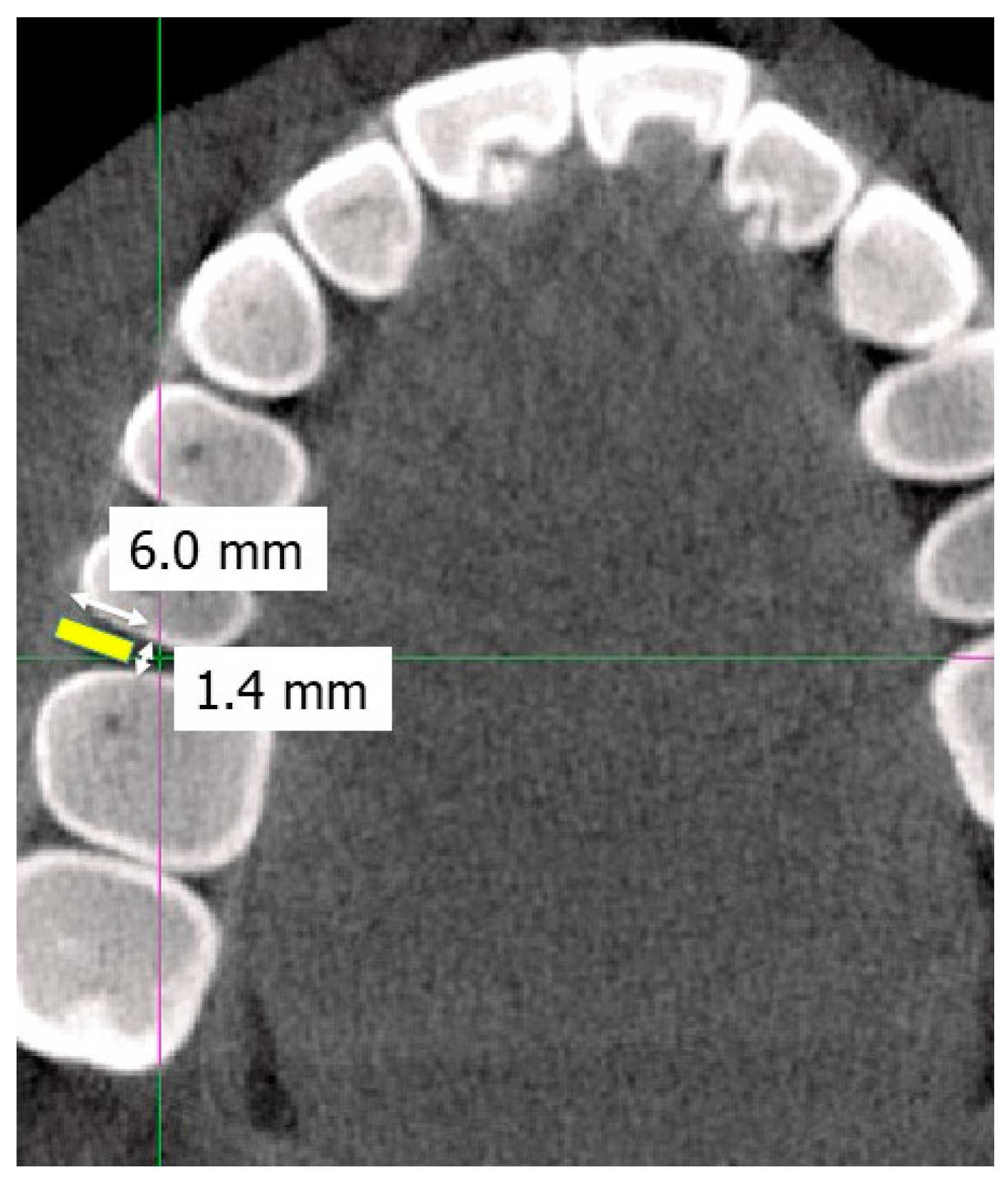
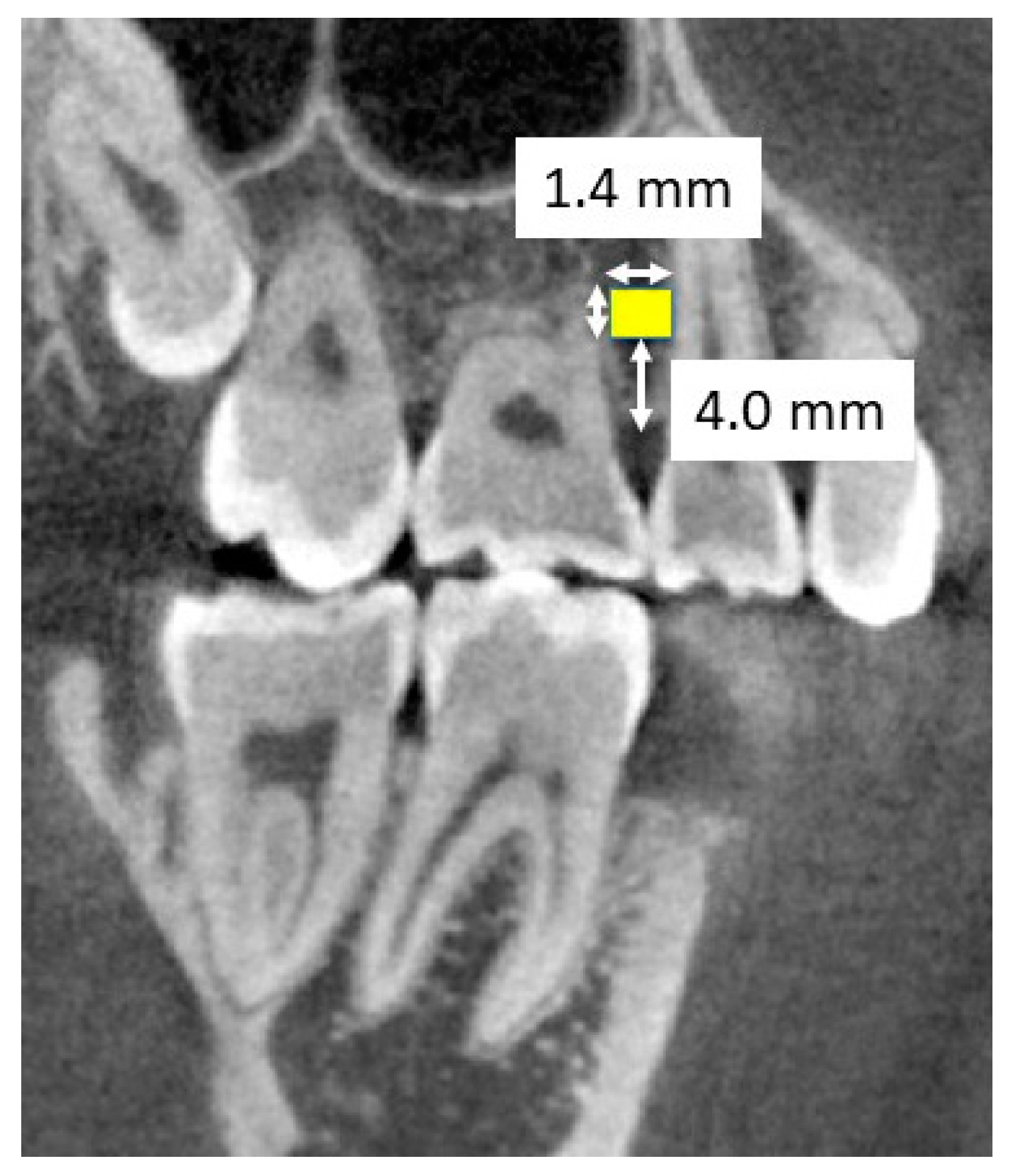
CBCT imaging was performed for each patient under the following conditions: a dental CBCT machine (Finecube, Yoshida Seisakusho Co., Ltd., Tokyo, Japan) was used for imaging in the normal mode (tube voltage, 90 kV; tube current, 4 mA; imaging time, 16.8 s) with the patient’s Frankfurt plane parallel to the floor. CBCT images were recorded with a slice thickness of 0.147 mm, a field of view of 81 × 74 mm, and a voxel size of 0.146 mm. The images were converted to Digital Imaging and Communication in Medicine (DICOM) files, and the DICOM data were transferred to a computer for analysis. DICOM analysis software (OsiriX MD, version 8.0.2, Pixmeo, Switzerland) was used for linear measurements and bone analysis software (TRI/3D-BON, RATOC, Tokyo, Japan) was used for bone mineral density analysis.

2.3. Measurements

The habitual chewing side was identified through a mandibular kinesiograph (MKG) taken before orthodontic treatment, wherein the patient chewed the gum freely (Figure 1). Of the 10 strokes from the 4th to 13th strokes, when ≥70% of the strokes were clearly biased toward one chewing side, that side was defined as the habitual chewing side [26]. Labial cortical bone thickness was measured in a horizontal section at a depth of 4 mm from the top of the alveolar bone where the orthodontic mini-screw was placed (Figure 2). As to trabecular bone analysis, we used trabecular bone analysis software (TRI/3D-BON) according to the manufacturer’s instructions. Alveolar cancellous bone density (mg/cm3) was also measured in the regions of interest set at the site of orthodontic mini-screw implantation in a cube 1.4 mm square at the base and 6 mm high at a depth of 4 mm from the top of the alveolar bone (with the height in the buccolingual direction) under the instructions (Figure 3).
For multivariate analysis, the bone thickness and bone density were grouped equally into three categories in the order of descending and increasing (Grp. 1, Grp. 2, and Grp. 3), respectively. The implantation sites were limited to two locations: between the maxillary second premolar and the maxillary first molar and between the maxillary first molar and the maxillary second molar. The cases were categorized into two groups according to the insertion site. The clinical experience of the orthodontists (1–27 years) who placed the mini-screw was also grouped into three categories. All measurements were performed twice with an interval of 14 days by the same blinded investigator to examine the intraclass correlation coefficient (ICC).

2.4. Statistical Analysis

First, a paired t-test was used to compare the mean values of the cortical bone thickness and bone density between the successful and unsuccessful sides. In our data set, individuals had multiple sites. Therefore, we then conducted multivariable multilevel Poisson regression, considering that the structure of each site was nested in a patient to estimate the prevalence ratio (PR) and its 95% confidence interval (95% CI) for mini-screw failure. The reason why, we applied Poisson regression rather than logistic regression because the prevalence of failure in this study was high, and logistic regression could overestimate the association between cortical bone thickness and bone density on the successful and unsuccessful side [29]. Then, univariate and multivariate analyses were used to examine the association of the habitual chewing side, cortical bone thickness, bone density, implantation site, and years of experience of the orthodontists who placed the mini-screw with the success or failure of the mini-screw. Statistical analysis software STATA v.17 (StataCorp LLC, College Station, TX, USA) and SPSS version 26.0 (IBM Corporation, Armonk, NY, USA) were used for the statistical evaluation. Statistical significance was set at p < 0.05.

3. Results

Our analysis included 43 patients (10 men and 33 women; age range: 18–44 years; mean age: 28.8 ± 11.2 years). ICC was measured twice by the same examiner at 14-day intervals. The ICCs (1.1) for cortical bone thickness on the successful and unsuccessful sides were 0.988 and 0.993, respectively. Likewise, the ICC (1.1) values for alveolar bone density on the successful and unsuccessful sides were 0.999 and 0.999, respectively. These results confirmed the validity of the measurements.
Table 1 and Table S1 present the descriptive statistics for the successful and unsuccessful sides. The cortical bone thickness was 1.01 ± 0.27 mm on the successful side and 0.93 ± 0.27 mm on the unsuccessful side without a significant difference. In addition, Grp. 1 (0.71 ± 0.11 mm, range: 0.47–0.85 mm) had 31 (successful: 11, unsuccessful: 20) sides, Grp. 2 (0.97 ± 0.054 mm, range: 0.86–1.05 mm) had a total of 27 (successful: 17, unsuccessful: 10) sides, and Grp. 3 (1.27 ± 0.20 mm, range: 1.07–1.78 mm) had a total of 28 (successful: 15, unsuccessful: 13) sides (Table S2). There was no significant correlation between mini-screw stability and cortical bone thickness (Table 2).
The bone density was 1317.89 ± 332.23 mg/cm3 on the successful side and 1059.64 ± 202.64 mg/cm3 on the unsuccessful side (Table S1). It was significantly higher on the successful side than on the unsuccessful side. Meanwhile, Grp. 1 (891.72 ± 110.44 mg/cm3, range: 674.7–1073.1 mg/cm3) had 29 sides (successful: 13, unsuccessful: 16); Grp. 2 (1149.22 ± 46.66 mg/cm3, range: 1078.6–1228.9 mg/cm3), 29 (successful: 9, unsuccessful: 20) sides; and Grp. 3 (1506.65 ± 238.91 mg/cm3, range: 1229.4–1910.3 mg/cm3), 28 (successful: 21, unsuccessful: 7) sides (Table S2). The regression analysis shows there was no significant correlation between mini-screw stability and bone density (Table 2).
As for the habitual chewing side, 39 out of 43 patients had a habitual chewing side, while 4 had no chewing preference. Of the 39 patients with a habitual chewing side, 27 had a successful mini-screw on the habitual chewing side, while 12 had an unsuccessful mini-screw on the habitual chewing side. There was a significant association between screw stability and habitual chewing side (Table 2). Compared to the preferred side, the non-preferred side had a 2.25 (95% CI = 1.14–4.442) times significantly higher PR for failure. This association remained significant after all variables were included in the same model (PR = 2.22, 95% CI = 1.12–4.41).
Furthermore, the mini-screw was inserted between the second premolar and first molar in 38 patients and between the first and second molars in 5 patients. There was no significant correlation between screw stability and the implantation site (Table 1 and Table 2).
Based on the orthodontists’ experience of mini-screw insertion, 22 orthodontists were subgrouped into Grp. 1 (minimum 2 years and maximum 3 years), 16 into Grp. 2 (minimum 4 years and maximum 9 years), and 26 into Grp. 3 (>10 years). There was no significant correlation between mini-screw stability and orthodontist experience (Table 1 and Table 2).

4. Discussion

Our null hypothesis was rejected because the multivariate analysis showed that the alveolar bone density on the unsuccessful side was significantly lower than on the successful side, with a significant correlation between the success or failure of the mini-screw and the sidedness of habitual chewing. To our knowledge, our study is the first to elucidate the intra-individual factors using a split-mouth design [30], wherein the orthodontic mini-screws were inserted bilaterally, with mini-screw loss only on one side, which helped diminish the influence of the systemic host factors, the inter-individual factors, that act as confounders. Our findings strongly support those of previous studies that concluded that bone density is related to the stability of mini-screws [8,11,12]. This is also the first study to examine the stability of orthodontic anchor screws in relation to masticatory functions, such as habitual chewing. Therefore, our findings may be meaningful for clinicians.
There was no significant difference in bilateral alveolar cortical bone thickness in the same regions in an individual [31]. In our study, the alveolar cortical bone thickness was 0.97 ± 0.29 mm on the right side and 0.97 ± 0.25 mm on the left side (Table S3), without a significant difference between the two sides, which agrees with a previous study [31]. However, there was a tendency for the successful side to have a larger width diameter of 1.01 ± 0.27 mm, when compared to the 0.93 ± 0.27 mm width diameter on the unsuccessful side. Miyawaki et al. [8] reported that the mini-screw loss rate increased significantly when alveolar bone thickness was less than 1.0 mm. Considering that the mean cortical bone thickness of the unsuccessful side in the present study was 0.93 mm, the sample size should be increased to investigate the cortical bone thickness in detail required for mini-screw stability. The alveolar bone density was significantly greater on the successful side than on the unsuccessful side, suggesting that bone density may be related to mini-screw stability, consistent with a previous study [15]. As alveolar bone density has been correlated with sex, body mass index, and age [32], it is important to investigate and compare other host factors that contribute to mini-screw stability in a larger sample.
Sato et al. [18] reported that bone density decreases when masticatory function weakens, while Loginova et al. [17] reported that bone mineral density becomes significantly greater on the chewing side than on the non-chewing side during gum training. This study focused on habitual chewing, and found that patients whose habitual chewing side was on the successful side were less than those whose habitual chewing side was on the unsuccessful side, as well as those who did not have a habitual chewing side. This suggests that masticatory function may affect bone density [16]. Therefore, when planning orthodontic treatment with mini-screws for patients with an obvious habitual chewing side, special attention is recommended based on our study results. Moreover, patients suspected of having a habitual chewing side according to the questionnaires, oral conditions, and functional examinations, such as the mandibular kinesiograph and electromyography should be encouraged to chew on the non-habitual side of mastication, (e.g., through the elimination of dental caries and gum training) to increase alveolar bone density before mini-screw placement. In addition, changing the implantation site to the palate may be considered when mini-screw insertion into the alveolar bone is unsuccessful [17].
In this study, mini-screws of the same size were placed bilaterally at the same site and in the same patient using motorized screwdrivers. Therefore, we analyzed the results without considering systemic factors, such as age, jaw shape, screw diameter, screw length, and individual techniques [16,18]. However, the present study had several limitations. First, we did not compare cases with bilateral failure and those with bilateral success to further examine the relationship between bone density and mini-screw failure/stability in order to focus on the investigation of the intra-individual factors. Second, because previous studies have shown greater cortical bone thickness and alveolar bone density in the mandible than in the maxilla [12,33,34], a further comparison between the two jaws should also be necessary in future studies. Finally, the CBCT images were used to measure BMD in this clinical study. Although the CBCT examination is often used to evaluate bone quality before dental implantation, it has been reported that the CBCT image has low reliability of reconstructed density values, such as BMD [35].
This study included patients with unilateral failure of mini-screw insertion to assess the contribution of bone quality and habitual chewing to mini-screw stability. Regarding the experience of orthodontists, it has been reported that the number of previous mini-screw placements by orthodontists is not a significant confounding factor for the success rate [36]. Therefore, although doctors’ experience was not considered a factor affecting the success rate in the present study, it should be considered in the future. Moreover, there are reports that alveolar bone density decreases during orthodontic treatment [37,38]. Hence, future studies should evaluate the effect of orthodontic treatment duration on these findings. In addition, the comparison method used in this study employed a split-mouse design. This statistical method allows the exclusion of systemic factors, but does not consider crossover effects. It is necessary to consider the possibility of changes in chewing patterns and the dominant hand of the patients associated with tooth brushing manner during orthodontic treatment.

5. Conclusions

In conclusion, this regression analysis shows that cortical bone thickness, bone density, site of mini-screw implantation, and practitioner experience are not significantly correlated with mini-screw stabilization. Meanwhile, habitual chewing side is related to mini-screw stability.

Supplementary Materials

The following supporting information can be downloaded at: https://www.mdpi.com/article/10.3390/app14073041/s1, Table S1: Descriptive statistics for the successful and unsuccessful sides, Table S2: Descriptive statistics for the divided three groups, Table S3: Descriptive statistics for the right and left sides.

Author Contributions

Conceptualization, M.O.-I., Y.I. and T.O.; methodology, Y.I. and J.A.; investigation, M.O.-I.; resources, A.I.-T. and C.S.-T.; data curation, M.O.-I.; writing—original draft preparation, M.O.-I.; writing—review and editing, Y.I., J.A. and T.O.; project administration, T.O. All authors have read and agreed to the published version of the manuscript.

Funding

This research received no external funding.

Institutional Review Board Statement

Institution Ethical Committee of Tokyo Medical and Dental University (TMDU) (approval numbers: No. D2015-609).

Informed Consent Statement

Informed consent was obtained from all subjects involved in the study.

Data Availability Statement

The raw data supporting the conclusions of this article will be made available by the authors on request.

Conflicts of Interest

The authors declare no conflicts of interest.

References

  1. Li, F.; Hu, H.K.; Chen, J.W.; Liu, Z.P.; Li, G.F.; He, S.S.; Zou, S.J.; Ye, Q.S. Comparison of anchorage capacity between implant and headgear during anterior segment retraction. Angle Orthod. 2011, 81, 915–922. [Google Scholar] [CrossRef] [PubMed]
  2. Park, H.S.; Kwon, T.G.; Sung, J.H. Nonextraction treatment with micro-screw implants. Angle Orthod. 2004, 74, 539–549. [Google Scholar]
  3. Arslan, A.; Nalbantgil Ozdemir, D.; Gursoy-Mert, H.; Malkondu, O.; Sencift, K. Intrusion of an overerupted mandibular molar using mini-screws and mini-screw implants: A case report. Aust. Dent. J. 2010, 55, 457–461. [Google Scholar] [CrossRef] [PubMed]
  4. Chung, K.R.; Kim, S.H.; Chaffee, M.P.; Nelson, G. Molar distalization with a partially integrated mini-implants to correct unilateral Class II malocclusion. Am. J. Orthod. Dentofacial. Orthop. 2010, 138, 810–819. [Google Scholar] [CrossRef] [PubMed]
  5. Janeth, P.; Helen, P.; Manuel, B. Rapid maxillary expansion assisted by mini-implants anchorage: A case report. Int. Orthod. 2019, 17, 159–169. [Google Scholar]
  6. Cousley, R.R.J. Molar intrusion in the management of anterior openbite and ‘high angle’ Class II malocclusions. J. Orthod. 2014, 41, S36–S46. [Google Scholar] [CrossRef]
  7. Ayadi, I.; Dallel, I.; Ben Rejeb, S.; Tobji, S.; Ben Amor, F.; Ben Amor, A. Orthodontic intrusion using mini-screws. Orthod. Fr. 2018, 89, 397–410. [Google Scholar] [CrossRef] [PubMed]
  8. Miyawaki, S.; Koyama, I.; Inoue, M.; Mishima, K.; Sugahara, T.; Takano-Yamamoto, T. Factors associated with the stability of titanium screws placed in the posterior region for orthodontic anchorage. Am. J. Orthod. Dentofacial. Orthop. 2003, 124, 373–378. [Google Scholar] [CrossRef] [PubMed]
  9. Tseng, Y.-C.; Hsieh, C.-H.; Shen, Y.-S.; Huang, I.-Y.; Chen, C.-M. The application of mini-implants for orthodontic anchorage. Int. J. Oral. Maxillofac. Surg. 2006, 35, 704–707. [Google Scholar] [CrossRef]
  10. Kuroda, S.; Sugawara, Y.; Deguchi, T.; Kyung, H.-M.; Takano-Yamamoto, T. Clinical use of miniscrew implants as orthodontic anchorage: Success rates and postoperative discomfort. Am. J. Orthod. Dentofacial. Orthop. 2007, 131, 9–15. [Google Scholar] [CrossRef]
  11. Lee, M.Y.; Park, J.H.; Kim, S.C.; Kang, K.H.; Cho, J.H.; Cho, J.W.; Chang, N.-Y.; Chae, J.-M. Bone density effects on the success rate of orthodontic microimplants evaluated with cone-beam computed tomography. Am. J. Orthod. Dentofacial. Orthop. 2016, 149, 217–224. [Google Scholar] [CrossRef] [PubMed]
  12. Motoyoshi, M.; Yoshida, T.; Ono, A.; Shimizu, N. Effect of cortical bone thickness and implant placement torque on stability of orthodontic mini-implant. Int. J. Oral Maxillofac. Surg. 2007, 22, 779–784. [Google Scholar]
  13. Ashton, K.Y.; Jiang, S.S.; Melo, M.A.; Bosio, J.A. International investigation on temporary anchorage device use: A survey of orthodontists. J. World Fed. Orthod. 2023, 12, 93–104. [Google Scholar] [CrossRef] [PubMed]
  14. Wilmes, B.; Rademacher, C.; Olthoff, G.; Drescher, D. Parameters affecting primary stability of orthodontic mini-implants. J. Orofac. Orthop. 2006, 3, 162–174. [Google Scholar] [CrossRef] [PubMed]
  15. Chen, Y.J.; Chang, H.H.; Huang, C.Y.; Hung, H.C.; Lai, E.H.H.; Yao, C.C.J. A retrospective analysis of the failure rate of three different orthodontic skeletal anchorage systems. Clin. Oral Implant. Res. 2007, 18, 768–775. [Google Scholar] [CrossRef] [PubMed]
  16. Ozdemir, F.; Tozlu, M.; Germec-Cakan, D. Cortical bone thickness of the alveolar process measured with cone-beam computed tomography in patients with different facial types. Am. J. Orthod. Dentofacial. Orthop. 2013, 143, 190–196. [Google Scholar] [CrossRef] [PubMed]
  17. Uesugi, S.; Kokai, S.; Kanno, Z.; Ono, T. Stability of secondarily inserted orthodontic miniscrews after failure of the primary insertion for maxillary anchorage; Maxillary buccal area vs midpalatal suture area. Am. J. Orthod. Dentofacial. Orthop. 2018, 153, 54–60. [Google Scholar] [CrossRef] [PubMed]
  18. Thongudomporn, U.; Chongsuvivatwong, V.; Geater, A. The effect of maximum bite force on alveolar bone morphology. Orthod. Craniofac. Res. 2009, 12, 1–8. [Google Scholar] [CrossRef] [PubMed]
  19. Loginova, N.K.; Veĭsgeĭm, L.D.; Churina, S.V. Influence of course use of chewing gum on alveolar bone density. Stomatologiia 2006, 85, 22–24. [Google Scholar]
  20. Sato, H.; Kawamura, A.; Yamaguchi, M.; Kasai, K. Relationship between masticatory function and internal structure of the mandible based on computed tomography findings. Am. J. Orthod. Dentofacial. Orthop. 2005, 128, 766–773. [Google Scholar] [CrossRef]
  21. Inoue, M.; Ono, T.; Kameo, Y.; Sasaki, F.; Ono, T.; Adachi, T.; Nakashima, T. Forceful mastication activates osteocytes and builds a stout jawbone. Sci. Rep. 2019, 9, 4404. [Google Scholar] [CrossRef] [PubMed]
  22. Pond, L.H.; Barghi, N.; Barnwel, G.M. Occlusion and chewing side preference. J. Prosthet. Dent. 1986, 55, 498–500. [Google Scholar] [CrossRef] [PubMed]
  23. Wilding, R.J.; Lewin, A. A model for optimum functional human jaw movements based on values associated with preferred chewing patterns. Arch. Oral Biol. 1991, 36, 519–523. [Google Scholar] [CrossRef] [PubMed]
  24. Rovira-Lastra, B.; Flores-Orozco, E.I.; Ayuso-Montero, R.; Peraire, M.; Martinez-Gomis, J. Peripheral functional and postural asymmetries related to the preferred chewing side in adults with natural dentition. J. Oral Rehabil. 2016, 43, 279–285. [Google Scholar] [CrossRef]
  25. Matines-Gomis, J.; Lujan-Climent, M.; Palau, S.; Bizar, J.; Salsench, J.; Peraire, M. Relationship between chewing side preference and handedness and lateral asymmetry or peripheral factors. Arch. Oral Biol. 2009, 54, 101–107. [Google Scholar] [CrossRef] [PubMed]
  26. Minato, A.; Ono, T.; Miyamoto, J.J.; Honda, E.-I.; Kurabayashi, T.; Moriyama, K. Preferred chewing side-dependent two-point discrimination and cortical activation pattern of tactile tongue sensation. Behav. Brain Res. 2009, 203, 118–126. [Google Scholar] [CrossRef]
  27. Nissan, J.; Gross, M.D.; Shifman, A.; Tzadok, L.; Assif, D. Chewing side preference as a type of hemispheric laterality. J. Oral Rehabil. 2004, 31, 412–416. [Google Scholar] [CrossRef] [PubMed]
  28. Arai, S.; Kato, C.; Watari, I.; Ono, T. Does Orthodontic Treatment Change the Preferred Chewing Side of Patients with Malocclusion? J. Clin. Med. 2022, 11, 6343. [Google Scholar] [CrossRef] [PubMed]
  29. Barros, A.J.D.; Hirakata, V.N. Alternatives for logistic regression in cross-sectional studies. An empirical comparison of models that directly estimate the prevalence ratio. BMC Med. Res. Methodol. 2003, 3, 21. [Google Scholar] [CrossRef]
  30. Pandis, N.; Walsh, T.; Polychronopoulou, A.; Katsaros, C.; Eliades, T. Split-mouth designs in orthodontics: An overview with applications to orthodontic clinical trials. Eur. J. Orthod. 2013, 35, 783–789. [Google Scholar] [CrossRef]
  31. Deguchi, T.; Nasu, M.; Murakami, K.; Yabuuchi, T.; Kamioka, H.; Takano-Yamamoto, T. Quantitative evaluation of cortical bone thickness with computed tomographic scanning for orthodontic implants. Am. J. Orthod. Dentofacial. Orthop. 2006, 129, 721.e7–721.e12. [Google Scholar] [CrossRef] [PubMed]
  32. Ohiomoba, H.; Sonis, A.; Yansane, A.; Friedland, B. Quantitative evaluation of maxillary alveolar cortical bone thickness and density using computed tomography imaging. Am. J. Orthod. Dentofacial. Orthop. 2016, 151, 82–91. [Google Scholar] [CrossRef]
  33. Park, H.S.; Lee, Y.J.; Jeong, S.H.; Kwon, T.G. Density of the alveolar and basal bone of the maxilla and the mandible. Am. J. Orthod. Dentofacial. Orthop. 2008, 133, 30–37. [Google Scholar] [CrossRef] [PubMed]
  34. Chugh, T.; Ganeshkar, S.V.; Revankar, A.; Jain, A.K. Quantitative assessment of interred bone density in the maxilla and mandibule: Implications in clinical orthodontics. Prog. Orthod. 2013, 14, 38. [Google Scholar] [CrossRef]
  35. Schulze, R.; Heil, U.; Groß, D.; Bruellmann, D.; Dranischnikow, E.; Schwanecke, U.; Schoemer, E. Artefacts in CBCT: A review. Dentomaxillofac. Radiol. 2011, 40, 265–273. [Google Scholar] [CrossRef] [PubMed]
  36. Lim, H.J.; Eun, C.S.; Cho, J.H.; Lee, K.H.; Hwang, H.S. Factors associated with initial stability of miniscrews for orthodontic treatment. Am. J. Orthod. Dentofacial. Orthop. 2009, 136, 236–242. [Google Scholar] [CrossRef] [PubMed]
  37. Yu, J.H.; Huang, H.L.; Liu, C.F.; Wu, J.; Li, Y.F.; Tsai, M.T.; Hsu, J.T. Does Orthodontic Treatment Affect the Alveolar Bone Density? Medicine 2016, 95, e3080. [Google Scholar] [CrossRef]
  38. Chang, H.W.; Huang, H.L.; Yu, J.-H.; Hsu, J.-T.; Li, Y.-F.; Wu, Y.-F. Effect of orthodontic tooth movement on alveolar bone density. Clin. Oral Investig. 2012, 16, 679–688. [Google Scholar] [CrossRef]
Figure 1. Determination of the habitual chewing side by a mandibular kinesiograph. Of the 10 strokes from the 4th to 13th strokes, when 70% or more of the strokes were biased to one side, that side was defined as the habitual chewing side. (A) Sagittal view; (B) frontal view; closing (red), opening (blue/green).
Applsci 14 03041 g001
Figure 2. Alveolar bone density measurement on a horizontal plane section of a cone beam computed tomography image. The horizontal plane section was taken parallel to the occlusal plane.
Applsci 14 03041 g002
Figure 3. Alveolar bone density measurement on a sagittal plane section of a cone beam computed tomography image. The sagittal plane section was parallel to the axis of the upper second premolar.
Applsci 14 03041 g003
Table 1. Cross table with comparison of the successful and unsuccessful sides in patients with unilateral mini-screw failure.
SuccessUnsuccessfulTotal (n)
Habitual chewing (n)434386
Preferred side (%)69.230.839
Non-preferred side (%)30.869.239
No preference (%)50508
Cortical bone thickness (n)434386
Grp. 1 (%)35.564.531
Grp. 2 (%)633727
Grp. 3 (%)53.646.428
Bone density (n)434386
Grp. 1 (%)44.855.229
Grp. 2 (%)316929
Grp. 3 (%)752528
Insertion site (n)434386
Between U5 and U6 (%)505076
Between U6 and U7 (%)505010
Years of experience (n)434386
Grp. 1 (%)505044
Grp. 2 (%)505016
Grp. 3 (%)505026
Abbreviations: U5, upper second premolar; U6, upper first molar; U7, upper second molar.
Table 2. Multilevel Poisson regression analysis of the association of each factor with unilateral mini-screw loss. *: p < 0.05.
UnivariateMultivariate
Failure Rate (%)95% CI95% CI
PRMinMaxp-ValuePRMinMaxp-Value
Habitual chewing
Preferred side30.8111 111
Non-preferred side69.22.251.144.442* 0.0192.221.124.41* 0.02
No preference501.6250.5245.0390.41.40.444.40.57
Cortical bone thickness
Grp. 164.5111 111
Grp. 237.10.5740.2691.2260.1520.650.291.480.31
Grp. 346.40.720.3581.4470.3560.750.361.570.45
Bone density
Grp. 155.2111 111
Grp. 2691.250.6482.4120.5061.060.512.20.89
Grp. 3250.4530.1861.1010.0810.40.161.010.05
Insertion site
Between U5 and U650111 111
Between U6 and U75010.3942.54110.271.980.54
Years of experience
Grp. 150111 111
Grp. 25010.4452.24611.040.442.430.94
Grp. 35010.5041.98511.230.62.510.57
Cons 0.450.21.020.06
Abbreviations: CI, confidence interval; PR, prevalence ratio; min, minimum; max, maximum.
Disclaimer/Publisher’s Note: The statements, opinions and data contained in all publications are solely those of the individual author(s) and contributor(s) and not of MDPI and/or the editor(s). MDPI and/or the editor(s) disclaim responsibility for any injury to people or property resulting from any ideas, methods, instructions or products referred to in the content.

