Article

The Efficacy of Utilizing Platelet-Rich Fibrin for Managing Periodontal Intrabony Defects in Conjunction with Graft Material: A Systematic Review and Meta-Analysis

1
Department of Oral and Maxillofacial Surgery, Seoul St. Mary’s Hospital, College of Medicine, The Catholic University of Korea, Seoul 06591, Republic of Korea
2
Department of Orthodontics, Seoul St. Mary’s Hospital, College of Medicine, The Catholic University of Korea, Seoul 06591, Republic of Korea
3
Medical Library, College of Medicine, The Catholic University of Korea, Seoul 06591, Republic of Korea
4
Department of Periodontics, College of Medicine, The Catholic University of Korea, Seoul 06591, Republic of Korea
5
Dental Implantology, Graduate School of Clinical Dental Science, The Catholic University of Korea, Seoul 06591, Republic of Korea
6
Department of Medicine, Graduate School, The Catholic University of Korea, Seoul 06591, Republic of Korea
*
Author to whom correspondence should be addressed.
These authors contributed equally to this work.
Appl. Sci. 2024, 14(8), 3371; https://doi.org/10.3390/app14083371
Submission received: 6 March 2024 / Revised: 6 April 2024 / Accepted: 14 April 2024 / Published: 17 April 2024

Abstract

:
Platelet-rich fibrin has become increasingly popular in recent years due to its remarkable capacity to accelerate the post-surgery wound healing process, reduce inflammation, and promote tissue repair. This study aimed to perform a meta-analysis to evaluate the effect of platelet-rich fibrin in mixture form with bone substitute, as a membrane, or in combination. A comprehensive search using a combination of controlled vocabulary (MeSH) and free-text terms was undertaken by two reviewers to identify published randomized clinical trials. Three major electronic databases (Medline via PubMed, Cochrane database, and Embase) and the clinical trials registry (clinicaltrials.gov) were searched up to 9 July 2023. The results of the meta-analysis showed that the pooled standardized mean difference of probing depth for platelet-rich fibrin was 0.61 (95% CI, 0.33 to 0.88). The results of the meta-analysis showed that the mean difference in clinical attachment level for platelet-rich fibrin was 0.68 (95% CI, 0.35 to 1.01). The results of the meta-analysis showed that the mean difference in bone fill for platelet-rich fibrin was 0.50 (95% CI, 0.23 to 0.78). In conclusion, the study found that platelet-rich fibrin was effective as adjunct to periodontal regeneration.

1. Introduction

Platelet-rich fibrin and similar biological products are obtained from the plasma portion of one’s own blood, exhibiting a platelet concentration higher than that found in the initial blood sample [1]. Platelet-rich fibrin is distinguished from other platelet concentrates, such as platelet-rich plasma and platelet-rich growth factors, by its distinct preparation process and clinical applicability [2]. Platelet-rich fibrin does not require the addition of anticoagulants or thrombin during its preparation when compared with platelet-rich plasma and platelet-rich growth factors [3]. Platelet-rich fibrin has become increasingly popular in recent years due to its remarkable capacity to accelerate the post-surgery wound healing process, reduce inflammation, and promote tissue repair [4,5]. Platelet-rich fibrin finds extensive applications in dentistry, with dental implantology, oral and maxillofacial surgery, endodontics, and cosmetic dentistry witnessing a surge in its popularity [6]. The use of platelet-rich fibrin, with or without biomaterials, demonstrated superior efficacy compared to open flap debridement [7]. There was a notable enhancement in clinical indications when platelet-rich fibrin was combined with demineralized bone matrix, as opposed to using platelet-rich fibrin alone [7,8]. Additionally, the group receiving both platelet-rich fibrin and demineralized bone matrix displayed a greater degree of bone fill on radiographs [9]. Platelet-rich fibrin offers the benefit of maintaining a continuous release of growth factors and facilitating cell migration by stimulating the expression of type I collagen and transforming growth factor mRNA [10]. The secretomes of platelet-rich fibrin represent an innovative growth factor-based approach for promoting bone and periodontal regeneration, and differentially expressed proteins have been identified from the platelet-rich fibrin, including key growth factors, cytokines, and components of the extracellular matrix that play crucial roles in the process of wound healing [11]. The proliferation of human gingival fibroblast cells exhibited a direct correlation with the concentration of platelet-rich fibrin, demonstrating a dose-dependent effect [12].
Hence, the impact of platelet-rich fibrin on periodontal regeneration has remained a topic of continuous discussion. The rationale behind the functioning of platelet-rich fibrin can be elucidated as follows: The reasoning behind the use of platelet-rich fibrin lies in its role as a biomaterial, facilitating the delivery of essential growth factors and cytokines sourced from platelet granules to the specific area of focus, and this process, in turn, fosters tissue regeneration across a range of tissues, including bone regeneration [13]. In addition to utilizing platelet-rich fibrin and graft materials in managing periodontal intrabony defects, a range of other treatment methods, such as targeted delivery of antibiotics, the application of laser therapy, and the use of various medications, can be employed for the treatment of periodontal disease [14,15]. As a result of its additional benefits, platelet-rich fibrin appears to be a naturally appropriate complement in bone regenerative surgery, leading to positive results with minimal associated risks [16]. Therefore, this study aims to perform a meta-analysis to evaluate the effect of platelet-rich fibrin in mixture form with bone substitute, as a membrane, or in combination. The null hypothesis posited that there would be no noteworthy distinction in the effect of platelet-rich fibrin on periodontal regeneration. The primary goal of this research is to assess the extent of periodontal regeneration that can be attained with platelet-rich fibrin by examining improvements in reductions in periodontal probing, clinical attachment levels, and the attainment of bone fill. Additionally, the secondary objective is to carry out a subgroup analysis that categorizes the findings based on the type of graft material used.

2. Materials and Methods

2.1. Protocol and Registration

This present systematic review adheres to the guidelines outlined in the Preferred Reporting Items for Systematic Reviews and Meta-Analysis (PRISMA) Statement [17], as outlined in reference.

2.2. Eligibility Criteria

Question: Does the application of platelet-rich fibrin enhance periodontal regeneration when compared to a group where it is not applied?
Participants: Patients with intrabony defects.
Interventions: Surgical procedures involving a flap, with the addition of platelet-rich fibrin.
Comparisons: Periodontal regeneration without the application of platelet-rich fibrin.
Outcomes: Pre-operative and 6-month post-operative periodontal probing depth, clinical attachment level, and bone fill.
Study design: Randomized controlled trials.
The following criteria were required for studies to be included in this study: (1) studies with randomized controlled trials; (2) studies involving the use of platelet-rich fibrin in the management of periodontal intrabony defects; and (3) studies making a comparison between interventions and control groups. We excluded the following from our study: in vitro and animal studies, studies with a sample size of less than six, studies that involved additional procedures such as LASER application to enhance healing, literature reviews, case reports, case-control study designs, retrospective study designs, and studies published in languages other than English.

2.3. Sources of Information and Search Methodology

A single reviewer (NJK, library-affiliated searching personnel) conducted an extensive search using a combination of controlled vocabulary (MeSH) and free-text terms to locate published systematic reviews. The reviewer (SHH) conducted searches on three major electronic databases (Medline via PubMed, Cochrane database, and Embase), and another reviewer (WJP) conducted a search on the clinical trials registry known as clinicaltrials.gov up to 9 July 2023. The search results were meticulously transferred to the EndNote reference management software (Version 21, Clarivate, Philadelphia, PA, USA) for a thorough deduplication process. This step was essential to ensure that no duplicate entries or redundant references would compromise the integrity of the research findings. To maximize the accuracy and relevance of our search, the search strategy employed was thoughtfully customized to align with the distinct criteria and nuances of each database under consideration. This strategic adaptation allowed us to effectively harness the full potential of each database, optimizing the retrieval of pertinent and valuable information for our research objectives. Details of the search strategy are shown in Supplementary Tables S1–S3.

2.4. Assessment of the Risk of Bias

The reviewers employed the Cochrane Risk-Of-Bias (ROB 2.0) tool for randomized studies, which comprises a checklist with questions pertaining to various aspects of bias. This checklist covers aspects such as the randomization process (selection bias), deviations from the intended interventions (performance bias), missing outcome data (attrition bias), measurement of the outcome (detection bias), selection of the reported result (reporting bias), and an overall assessment of bias. The risk of bias for the included studies was assessed and categorized as low risk, some concerns, or high risk. The evaluation of study quality was conducted by two reviewers, namely WJP and SHH.

2.5. Missing Data Imputation

Both the mean change in variables and the standard deviation of this change need to be imputed if they are missing. The missing mean change in variables was filled by calculating the difference between the mean follow-up and mean baseline data. We used the formula from the Cochrane Handbook to estimate missing standard deviations [18]. The data from the selected papers, where the mean and standard deviation for baseline, follow-up, and change were all provided, were used to calculate the mean correlation for the control group. The obtained correlation values were 0.65 for probing depth, 0.8 for clinical attachment level, and 0.85 for bone gain, and these values were used to estimate missing standard deviations.

2.6. Synthesis and Analysis of Data

We conducted a meta-analysis utilizing R (Version 3.5.0; R Project for Statistical Computing). Summary statistics were represented by the mean difference and a 95% confidence interval. A random-effects model was employed for the meta-analysis, and the significance level was set at 0.05. To assess heterogeneity across studies, we calculated I2 and performed a chi-square test.

3. Results

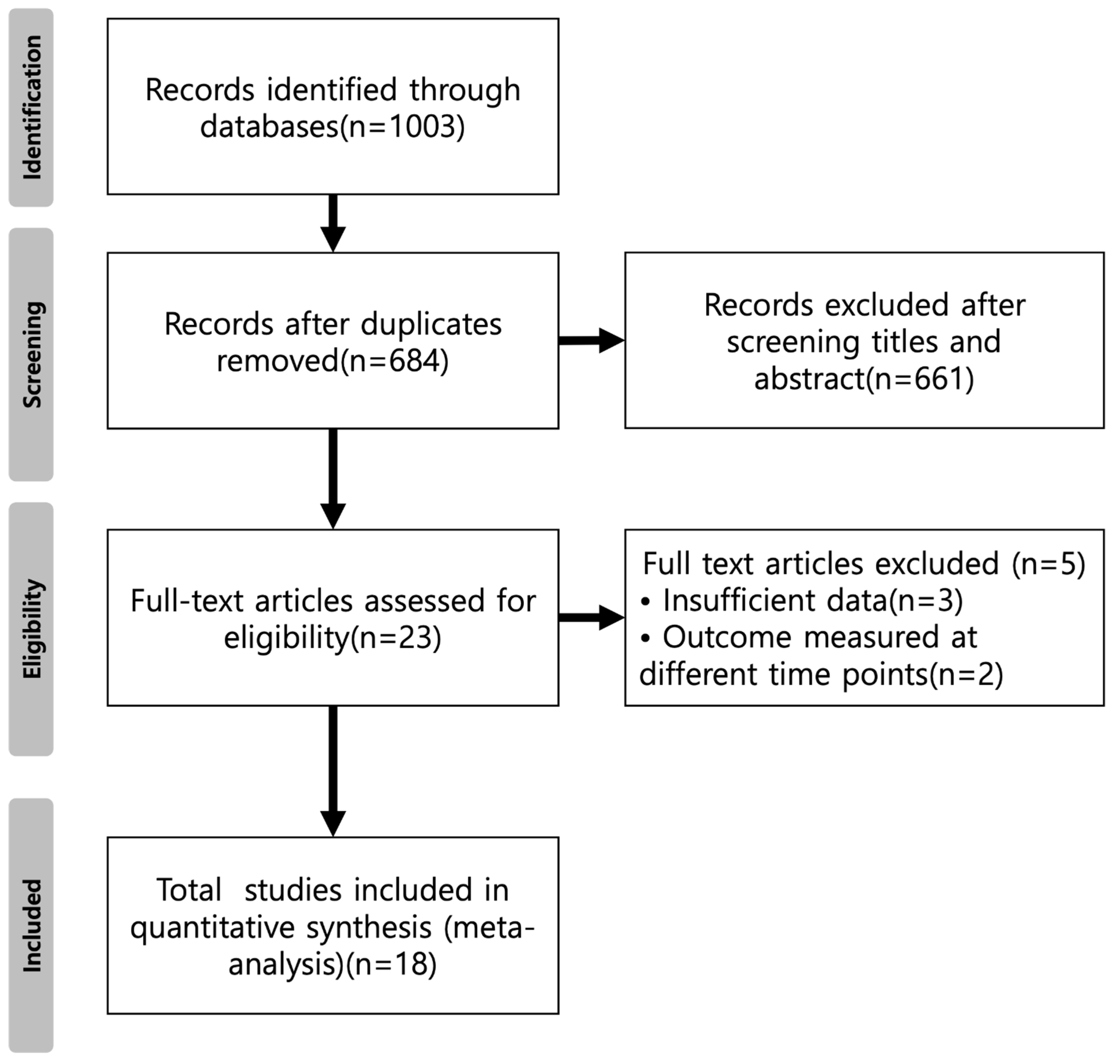
3.1. Study Selection
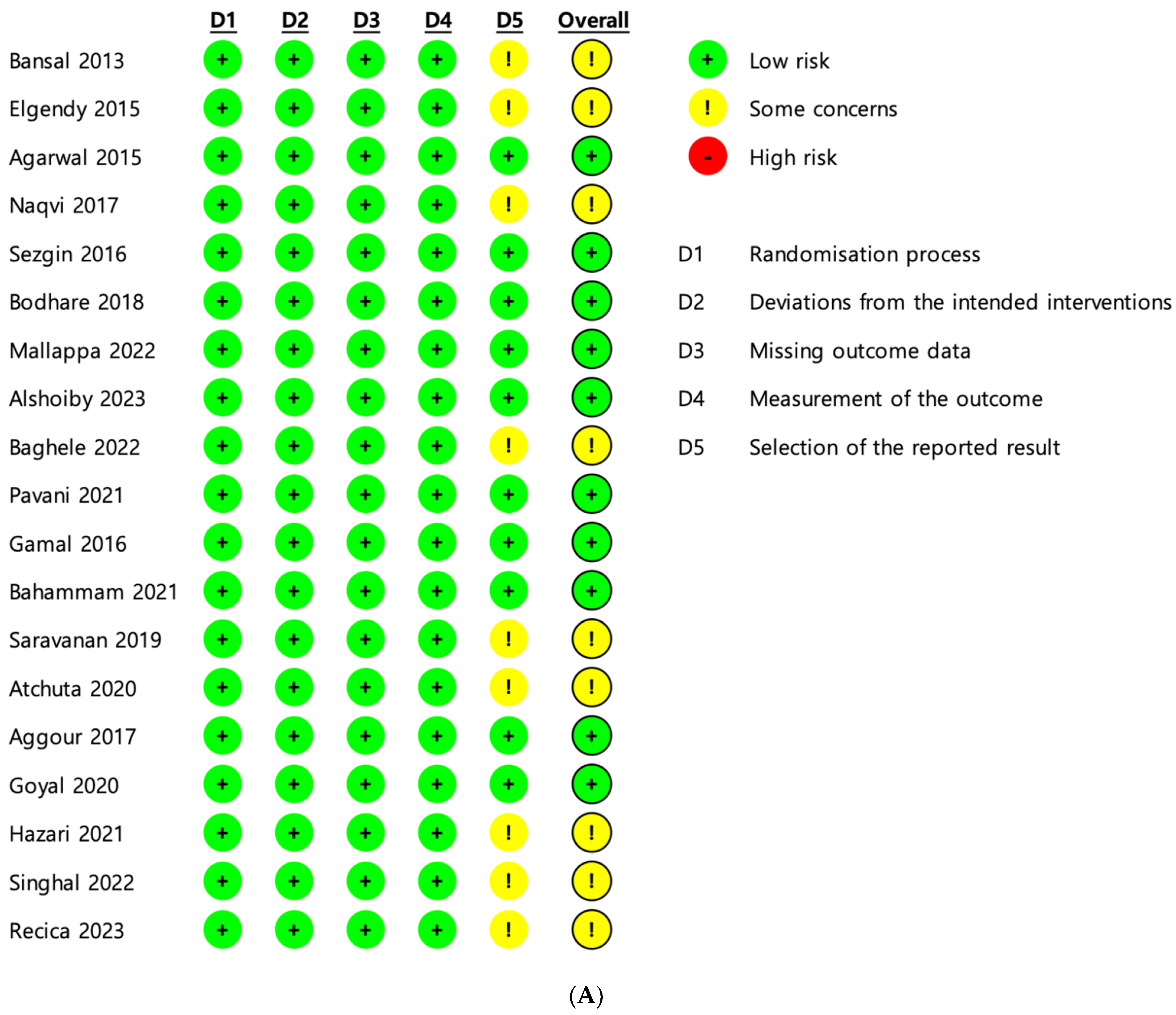
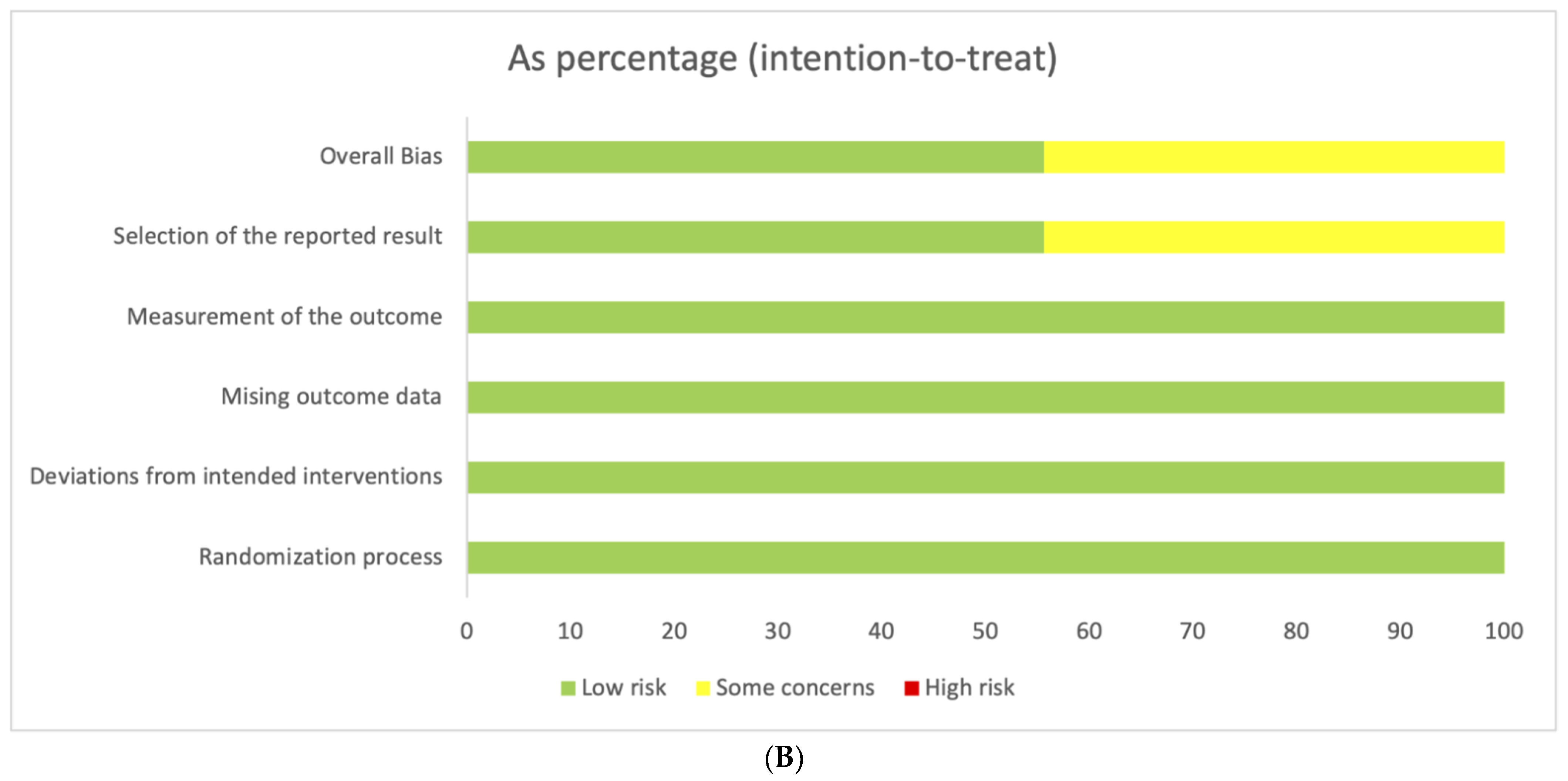
The initial search yielded 1003 articles, and after removing 661 duplicates, 684 studies were taken into consideration. The abstracts were reviewed by two independent reviewers (WJP and SHH), finding that 661 articles did not meet the inclusion criteria. Subsequently, the inclusion and exclusion criteria were applied to the 23 full-text articles, leading to the exclusion of 5 that did not meet the inclusion criteria. Ultimately, 18 studies were assessed for eligibility. A visualization of this literature-screening process is shown in Figure 1.

3.2. Risk of Bias

The overview of bias risk and the overall bias score for each domain within the included articles are displayed in Figure 2A,B. Out of the total, nine trials were categorized as having a low risk of bias, while nine raised some concerns regarding bias. Most of the trials employed an appropriate randomization process. Nevertheless, in nine studies, the reported results were influenced by an inadequate selection of reported results. Figure 2B illustrates the collective bias risk score across all fields.

3.3. Meta-Analysis

Table 1 shows the main characteristics of the included studies. Periodontal regeneration without the use of platelet-rich fibrin was considered the control group. Supplementary Table S4 lists the outcome parameters of the included studies, including periodontal probing depth, clinical attachment level, and bone filling.

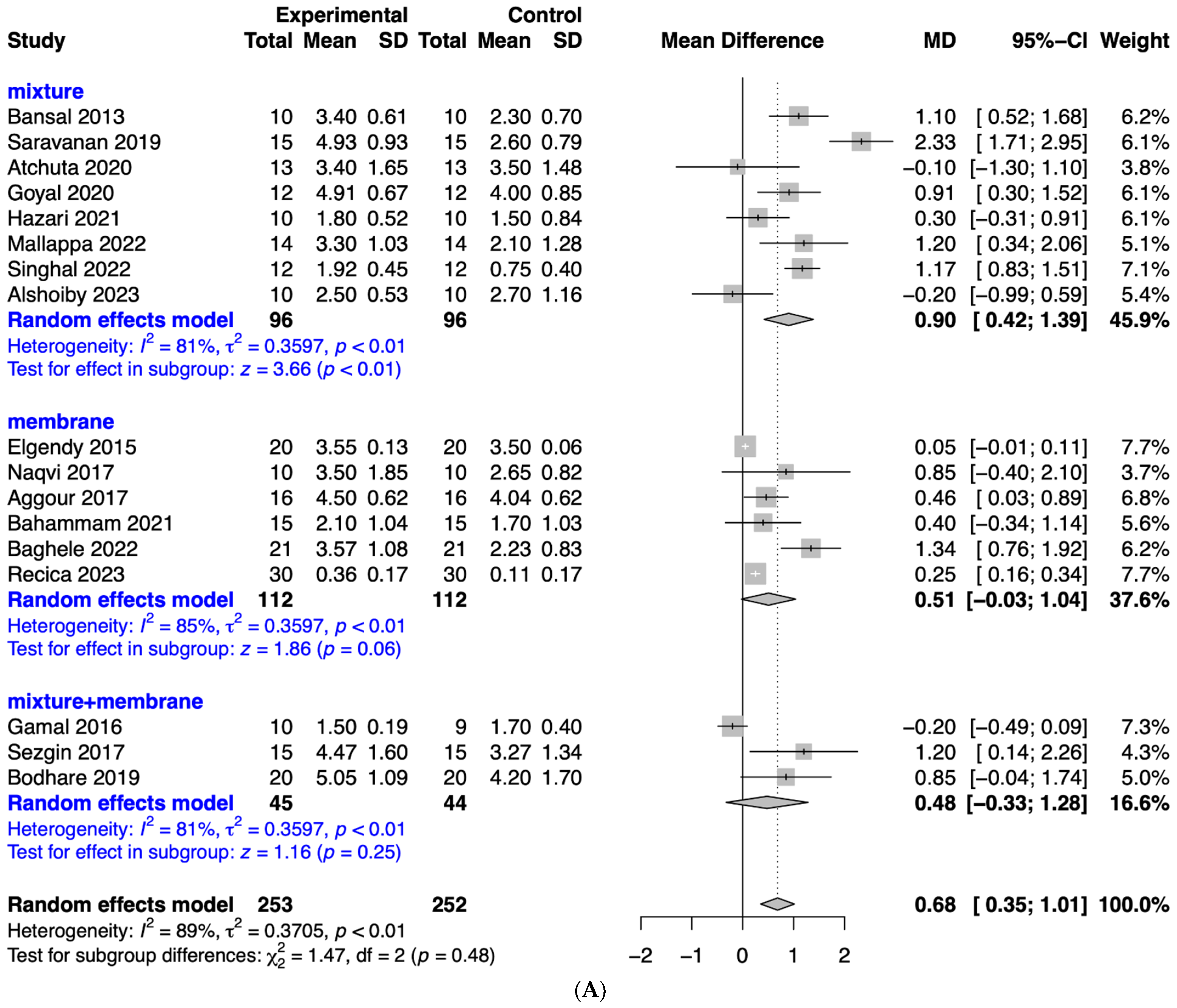
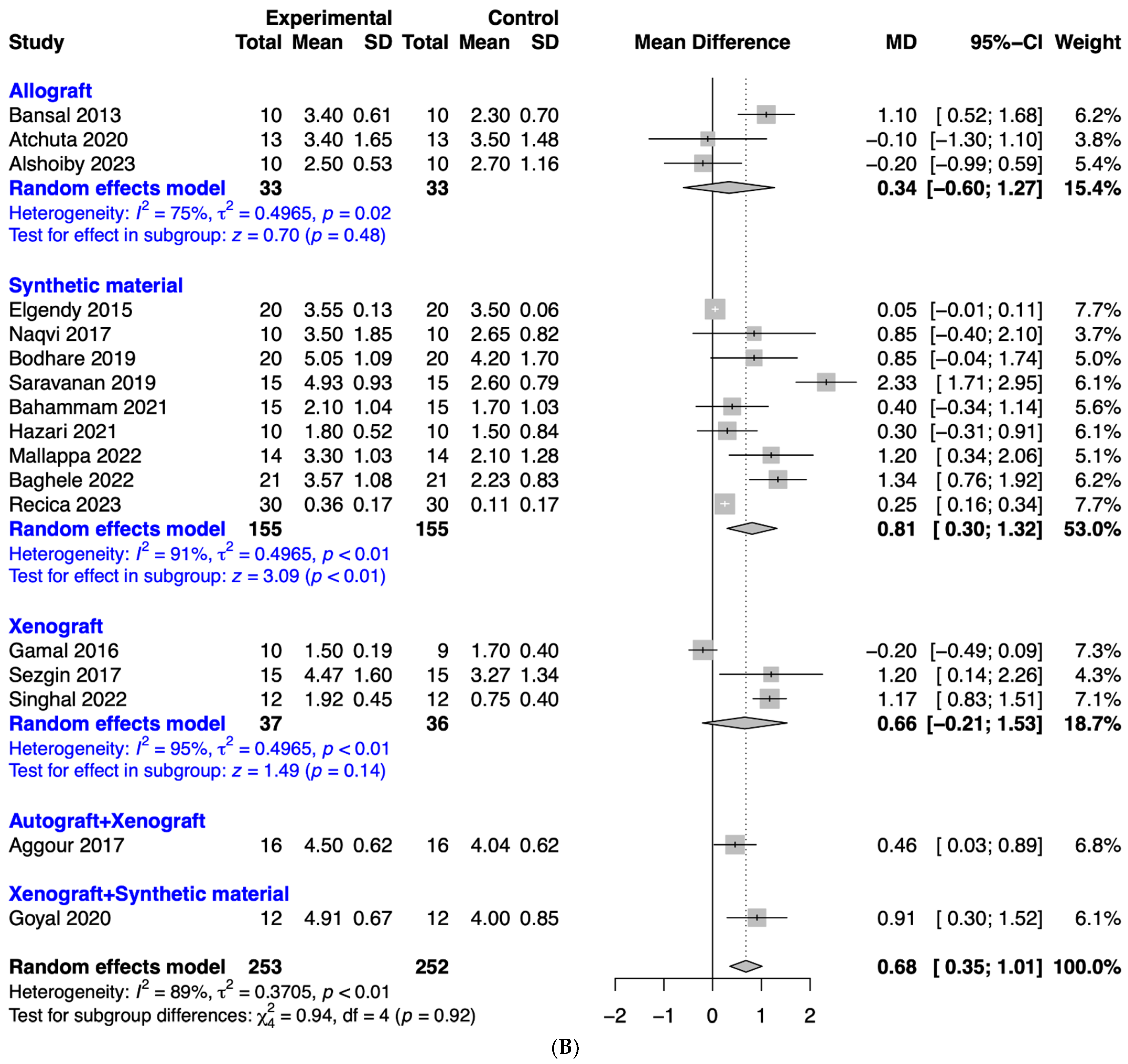
3.4. Evaluation of the Effects of Platelet-Rich Fibrin on Periodontal Probing Depth

Figure 3A provides a forest plot illustrating the comparison between platelet-rich fibrin and control for reduction in periodontal probing depth. This forest plot serves as a valuable tool for comprehending the nuances of the research findings. The results of the meta-analysis showed that the mean difference in probing depth for platelet-rich fibrin was 0.61 (95% confidence interval, 0.33 to 0.88). Subgroup analysis categorized by application method showed that platelet-rich fibrin, which is diced and mixed with the graft material, resulted in a mean difference of probing depth of 0.88 (95% confidence interval, 0.53 to 1.23; p < 0.01). The mean difference in probing depth for platelet-rich fibrin applied as a membrane was 0.34 (95% confidence interval, −0.07 to 0.76; p = 0.11). Platelet-rich fibrin applied as a mixture with the graft material and additional application as a membrane showed the mean difference in probing depth for platelet-rich fibrin was 0.31 (95% confidence interval, −0.31 to 0.93; p = 0.32). Subgroup analysis categorized by application method did not reach statistical significance.
Figure 3B shows a subgroup analysis categorized by types of graft material. The results of the meta-analysis showed that the mean difference in probing depth for platelet-rich fibrin applied with allograft was 0.42 (95% confidence interval, −0.34 to 1.18; p = 0.28). Subgroup analysis categorized by graft material of synthetic material resulted in the mean difference of probing depth for platelet-rich fibrin being 0.78 (95% confidence interval, 0.39 to 1.16; p < 0.01). Platelet-rich fibrin applied with xenograft showed the mean difference in probing depth for platelet-rich fibrin was 0.46 (95% confidence interval, −0.22 to 1.14; p = 0.18). Subgroup analysis categorized by types of graft material did not reach a significant difference (p = 0.68).

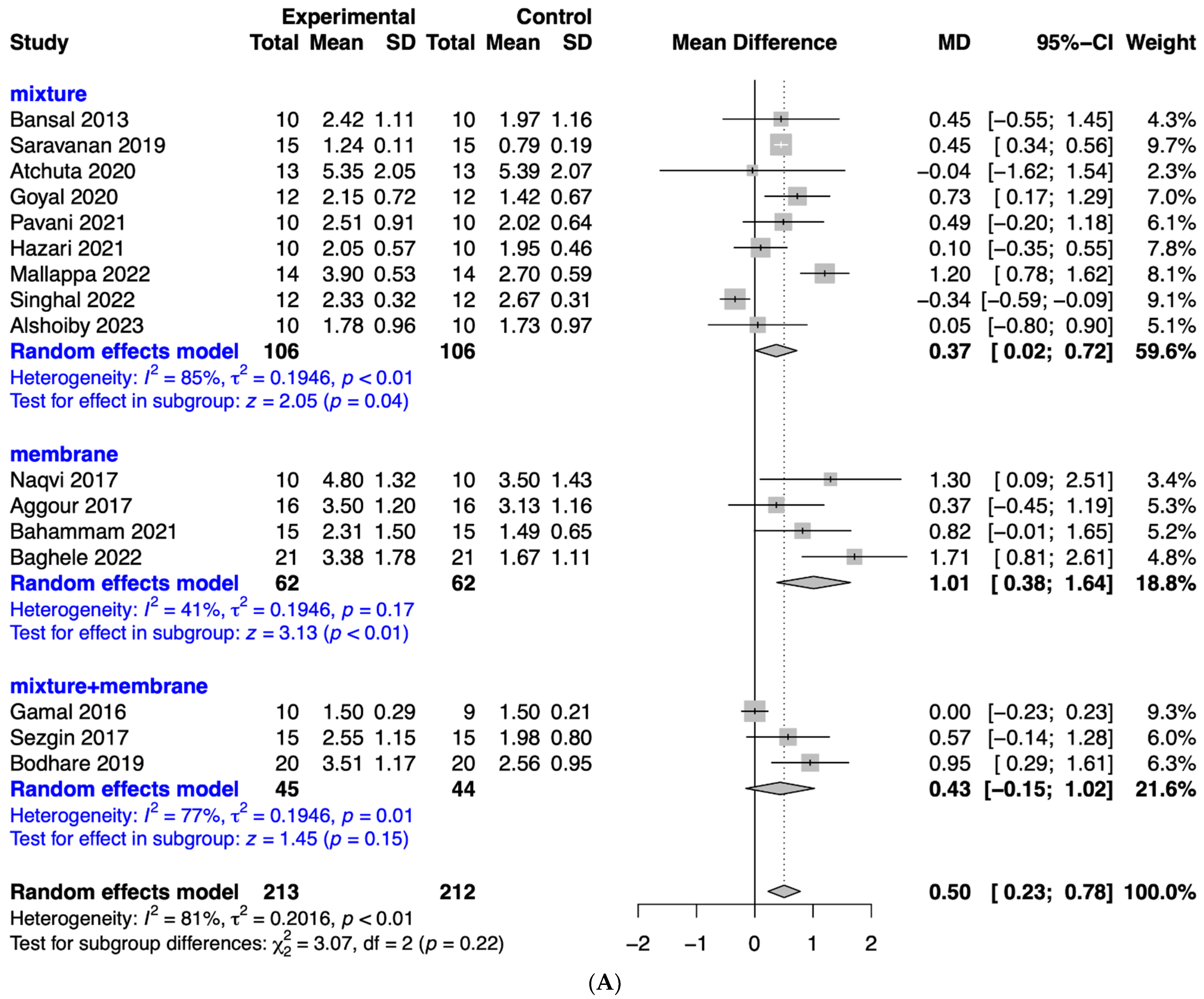
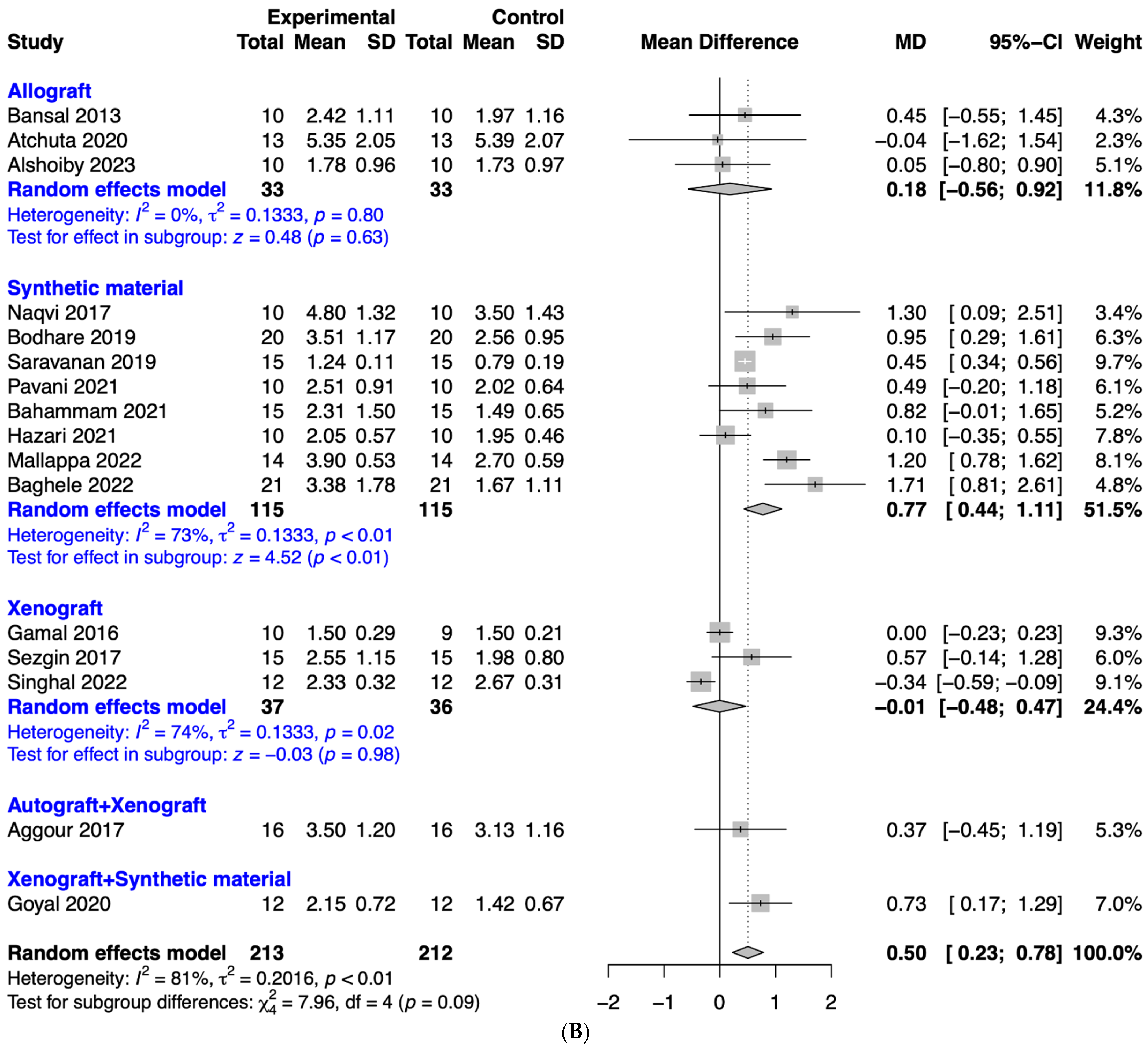
3.5. Analysis of the Effects of Platelet-Rich Fibrin on Clinical Attachment Level

Figure 4A shows a forest plot meticulously crafted to provide a visual representation of the comparison between platelet-rich fibrin and the control group concerning their impact on enhancing clinical attachment level. The analysis has yielded a mean difference of 0.68, with a 95% confidence interval ranging from 0.35 to 1.01. This statistically robust finding underscores the potential clinical benefits associated with the utilization of platelet-rich fibrin in improving clinical attachment levels. Subgroup analysis categorized by application method showed that platelet-rich fibrin, which is diced and mixed with the graft material, resulted in a mean difference in clinical attachment level for platelet-rich fibrin of 0.90 (95% confidence interval, 0.42 to 1.39; p < 0.01). The mean difference in clinical attachment level for platelet-rich fibrin applied as a membrane was 0.51 (95% confidence interval, −0.03 to 1.04; p = 0.06). Platelet-rich fibrin applied as a mixture with the graft material and additional application as a membrane showed the mean difference in clinical attachment level for platelet-rich fibrin was 0.48 (95% confidence interval, −0.33 to 1.28; p = 0.25).
Figure 4B shows a subgroup analysis categorized by types of graft material. The results of the meta-analysis showed that the mean difference in clinical attachment level for platelet-rich fibrin for allografts was 0.34 (95% confidence interval, −0.60 to 1.27; p = 0.48). Subgroup analysis categorized by graft material of synthetic material resulted in the mean difference of clinical attachment level for platelet-rich fibrin being 0.81 (95% confidence interval, 0.30 to 1.32; p < 0.01). The mean difference in clinical attachment level for platelet-rich fibrin applied as a membrane was 0.66 (95% confidence interval, −0.21 to 1.53; p = 0.14).

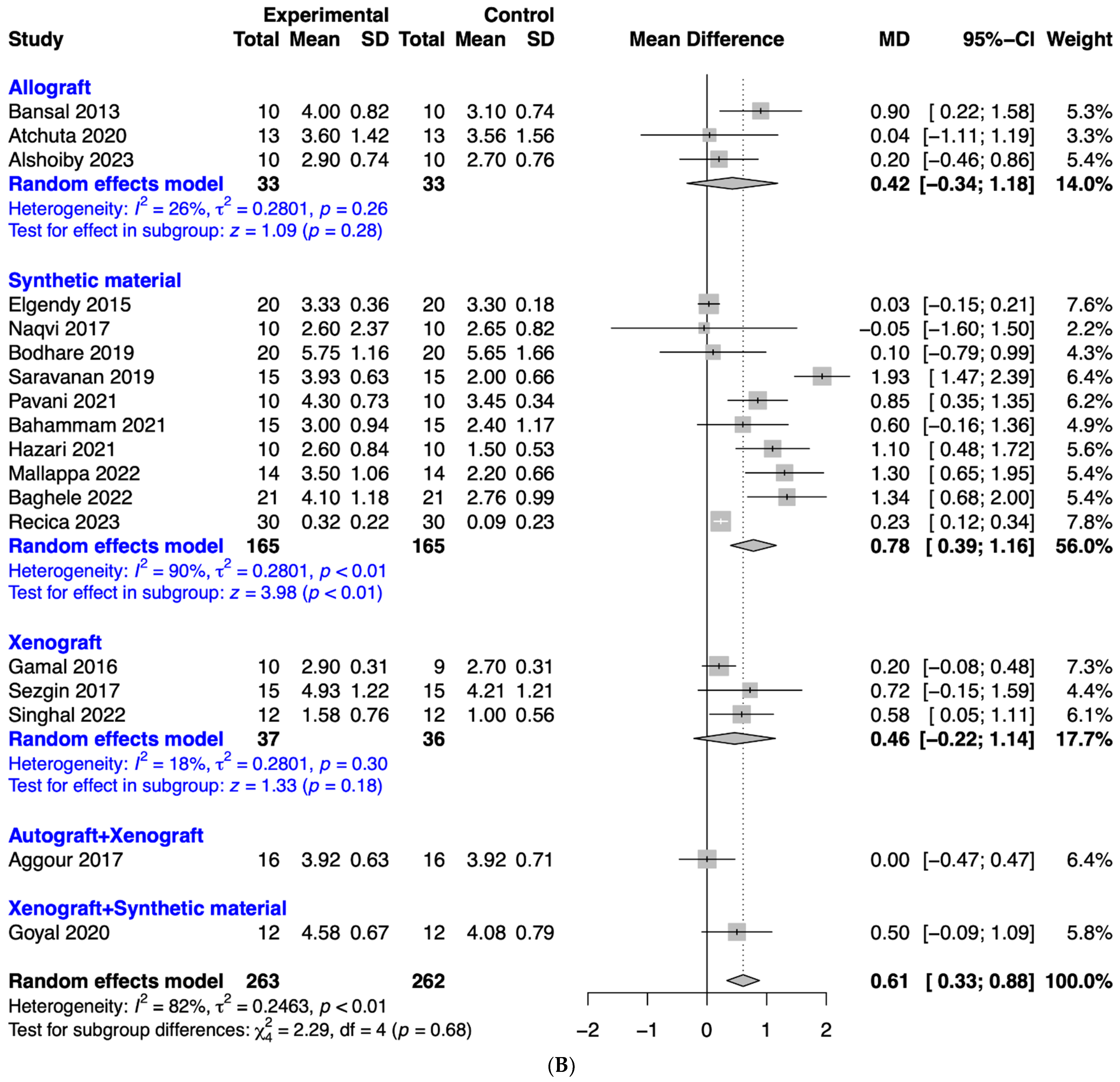
3.6. Evaluation of the Effects of Platelet-Rich Fibrin on Bone Fill

In Figure 5A, a forest plot that visually represents the comparison between platelet-rich fibrin and the control group in terms of achieving bone fill is presented. The mean difference, a robust marker of the efficacy of platelet-rich fibrin, stands resolute at an impressive 0.50. This value, encased within the reassuring confines of a 95% confidence interval that stretches from 0.23 to 0.78, signifies a noteworthy effect size. The magnitude of this finding cannot be overstated, for it points to a clear and statistically significant advantage in favor of platelet-rich fibrin in the realm of achieving bone fill. Subgroup analysis categorized by application method showed that platelet-rich fibrin, which is diced and mixed with the graft material, resulted in a mean difference of 0.37 (95% confidence interval, 0.02 to 0.72; p = 0.04). The mean difference in bone fill for platelet-rich fibrin applied as a membrane was 1.01 (95% confidence interval, 0.38 to 1.64; p < 0.01). Platelet-rich fibrin applied as a mixture with the graft material and additional application as a membrane showed the mean difference in bone fill for platelet-rich fibrin was 0.43 (95% confidence interval, −0.15 to 1.02; p = 0.15). Subgroup analysis categorized by application method did not reach statistical significance (p = 0.22).
Figure 5B shows a subgroup analysis categorized by types of graft material. The results of the meta-analysis showed that the mean difference in bone fill for platelet-rich fibrin applied with allograft was 0.18 (95% confidence interval, −0.56 to 0.92; p = 0.63). Subgroup analysis categorized by graft material of synthetic material resulted in the mean difference of bone fill for platelet-rich fibrin being 0.77 (95% confidence interval, 0.44 to 1.11; p < 0.01). Platelet-rich fibrin applied with xenograft showed the mean difference in bone fill for platelet-rich fibrin was −0.01 (95% confidence interval, −0.48 to 0.47; p = 0.98). However, subgroup analysis categorized by types of graft material did not reach significant difference (p = 0.09).

4. Discussion

In this study, a meta-analysis was conducted to analyze the effect of platelet-rich fibrin in mixture form with bone substitute, as a membrane, or in combination. The study demonstrated the effectiveness of platelet-rich fibrin as a valuable addition to periodontal regeneration procedures.
In general, this meta-analysis demonstrated that platelet-rich fibrin offered substantial advantages in periodontal regeneration, as evidenced by a reduction in probing depth, an improvement in clinical attachment level, and an enhancement of bone fill. The application of a mixture, membrane, or a combination of both produced varying outcomes, which need to be considered clinically. The importance of customizing platelet-rich fibrin applications for particular clinical situations becomes evident due to the variability observed and the need to achieve optimal healing and regeneration. The utilization of platelet-rich fibrin in a mixture could potentially provide significant advantages due to its increased adaptability and interaction with the treatment site. This allows for a more direct and concentrated release of growth factors and cytokines, which are crucial for tissue regeneration. The use of platelet-rich fibrin as a membrane may be limited by its physical form, which could impede its ability to integrate and interact with the host tissue, potentially reducing its effectiveness. The application of the combined approach did not produce enhanced effects. Similarly, previous research indicated that the pairing of bone grafting material and enamel matrix derivative did not offer any additional advantages compared to using enamel matrix derivative alone [37,38].
Different outcomes emerged from using various bone graft materials, such as synthetic materials, allografts, and xenografts. The employment of synthetic materials as the graft choice yielded the most favorable outcomes when applied with platelet-rich fibrin. A previous report demonstrated the significance of choosing appropriate graft materials for successful treatment [39]. Synthetic materials are inorganic, biocompatible substitutes designed to mimic the properties of natural bone, effectively filling and repairing bone defects [40]. Indeed, beyond biocompatibility, the versatility of synthetic materials lies in their ability to be tailored in terms of pore diameter, porosity, and interconnectivity [41]. These modifications can significantly influence the material’s integration with surrounding tissues, promoting better bone ingrowth and vascularization, which are essential for effective bone regeneration and healing. Through this study, we propose that the type of bone graft should also be considered when applying platelet-rich fibrin.
A biological substance called platelet-rich fibrin, which is created from a patient’s own blood, has been employed in a number of surgical and dental procedures, including the repair of periodontal defects [42]. In an earlier study, platelet-rich fibrin was utilized to address endo-perio lesions, and the findings demonstrated an improvement in periodontal attachment along with a reduction in the depth of periodontal pockets [43]. A systematic review and meta-analysis assessing the impact of platelet-rich fibrin on periapical healing and the alleviation of clinical symptoms in patients undergoing periapical surgery revealed significant advantages, suggesting it could be regarded as a valuable complement to periapical surgical procedures [44]. Platelet-rich fibrin finds application in regenerative endodontic therapy due to its capacity to stimulate cellular proliferation and differentiation [45,46]. The utilization of platelet-rich fibrin has been linked to notable enhancements in various wound healing parameters, resulting in a more favorable outcome in terms of scar formation and overall scar appearance [47]. The combination of platelet-rich fibrin with nanosilver particles exhibited superior antimicrobial effectiveness compared to platelet-rich plasma with nanosilver [48]. Furthermore, when tested against anaerobic bacteria E. faecalis and yeast-like fungi Candida albicans, both platelet-rich fibrin and simple platelet-rich plasma demonstrated equivalent performance [48]. The utilization of platelet-rich fibrin in various dental fields is experiencing a significant increase. Platelet-rich fibrin has been observed to demonstrate noteworthy effectiveness when incorporated into dental implant procedures. The impact of platelet-rich fibrin on the stability of dental implants was evaluated, and it was shown that platelet-rich fibrin enhanced secondary implant stability, offering potential implications for clinical application [49]. The platelet-rich fibrin matrix was combined with peripheral blood mesenchymal stem cells to enhance implant stability, which was evaluated by measuring bone-to-implant contact using resonance frequency analysis [50,51]. This approach showed promise as a regenerative material for improving and reinforcing bone-to-implant contact, ultimately enhancing implant stability. Utilizing platelet-rich fibrin in the alveolus following tooth extraction effectively diminished the discomfort associated with alveolar osteitis [52].
The variations in the impacts of platelet-rich fibrin may arise from differences in factors such as relative centrifugal force or revolutions per minute, the use of low-speed centrifugation versus high-speed centrifugation, as well as variations in centrifugation angles and platelet concentrations [4]. Platelet-rich fibrin was utilized in the management of multiple gingival recessions using the tunnel technique [53]. Platelet-rich fibrin has the potential to contribute to an increase in the percentage of mean root coverage [54]. The positive outcomes indicate that participants receiving platelet-rich fibrin may have benefited from adherence to oral hygiene instructions [55]. Different probing depths could potentially affect the outcomes when using platelet-rich fibrin in conjunction with graft material [56]. Moreover, due to individual differences in the activity of fibrin-rich platelets obtained from participants, standardization may be difficult.
The group that underwent open flap debridement along with the application of platelet-rich fibrin showed a notable reduction in their inflammatory score when compared to both the group that received open flap debridement alone and the group that only had platelet-rich fibrin applied [57]. The combination of platelet-rich fibrin with calcium carbonate nanoparticles has not only slowed down its resorption rate but also heightened its osteogenic and osteoinductive qualities [58]. An earlier publication indicated that the combination of platelet-rich fibrin with bone mesenchymal stem cells had a greater capacity to reduce the expression of Notch1 and Wnt3a while simultaneously activating the Notch1/Wnt3a signaling pathway more efficiently [59]. Furthermore, the use of platelet-rich fibrin also led to a significant decrease in the expression of the inflammatory cytokines TNFA and IL1B [57]. A split-mouth clinical trial conducted across multiple centers, using randomization, demonstrated that the application of platelet-rich fibrin resulted in a reduction in post-surgical swelling and pain [60].
This study contains several limitations. One of the most important aspects to consider is the heterogeneity of the included studies. The studies included in this meta-analysis may vary in terms of design, sample size, and methodological quality, which could affect the generalizability of the findings [61]. Variability in the studies, including variations in the local administration of platelet-rich fibrin, restricts our ability to draw clear conclusions from the data. Secondly, there may be a lack of standardization in the surgical procedures, proficiency level, and outcome measures. Publication bias has to always be considered, too. As with this meta-analyses, there is the potential for publication bias, where studies with positive results are more likely to be published than those with negative or inconclusive results [62]. To investigate publishing bias, we constructed a funnel plot and observed asymmetries that indicated some degree of publishing bias, even within the statistically significant region (Supplementary Figure S1). We evaluated the results after adjusting for publication bias using a trim-and-fill method. Before adjustment, all outcomes were significant, including decreased periodontal probing depth, improved clinical attachment level, and bone fill, but after applying trim and fill, the significance decreased (periodontal probing depth: p = 0.1316, clinical attachment level: p = 0.3189, and bone fill: p = 0.0593). Nevertheless, the initially observed significant results suggest a potential benefit of platelet-rich fibrin, and clinical application cannot be completely ruled out. These findings raise awareness of the impact publication bias can have on results and emphasize the importance of unpublished studies or studies reporting non-significant results. The significant impact of platelet-rich fibrin observed in the initial analysis could still be considered clinically important, suggesting that more extensive clinical studies are needed to provide further evidence.
This study underscores the therapeutic potential of this intervention and its potential to positively impact the fields of probing depth, clinical attachment level enhancement, and bone fill. Drawing from the existing body of literature, it can be inferred that platelet-rich fibrin is effective as an adjunct to periodontal regeneration if applied in mixture form. Given the promising results observed in this meta-analysis on the efficacy of platelet-rich fibrin in periodontal regeneration, further investigation is needed to expand the understanding and application of platelet-rich fibrin in the dental field. Based on the conclusions obtained in this study, it is expected that the application of platelet-rich fibrin can be further expanded and the range of possible applications can be expanded.

Supplementary Materials

The following supporting information can be downloaded at: https://www.mdpi.com/article/10.3390/app14083371/s1, Figure S1: Contour-enhanced funnel plots; Table S1: Search strategy of the online databases; Table S2: Excluded studies from full-text reading; Table S3: Risk of bias. References [63,64,65,66,67] are cited in the supplementary materials.

Author Contributions

Conceptualization, W.-J.P., S.-H.H., N.J.K. and J.-B.P.; formal analysis, W.-J.P., S.-H.H., N.J.K. and J.-B.P.; writing—original draft preparation, W.-J.P., S.-H.H., N.J.K. and J.-B.P.; and writing—review and editing, W.-J.P., S.-H.H., N.J.K. and J.-B.P. All authors have read and agreed to the published version of the manuscript.

Funding

This research received no external funding.

Data Availability Statement

The original contributions presented in the study are included in the article/Supplementary Material, further inquiries can be directed to the corresponding author.

Conflicts of Interest

The authors declare no conflict of interest.

References

  1. Egierska, D.; Perszke, M.; Mazur, M.; Duś-Ilnicka, I. Platelet-rich plasma and platelet-rich fibrin in oral surgery: A narrative review. Dent. Med. Probl. 2023, 60, 177–186. [Google Scholar] [CrossRef] [PubMed]
  2. Pavlovic, V.; Ciric, M.; Jovanovic, V.; Trandafilovic, M.; Stojanovic, P. Platelet-rich fibrin: Basics of biological actions and protocol modifications. Open Med. 2021, 16, 446–454. [Google Scholar] [CrossRef] [PubMed]
  3. Narayanaswamy, R.; Patro, B.P.; Jeyaraman, N.; Gangadaran, P.; Rajendran, R.L.; Nallakumarasamy, A.; Jeyaraman, M.; Ramani, P.; Ahn, B.C. Evolution and Clinical Advances of Platelet-Rich Fibrin in Musculoskeletal Regeneration. Bioengineering 2023, 10, 58. [Google Scholar] [CrossRef] [PubMed]
  4. Miron, R.J.; Fujioka-Kobayashi, M.; Sculean, A.; Zhang, Y. Optimization of platelet-rich fibrin. Periodontol. 2000, 2023; early view. [Google Scholar] [CrossRef]
  5. Mourão, C.F.; Lowenstein, A.; Mello-Machado, R.C.; Ghanaati, S.; Pinto, N.; Kawase, T.; Alves, G.G.; Messora, M.R. Standardization of Animal Models and Techniques for Platelet-Rich Fibrin Production: A Narrative Review and Guideline. Bioengineering 2023, 10, 482. [Google Scholar] [CrossRef] [PubMed]
  6. Agrawal, A.A. Platelet rich fibrin is not a barrier membrane! Or is it? World J. Clin. Cases 2023, 11, 2396–2404. [Google Scholar] [CrossRef] [PubMed]
  7. Ye, L.; Mashrah, M.A.; Ge, L.; Fang, Y.; Guo, X.; Ge, Q.; Wang, L. Network meta-analysis of platelet-rich fibrin in periodontal intrabony defects. J. Oral. Pathol. Med. Off. Publ. Int. Assoc. Oral. Pathol. Am. Acad. Oral. Pathol. 2023, 52, 206–215. [Google Scholar] [CrossRef] [PubMed]
  8. Alrayyes, Y.; Al-Jasser, R. Regenerative Potential of Platelet Rich Fibrin (PRF) in Socket Preservation in Comparison with Conventional Treatment Modalities: A Systematic Review and Meta-Analysis. Tissue Eng. Regen. Med. 2022, 19, 463–475. [Google Scholar] [CrossRef]
  9. Patel, B.; Joshi, S.; Nagrani, T.; Girdhar, G.A.; Patel, H.; Sinha, S.; Haque, M.; Kumar, S.; Haq, M.A. Clinical and Radiographic Evaluation of Autologous Platelet-Rich Fibrin With or Without Demineralized Bone Matrix in the Treatment of Grade II Furcation Defects. Cureus 2023, 15, e44394. [Google Scholar] [CrossRef]
  10. Gollapudi, M.; Bajaj, P.; Oza, R.R. Injectable Platelet-Rich Fibrin—A Revolution in Periodontal Regeneration. Cureus 2022, 14, e28647. [Google Scholar] [CrossRef]
  11. Al-Sharabi, N.; Gruber, R.; Sanz, M.; Mohamed-Ahmed, S.; Kristoffersen, E.K.; Mustafa, K.; Shanbhag, S. Proteomic Analysis of Mesenchymal Stromal Cells Secretome in Comparison to Leukocyte- and Platelet-Rich Fibrin. Int. J. Mol. Sci. 2023, 24, 13057. [Google Scholar] [CrossRef]
  12. Mudalal, M.; Wang, Z.; Mustafa, S.; Liu, Y.; Wang, Y.; Yu, J.; Wang, S.; Sun, X.; Zhou, Y. Effect of Leukocyte-Platelet Rich Fibrin (L-PRF) on Tissue Regeneration and Proliferation of Human Gingival Fibroblast Cells Cultured Using a Modified Method. Tissue Eng. Regen. Med. 2021, 18, 895–904. [Google Scholar] [CrossRef]
  13. Xu, J.; Gou, L.; Zhang, P.; Li, H.; Qiu, S. Platelet-rich plasma and regenerative dentistry. Aust. Dent. J. 2020, 65, 131–142. [Google Scholar] [CrossRef] [PubMed]
  14. Martu, M.A.; Maftei, G.A.; Luchian, I.; Stefanescu, O.M.; Scutariu, M.M.; Solomon, S.M. The Effect of Acknowledged and Novel Anti-Rheumatic Therapies on Periodontal Tissues-A Narrative Review. Pharmaceuticals 2021, 14, 1209. [Google Scholar] [CrossRef]
  15. Park, J.B.; Kim, I.; Lee, W.; Kim, H. Evaluation of the regenerative capacity of stem cells combined with bone graft material and collagen matrix using a rabbit calvarial defect model. J. Periodontal Implant. Sci. 2023, 53, 467–477. [Google Scholar] [CrossRef]
  16. Cortese, A.; Pantaleo, G.; Borri, A.; Caggiano, M.; Amato, M. Platelet-rich fibrin (PRF) in implant dentistry in combination with new bone regenerative technique in elderly patients. Int. J. Surg. Case Rep. 2016, 28, 52–56. [Google Scholar] [CrossRef]
  17. Page, M.J.; McKenzie, J.E.; Bossuyt, P.M.; Boutron, I.; Hoffmann, T.C.; Mulrow, C.D.; Shamseer, L.; Tetzlaff, J.M.; Akl, E.A.; Brennan, S.E.; et al. The PRISMA 2020 statement: An updated guideline for reporting systematic reviews. Syst. Rev. 2021, 10, 89. [Google Scholar] [CrossRef]
  18. Higgins, J.P.T.; Green, S. Cochrane Handbook for Systematic Reviews of Interventions Version 6.4. 2023. [Updated August 2023]. Available online: https://training.cochrane.org/handbook (accessed on 17 September 2023).
  19. Bansal, C.; Bharti, V. Evaluation of efficacy of autologous platelet-rich fibrin with demineralized-freeze dried bone allograft in the treatment of periodontal intrabony defects. J. Indian Soc. Periodontol. 2013, 17, 361–366. [Google Scholar] [CrossRef] [PubMed]
  20. Elgendy, E.A.; Abo Shady, T.E. Clinical and radiographic evaluation of nanocrystalline hydroxyapatite with or without platelet-rich fibrin membrane in the treatment of periodontal intrabony defects. J. Indian Soc. Periodontol. 2015, 19, 61–65. [Google Scholar] [CrossRef]
  21. Gamal, A.Y.; Abdel Ghaffar, K.A.; Alghezwy, O.A. Crevicular Fluid Growth Factors Release Profile Following the Use of Platelet-Rich Fibrin and Plasma Rich Growth Factors in Treating Periodontal Intrabony Defects: A Randomized Clinical Trial. J. Periodontol. 2016, 87, 654–662. [Google Scholar] [CrossRef] [PubMed]
  22. Naqvi, A.; Gopalakrishnan, D.; Bhasin, M.T.; Sharma, N.; Haider, K.; Martande, S. Comparative Evaluation of Bioactive Glass Putty and Platelet Rich Fibrin in the Treatment of Human Periodontal Intrabony Defects: A Randomized Control Trial. J. Clin. Diagn. Res. 2017, 11, ZC09–ZC13. [Google Scholar] [CrossRef]
  23. Sezgin, Y.; Uraz, A.; Taner, I.L.; Culhaoglu, R. Effects of platelet-rich fibrin on healing of intra-bony defects treated with anorganic bovine bone mineral. Braz. Oral. Res. 2017, 31, e15. [Google Scholar] [CrossRef] [PubMed]
  24. Aggour, R.L.; Abd El-Hady, H.M.G. Platelet-Rich Fibrin for the Treatment of Intrabony Periodontal Defects in Patients with Generalized Aggressive Periodontitis: A Randomized Controlled Clinical Study. J. Int. Acad. Periodontol. 2017, 19, 28–34. [Google Scholar] [PubMed]
  25. Bodhare, G.H.; Kolte, A.P.; Kolte, R.A.; Shirke, P.Y. Clinical and radiographic evaluation and comparison of bioactive bone alloplast morsels when used alone and in combination with platelet-rich fibrin in the treatment of periodontal intrabony defects-A randomized controlled trial. J. Periodontol. 2019, 90, 584–594. [Google Scholar] [CrossRef] [PubMed]
  26. Saravanan, D.; Rethinam, S.; Muthu, K.; Thangapandian, A. The Combined Effect of Bioactive Glass and Platelet-Rich Fibrin in Treating Human Periodontal Intrabony Defects—A Clinicoradiographic Study. Contemp. Clin. Dent. 2019, 10, 110–116. [Google Scholar] [CrossRef] [PubMed]
  27. Atchuta, A.; Gooty, J.R.; Guntakandla, V.R.; Palakuru, S.K.; Durvasula, S.; Palaparthy, R. Clinical and radiographic evaluation of platelet-rich fibrin as an adjunct to bone grafting demineralized freeze-dried bone allograft in intrabony defects. J. Indian Soc. Periodontol. 2020, 24, 60–66. [Google Scholar] [CrossRef] [PubMed]
  28. Goyal, J.; Sachdeva, S.; Salaria, S.K.; Vakil, N.; Mittal, A. Comparative assessment of periodontal regeneration in periodontal intraosseous defects treated with PepGen P-15 unaided or in blend with platelet-rich fibrin: A clinical and high-resolution computed tomography scan-assisted volumetric analysis. J. Indian Soc. Periodontol. 2020, 24, 156–162. [Google Scholar] [CrossRef] [PubMed]
  29. Pavani, M.P.; Reddy, K.; Reddy, B.H.; Biraggari, S.K.; Babu, C.H.C.; Chavan, V. Evaluation of platelet-rich fibrin and tricalcium phosphate bone graft in bone fill of intrabony defects using cone-beam computed tomography: A randomized clinical trial. J. Indian Soc. Periodontol. 2021, 25, 138–143. [Google Scholar] [CrossRef] [PubMed]
  30. Bahammam, M.A.; Attia, M.S. Expression of Vascular Endothelial Growth Factor Using Platelet Rich Fibrin (PRF) and Nanohydroxyapatite (nano-HA) in Treatment of Periodontal Intra-Bony Defects—A Randomized Controlled Trial. Saudi J. Biol. Sci. 2021, 28, 870–878. [Google Scholar] [CrossRef] [PubMed]
  31. Hazari, V.; Choudhary, A.; Mishra, R.; Chandrashekar, K.T.; Trivedi, A.; Pathak, P.K. Clinical and Radiographic Analysis of Novabone Putty with Platelet-Rich Fibrin in the Treatment of Periodontal Intrabony Defects: A Randomized Control Trial. Contemp. Clin. Dent. 2021, 12, 150–156. [Google Scholar] [CrossRef]
  32. Mallappa, J.; Vasanth, D.; Gowda, T.M.; Shah, R.; Gayathri, G.V.; Mehta, D.S. Clinicoradiographic evaluation of advanced-platelet rich fibrin block (A PRF + i PRF + nanohydroxyapatite) compared to nanohydroxyapatite alone in the management of periodontal intrabony defects. J. Indian Soc. Periodontol. 2022, 26, 359–364. [Google Scholar] [CrossRef]
  33. Baghele, O.N.; Thorat, M.S.; Malpani, P.S. Clinical and radiographic evaluation of platelet rich fibrin and bone graft material (beta-tricalcium phosphate + hydroxyapatite) in the treatment of intrabony defects of periodontitis patients—A randomized controlled trial. Quintessence Int. 2023, 54, 472. [Google Scholar] [CrossRef] [PubMed]
  34. Singhal, D.; Bali, S.; Aggarwal, P.; Nautiyal, A.; Pal, K. Comparative Evaluation of Anorganic Bovine Bone Matrix (ABBM) with or without Platelet Rich Fibrin intreatment of Intrabony Defects:A Randomized Controlled Trial. J. Pharm. Negat. Results 2022, 224, 1703–1710. [Google Scholar] [CrossRef]
  35. Alshoiby, M.M.; Fawzy El-Sayed, K.M.; Elbattawy, W.; Hosny, M.M. Injectable platelet-rich fibrin with demineralized freeze-dried bone allograft compared to demineralized freeze-dried bone allograft in intrabony defects of patients with stage-III periodontitis: A randomized controlled clinical trial. Clin. Oral. Investig. 2023, 27, 3457–3467. [Google Scholar] [CrossRef] [PubMed]
  36. Recica, B.; Bukleta, D.; Popovska, M.; Tefiku, U.; Ahmedi, J.; Stubljar, D. Comparative analysis of the effect of 4MATRIX with and without PRF on regeneration of infrabony periodontal pockets. Saudi Dent. J. 2023, 35, 244–250. [Google Scholar] [CrossRef] [PubMed]
  37. Lekovic, V.; Camargo, P.M.; Weinlaender, M.; Nedic, M.; Aleksic, Z.; Kenney, E.B. A comparison between enamel matrix proteins used alone or in combination with bovine porous bone mineral in the treatment of intrabony periodontal defects in humans. J. Periodontol. 2000, 71, 1110–1116. [Google Scholar] [CrossRef] [PubMed]
  38. Zucchelli, G.; Amore, C.; Montebugnoli, L.; De Sanctis, M. Enamel matrix proteins and bovine porous bone mineral in the treatment of intrabony defects: A comparative controlled clinical trial. J. Periodontol. 2003, 74, 1725–1735. [Google Scholar] [CrossRef]
  39. Finkemeier, C.G. Bone-grafting and bone-graft substitutes. J. Bone Jt. Surg. Am. Vol. 2002, 84, 454–464. [Google Scholar] [CrossRef]
  40. Cheah, C.W.; Al-Namnam, N.M.; Lau, M.N.; Lim, G.S.; Raman, R.; Fairbairn, P.; Ngeow, W.C. Synthetic Material for Bone, Periodontal, and Dental Tissue Regeneration: Where Are We Now, and Where Are We Heading Next? Materials 2021, 14, 6123. [Google Scholar] [CrossRef]
  41. Zhao, R.; Yang, R.; Cooper, P.R.; Khurshid, Z.; Shavandi, A.; Ratnayake, J. Bone Grafts and Substitutes in Dentistry: A Review of Current Trends and Developments. Molecules 2021, 26, 3007. [Google Scholar] [CrossRef]
  42. Mohan, S.P.; Jaishangar, N.; Devy, S.; Narayanan, A.; Cherian, D.; Madhavan, S.S. Platelet-Rich Plasma and Platelet-Rich Fibrin in Periodontal Regeneration: A Review. J. Pharm. Bioallied Sci. 2019, 11, S126–S130. [Google Scholar] [CrossRef]
  43. Varshney, S.; Dwivedi, A.; Dwivedi, V. Comparing efficacies of autologous platelet concentrate preparations as mono-therapeutic agents in intra-bony defects through systematic review and meta-analysis. J. Oral. Biol. Craniofacial Res. 2023, 13, 671–681. [Google Scholar] [CrossRef] [PubMed]
  44. Sinha, A.; Jain, A.K.; Rao, R.D.; Sivasailam, S.; Jain, R. Effect of platelet-rich fibrin on periapical healing and resolution of clinical symptoms in patients following periapical surgery: A systematic review and meta-analysis. J. Conserv. Dent. 2023, 26, 366–376. [Google Scholar]
  45. Rahul, M.; Lokade, A.; Tewari, N.; Mathur, V.; Agarwal, D.; Goel, S.; Keshari, P.; Sharma, S.; Bansal, K. Effect of Intracanal Scaffolds on the Success Outcomes of Regenerative Endodontic Therapy—A Systematic Review and Network Meta-analysis. J. Endod. 2023, 49, 110–128. [Google Scholar] [CrossRef] [PubMed]
  46. Riaz, A.; Shah, F.A. Regenerating the Pulp-Dentine Complex Using Autologous Platelet Concentrates: A Critical Appraisal of the Current Histological Evidence. Tissue Eng. Regen. Med. 2021, 18, 37–48. [Google Scholar] [CrossRef]
  47. Long, T.; Li, C.; Xu, F.; Xiao, J. Therapeutic efficacy of platelet-rich fibrin on surgical site wound healing in patients undergoing oral carcinoma resection: A meta-analysis. Int. Wound J. 2023, 21, e14386. [Google Scholar] [CrossRef] [PubMed]
  48. Zafar, A.; Tripathi, V.; Khan, M.; Manglam, K.K.; Rastogi, P.; Almotreb, A.M. To Evaluate the Antimicrobial Activity of PRP and PRF with and without Nanosilver. J. Pharm. Bioallied Sci. 2023, 15, S892–S894. [Google Scholar] [CrossRef]
  49. Tabassum, S.; Raj, S.C.; Rath, H.; Mishra, A.K.; Mohapatra, A.; Patnaik, K. Effect of platelet rich fibrin on stability of dental implants: A systematic review and meta-analysis. Int. J. Health Sci. 2022, 16, 58–68. [Google Scholar]
  50. Cheng, B.; Feng, F.; Shi, F.; Huang, J.; Zhang, S.; Quan, Y.; Tu, T.; Liu, Y.; Wang, J.; Zhao, Y.; et al. Distinctive Roles of Wnt Signaling in Chondrogenic Differentiation of BMSCs under Coupling of Pressure and Platelet-Rich Fibrin. Tissue Eng. Regen. Med. 2022, 19, 823–837. [Google Scholar] [CrossRef]
  51. Singhal, L.; Belludi, S.A.; Pradhan, N.; Manvi, S. A comparative evaluation of the effect of platelet rich fibrin matrix with and without peripheral blood mesenchymal stem cells on dental implant stability: A randomized controlled clinical trial. J. Tissue Eng. Regen. Med. 2022, 16, 422–430. [Google Scholar] [CrossRef] [PubMed]
  52. La Rosa, G.R.M.; Marcianò, A.; Priolo, C.Y.; Peditto, M.; Pedullà, E.; Bianchi, A. Effectiveness of the platelet-rich fibrin in the control of pain associated with alveolar osteitis: A scoping review. Clin. Oral. Investig. 2023, 27, 3321–3330. [Google Scholar] [CrossRef]
  53. Carrera, T.M.I.; Machado, L.M.; Soares, M.T.R.; Passos, G.P.; Oliveira, G.J.P.; Ribeiro Júnior, N.V.; Soares, P.B.F.; Pigossi, S.C. Root coverage with platelet-rich fibrin or connective tissue graft: A split-mouth randomized trial. Braz. Oral. Res. 2023, 37, e084. [Google Scholar] [CrossRef] [PubMed]
  54. Öngöz Dede, F.; Bozkurt Doğan, Ş.; Çelen, K.; Çelen, S.; Deveci, E.T.; Seyhan Cezairli, N. Comparison of the clinical efficacy of concentrated growth factor and advanced platelet-rich fibrin in the treatment of type I multiple gingival recessions: A controlled randomized clinical trial. Clin. Oral. Investig. 2023, 27, 645–657. [Google Scholar] [CrossRef]
  55. Al-Hamed, F.S.; Tawfik, M.A.-M.; Abdelfadil, E. Clinical effects of platelet-rich fibrin (PRF) following surgical extraction of lower third molar. Saudi J. Dent. Res. 2017, 8, 19–25. [Google Scholar] [CrossRef]
  56. Madi, M.; Elakel, A.M. The clinical implications of platelet-rich fibrin on periodontal regeneration: A systematic review. Saudi Dent. J. 2021, 33, 55–62. [Google Scholar] [CrossRef]
  57. Kornsuthisopon, C.; Pirarat, N.; Osathanon, T.; Kalpravidh, C. Autologous platelet-rich fibrin stimulates canine periodontal regeneration. Sci. Rep. 2020, 10, 1850. [Google Scholar] [CrossRef] [PubMed]
  58. Abd-Elkawi, M.; Sharshar, A.; Misk, T.; Elgohary, I.; Gadallah, S. Effect of calcium carbonate nanoparticles, silver nanoparticles and advanced platelet-rich fibrin for enhancing bone healing in a rabbit model. Sci. Rep. 2023, 13, 15232. [Google Scholar] [CrossRef]
  59. Li, T.; Long, H.; Niu, W.; Feng, B. The repair and regeneration mechanism of platelet-rich fibrin-promoting tissue after alveolar bone defect through the notch pathway. Cell. Mol. Biol. 2023, 69, 85–90. [Google Scholar] [CrossRef]
  60. Ozgul, O.; Senses, F.; Er, N.; Tekin, U.; Tuz, H.H.; Alkan, A.; Kocyigit, I.D.; Atil, F. Efficacy of platelet rich fibrin in the reduction of the pain and swelling after impacted third molar surgery: Randomized multicenter split-mouth clinical trial. Head. Face Med. 2015, 11, 37. [Google Scholar] [CrossRef]
  61. Sánchez-Meca, J.; Marín-Martínez, F.; López-López, J.A.; Núñez-Núñez, R.M.; Rubio-Aparicio, M.; López-García, J.J.; López-Pina, J.A.; Blázquez-Rincón, D.M.; López-Ibáñez, C.; López-Nicolás, R. Improving the reporting quality of reliability generalization meta-analyses: The REGEMA checklist. Res. Synth. Methods 2021, 12, 516–536. [Google Scholar] [CrossRef]
  62. Sutton, A.J.; Duval, S.J.; Tweedie, R.L.; Abrams, K.R.; Jones, D.R. Empirical assessment of effect of publication bias on meta-analyses. BMJ 2000, 320, 1574–1577. [Google Scholar] [CrossRef]
  63. Liu, K.; Huang, Z.; Chen, Z.; Han, B.; Ouyang, X. Treatment of periodontal intrabony defects using bovine porous bone mineral and guided tissue regeneration with/without platelet-rich fibrin: A randomized controlled clinical trial. J. Periodontol. 2021, 92, 1546–1553. [Google Scholar] [CrossRef] [PubMed]
  64. Agrawal, I.; Chandran, S.; Nadig, P. Comparative evaluation of the efficacy of platelet-rich fibrin and calcium phosphosilicate putty alone and in combination in the treatment of intrabony defects: A randomized clinical and radiographic study. Contemp. Clin. Dent. 2017, 8, 205–210. [Google Scholar] [CrossRef] [PubMed]
  65. Mlachkova, A.; Dosseva-Panova, V.; Ivanov, I.; Parvanov, D.; Maynalovska, H. Application of platelet rich fibrin in surgical periodontal therapy: A controlled clinical trial. J. IMAB 2022, 28, 4–9. [Google Scholar] [CrossRef]
  66. Chaudhary, B.; Singh, R.; Manjunath, R.; Subramanyam, S. Injectable platelet-rich fibrin polymerized with hydroxyapatite bone graft for the treatment of three-wall intrabony defects: A randomized control clinical trial. J. Indian Soc. Periodontol. 2023, 27, 174–179. [Google Scholar]
  67. Agarwal, A.; Gupta, N.; Jain, A. Platelet rich fibrin combined with decalcified freeze-dried bone allograft for the treatment of human intrabony periodontal defects: A randomized split mouth clinical trail. Acta odontol. Scand. 2016, 74, 36–43. [Google Scholar] [CrossRef]
Figure 1. Flow chart illustrating the process regarding the articles that have been encompassed within the systematic reviews.
Applsci 14 03371 g001
Figure 2. Risk of bias. (A) Summary of the risk of bias in the included studies. (B) Overall risk of bias score for each field.
Applsci 14 03371 g002aApplsci 14 03371 g002b
Figure 3. Forest plot illustrating the comparison between platelet-rich fibrin and control for reduction in periodontal probing depth [19,20,21,22,23,24,25,26,27,28,29,30,31,32,33,34,35,36]. (A) Subgroup analysis categorized by application method. (B) Subgroup analysis categorized by types of graft material.
Applsci 14 03371 g003aApplsci 14 03371 g003b
Figure 4. Forest plot illustrating the comparison between platelet-rich fibrin and control for improving clinical attachment level [19,20,21,22,23,24,25,26,27,28,30,31,32,33,34,35,36]. (A) Subgroup analysis categorized by application method. (B) Subgroup analysis categorized by types of graft material.
Applsci 14 03371 g004aApplsci 14 03371 g004b
Figure 5. Forest plot illustrating the comparison between platelet-rich fibrin and control for obtaining bone fill [19,20,21,22,23,24,25,26,27,28,30,31,32,33,34,35,36]. (A) Subgroup analysis categorized by application method. (B) Subgroup analysis categorized by types of graft material.
Applsci 14 03371 g005aApplsci 14 03371 g005b
Table 1. Main characteristics of the included studies.
AuthorStudy DesignSample Size (Control:Intervention)Type of Graft MaterialPlatelet-Rich Fibrin Preparation Method
(Centrifuge System)
Application MethodFollow-Up Period
Bansal 2013 [19]RCT (split-mouth)10:10Allograft (DFDBA)3000 rpm for 10 min
(NR)
Mixture of PRF with graft material6 months
Elgendy 2015 [20]RCT (split-mouth)20:20Synthetic material3000 rpm for 10 min
(NR)
Membrane6 months
Gamal 2016 [21]RCT (parallel)9:10Xenograft2500 rpm for 10 min
(NR)
Mixture of PRF with graft material & membrane6, 9 months
Naqvi 2017 [22]RCT (split-mouth)10:10Synthetic material400× g for 10 min
(NR)
Membrane3, 6, 9 months
Sezgin 2017 [23]RCT(split-mouth)15:15Xenograft2700 rpm (approximately 400× g) for 12 min
(PC-02 table centrifuge, Process for PRF, Nice, France))
Mixture of PRF with graft material & membrane6 months
Aggour 2017 [24]RCT (split-mouth)16:16Autograft + xenograft400× g for 10 min
(tabletop centrifuge, Shanghai Medical Instruments, Shanghai, China)
Membrane6 months
Bodhare 2019 [25]RCT (split-mouth)20:20Synthetic material3000 rpm for 10 min (REMI® Laboratories, Mumbai, Maharashtra, India)Membrane & graft material mixed with few drops of top layer of straw-colored acellular plasma3, 6 months
Saravanan 2019 [26]RCT (split-mouth)15:15Synthetic material3000 rpm for 10 min
(NR)
Mixture of PRF with graft material6 months
Atchuta 2020 [27]RCT (parallel)13:13Allograft (DFDBA)3000 rpm for 10 min
(NR)
Mixture of PRF with graft material3, 6 months
Goyal 2020 [28]RCT (split-mouth)12:12Xenograft + synthetic material2700 rpm for 10 min
(REMI® Laboratories, Mumbai, Maharashtra, India)
Mixture of PRF with graft material3, 6 months
Pavani 2021 [29]RCT (parallel)10:10Synthetic material3000 rpm for 10 min
(NR)
Mixture of PRF with graft material6 months
Bahammam 2021 [30]RCT (parallel)15:15Synthetic material3000 rpm for 10 min
(NR)
Membrane6 months
Hazari 2021 [31]RCT (parallel)10:10Synthetic material3000 rpm for 10 min
(NR)
Mixture of PRF with graft material3, 6 months
Mallappa 2022 [32]RCT (parallel)14:14Synthetic material1500 rpm for 14 min (A-PRF)
700 rpm for 3 min (i-PRF)
(Process for PRF, Nice, France)
Mixture of A-PRF, i-PRF and graft material6 months
Baghele 2022 [33]RCT (parallel)21:21Synthetic material3000 rpm for 10 min
(NR)
Membrane6 months
Singhal 2022 [34]RCT (parallel)12:12Xenograft3000 rpm for 12 min
(NR)
Mixture of PRF with graft material3, 6 months
Alshoiby 2023 [35]RCT (parallel)10:10Allograft (DFDBA)60× g (700 rpm) for 3 min (i-PRF)
(VE-4000, Velab, Pharr, TX, USA)
mixture of PRF with graft material6, 9 months
Recica 2023 [36]RCT (split-mouth)30:30Synthetic material(NR)Membrane6, 12, 18 months
PRF: platelet-rich fibrin; RCT: randomized controlled trial; DFDBA: demineralized freeze-dried bone allograft; NR: not reported.
Disclaimer/Publisher’s Note: The statements, opinions and data contained in all publications are solely those of the individual author(s) and contributor(s) and not of MDPI and/or the editor(s). MDPI and/or the editor(s) disclaim responsibility for any injury to people or property resulting from any ideas, methods, instructions or products referred to in the content.

