Review

Quantitative 31P NMR Spectroscopy: Principles, Methodologies, and Applications in Phosphorus-Containing Compound Analysis

Department of Chemistry, Zhejiang University, Hangzhou 310058, China
*
Author to whom correspondence should be addressed.
Appl. Sci. 2025, 15(1), 323; https://doi.org/10.3390/app15010323
Submission received: 1 December 2024 / Revised: 27 December 2024 / Accepted: 30 December 2024 / Published: 31 December 2024

Abstract

:
Nuclear magnetic resonance (NMR) spectroscopy is gaining prominence as a vital quantitative method for sample analysis, with significant progress being made in the investigation of heteronuclei like phosphorus, a key element in numerous physiological functions. This paper provides a comprehensive review of the principles, methodologies, and applications of quantitative 31P nuclear magnetic resonance (qNMR) spectroscopy. It begins with an introduction to the fundamental principles of NMR spectroscopy, highlighting the specific advantages of qNMR and the unique properties of the 31P nucleus, including its high natural abundance and broad chemical shift range. While 1H qNMR is widely used, signal overlap in complex mixtures can limit its accuracy. Additionally, this work explores diverse applications of 31P qNMR across fields such as food analysis, pharmaceuticals, and biology, emphasizing its contributions to real-time drug quantification, metabolomics, and environmental analysis. A key advantage of 31P NMR is its ability to provide exclusive detection and direct quantification of phosphorus in phosphorus-containing compounds. The internal standard method is favored for its simplicity, as it avoids the need for calibration curves, while the external standard method is better suited for natural products with established reference materials. This review aims to consolidate the applied prospects of 31P qNMR, emphasizing its potential to expand the horizons of quantitative detection technologies and facilitate advancements in future research and practical applications.

1. Introduction

Nuclear magnetic resonance (NMR) has been widely recognized as a powerful and effective method for chemical structure identification and analysis since its discovery in the early 1940s [1,2,3]. The continuous advancements in superconducting pulse Fourier transform technology have significantly improved the sensitivity of NMR quantitative analysis and greatly reduced analysis time, making it widely adopted by the 1970s [4]. Quantitative nuclear magnetic resonance (qNMR) has garnered significant attention and applications in international metrological comparisons since the early 21st century [5,6,7,8]. Conventional techniques like high-performance liquid chromatography and UV-visible spectrophotometry are widely employed for quantification. Nonetheless, NMR has gained prominence for its non-invasive properties, rapid analysis, and high throughput, earning its place in numerous national pharmacopeias.
In detail, the broad acceptance of NMR for quantitative analysis stems from a range of distinct advantages, including the following: (i) The capability to analyze molecular structures: NMR spectroscopy provides diverse information about molecular structures, allowing for precise identification and analysis of compounds. (ii) Direct proportionality of signal area to the number of nuclei: When calculating ratios, NMR eliminates the need for intensity calibrations because the signal area directly correlates with the number of nuclei present. (iii) Faster analysis: NMR offers relatively quick measurement times, improving the efficiency of analytical workflows. (iv) Preservation of samples: As a non-destructive method, NMR maintains the sample’s integrity, allowing for additional analyses or future use. (v) No requirement for prior isolation of the analyte in a mixture: NMR can analyze components within a mixture without necessitating prior isolation of the analyte. (vi) Simultaneous determination of multiple analytes: NMR allows for the simultaneous determination of multiple analytes within a mixture, streamlining the analytical process. (vii) Easy sample preparation and handling: Sample preparation for NMR is generally straightforward, facilitating ease of use in various applications [9]. These advantages contribute to the widespread adoption of NMR in quantitative measurements across diverse fields.
qNMR is a highly accurate absolute quantification method widely used in various fields to assess the purity of compounds. While 1H qNMR is common, signal overlap in complex compounds can hinder accuracy. Therefore, 31P-qNMR was investigated as an alternative because of its straightforward approach and greater sensitivity compared to other NMR-active nuclei. This article introduces the unique advantages of 31P in nuclear magnetic resonance as well as recent research advancements in 31P qNMR methods. It also discusses recent progress in the application of 31P qNMR across various fields.
This paper is organized into four sections, each describing different aspects of the outline and application of phosphorus-containing compounds in quantitative 31P NMR spectroscopy. The first section covers the fundamental principles of NMR spectroscopy, including the structure and components of NMR spectrometers. The second section provides a brief description of the specific advantages of qNMR and the unique attributes of 31P nuclei. The third section explores various applications of 31P qNMR across fields such as food analysis, pharmaceuticals, and biology. The final section concludes by addressing the future prospects of 31P qNMR, highlighting its potential for broader applications with ongoing advancements in NMR technology and sensitivity enhancements.

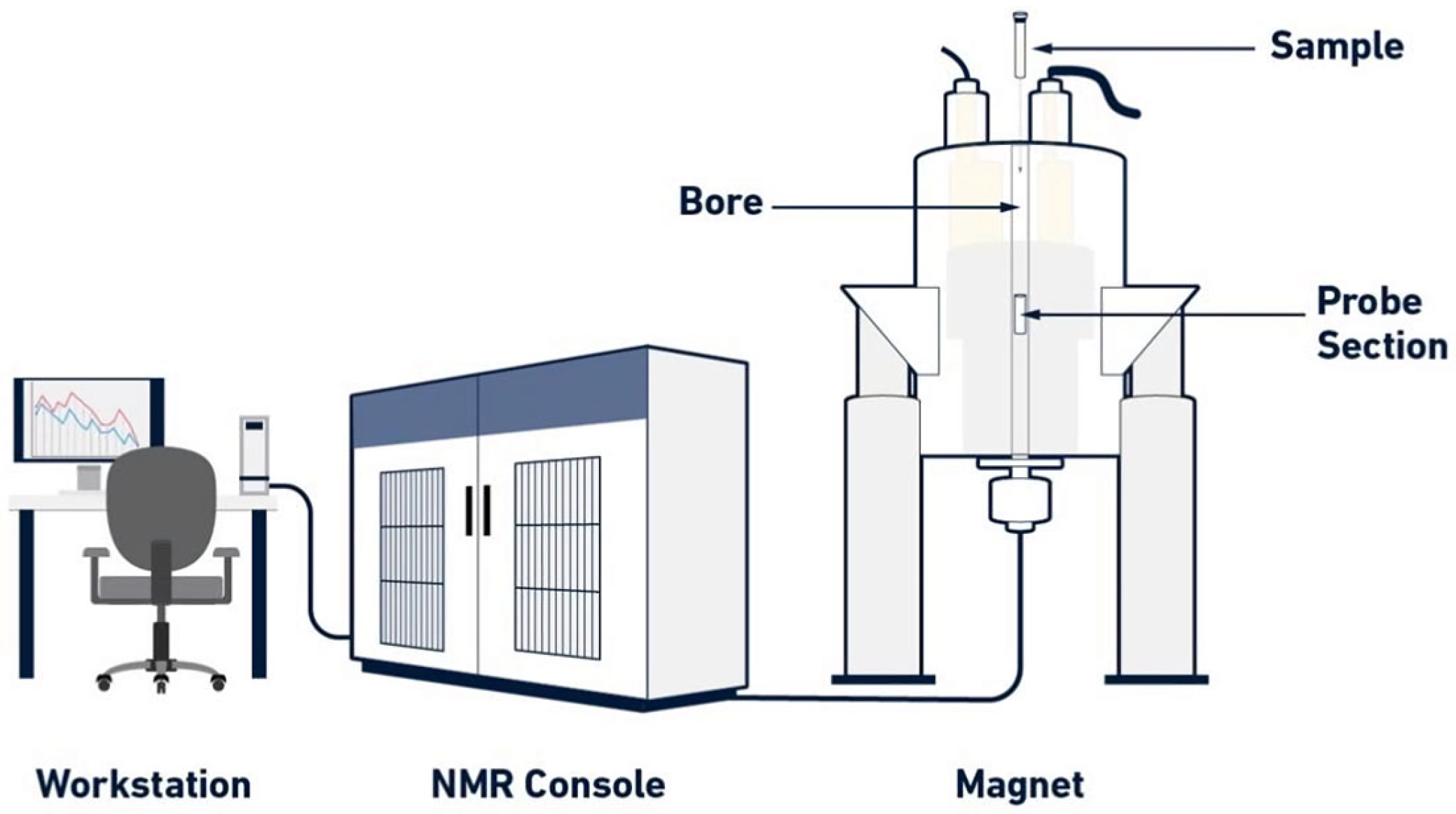
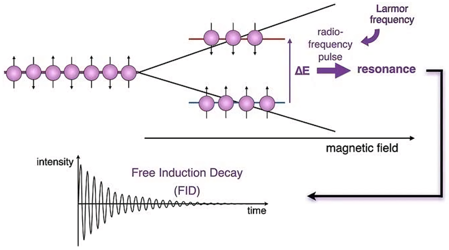
2. General Principles of NMR

NMR spectrometers are composed of three primary components: a superconducting magnet, a probe, and a sophisticated electronic system (console) managed through a workstation (Figure 1). The superconducting magnet generates a highly uniform and stable magnetic field to house the sample. Key sections such as the probe section and magnet bore should be labeled. The NMR console, a rectangular control unit, is typically linked to the magnet. The workstation, which is a computer or display for observing the NMR spectrum, connects to the console.
Accordingly, Figure 2 depicts the internal components of a superconducting magnet for an NMR spectrometer, focusing on the cryogenic cooling system and the probe setup used in NMR experiments. The key parts of the NMR magnet shown are the vacuum chamber, liquid nitrogen (N₂) tank, liquid helium (He) tank, and bore. The vacuum chamber is used to insulate the cryogenic system, helping maintain the very low temperatures necessary for the superconducting magnets to function. The liquid nitrogen (N₂) tank holds liquid nitrogen, which is used as part of the cooling system to reduce the temperature inside the spectrometer and typically serves as the first stage of cooling. The liquid helium (He) tank provides a much lower temperature than liquid nitrogen, cooling the superconducting magnet to around 4 K. Ensuring the superconductivity of the magnet is vital for generating the powerful magnetic fields required for NMR. The central bore is where the sample is placed for analysis, allowing it to pass through the magnetic field produced by the superconducting magnet. The field coil, an integral part of the superconducting magnet, creates the magnetic field that interacts with the sample’s nuclear spins. Inside the magnet’s bore, the sample is encased by the probe system, which subjects it to both the magnetic field and radiofrequency pulses during the measurement process. The radiofrequency probe delivers pulses to excite the nuclei in the sample. It forms the core of the detection system in NMR. After the nuclei in the sample are excited by the radiofrequency probe, they emit signals. The recording coil detects these signals, which are then processed to produce the NMR spectra.
The fundamental concept of nuclear magnetic resonance spectroscopy is illustrated in Figure 3. In this figure, atomic nuclei possess spins (depicted by arrows) that can align either parallel or antiparallel to the applied magnetic field. These alignments correspond to two distinct energy states: a lower energy state (spins aligned with the magnetic field) and a higher energy state (spins aligned against it). The energy difference between these states is represented as ΔE. A radiofrequency (RF) pulse is applied to the system at the Larmor frequency, which corresponds to the energy gap between the two spin states of spin-1/2 nuclei only. When the RF pulse resonates with this frequency, nuclei in the lower energy state absorb the energy and transition to the higher energy state–a phenomenon known as resonance. After the RF pulse is turned off, the spins relax to their thermal equilibrium corresponding to a Boltzmann distribution, which is here close to an equal distribution over all levels.

3. Overview of 31P qNMR for Phosphorus Compounds

Phosphorus, a key element in phosphoric acid esters and DNA molecules, plays a crucial role in physiological processes. It is vital for metabolic regulation, as phosphorylation and dephosphorylation reactions occur rapidly at the protein level. Investigating physiological pathways, metabolomics, kinetics, disease processes, and biomarker identification constitutes a major focus of contemporary research. [10]. Various analytical techniques are employed to analyze phosphorylation and dephosphorylation processes, offering both qualitative and quantitative detection. These methods include fluorescence, electrophoresis, label-free approaches like flow cytometry, stable isotope labeling, and mass spectrometry [11]. While these methods have certain advantages and challenges, the exploration of alternative quantification approaches remains a central focus.
Phosphorylated compounds such as metabolites can be measured by nuclear magnetic resonance spectroscopy [12]. 1H NMR spectroscopy of liver extracts is highly sensitive but challenging to interpret due to the full complexity of metabolic pathways and significant signal overlap, combined with strong background signals from water. This complexity makes it difficult to extract clear and quantifiable data from liver extracts using 1H NMR, even when focusing specifically on phosphorylated metabolites. In contrast, 31P NMR offers significant advantages, including relatively high sensitivity and a broader signal dispersion that minimizes signal overlap [13]. Quantitative 31P NMR spectroscopy has been utilized to study phosphorylated metabolites in liver extracts [14].
Only a limited number of methods can utilize a universal reference standard for quantification. Quantitative nuclear magnetic resonance (qNMR) stands out by enabling direct comparison of the analyte’s signal with an internal or external reference standard. When primary reference materials are employed, qNMR delivers highly precise measurements with minimal uncertainty, while also facilitating the certification of reference materials for 1H or other nuclei such as 31P. Over recent decades, the significance of qNMR has grown substantially [15,16,17,18].
As qNMR expands into new areas, including metabolomics, environmental analysis, and physiological research, the range of substances requiring analysis and the development of certified reference materials has broadened considerably. This includes complex biomolecules, proteins, and metabolites. These developments present growing challenges for 1H qNMR, the most commonly utilized method in qNMR, requiring enhanced reliability, improved precision, and accurate quantification of increasingly complex molecules [19].
The development of 31P NMR has opened new application areas for studying phosphorus-containing compounds and 31P qNMR has many advantages, making it an important qNMR method. Both 1H and 31P are spin-1/2 nuclei with high natural abundance. In molecular structures, the number of phosphorus atoms is typically low, leading to only one or a few signals in the entire nuclear magnetic resonance spectrum. This minimizes the risk of overlap between the signals of the sample and the standard in mixtures. The broad chemical shift range of 31P NMR (approximately 2000 ppm) allows for better separation of signals [20]. Moreover, commonly used deuterated NMR solvents lack 31P, eliminating interference from residual solvent or water peaks. Quantitative NMR (qNMR) studies focusing on 31P have been previously reported [21,22].
Quantitative nuclear magnetic resonance (qNMR) spectroscopy operates on the principle that the area under an NMR signal is directly proportional to the number of nuclei contributing to that resonance. This proportionality enables highly precise quantification, as the integrated signal intensity reflects the concentration of the corresponding nuclear species. qNMR relies on chemical shifts and peak areas for quantification. It does not require standard curves, minimizes sample destruction, and is particularly effective for the quantification of complex natural products. Therefore, it usually requires the use of certified reference materials as standards to achieve quantification [23]. Quantitative nuclear magnetic resonance (qNMR) encompasses two primary methodologies: the internal standard method and the external standard method. In the internal standard method, a reference compound is added directly to the sample solution, facilitating accurate quantification by comparing the integrals of the analyte and the standard within the same spectrum. Conversely, the external standard method involves preparing the reference standard and the analyte in separate solutions, with quantification achieved by comparing their respective spectra. Each method has its advantages; the internal standard method often offers higher precision, while the external standard method is beneficial when sample recovery is essential, such as when analyzing expensive materials (Figure 4) [24].
As mentioned in Figure 4, in the resulting spectrum of the internal standard method, the reference material and sample produce distinct peaks at different positions along the ppm (parts per million) scale. This method allows for direct comparison within a single spectrum by using a known internal reference within the same sample solution. In the external standard method, separate test tubes are used for the sample (green star) and reference material (yellow dot), indicating independent preparation. This method requires a separate analysis of the sample and reference, with results combined by referencing the separate spectra. The diagram compares how each method handles sample and reference material positioning and peak interpretation in ppm spectra. The internal standard method involves direct comparison within a single solution, while the external standard method analyzes each separately.

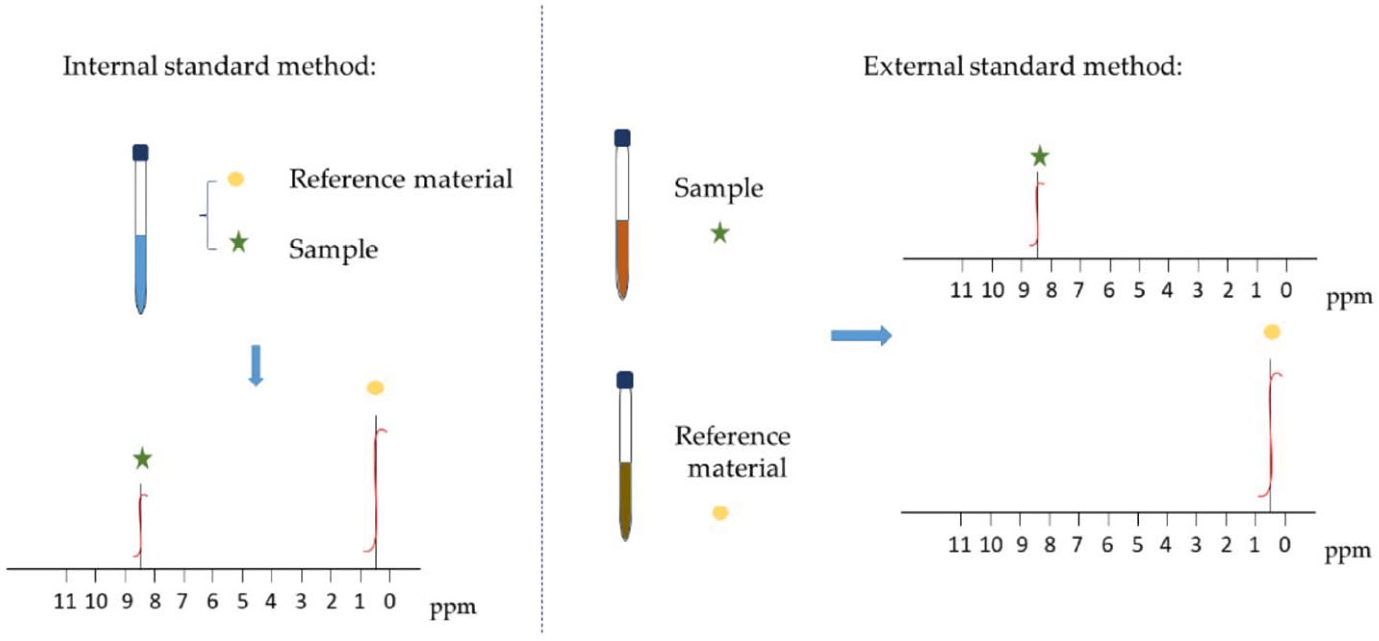
3.1. Internal Standard Method for qNMR

In the internal standard method of quantitative nuclear magnetic resonance (qNMR), a compound with a precisely known concentration is added to the sample as an internal standard. By measuring the ratio of the integrated signal areas between the internal standard and the analyte, the concentration of the analyte can be accurately determined. This approach is widely used in qNMR due to its precision and reliability. For instance, Luan et al. employed the internal standard method to determine the purity of a thiopeptin reference standard using sulfadoxine as the internal standard [25]. Similarly, Schleiff et al. developed a quantitative proton NMR method to analyze pregnenolone, utilizing an internal standard for accurate quantification [26]. The concentration of the analyte can be calculated using the following equation [15]:
m x = I x I s t d   N s t d N x   M x M s t d   P s t d   m s t d
where Ix is the integral of the analyte signal, Istd is the integral of the internal standard signal, Nx is the number of nuclei contributing to the analyte signal, Nstd is the number of nuclei contributing to the internal standard signal, Mx is the molar mass of the analyte, Mstd is the molar mass of the internal standard, Pstd is the purity of the internal standard, and mstd is the mass of the internal standard. By employing quantitative nuclear magnetic resonance (qNMR) with an internal standard, the necessity for calibration curves is eliminated. This technique involves adding a reference material of known concentration directly to the analyte solution or using a coaxial insert. The internal standard method is a well-established primary ratio analytical approach, extensively utilized in organic chemical analysis [27].

3.2. External Standard Method for qNMR

The external standard method involves preparing a series of standard solutions with different concentrations by using a known quantity of standard samples [28]. Under the same experimental conditions, a standard curve is generated, with the characteristic peak area on the vertical axis and the concentration on the horizontal axis, to measure the nuclear magnetic resonance spectrum of an unknown sample solution. The corresponding peak area is calculated, and the concentration of the unknown sample is determined from the standard curve. Nishizaki et al. [29] systematically evaluated the data acquisition workflow using certified reference materials (caffeine as the analyte and dimethyl sulfoxide as the calibrator) to identify key factors for accuracy and precision in external standard qNMR. Their study highlighted that the external standard method, wherein the standard material is measured separately from the samples, offers significant advantages. Notably, it minimizes artifacts resulting from internal mixing and prevents contamination of analyte samples by the reference compound.
Quantitative nuclear magnetic resonance (qNMR) spectroscopy presents numerous benefits compared to traditional chromatographic methods, such as superior specificity, rapid processing, high accuracy, excellent precision, and versatility. Its core principle relies on the direct proportionality between the integral of each NMR signal and the number of nuclei represented by that signal in the sample, ensuring a linear response without requiring calibration curves. For reliable quantification through qNMR, it is critical that the target compound is fully soluble; does not interact negatively with solvents, matrices, or internal standards; and exhibits well-separated quantitative peaks for both the analyte and internal standards. Moreover, qNMR facilitates quantification without necessitating the physical separation of sample components and allows the analysis of multiple compounds using a single internal standard [30]. The internal standard method is particularly popular for its simplicity, as it eliminates the need for calibration curves. Meanwhile, the external standard method is better suited for natural products when appropriate reference materials are available.

3.3. 31P Quantitative NMR Method

31P nuclear magnetic resonance spectroscopy is a valuable analytical technique for examining phosphorus-containing compounds. The 31P nucleus has a natural abundance of 100% and a high gyromagnetic ratio, resulting in excellent NMR sensitivity. Additionally, 31P NMR exhibits a broad chemical shift range, allowing for better separation and identification of different phosphorus environments [9]. These features make 31P NMR particularly beneficial for quantitative analysis. Typically, 31P NMR produces sharp signals, often corresponding to a single phosphorus atom within a molecule, facilitating precise quantification [31,32,33]. Compared to 1H NMR, 31P NMR can often avoid complex resonance interference, leading to clearer spectra [20]. Moreover, 31P NMR does not require expensive deuterated solvents, simplifying the sample preparation process [34].
31P qNMR is a highly effective technique for analyzing phosphorus-containing compounds. It has demonstrated the capability to measure trace amounts of these molecules in vivo [35]. Studies have shown that the accuracy and precision of 31P qNMR are comparable to those of chromatographic methods, making it a reliable tool for both pharmaceutical and food analysis [36,37,38]. Many researchers have improved and optimized 31P quantitative NMR, developed new methods, and refined the use of 31P nuclei for NMR quantification.
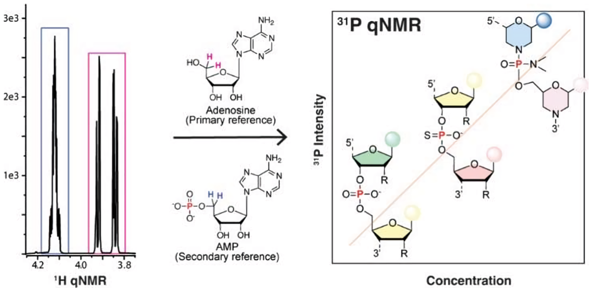
  • Research on 31P qNMR standard substances. Weber et al. [39] investigated 31P qNMR internal standards with metrological traceability, such as triphenyl phosphate and phosphoacetic acid, and their practical applications as shown in Figure 5. The study assessed the purity of tris(2-chloroethyl) phosphate using different traceability schemes and solvent systems. The findings demonstrated consistent results within measurement uncertainty ranges, highlighting the reliability of the certification approach. The application of these certified reference materials was further exemplified through measurements of tris(2-chloroethyl) phosphate. Additionally, researchers have used the 31P nucleus for quantitative NMR, testing various phosphorus-containing standards based on solubility and application needs. Internal standards, such as triphenyl phosphate and sodium phosphate, or external standards like phosphoric acid, are commonly employed to prevent reactions with analytes [40].
  • Development of Internal Standards for Coaxial Insertion. Henderson et al. [41] developed an absolute quantification method for 31P NMR using coaxial nuclear magnetic resonance tube inserts containing internal standards. This approach physically separates the reference material from the test sample, avoiding chemical interactions and mixing errors. The method demonstrated precision and accuracy exceeding 1% for purity determinations and detected impurities at concentrations as low as 25 µg/mL. Compared to traditional chromatographic methods, it eliminates the need for extensive sample preparation and reference standards, providing significant cost-effectiveness and operational simplicity.
  • Integration of 31P qNMR with Other Analytical Techniques. Atanassova et al. [42] studied the interaction between two extractants in deuterated chloroform using 1H, 13C, and 31P nuclear magnetic resonance spectra, along with NOESY experiments. Derewinski et al. [43] applied quantitative NMR techniques, including 27Al, 31P, and 1H magic-angle spinning NMR, together with temperature-programmed desorption to study the impact of various treatment processes on materials. NMR spectroscopy played a key role in this study, offering detailed characterization of transformations in aluminum, phosphorus, and acid sites. The study revealed the mechanisms underlying phosphorus modification and its partial reversibility, offering critical insights for advancing the understanding and optimization of zeolite-based catalysts in industrial applications.

4. Applications of 31P qNMR for Phosphorus-Containing Compounds

Quantitative nuclear magnetic resonance is a widely recognized method for analyzing phosphorus compounds, with applications ranging from food analysis [44,45,46,47], pharmaceuticals [9,48,49,50,51,52], and biological research [53,54,55] to energy materials and polymers. Compared to 1H and 13C qNMR techniques, 31P qNMR offers notable advantages, including rapid and precise identification of phosphorus-containing compounds, enhanced sensitivity, and broad signal dispersion.

4.1. In the Field of Food

31P qNMR is widely used to analyze diverse matrices, including complex natural products, dietary supplements, fungicides, and traditional medicines. Belmonte-Sanchez et al. [44] developed a rapid and reliable quantitative method for hydroxyethyl diphosphonic acid during vegetable washing using 31P NMR, which has been successfully applied to determine hydroxyethyl diphosphonic acid in 10 agricultural food processing industrial water samples used for fruit and vegetable washing. Moreover, Kato et al. [45] demonstrated the successful application of 31P NMR for the quantitative analysis of phospholipid-rich and moderately phospholipid-rich samples, such as soybeans, egg yolks, and dietary supplements. When compared to the two-dimensional thin-layer chromatography method, the total phospholipid contents of polar lipids determined by 31P NMR aligned closely with those obtained via molybdenum blue colorimetry. However, the quantitative values for specific phospholipid classes varied depending on their content, especially when exceeding or falling below 10%. Furthermore, Doyoon et al. [47] established a 31P qNMR method for the simultaneous quantification of the active forms of vitamins B1, B2, and B6. The 31P NMR signals corresponding to the analytes and the internal standard were clearly resolved and distinctly identifiable. EDTA and pH optimization (to 8.0) were utilized to enhance signal resolution and eliminate interference from metal ions in complex matrices. The proposed method uses phosphorus-31 NMR to target the phosphate group present in the active forms of vitamins. The advantages include simultaneous analysis of multiple active vitamins, which reduces complexity and sample preparation time compared to conventional methods like HPLC or LC-MS. Additionally, the high sensitivity and reproducibility make it suitable for routine quality control in the food and supplement industry, ensuring the accurate quantification of active vitamin content in complex multivitamin formulations. Kovacs et al. [47] report the use of quantitative 31P NMR spectroscopy to measure the phosphorus content in a cola beverage sample. The single 31P resonance, referred to as the "phosphate peak," is the concentration-weighted average signal of the phosphate species (PO43−, HPO42−, H2PO4, H3PO4). Since the 0 ppm reference is the 85% H3PO4 solution, the lower the pH, the smaller the chemical shift of the phosphate peak becomes. External calibration involves creating a dilution series of sodium phosphate solutions to establish a calibration curve. The experiment compares the results of external calibration and standard addition methods using peak height and integral values.

4.2. In the Field of Pharmaceuticals

31P qNMR provides precise, real-time, and direct drug quantification, supporting reliable in vitro/in vivo correlations. Agrahari et al. [9] designed a targeted 31P qNMR approach for the direct real-time measurement of therapeutic molecules in biological fluids. The results indicate that the method is simple and accurate and can quantify phosphorus-containing drug molecules from various biological fluids, such as vaginal fluid mimetics, semen mimetics, and human plasma.
Moreover, Khatun et al. [48] introduced an innovative qNMR approach for quantifying total aluminum and phosphorus in formulated vaccine products. This method was applied to a commercial adjuvanted combination vaccine as well as individual adsorbed antigen drug substances used in its production. With minimal sample preparation, this method delivers rapid, quantitative analysis, making it ideal for routine monitoring of vaccine adjuvant and formulation processes. The qNMR technique is well-suited for optimizing and controlling vaccine formulations. Its adaptability to various aluminum phosphate adjuvants highlights its potential for broader use in vaccine development and manufacturing. Offering a faster, simpler alternative to conventional techniques, it enables the simultaneous analysis of free and total phosphate in a single workflow, reduces preparation complexity, and enhances accuracy.
Glunde et al. [49] utilized 31P NMR to track malignant human mammary epithelial cells simultaneously during treatment with the anti-inflammatory drug indomethacin. Additionally, Uchiyama et al. [50] employed 31P qNMR and 1H qNMR to assess the purity of the anti-hepatitis C virus medication sofosbuvir, finding that the results from 31P qNMR were consistent with those obtained from 1H qNMR. Two solvents were evaluated, and these findings highlight that DMSO-d6 is a more suitable solvent for 31P qNMR analysis. The study validates the efficacy of 31P qNMR for determining the purity of organophosphorus compounds, particularly complex substances. Non-protic solvents, like DMSO-d6, are recommended for analyzing compounds with exchangeable protons to avoid deuterium exchange effects. The development of new reference standards and higher-sensitivity probes for 31P qNMR is suggested to expand its applicability. This study provides a robust framework for applying qNMR in pharmaceutical development and quality control, especially for complex organic compounds.
Phosphorus, present in all nucleotide units, has an abundant isotope that is NMR-active. Past 31P NMR studies on oligonucleotides have focused on peak assignment, chemical identification through the sensitivity of 31P chemical shifts to the phosphor chemical bond, and backbone conformation analysis. The 31P NMR could be applied to small-molecule drugs like cyclophosphamide. The capability of using 31P NMR for accurate quantification of oligonucleotide drugs was recently demonstrated using an internal reference standard in a mixture with a small interference siRNA drug substance and product. Chen and coworkers [52] report an external referenced qNMR method using 31P to directly characterize various representative oligonucleotide drug products in formulation, independent of any chemical modifications of phosphorothioate or phosphorodiamidate morpholino oligomer, duplex formation of siRNA, or process-associated ions (Figure 6). The 31P apparent T1 values were estimated by varying recycle delay from 0.1 s to 3 s. The accuracy of this measurement technique was better than the UV method, and its simplicity was much higher than the UV method.

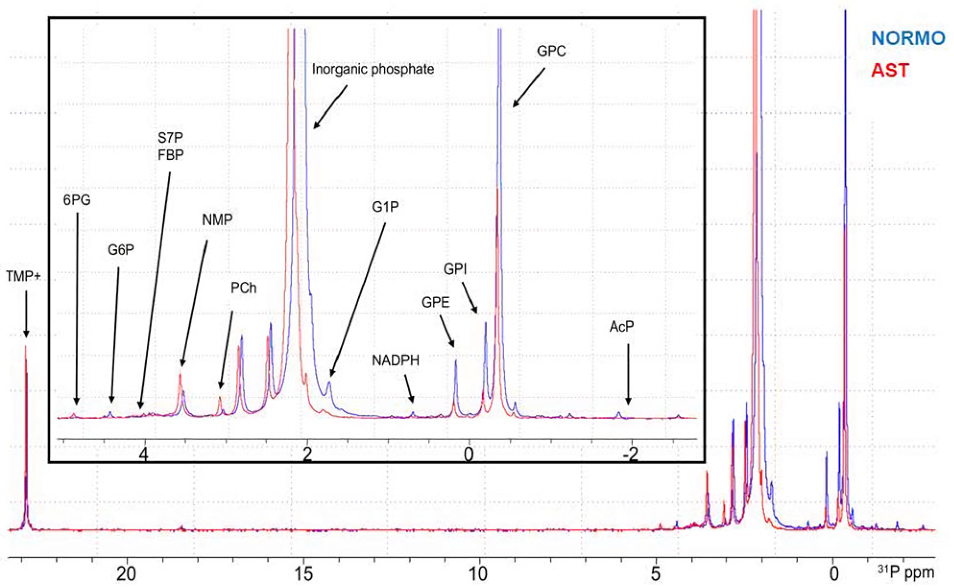
4.3. In the Field of Biology

Quantitative 31P NMR kinetic analysis is applicable to phosphate transfer reactions and has garnered widespread attention in biology, biotechnology, and biomedical sciences [56]. Bettjeman et al. [57] used phosphorus nuclear magnetic resonance (31P NMR) to determine the total phospholipid (PL) absolute concentration and PL spectrum. This technology is increasingly being used for the qualitative and quantitative analysis of PL in various biological matrices, including fish oil. Nevertheless, Matsumi et al. [56] developed a continuous and quantitative 31P NMR method for the analysis of (R)-5-hydroxyvaleric acid-5-phosphate formation. This method can be used to determine the efficacy of biocatalysts in the synthesis of enantiomerically pure (R)-mevalonic acid-5-phosphate, as well as to develop biocatalytic processes. Bjorstorp et al. [53] investigated the feasibility of quantifying siRNA content through a platform approach that integrates quantitative 31P nuclear magnetic resonance and the internal standard method. The platform method uses phosphonoacetic acid as the internal reference standard, which shows no signal overlap or interaction with siRNA. The optimization includes adjusting the acquisition time to remove spectral artifacts and setting a relaxation delay based on longitudinal relaxation times for accurate signal integration. The method demonstrates high precision, with a %RSD of 0.9%, outperforming UV spectroscopy. 31P-qNMR provides more accurate siRNA quantifications, closer to the theoretical mass balance (100% w/w) after adjusting for sodium and water content. 31P qNMR avoids uncertainties related to extinction coefficients and dilution errors. The method is versatile and applicable to both drug substance and drug product formulations, with minimal differences in preparation protocols. Furthermore, 31P qNMR offers structural insights into siRNA, confirming the backbone’s theoretical group ratios. The 31P qNMR platform method is a superior alternative to UV spectroscopy for siRNA quantification, offering greater accuracy, efficiency, and robustness. It represents a promising tool for analyzing complex oligonucleotides in pharmaceutical development and quality control.
Taking a different approach, Serrano and colleagues [54] employed a phosphorometabolomics strategy to gain deeper insights into the variations in phosphorus-containing metabolites associated with low sperm motility. They performed a comparative 31P NMR analysis of human seminal plasma samples from asthenozoospermic and normozoospermic individuals (Figure 7). Quantitative 31P NMR was crucial in detecting, identifying, and quantifying phosphorus-containing metabolites in human semen. It provided valuable insights into the metabolic differences between normozoospermic and asthenozoospermic individuals, offering potential biomarkers for diagnosing male infertility and enhancing the understanding of sperm energy metabolism and motility. Quantitative data analysis was achieved due to the direct correlation between signal intensity and analyte concentration. To enable comparison of metabolite levels across different samples, normalization was carried out. For seminal plasma and whole semen, metabolite quantities were normalized per volume and expressed in nmol/μL. In the case of spermatozoa analysis, metabolite abundance was normalized based on the number of cells, expressed as nmol per million cells.
Interestingly, Gouilleux et al. [55] explored the use of low-field 31P NMR with simple, maintenance-free benchtop spectrometers to analyze phospholipids in complex mixtures (Figure 8). The study assessed the analytical performance of 31P benchtop NMR for both relative and absolute quantification of individual phospholipid types in mixtures. It highlights the potential of low-field 31P NMR as a powerful tool for identifying and quantifying phospholipids, leveraging the distinct chemical shift dispersion of 31P to analyze phospholipid headgroups and mixtures effectively. Both proton-decoupled 31P NMR and heteronuclear cross-polarization experiments were evaluated in the study. Phospholipid mixtures were dissolved in specialized solvent systems to ensure optimal resolution and signal clarity. Heteronuclear cross-polarization significantly enhances signal-to-noise ratios compared to standard pulse experiments and allows for the unambiguous identification of phospholipid headgroups through 2D TOCSY experiments. Quantitative experiments at 1 T and 2 T delivered precision below 5% and accuracy better than 8%, with performance improving at higher fields. The method demonstrates strong potential for routine lipidomic applications, including the relative and absolute quantification of phospholipids in food science and biomedical research.

4.4. Others

31P NMR spectroscopy is also a convenient and widely used analytical method for investigating phosphorus-containing organic compounds or metal complexes. Wang et al. [58] studied the degradation behavior and mechanism of black phosphorus crystals in an atmospheric environment using phosphorus nuclear magnetic resonance spectroscopy. Mazumder et al. [59] developed a high-resolution 31P qNMR for the detection, identification, and quantification of non-phosphorus markers for toxic nerve agents, foaming agents, and incapacitation agents. Wan et al. [60] demonstrated a novel approach combining 1H NMR and 31P NMR for the first time to determine the level of purity in the assessment of tripropyl phosphate using NMR.
Chiral recognition of phosphines was reported with metal-based sensors by utilizing the metal chelation ability of phosphines. Kim et al. [61] developed an efficient chiral recognition of phosphine oxides formed via in situ oxidation of phosphines by optimizing the chiral metal complexes for the efficient 31P NMR analysis of in situ-prepared phosphine oxides (Figure 9). This chiral 31P NMR analysis can be used as a universal tool to measure the optical purity of chiral phosphines. The use of 31P NMR spectroscopy for chiral analysis, such as the determination of enantiomeric excess or absolute chirality, can be a direct, complementary, and convenient analytical protocol.

5. Conclusions

In summary, this paper highlights the unique advantages of quantitative 31P nuclear magnetic resonance (qNMR) for analyzing phosphorus-containing compounds. With features such as high natural abundance, wide chemical shift range, and the ability to produce clear, distinct signals, 31P qNMR is valuable in diverse fields, from food analysis and pharmaceuticals to biology study. The methodology of 31P qNMR, encompassing both internal and external standard techniques, offers precise and reliable quantification without the need for complex sample preparation, making it a powerful alternative to traditional analytical techniques. 31P qNMR was proven to be a superior and more efficient alternative to UV spectroscopy, providing accurate, reliable, and reproducible results for quantification in pharmaceutical contexts. Despite current sensitivity challenges and accessibility of NMR instrumentation, advances in high-sensitivity probes and broader adoption of NMR technology are expected to drive the prospects of 31P qNMR applications. In the future, this technique holds great promise for the accurate quantification of complex samples, further solidifying its role as a critical tool in chemical, biological, and medical research.

Author Contributions

Writing the manuscript, Y.L.; Assisting in editing the manuscript, L.G. and Z.Y. All authors have read and agreed to the published version of the manuscript.

Funding

This work was funded by the General Project of the Experimental Technology Program of Zhejiang University, grant number SYBJS202404.

Data Availability Statement

Not applicable.

Acknowledgments

We thank the Chemistry Instrumentation Center of Zhejiang University for providing the NMR research platform.

Conflicts of Interest

The authors declare no conflicts of interest.

Abbreviations

NMRNuclear magnetic resonance spectroscopy
31P qNMRQuantitative phosphorus nuclear magnetic resonance spectroscopy
FIDFree induction decay
RFRadio frequency
PLPhospholipid
AQAcquisition time

References

  1. Rabi, I.I.; Millman, S.; Kusch, P.; Zacharias, J.R. The Magnetic Moments of 3Li6, 3Li7 and 9F19. Phys. Rev. 1939, 55, 495. [Google Scholar]
  2. Purcell, E.; Torrey, H.; Pound, R. Resonance Absorption by Nuclear Magnetic Moments in a Solid. Phys. Rev. 1946, 69, 37–38. [Google Scholar] [CrossRef]
  3. Bloch, F.; Hansen, W.; Packard, M. Nuclear Induction. Phys. Rev. 1946, 69, 127. [Google Scholar] [CrossRef]
  4. Malet-Martino, M.; Holzgrabe, U. NMR Techniques in Biomedical and Pharmaceutical Analysis. J. Pharm. Biomed. Anal. 2011, 55, 1–15. [Google Scholar] [CrossRef]
  5. Holzgrabe, U.; Deubner, R.; Schollmayer, C.; Waibel, B. Quantitative NMR Spectroscopy Applications in Drug Analysis. J. Pharm. Biomed. Anal. 2005, 38, 806–812. [Google Scholar] [CrossRef]
  6. Wood, J.S.; Poggetto, G.D.; Wang, X.; Reibarkh, M.; Williamson, R.T.; Cohen, R.D. Quantitative Nuclear Magnetic Resonance of Chloride by an Accurate Internal Standard Approach. Magn. Reson. Chem. 2023, 61, 22–31. [Google Scholar] [CrossRef] [PubMed]
  7. Malz, F.; Jancke, H. Validation of Quantitative NMR. J. Pharm. Biomed. Anal. 2005, 38, 813–823. [Google Scholar] [CrossRef]
  8. Zhang, C.; Zhang, T.; Oyler, N.A.; Youan, B.B. Direct and Real-time Quantification of Tenofovir Release from pH-sensitive Microparticles into Simulated Biological Fluids using 1H Nuclear Magnetic Resonance. J. Pharm. Sci. 2014, 103, 1170–1177. [Google Scholar] [CrossRef]
  9. Agrahari, V.; Meng, J.; Sudhaunshu, S.; Nathan, A.; Youan, B.B. Real-Time Analysis of Tenofovir Release Kinetics using Quantitative Phosphorus (31P) Nuclear Magnetic Resonance Spectroscopy. J. Pharm. Sci. 2017, 106, 3005–3015. [Google Scholar] [CrossRef] [PubMed]
  10. Lewis, G.D.; Asnani, A.; Gerszten, R.E. Application of Metabolomics to Cardiovascular Biomarker and Pathway Discovery. J. Am. Coll. Cardiol. 2008, 52, 117–123. [Google Scholar] [CrossRef]
  11. Bonilla, L.; Means, G.; Lee, K.; Patterson, S. The Evolution of Tools for Protein Phosphorylation Site Analysis: From Discovery to Clinical Application. Biotechniques 2008, 44, 671–679. [Google Scholar] [CrossRef] [PubMed]
  12. Kumar, G.S.; Page, R.; Peti, W. Preparation of Phosphorylated Proteins for NMR Spectroscopy. Methods Enzymol. 2019, 614, 187–205. [Google Scholar]
  13. Emwas, A.H.; Roy, R.; McKay, R.T.; Tenori, L.; Saccenti, E.; Gowda, G.A.N.; Raftery, D.; Alahmari, F.; Jaremko, L.; Jaremko, M. NMR Spectroscopy for Metabolomics Research. Metabolites 2019, 9, 123. [Google Scholar] [CrossRef]
  14. Bernardo-Seisdedos, G.; Bilbao, J.; Fernández-Ramos, D.; Lopitz-Otsoa, F.; Gutierrez de Juan, V.; Bizkarguenaga, M.; Mateos, B.; Fondevila, M.F.; Abril-Fornaguera, J.; Millet, O.; et al. Metabolic Landscape of the Mouse Liver by Quantitative 31P Nuclear Magnetic Resonance Analysis of the Phosphorome. Hepatology 2021, 74, 148–163. [Google Scholar] [CrossRef]
  15. Fujiwara, S.; Wainai, T. Analysis of Tar Products by Nuclear Magnetic Resonance Spectrometry. Anal. Chem. 1961, 33, 1085–1087. [Google Scholar] [CrossRef]
  16. Jungnickel, J.L.; Forbes, J.W. Quantitative Measurement of Hydrogen Types by Integrated Nuclear Magnetic Resonance Intensities. Anal. Chem. 1963, 35, 938–942. [Google Scholar] [CrossRef]
  17. Hollis, D.P. Quantitative Analysis of Aspirin, Phenacetin, and Caffeine Mixtures by Nuclear Magnetic Resonance Spectrometry. Anal. Chem. 1963, 35, 1682–1684. [Google Scholar] [CrossRef]
  18. Wells, R.J.; Cheung, J.; Hook, J.M. Dimethylsulfone as a Universal Standard for Analysis of Organics by QNMR. Accred. Qual. Assur. 2004, 9, 450–456. [Google Scholar] [CrossRef]
  19. Barding, G.A.; Salditos, R.; Larive, C.K. Quantitative NMR for Bioanalysis and Metabolomics. Anal. Bioanal. Chem. 2012, 404, 1165–1179. [Google Scholar] [CrossRef] [PubMed]
  20. Rigger, R.; Ruck, A.; Hellriegel, C.; Sauermoser, R.; Morf, F.; Breitruck, K.; Obkircher, M. Certified Reference Material for Use in 1H, 31P, and 19F Quantitative NMR, Ensuring Traceability to the International System of Units. J. Aoac. Int. 2017, 100, 1365–1375. [Google Scholar] [CrossRef]
  21. Maniara, G.; Rajamoorthi, K.; Rajan, S.; Stockton, G.W. Method Performance and Validation for Quantitative Analysis by 1H and 31P NMR Spectroscopy. Applications to Analytical Standards and Agricultural Chemicals. Anal. Chem. 1998, 70, 4921–4928. [Google Scholar] [CrossRef] [PubMed]
  22. Deen, T.S.A.; Hibbert, D.B.; Hook, J.M.; Wells, R.J. Quantitative Nuclear Magnetic Resonance Spectrometry. Anal. Chim. Acta 2002, 474, 125–135. [Google Scholar] [CrossRef]
  23. Pearce, J.M.; Komoroski, R.A.; Mrak, R.E. Phospholipid Composition of Postmortem Schizophrenic Brain by 31P NMR Spectroscopy. Magn. Reson. Med. 2010, 61, 28–34. [Google Scholar] [CrossRef] [PubMed]
  24. Wang, S.Q. Research Progress of NMR in Natural Product Quantification. Molecules 2021, 26, 6308. [Google Scholar] [CrossRef]
  25. Luan, J.; Feng, R.; Chen, Y.; Wu, X.; Su, M. Quantitative Assessment of the Absolute Purity of Thiopeptcin Reference Standard by 1H-NMR. Anal. Sci. 2018, 34, 1093–1098. [Google Scholar] [CrossRef] [PubMed]
  26. Schleiff, M.; Sommers, C.; Yang, J.; Shen, X.; Rodriguez, J.D.; Shu, Q. Development and Validation of a Quantitative Proton NMR Method for the Analysis of Pregnenolone. J. Pharm. Biomed. Anal. 2023, 222, 115073. [Google Scholar] [CrossRef] [PubMed]
  27. Wells, R.J.; Hook, J.M.; Al-Deen, T.S.; Hibbert, D.B. Quantitative Nuclear Magnetic Resonance (QNMR) Spectroscopy for Assessing the Purity of Technical Grade Agrochemicals: 2,4-dichlorophenoxyacetic Acid (2,4-D) and Sodium 2,2-dichloropropionate. J. Agric. Food Chem. 2002, 50, 3366–3374. [Google Scholar] [CrossRef] [PubMed]
  28. Li, C.; Bhavaraju, S.; Thibeault, M.P.; Melanson, J.; Blomgren, A.; Rundlöf, T.; Kilpatrick, E.; Swann, C.J.; Rudd, T.; Aubin, Y. Survey of Peptide Quantification Methods and Comparison of Their Reproducibility: A Case Study using Oxytocin. J. Pharm. Biomed. Anal. 2019, 166, 105–112. [Google Scholar] [CrossRef]
  29. Nishizaki, Y.; Lankin, D.C.; Chen, S.; Pauli, G.F. Accurate and Precise External Calibration Enhances the Versatility of Quantitative NMR (qNMR). Anal. Chem. 2011, 93, 2733–2741. [Google Scholar] [CrossRef]
  30. Castaing-Cordier, T.; Ladroue, V.; Besacier, F.; Bulete, A.; Jacquemin, D.; Giraudeau, P.; Farjon, J. High-field and Benchtop NMR Spectroscopy for the Characterization of New Psychoactive Substances. Forensic Sci. Int. 2021, 321, 110718. [Google Scholar] [CrossRef]
  31. Sun, L.; Fan, Y.; Wang, Q.; Xiang, L.; Han, H.; Chen, D. Validated Quantitative 31P NMR Spectroscopy for Positional Isomeric Impurity Determination in L-α-glycerylphosphorylcholine (L-α-GPC). J. Pharm. Biomed. Anal. 2022, 221, 115067. [Google Scholar] [CrossRef] [PubMed]
  32. Igarashi, H.; Yoshida, A.; Tanaka, K. Comparison of Pig Cornea Preservation Solutions using Quantitative 31P NMR Spectroscopy. Magn. Reson. Med. 1993, 29, 107–109. [Google Scholar] [CrossRef] [PubMed]
  33. Uchiyama, N.; Hosoe, J.; Komatsu, T.; Sugimoto, N.; Ishizuki, K.; Koide, T.; Murabayashi, M.; Shinozaki, T.; Kobayashi, K.; Fujimine, Y.; et al. Quantitative 31P NMR for the Purity Determination of the Organophosphorus Compound Brigatinib and Its Method Validation. Chem. Pharm. Bull. 2024, 72, 36–40. [Google Scholar] [CrossRef] [PubMed]
  34. Jiang, H.; Chen, H.; Cai, N.; Zou, J.; Ju, X. Quantitative 31P-NMR Spectroscopy for the Determination of Fosfomycin and Impurity A in Pharmaceutical Products of Fosfomycin Sodiumor Calcium. Magn. Reson. Chem. 2015, 53, 454–459. [Google Scholar] [CrossRef] [PubMed]
  35. Gupta, A. Cardiac 31P MR Spectroscopy: Development of the Past Five Decades and Future Vision-will it Be of Diagnostic Use in Clinics? Heart Fail Rev. 2023, 28, 485–532. [Google Scholar] [CrossRef] [PubMed]
  36. Gard, D.R.; Burquin, J.C.; Gard, J.K. Quantitative Analysis of Short-Chain Phosphates by Phosphorus-31 Nuclear Magnetic Resonance and Interlaboratory Comparison with Infrared and Chromatographic Methods. Anal. Chem. 1992, 64, 557–561. [Google Scholar] [CrossRef]
  37. Holzgrabe, U. Quantitative NMR Spectroscopy in Pharmaceutical Applica-tions. Prog. Nucl. Mag. Res. Sp. 2010, 57, 229–240. [Google Scholar] [CrossRef]
  38. Martino, R.; Gilard, V.; Desmoulin, F.; Malet-Martino, M. Fluorine-19 or Phosphorus-31 NMR Spectroscopy: A Suitable Analytical Technique for Quantitative in Vitro Metabolic Studies of Fluorinated or Phosphorylated Drugs. J. Pharmaceut. Biomed. 2005, 38, 871–891. [Google Scholar] [CrossRef] [PubMed]
  39. Weber, M.; Hellriegel, C.; Rueck, A.; Wuethrich, J.; Jenks, P.; Obkircher, M. Method Development in Quantitative NMR Towards Metrologically Traceable Organic Certified Reference Materials Used as 31P qNMR Standards. Anal. Bioanal. Chem. 2015, 407, 3115–3123. [Google Scholar] [CrossRef]
  40. Gauss, D.; Sanchez-Moreno, I.; Oroz-Guinea, G.J.I.; Wohlgemuth, R. Phosphorylation Catalyzed by Dihydroxyacetone Kinase. Eur. J. Org. Chem. 2018, 23, 2892–2895. [Google Scholar] [CrossRef]
  41. Henderson, T.J. Quantitative NMR Spectroscopy using Coaxial Inserts Containing a Reference Standard: Purity Determinations for Military Nerve Agents. Anal. Chem. 2002, 74, 191–198. [Google Scholar] [CrossRef]
  42. Derewinski, M.; Sarv, P.; Sun, X.; Muller, S.; Van Veen, A.C.; Lercher, J.A. Reversibility of the Modification of HZSM-5 with Phosphate Anions. J. Phys. Chem. C 2014, 118, 6122–6131. [Google Scholar] [CrossRef]
  43. Atanassova, M.; Kurteva, V. Synergism in the Solvent Extraction of Europium(III) with Thenoyltrifluoroacetone and CMPO in Methylimidazolium Ionic liquids. J. Solution Chem. 2019, 48, 15–30. [Google Scholar] [CrossRef]
  44. Belmonte-Sanchez, J.R.; Aguilera-Saez, L.M.; Romero-Gonzalez, R.; Vidal, J.L.M.; Frenich, A.G. Determination of Etidronic Acid in Vegetable-washing Water by a Simple and Validated Quantitative 31P Nuclear Magnetic Resonance Method. Microchem. J. 2019, 150, 104083. [Google Scholar] [CrossRef]
  45. Kato, T.; Nishimiya, M.; Kawata, A.; Kishida, K.; Suzuri, K.; Saito, M.; Fujita, K.; Igarashi, T.; Inagaki, M. Quantitative 31P NMR Method for Individual and Concomitant Determination of Phospholipid Classes in Polar Lipid Samples. J. Oleo Sci. 2018, 67, 1279–1289. [Google Scholar] [CrossRef]
  46. Loening, N.M.; Chamberlin, A.M.; Zepeda, A.G.; Gonzalez, R.G.; Cheng, L.L. Quantification of Phosphocholine and Glycerophosphocholine with 31P edited 1H NMR Spectroscopy. NMR Biomed. 2005, 18, 413–420. [Google Scholar] [CrossRef]
  47. Kovacs, D.; Sebak, F.; Szabo, C.L.; Bodor, A. Determination of Phosphorus Content in Cola Drinks by 31P NMR Spectroscopy using External Calibration and Standard Addition Approaches. J. Chem. Educ. 2024, 101, 2524–2532. [Google Scholar] [CrossRef]
  48. Khatun, R.; Hunter, H.N.; Sheng, Y.; Carpick, B.W.; Kirkitadze, M.D. 27Al and 31P NMR Spectroscopy Method Development to Quantify Aluminum Phosphate in Adjuvanted Vaccine Formulations. J. Pharm. Biomed. Anal. 2018, 159, 166–172. [Google Scholar] [CrossRef] [PubMed]
  49. Glunde, K.; Ackerstaff, E.; Natarajan, K.; Artemov, D.; Bhujwalla, Z.M. Real-time Changes in 1H and 31P NMR Spectra of Malignant Human Mammary Epithelial Cells during Treatment with the Anti-inflammatory Agent Indomethacin. Magn. Reson. Med. 2010, 48, 819–825. [Google Scholar] [CrossRef] [PubMed]
  50. Uchiyama, N.; Kiyota, K.; Hosoe, J.; Komatsu, T.; Sugimoto, N.; Ishizuki, K.; Koide, T.; Murabayashi, M.; Kobayashi, K.; Fujimine, Y. Quantitative 31P NMR for Purity Determination of Sofosbuvir and Method Validation. Chem. Pharm. Bull. 2022, 70, 892–900. [Google Scholar] [CrossRef] [PubMed]
  51. Doyoon, P.; Seokyoon, K.; Hyeona, L.; Hyo-Yeon, Y.; Sangdoo, A. Development and Validation of a Method for the Simultaneous Quantitative Analysis of the Active Forms of Vitamins B1, B2, and B6 using 31P qNMR. Microchem. J. 2024, 196, 109704. [Google Scholar]
  52. Li, J.; Chen, F.; Zhang, D.; Wang, Y.; Kozak, D.; Chen, K. An Accurate and Fast 31P qNMR Assay Method for Oligonucleotide Therapeutics. Anal. Chem. 2024, 96, 16514–16519. [Google Scholar] [CrossRef]
  53. Bjorstorp, S.; Malmstrom, J. Quantitative 31P NMR Spectroscopy Platform Method for the Assay of Oligonucleotides as Pure Drug Substances and in Drug Product Formulations Using the Internal Standard Method. Anal. Chem. 2024, 96, 11198–11204. [Google Scholar] [CrossRef] [PubMed]
  54. Serrano, R.; Martin-Hidalgo, D.; Bilbao, J.; Bernardo-Seisdedos, G.; Millet, O.; Garcia-Marin, L.J.; Bragado, M.J. Quantitative Analysis of the Human Semen Phosphorometabolome by 31P-NMR. Int. J. Mol. Sci. 2024, 25, 1682. [Google Scholar] [CrossRef]
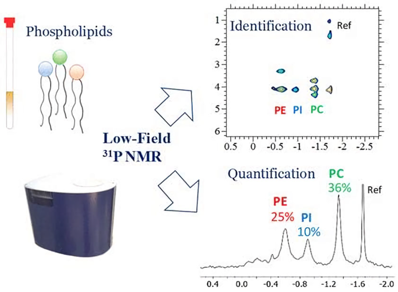
  55. Gouilleux, B.; Christensen, N.V.; Malmos, K.G.; Vosegaard, T. Analytical Evaluation of Low-Field 31P NMR Spectroscopy for Lipid Analysis. Anal. Chem. 2019, 91, 3035–3042. [Google Scholar] [CrossRef]
  56. Matsumi, R.; Hellriegel, C.; Schoenenberger, B.; Milesi, T.; Vanderoost, J.; Wohlgemuth, R. Biocatalytic Asymmetric Phosphorylation of Mevalonate. RSC Adv. 2014, 4, 12989–12994. [Google Scholar] [CrossRef]
  57. Bettjeman, B.I.; Hofman, K.A.; Burgess, E.J.; Perry, N.B.; Killeen, D.P. Seafood Phospholipids: Extraction Efficiency and Phosphorous Nuclear Magnetic Resonance Spectroscopy (31P NMR) Profiles. J. Am. Oil. Chem. Soc. 2018, 95, 779–786. [Google Scholar] [CrossRef]
  58. Wang, Y.; Yang, B.; Wan, B.; Xi, X.; Zeng, Z.; Liu, E.; Wu, G.; Liu, Z.; Wang, W. Degradation of Black Phosphorus: A Real-Time 31P NMR Study. 2D Mater. 2016, 3, 035025. [Google Scholar] [CrossRef]
  59. Mazumder, A.; Kumar, A.; Purohit, A.K.; Dubey, D.K. A High Resolution Phosphorus-31p Nuclear Magnetic Resonance (NMR) Spectroscopic Method for the Non-phosphorus Markers of Chemical Warfare Agents. Anal. Bioanal. Chem. 2012, 402, 1643–1652. [Google Scholar] [CrossRef]
  60. Wan, Y.; Li, K.; Li, X.; Li, X.; Chu, H.; Zhang, Q. Purity Assessment of Tripropyl Phosphate through Mass Balance and 1H and 31P Quantitative Nuclear Magnetic Resonance. Molecules 2024, 29, 1975. [Google Scholar] [CrossRef] [PubMed]
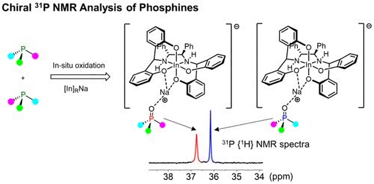
  61. Kwahk, E.J.; Jang, S.; Kim, H. Chiral Recognition of in Situ-Oxidized Phosphine Oxides with Octahedral Indium Complexes by 31P NMR Spectroscopy. Org. Lett. 2021, 23, 7829–7833. [Google Scholar] [CrossRef] [PubMed]
Figure 1. A schematic diagram illustrating the overall layout of an NMR spectrometer and its principal components. Copyright (2021) Technology networks.
Applsci 15 00323 g001
Figure 2. Internal components of a superconducting magnet for an NMR spectrometer, including a detailed view of the probe. Copyright (2021) Technology networks.
Applsci 15 00323 g002
Figure 3. Principle of nuclear magnetic resonance. Copyright (2024) Mariabaias network.
Applsci 15 00323 g003
Figure 4. Internal standard and external standard method of quantitative NMR. Copyright (2021) MDPI.
Applsci 15 00323 g004
Figure 5. The 31P NMR spectra include ammonium dihydrogen phosphate in D2O (brown), triphenyl phosphate in CDCl3 (blue), phosphonoacetic acid in D2O (red), and tris(2-chloroethyl) phosphate in CDCl3 (black). Copyright (2015) Springer.
Applsci 15 00323 g005
Figure 6. The external referenced qNMR method using 31P to directly characterize various representative oligonucleotide drug products. Copyright (2024) American Chemical Society.
Applsci 15 00323 g006
Figure 7. Representative 31P-NMR spectra of seminal plasma from asthenozoospermic (AST, red) and normozoospermic (NORMO, blue) groups, highlighting peak dispersion and metabolite assignments. The 5 to −3 ppm region is displayed with the corresponding metabolite identifications. Copyright (2024) MDPI.
Applsci 15 00323 g007
Figure 8. Identification and quantification analysis of phospholipids using low-field 31P NMR spectroscopy from phosphatidylcholine (PC), phosphatidyl-ethanolamine (PE), and phosphatidylinositol (PI). Copyright (2019) American Chemical Society.
Applsci 15 00323 g008
Figure 9. Chiral receptors for phosphines and phosphine oxides designed using 31P NMR analysis. Copyright (2021) American Chemical Society.
Applsci 15 00323 g009
Disclaimer/Publisher’s Note: The statements, opinions and data contained in all publications are solely those of the individual author(s) and contributor(s) and not of MDPI and/or the editor(s). MDPI and/or the editor(s) disclaim responsibility for any injury to people or property resulting from any ideas, methods, instructions or products referred to in the content.

