Review

Reviewing Breakthroughs and Limitations of Implantable and External Medical Device Treatments for Spinal Cord Injury

by
Tooba Wallana
,
Konstantinos Banitsas
* and
Wamadeva Balachandran
Electronic and Electrical Engineering, Brunel University of London, London UB8 3PH, UK
*
Author to whom correspondence should be addressed.
Appl. Sci. 2025, 15(15), 8488; https://doi.org/10.3390/app15158488
Submission received: 9 May 2025 / Revised: 21 July 2025 / Accepted: 28 July 2025 / Published: 31 July 2025

Abstract

Spinal cord injury (SCI) is a major disability that, to this day, does not have a permanent cure. The spinal cord extends caudally through the body structure of the vertebral column and is part of the central nervous system (CNS). The spinal cord enables neural communication and motor coordination, so injuries can disrupt sensation, movement, and autonomic functions. Mechanical and traumatic damage to the spinal cord causes lesions to the nerves, resulting in the disruption of relayed messages to the extremities. Various forms of treatment for the spinal cord include functional electrical stimulation (FES), epidural electrical stimulation (EES), ‘SMART’ devices, exoskeleton and robotic systems, transcranial magnetic stimulation, and neuroprostheses using AI for the brain–computer interface. This research is going to analyse and review these current treatment methods for spinal cord injury and identify the current gaps and limitations in these, such as long-term biocompatibility, wireless adaptability, cost, regulatory barriers, and risk of surgery. Future advancements should work on implementing wireless data logging with AI algorithms to increase SCI device adaptability, as well as maintaining regulatory and health system integration.

1. Introduction

Spinal cord injury (SCI) currently impacts 50,000 people in the UK with the addition of 2500 people every year [1]. Despite these rising figures, there is still no permanent cure for this injury [2]. Not only does it massively influence the quality of life of the individual, but it also massively drains limited healthcare budgets with increasing surgeries and multiple failed treatments, as these patients need to be monitored much more frequently. SCI increases societal demands by requiring additional resources to support the incapacitated individual. Since the Egyptian times, SCI has been neglected due to a lack of facilities and advances in medical engineering. In pharmacology and medical engineering, SCI has been labelled a terminal illness and surgery has been accepted as the sole option. In recent times, there has been a revelation that if the right intervention is applied, SCI is treatable and can significantly reduce the mortality rate as well as offer an increased quality of life for the individuals. Though SCI remains one of the most complex neurological conditions, device-based treatments offer promising avenues for functional restoration. While numerous technologies, from functional electrical stimulation (FES) to exoskeletons, have demonstrated significant efficacy, the field lacks a cohesive framework to evaluate these treatments across dimensions of invasiveness, longevity, and patient-centred outcomes.
This review introduces a comparative view of recent breakthrough methods in spinal cord injury treatments, including implantable and external devices, emphasising trade-offs in biocompatibility, power source limitations, and user accessibility. Implantable devices, such as electrical epidural stimulators and smart sensors, offer precision and long-term integration, yet face challenges in surgical risk and regulatory hurdles. External devices, including robotic exoskeletons and surface FES systems, provide non-invasive alternatives; however, they often suffer from the challenge of bulkiness and limited personalisation. These factors will be mentioned in more detail in the following review, as well as emerging directions, including closed-loop systems and AI-driven neuroprostheses, reshaping SCI treatments. These innovations show promise in bridging the gap between device functionality and neural adaptability, offering dynamic, responsive interventions, personalised to everyone’s neurophysiological profiles.

1.1. The Spinal Cord

The spinal cord is the main communication link between the brain and body via the central nervous system (CNS). It can also independently process reflexes and repetitive motor tasks through central pattern generators (CPGs). It begins at the medulla oblongata, passes through the foramen magnum [3], and runs from cervical vertebrae C1–C8, through thoracic T1–T12, to lumbar L1–L2 [4], ending at the conus medullaris and cauda equina [3]. Each vertebral level connects to a spinal nerve that controls a myotome (muscles) and a dermatome (skin) [3]. Sensory (afferent) neurons enter via dorsal roots containing dorsal root ganglia, while motor (efferent) neurons exit via ventral roots [5]. These roots join to form spinal nerve pathways throughout the spinal cord as shown in Figure 1 [3]. Cervical nerves control the head, diaphragm, arms, and hands; thoracic nerves handle the chest and abdomen; lumbar nerves control the legs; sacral nerves manage bowel, bladder, and sexual functions; and the coccygeal nerve serves the skin over the tailbone. Protected by vertebrae and meninges, the spinal cord is cushioned by cerebrospinal fluid and connective tissue. It contains central grey matter (neuron bodies) and surrounding white matter (myelinated axons) [3]. Ascending tracts carry sensory data to the brain, while descending tracts send motor commands to the body [6].

1.2. Spinal Cord Injury

Spinal cord injury (SCI) is classified as either traumatic—such as from falls, violence, or traffic accidents—or nontraumatic, resulting from autoimmune disorders, tumours, or degenerative diseases. Regardless of cause, SCI pathology is divided into primary and secondary injuries [7]. Primary injury refers to the immediate mechanical damage to the spinal cord, often due to compression from fractures, malignancies, or abscesses. Another form, distraction injury, occurs when two vertebrae separate, stretching or tearing the spinal cord in the axial plane [7]. Primary injuries often trigger a secondary injury cascade, which unfolds over time and is divided into acute, subacute, and chronic phases. Secondary injury begins within 2–48 h post-injury, involving inflammation, oedema, or haemorrhage. The acute phase (up to ~2 weeks) includes free radical formation, ionic imbalance, vascular damage, and immune-related toxicity. These factors lead to mitochondrial dysfunction, abnormal glutamate signalling, and the release of proinflammatory cytokines like interferon-gamma, transitioning the injury to the subacute phase [4,7]. During the subacute phase (~days 2–14), phagocytes remove debris, astrocytes multiply, and ionic balance begins to restore. The blood–brain barrier limits immune cell entry, but scar formation begins, restricting axonal regrowth. Between weeks 2 and 24, this intermediate phase includes scar maturation and early axonal sprouting. The chronic phase starts around 6 months post-injury and is marked by continued scarring, syrinx formation, Wallerian degeneration, and cystic cavitations [4]. These chronic changes can result in persistent neurological deficits, including sensory and motor loss, neuropathic pain, bladder and bowel dysfunction, and sexual impairment [2].

2. Study Selection Criteria

To ensure the relevant amalgamation of device-based treatments for SCI, this review applied the following inclusion and exclusion criteria: the studies must have been published between 2010 and 2025, to exhibit the most recent technological advancements, and they must have been published in the English language. The type of studies included were peer-reviewed clinical trials, pilot studies, systemic reviews, and translational animal studies that have a direct relevance to device-based SCI. The intervention focus was on studies evaluating implantable or external medical devices (e.g., FES, EES, exoskeletons, brain–computer interface, TMS, SMART implants). The human patients used in the studies all had traumatic or nontraumatic SCI, and the animal studies had subjects with simulated SCI. All the studies selected reported motor function recovery, sensory improvement, neuroplasticity, or quality of life improvement. Various studies were assessed with these selection criteria, and 17 papers were chosen in this review. Studies excluded from this review focused solely on pharmacological, stem cell, or biological surgical interventions with device integration. Articles lacking clinically sound quantitative or qualitative outcome data and non-peer-reviewed sources, editorials, or opinion pieces were not included in this review.

3. Treatments for Spinal Cord Injury

There are various forms of treatment for the spinal cord. This research paper will be analysing and comparing implantable smart medical devices and external device treatment options rather than pharmaceutical and biological methods, as these have been shown to have the most positive outcomes. Neuroprostheses cover one of the main treatment ranges of spinal cord surgery and involve restoring function to the damaged part of the nervous system [8]. This is achieved through a brain–computer interface, where messages from the brain are sent to an external computer, where the signals are decoded and resent to dormant parts of the body through an external simulator [9]. Types of neuroprostheses include functional electrical stimulation (FES) systems, epidural electrical systems, spinal cord bridges, and smart implants/devices. Other forms of external medical device treatments include exoskeletons and robotic systems, which have had a huge impact in allowing movement in patients without a limb or patients with paralysis who are not able to withstand internal medical devices for any given reason [10].

3.1. Functional Electrical Simulation (FES) Systems

Functional electrical stimulation is a method of neuroprostheses. FES applies an electrical stimulus to the damaged nerve fibre in the spinal cord, in turn activating the paralysed nerve muscle and restoring the function of the dormant limbs [11]. The application of electrical stimulus, in turn, produces a contraction in the muscle. This contraction, however, is different from a natural physiological contraction in two main ways. The first difference is that in an electrically driven contraction, the action potential generated will be anterograde when travelling to the neuromuscular junction and retrograde when travelling to the anterior horn cell. Secondly, electrical stimulation is unable to process motor neurons from small to large, as is performed in voluntary contraction. Instead, they process them from large to small, as the large diameter of the motor units is easier to activate electrically. This results in a lack of smooth conversion between active and inactive motor units and recruits motor units judging by their size and the proximity to the electrode attached to the spinal cord. In turn, this produces a staggered response in the form of movement in the limb instead of a smooth and gradual movement. The most effective way to use electrical stimulation is to use it in combination with the natural voluntary response so that the electrical stimulation can recruit motor units when the voluntary contraction does not.
FES systems have been used in clinical studies to treat patients with cervical spinal cord injury. A trial performed at the Cleveland Functional Electrical Stimulation Centre in 1992 on implanted FES systems was studied in the paper ‘Functional Electrical Stimulation and Spinal Cord Injury’ by Chester H Ho et al., 2014 [12]. This trial was one of the largest clinical studies of neuroprostheses of the upper extremities and was called the Freehand trial. During this trial, a receiver–stimulator that consisted of eight channels was implanted into the cervical spinal cord at the damaged position. The implant worked using the level of elevation in the patient’s shoulder to gain control of the opening and closing of the patient’s grasp. Following the limitations of only eight channels in this trial, the authors proceeded to produce a second-generation implant called the ‘Implantable Stimulator Telemeter Twelve-channel System’ (IST-12). This allowed the patients to move both their hands and use myoelectric signals emitted from the wrist to improve grasp. Though this trial was able to increase the movement in the patient’s hands with the second-generation implant, it was still not a fully implanted device. This meant that there were still wires that protruded from the patient, affecting their quality of life. The use of myoelectric control algorithms could have had a more major role in this implant to allow for the patient’s limbs to be multifunctional [12].
Other treatments with functional electrical stimulation include FES cycling. This is a piece of equipment that aids in the stimulation of motor neurons and muscle fibres that have been damaged using electrical pulses. The electrical stimulation from every single pulse activates all the muscles in the leg, which will be recorded on the FES bicycle. It records the muscle’s power output and allows the patients to keep track of which muscles are weaker. Consistently using the FES bicycle 2–3 times a week is proven to improve the motor and sensory neurons in the lower limbs, relative to the ASIA Impairment Scale Score [13]. The frequency the FES cycle uses for effective simulation of muscles is 30 Hz; however, for optimal activation, 50–60 Hz is used for 30 min per session [14]. The electrodes used for FES cycling can be externally attached to the skin as well as be implanted into the body; however, implanting this type of implant can be very costly [15]. Additional limitations to FES cycling may be the cost of the equipment itself [16]. Furthermore, for a patient to spend extended periods on a stationary bicycle multiple times a week can be quite intrusive to one’s lifestyle. If the patient were to forget to do the activity or miss a session, it would mean the muscle fibres would begin to deteriorate again due to the significant spinal cord injury. To improve this treatment for SCI, it may be more effective to minimise the number of sessions and increase the electrical stimulation power to increase the longevity of the results.
The most recent FES device that has currently been limited to clinical trials is the battery-free ultrasonically powered FES device, used for the restoration of motor neurons for patients with paralysis post spinal cord injury [16]. This study investigated the development of an implantable wireless functional electrical stimulator for spinal cord injury rehabilitation, focusing on the selection of piezoelectric materials. Key materials tested include lead zirconate titanates (PZT4, PZT5, PZT8) and barium titanate (BaTiO3), each with a diameter of 10 mm and an optimum operating frequency of approximately 1 MHz. The performance of these materials was evaluated in a benchtop setup, measuring the piezoelectric voltage generated under ultrasound irradiation, which informed the creation of a conditioning circuit to convert this output into effective stimulation pulses.
The final prototype was coated with biocompatible materials and implanted into spinal-injured rats following a T7 spinal hemisection. Figure 2 shows the testing procedure of the device after being installed into the rat. This enabled targeted muscle stimulation, engaging movement restoration through functional electrical pulses. The self-powered property of the device allows for continuous monitoring and stimulation without permanently attached external power sources, significantly enhancing rehabilitation options for spinal cord injury patients. The technology shows promise for transforming recovery practices by providing reliable methods for restoring motor function and improving patients’ quality of life through innovative, implantable solutions. Despite this device showing promise for the wireless bridging of spinal cord injury, there are some limitations to this method. In addition to this ultrasonically powered device, a sister device first requires implantation in a small pocket of the patient’s skull, increasing mortality risks. Furthermore, though the device has a limited battery power, it is still dependent on an external ultrasonic probe to send electrical signals across the damaged site. The patient runs the risk of displacing the ultrasonic probe or not using the probe consistently enough for longevity of usability [16].
Furthermore, a research paper by Karamian et al. evaluates different techniques of electrical stimulation, including the role of functional electrical stimulation for the rehabilitation of SCI [17]. The author discusses the clinical applications, such as muscle activation, neuroplasticity, and functional recovery. The study highlighted FES and showed the highest neuroplastic benefits in patients with SCI due to the significant increase in muscle activation and coordination [18]. The use of an implanted FES device provided more long-term benefits, improving mobility and independence. Although, the paper was clear that despite some positive results, different levels of SCI affected the rehabilitation outcomes, depending on the position of the SCI and the level of paralysis the patient initially experienced [17]. Though this research paper gathered breakthrough knowledge of FES, it did not include any experimental results or specific trial data, limiting the validity of the highlighted results. However, the research paper expresses further direction for delivering FES for outpatient cases as a wearable garment. This technology combines cloth with silver thread for conductivity to allow for electrodes to be embedded into the material, providing functional electrical stimulation. Yet, further work is required to test the effectiveness of these garments on patients with paralysis [19].
Balbinot et al. further investigated the impact of FES on patients with SCI affecting the upper extremities. This was a pilot study conducted at a tertiary spinal cord rehabilitation centre that specialised in SCI care [20]. The study examined 29 muscles from four individuals with different levels of spinal cord injury (chronic, cervical, and incomplete). FES therapy was implemented for the upper limb muscles, using the MyndMove stimulation device [21]. This device allowed for pre-programmed commands to stimulate movement of the upper extremities. The device was used to measure muscle activation, strength, and coordination in patients. The study also focused on neuromuscular changes, including motor unit recruitment and cortical drive. Results showed improvements in muscle strength and activation due to the use of FES. Some patients showed an enhanced ability to sustain voluntary contractions and reduced co-contractions of antagonist muscles. FES therapy further demonstrated potential for improving fine motor control, especially grasping and reaching movements [20]. However, limitations to the study included a small sample size, as only four patients were included, limiting the generalisability. Yet, this study still showed valuable insights into FES therapy for SCI rehabilitation as it highlighted the potential benefits of using an FES device.

3.2. Epidural Electrical Stimulation (EES)

Epidurals for spinal cord injury have been used in the past to relieve pain. However, they are now being used to improve motor function in patients with SCI [22]. EES treatment involves implanting an epidural stimulator between the spinal cord and the vertebrae, also known as the epidural space. This implant is placed on the lumbar region of the spinal cord, and it aims to release electrical impulses at the damaged site to invoke a voluntary response in the lower limb [23]. A study was performed by Dimitrijevic et al., which showed the effects of epidural electrical stimulation and its correlation with the positioning of the electrodes relative to the SCI. This study showed that the closer the electrode of the EES was to the lesion site on the spinal cord, the more effective the electrical impulses were when causing leg spasms. Dimitrijevic et al. further used a central pattern generator (CPG), which used neuronal circuits to be able to produce rhythmic locomotor patterns upon activation. This was used in conjunction with the EES to improve volitional motor control. In the paper, ‘Epidural Spinal Cord Simulation of Spinal Cord Injury in Humans: A Systemic Review’, Chalif et al. describe the implantation approach of the EES device and the impact it has on spinal networks and reflex pathways. The electrodes on the EES device are implanted with a surgical or percutaneous approach as they are attached to the dorsum of the dura. The surgical approach is performed through the laminotomy method, which requires various individual multi-column paddle leads consisting of 16 contacts. On the other hand, the percutaneous approach requires multi-contact leads that only use eight independent programmable contacts. Once either of these methods is used to implant the device, it is implanted into the deep tissue and is attached to either a subcutaneous pulse generator or an external pulse generator. The study tested the EES device on animals and then on patients with SCI. The results showed that there was a high success rate when measuring locomotor function [22]. However, a limitation of this device is that the electrode itself is attached to a pulse generator. This generator also needs to be implanted, which increases the risk during surgery, as well as the risk of human error. Furthermore, this pulse generator would supposedly have an average battery life of an implantable device, which is estimated to be 5 years, after which it would need to be removed and a new one implanted.
Furthermore, Ren et al. conducted a study including 21 incomplete spinal cord injury patients, 11 of who received EES combined with physical therapy (PT), while the remaining 10 patients only received PT as a control group [24]. All 21 patients had 1.5 T MRI scans pre-surgery to receive sagittal and axial images of their respective spinal cord injury sites to help guide the implantation of the EES electrode, as well as CT scans to rule out the chances of spinal cord compression. The patients then underwent an Electromyography/Motor Somatosensory Evoked Potentials (EMG/SEP) examination to assess lower limb functions to eliminate patients with complete SCI. All patients received PT for 4–5 h every day for the length of the study (19–25 months), which included a range of lower limb training such as cycling, quadricep training, and reduced-weight treadmill training. The EES surgery involved the implantation of a Medtronic spinal cord electrode (Model 39565) and a Medtronic EES stimulator (Model 37714). Figure 3, below, shows an example of the device implantation for a patient with a lumbar enlargement between T11 and T12. Each patient was anatomically different at the site of enlargement. After 7–10 days of success, an implantable stimulator was placed between the subcutaneous fat and the muscular fascial space in the abdomen. The stimulator was then connected to the electrode subcutaneously, and the external connection to the temporary stimulator was taken off [24].
All 11 patients in the EES and PT group showed significant signs of improvement in sensory function (p < 0.01) and showed a reduction in muscle plasticity (p < 0.0001). There was evidence of urinary tract function in six out of eleven patients, and from the five patients who experienced neuropathic pain, four experienced reduced pain scores [24]. Though these results show an overall positive outcome from the results, the sample size used may be too small, which limits the validity and generalisability of the findings. The findings also displayed that patients who had less severe injuries responded better to EES, suggesting that EES may be less effective for patients with severe SCI. Additionally, there are surgical risks to the implantation of a medical device, and in this case, the surgical risk is increased with the implantation of an electrode, as well as a stimulator. Due to the device dependency on the pulse-generating stimulator, the battery life of the device is limited to an average of 5 years.
Furthermore, a systemic review by Choi et al. explored the efficacy of EES in spinal cord injury rehabilitation. The review included 64 studies with 306 patients who underwent EES [25]. To assess each study, researchers evaluated the locomotor, cardiovascular, pulmonary, and genitourinary functions to determine the impact of EES on neurological recovery. The patients included in each study had chronic SCI; thus, the focus remained on gaining motor and autonomic function improvements. The EES device was implanted onto the dorsal aspect of the lumbosacral spinal cord dura mater, and the electrodes were placed near the lesion site, optimising neuromodulation effects. The device was powered by a pulse generator that was connected to either a subcutaneous or external pulse generator, which allowed for specialised programmable stimulation settings. Within this review, a study by Grahn et al. showed that the combination of EES and physical training allowed patients with complete SCI to initiate step-like movements while lying on their side or suspended by a body weight [26]. However, a later study by Gill et al. demonstrated that patients with the same complete spinal cord injury were able to initiate physical positions such as independent standing, different sensorimotor engagement between stepping and standing, as well as bilateral stepping on a treadmill with the addition of EES [27].
Harkema et al. reported results from a paraplegic man of 3 years, retaining some sensory function after a 3-month training and EES. Due to this combination, the patients were able to achieve control of some leg movements and full weight bearing with assistance for balance [28]. In addition, a more recent study by Angeli et al. was able to make great improvements in the application of EES. This study assessed four patients with complete SCI. Two of these patients reported restoration of walking ability. It was prominent from the study that the ability to walk came from the addition of EES, which illustrates the positive impact EES had on activating intraneuronal lumbosacral spinal networks [29]. This review discussed various significant advances in the advancement of EES in the rehabilitation of SCI. Future direction in EES requires the establishment of specific parameters for the combination of EES and physical training techniques for optimal results for SCI [25].

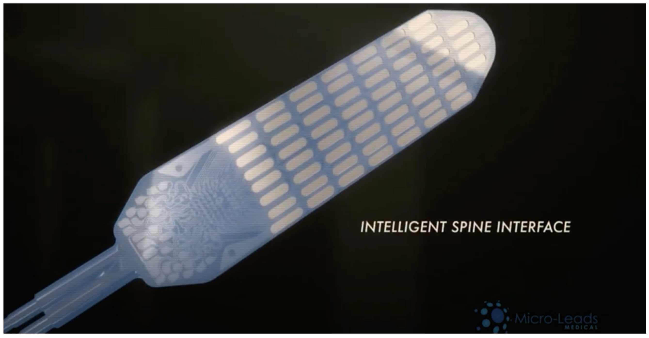
3.3. ‘SMART’ Implants and Devices

Researchers at the University of Pittsburgh, Swansea, have been developing a ‘SMART’ spinal implant device [30]. This study introduces a “self-aware” metamaterial implant designed for spinal cord injury treatment, integrating energy-harvesting and self-sensing functionalities. Utilising triboelectric nanogenerators (TENGs), the implant converts body motion into electrical energy, allowing it to operate without external power sources [31]. Constructed with triboelectric auxetic microstructures, the device autonomously collects real-time data on its environment, enhancing its monitoring capabilities. The prototype consisted of an interbody fusion cage, which demonstrated success in synthetic and cadaveric spinal models by generating up to 9.2 volts and 4.9 nanoamperes while monitoring bone healing. Mechanical fatigue testing revealed a reduction in elastic modulus (from 1.76 MPa to 1.4 MPa) and voltage output (from 2.69 volts to approximately 1 volt) after 40,000 loading cycles, indicating the need for more robust fabrication methods. By providing continuous, real-time tracking of spinal fusion progress, this device can record data that can be obtained with an ultrasonic probe [31]. This self-powered implant addresses critical gaps in spinal cord injury care. Its ability to function without external power enhances patient monitoring and recovery outcomes. Though this is a self-aware implant that can gather data in response to mechanical stimulus, these data cannot be wirelessly logged, making it difficult to adapt the implant to the patient’s unique movements. In future designs, this device may be tested on a larger cohort of clinical human trials and have a more unique patient-specific approach to spinal cord injury.
Another ‘SMART’ device was produced by a group of researchers at the University of Cambridge. This study tested a flexible circumferential spinal cord implant, enabling it to simulate and record 360 degrees around the spinal cord [32]. The device itself stimulates the spinal cord through ventral stimulation, which has been shown to be more efficient in muscle activation, through a 32-electrode array organised in a staggered linear configuration. Figure 4, below, illustrates the device concept and optical images. The ‘i360’ device was fabricated from a parylene-C-based array, which included titanium and gold electrical structures. Furthermore, to enhance the performance of the device, the impedances were decreased as polymer poly (3,4-ethylene) dioxythiophene (PEDOT) was used with a counterion, poly (styrene sulfonate) (PSS). Due to the thin nature of the device, at 4 μm, it was able to completely rotate 360 degrees around the spinal cord implant. The implant was tested on rats and human cadavers.
The animal spinal cord recording showed signal altitudes across the 32 channels, mapping the diverse patterns of evoked potentials at equal intervals. The researchers used a thresholding method to allow for the filtration of these signals based on peak amplitude and then the extraction of the signals in preparation for data analysis. The study used a supervised machine learning approach, which used a k-nearest neighbours (KNN) algorithm to locate the origins of the evoked potentials. As a result, the device showed very low latency while stimulating limb movement in a rat, which showed that the device may be able to record signals and stimulation of the spinal cord in humans. The potential application for this device includes treating paralysis, as it does not require brain surgery and could allow for spinal cord signals to be sent across a damaged spinal cord site [33]. However, there are a few limitations to this method. The study is in early stages, meaning clinical trials on humans have not yet been conducted; therefore, the effectiveness on a live human remains unknown. Furthermore, the longevity and durability of this device has not been mentioned in the research, nor has the power source for the device. This could mean various wires emitting from the patient’s spinal cord. Finally, though the implant provides real-time monitoring, it has not yet integrated wireless data logging, which makes it difficult for the device to adapt to everyone’s movements [32].

3.4. Exoskeletons and Robotic Systems

Exoskeletons have been used to increase the independence of a patient with spinal cord injury [34]. This method is predominantly used in patients who have control over their hip flexion but do not have control over the knee and ankle muscles. A study performed by Josep M. Front-Llangunes developed two robotic orthoses as part of an exoskeleton that allows for knee flexion and extension with a motor-harmonic drive actuation software [35]. Regarding the transportability of this method, the patient can carry a specifically designed backpack that contains the computer boards, various motor drives, and the battery that powers the equipment, as shown in Figure 5. Movement of the knee flexion and extension is gained through the stance-to-swing transition, which will be distinguished by the inertial recording units that are placed on top of each tibial support. The motor drivers will initiate the corresponding flexion extension that has been previously uploaded, particularly to the patient’s motor functions. This equipment was tested on a female patient who was suffering from SCI at the T11 vertebrae. Overall, the results showed an increase in gait symmetry and a significant increase in walking capability. However, there are various limitations to this design. The exoskeleton itself is bulky and has various parts to its structure. Although it aids in walking, the quality of life for the patient may be diminished by the sheer volume of requirements and materials the patient must wear to be able to do so. Furthermore, the backpack that contains the electrical components of the equipment contains a battery that must be charged, creating a window of time where the patient is not able to use the equipment, therefore diminishing the longevity of usage.
Moreover, a randomised clinical trial by Suhalka et al. studied robotic exoskeleton gait training (RGT) post spinal cord injury as a rehabilitation approach [36]. This approach used dosing through the robotic exoskeleton to maximise neuroplastic potential in patients after spinal cord injury. There were 144 participants with motor incomplete spinal cord injury, obtained within six months of injury. Participants were randomised and categorised into three groups (high, moderate, low) and a control group who only received the usual care. Participants of each group completed 24 RGT sessions with assessments until nine months post SCI. Each participant’s outcomes were measured on walking ability via walking index for spinal cord injury; health outcomes, which included gait speed, pain levels, fatigue, physical activity, general health, and quality of life; and neuroplastic effects, assessing the motor-evoked potential amplitudes. The results show that high-frequency RGT sessions led to more significant improvements in walking ability and neuroplasticity. These patients further showed a significant gain in gait speed and independence in comparison to the control group. Finally, the neuroplastic assessment results suggested that RTG enhanced spinal cord recovery [36]. The spinal cord injury evaluated was motor-incomplete, meaning the impact on complete spinal cord injury is still unknown. Though this study showed significant effectiveness in reducing the effects of spinal cord injury, there are various limitations to this research, including duration of study and battery life concerns. This means there will be periods of time where the patients are not able to use the device, reverting to paralysis.

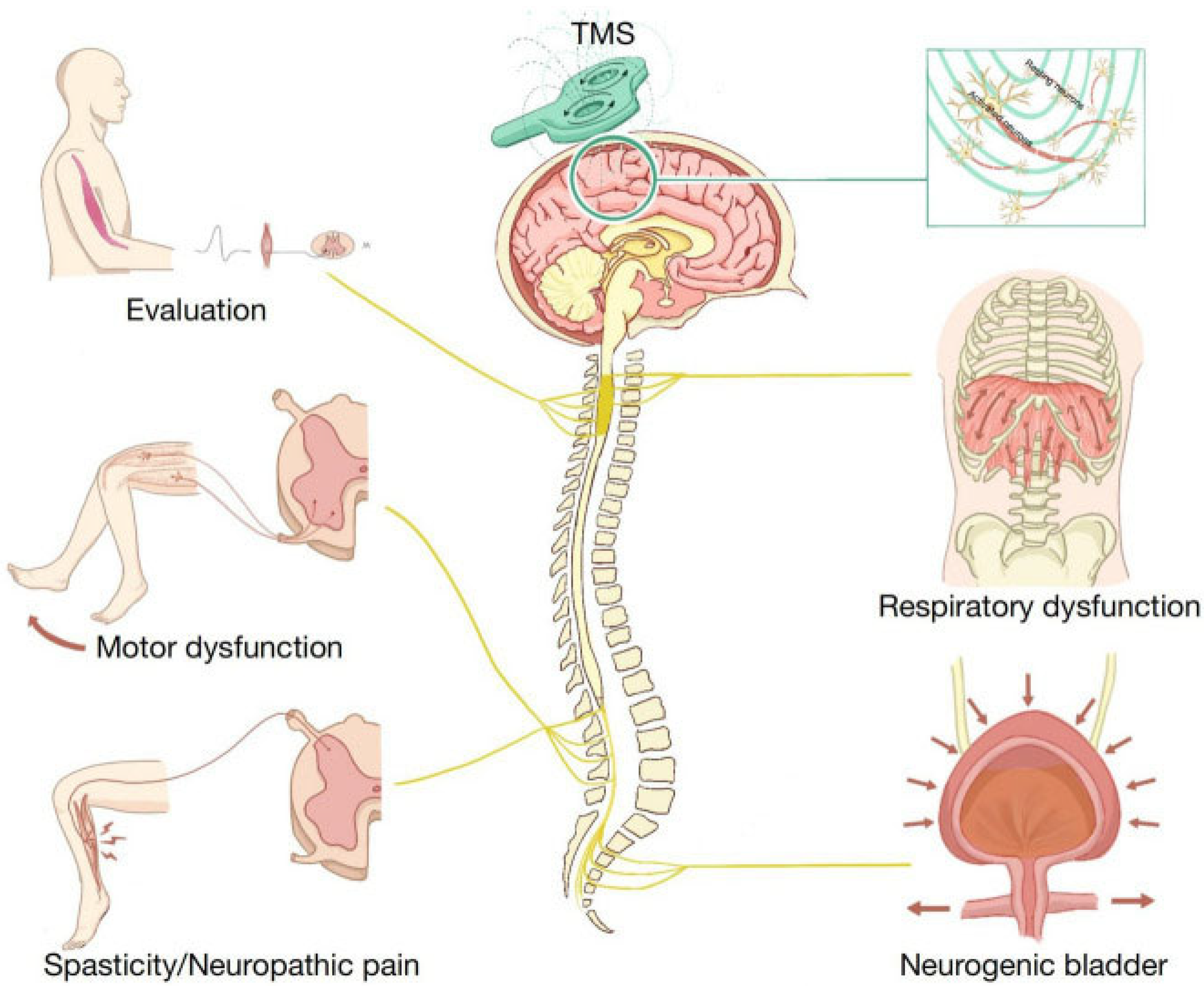
3.5. Transcranial Magnetic Stimulation (TMS)

Transcranial magnetic stimulation is a non-invasive technique that initiates cortical stimulation for the treatment of spinal cord injury [37]. The device itself, as shown in Figure 6, transmits magnetic pulses that affect the local neurons in the brain through the copper coils in the device. There are three methods of TMS.
The first method is single-pulse stimulation, which results in single magnetic pulses that are released to simulate the primary motor cortex, in turn activating the contralateral muscle [38]. This generates motor-evoked potentials (MEPs) that allow for the analysis of cortical motor control, as well as the corticospinal conduction time. MEPs can be used for neuronavigational methods such as functional mapping, which is used for muscle representation inside the motor cortex. The second method is paired-pulse TMS, which aids in the analysis of intracortical or intercortical excitability with a paired simulation. The result of this method avoids affecting the cortex through nerve feedback as the TMS is focused on a singular point, for example, the motor cortex. This method is practical for evaluating cortical excitability; however, it is not effective if magnetic stimulation is given [37]. The final method is repetitive transcranial magnetic stimulation (rTMS), which is the most common simulation method. rTMS is widely used in therapeutic research to modulate cerebral cortex activity by varying stimulation parameters. Specifically, high-frequency rTMS (≥5 Hz) increases motor cortex excitability, while low-frequency rTMS (≤1 Hz) decreases it, making it useful for controlling overactivity in targeted brain regions [38]. Paired associative stimulation (PAS) combines single-pulse peripheral nerve electrical stimulation with single-pulse TMS of the related motor cortex to alter excitability along the corticospinal pathway. The timing of these stimuli is critical: when peripheral stimulation reaches the cortex before or at the same time as TMS, corticospinal excitability increases; otherwise, it is reduced. PAS has demonstrated efficacy in promoting motor cortex plasticity and excitability, offering promising therapeutic potential.
The type of coil used in TMS significantly affects stimulation depth and precision. Circular coils provide maximum stimulation at the coil’s edge but none at the centre, resulting in broad but shallow stimulation limited to areas within 2–2.5 cm of the scalp. In contrast, Figure 6 coils deliver a more focused stimulation at the centre, also limited to superficial cortical regions. For deeper brain regions (3–6 cm), specialised coils like double cone, H-coils, and HCA coils have been developed, allowing greater penetration to target deeper neural structures. The choice of coil and stimulation parameters is critical in tailoring rTMS and PAS for specific therapeutic applications, maximising efficacy based on the target depth and desired effect [38].
A study was performed by Katarzyna Leszczynska and Juliusz Huber at the Poznan University of Medical Sciences, in which they evaluated the impact of rTMS as a treatment for patients with incomplete spinal cord injury (iSCI) [39]. The therapy protocol used rTMS on the motor cortex to enhance motor function for individuals with spinal cord injuries. The treatment averaged eight months, with patients receiving 3–5 sessions monthly. MagPro R30 and MagPro X100 stimulators were used to target lower limb regions in the motor cortex, applying frequencies between 15 and 25 Hz at an intensity of 70–80% of each patient’s resting motor threshold (RMT). Each session delivered 800 pulses per hemisphere. Before therapy, RMT values were assessed individually to personalise the stimulation, ensuring that each patient received optimal intensity without exceeding 80% of their RMT. This tailored approach aimed to maximise therapeutic outcomes by aligning stimulation intensity with each patient’s unique neurophysiological response, potentially aiding mobility and recovery in those with spinal cord injuries. Although this study showed a possibility of aiding with mobility, it does not show a significant enough success rate for long-term use. The patient, in this case, is also dependent on receiving multiple sessions of treatment a month, which may interfere with the patient’s quality of life. Overall, this method is not considered a long-term solution for spinal cord injury [39].
Moreover, Jung et al. at King’s College London were able to study the current evidence, challenges, and possible future directions for the use of negative transcranial magnetic stimulation (nTMS) in spinal cord injury [40]. This study evaluated nine clinical studies involving the use of TMS for rehabilitation in spinal cord injury. Each study had 11–115 participants and focused on incomplete and traumatic spinal cord injury across 4–8 weeks. Various TMS protocols were used in each study, including PAS, intermittent theta bursts (iTBS), and different combinations of trans-spinal direct current stimulation (tsDCS). Most of the studies analysed showed an improvement in the lower extremity motor score (LEMS), muscle activation patterns, and neuroplasticity. Though there was improvement, the study emphasised the need for standardised protocols to integrate into clinical practice as various stimulation parameters and treatment durations were assessed [40]. This research provides a valuable insight into nTMS therapy for SCI rehabilitation, yet further research is needed to refine treatment protocols and patient selection criteria.
Norgueira et al. further assessed the use of TMS in a pilot randomised clinical trial to investigate the efficacy of combining repetitive TMS (rTMS) with body weight-supported treadmill training (BWSTT) to enhance walking function in individuals with chronic incomplete spinal cord injury (iSCI) [41]. The use of rTMS was discussed as a non-invasive brain stimulation device, which resulted in the modulation of cortical excitability [42]. On the other hand, BWSTT was used, as it allows for the recurrence of a complete gait cycle and initiation of the spinal central pattern generator [43]. The study evaluated the neuromuscular activation, functional independence, and gait recovery over 4 weeks. The rTMS device used was a non-invasive electromagnetic coil that was placed over the primary motor cortex to deliver pulsed magnetic stimulation. There were 15 individuals with chronic iSCI, divided into the real rTMS and BWSTT and the mock rTMS and BWSTT groups. The real group received a 10 Hz stimulation with 1800 pulses per session, while the mock group received placebo stimulation. The BWSTT protocol included 15–20 min of moderate-intensity treadmill training per session with the assistance of a reduced weight-bearing harness. Results of the study illustrated that after twelve sessions, the real rTMS group showed significantly more positive results than the mock rTMS group. The motor function recovery was improved through enhanced neuromuscular activity for the real rTMS group, compared to the mock rTMS group [41]. Limitations to this include a small sample size of only 15 patients and the short experimental duration of four weeks, which limit generalisability and do not assess the long-term effects. Furthermore, the device used was not an implantable device, which means the effects of the device would be temporary and dependent on battery charging, resulting in a period when the patient reverts to the original SCI state.

3.6. Brain–Computer Interface Technology Using AI

The Defense Advanced Research Projects Agency (DARPA) has innovated a brain–computer interface (BCI) that bridges the gap in spinal cord injury [44]. The process of bridging the gap involves an electrode on the spinal cord just above the damaged site that collects the information the brain is sending and feeds it back to a computer. This computer then uses machine learning to recognise the signals and match them to various motor and sensory actions in the body. Once the action is calculated, it is sent back to the spinal cord through another electrode that is attached to the other side of the spinal cord damage. Messages are received by the computer, sent down the spinal cord, and relayed to the muscles. The device itself is a 64-channel electrode (as shown in Figure 7) with two wires coming out of the device externally on either side of the spinal cord injury and allows for the bidirectional flow of information. Though this device is revolutionising spinal cord injury treatments by using machine learning to directly decode brain waves into motions on the other side of the damage, there is still a major limitation. The device has two wires that protrude from the body and are connected to a computer. To increase the usability and discreteness of this device, it would require battery power to also be implanted with the electrode and embedded in a wireless connection to a computer.
Additionally, a study was conducted by researchers at Tsinghua University and Xuanwu Hospital that focused on using epidurals for a minimally invasive brain–computer interface (BCI) to reclaim motor functions after complete SCI [46]. The BCI included a wireless device called NEO, comprising a titanium implant and two electrode adaptors sealed in silicone, which was implanted into the brain with the electrode adaptors facing outwards. Externally, the epidural electrodes are connected to the implanted adaptors, which allows the device to record and stimulate pulses. The electrodes do this through two sets of coils embedded into the device. One of the sets used near-field coupling for the power supply, and the other set used Bluetooth to transmit and collect neural signals. This means the first device does not require a battery, which allows for the longevity of the device. The device was trialled on a patient with complete C4 SCI from a car accident 10 years prior, which resulted in limited movement and motor functions in the hands. Post implantation of the device, the calibration time took less than 10 min and was able to maintain a significantly high grasping score over the nine-month evaluation period of home use. Other tests performed included object manipulation, in which the patients achieved a 100% success rate in the object transfer test and the action research arm test (ARAT), which illustrated significant functional gains [46]. Though the outcomes of this approach were deemed increasingly positive, this was a single-patient clinical trial, limiting the generalisability. Furthermore, despite the procedure being minimally invasive, brain surgery still comes with high risk factors such as infection and device migration. As the process is still in the early development stages, widespread clinical implantation may not be feasible yet. This study presents a promising new approach for spinal cord injury rehabilitation, yet further clinical trials with larger sample sizes are needed to confirm its effectiveness.

4. Table of Studies

The Table 1 of studies mentioned below shows a concise representation of all 17 studies mentioned above. The table categorises each study into SCI treatment methods, research team, mechanisms of action, targeted injury level, stage of development, success metrics/validity, long term viability, safety and side effects and challenges or problems.

5. Discussion and Future Directions

This review covers current device-based interventions for spinal cord injury (SCI), highlighting both technological breakthroughs and persistent limitations. While the diversity of the approaches mentioned reflects innovation, many of the studies lack methodological thoroughness, and device limitations are often underexplored or inconsistently reported.

5.1. Methodological Quality and Evidence Gaps

In this review, several studies seem to suffer from small sample sizes, short follow-up durations, and inconsistent outcome reporting. Most early studies (2011–2018) were single-subject or small case series, limiting generalisability. Many of the studies mentioned in this review were either tested on animals or cadavers, were pilot studies, or were early phase feasibility studies, which explains the lack of sample sizes. For example, the pilot study on upper limb FES therapy included only four participants, limiting statistical significance and generalisability. Similarly, the NEO brain–computer interface was tested on a single patient, making it difficult to draw broader and more reliable conclusions. Many studies fail to report adverse event rates, dropout data, or standardised outcome measures such as ASIA scores or neurophysiological indices, which hinders cross-study comparison and meta-analysis [47]. Moreover, it is common in most of the studies reviewed that device efficacy is often reported without stratification by injury level, chronicity, or comorbidities—factors known to influence rehabilitation outcomes. These findings reinforce the need for standardised protocols, higher-powered studies, and broader inclusion criteria in future research to ensure device efficacy across diverse SCI populations. Future clinical studies may be performed with a larger cohort across a varied geographical area.

5.2. Limitations of SCI Device-Based Therapies

5.2.1. Power Supply and Device Longevity

Many of the SCI devices mentioned rely on electrical stimulation and motorised components, making power supply a universal challenge. Implantable devices such as FES, EES, and BCIs are often dependent on internal batteries with limited lifespans of around 3–5 years, which means they require surgical replacement, increasing the procedural risk. External systems, such as exoskeletons and surface FES, need frequent charging as the battery life is limited. The time needed to charge the devices creates interference in the therapy and reduces overall efficacy. Lastly, emerging technologies such as ultrasonically powered stimulators and triboelectric nanogenerators offer alternative power sources; however, these methods are still in early development phases and face the limitation of energy density and reliability [48].

5.2.2. Wireless Data Logging and Adaptability

Wireless data logging is increasingly recognised as a critical feature in modern medical devices, allowing for real-time monitoring, adaptive feedback, and personalised rehabilitation strategies. Yet, most SCI devices reviewed in this paper lack wireless data logging capabilities, which significantly limits the clinical usability and long-term adaptability. The lack of wireless logging comes with several limitations. One of the key limitations is in device evaluation and troubleshooting, as without data logging, device engineers do not have enough information to make improvements to drive innovation and progression. Without real-time data, devices cannot adjust stimulation and parameters based on patient-specific responses, which limits their ability to tailor the therapy to the individual’s needs [49]. This inability to adapt to patient response can delay clinical decision making when treating SCI. Manual data retrieval delays feedback loops, preventing timely adjustments in therapy and reducing the responsiveness to change in the patient’s condition [50].
As an exception, MyndMove FES systems and the EksoGT exoskeleton both allow for non-real-time wireless data logging through a cloud-based tracking system or software platforms [20,21,36]. Furthermore, the NEO wireless SCI has partial wireless logging capabilities. Although this device uses Bluetooth for signal transmission, it still requires external electrode adaptors and lacks full integration with any wireless data platforms [46]. This limits the device’s ability to personalise patient therapy over time. While the ultrasonically powered FES implant is self-powered and biocompatible, it is dependent on an external ultrasonically powered probe to activate the device. The device lacks wireless telemetry, which means that it cannot be logged or transmitted in real time. This will limit the device’s adaptability and limit patient progress. The SMART implant mentioned by Barri et al. [31] can harvest energy from body motion and monitors spinal fusion progress. Yet, it does not have wireless data transmission, as it requires a manual probe-based interrogation. This prevents the ability to track healing dynamically or integrate with remote monitoring systems. The i360 device, although capable of 360-degree stimulation and recording, lacks wireless data logging and must be manually accessed for signal extraction. This reduces the potential for real-time feedback and patient-specific stimulation protocols. Similarly, DARPA BCI has a 64-channel system; however, it needs wires to log data back to the computer. Without wireless logging, the system cannot adapt stimulation based on real-time neural feedback, reducing its usability and adaptive potential.

5.2.3. Surgical Risk and Anatomical Constraints

Implantable devices mentioned in this review pose significant surgical challenges. EES and BCI systems require precise electrode placement near spinal lesions or cortical regions, which increases the risk of infection, device migration, and tissue damage. Similarly, SMART implants and circumferential arrays must conform to spinal anatomy, yet long-term biocompatibility and fibrotic encapsulations remain as unresolved issues. There is a further surgical risk when surgically implanting devices, as the patient may reject the implant, causing it to malfunction and creating bigger problems. Patient viability in spinal cord morphology, injury level, and comorbidities complicates device standardisation and surgical planning.

5.2.4. Usability, Training, and Maintenance

The successful implementation of an SCI device depends on the user’s competency level as well as the continuous system upkeep. Exoskeletons require extensive training for safe use, which may take extra patient time, energy, and determination. Misalignment with joint axes can cause discomfort or injury. FES systems demand calibration and electrode placement expertise, which may not be available in a clinical setting. Not having the right professional available to the patient may mean malfunction of the device. There are further maintenance burdens, which include software updates, battery management, and hardware servicing, which can overwhelm the rehabilitation teams without the dedicated technical support [48].

5.2.5. Cost and Accessibility

SCI devices require extensive equipment and top-of-the-range biomaterials, which results in high development and operational costs. This tends to limit widespread use. Advanced devices like BCIa and robotic exoskeletons are expensive to produce and maintain, with limited insurance coverage or reimbursement pathways. Similarly, implantable systems require surgical infrastructure and follow-up care, making them inaccessible in low-resource settings. Few studies include cost-effectiveness analysis or address economic strains in device availability. For example, the Alam et al. study for the ultrasonically powered FES device implies cost reduction due to the elimination of external batteries, as well as reducing the long-term cost due to the reduction of surgical replacements and external hardware [16]. The traditional FES device, in a separate randomised control trial, had shown to cost GBP 14,300 per quality-adjusted life year, over two years, which is lower than the GBP 20,000 NHS quota, making it cost-effective for the NHS [51]. Suhalka et al. covered robotic gait training with exoskeletons; however, they did not mention cost. The cost of this was analysed in a study performed in Singapore, which showed chronic stage treatment cost SGD 440,388 (equivalent to GBP 256,233), deeming it very unfavourable [52]. Wearable FES garments, by Moineau et al., have emphasised low-cost mass production potential; however, they do not provide the unit price of the device. Yet the reusability of the product proves a future cost-effectiveness [19]. It is important to note that most studies do not report direct device prices, especially for prototypes or implants, hence why the other studies mentioned in this review do not have a cost analysis.

5.2.6. Regulatory and Health System Integration

Despite promising technological advances, many spinal cord injury devices face significant regulatory hurdles before clinical use. Especially implantable devices must undergo rigorous safety testing, biocompatibility assessments, and long-term monitoring to meet the standards set by agencies such as the FDA (U.S) or EMA (Europe). These processes can delay large-scale production and may increase development costs. These regulatory processes lack global consistency, which creates more issues with global development [53].
Implantable devices such as epidural electrical stimulators, brain–computer interfaces, and SMART implants are typically classified as Class III high-risk devices, requiring extensive premarket approval. In the United States, the FDA mandates compliance with Good Manufacturing Practices (GMP) and Quality System Regulations (QSR), while in Europe, there are rigorous conformity assessments and clinical evaluations [54]. Devices like DARPA’s 64-channel brain–computer interface must undergo both neurological safety testing and software validation. These systems often integrate AI and wireless communication, triggering additional scrutiny around cybersecurity, data privacy, and human–machine interfacing. Regulatory frameworks such as ISO 13485 (Quality Management) and ISO 14971 (Risk Management) are essential but not always harmonised globally [44]. To overcome these barriers, future SCI device development should align early with international standards (ISO 13485 and ISO 14971), incorporate cybersecurity protocols (IEC 62304, GDPR compliance), engage with regulatory bodies during design and testing phases, and prioritise modular and scalable device architectures to ease certification.

5.3. Future Directions

Future engineering advancements in SCI treatment are likely to focus on the miniaturisation of devices, the increased biocompatibility of implants, and more sophisticated algorithms for decoding brain signals and predicting motor intentions. Developing flexible and stretchable electronics will also be critical, allowing implants to more seamlessly interface with the spinal cord or brain, enhancing the longevity and comfort of the devices. Additionally, advances in wireless power and data transfer could enable fully implantable systems, eliminating the need for external power sources and further improving patient mobility and adaptability. Integrating wireless data transfer with AI and adaptive algorithms can increase device usability by using machine learning models that can predict motor intentions or optimise stimulation patterns. Future research should not only focus on the technical performance of the SCI device but also on designing devices that align with the evolving regulatory frameworks to ensure safe and effective clinical practice. As these technological improvements evolve, biomedical engineering will continue to play a crucial role in advancing SCI treatment, potentially leading to more widespread functional recovery for individuals with spinal cord injuries.

6. Conclusions

While spinal cord injury (SCI) devices hold promise for improving patient outcomes, their implantations remain constrained by the complexity of regulatory, economic, and clinical factors. This review highlights key barriers—long-term biocompatibility, wireless adaptability, cost, regulatory barriers, and risk of surgery—that collectively hinder the widespread implementation of these SCI devices. Rather than portraying that these challenges are what block the progress of said devices, it is more accurate to acknowledge that their impact varies across device type, patient populations, and healthcare contexts.
Importantly, this review reveals gaps in the current research; only a few studies offer integrated solutions that address multiple barriers simultaneously. For example, hybrid rehabilitation approaches combining robotics and neuroprostheses show potential; however, they lack standardised protocols and long-term efficacy data. Similarly, closed-loop systems and AI-driven personalisation remain underexplored in SCI contexts, despite their success in other neurotechnological areas.
To advance the field, future work should prioritise the collaboration of multiple treatment methods and bring together engineers, clinicians, regulators, and patients to co-design devices that are not only technically feasible but also clinically meaningful, as well as economically viable for patients. There should be emphasis on developing an adaptive personalised device with scalable manufacturing models for long-term and inclusive rehabilitation that reflect the diverse needs of SCI patients.

Author Contributions

Conceptualization, T.W. and K.B.; methodology, T.W.; software, T.W.; validation, T.W.; formal analysis, T.W.; investigation, T.W.; resources, T.W.; data curation, T.W.; writing—original draft preparation, T.W.; writing—review and editing, T.W. and K.B.; visualization, T.W. and K.B.; supervision, K.B. and W.B.; project administration, T.W. and K.B.; funding acquisition, K.B. All authors have read and agreed to the published version of the manuscript.

Funding

This research received no external funding.

Conflicts of Interest

The authors declare no conflict of interest.

References

  1. Rhind, J. How Many Spinal Cord Injuries Occur Each Year? JMW Solicitors. 2023. Available online: https://www.jmw.co.uk/articles/spinal-injuries/how-many-spinal-cord-injuries-each-year (accessed on 23 January 2025).
  2. Anwar, M.A.; Al Shehabi, T.S.; Eid, A.H. Inflammogenesis of Secondary Spinal Cord Injury. Front. Cell. Neurosci. 2016, 10, 98. [Google Scholar] [CrossRef]
  3. Lima, R.; Monteiro, A.; Salgado, A.J.; Monteiro, S.; Silva, N.A. Pathophysiology and Therapeutic Approaches for Spinal Cord Injury. Int. J. Mol. Sci. 2022, 23, 13833. [Google Scholar] [CrossRef]
  4. Bennett, J.; Das, J.M.; Emmady, P.D. Spinal Cord Injuries; StatPearls Publishing: Treasure Island, FL, USA, 2024. Available online: https://www.ncbi.nlm.nih.gov/books/NBK560721/ (accessed on 1 May 2025).
  5. Dowlati, E. Spinal cord anatomy, pain, and spinal cord stimulation mechanisms. Semin. Spine Surg. 2017, 29, 136–146. [Google Scholar] [CrossRef]
  6. Harrow-Mortelliti, M.; Jimsheleishvili, G.; Reddy, V. Physiology, Spinal Cord; StatPearls Publishing: Treasure Island, FL, USA, 2023. Available online: https://www.ncbi.nlm.nih.gov/books/NBK544267/ (accessed on 1 May 2025).
  7. Fallahi, M.S.; Azadnajafabad, S.; Maroufi, S.F.; Pour-Rashidi, A.; Khorasanizadeh, M.; Sattari, S.A.; Faramarzi, S.; Slavin, K.V. Application of Vagus Nerve Stimulation in Spinal Cord Injury Rehabilitation. World Neurosurg. 2023, 174, 11–24. [Google Scholar] [CrossRef]
  8. Collinger, J.L.; Foldes, S.; Bruns, T.M.; Wodlinger, B.; Gaunt, R.; Weber, D.J. Neuroprosthetic technology for individuals with spinal cord injury. J. Spinal Cord Med. 2013, 36, 258–272. [Google Scholar] [CrossRef]
  9. Gupta, A.; Vardalakis, N.; Wagner, F.B. Neuroprosthetics: From sensorimotor to cognitive disorders. Commun. Biol. 2023, 6, 14. [Google Scholar] [CrossRef]
  10. Contreras-Vidal, J.L.; Bhagat, N.A.; Brantley, J.; Cruz-Garza, J.G.; He, Y.; Manley, Q.; Nakagome, S.; Nathan, K.; Tan, S.H.; Zhu, F.; et al. Powered exoskeletons for bipedal locomotion after spinal cord injury. J. Neural Eng. 2016, 13, 031001. [Google Scholar] [CrossRef]
  11. Martin, R.; Sadowsky, C.; Obst, K.; Meyer, B.; McDonald, J. Functional Electrical Stimulation in Spinal Cord Injury: From Theory to Practice. Top. Spinal Cord Inj. Rehabil. 2012, 18, 28–33. [Google Scholar] [CrossRef]
  12. Ho, C.H.; Triolo, R.J.; Elias, A.L.; Kilgore, K.L.; DiMarco, A.F.; Bogie, K.; Vette, A.H.; Audu, M.L.; Kobetic, R.; Chang, S.R.; et al. Functional Electrical Stimulation and Spinal Cord Injury. Phys. Med. Rehabil. Clin. N. Am. 2014, 25, 631–654. [Google Scholar] [CrossRef]
  13. Griffin, L.; Decker, M.; Hwang, J.; Wang, B.; Kitchen, K.; Ding, Z.; Ivy, J. Functional electrical stimulation cycling improves body composition, metabolic and neural factors in persons with spinal cord injury. J. Electromyogr. Kinesiol. 2009, 19, 614–622. [Google Scholar] [CrossRef]
  14. Functional Electrical Stimulation Cycling for Spinal Cord Injury. Physiopedia. 2012. Available online: https://www.physio-pedia.com/Functional_Electrical_Stimulation_Cycling_for_Spinal_Cord_Injury (accessed on 1 May 2025).
  15. Berkelmans, R. Fes cycling. J. Autom. Control 2008, 18, 73–76. [Google Scholar] [CrossRef]
  16. Alam, M.; Li, S.; Ahmed, R.U.; Yam, Y.M.; Thakur, S.; Wang, X.-Y.; Tang, D.; Ng, S.; Zheng, Y.-P. Development of a battery-free ultrasonically powered functional electrical stimulator for movement restoration after paralyzing spinal cord injury. J. Neuroeng. Rehabil. 2019, 16, 36. [Google Scholar] [CrossRef]
  17. Karamian, B.A.; Siegel, N.; Nourie, B.; Serruya, M.D.; Heary, R.F.; Harrop, J.S.; Vaccaro, A.R. The role of electrical stimulation for rehabilitation and regeneration after Spinal Cord Injury. J. Orthop. Traumatol. 2022, 23, 2. [Google Scholar] [CrossRef]
  18. Gorgey, A.S.; Khalil, R.E.; Lester, R.M.; Dudley, G.A.; Gater, D.R. Paradigms of lower extremity electrical stimulation training after spinal cord injury. J. Vis. Exp. 2018, 132, e57000. [Google Scholar] [CrossRef]
  19. Moineau, B.; Marquez-Chin, C.; Alizadeh-Meghrazi, M.; Popovic, M.R. Garments for functional electrical stimulation: Design and proofs of concept. J. Rehabil. Assist. Technol. Eng. 2019, 6, 2055668319854340. [Google Scholar] [CrossRef]
  20. Balbinot, G.; Li, G.; Gauthier, C.; Musselman, K.E.; Kalsi-Ryan, S.; Zariffa, J. Functional electrical stimulation therapy for upper extremity rehabilitation following Spinal Cord Injury: A pilot study. Spinal Cord Ser. Cases 2023, 9, 11. [Google Scholar] [CrossRef]
  21. Anderson, K.D.; Wilson, J.R.; Korupolu, R.; Pierce, J.; Bowen, J.M.; O’REilly, D.; Kapadia, N.; Popovic, M.R.; Thabane, L.; Musselman, K.E. Multicentre, single-blind randomised controlled trial comparing MyndMove neuromodulation therapy with conventional therapy in Traumatic Spinal Cord Injury: A protocol study. BMJ Open 2020, 10, e039650. [Google Scholar] [CrossRef]
  22. Chalif, J.I.; Chavarro, V.S.; Mensah, E.; Johnston, B.; Fields, D.P.; Chalif, E.J.; Chiang, M.; Sutton, O.; Yong, R.; Trumbower, R.; et al. Epidural Spinal Cord Stimulation for Spinal Cord Injury in Humans: A Systematic Review. J. Clin. Med. 2024, 13, 1090. [Google Scholar] [CrossRef]
  23. Spinal Cord Injury Epidural Stimulation. Mayo Clinic. 2021. Available online: https://www.mayo.edu/research/clinical-trials/cls-20167853 (accessed on 1 May 2025).
  24. Ren, Y.; Mo, L.; Lu, J.; Zhu, P.; Yin, M.; Jia, W.; Liang, F.; Han, X.; Zhao, J. Epidural electrical stimulation for functional recovery in incomplete spinal cord injury. Cyborg Bionic Syst. 2025, 6, 314. [Google Scholar] [CrossRef]
  25. Oh, M.Y.; Choi, E.H.; Gattas, S.; Brown, N.J.; Hong, J.D.; Limbo, J.N.; Chan, A.Y. Epidural electrical stimulation for spinal cord injury. Neural Regen. Res. 2021, 16, 2367–2375. [Google Scholar] [CrossRef]
  26. Grahn, P.J.; Lavrov, I.A.; Sayenko, D.G.; Van Straaten, M.G.; Gill, M.L.; Strommen, J.A.; Calvert, J.S.; Drubach, D.I.; Beck, L.A.; Linde, M.B.; et al. Enabling task-specific volitional motor functions via spinal cord neuromodulation in a human with paraplegia. Mayo Clin. Proc. 2017, 92, 544–554. [Google Scholar] [CrossRef]
  27. Gill, M.L.; Grahn, P.J.; Calvert, J.S.; Linde, M.B.; Lavrov, I.A.; Strommen, J.A.; Beck, L.A.; Sayenko, D.G.; Van Straaten, M.G.; Drubach, D.I.; et al. Neuromodulation of lumbosacral spinal networks enables independent stepping after complete paraplegia. Nat. Med. 2018, 24, 1677–1682. [Google Scholar] [CrossRef]
  28. Harkema, S.; Gerasimenko, Y.; Hodes, J.; Burdick, J.; Angeli, C.; Chen, Y.; Ferreira, C.; Willhite, A.; Rejc, E.; Grossman, R.G.; et al. Effect of epidural stimulation of the lumbosacral spinal cord on voluntary movement, standing, and assisted stepping after Motor Complete Paraplegia: A Case Study. Lancet 2011, 377, 1938–1947. [Google Scholar] [CrossRef]
  29. Angeli, C.A.; Edgerton, V.R.; Gerasimenko, Y.P.; Harkema, S.J. Altering spinal cord excitability enables voluntary movements after chronic complete paralysis in humans. Brain 2014, 137, 1394–1409. [Google Scholar] [CrossRef]
  30. PelicanSSNadmin. Smart Implants Used to Monitor Spinal Fusion Healing—Spinal Surgery News. Spinal Surgery News, 28 June 2022. Available online: https://www.spinalsurgerynews.com/2022/06/smart-implants-used-to-monitor-spinal-fusion-healing/95622 (accessed on 1 May 2025).
  31. Barri, K.; Zhang, Q.; Swink, I.; Aucie, Y.; Holmberg, K.; Sauber, R.; Altman, D.T.; Cheng, B.C.; Wang, Z.L.; Alavi, A.H. Patient-Specific Self-Powered Metamaterial Implants for Detecting Bone Healing Progress. Adv. Funct. Mater. 2022, 32, 2203533. [Google Scholar] [CrossRef]
  32. Woodington, B.J.; Lei, J.; Carnicer-Lombarte, A.; Güemes-González, A.; Naegele, T.E.; Hilton, S.; El-Hadwe, S.; Trivedi, R.A.; Malliaras, G.G.; Barone, D.G. Flexible circumferential bioelectronics to enable 360-degree recording and stimulation of the spinal cord. Sci. Adv. 2024, 10, eadl1230. [Google Scholar] [CrossRef] [PubMed]
  33. Wraparound Implants Represent New Approach to Treating Spinal Cord Injuries. University of Cambridge. 2024. Available online: https://www.cam.ac.uk/research/news/wraparound-implants-represent-new-approach-to-treating-spinal-cord-injuries (accessed on 15 June 2025).
  34. Spinal Cord Injury Braces & Walking Systems. Reeve Foundation. Christopher & Dana Reeve Foundation. 2025. Available online: https://www.christopherreeve.org/todays-care/living-with-paralysis/rehabilitation/living-with-paralysis-rehabilitation-exoskeletons/ (accessed on 1 May 2025).
  35. Font-Llagunes, J.M.; Lugrís, U.; Clos, D.; Alonso, F.J.; Cuadrado, J. Design, Control, and Pilot Study of a Lightweight and Modular Robotic Exoskeleton for Walking Assistance After Spinal Cord Injury. J. Mech. Robot. 2020, 12, 031008. [Google Scholar] [CrossRef]
  36. Suhalka, A.; Areas, F.Z.d.S.; Meza, F.; Ochoa, C.; Driver, S.; Sikka, S.; Hamilton, R.; Goh, H.-T.; Callender, L.; Bennett, M.; et al. Dosing overground robotic gait training after Spinal Cord Injury: A randomized clinical trial protocol. Trials 2024, 25, 690. [Google Scholar] [CrossRef]
  37. Lefaucheur, J.-P. Chapter 37—Transcranial magnetic stimulation. In Handbook of Clinical Neurology; Levin, K.H., Chauvel, P., Eds.; Elsevier: Amsterdam, The Netherlands, 2019; Available online: https://www.sciencedirect.com/science/article/abs/pii/B9780444640321000370 (accessed on 1 May 2025).
  38. Wang, Y.; Dong, T.; Li, X.; Zhao, H.; Yang, L.; Xu, R.; Fu, Y.; Li, L.; Gai, X.; Qin, D. Research progress on the application of transcranial magnetic stimulation in spinal cord injury rehabilitation: A narrative review. Front. Neurol. 2023, 14, 1219590. [Google Scholar] [CrossRef]
  39. Leszczyńska, K.; Huber, J. The Role of Transcranial Magnetic Stimulation, Peripheral Electrotherapy, and Neurophysiology Tests for Managing Incomplete Spinal Cord Injury. Biomedicines 2023, 11, 1035. [Google Scholar] [CrossRef] [PubMed]
  40. Jung, J.; Patel, S.; Khan, A.; Baamonde, A.D.; Mirallave-Pescador, A.; Chowdhury, Y.A.; Bell, D.; Malik, I.; Thomas, N.; Grahovac, G.; et al. NTMS in spinal cord injury: Current evidence, challenges and a future direction. Brain Spine 2025, 5, 104234. [Google Scholar] [CrossRef]
  41. Nogueira, F.; Shirahige, L.; Brito, R.; Lima, H.; Victor, J.; Sanchez, M.P.; Ilha, J.; Monte-Silva, K. Repetitive transcranial magnetic stimulation with body weight-supported treadmill training enhances independent walking of individuals with chronic incomplete spinal cord injury: A pilot randomized clinical trial. Brain Topogr. 2024, 37, 1232–1241. [Google Scholar] [CrossRef]
  42. Valero-Cabré, A.; Amengual, J.L.; Stengel, C.; Pascual-Leone, A.; Coubard, O.A. Transcranial magnetic stimulation in basic and clinical neuroscience: A comprehensive review of fundamental principles and novel insights. Neurosci. Biobehav. Rev. 2017, 83, 381–404. [Google Scholar] [CrossRef]
  43. Minassian, K.; Hofstoetter, U.S.; Dzeladini, F.; Guertin, P.A.; Ijspeert, A. The Human Central Pattern Generator for locomotion: Does it exist and contribute to walking? Neurosci 2017, 23, 649–663. [Google Scholar] [CrossRef]
  44. Miranda, R.A.; Casebeer, W.D.; Hein, A.M.; Judy, J.W.; Krotkov, E.P.; Laabs, T.L.; Manzo, J.E.; Pankratz, K.G.; Pratt, G.A.; Sanchez, J.C.; et al. DARPA-funded efforts in the development of novel brain–computer interface technologies. J. Neurosci. Methods 2015, 244, 52–67. [Google Scholar] [CrossRef]
  45. DARPA. Bridging the Gap (BG+): AI-Enabled Solutions for Spinal Cord Injury, YouTube. 2024. Available online: https://www.youtube.com/watch?v=tyJcm-CfusI (accessed on 27 January 2025).
  46. Liu, D.; Shan, Y.; Wei, P.; Li, W.; Xu, H.; Liang, F.; Liu, T.; Zhao, G.; Hong, B. Reclaiming hand functions after complete spinal cord injury with epidural brain-computer interface. medRXiv 2024, preprint. [Google Scholar] [CrossRef]
  47. Torii, T.; Sato, A.; Iwahashi, M.; Iramina, K. Using repetitive paired-pulse transcranial magnetic stimulation for evaluation motor cortex excitability. AIP Adv. 2019, 9, 125224. [Google Scholar] [CrossRef]
  48. Eko, S. 11 Challenges in Medical Device Design and How to Solve Them. iBrandStudio. 2023. Available online: https://ibrandstudio.com/articles/challenges-in-medical-device-design-how-to-solve (accessed on 19 July 2025).
  49. Huang, Y.; Millar, J.; Long, Y.; Zhao, Y.; Haddadi, H. Towards low-energy adaptive personalization for resource-constrained devices. In Proceedings of the 4th Workshop on Machine Learning and Systems, Athens, Greece, 22 April 2024; pp. 73–80. [Google Scholar] [CrossRef]
  50. Zand, P.; Chatterjea, S.; Das, K.; Havinga, P. Wireless Industrial Monitoring and control networks: The journey so far and the road ahead. J. Sens. Actuator Netw. 2012, 1, 123–152. [Google Scholar] [CrossRef]
  51. Renfrew, L.; Paul, L.; McFadyen, A.; Rafferty, D.; Moseley, O.; Lord, A.C.; Bowers, R.; Mattison, P. The clinical- and cost-effectiveness of functional electrical stimulation and ankle-foot orthoses for foot drop in multiple sclerosis: A multicentre randomized trial. Clin. Rehabil. 2019, 33, 1150–1162. [Google Scholar] [CrossRef]
  52. Shankar, R.; Tang, N.; Shafawati, N.; Phan, P.; Mukhopadhyay, A.; Chew, E. Cost-effectiveness analysis of robotic exoskeleton versus conventional physiotherapy for stroke rehabilitation in Singapore from A Health System Perspective. BMJ Open 2025, 15, e095269. [Google Scholar] [CrossRef] [PubMed]
  53. Kumbhar, S.; Khadtare, S.; Zoman, S.; Rane, D.; Patel, V.; Bhinge, S. Regulatory complexities in Mdsap: Challenges and pathways for implantable medical device approval. Biomed. Mater. Devices 2025, 3. [Google Scholar] [CrossRef]
  54. European Commission. Regulation (EU) 2017/745 of the European Parliament and of the Council on Medical Devices. Off. J. Eur. Union. 2021, 60, 117–176. Available online: https://eur-lex.europa.eu/legal-content/EN/TXT/?uri=CELEX%3A32017R0745 (accessed on 1 May 2025).
Figure 1. Cross-section of a spinal cord, showing the distinction between the grey and white matter and the various neural root pathways surrounding the spinal cord. Arrows on cross section indicate direction of blood flow [3].
Applsci 15 08488 g001
Figure 2. The testing process of the ultrasonically powered battery-free spinal cord implant in a rat [16].
Applsci 15 08488 g002
Figure 3. (a) X-ray positioning during surgery for a T11-T12 implantation. Image shows a postoperative CT scan in the sagittal, (b) a CT scan of the coronal, (c) and a CT of the general view. (d) MRI scan of the sagittal view performed after the surgery. The red square shows the site of injury in each image, which is the focal point for analysis in this data [24].
Applsci 15 08488 g003
Figure 4. (A) A design image of the i360 device. (B) An image showing the implanted device wrapped around the spinal cord during surgery [32].
Applsci 15 08488 g004
Figure 5. Example of an exoskeleton on a female patient [34].
Applsci 15 08488 g005
Figure 6. The structure of a transcranial magnetic stimulation device. This example shows how the magnetic signals from the copper coils in the device impact the local neurons in the brain and travel through the spinal cord to illustrate various actions [37].
Applsci 15 08488 g006
Figure 7. A 64-channel brain–computer interface implantable device created by DARPA [45].
Applsci 15 08488 g007
Table 1. Concise representation of all 17 studies.
SCI Treatment MethodsAuthor + YearMechanisms of Action Targeted Injury LevelStage of Development Success Metrics/Validity Long-Term Viability Safety and Side Effects Challenges/Problems
FES Systems-Freehand and IST-12Cleveland FES Centre, 1992; Ho et al., 2014 [12]Implanted receiver-stimulator with 8–12 channels; uses shoulder elevation and myoelectric signals for hand grasp control.Cervical SCI, upper limb function restorationSecond-generation implants under developmentEnhanced grasp movement in upper limbs using myoelectric controlSecond-generation device improved function but lacked full implantation.Wires protruding from device negatively impact quality of life.Not fully implanted; external wiring is cumbersome and reduces patient satisfaction.
FES CyclingGriffin et al., 2009; Functional Electrical Stimulation Cycling for Spinal Cord Injury, 2012 [13]Electrically stimulates motor neurons and muscle fibres via pulses; tracks muscle power output during cycling.SCI with lower limb impairmentCommercially available with ongoing improvementsImproves motor and sensory neurons (ASIA scores); effectiveness at 30–60 Hz for 30-min sessions.Regular use is required to prevent muscle deterioration.High costs of equipment; time-consuming for patients.Intrusive lifestyle demands; limited results if sessions are skipped. High equipment and implantation costs.
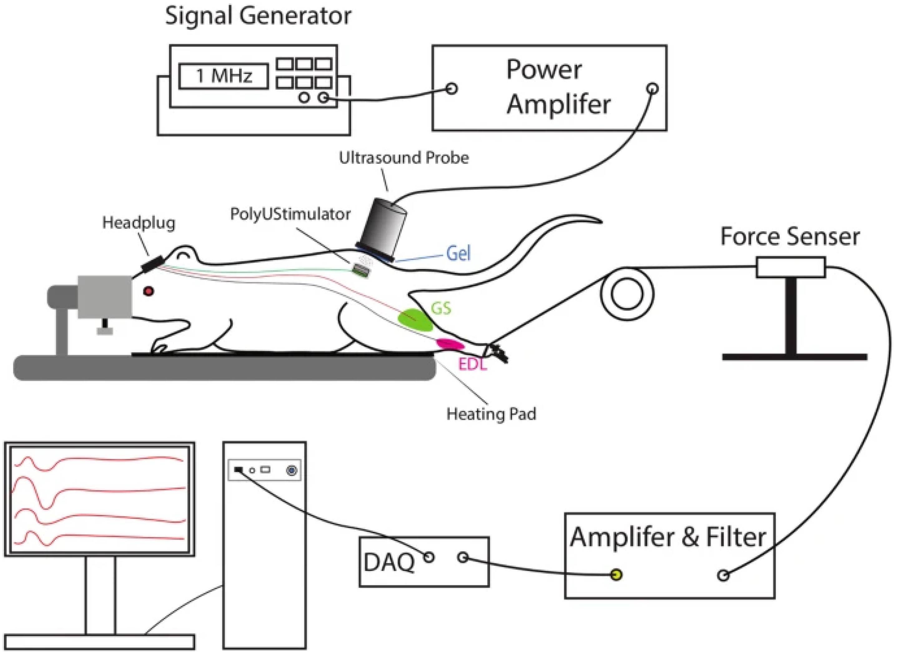
Battery-freeUltrasonically Powered FES DeviceAlam et al. Journal of NeuroEngineering and Rehabilitation, 2019 [16]Wireless stimulation using piezoelectric materials (e.g., PZT and BaTiO3) for targeted motor neuron restoration; powered via external ultrasound probe.T7 spinal cord injuries (tested on rats)Preclinical animal trialsShowed effective movement restoration in rat models; biocompatible materials used in implants.Self-powered and wireless design reduces maintenance requirements.Requires secondary implantation in the skull; risks displacement of ultrasonic probe.Dependence on external probe; risks of inconsistent use. Mortality risks due to skull implantation.
Functional Electrical Stimulation (FES)Karamian et al. [17]Electrical stimulation to activate muscles and promote neuroplasticity; implanted FES improves mobility and coordination; proposed wearable version integrates silver-threaded fabricVaries by level of SCI; specific outcomes influenced by injury location and degree of paralysis. Literature review with conceptual future directions.FES highlighted as having highest neuroplastic benefit among stimulation; no original trial data included.Implanted FES shows strong potential for long-term mobility and independence improvements.No adverse effects reported, but no human trials for wearable version yet.No experimental trials; individual variability due to SCI level: wearable FES requires future testing for efficacy and safety.
Functional Electrical Stimulation (FES) using MyndMoveBalbinot et al. [20]Application of FES to upper limb muscle via MyndMove device; uses pre-programmed protocols to stimulate muscle contraction and enhance motor control Cervical SCI (upper extremity impairment) Pilot study at tertiary SCI rehabilitation centreImprovements in muscle strength, sustained voluntary contraction, and reduction in antagonist co-contraction; enhanced fine motor control. Shows promise for restoring hand and arm function; not evaluated for long-term use or retention.No major adverse effects reported, but small sample precludes full safety profile.Small sample size limits generalisability: effectiveness may vary by SCI level; further trials needed for clinical translation.
Epidural Electrical Stimulation (EES)Chalif et al.; Dimitrijevic et al., Journal of Clinical Medicine, 2024 [22]Implantation of an epidural stimulator in the lumbar region to release electrical impulses, activating motor circuits and voluntary responses. CPGs enhance rhythmic locomotor patterns.Lumbar SCI (lower limbs)Clinical trials and patient studiesHigh success in restoring locomotor function; electrode positioning critical for effectivenessRequires battery replacement every ~5 years; could support long-term functional improvement.Risks of surgery include infection and human error. Implanted pulse generator may require replacement surgery every 5 years.Battery life limitations requiring repeated surgeries; risks from electrode attachment and implantation of pulse generator impacting mortality.
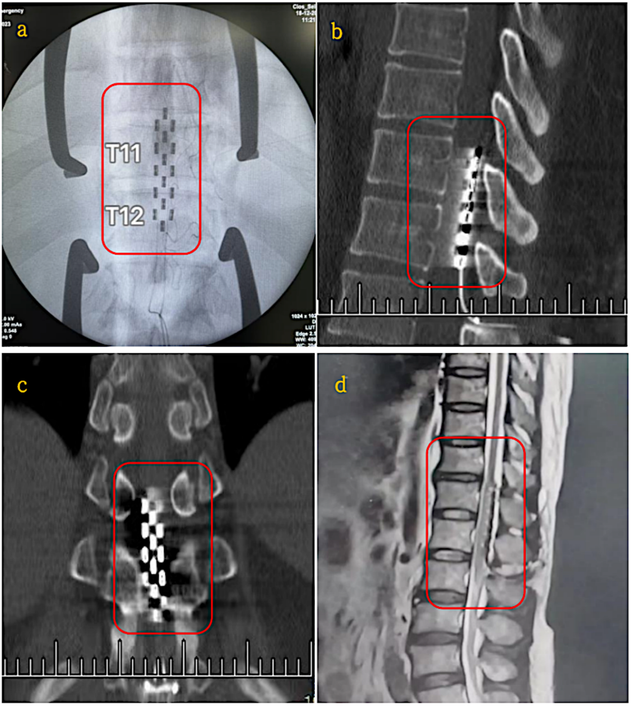
Epidural Electrical Stimulation (EES)Yihang Ren et al. [24]Electrical stimulation via implanted electrodes to activate neural pathways and enhance motor function.Incomplete SCI (Lumbar region, T11–T12) Clinical Trial (19–25 month study period) Significant improvements in sensory function (p < 0.01), muscle plasticity reductions (p < 0.0001), urinary functions in 6/11 patients and neuropathic pain relief in 4/5 patients.Battery life limited to 5 years, requiring replacement surgerySurgical risks include implantation complications, infection and device dependency.Small sample size limits generalisability, effectiveness varies with SCI severity, requires wireless improvements to enhance usability.
Epidural Electrical Stimulation (EES)Choi et al. [25]Implanted electrodes on the dorsal lumbosacral dura matter deliver electrical impulses, activating spinal networks and voluntary motor responses; combined with training.Chronic SCI Systemic review of 64 studies with 306 patients Improvements in locomotion (stepping, standing), cardiovascular and autonomic function; standing/walking in complete SCI patients.Pulse generator enables long-term stimulation, yet battery life of 5 years limits extended viability without revision.Device migration, infection risk, and post-implant complications observed; additional balance support often required.Required surgery; battery needs replacement; variability in patient outcomes; need for standardisation and training parameters for optimal results.
SMART Spinal ImplantBarri et al., 2022; University of Pittsburgh Swansea, UK [31]Self-aware metamaterial implant using triboelectric nanogenerators (TENGs) to convert body motion into energy and collect real-time data.Spinal cord injuries and spinal fusionPreclinical testing on synthetic and cadaveric spinal modelsSuccessfully generated up to 9.2 V and 4.9 nA; monitored bone healing. Mechanical fatigue testing showed durability concerns (elastic modulus reduced from 1.76 MPa to 1.4 MPa; voltage dropped from 2.69 V to ~1 V after 40,000 cycles).Self-powered, eliminating reliance on external power; continuous real-time monitoring; durability requires improved fabrication methods.No direct safety issues noted in preclinical testing, but mechanical fatigue affects performance.Cannot wirelessly log data; adaptation to patient-specific movements is limited. Larger human clinical trials and personalised designs are needed.
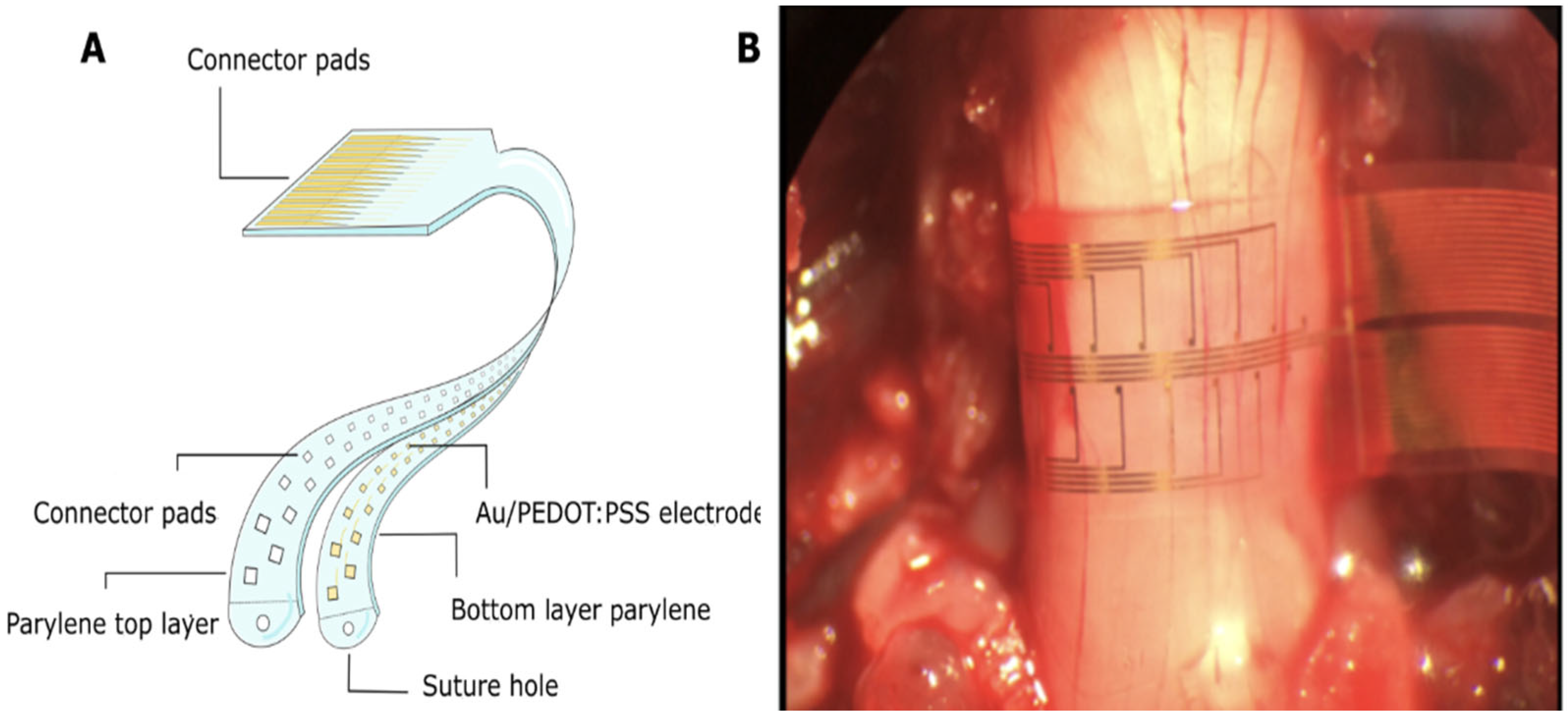
SMART-Flexible Circumferential Spinal Cord Implant (i360)Woodington et.al.
University of Cambridge Research Team [32]
32-electrode array providing 360-degree stimulation and signal recording; ventral stimulation enhances muscle activation Tested on rats and human cadavers (potential for human trials) Preclinical stage (No human clinical trials yet conducted) Low latency observed in stimulating limb movement in rats, showing potential for human use.Longevity and durability not yet tested; no mention of device power source.No brain surgery required, reducing patient risk; unknown long term safety profile. Lack of human trials, unknown long-term viability, potential wiring complexity, and no wireless data logging, limiting device adaptability.
Exoskeletons for SCIFont-Llagunes et al., 2020; Spain, Journal of Mechanisms and Robotics [35]Robotic orthoses with motor-harmonic drive actuation for knee flexion/extension; powered by backpack with computer boards, motor drives, and battery.T11 spinal cord injuryClinical testing on a female SCI patientIncreased gait symmetry and walking capability in patients with hip flexion control but no knee/ankle control.Limited by bulky design; battery requires frequent charging, creating downtime when equipment is unavailable.No significant side effects reported, but the bulkiness may negatively impact quality of life.Bulky structure; limited transportability; requires frequent battery recharging, reducing continuous use and patient convenience.
Robotic Exoskeleton Gait Training (RGT) Suhalka et al. [36]Exoskeleton-assisted movement training to stimulate neuroplasticity and improve walking ability. Motor-incomplete SCI (obtained within 6 moths of injury) Clinical Trial (144 patients, 9-month assessment period) High frequency RGT sessions led to significant improvements in walking ability, gait speed, independence, and neuroplasticity compared to control group.Battery required charging downtime, limiting continuous use.No major reported adverse effects, yet long-term impacts remain unknownSmall sample size, short term follow up, effectiveness in complete SCI unknown, battery limitations causing downtime.
Transcranial Magnetic Stimulation (TMS)Katarzyna Leszczyńska and Juliusz Huber, Poznan University of Medical Sciences, Poland, 2023 [39]Non-invasive cortical stimulation through magnetic pulses; techniques include single-pulse TMS, paired-pulse TMS, and rTMS for motor cortex modulation.Incomplete SCI (iSCI)Evaluated for iSCI therapy through clinical trials.Demonstrated potential for improved motor function through personalised stimulation of the motor cortex; used MagPro R30/X100 stimulators.Not a long-term solution due to dependency on repeated sessions; quality of life may be impacted by treatment schedule.No major safety concerns reported, but treatment requires careful intensity calibration to avoid exceeding RMT.Limited success rate for long-term outcomes; dependency on frequent sessions; not effective for deeper brain regions without specialised coils.
Negative Transcranial Magnetic Stimulation (nTMS)Jung et al.
Kings College London [40]
Uses magnetic stimulation to enhance neuroplasticity and muscle activation through various TMS protocols (PAS, iTBS, tsDCS).Incomplete and traumatic SCIClinical research review (Evaluated 9 studies)Majority of studies showed improvement in lower extremity motor score (LEMS), muscle activation patterns, and neuroplasticity. Requires standardised protocols for clinical integration.No major safety concerns reported, but effectiveness caries based on stimulation protocols. Variability in protocols, short trial duration (4–8 weeks), and small participant groups (11–115) limit generalisability.
Repetitive TMS (rTMS) combined with BWSTTNorgueira et al. [41]Non-invasive rTMS modulates cortical excitability via magnetic pulses over primary motor cortex; BWSTT promotes gait cycle and initiates central pattern generator activation Chronic incomplete SCI (iSCI), primarily cervical or thoracicPilot randomised clinical trial Real rTMS group showed greater motor function and neuromuscular activation versus placebo group after 12 sessions.Effects are temporary; dependent on session frequency and device power; no implant ensure no long term sustained impact.No serious adverse effects reported; well tolerated by participants. Small sample size; short study duration (4 weeks); non-implantable benefits require on going use; battery charging gaps may interrupt therapeutic gains.
Brain-Computer Interface (BCI)DARPA, USA, 2015 [45]64-channel electrode collects signals from the brain, processes them via machine learning, and relays them across spinal damage using electrodes.SCI (general)Prototype demonstrated; preclinical trials.Demonstrated bidirectional flow of information across spinal cord damage; uses machine learning for motor/sensory decoding.Not yet viable for long-term use due to external wires and lack of embedded wireless or battery-powered operation.External wires pose infection risk and reduce usability; no significant safety concerns with current setup.Requires battery-powered implant and wireless connection for greater usability; current design has external components, reducing practicality.
Brain Computer Interface (BCI) NEO DeviceLiu et al.
Tsinghua University and Xuanwu Hospital [46]
Wireless device (NEO) implanted in the brain; electrode adaptors record and stimulate neural signals via near-field coupling (power supply) and Bluetooth (signal transmission). Complete C4 SCIEarly development stage (Single patient trial) 100% object transfer success rate, high grasping score over 9-months, significant functional gains (ARAT improvement) Battery-free design allows for longer usability, yet long-term stability remains untested. Minimally invasive, yet still requires brain surgery, risk of infection and device migration. Single patient trial limits generalisability, clinical implantation not yet feasible, further trials needed for validation.
Disclaimer/Publisher’s Note: The statements, opinions and data contained in all publications are solely those of the individual author(s) and contributor(s) and not of MDPI and/or the editor(s). MDPI and/or the editor(s) disclaim responsibility for any injury to people or property resulting from any ideas, methods, instructions or products referred to in the content.

