Article

Fracture Resistance of CAD/CAM Onlays Versus Direct Composite Repairs for Ceramic Crown Chipping

by
Mariona Rodeja-Vazquez
,
Oscar Figueras-Álvarez
*,
Alma Aschkar-Carretero
,
Cristina Corominas-Delgado
,
Santiago Costa-Palau
,
Josep Cabratosa-Termes
and
Francisco Real-Voltas
Department of Integrated Dentistry, Faculty of Dentistry, Universitat Internacional de Catalunya, 08195 Sant Cugat del Vallès, Barcelona, Spain
*
Author to whom correspondence should be addressed.
Appl. Sci. 2025, 15(19), 10706; https://doi.org/10.3390/app151910706
Submission received: 17 September 2025 / Revised: 30 September 2025 / Accepted: 30 September 2025 / Published: 3 October 2025
(This article belongs to the Special Issue Recent Development and Emerging Trends in Dental Implants)

Abstract

Featured Application

This study provides clinicians with experimental evidence supporting the use of CAD/CAM onlays and direct composite resin as reliable and conservative alternatives for the repair of fractured metal–ceramic crowns. The findings highlight practical applications in daily dental practice, demonstrating that these techniques can withstand posterior occlusal forces and may reduce the need for complete crown replacement.

Abstract

This in vitro study evaluated the fracture resistance of metal–ceramic crowns repaired with milled hybrid resin, printed hybrid resin, lithium disilicate, and direct composite resin. One hundred crowns were fabricated, fractured under controlled loading, and 80 with standardized defects were randomly assigned to four groups (n = 20). Repairs were performed using CAD/CAM onlays or direct composite, followed by compressive testing until fracture. Mean fracture resistance values ranged from 1858.95 N to 1997 N across all groups, exceeding typical posterior occlusal forces (700–900 N). No statistically significant differences were found among groups (p = 0.200). Most failures were cohesive. These results indicate that both digital (milled and printed) and direct techniques offer sufficient strength to serve as minimally invasive and cost-effective alternatives to full crown replacement. Although limited by the in vitro design, this study supports the applicability of modern repair approaches in daily practice.

1. Introduction

Although the use of all-ceramic crowns has increased in recent years due to their superior esthetics, metal–ceramic crowns are still widely present in clinical practice. Their long history of success, favorable survival rates, and cost-effectiveness make them a reliable option, particularly in cases with limited occlusal space, parafunctional habits, or high functional demands where all-ceramic restorations may not be the best choice. For these reasons, evaluating repair strategies for metal–ceramic crowns remains clinically relevant, as a large number of such restorations are already in service and still commonly indicated in specific situations.
Digital dentistry has significantly transformed restorative dental care by enabling the fabrication of highly precise, durable, and esthetically pleasing restorations, all while reducing chair time and enhancing patient outcomes [1,2,3]. Through computer-aided design and computer-aided manufacturing (CAD/CAM) technologies, clinicians now have access to efficient and predictable treatment modalities that have elevated the standard of care in prosthodontics [3].
Despite these advancements, one of the ongoing clinical challenges in fixed prosthodontics is the management of ceramic chipping or fractures, particularly in posterior crowns, where occlusal forces are greatest and material fatigue is more likely [4]. Traditionally, such failures often led to full crown replacement, an invasive approach that may result in unnecessary removal of sound tooth structure, additional cost, and increased patient discomfort [5]. With the advent of modern adhesive dentistry and improved restorative materials, more conservative alternatives have emerged—allowing clinicians to repair rather than replace defective restorations, thereby preserving healthy dental tissues while restoring function and esthetics [6,7,8].
CAD/CAM systems enable the fabrication of partial restorations such as onlays, inlays, and overlays from high-performance materials like lithium disilicate and resin nanoceramics [9,10,11]. These materials exhibit excellent mechanical properties—including high flexural strength, superior wear resistance, and fracture toughness—making them ideal candidates for the restoration of chipped or fractured ceramic crowns, particularly in high-load bearing posterior regions [12,13,14]. Additionally, they offer excellent marginal adaptation and esthetic integration, further enhancing their clinical appeal [15].
In contrast, direct composite resin remains a widely used, minimally invasive solution that can be applied intraorally in a single appointment. This approach is often preferred for small to moderate ceramic defects due to its simplicity, cost-effectiveness, and conservative nature [16]. However, the success of direct composite repairs largely depends on meticulous surface conditioning, adhesive protocol, and material compatibility with the underlying ceramic substrate [16,17,18,19]. Techniques such as hydrofluoric acid etching, sandblasting with aluminum oxide particles, and the application of silane coupling agents or functional monomers like 10-MDP are critical for achieving a stable and durable bond at the interface [20,21,22].
To the best of our knowledge, this is the first study to directly compare milled, 3D-printed, and direct composite repairs for ceramic chipping in metal–ceramic crowns. This innovative approach highlights the potential of emerging digital workflows to provide minimally invasive and cost-effective alternatives to full crown replacement.
Among the various factors influencing clinical outcomes, fracture resistance is a fundamental parameter in determining the long-term viability of repaired restorations. Masticatory forces in the posterior region typically range from 700 to 900 N during normal function, with parafunctional habits potentially generating even higher loads [23,24]. CAD/CAM materials have demonstrated fracture resistance values that often exceed 1400 N, suggesting their suitability for stress-bearing areas [25,26]. Nonetheless, the comparative performance of different CAD/CAM materials versus direct composite resin in the context of crown repair remains insufficiently documented in the literature.
Therefore, this study aims to compare the fracture resistance of ceramic crowns repaired with three types of CAD/CAM onlays and those repaired using direct composite resin.

2. Materials and Methods

2.1. Sample Preparation

Standardized metal–ceramic crowns were fabricated on abutments with premolar morphology (7 mm height, 5 mm occlusal diameter, 6° taper, 1 mm shoulder). Premolars were selected because they represent a common clinical scenario, are subject to significant occlusal forces, and allow standardized geometry for reliable fracture testing. These abutments served as the foundation for the fabrication of all one hundred crowns.
Each crown was sequentially numbered from 1 to 100 to facilitate accurate data tracking using an Excel spreadsheet. Subsequently, all crowns were subjected to controlled loading until a chipping fracture of the ceramic occurred. Crowns that exhibited fractures involving more than 50% of the crown surface—corresponding to fractures extending beyond half of the restoration—were excluded from the study.
As a result, a total of 80 metal–ceramic crowns with standardized ceramic chipping were selected for further analysis. All selected specimens presented fractures with a maximum metal exposure of less than 10%, ensuring uniformity in the defect characteristics among the remaining samples.

2.2. Sample Size Calculation

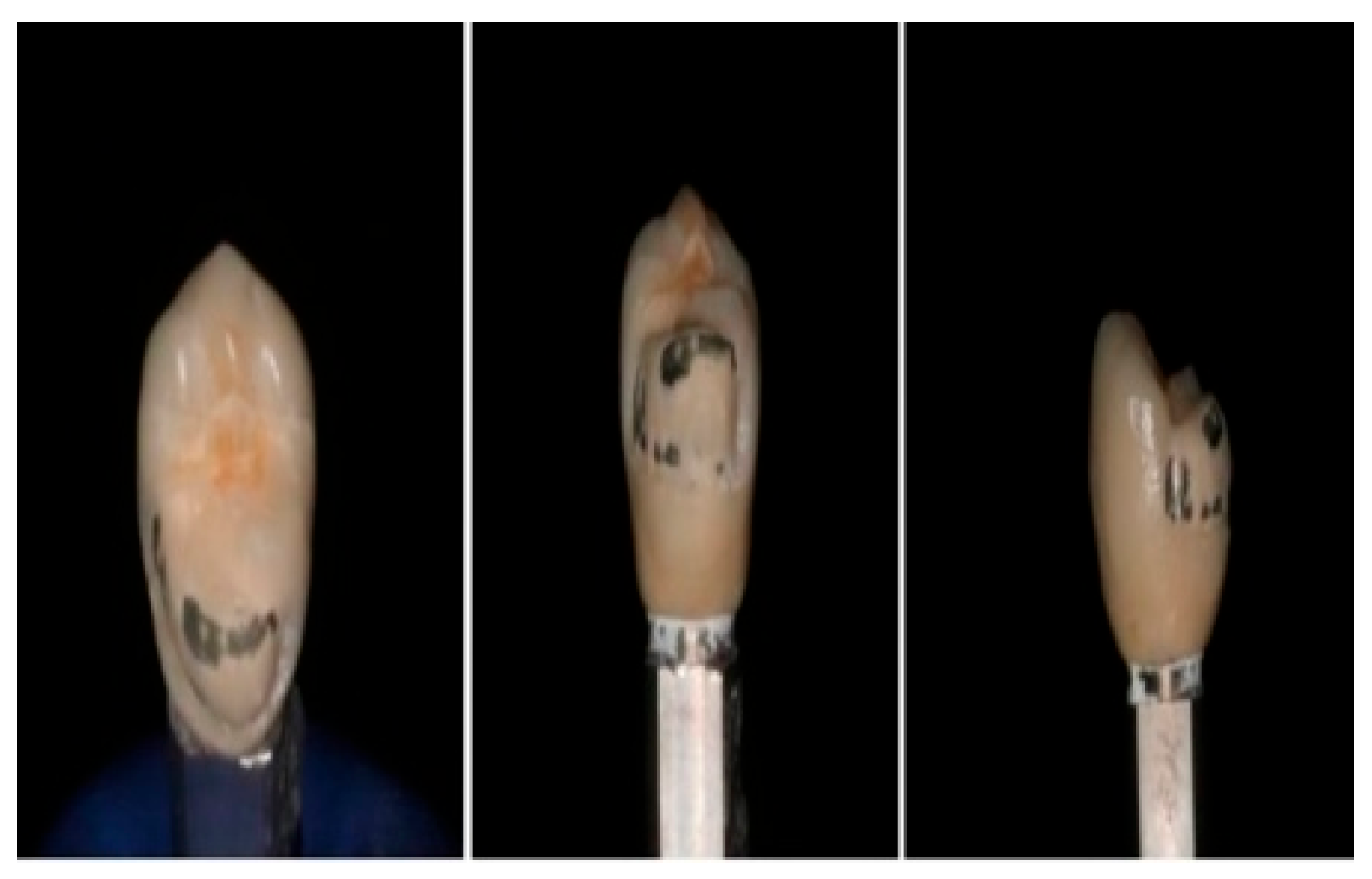
The initial sample size was set at 100 crowns; however, after excluding 20 samples due to extensive fractures, the final sample size was reduced to 80 fractured crowns. These remaining crowns were then randomly allocated into four groups of 20 (Figure 1).
The sample size for this study was determined based on prior research on dental fracture resistance. A sample size calculator from https://www.datarus.eu/aplicaciones/granmo/ (accessed on 20 June 2024) was used, incorporating data obtained from a pilot study, with an alpha risk of 0.05 and a beta risk of 0.2 in a two-sided test.
The calculation indicated that 19 samples per group were required to detect a statistically significant difference of 0.05 units or more. The standard deviation was assumed to be 605.9, and the anticipated dropout rate was 0%. This methodology aligns with the approach of Beji Vijayakumar et al. [27]. who used 20 samples per group to assess the fracture resistance of resin-based and lithium disilicate endocrowns, justifying the sample size for detecting significant differences between groups [28]. Similarly, Kavu et al. [28] employed 20 crowns per group to examine the effect of core thickness and resin cement on the fracture strength of zirconia-based crowns, obtaining valid results despite the small sample size [29].

2.3. Crown Repair Process

The 80 fractured crowns, irrespective of fracture extent, were randomly distributed into four groups of 20. They were then repaired using onlay restorations made of different materials. These included 20 milled hybrid resin onlays (Group 1: samples 1 to 20), 20 printed hybrid resin onlays (Group 2: samples 21 to 40), 20 milled disilicate onlays (Group 3: samples 41 to 60), and 20 direct resin onlays (Group 4: samples 61 to 80).
The onlay preparation involved high-speed rotary instruments (NSK Turbine, PANA MAX, NAKANISHI INC. (NSK Dental), Kanuma, Japan) and diamond burs, minimizing the exposed material and allowing the onlays enough space to fit a restoration with a minimal thickness of 1.5 mm on axial surfaces and 1 mm at the margins (Figure 2).
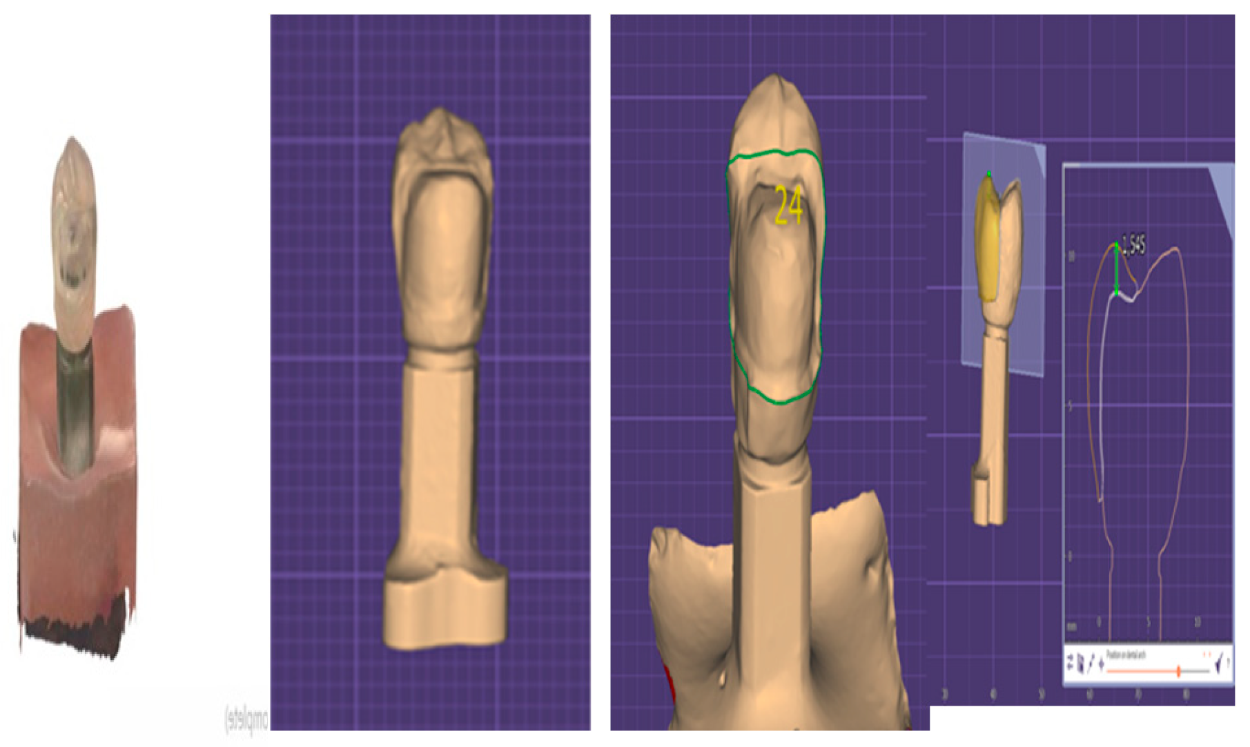
The preparations were then scanned with the Prime Scan intraoral scanner, and the scans were uploaded into proprietary dental design software (DentalCAD 3.1, exocad GmbH, Darmstadt, Germany). The onlays were designed based on the initial crown anatomy using the software’s best-fit alignment tool (Figure 3).

2.4. Restoration Fabrication

The restoration fabrication process varied by group. For Group 1, the milled hybrid resin onlays were created from Cerasmart blocks using the Sirona Cerec MC XL milling machine (Dentsply Sirona, Charlotte, NC, USA). For Group 2, the printed hybrid resin onlays were printed and post-processed using the SprintRay printer ecosystem (SprintRay, Los Angeles, CA, USA) and VarseoCrown permanent resin (BEGO, Bremen, Germany). Group 3’s milled disilicate onlays were created by milling IPS e.max CAD blocks in the Sirona Cerec MC XL milling machine and firing the resultant restoration in the recommended oven (Programat P710, Ivoclar Vivadent, Schaan, Liechtenstein). Finally, for Group 4, the direct resin onlays were manufactured using a direct technique in which twenty models were printed, and a clear silicone key (GC EXACLEAR, GC Europe NV, GC Corporation, Tokyo, Japan) was used to stamp the Gradia Plus One Body resin onto the crowns.
This material was chosen because it is a nanohybrid resin composite widely validated for direct and indirect restorative techniques. Its handling properties, high filler content, and mechanical performance make it suitable for reproducing the occlusal anatomy of fractured crowns under standardized laboratory conditions.

2.5. Cementation Process

The cementation process differed according to the materials used for the fractured crowns and onlays. The exposed metal of the fractured metal–ceramic crowns was first sandblasted, and then 4.6% hydrofluoric acid (IPS Ceramic Etching Gel, Ivoclar Vivadent, Schaan, Liechtenstein) was applied to the bonding surfaces for 2 min to achieve adequate mechanical retention. The acid was then rinsed off with water for 1 min and dried. Silane with 10-MDP (G-Multi Primer, GC Corporation, Tokyo, Japan) was applied for 60 s to achieve chemical retention, followed by drying. A dual-cure adhesive (Excite DSC F, Ivoclar Vivadent Schaan, Liechtenstein) was used.
For Group 1, the Cerasmart 270 (GC Corporation, Tokyo, Japan) onlays were sandblasted with aluminum oxide particles, cleaned, and dried. A silane coupling agent (G-Multi Primer, GC Corporation, Tokyo, Japan) was then applied to the bonding surface and dried with an air syringe. Self-adhesive resin cement (G-CEM ONE, GC Corporation, Tokyo, Japan) was then applied, and moderate pressure was exerted on the pretreated fractured crown for about 3 min. The onlay was light-cured for 1 min, excess cement was removed, and the onlay was light-cured again, with glycerin gel spread along all margins.
For Group 2, the VarseoSmile Crown Plus (BEGO, Bremen, Germany) onlays were treated similarly to the milled onlays. They were sandblasted with 60 microns of aluminum oxide particles at 2 bar for 10 s, cleaned, and dried. A silane coupling agent (G-Multi Primer, GC Corporation, Tokyo, Japan) was applied to the bonding surface and dried with an air syringe. G-CEM ONE self-adhesive resin cement was then applied, and manual pressure was applied with a metal ball instrument to ensure proper restoration seating. The onlay was light-cured for 1 min, excess cement was removed, and the onlay was light-cured again, with glycerin gel spread along all margins.
For Group 3, the lithium disilicate onlays were treated by applying 4.6% hydrofluoric acid (IPS Ceramic Etching Gel, Ivoclar Vivadent, Schaan, Liechtenstein) to the bonding surfaces of the IPS e.Max CAD onlays for 20 s to achieve mechanical retention. The acid was then rinsed off with water for 1 min and dried. Silane (Monobond Plus, Ivoclar Vivadent, Schaan, Liechtenstein) was applied for 60 s to achieve chemical retention, followed by drying. A dual-cure adhesive (Excite DSC F, Ivoclar Vivadent, Schaan, Liechtenstein) was applied (G-CEM ONE, GC Corporation, Tokyo, Japan) cement was used to bond the onlay onto the crown. The cement was light-cured for 2 s, and after excess cement removal, each side of the onlay was light-cured for 40 s, with glycerin gel spread along all margins.
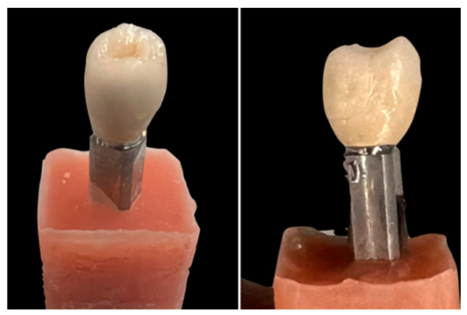
For Group 4, after treating the fractured metal–ceramic crown following the steps mentioned in the study, the composite was stamped using transparent silicone and polymerized for 20 s per side. Finally, glycerin was applied, and the surface was polished (Figure 4).

2.6. Fracture Resistance Testing

The fracture resistance of the different repair materials was assessed by applying a compressive force to all samples using a universal testing machine (Zwick Roell Z2.5, GmbH & Co. KG, Ulm, Germany) at a constant speed of 5 mm/min until a fracture occurred. Three rubber dams (Dental Dam, Jalisco, Mexico) were placed between the testing machine’s tip and the specimens to achieve a thickness of 0.6 mm, ensuring a good distribution of the compressive forces (Figure 5).
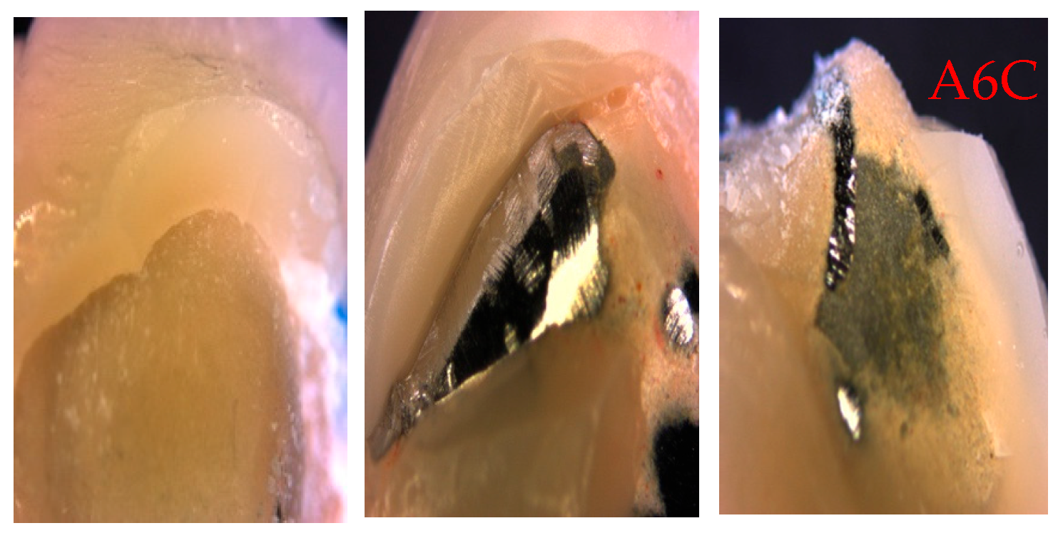
The amount of applied compression force until fracture, measured in Newtons, was recorded in an Excel spreadsheet. Finally, the 80 fractured samples were examined under a stereoscopic microscope, where photographs from the same microscope were taken to evaluate whether the fractures were adhesive or cohesive (Figure 6).

2.7. Statistical Analysis

Data analysis was conducted using IBM SPSS Statistics (Version 27.0, IBM Corp., Armonk, NY, USA). Descriptive statistics, including mean, standard deviation, and standard error, were calculated for each group. A one-way analysis of variance (ANOVA) was conducted to assess differences between the groups. A post hoc Tukey test was used to further investigate specific group differences. Normality tests (Shapiro–Wilk) and homogeneity of variance (Levene’s test) were performed to ensure the validity of the ANOVA results.

3. Results

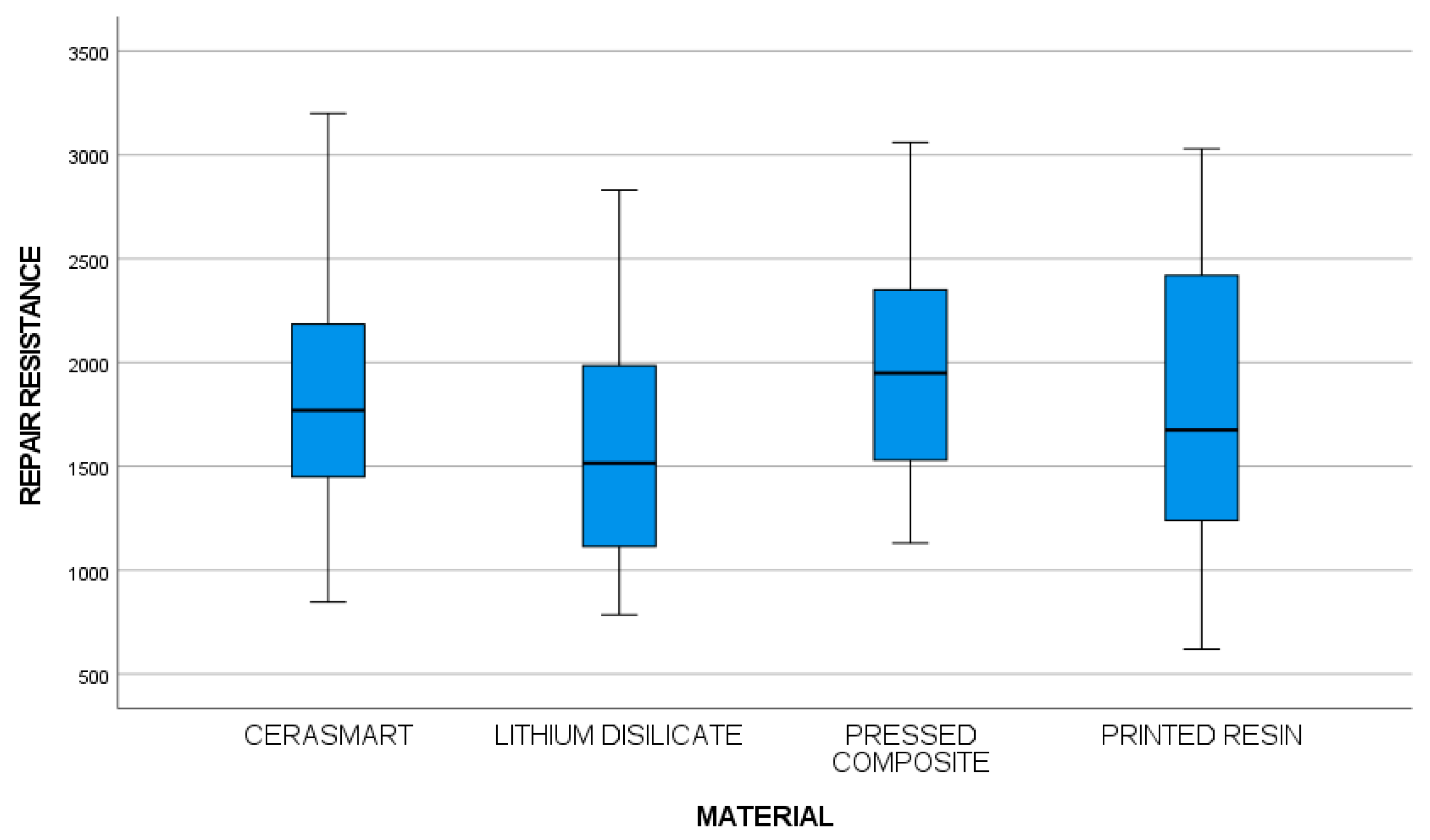
The fractured crowns underwent repair with four types of onlays (20 samples per group) and were similarly tested for fracture resistance. The fracture data, recorded in Newtons, for fracture crowns and repaired crowns with onlays made of hybrid resin, printed hybrid resin, milled disilicate, and direct resin, together with the descriptive statistics, are shown in Table 1 and Figure 7.
The data exhibited normal distribution, confirmed by standardized skewness and kurtosis, and Levene’s test confirmed the homogeneity of variances. ANOVA analysis revealed no significant differences in the fracture resistance among the groups (F-ratio = 1.58; p = 0.200).
Regarding the fracture type, most fractures were cohesive, with adhesive fractures observed only in the hybrid resin and disilicate groups. A frequency analysis was performed for both groups, with Fisher’s accuracy test indicating no significant differences.

4. Discussion

This in vitro investigation evaluated the fracture resistance of metal–ceramic crowns repaired using three types of CAD/CAM onlays—milled hybrid resin, printed hybrid resin, and lithium disilicate—as well as a conventional direct composite technique. The selection of Gradia Plus One Body for direct repairs reflects its clinical popularity, favorable mechanical properties, and ease of intraoral application. The objective was to determine whether these restorative modalities could serve as reliable alternatives to complete crown replacement in cases of ceramic chipping—a frequent complication observed in daily clinical practice, particularly in posterior restorations subjected to high occlusal loads.
Ceramic chipping in metal–ceramic crowns remains a prevalent issue, with reported incidence rates ranging from 4% to 25%, depending on material type, occlusal scheme, and clinical variables [30,31]. In many cases, full crown replacement may be excessive and invasive, especially when the underlying metal infrastructure remains intact. The current trend in restorative dentistry emphasizes minimally invasive techniques that preserve sound tooth structure, reduce treatment time, and improve cost-effectiveness—all of which can be achieved through partial repair strategies utilizing advanced adhesive protocols and digital workflows [30].
In the present study, static load-to-fracture testing was selected instead of cyclic fatigue loading. While cyclic loading better simulates intraoral functional conditions, static fracture testing provides a standardized and reproducible method that allows direct comparison among different restorative materials. Moreover, static testing remains the most widely used methodology in the literature for initial screening of fracture resistance in restorative materials, offering valuable baseline information before more complex fatigue protocols are undertaken.
CAD/CAM technology, through both milling and 3D printing, allows for the fabrication of restorations with high precision, reproducibility, and material consistency, which are critical in achieving predictable outcomes. In the present study, all tested materials exhibited fracture resistance values well above physiological masticatory forces in the posterior region (700–900 N), with values ranging from 1858.95 N to 1997 N [23,24]. These findings exceed normal posterior masticatory forces, which typically range between 700 and 900 N during function. Such high forces are rarely observed in vivo, except in parafunctional conditions such as bruxism. This suggests that all tested repair techniques can provide adequate strength to withstand physiological loads under clinical conditions.
The absence of statistically significant differences in fracture resistance among the four groups (F-ratio = 1.58; p = 0.200) suggests that milled, printed, and direct techniques offer comparable mechanical performance under controlled conditions. This is clinically relevant because it provides flexibility in material selection based on individual case factors such as cost, esthetics, availability, and operator experience. For example, milled hybrid resins like Cerasmart consistently demonstrated high resistance values and have been extensively validated in the literature for their wear resistance, toughness, and ease of handling [32]. However, studies such as those by Hofsteenge et al. [33]. highlight that factors like preparation design, restoration thickness, and occlusal morphology are critical to achieving optimal clinical results, regardless of the material used.
Notably, printed hybrid resins, a relatively new option in restorative dentistry, performed comparably well in this study. The VarseoCrown material yielded clinically acceptable fracture resistance, confirming its potential as a viable alternative in digitally guided restorative workflows. Although promising, emerging evidence Refaie et al. [34]. suggests that 3D-printed materials may undergo gradual degradation over time, warranting further research into long-term aging and fatigue performance [35].
Lithium disilicate (IPS e.max CAD), widely regarded as the gold standard for esthetic and high-strength ceramic restorations, showed a wide range of fracture resistance values, from 785 N to 2831 N. These variations may be attributed to differences in material thickness and internal adaptation. The high-end values observed here reinforce lithium disilicate’s suitability for posterior restorations, especially when adequate occlusal thickness and bonding are achieved [36].
Direct composite resin repairs also demonstrated favorable fracture resistance values (mean 1997 N), exceeding those typically reported for anterior single-unit crowns [36]. Their immediate applicability, minimally invasive nature, and cost-effectiveness make them particularly attractive in emergency or time-limited clinical settings. However, their performance is more dependent on operator skill, and their success hinges on strict adherence to surface pretreatment protocols—such as microabrasion, silanization, and the use of functional monomers like 10-MDP [37,38].
Fractographic analysis revealed predominantly cohesive failures, indicating that the tested materials themselves possess adequate internal strength. The presence of some adhesive failures, particularly in the hybrid resin and lithium disilicate groups, underscores the critical role of surface conditioning and bonding agents in repair longevity [36,37,38].
Despite the promising results, the in vitro nature of this study introduces certain limitations. Key clinical variables—such as thermal cycling, fatigue loading, moisture contamination, and biofilm presence—were not simulated. These factors can significantly influence the longevity and performance of restorations in the oral environment [37,38,39]. Moreover, although the sample size was statistically sufficient for the objectives of this study, it may not capture the full spectrum of clinical scenarios encountered in practice. Another limitation of this study is the lack of surface roughness analysis after different surface treatments. Such analysis could offer additional insights into the quality of adhesion between materials and should be included in future studies.
Nevertheless, the findings offer valuable insight into how these materials may perform under ideal conditions and provide a foundation for clinical application. From a practical standpoint, this study supports the use of CAD/CAM onlays and direct composites as effective alternatives for the conservative repair of chipped crowns. Dentists may select the appropriate repair strategy based on patient-specific factors, available materials, esthetic demands, and economic considerations.
Recent studies have also explored the integration of antimicrobial properties into CAD/CAM restorative materials, including novel resins containing plant-derived extracts. Although not evaluated in the present study, such approaches may enhance the biological performance of restorations by reducing biofilm accumulation and secondary caries. Future research could investigate whether combining fracture resistance with antimicrobial functionality further expands the clinical applicability of CAD/CAM-based repair strategies.

5. Conclusions

This in vitro study demonstrates that fractured metal–ceramic crowns can be effectively repaired using CAD/CAM-fabricated onlays (milled or printed) and direct composite resin restorations. All tested materials exhibited fracture resistance values that exceeded the maximum reported physiological masticatory forces, supporting their mechanical viability for clinical application.
Among the materials evaluated, milled hybrid resin showed the highest average resistance, confirming its reliability in high-load clinical scenarios. However, no statistically significant differences were observed among groups, indicating that all repair strategies tested are mechanically viable. Future studies should assess the long-term clinical behavior of these materials under cyclic loading, and explore the development of CAD/CAM resins with additional biofunctional properties, such as antimicrobial activity, to further improve restoration longevity.
These findings suggest that both digital and conventional techniques can provide reliable outcomes, offering clinicians a range of conservative, cost-effective, and efficient alternatives to full crown replacement.
While further in vivo research is required to validate long-term clinical performance, the present results contribute to the growing body of evidence supporting modern, minimally invasive repair strategies—reinforcing the practical integration of digital dentistry into everyday restorative protocols.

Author Contributions

Each author played a specific role that was essential for the result. M.R.-V. and F.R.-V. made the study itself. They were responsible for the clinical protocol. In addition, J.C.-T. controlled the suitable implementation of the protocol. Also, O.F.-Á. was the responsible for the statistical part of the study. A.A.-C. and S.C.-P. collected all the clinical information, and C.C.-D. wrote the article. Teamwork and good communication brought us to this submission, so we highly appreciate your consideration. All authors have read and agreed to the published version of the manuscript.

Funding

This research received no external funding.

Institutional Review Board Statement

Not applicable.

Informed Consent Statement

Not applicable.

Data Availability Statement

The data presented in this study are available on request from the corresponding author. The data are not publicly available due to ethical restrictions.

Conflicts of Interest

The authors declare no conflicts of interest.

Abbreviation

The following abbreviation is used in this manuscript:
CAD/CAMcomputer-aided design and computer-aided manufacturing

References

  1. Aziz, A.; El-Mowafy, O. Six-year clinical performance of lithium disilicate glass-ceramic CAD-CAM versus metal-ceramic crowns. J. Adv. Prosthodont. 2023, 15, 44–54. [Google Scholar] [CrossRef] [PubMed]
  2. Ille, C.E.; Jivanescu, A.; Pop, D.; Stoica, E.T.; Flueras, R.; Talpos-Niculescu, I.C.; Cosoroaba, R.M.; Popovici, R.A.; Olariu, I. Exploring the Properties and Indications of Chairside CAD/CAM Materials in Restorative Dentistry. J. Funct. Biomater. 2025, 16, 46. [Google Scholar] [CrossRef] [PubMed]
  3. Rexhepi, I.; Santilli, M.; D’Addazio, G.; Tafuri, G.; Manciocchi, E.; Caputi, S.; Sinjari, B. Clinical Applications and Mechanical Properties of CAD-CAM Materials in Restorative and Prosthetic Dentistry: A Systematic Review. J. Funct. Biomater. 2023, 14, 431. [Google Scholar] [CrossRef]
  4. Guth, J.F.; Erdelt, K.; Keul, C.; Burian, G.; Schweiger, J.; Edelhoff, D. In vivo wear of CAD-CAM composite versus lithium disilicate full coverage first-molar restorations: A pilot study over 2 years. Clin. Oral Investig. 2020, 24, 4301–4311. [Google Scholar] [CrossRef]
  5. Suganna, M.; Kausher, H.; Tarek Ahmed, S.; Sultan Alharbi, H.; Faraj Alsubaie, B.; Ds, A.; Haleem, S.; Meer Rownaq Ali, A.B. Contemporary Evidence of CAD-CAM in Dentistry: A Systematic Review. Cureus 2022, 14, e31687. [Google Scholar] [CrossRef]
  6. Gracis, S.; Thompson, V.P.; Ferencz, J.L.; Silva, N.R.; Bonfante, E.A. A new classification system for all-ceramic and ceramic-like restorative materials. Int. J. Prosthodont. 2015, 28, 227–235. [Google Scholar] [CrossRef]
  7. Sulaiman, T.A.; Suliman, A.A.; Abdulmajeed, A.A.; Zhang, Y. Zirconia restoration types, properties, tooth preparation design, and bonding. A narrative review. J. Esthet. Restor. Dent. 2024, 36, 78–84. [Google Scholar] [CrossRef]
  8. Yeslam, H.E.; Freifrau von Maltzahn, N.; Nassar, H.M. Revolutionizing CAD/CAM-based restorative dental processes and materials with artificial intelligence: A concise narrative review. PeerJ 2024, 12, e17793. [Google Scholar] [CrossRef]
  9. Albergardi, A.B.S.; Limirio, J.; Gomes, J.M.L.; Pesqueira, A.A.; Pellizzer, E.P. Effect of surface treatments on the bond strength of resin-repaired resin matrix CAD-CAM ceramic: A scoping review. J. Dent. 2025, 154, 105594. [Google Scholar] [CrossRef]
  10. Emam, M.; Eldimeery, A.; Salah, T. Fracture Resistance of Custom Lithium Disilicate Implant Restorations with Two Fabrication Techniques and Two Designs. Int. J. Oral Maxillofac. Implants 2022, 37, 677–684. [Google Scholar] [CrossRef] [PubMed]
  11. Lee, J.H.; Son, K.; Lee, K.B. Marginal and Internal Fit of Ceramic Restorations Fabricated Using Digital Scanning and Conventional Impressions: A Clinical Study. J. Clin. Med. 2020, 9, 4035. [Google Scholar] [CrossRef]
  12. Alghazzawi, T.F. Advancements in CAD/CAM technology: Options for practical implementation. J. Prosthodont. Res. 2016, 60, 72–84. [Google Scholar] [CrossRef]
  13. Belli, R.; Wendler, M.; Zorzin, J.I.; Lohbauer, U. Practical and theoretical considerations on the fracture toughness testing of dental restorative materials. Dent. Mater. 2018, 34, 97–119. [Google Scholar] [CrossRef]
  14. Sulaiman, T.A. Materials in digital dentistry—A review. J. Esthet. Restor. Dent. 2020, 32, 171–181. [Google Scholar] [CrossRef]
  15. Abu Alhuda, S.; Arossi, G.A.; Anagnostopoulos-King, F.; Alkhubaizi, Q.; Mokeem, L.S.; Melo, M.A.S.d. Current Evidence and Advances in CAD-CAM Resin Composite Blocks for Chairside Dental Restorations: Where Are We Now? A Scoping Review. Appl. Sci. 2024, 14, 10423. [Google Scholar] [CrossRef]
  16. Cui, X.; Wang, F.; Tang, K.; Dai, S.; Duan, L.; Niu, L.; Vyas, Y.; Tay, F.; Chen, J.; Zhang, X. A narrative review of the factors that affect the mechanical properties of polymer-infiltrated ceramics used for dental restorations. J. Dent. 2025, 157, 105732. [Google Scholar] [CrossRef]
  17. Lupu, I.C.; Tatarciuc, M.S.; Vitalariu, A.M.; Bobu, L.; Diaconu, D.A.; Vasluianu, R.I.; Stamatin, O.; Cretu, C.I.; Dima, A.M. Bonding Strategies for Zirconia Fixed Restorations: A Scoping Review of Surface Treatments, Cementation Protocols, and Long-Term Durability. Biomimetics 2025, 10, 632. [Google Scholar] [CrossRef] [PubMed]
  18. Moreschi, L.; De Carli, J.P.; Fuhr, M.C.S.; Dos Santos, R.B.; da Silva, P.R.P.; Corazza, P.H. Evaluation of compressive and adhesive strengths of a hybrid ceramic and 5Y-PSZ zirconia cemented with three different materials. Eur. J. Oral Sci. 2025, 133, e70017. [Google Scholar] [CrossRef] [PubMed]
  19. Szawioła-Kirejczyk, M.; Chmura, K.; Gronkiewicz, K.; Gala, A.; Loster, J.E.; Ryniewicz, W. Adhesive Cementation of Zirconia Based Ceramics-Surface Modification Methods Literature Review. Coatings 2022, 12, 1067. [Google Scholar] [CrossRef]
  20. Alsaeed, A.Y. Bonding CAD/CAM materials with current adhesive systems: An overview. Saudi Dent. J. 2022, 34, 259–269. [Google Scholar] [CrossRef]
  21. Cuzic, C.; Rominu, M.; Pricop, A.; Urechescu, H.; Pricop, M.O.; Rotar, R.; Cuzic, O.S.; Sinescu, C.; Jivanescu, A. Clinician’s Guide to Material Selection for All-Ceramics in Modern Digital Dentistry. Materials 2025, 18, 2235. [Google Scholar] [CrossRef]
  22. Nogueira, I.O.; Oliveira, P.F.G.; Magno, M.B.; Ferreira, D.; Maia, L.C.; Rabello, T.B. Does the application of an adhesive layer improve the bond strength of etched and silanized glass-ceramics to resin-based materials? A systematic review and meta-analysis. J. Prosthet. Dent. 2021, 125, 56–64. [Google Scholar] [CrossRef] [PubMed]
  23. Alenezi, A.; Alsweed, M.; Alsidrani, S.; Chrcanovic, B.R. Long-Term Survival and Complication Rates of Porcelain Laminate Veneers in Clinical Studies: A Systematic Review. J. Clin. Med. 2021, 10, 1074. [Google Scholar] [CrossRef] [PubMed]
  24. Sailer, I.; Strasding, M.; Valente, N.A.; Zwahlen, M.; Liu, S.; Pjetursson, B.E. A systematic review of the survival and complication rates of zirconia-ceramic and metal-ceramic multiple-unit fixed dental prostheses. Clin. Oral Implants Res. 2018, 29 (Suppl. 16), 184–198. [Google Scholar] [CrossRef]
  25. Harouny, R.; Hardan, L.; Harouny, E.; Kassis, C.; Bourgi, R.; Lukomska-Szymanska, M.; Kharouf, N.; Ball, V.; Khairallah, C. Adhesion of Resin to Lithium Disilicate with Different Surface Treatments before and after Salivary Contamination-An In-Vitro Study. Bioengineering 2022, 9, 286. [Google Scholar] [CrossRef]
  26. Blatz, M.B.; Alvarez, M.; Sawyer, K.; Brindis, M. How to Bond Zirconia: The APC Concept. Compend. Contin. Educ. Dent. 2016, 37, 611–617; quiz 618. [Google Scholar]
  27. Beji Vijayakumar, J.; Varadan, P.; Balaji, L.; Rajan, M.; Kalaiselvam, R.; Saeralaathan, S.; Ganesh, A. Fracture resistance of resin based and lithium disilicate endocrowns. Which is better?—A systematic review of in-vitro studies. Biomater. Investig. Dent. 2021, 8, 104–111. [Google Scholar] [CrossRef]
  28. Kavut, I.; Kulunk, S. Evaluation of the core thickness and resin cement on the fracture strength of zirconia-based multilayer computer-aided design/computer-aided manufacturing ceramic crowns. J. Int. Dent. Med. 2019, 7, 16. [Google Scholar] [CrossRef]
  29. Malpartida-Carrillo, V.; Tinedo-Lopez, P.L.; Ortiz-Culca, F.; Guerrero, M.E.; Amaya-Pajares, S.P.; Ozcan, M. Fracture Resistance of Cement-retained, Screw-retained, and Combined Cement- and Screw-retained Metal-ceramic Implant-supported Molar Restorations. J. Contemp. Dent. Pract. 2020, 21, 868–873. [Google Scholar]
  30. Sailer, I.; Makarov, N.A.; Thoma, D.S.; Zwahlen, M.; Pjetursson, B.E. All-ceramic or metal-ceramic tooth-supported fixed dental prostheses (FDPs)? A systematic review of the survival and complication rates. Part I: Single crowns (SCs). Dent. Mater. 2015, 31, 603–623. [Google Scholar] [CrossRef]
  31. Teichmann, M.; Gockler, F.; Weber, V.; Yildirim, M.; Wolfart, S.; Edelhoff, D. Ten-year survival and complication rates of lithium-disilicate (Empress 2) tooth-supported crowns, implant-supported crowns, and fixed dental prostheses. J. Dent. 2017, 56, 65–77. [Google Scholar] [CrossRef]
  32. Ashour, A.M.; El-Kateb, M.M.; Azer, A.S. The effect of two preparation designs on the fracture resistance and marginal adaptation of two types of ceramic crowns using CAD/CAM technology (In vitro study). BMC Oral Health 2024, 24, 1065. [Google Scholar] [CrossRef]
  33. Hofsteenge, J.W.; Carvalho, M.A.; Botenga, E.L.F.; Cune, M.S.; Ozcan, M.; Magne, P.; Gresnigt, M.M.M. Effect of preparation design on fracture strength of compromised molars restored with direct composite resin restorations: An in vitro and finite element analysis study. J. Prosthet. Dent. 2024, 131, 1150–1158. [Google Scholar] [CrossRef] [PubMed]
  34. Refaie, A.; Bourauel, C.; Fouda, A.M.; Keilig, L.; Singer, L. The effect of cyclic loading on the fracture resistance of 3D-printed and CAD/CAM milled zirconia crowns-an in vitro study. Clin. Oral Investig. 2023, 27, 6125–6133. [Google Scholar] [CrossRef] [PubMed]
  35. Gungor, M.B.; Nemli, S.K.; Bal, B.T.; Tamam, E.; Yilmaz, H.; Aydin, C. Fracture resistance of monolithic and veneered all-ceramic four-unit posterior fixed dental prostheses after artificial aging. J. Oral Sci. 2019, 61, 246–254. [Google Scholar] [CrossRef] [PubMed]
  36. Channarong, W.; Lohawiboonkij, N.; Jaleyasuthumkul, P.; Ketpan, K.; Duangrattanaprathip, N.; Wayakanon, K. Fracture resistance of bonded ceramic overlay restorations prepared in various designs. Sci. Rep. 2022, 12, 16599. [Google Scholar] [CrossRef]
  37. Oyar, P.; Durkan, R. Effect of cavity design on the fracture resistance of zirconia onlay ceramics. Niger. J. Clin. Pract. 2018, 21, 687–691. [Google Scholar] [CrossRef]
  38. Alassar, R.M.; Samy, A.M.; Abdel-Rahman, F.M. Effect of cavity design and material type on fracture resistance and failure pattern of molars restored by computer-aided design/computer-aided manufacturing inlays/onlays. Dent. Res. J. 2021, 18, 14. [Google Scholar] [CrossRef]
  39. Jurado, C.A.; Mekled, S.; Abuhammoud, S.; Azpiazu-Flores, F.X.; Vegh, D.; Wang, T.; Morton, D. Fracture resistance of partial and complete coverage veneers and ceramic crowns for maxillary central incisors. J. Prosthet. Dent. 2024, 132, 420.e1–420.e6. [Google Scholar] [CrossRef]
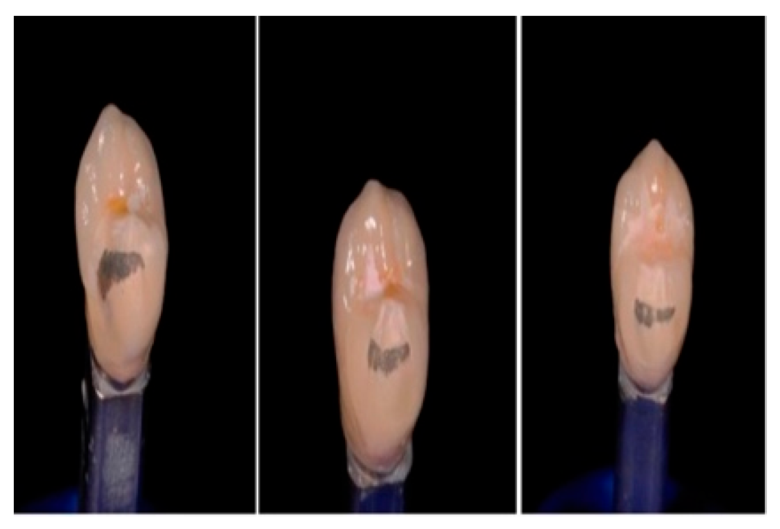
Figure 1. Metal–ceramic crowns fracture.
Applsci 15 10706 g001
Figure 2. Preparation of the onlays on the fractured metal ceramic crowns.
Applsci 15 10706 g002
Figure 3. The scanning process with PrimeScan and subsequent CAD design of the onlay using DentalCAD 3.1 software (exocad GmbH), including the preparation scan, STL generation, margin determination, and final onlay design.
Applsci 15 10706 g003
Figure 4. Onlays cemented on the metal–ceramic crowns.
Applsci 15 10706 g004
Figure 5. Fractures of the onlays used to repair the metal–ceramic crowns.
Applsci 15 10706 g005
Figure 6. Different types of onlay fractures observed under the microscope, including the fracture of a crown repair with a Cerasmart onlay, a printed composite onlay, and a lithium disilicate onlay.
Applsci 15 10706 g006
Figure 7. Fracture resistance (in N) of the repair crowns using different materials.
Applsci 15 10706 g007
Table 1. Descriptive analysis of the data. Group A: The original metal–ceramic crowns; Group B: The group with the repair hybrid milled onlays; Group C: The group with the disilicate ceramic milled onlays; Group D: The group with the pressed resin onlays; Group E: The group with the printed resin onlays.
SampleAverageMedianStandard DeviationMinimumMaximumRangeStandardized KurtosisStandardized Skew
GROUP A: Original metal ceramic crowns (initial crowns)1002969.883033.50742.91136341532790−0.62−0.37
GROUP B: Repair hybrid milled (Cerasmart) onlay crowns201901.351770.00668.54847.03200.02353.0−0.240.75
GROUP C: Repair disilicate ceramic onlay crowns201578.81444.55,185,97878528312046.00.450.23
GROUP D: Repair pressed resin onlay crowns20199718755,665,2311130306019300.740.21
GROUP E: Repair printed resin onlay crowns201826.81480761,221620303024100.870.16
Disclaimer/Publisher’s Note: The statements, opinions and data contained in all publications are solely those of the individual author(s) and contributor(s) and not of MDPI and/or the editor(s). MDPI and/or the editor(s) disclaim responsibility for any injury to people or property resulting from any ideas, methods, instructions or products referred to in the content.