Article

Optimizing Growth Conditions and Biochemical Properties of Chondracanthus acicularis (Rhodophyta) in Laboratory Settings

1
MARE-Marine and Environmental Sciences Centre/ARNET-Aquatic Research Network, ESTM, Polytechnic University of Leiria, Edifício Cetemares, Av. Porto de Pesca, 2520-641 Peniche, Portugal
2
School of Tourism and Maritime Technology, Polytechnic University of Leiria, Rua do Conhecimento 4, 2520-614 Peniche, Portugal
3
MARE-Marine and Environmental Sciences Centre/ARNET-Aquatic Research Network, Department of Life Sciences, University of Coimbra, Calçada Martim de Freitas, 3000-456 Coimbra, Portugal
*
Author to whom correspondence should be addressed.
Appl. Sci. 2025, 15(2), 810; https://doi.org/10.3390/app15020810
Submission received: 10 December 2024 / Revised: 9 January 2025 / Accepted: 14 January 2025 / Published: 15 January 2025
(This article belongs to the Special Issue Harnessing Microalgae and Seaweed for the Food Sector)

Abstract

:
This study aimed to evaluate the laboratory cultivation of Chondracanthus acicularis, focusing on key environmental parameters such as nutrient levels and light exposure. The results provide insights into the optimal growth conditions and biochemical composition of C. acicularis, which are crucial for its sustainable exploitation in industrial applications. Significant differences in the relative growth rate (RGR) and productivity (Y) were found between the different treatments. Seaweed grown on Provasoli (PES) Medium with white LED light and red LED light showed the best growth rates. Negative growth was observed in treatments with Nutribloom plus®, and blue LED light. The proximate composition analysis revealed a high moisture content across all treatments, with significant differences in ash and organic matter content between the treatments. The use of LED light played a crucial role in optimizing growth by influencing photosynthetic efficiency and pigment production. The proximate composition varied significantly between treatments, especially ash and organic matter. Light and nutrient conditions also influenced pigmentation and colour characteristics, with significant changes in phycoerythrin, phycocyanin, and chlorophyll concentration. PES treatments consistently showed the highest colour variation. These findings highlight the influence of environmental conditions on seaweed growth, productivity, pigmentation, and proximate composition, and provide valuable insights for optimized cultivation strategies.

1. Introduction

Oceans are a unique source of natural resources, offering a wealth of biodiversity that has long inspired scientific exploration and innovation [1]. However, until the mid-20th century, the challenge of reaching the depths of the oceans limited our understanding of many marine species. Advances in diving technology in the 1970s and 1980s allowed scientists to study previously unexplored marine organisms, including a wide range of algae and invertebrates. Today, the rapid growth in the human population, climate change, and environmental degradation emphasize the urgent need for the sustainable management of marine resources [2]. Among marine organisms, seaweeds have traditionally been harvested from the wild for a variety of uses, including food, animal feed, and soil enhancers [3,4]. Seaweeds are renowned for their high nutritional value, with carbohydrate, protein, lipid, and ash contents ranging approximately from 31 to 68%, 9 to 47%, 0.1 to 9.8%, and 7 to 39%, respectively [5]. In recent decades, however, seaweeds have become a prominent source of bioactive compounds with applications in the food, medical, and biotechnological industries [5].
The intertidal environment, in which many seaweeds thrive, exposes these organisms to fluctuating conditions of humidity, temperature, salinity, sunlight, and water movement, which in turn influences their biochemical composition and structural adaptations [6,7]. Among the diverse seaweed species, red algae (Rhodophyta) are particularly notable for their high-value polysaccharides, such as carrageenans [8]. The compounds derived from red seaweeds such as Chondrus crispus, Eucheuma spp., Kapaphyccus spp., Gigartina spp., Chondracanthus acicularis, and other Gigartinales possess thickening, gelling, and stabilizing properties, making them highly sought after in the pharmaceutical and cosmetic industries [9,10,11]. In addition, red seaweeds are a rich source of unique bioactive compounds, including phycobilins, mycosporine-like amino acids (MAAs), and various other secondary metabolites. Phycobilins, such as phycoerythrin and phycocyanin, are water-soluble pigments that act as accessory light-harvesting molecules, allowing red algae to absorb and utilize a broader spectrum of light. These pigments possess potent antioxidant and anti-inflammatory properties, making them valuable compounds in nutraceuticals and medical formulations, and their fluorescent properties enable their use as markers in biomedical imaging and diagnostics [12,13,14]. Mycosporine-like amino acids, which protect algae from ultraviolet (UV) radiation, are particularly promising as natural UV filters in sunscreens, providing a safe alternative to synthetic UV-blocking agents [15,16,17]. Carrageenans are versatile polysaccharides with a wide range of applications in various industries, as they have unique properties such as biodegradability, biocompatibility and functional versatility and have been widely used in the food industry as gelling, thickening, and stabilizing agents [18,19]. Additionally, red seaweeds are rich in other bioactive compounds including other polysaccharides, polyphenols, and fatty acids, exhibiting antimicrobial, antiviral, and antitumor activities. Together, these compounds not only contribute to the survival and resilience of red seaweeds in harsh marine environments but also provide a sustainable source of high-value bioactive molecules with applications in pharmaceuticals, cosmetics, and functional foods [12,20].
The carrageenan industry has expanded rapidly in recent years, with demand now far outstripping what can be sustainably supplied from wild seaweed stocks and growing at an annual rate of over 7.5%. This demand is increasingly being met by aquaculture, as wild harvesting becomes less viable [21]. With global seaweed production set to reach a record 37.8 million tons in 2022, the sustainable aquaculture of seaweed has emerged as an essential strategy to meet increasing demand while preserving natural habitats [22]. Beyond supporting food, pharmaceutical, and material supply chains, seaweed aquaculture contributes to carbon sequestration, habitat restoration, and nutrient cycling, making it a promising approach to achieving environmental and economic sustainability [23].
Cultivating seaweed has become essential to ensure a sustainable and abundant biomass supply to meet the rising demand from various industries. Traditionally harvested from natural habitats, red seaweed harvesting has reached a point where continued wild collection could threaten local ecosystems and reduce biodiversity. Transitioning to aquaculture produces high-quality biomass on a large scale, safeguarding wild populations while supporting blue economy industries [21,23]. Also, cultivating indigenous seaweed species, rather than introducing non-native varieties, is essential to protect ecosystem integrity and prevent potential invasions that can disrupt native biodiversity and marine habitats [24,25]. However, the main commercial sources of carrageenan are the Asian species Eucheuma and Kappaphycus species, which account for over 90% of global production [9]. As there are only a few cultivated species, it is important to find indigenous species that can be domesticated to increase the number of cultivated species and provide the market with a wide range of biological raw materials [25].
The genus Chondracanthus has previously been cultivated in North America [26], Latin America [27,28], and in the Iberian Peninsula [29]. The interest in the cultivation of Chondracanthus arises from its richness in carrageenans, with yields of up to 61.1% with the gametophyte-producing hybrid kappa-iota carrageenans [30,31]. The high protein and fibre contents of the genus, together with the mineral content, also highlight the possibility of using Chondracanthus as a nutraceutical [32]. In addition, the genus has antifungal [33] and anti-hypertensive [34] activities.
As a cosmopolitan perennial red alga with a wide Atlantic and Mediterranean distribution [23], Chondracanthus acicularis (formerly Gigartina acicularis) is usually found in abundance in the intertidal zone, where it forms a dense turf (Figure 1a). It has also been indicated that the species is resistant to urbanization [35]. The species exhibits a dense, cartilaginous morphology, with irregular branching (Figure 1b) that enables it to attach firmly to substrates in high-energy coastal environments, contributing to its resilience and adaptability [36,37]. The species reproduces mostly by vegetative reproduction, while sporogenesis is uncommon [38] C. acicularis shows a combination of photoperiod and temperature control of gametogenesis, and cystocarps and tetrasporangia are only recorded between late autumn and winter, being influenced by temperature and photoperiod [24]. The species is known to grow between 13 and 20 °C and produces tetrasporangial sori between 16 and 20 °C. At 16 °C, the tetrasporangia formation shows a true photoperiodic response, with spore release being higher during shorter daylengths [38]. Gametogenesis is also confined to a relatively narrow temperature and photoperiod (14–18 °C and 12:12 h) [39]. Hence, a high seasonality can be observed, with populations increasing in the number of tetraspores in autumn while growing later in spring [36,40].
C. acicularis is an important species owing to its high yield of co-polymers of kappa-iota carrageenan [31,41,42], while lambda-type carrageenan has been identified by FTIR [43]. Due to their acknowledged applications, C. acicularis carrageenans have been tested for other applications besides food supplements, such as in food packaging [44], for biostimulant effect on the germination and growth of plants [45,46,47], and for biomedical applications, namely for their antiviral properties [48,49].
Despite its commercial potential, large-scale cultivation of C. acicularis presents challenges due to its complex, isomorphic, triphasic life cycle [37]. The study by Bodian et al. [50] investigated in vitro propagation methods to support the sustainable production of C. acicularis, aiming to meet rising market demand while conserving wild populations. Understanding the impact of environmental factors on the growth and physiology of C. acicularis in controlled settings could pave the way for more efficient cultivation practises and broaden its potential industrial applications.
This study aimed to evaluate the laboratory cultivation and biochemical characterization of Chondracanthus acicularis. To do so, key environmental parameters such as nutrient levels and light exposure were analyzed. Additionally, the biochemical analysis of both cultivated and wild biomass was also assessed, providing insights into the adaptive mechanisms of this species in response to varying nutrient and light conditions. By advancing knowledge of the optimal growth conditions for C. acicularis, this research seeks to support the sustainable exploitation of this species for industrial applications, particularly in fields reliant on high-quality and high-value bioactive compounds.

2. Materials and Methods

2.1. Sampling and Acclimatization

The biomass of Chondracanthus acicularis (Roth) Fredericq 1993 used in this study was collected at Barcos beach, in Baleal (39°22′35″ N, 9°20′24″ W); at Praia Porto Batel beach, in Atouguia da Baleia (39°19′09″ N, 9°21′21″ W); and at Tamargueira beach, in Figueira da Foz (40°09′58″ N, 08°53′01″ W), in Portugal, during low tide, and was transported to the laboratory in dark, cool boxes. In the laboratory, the biomass was carefully washed with filtered seawater and then cleaned to remove epiphytes, debris, and necrotic parts.
Part of the biomass was frozen to characterize the wild seaweed’s biomass and the rest was acclimatized to laboratory conditions for at least 5 days, exposed to artificial white LED light (40 ± 1 µmol photons m−2 s−1) (Input Kilight 18 W 1900 lm G13 AC175-265V 300° 6000K 032109 RoHS) provided by two horizontal LED bulbs at the level of the culture flask (Figure 2a), under a 12:12 h photoperiod (light/dark), with constantly aerated seawater (35 ± 2 psu) in a temperature-controlled room at 20 ± 1 °C, in open 5 L trays. The acclimatized biomass was then prepared for the subsequent trials.

2.2. Cultivation Methods

For each test, 24 g of fresh algal biomass was weighed and placed in a 6 L flat-bottom flask (density 4 g·L−1) with constant aeration by air filtered through a 20 μm filter (Sartorius StedimBiotech GmbH, Göttingen, Germany). The culture conditions were the same as detailed above. Different tests were carried out, as summarized in Table 1. Three flasks were used for each medium and light source (n = 3). All the trials lasted 14 days, with the medium being renewed on the seventh day.
The nutrient assay tested the growth of the algae when exposed to three different culture media: Modified Von Stosch Enriched Medium (VSE), suitable for the cultivation of macroalgae, especially red algae, and for the isolation of spores, using 1 mL of medium per litre of salt water [51]; Provasoli Enriched Seawater (PES) Medium, which is often used to grow kelps and red macroalgae [52], using 10 mL of medium per litre of seawater; and Nutribloom plus® (NB), a commercial culture medium (Necton, Olhão, Portugal), at a concentration of 1 mL per litre of seawater [53]. The chemical compositions of the three nutrient media are stated in the Supplementary Materials (Table S1). The air was filtered through a 20 μm filter (Sartorius StedimBiotech GmbH, Göttingen, Germany) and germanium dioxide (GeO2, 1 mL red seaweeds L−1) was added to all media to prevent the proliferation of diatoms.
Once the best medium for the growth of C. acicularis was found, a light wavelength test was carried out, testing the growth of the algae when exposed to red LED light (40 ± 1 µmol m−2 s−1, 657 nm) or blue LED light (40 ± 1 µmol m−2 s−1, 455 nm). Red and blue LED lights (AquaBar T-Series Single 1200 mm COLOURPLUS) were arranged in a horizontal row, at the level of the culture flask (~10 cm high), approximately 5 cm from the flasks. The experimental setup can be seen in Figure 2.

2.3. Growth Measurements

Biomass weight was recorded at the beginning, middle, and end of each trial using an analytical scale (Sartorius, TE124S—120 g × 0.0001 g). Excess water was carefully removed with paper towels before each weighing. The relative growth rate (RGR) measures the speed at which an organism or culture grows relative to its initial size over a given period. Productivity (Y) refers to the total amount of biomass generated per unit volume (or area) over a given period and is crucial for assessing the ability of a production system to generate an economic or biological yield. The RGR and Y were calculated using Equations (1) and (2), respectively, adapted from Patarra et al. [54]:
R G R %   f w   d a y 1 = ln f w ln i w ÷ t × 100
Y ( g   d w   m 3 d a y 1 ) = 0.2876 × [ ( f w i w ) ÷ t ) ] ÷ V
where iw and fw stand, respectively, for the initial and final fresh weights expressed in grams; t stand as time in days; 0.2876 (based on 42 samples) is the ratio between the dry weight and the fresh weight of C. acicularis; and V is the volume of the 6 L flasks used, in m3.

2.4. CIELAB Colour System

Colour measurements were performed using a Konica Minolta CR-400 Chroma Meter (Minolta Camera Co., Marunouchi, Chiyoda, Japan). Fresh samples were assessed at the beginning and end of each trial and each medium renewal, with the entire sensor surface pressed carefully onto the seaweed, which had been pre-dried with paper towels. Tristimulus colour coordinates (CIELAB-system) were used to measure the degree of lightness, which stands for perpetual lightness ranging from black to white (L* = 0: black; L* = 100: white); redness (a*), which represents the coordinate along the red to green (+a* = red, −a* = green); and yellowness (b*), which is the coordinate along the yellow to blue (+b* = yellow, −b* = blue). All measurements were made in triplicate.
These values were used to calculate chromaticity (C*), which is the saturation of the colour (Equation (3)), and hue (° h), which is the tone of the biomass Equation (4) [55]:
C * = a * 2 + b * 2
° h = a r c t g   ( b ÷   a )
Finally, to assess the effect of the different treatments (nutrients and wavelength) on seaweed colour, the total colour difference was calculated using Equation (5) [55]:
ΔE = [(L* − L0*)2 + (a* − a0*)2 + (b* − b0*)2]1/2
where ΔE quantifies the overall colour difference in a sample (L*, a*, b*) when compared to wild biomass used at the beginning of the trial (L0*, a0*, b0*).

2.5. Biochemical Profiling

The biomass from each trial and wild biomass were analyzed to determine the biochemical composition of the species. Fresh biomass was used to assess fresh weight, organic matter, ash, and pigment content. All biochemical analyses were carried out in duplicate.

2.5.1. Moisture, Organic Matter, and Ash Content

The AOAC method was used to determine the moisture and ash content of the seaweed [56]. Briefly, 1 g of fresh biomass was weighed and placed in an oven (FD115, Binder, Germany) at 105 °C for 48 h. The biomass was then cooled to a constant weight and reweighted. The moisture content was expressed as a percentage of the fresh weight (fw).
The dried biomass was then placed in a muffle furnace (B170, Nabertherm, Lilienthal, Germany) with a heating ramp of 4 h and a plateau of 5 h at 525 °C. After cooling in a desiccator to a stable weight, the ash and organic matter percentages were calculated on a dry weight basis. Organic matter was calculated by subtracting the ash and moisture percentages from 100.

2.5.2. Extraction and Quantification of Pigments

Red seaweeds contain chlorophyll a, carotenoids, and soluble phycobilins, mostly phycoerythrin and phycocyanin [13]. To quantify phycoerythrin and phycocyanin, 1 g of fresh, previously frozen seaweed (−20 °C) was weighed. The solvent and mortar were also cooled to preserve temperature-sensitive pigments. Phosphate buffer (0.1 M, pH 7.0) was added at a ratio of 1:20 (biomass), and the biomass was grounded for 10 min in a cold mortar on ice. The samples were then stirred in the dark for 30 min, centrifuged at 12,500× g at 4 °C for 20 min, and afterwards the liquid fraction was collected. Absorbance was read between 280 and 900 nm, on a UV–visible spectrophotometer (Epoch 2, BioTek, Winooski, VT, USA).
Phycoerythrin (R-PE) and phycocyanin (R-PC) concentrations were calculated as described by Beer and Eshel [57], using Equations (6) and (7), respectively.
R P E   ( m g / m L ) = [ ( A b s 565 A b s 592 ) ( A b s 455 A b s 592 ) × 0.20 ] × 0.12
R P C ( m g / m L ) = [ ( A b s 618 A b s 645 ) ( A b s 592 A b s 645 ) × 0.51 ] × 0.15
For chlorophyll a and carotenoids, 1 g of fresh frozen algae was weighed, and 90% acetone was added at a ratio of 1:20. Samples were grounded in a mortar for 10 min, stirred in the dark for 30 min, and then centrifuged at 8000× g for 20 min at room temperature. Absorbance was read at 280–900 nm using a UV–visible spectrophotometer. Chlorophyll a (Equation (8)) and total carotenoid (Equation (9)) concentrations were calculated as per Kirk and Allen and Osório et al. [58,59].
C h l   a   µ g m L = 0.3319 × A b s 630 A b s 750 1.7465 × A b s 647 A b s 750 + 11.9442 × A b s 664 A b s 750 1.4306 × ( A b s 691 A b s 750 )
C a r o t e n o i d s   µ g m L = A b s 480 + 0.114 × A b s 663 0.638 × A b s 645

2.6. Statistical Analysis

First, normality (Shapiro–Wilk’s test) and variance homogeneity (Levene’s test) were tested. If the assumptions were met, a one-way analysis of variance (ANOVA) was conducted, followed by a post hoc Tukey HSD test to analyze the significant differences or interactions. Otherwise, the non-parametric Kruskal–Wallis test was used for independent samples. Differences were considered significant at a p-value < 0.05. Data were expressed as mean ± standard deviation. The statistical analysis was performed using the software SPSS Statistics 29 software (IBM Corporation, New York, NY, USA).

3. Results

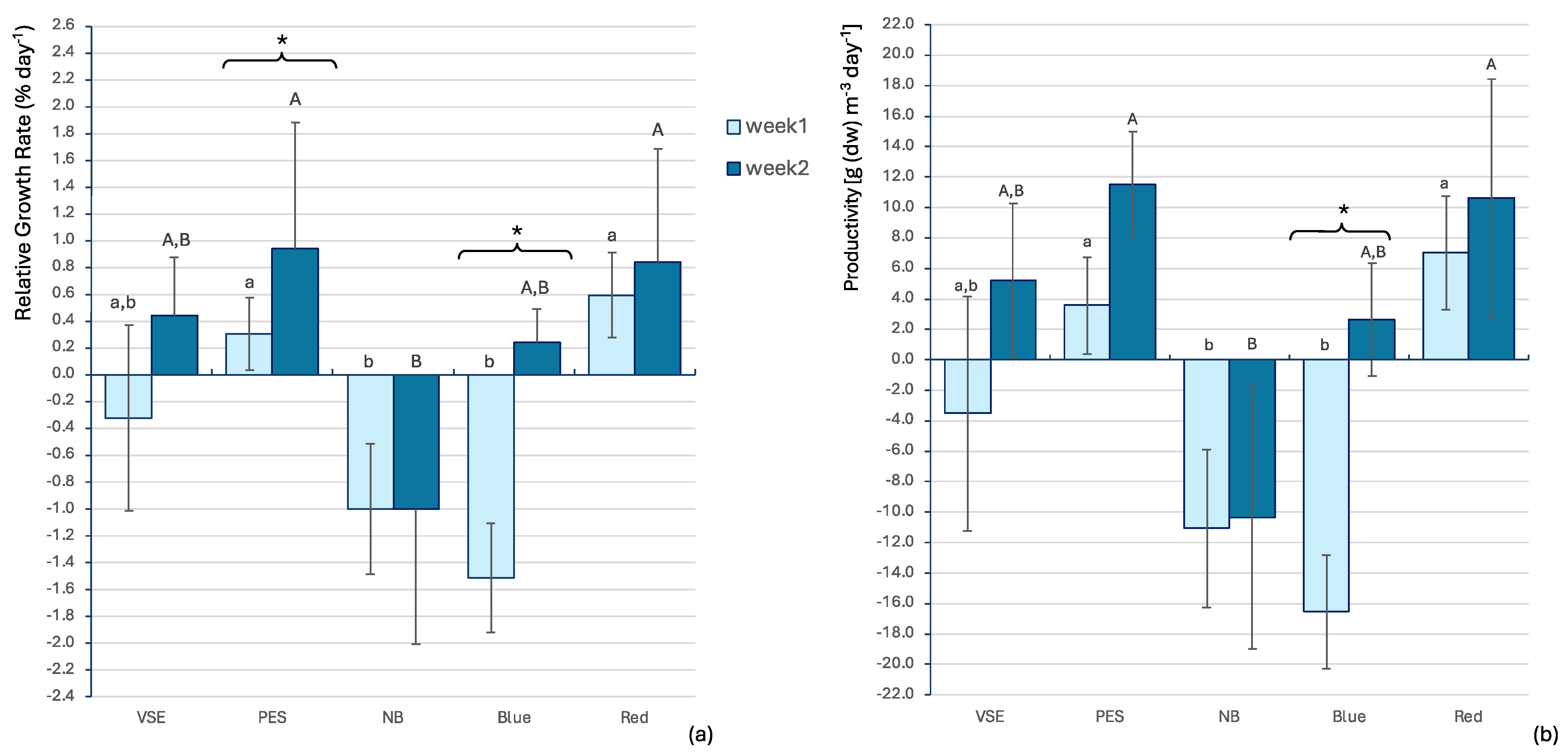
The relative growth rate (RGR) and productivity (Y) were used to assess the performance of the seaweeds in laboratory cultivation. There were statistically significant differences in ANOVA for both RGR and Y, for both weeks between the treatments. Overall, for the first week, RGR presented a test statistic F(4, 10) = 10.997, p < 0.001 and Y showed a test statistic F(4, 10) = 10.840, p < 0.001, while for the second week, RGR exhibited a test statistic F(4, 10) = 6.265, p = 0.009 and Y displayed a test statistic F(4, 10) = 6.262, p = 0.009. Therefore, the Tukey HSD test was conducted between the different treatments for each week of growth, and the significant differences are shown in Figure 3a for RGR and Figure 3b for Y. In the first week, the seaweed grown with Provasoli (PES) Medium and red LED light showed the highest RGR (0.595% day−1), but at the end of the second week, the seaweed grown with PES and white LED light showed the highest growth rate (0.945% day−1). There were no significant differences between these two treatments. The other treatments (VSE, NB, and blue LED) showed negative growth in the first week and some recovery (if any) in the second week.
As to productivity, the same pattern was observed. The seaweed grown with PES and white LED light showed the best growth at the end of the second week (11.503 g(dw) m−1 day−1), while in the first week PES and red LED light allowed for the highest Y (7.025 g(dw) m−1 day−1). Therefore, the seaweeds grown on PES medium, both with white LED light and red light, showed the best performance. Again, the NB treatment exhibited a negative yield over the two weeks, while the blue LED light had a negative impact on the productivity of the seaweed during the first week, recovering during the second week.
Analyzing each treatment over the two weeks of the trial, it was observed that both the relative growth rate and productivity increased consistently from week 1 to week 2. However, significant differences were only noted for the RGR in seaweeds treated with PES and white LED light and those exposed to blue LED light (F(1, 4) = 8.33, p = 0.045 and F(1, 4) = 31.99, p = 0.005, respectively). Additionally, significant differences were found for Y in seaweeds treated with blue LED light (F(1, 4) = 34.82, p = 0.004).
Overall, the growth and yield were significantly higher for the seaweeds treated with PES and white LED light than for those exposed to PES and red LED light.
The proximate composition of C. acicularis both for cultivated and wild specimens is shown in Figure 4.
Moisture values were always high and varied between 73.88 ± 0.007% in the seaweeds cultivated with PES and the red LED light and 68.79 ± 1.01% in the algae cultivated with NB. The Kruskal–Wallis test showed no statistically significant differences between the treatments nor with the wild biomass for the moisture and the ash content.
As to the ash content in the fresh biomass the analysis revealed a test statistic H(10) = 19.840, with a p-value of 0.031, indicating a significant difference between the groups. The highest value was recorded in the wild sample (9.08 ± 0.18% fw) and the lowest value was registered at the end of the two weeks of cultivation with PES (7.57 ± 0.01% fw). The organic matter was also statistically analyzed, validating the normality and homogeneity of the samples. Therefore, the univariate ANOVA conducted showed significant differences between the samples (F(10,15) = 3.576, p-value = 0.013). The seaweed cultivated after 7 days with NB had a significantly higher concentration of organic matter (22.86 ± 0.79% fw) when compared to all the other samples, while both samples cultivated with PES and red LED light (after the first week: 18.74 ± 0.77 and after the second week: 17.78 ± 0.07% fw) showed a significantly lower concentration of organic matter compared to all the other samples.
Regarding the dry weight, the Kruskal–Wallis test did not deliver any differences between the samples, which varied from 27.01% to 31.21% (Table 2). As to the organic matter and the ash content in the dry biomass the Kruskal–Wallis test showed significant differences between the samples.
Nevertheless, some differences were noticed in dry weight for organic matter and ash (H(10) = 19.128, p-value = 0.091). For both analyses, on the seventh day of cultivation, the red LED light and the wild biomass were significantly different from those cultivated with VSE, PES, and NB. At the end of the trial, however, only the biomass cultivated with PES was significantly different, achieving the highest organic matter (73.33%) and the lowest ash content (26.67%).
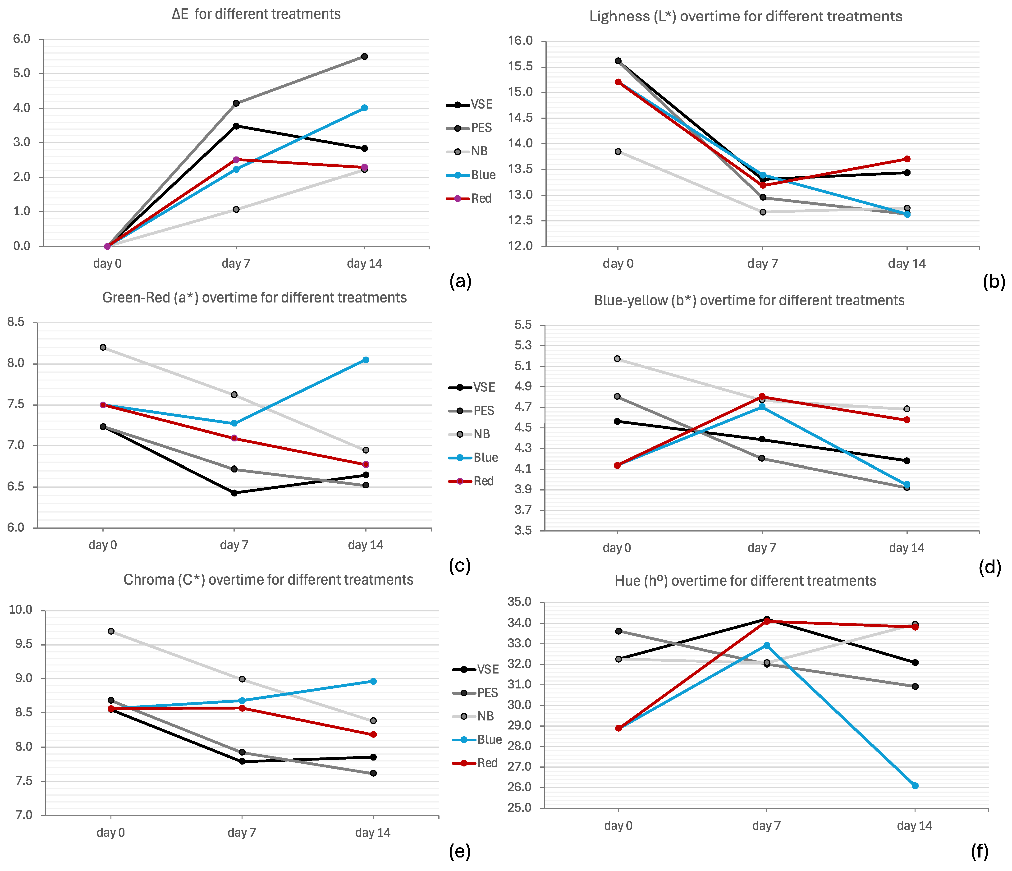
The CIELAB colour space consists of several components. The ΔE values show a noticeable colour difference over time, with increasing values (Figure 5a). The Kruskal–Wallis test delivered statistically significant differences between PES and NB (H(4) = 14.809, p-value = 0.005), with PES presenting the highest colour variation (5.51 ± 2.25) and NB the lowest (2.22 ± 0.60). The results indicate that all treatments show a slight darkening (decrease in L*), with values decreasing slightly over time, with no statistical differences (Figure 5b). A shift towards green (decrease in a*) can be noticed, except for the blue LED light treatment which shifts towards red (increase in a*) (Figure 5c). The statistical test with ANOVA indicates significant differences in a* (F(4, 40) = 4.767, p-value = 0.003), with the two NB and blue LED light treatments being significantly higher than VSE and PES.
Minor b* fluctuations can be seen but generally decrease, which indicates slight shifts towards blue; however, no statistical differences were found (Figure 5d). The chroma values C* decreased, indicating a reduction in colour intensity (Figure 5e). The Kruskal–Wallis test showed statistical differences (F(4, 40) = 43.713, p-value = 0.012), with the NB treatment being significantly higher than the other two nutrient media VSE and PES, while significant differences were neither shown for blue LED light or red LED light. Finally, the hue angle (h°) values remain relatively stable within the red–yellow hues throughout the cultivation of C. acicularis (Figure 5f). The higher values lean towards the red and lower values towards the yellow, with the blue LED light producing the highest variation. Overall, there are no significant differences between week 1 and week 2 for any of the treatments.
Pigments such as phycoerythrin, phycocyanin, and chlorophyll a play crucial role in the photosynthetic processes and colouration of red seaweed, with variations in their concentrations influenced by environmental factors such as light quality and nutrient availability. It is certainly possible that the concentration of these pigments may be responsible for the colour changes analyzed above. There were significant differences in the phycoerythrin content between the different treatments and between the wild and cultivated biomass (F(5, 12) = 14.127, p-value < 0.001), with all the treatments except for the red LED light significantly increasing the concentration of phycoerythrin (Figure 6).
The same pattern was observed for phycocyanin, with all treatments producing significantly higher pigment concentrations than the wild biomass, except for PES (F(5,12) = 11.817, p-value < 0.001). For chlorophyll a, only the VSE treatment was significantly higher than the wild biomass (F(5,12) = 8.189, p-value = 0.001). It is also noteworthy that, generally, the concentration of carotenoids in red seaweeds, primarily lutein and zeaxanthin [37], is low. However, no significant differences in carotenoid levels were observed.

4. Discussion

This study aimed to evaluate the laboratory cultivation of Chondracanthus acicularis, focusing on key environmental parameters such as nutrient levels and light exposure. The results provide some insights into the optimal conditions for the growth and biochemical composition of C. acicularis, which are crucial for its sustainable exploitation in industrial applications.
The study found significant differences in the relative growth rate (RGR) and productivity (Y) of C. acicularis under different growth conditions. The seaweed grown on Provasoli (PES) Medium and under red LED light showed the highest RGR during the first week, while those grown on PES and with white LED light exhibited the best growth at the end of the second week. This indicates that nutrient medium and light quality significantly influence the growth rate of C. acicularis [60]. In the first week, the combination of PES nutrient medium and red LED light likely provided optimal conditions for the initial growth phases, possibly due to the red light’s influence on photosynthetic efficiency and energy absorption. Red LED light has been shown to enhance growth in specific seaweed species, as it supports higher photosynthetic pigment production [61]. In addition, the PES nutrient medium provides essential nutrients that support rapid cell division and growth. However, as the cultivation progressed into the second week, white LED light, in conjunction with the PES medium, became more favourable, likely due to its broader spectrum, which supports a wider range of photosynthetic processes and pigment synthesis [61]. The productivity results mirrored the RGR findings, with the highest Y being observed in seaweeds grown with PES and white LED light by the end of the second week. This suggests that white LED light, in combination with PES medium, provides the most favourable conditions for the growth of C. acicularis. These results are consistent with those of Ismail et al. [62], who also found that PES was the most adequate nutrient medium for the growth of Gigartinales. However, our results point to a maximum growth of 0.945% day−1. These values are much lower than those found for Chondracanthus teedei by López-Campos et al. [29], who reported a maximum growth of 4.68% day−1 in similar conditions. These differences may be attributed to the different species and physiological states of the algae used [23,25].
The negative growth observed in the Von Stoch Medium (VSE), Nutribloom plus® (NB), and blue LED light treatments during the first week, followed by some recovery during the second week, highlights the importance of selecting appropriate light and nutrient conditions for optimal seaweed cultivation [63]. In general, macroalgal nitrogen growth kinetics follow typical growth–saturation curves, meaning that macroalgal growth rates increase positively with increasing nitrogen concentration, and then stabilize after nitrogen concentration reaches saturation [64]. However, NB has a much higher nitrate content than the other two media, which might have stressed the red seaweed, causing growth inhibition due to nutrient imbalances and ammonium toxicity [65]. A balanced supply of macronutrients such as nitrogen, phosphorus, and potassium, together with trace elements like iron, zinc, and manganese, is essential for the growth and development of red seaweeds, as they play a critical role in the metabolic processes, pigment synthesis, and overall health of the algae [66,67,68].
The proximate composition analysis of C. acicularis revealed a high moisture content across all treatments, ranging from 68.79% to 73.65%, but with no significant differences found between samples. According to Ganesan et al. [69], seaweeds tend to retain a high water content when grown under optimal culture conditions. The cultivation conditions were generally suitable for seaweed growth, as indicated by the consistent moisture levels. However, the NB medium showed the lowest moisture content, suggesting that it was the worst culture media to grow this seaweed.
In contrast to moisture, significant differences in ash content were observed, ranging from 7.28% fw (27.01% dw), for the VSE cultivated biomass, to 9.08% fw (30.80% dw), for the wild biomass. These values are within the expected values for Gigartinales (16.55%) [70] and for the red seaweed Gracilaria spp. (19% to 25%) [71,72]. Algae ash content varies between locations and cultivation sites due to differences in key factors such as salinity, temperature, and nutrient availability [73]. Therefore, the lower ash content of cultivated seaweeds compared to the wild may be attributed to optimized nutrient solutions that might limit certain minerals, reducing mineral uptake and, thus, the ash content [37,74].
The organic matter content showed significant differences between treatments, with seaweeds cultivated with NB exhibiting the highest concentration after 7 days. Conversely, seaweeds grown with PES and red LED light showed the lowest concentrations of organic matter. Cultivated seaweeds often have higher ash content and moisture levels due to controlled nutrient availability and environmental conditions, which can lead to a lower percentage of organic matter and dry weight when compared to wild specimens [74].
Red algae are rich in the pink pigment phycoerythrin, which absorbs light over a wide blue–green light spectral range, roughly 460–570 nm, including spectral regions which are not available for other photosynthetic pigments [75]. Also, red seaweeds exhibit rapid phycobilin acclimatory responses to changes in light, enhancing light harvesting [76]. Thus, the use of LED lights plays a crucial role in optimizing the growth of red seaweeds by influencing their photosynthetic efficiency and pigment production. Different light intensities and wavelengths can be used as a tool to enhance the concentration of valuable compounds in seaweed aquaculture, such as photosynthetic pigments. Our results show that white LED light delivered the best growth, providing a broad spectrum (a narrow peak at 450 nm and a wide peak between 550 and 620 nm) which supports balanced growth and enhances the overall biomass production of seaweeds [77]. Red LED light also showed significantly higher growth and yield. Red LED light (narrow peak at ~655 nm), although less effective for some species, can significantly boost the production of phycoerythrin, thereby enhancing the red colouration of the seaweed. As to blue LED light (narrow peak at ~455 nm), although it has been reported that it is particularly effective in promoting the synthesis of chlorophyll and other photosynthetic pigments [78], it did not prove to be adequate for the growth of C. acicularis. Hence, while beneficial for certain species, blue LED light may not provide the optimal spectrum for this species, leading to reduced photosynthetic efficiency [79]. However, studies have shown that the combination of these LED lights can lead to improved growth rates and higher concentrations of valuable bioactive compounds in red seaweeds, making them a promising tool for sustainable aquaculture practises [60,80,81,82,83,84,85].
The different chemical compositions of the biomasses are denoted by the colour of the biomass, as analyzed through CIE Lab components. The increasing ΔE values indicate that there was a noticeable colour difference over time compared to the initial state. The increase in the biomass grown with PES showed the highest variation, easily visible to the naked eye [86]. This means that the seaweed underwent significant colour changes, which could indicate changes in the biochemical composition of the biomasses submitted to different treatments, namely in the concentration of pigments [87]. Indeed, different culture media and distinct light qualities can significantly influence photosynthesis and pigment production in seaweeds [88]. The changes in ΔE suggest a darkening of the biomass, which is due to the increase in the pigment content [89]. The changes in a* values suggest that the red seaweed grown under NB and blue LED light shifts towards the green spectrum, which may indicate a decrease in phycobilin content. As stated, the high nitrate content in the NB medium may cause nutrient imbalances, potentially affecting the production of phycobilins, and blue LED light might also cause stress, reducing the efficiency of light-harvesting pigment [76,79]. As to the b* values, although not significant, their slight decrease indicates a shift towards the blue spectrum, probably due to an increase in the blue pigment phycocyanin registered in all samples compared to the wild. The chromaticity (C*) values decreased except for the blue LED light medium, for which the hue (°h) decreased. Phycobilisomes have an intensity-dependent photoprotection mechanism, so the production of phycobilins is light-dependent. Exposure to intense light increases the blue structural colour and simultaneously favours the capture of light at green and red wavelengths by the phycobilisomes [90]. On the other hand, less intense light tends to reduce the production of photoprotective pigments. The relatively stable hue angle values indicate that, despite the changes in lightness and chromaticity, the overall hue of the seaweed remains consistent. This suggests that the primary colour characteristics of the seaweed are maintained. Hence, white LED light appears to provide a broad spectrum that can support balanced growth and pigment production, while blue LED light shows the greatest colour shifts, indicating major variation in the chemical composition of the seaweeds, probably due to stress-induced conditions.
These findings are consistent with the pigment content of the samples. There was a significant increase in the production of phycoerythrin and phycocyanin, while chlorophyll a was higher in NB. These changes were induced by the availability of nutrients and by light intensity and wavelength, as stated by several authors, namely Dawes et al. [91], Bonomi-Barifi et al. [92], and Idowu [87]. Different wavelengths activate specific photosynthetic pathways, with red light primarily activating Photosystem I (PSI) while blue light activates both Photosystem I (PSI) and Photosystem II (PSII) [76]. The increase in phycoerythrin under red light and of phycocyanin under blue light optimized light absorption for these pathways.

5. Conclusions

This study demonstrates that the quality of nutrient medium and light have a significant effect on the growth, productivity, pigmentation, and proximate composition of Chondracanthus acicularis under laboratory cultivation. Provasoli (PES) medium has been shown to provide a well-balanced mix of essential macronutrients such a nitrogen, phosphorus, and potassium, as well as essential trace elements and vitamins that support metabolic processes and enhance the overall growth and development of C. acicularis. Further studies are needed to fine-tune the concentrations of macronutrients and trace elements to match the specific needs of the seaweed. In addition, improving the light spectrum and intensity can also significantly impact growth. Experimenting with different light conditions, such as mixing red and white LED lights, can help identify the most effective setup for the species’ growth.
These findings highlight the importance of selecting appropriate environmental parameters for optimal seaweed cultivation. The results also indicate that controlled nutrient availability and environmental conditions in cultivation can lead to higher ash content and moisture levels, but lower organic matter and dry weight compared to wild specimens. By identifying the optimal light and nutrient conditions, this research supports the development of sustainable cultivation practises that can improve the yield and quality of C. acicularis biomass. This is particularly relevant for industries that rely on high-quality bioactive compounds, such as the pharmaceutical, nutraceutical, and cosmetics industries.

Supplementary Materials

The following supporting information can be downloaded at: https://www.mdpi.com/article/10.3390/app15020810/s1, Table S1: Chemical composition of the three nutrient media used in the trials.

Author Contributions

Conceptualization T.M.; methodology, T.M. and C.A.; validation, T.M., M.V.F. and C.A.; formal analysis, A.S., T.M. and C.A.; investigation, A.S. and M.V.F.; resources, T.M. and C.A.; data curation, T.M. and C.A.; writing—original draft preparation, T.M.; writing—review and editing, C.A. and M.V.F.; visualization, T.M.; supervision, T.M. and C.A.; project administration, T.M. and C.A.; funding acquisition, T.M. and C.A. All authors have read and agreed to the published version of the manuscript.

Funding

This work was funded by national funds through FCT—Fundação para a Ciência e a Tecnologia, I.P., within the scope of the projects MARE—Centro de Ciências do Mar e do Ambiente, UID/04292/MARE) and ARNET—Aquatic Research Network, LA/P/0069/2020 and the doctoral grant awarded to Marta V. Freitas, UI/BD/150957/2021.

Data Availability Statement

Data will be made available upon request.

Acknowledgments

The authors would like to express their gratitude to Joaquina Pinheiro for her invaluable assistance with the CIE Lab measurements and interpretation.

Conflicts of Interest

The authors declare no conflicts of interest.

References

  1. Selnes, T.; Giesbers, E.; van den Burg, S.W.K. Innovation and Collaboration: Opportunities for the European Seaweed Sector in Global Value Chains. J. Mar. Sci. Eng. 2021, 9, 693. [Google Scholar] [CrossRef]
  2. Yang, Y.; Zhang, M.; Alalawy, A.I.; Almutairi, F.M.; Al-Duais, M.A.; Wang, J.; Salama, E.-S. Identification and Characterization of Marine Seaweeds for Biocompounds Production. Environ. Technol. Innov. 2021, 24, 101848. [Google Scholar] [CrossRef]
  3. Cavaco, M.; Duarte, A.; Bernardino, S.; Afonso, C.; Mouga, T. Sustainable Use of Seaweeds from S. Martinho Do Porto, Portugal–Past, Present, and Future Perspective. In Conference Proceedings ICoWEFS 2021, Proceedings of the 1st International Conference on Water Energy Food and Sustainability (ICoWEFS 2021), Leiria, Portugal, 10–12 May 2021; da Costa Sanches Galvão, J.R., Duque de Brito, P.S., dos Santos Neves, F., da Silva Craveiro, F.G., de Amorim Almeida, H., Correia Vasco, J.O., Pires Neves, L.M., de Jesus Gomes, R., de Jesus Martins Mourato, S., Santos Ribeiro, V.S., Eds.; Springer International Publishing: Leiria, Portugal, 2021; pp. 216–226. [Google Scholar]
  4. Pereira, L.; Cotas, J. Historical Use of Seaweed as an Agricultural Fertilizer in the European Atlantic Area. In Seaweeds as Plant Fertilizer, Agricultural Biostimulants and Animal Fodder; Pereira, L., Bahcevandziev, K., Joshi, N.H., Eds.; CRC Press Taylor & Francis Group: Boca Raton, FL, USA, 2019; pp. 1–22. ISBN 9781138597068. [Google Scholar]
  5. Pereira, L. Edible Seaweeds of the World; CRC Press: Boca Raton, FL, USA, 2016; ISBN 9780429154041. [Google Scholar]
  6. Flores-Molina, M.R.; Thomas, D.; Lovazzano, C.; Nez, A.N.; Zapata, J.; Kumar, M.; Correa, J.A.; Contreras-Porcia, L. Desiccation Stress in Intertidal Seaweeds: Effects on Morphology, Antioxidant Responses and Photosynthetic Performance. Aquat. Bot. 2014, 113, 90–99. [Google Scholar] [CrossRef]
  7. Sampath-Wiley, P.; Neefus, C.D.; Jahnke, L.S. Seasonal Effects of Sun Exposure and Emersion on Intertidal Seaweed Physiology: Fluctuations in Antioxidant Contents, Photosynthetic Pigments and Photosynthetic Efficiency in the Red Alga Porphyra Umbilicalis Kützing (Rhodophyta, Bangiales). J. Exp. Mar. Biol. Ecol. 2008, 361, 83–91. [Google Scholar] [CrossRef]
  8. Pereira, L.; Sousa, A.; Coelho, H.; Amado, A.M.; Ribeiro-Claro, P.J.A. Use of FTIR, FT-Raman and 13C-NMR Spectroscopy for Identification of Some Seaweed Phycocolloids. Biomol. Eng. 2003, 20, 223–228. [Google Scholar] [CrossRef] [PubMed]
  9. Rupert, R.; Rodrigues, K.F.; Thien, V.Y.; Yong, W.T.L. Carrageenan from Kappaphycus alvarezii (Rhodophyta, Solieriaceae): Metabolism, Structure, Production, and Application. Front. Plant Sci. 2022, 13, 859635. [Google Scholar] [CrossRef]
  10. Rahmawati, I.; Liviawaty, E.; Pratama, R.I. Carrageenan in Seaweed (Eucheuma sp.) and Use of Carrageenan in Fishery Food Products: A Review. Asian J. Fish. Aquat. Res. 2023, 23, 1–10. [Google Scholar] [CrossRef]
  11. Cardoso, S.S.M.; Carvalho, L.L.G.L.; Silva, P.P.J.; Rodrigues, M.S.M.; Pereira, O.; Pereira, L. Bioproducts From Seaweeds: A Review With Special Focus On The Iberian Peninsula. Curr. Org. Chem. 2014, 18, 896–917. [Google Scholar] [CrossRef]
  12. Freitas, M.V.; Inácio, L.G.; Martins, M.; Afonso, C.; Pereira, L.; Mouga, T. Primary Composition and Pigments of 11 Red Seaweed Species from the Center of Portugal. J. Mar. Sci. Eng. 2022, 10, 1168. [Google Scholar] [CrossRef]
  13. Freitas, M.V.; Pacheco, D.; Cotas, J.; Mouga, T.; Afonso, C.; Pereira, L. Red Seaweed Pigments from a Biotechnological Perspective. Phycology 2021, 2, 1–29. [Google Scholar] [CrossRef]
  14. Pereira, T.; Barroso, S.; Mendes, S.; Amaral, R.A.; Dias, J.R.; Baptista, T.; Saraiva, J.A.; Alves, N.M.; Gil, M.M. Optimization of Phycobiliprotein Pigments Extraction from Red Algae Gracilaria gracilis for Substitution of Synthetic Food Colorants. Food Chem. 2020, 321, 126688. [Google Scholar] [CrossRef]
  15. Carreto, J.I.; Carignan, M.O. Mycosporine-like Amino Acids: Relevant Secondary Metabolites. Chemical and Ecological Aspects. Mar. Drugs 2011, 9, 387–446. [Google Scholar] [CrossRef] [PubMed]
  16. Chrapusta, E.; Kaminski, A.; Duchnik, K.; Bober, B.; Adamski, M.; Bialczyk, J. Mycosporine-Like Amino Acids: Potential Health and Beauty Ingredients. Mar. Drugs 2017, 15, 326. [Google Scholar] [CrossRef] [PubMed]
  17. McReynolds, C.; Adrien, A.; Silvestre de Ferron, A.; Boussetta, N.; Grimi, N.; Pecastaing, L.; Fernandes, S.C.M. Extraction of Mycosporine-like Amino Acids and Proteins from the Agarophyte Gelidium corneum Using Pulsed Power Techniques. Foods 2023, 12, 1473. [Google Scholar] [CrossRef]
  18. Towle, G.A. Carrageenan. In Industrial Gums; Academic Press: Cambridge, MA, USA, 1973; pp. 83–114. [Google Scholar] [CrossRef]
  19. Udo, T.; Mummaleti, G.; Mohan, A.; Singh, R.K.; Kong, F. Current and Emerging Applications of Carrageenan in the Food Industry. Food Res. Int. 2023, 173, 113369. [Google Scholar] [CrossRef] [PubMed]
  20. Freitas, M.V.; Inácio, L.G.; Ruas, A.; Silva, I.A.; Mouga, T.; Pereira, L.; Afonso, C. Antioxidant and Antimicrobial Properties of Selected Red Seaweeds from Central Portugal. Appl. Sci. 2022, 13, 157. [Google Scholar] [CrossRef]
  21. Mouga, T. Seaweed Harvesting and Aquaculture: An Overview of the Past 70 Years. In Conference Proceedings ICoWEFS 2023, Proceedings of the 3rd International Conference on Water Energy Food and Sustainability (ICoWEFS 2023), Leiria, Portugal, 10–12 May 2023; Galvão, J.R.D.C.S., Brito, P., Neves, F.D.S., Almeida, H.D.A., Mourato, S.D.J.M., Nobre, C., Eds.; Springer Nature: Castelo Branco, Portugal, 2024; pp. 365–375. ISBN 9783031485329. [Google Scholar]
  22. FAO. The State of World Fisheries and Aquaculture 2024; FAO: Rome, Italy, 2024; ISBN 978-92-5-138763-4. [Google Scholar]
  23. Kim, J.K.; Yarish, C.; Hwang, E.K.; Park, M.; Kim, Y. Seaweed Aquaculture: Cultivation Technologies, Challenges and Its Ecosystem Services. Algae 2017, 32, 1–13. [Google Scholar] [CrossRef]
  24. Andreakis, N.; Schaffelke, B. Invasive Marine Seaweeds: Pest or Prize? In Seaweed Biology Novel Insights into Ecophysiology, Ecology and Utilization; Wiencke, C., Bischof, K., Eds.; Springer Nature: Berlin, Germany, 2012; pp. 235–262. [Google Scholar]
  25. Valero, M.; Guillemin, M.; Destombe, C.; Gachon, C.M.M.; Badis, Y.; Buschmann, A.H.; Camus, C.; Faugeron, S. Perspectives on Domestication Research for Sustainable Seaweed Aquaculture. Perspect. Phycol. 2017, 1, 33–46. [Google Scholar] [CrossRef]
  26. Mumford, T.W.J.R. Progress and Prospects for Field Cultivation of Iridaea cordata and Gigartina exasperata. In Proceedings of the Symposium on Seaweeds in the Warm Pacific, Pacific Grove, CA, USA, 6 March 1980. [Google Scholar]
  27. Bulboa, C.R.; Macchiavello, J.E.; Oliveira, E.C.; Fonck, E. First Attempt to Cultivate the Carrageenan-Producing Seaweed Chondracanthus chamissoi (C. Agardh) Kutzing (Rhodophyta; Gigartinales) in Northern Chile. Aquac. Res. 2005, 36, 1069–1074. [Google Scholar] [CrossRef]
  28. Buschmann, A.H.; Varela, D.; Cifuentes, M.; Del Carmen Hernández-González, M.; Henríquez, L.; Westermeier, R.; Correa, J.A. Experimental Indoor Cultivation of the Carrageenophytic Red Alga Gigartina skottsbergii. Aquaculture 2004, 241, 357–370. [Google Scholar] [CrossRef]
  29. López-Campos, M.; Pérez-Lloréns, J.L.; Barrena, F.; Pérez-González, C.M.; Hernández, I. Culture of Gracilaria gracilis and Chondracanthus teedei from Vegetative Fragments in the Field and Carpospores in Laboratory. J. Mar. Sci. Eng. 2022, 10, 1041. [Google Scholar] [CrossRef]
  30. Pereira, L.; Amado, A.M.; Critchley, A.T.; van de Velde, F.; Ribeiro-Claro, P.J.A. Identification of Selected Seaweed Polysaccharides (Phycocolloids) by Vibrational Spectroscopy (FTIR-ATR and FT-Raman). Food Hydrocoll. 2009, 23, 1903–1909. [Google Scholar] [CrossRef]
  31. Pereira, L. Population Studies and Carrageenan Properties in Eight Gigartinales (Rhodophyta) from Western Coast of Portugal. Sci. World J. 2013, 2013, 939830. [Google Scholar] [CrossRef] [PubMed]
  32. Pacheco, D.; Cotas, J.; Domingues, A.; Ressurreição, S.; Bahcevandziev, K.; Pereira, L. Chondracanthus teedei var. lusitanicus: The Nutraceutical Potential of an Unexploited Marine Resource. Mar. Drugs 2021, 19, 570. [Google Scholar] [CrossRef]
  33. Soares, F.; Fernandes, C.; Silva, P.; Pereira, L.; Gonçalves, T. Antifungal Activity of Carrageenan Extracts from the Red Alga Chondracanthus teedei var. lusitanicus. J. Appl. Phycol. 2016, 28, 2991–2998. [Google Scholar] [CrossRef]
  34. Vásquez, V.; Martínez, R.; Rodríguez-Núñez, K.; Bernal, C. Angiotensin-I Converting Enzyme (ACE) Inhibitory Bioaccessible Peptides Obtained from Protein Extracts of Macrocystis pyrifera and Chondracanthus chamissoi Seaweeds. Algal. Res. 2024, 81, 103575. [Google Scholar] [CrossRef]
  35. Tavares Pessoa Pinho de Vasconcelos, E.R.; Vasconcelos, J.B.; de Vasconcelos Reis, T.N.; de Lourdes Montenegro Cocentino, A.; Mallea, A.J.A.; Martins, G.M.; Neto, A.I.; Fujii, M.T. Macroalgal Responses to Coastal Urbanization: Relative Abundance of Indicator Species. J. Appl. Phycol. 2019, 31, 893–903. [Google Scholar] [CrossRef]
  36. Neto, A.I. Observations on the Biology and Ecology of Selected Macroalgae from the Littoral of São Miguel (Azores). Bot. Mar. 2000, 43, 483–498. [Google Scholar] [CrossRef]
  37. Bulboa Contador, C.; Massad, I.P.; Contreras-Porcia, L.; Zapata, J.; Castañeda, F.; Ramírez, M.E.; Gil-Kodaka, P. Concise Review of Genus Chondracanthus (Rhodophyta: Gigartinales). J. Appl. Phycol. 2020, 32, 773–785. [Google Scholar] [CrossRef]
  38. Guiry, M.D. Photoperiodic and Temperature Responses in the Growth and Tetrasporogenesis of Gigartina acicularis (Rhodophyta) from Ireland. Helgol. Meeresunters. 1984, 1, 335–347. [Google Scholar] [CrossRef]
  39. Santelices, B. Patterns of Reproduction, Dispersal and Recruitment in Seaweeds. Oceanogr. Mar. Biol. Annu. Rev. 1990, 28, 177–276. [Google Scholar]
  40. Bertocci, I.; Araújo, R.; Incera, M.; Arenas, F.; Pereira, R.; Abreu, H.; Larsen, K.; Sousa-Pinto, I. Benthic Assemblages of Rock Pools in Northern Portugal: Seasonal and between-Pool Variability. Sci. Mar. 2011, 76, 781–789. [Google Scholar] [CrossRef]
  41. Pereira, L.; Mesquita, J.F. Carrageenophytes of Occidental Portuguese Coast: 1-Spectroscopic Analysis in Eight Carrageenophytes from Buarcos Bay. Biomol. Eng. 2003, 20, 217–222. [Google Scholar] [CrossRef]
  42. Pereira, L.; Van De Velde, F. Portuguese Carrageenophytes: Carrageenan Composition and Geographic Distribution of Eight Species (Gigartinales, Rhodophyta). Carbohydr. Polym. 2011, 84, 614–623. [Google Scholar] [CrossRef]
  43. Gómez-Ordóñez, E.; Rupérez, P. FTIR-ATR Spectroscopy as a Tool for Polysaccharide Identification in Edible Brown and Red Seaweeds. Food Hydrocoll. 2011, 25, 1514–1520. [Google Scholar] [CrossRef]
  44. Wang, X.; Guo, C.; Guo, H. Progress of Carrageenan-Based Films and Coatings for Food Packaging Applications. Packag. Technol. Sci. 2024, 37, 533–550. [Google Scholar] [CrossRef]
  45. Mzibra, A.; Aasfar, A.; Benhima, R.; Khouloud, M.; Boulif, R.; Douira, A.; Bamouh, A.; Meftah Kadmiri, I. Biostimulants Derived from Moroccan Seaweeds: Seed Germination Metabolomics and Growth Promotion of Tomato Plant. J. Plant Growth Regul. 2021, 40, 353–370. [Google Scholar] [CrossRef]
  46. Mzibra, A.; Aasfar, A.; El Arroussi, H.; Khouloud, M.; Dhiba, D.; Kadmiri, I.M.; Bamouh, A. Polysaccharides Extracted from Moroccan Seaweed: A Promising Source of Tomato Plant Growth Promoters. J. Appl. Phycol. 2018, 30, 2953–2962. [Google Scholar] [CrossRef]
  47. Hossain, M.M.; Sultana, F.; Khan, S.; Nayeema, J.; Mostafa, M.; Ferdus, H.; Tran, L.S.P.; Mostofa, M.G. Carrageenans as Biostimulants and Bio-Elicitors: Plant Growth and Defense Responses. Stress Biol. 2024, 4, 3. [Google Scholar] [CrossRef] [PubMed]
  48. Montanha, J.A.; Bourgougnon, N.; Boustie, J.; Amoros, M. Antiviral Activity of Carrageenans from Marine Red Algae. Am. J. Pharm. 2009, 28, 443–451. [Google Scholar]
  49. Vrinda, S.K.; Niharika, A.S.; Ligin, M.; Sabitha, M. Carrageenan for Drug Delivery and Biomedical Applications. Nat. Biopolym. Drug Deliv. Tissue Eng. 2023, 57–76. [Google Scholar] [CrossRef]
  50. Bodian, M.Y.; Lafontaine, N.; Matard, M.; Mussio, I.; Rusig, A.M. Evaluation of the in Vitro Methods for Micropropagation of Chondracanthus acicularis (Roth) Fredericq (Gigartinales, Rhodophyta): Tissue Culture and Production of Protoplasts. J. Appl. Phycol. 2013, 25, 1835–1845. [Google Scholar] [CrossRef]
  51. Redmond, S.; Green, L.; Yarish, C.; Kim, J.; Neefus, C. New England Seaweed Culture Handbook-Nursery Systems; Redmond, S., Green, L., Yarish, C., Kim, J., Neefus, C., Eds.; Connecticut Sea Grant CTSG-14-01; Connecticut Sea Grant: Groton, CT, USA, 2014. [Google Scholar]
  52. Ferreira, L.B.; Barufi, J.B.; Plastino, E.M. Growth of Red and Green Strains of the Tropical Agarophyte Gracilaria cornea J. Agardh (Gracilariales, Rhodophyta) in Laboratory. Rev. Bras. Bot. 2006, 29, 187–192. [Google Scholar] [CrossRef]
  53. Concórdio-Reis, P.; Cardeira, M.; Macedo, A.C.; Ferreira, S.S.; Serra, A.T.; Coimbra, M.A.; Amorim, A.; Reis, M.A.M.; Freitas, F. Novel Exopolysaccharide Produced by the Marine Dinoflagellate Heterocapsa AC210: Production, Characterization, and Biological Properties. Algal. Res. 2023, 70, 103014. [Google Scholar] [CrossRef]
  54. Patarra, R.F.; Carreiro, A.S.; Lloveras, A.A.; Abreu, M.H.; Buschmann, A.H.; Neto, A.I. Effects of Light, Temperature and Stocking Density on Halopteris scoparia Growth. J. Appl. Phycol. 2017, 29, 405–411. [Google Scholar] [CrossRef]
  55. Castellar, M.; Obón, J.; Fernández-López, J. The Isolation and Properties of a Concentrated Red-purple Betacyanin Food Colourant from Opuntia stricta Fruits. J. Sci. Food Agric. 2006, 86, 122–128. [Google Scholar] [CrossRef]
  56. AOAC. Official Methods of Analysis of AOAC International, 20th ed.; Latimer, G.W., Jr., Ed.; Association of Official Analytical Chemists: Gaithersburg, MD, USA, 2016; ISBN 0935584870. [Google Scholar]
  57. Beer, S.; Eshel, A. Determining Phycoerythrin and Phycocyanin Concentrations in Aqueous Crude Extracts of Red Algae. Mar. Freshw. Res. 1985, 36, 785–792. [Google Scholar] [CrossRef]
  58. Osório, C.; Machado, S.; Peixoto, J.; Bessada, S.; Pimentel, F.B.; Alves, R.C.; Oliveira, M.B.P.P. Pigments Content (Chlorophylls, Fucoxanthin and Phycobiliproteins) of Different Commercial Dried Algae. Separations 2020, 7, 33. [Google Scholar] [CrossRef]
  59. Kirk, J.T.O.; Allen, R.L. Dependence of Chloroplast Pigment Synthesis on Protein Synthesis: Effect of Actidione. Biochem. Biophys. Res. Commun. 1965, 21, 523–530. [Google Scholar] [CrossRef] [PubMed]
  60. Kim, J.K.; Mao, Y.; Kraemer, G.; Yarish, C. Growth and Pigment Content of Gracilaria tikvahiae McLachlan under Fluorescent and LED Lighting. Aquaculture 2015, 436, 52–57. [Google Scholar] [CrossRef]
  61. Figueroa, F.L.; Aguilera, J.; Niell, F.X. Red and blue light regulation of growth and photosynthetic metabolism in Porphyra umbilicalis (Bangiales, Rhodophyta). Eur. J. Phycol. 1995, 30, 11–18. [Google Scholar] [CrossRef]
  62. Ismail, M.M.; El-Sheekh, M. Enhancement of Biochemical and Nutritional Contents of Some Cultivated Seaweeds Under Laboratory Conditions. J. Diet. Suppl. 2018, 15, 318–329. [Google Scholar] [CrossRef] [PubMed]
  63. Freitas, M.; Correia, A.; Pereira, C.; Santos, J.; Baptista, T.; Afonso, C.; Gil, M.; Pombo, A.; Tecelão, C.; Mendes, S.; et al. The Influence of Light and Culture Media on the Growth of the Red Seaweed Gracilaria gracilis (Rhodophyta, Gracilariales) under Laboratory Conditions. Front. Mar. Sci. 2018, 5, 2022. [Google Scholar] [CrossRef]
  64. Hurd, C.L.; Harrison, P.J.; Bischof, K.; Lobban, C.S. Seaweed Ecology and Physiology, 2nd ed.; Cambridge University Press: Cambridge, UK, 2014; ISBN 9780521145954. [Google Scholar]
  65. Wang, Q.; Lan, L.; Li, H.; Gong, Q.; Gao, X. Effects of Nitrogen Source and Concentration on the Growth and Biochemical Composition of the Red Seaweed Grateloupia turuturu (Halymeniaceae, Rhodophyta). Sustainability 2023, 15, 4210. [Google Scholar] [CrossRef]
  66. Lubsch, A.; Lansbergen, R.A. Seaweed Factsheet: Nutrient Uptake and Requirements; Wageningen University & Research: Wageningen, The Netherlands, 2020; pp. 1–4. [Google Scholar]
  67. Harrison, P.J.; Hurd, C.L. Nutrient Physiology of Seaweeds: Application of Concepts to Aquaculture. Cah. Biol. Mar. 2001, 42, 71–82. [Google Scholar]
  68. Zhao, Y.; Wang, Z.; Chen, M.; Huang, X.; Luo, Z. Effects of Nitrogen to Phosphorus Ratios on Algal Growth and Arsenate Metabolism by Microcystis aeruginosa with Dissolved Organic Phosphorus and Nitrate as Nutrients. Algal Res 2023, 69, 102922. [Google Scholar] [CrossRef]
  69. Ganesan, M.; Meena, R.; Siddhanta, A.K.; Selvaraj, K.; Chithra, K. Culture of the Red Alga Sarconema filiforme in Open Waters and Hybrid Carrageenan from the Cultivated Seaweeds. J. Appl. Phycol. 2015, 27, 1549–1559. [Google Scholar] [CrossRef]
  70. Arakaki, N.; Flores Ramos, L.; Oscanoa Huaynate, A.I.; Ruíz Soto, A.; Ramírez, M.E. Biochemical and Nutritional Characterization of Edible Seaweeds from the Peruvian Coast. Plants 2023, 12, 1795. [Google Scholar] [CrossRef]
  71. Freitas, M.V.; Mouga, T.; Correia, A.P.; Afonso, C.; Baptista, T. New Insights on the Sporulation, Germination, and Nutritional Profile of Gracilaria gracilis (Rhodophyta) Grown under Controlled Conditions. J. Mar. Sci. Eng. 2021, 9, 562. [Google Scholar] [CrossRef]
  72. Rodrigues, D.; Freitas, A.C.; Pereira, L.; Rocha-Santos, T.A.P.; Vasconcelos, M.W.; Roriz, M.; Rodríguez-Alcalá, L.M.; Gomes, A.M.P.; Duarte, A.C. Chemical Composition of Red, Brown and Green Macroalgae from Buarcos Bay in Central West Coast of Portugal. Food Chem. 2015, 183, 197–207. [Google Scholar] [CrossRef]
  73. Paiva, L.; Lima, E.; Neto, A.I.; Marcone, M.; Baptista, J. Nutritional and Functional Bioactivity Value of Selected Azorean Macroalgae: Ulva compressa, Ulva rigida, Gelidium microdon, and Pterocladiella capillacea. J. Food Sci. 2017, 82, 1757–1764. [Google Scholar] [CrossRef] [PubMed]
  74. Tibbetts, S.M.; Milley, J.E.; Lall, S.P. Nutritional Quality of Some Wild and Cultivated Seaweeds: Nutrient Composition, Total Phenolic Content and in Vitro Digestibility. J. Appl. Phycol. 2016, 28, 3575–3585. [Google Scholar] [CrossRef]
  75. Bidigare, R.R.; Ondrusek, M.E.; Morrow, J.H.; Kiefer, D.A. In-Vivo Absorption Properties of Algal Pigments. Conf. Proc. SPIE 1990, 1302, 290–302. [Google Scholar] [CrossRef]
  76. Voerman, S.E.; Ruseckas, A.; Turnbull, G.A.; Samuel, I.D.W.; Burdett, H.L. Red Algae Acclimate to Low Light by Modifying Phycobilisome Composition to Maintain Efficient Light Harvesting. BMC Biol. 2022, 20, 1–16. [Google Scholar] [CrossRef] [PubMed]
  77. Huang, S.; Li, K.; Pan, Y.; Yu, Y.; Wernberg, T.; de Bettignies, T.; Wu, J.; Zhou, C.; Huang, Z.; Xiao, X. Artificial Light Source Selection in Seaweed Production: Growth of Seaweed and Biosynthesis of Photosynthetic Pigments and Soluble Protein. PeerJ 2021, 9, e11351. [Google Scholar] [CrossRef]
  78. Torres, R.; Campos, A.M.; Goldman, J.; Barrote, I.; Mata, L.; Silva, J. Effects of Light Quality and Intensity on Growth and Bromoform Content of the Red Seaweed Asparagopsis taxiformis. J. Appl. Phycol. 2024, 36, 627–637. [Google Scholar] [CrossRef]
  79. Dishon, G.; Resetarits, H.M.; Tsai, B.; Jones, A.L.; Agarwal, V.; Smith, J.E. The Effect of Light Intensity, Spectrum, and Photoperiod on the Physiological Performance of Asparagopsis taxiformis Tetrasporophytes. Algal. Res. 2023, 76, 103304. [Google Scholar] [CrossRef]
  80. Carmona, R.; Vergara, J.J.; Lahaye, M.; Niell, F.X. Light Quality Affects Morphology and Polysaccharide Yield and Composition of Gelidium sesquipedale. J. Appl. Phycol. 1998, 10, 323–331. [Google Scholar] [CrossRef]
  81. Yago, T.; Arakawa, H.; Shigeto, S.; Ito, R.; Matsumoto, A.; Okumura, Y. Effects of Light Conditions on the Growth of Commercial Seaweed Undaria pinnatifida. Afr. J. Plant Sci. 2017, 11, 190–196. [Google Scholar] [CrossRef]
  82. Ak, I.; Yücesan, M. Effect of Light Intensity on the Pigment Composition of Gracilaria verrucosa (Rhodophyta). Fres. Environ. Bull. 2012, 21, 2126–2131. [Google Scholar]
  83. Singh, S.P.; Singh, P. Effect of Temperature and Light on the Growth of Algae Species: A Review. Renew. Sustain. Energy Rev. 2015, 50, 431–444. [Google Scholar] [CrossRef]
  84. Morgan, K.C.; Simpson, F.J. The Cultivation of Palmaria palmata. Effect of Light Intensity and Temperature on Growth and Chemical Composition. Bot. Mar. 1981, 24, 547–552. [Google Scholar] [CrossRef]
  85. Ghedifa, A.B.; Vega, J.; Korbee, N.; Mensi, F.; Figueroa, F.L.; Sadok, S. Effects of Light Quality on the Photosynthetic Activity and Biochemical Composition of Gracilaria gracilis (Rhodophyta). J. Appl. Phycol. 2021, 1, 1–13. [Google Scholar] [CrossRef]
  86. Delta E, Delta H, Delta T: What does it mean? Available online: https://help.fiery.com/fieryxf/KnowledgeBase/color/Delta%20E_H_T.pdf (accessed on 3 December 2024).
  87. Idowu, A.T.; Amigo-Benavent, M.; Whelan, S.; Edwards, M.D.; FitzGerald, R.J. Impact of Different Light Conditions on the Nitrogen, Protein, Colour, Total Phenolic Content and Amino Acid Profiles of Cultured Palmaria palmata. Foods 2023, 12, 3940. [Google Scholar] [CrossRef] [PubMed]
  88. Dawes, C.J. The Effect of Nutrient and Photon Fluence on the Photosynthetic Responses of Red and Green Pigmented Cultivars of Eucheuma denticulatum. Bot. Mar. 1995, 38, 323–327. [Google Scholar] [CrossRef]
  89. Simko, I. Predictive Modeling of a Leaf Conceptual Midpoint Quasi-Color (CMQ) Using an Artificial Neural Network. Sensors 2020, 20, 3938. [Google Scholar] [CrossRef] [PubMed]
  90. Fleitas, A.G.; Sardar, S.; Arnould-Pétré, M.M.; Murace, M.; Vignolini, S.; Brodie, J.; Lanzani, G.; D’Andrea, C. Influence of Structural Colour on the Photoprotective Mechanism in the Gametophyte Phase of the Red Alga Chondrus crispus. J. R. Soc. Interface 2024, 21, 20230676. [Google Scholar] [CrossRef] [PubMed]
  91. Dawes, C.; Kovach, C.; Friedlander, M. Exposure of Gracilaria to Various Environmental Conditions. II. The Effect on Fatty Acid Composition. Bot. Mar. 1993, 36, 289–296. [Google Scholar] [CrossRef]
  92. Bonomi Barufi, J.; Figueroa, F.L.; Plastino, E.M. Effects of Light Quality on Reproduction, Growth and Pigment Content of Gracilaria birdiae (Rhodophyta: Gracilariales). Sci. Mar. 2015, 79, 15–24. [Google Scholar] [CrossRef]
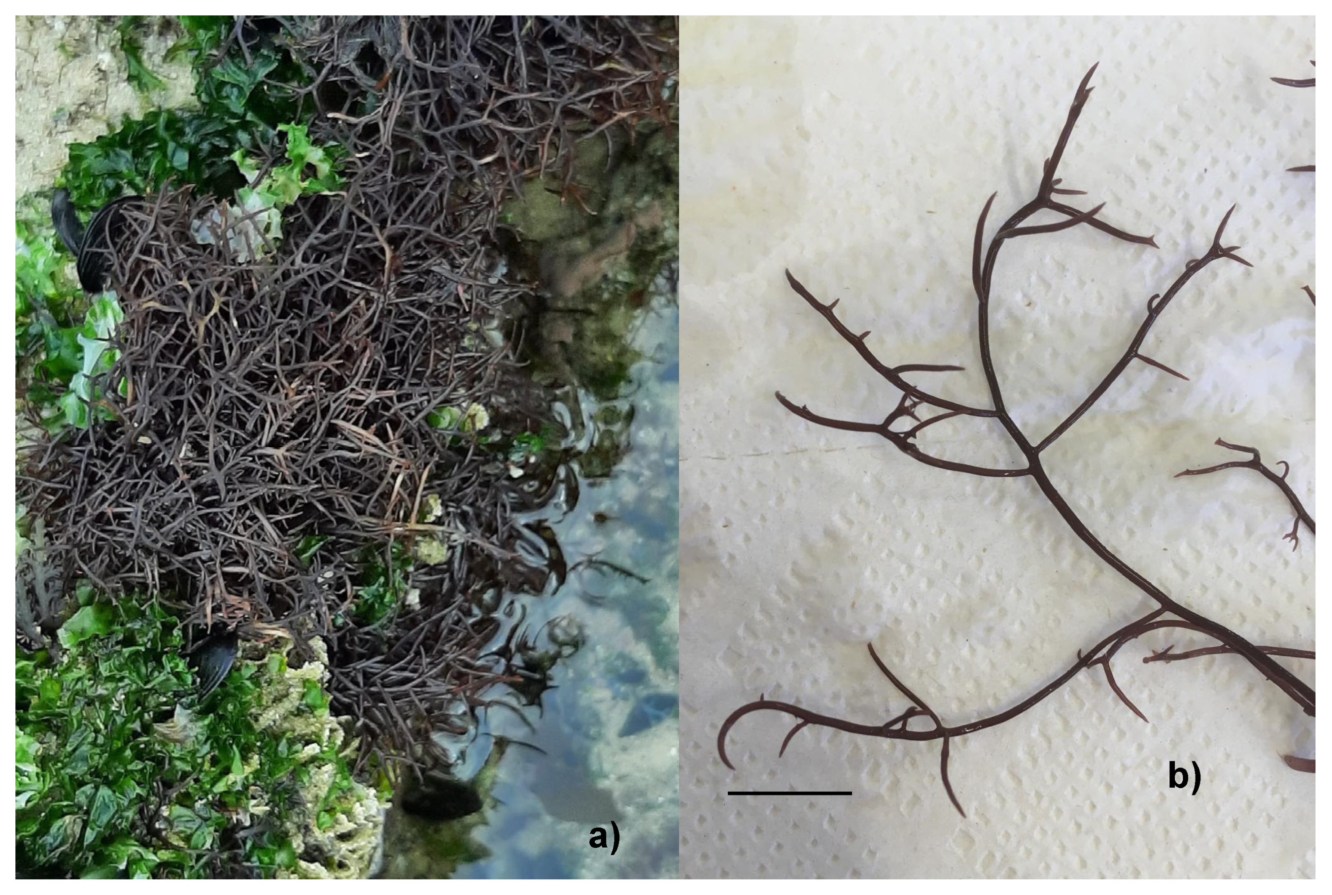
Figure 1. Chondracanthus acicularis specimens were captured: (a) in its natural habitat at Tamargueira beach in January 2024; (b) in the laboratory, highlighting the tip of the thallus (scale bar represents 0.5 cm).
Applsci 15 00810 g001
Figure 2. Experimental setup of the cultivation of Chondracanthus acicularis in 6 L flasks with PES medium, a density of 4 g L−1, temperature room set at 20 ± 1 °C, and aeration. (a) White LED light; (b) blue LED light; (c) red LED light.
Applsci 15 00810 g002
Figure 3. (a) Relative growth rate (RGR) and (b) productivity (Y) in the two-week trials of Chondracanthus acicularis. The bars represent the mean values and standard deviation (n = 3). VSE—Von Stoch Medium; PES—Provasoli Medium, NB—Nutribloom plus®, all grown with white LED light; blue—PES and blue LED light; red—PES and red LED light. Different letters above bars indicate significant differences (ANOVA, p-value < 0.05) between treatments, with lowercase letters for the first week and uppercase letters for the second week. An asterisk (*) above the bars indicates significant differences between weeks for each treatment.
Applsci 15 00810 g003
Figure 4. Proximate analysis of the wild and cultivated biomass of Chondracanthus acicularis, grown over 14 days, for percentage of moisture, ash, and organic matter on fresh weight. VSE—Von Stoch Medium; PES—Provasoli Medium, NB—Nutribloom plus®, all grown under white LED light; blue—PES and blue LED light; red—PES and red LED light. Different letters within the bars indicate statistical differences for organic matter between treatments (ANOVA, p < 0.05).
Applsci 15 00810 g004
Figure 5. Colour parameters of cultivated biomass of Chondracanthus acicularis when grown over 14 days. (a) ΔE–colour variation, (b) L*-lightness, (c) a*-green-red, (d) b*-blue-yellow, (e) C*-chromaticity, (f) h°-hue. VSE—Von Stoch Medium; PES—Provasoli Medium, NB—Nutribloom plus®, all grown with white LED light; blue—PES and blue LED light; red—PES and red LED light.
Applsci 15 00810 g005
Figure 6. Pigment content of wild and cultivated Chondracanthus acicularis when grown over 14 days. VSE–Von Stoch Medium; PES–Provasoli Medium, NB–Nutribloom plus®, all grown with white LED light; blue–PES and blue LED light; red—PES and red LED light. The bars represent the mean values and standard deviations (n = 3). Different letters above bars indicate significant differences (ANOVA, p-value < 0.05) between treatments.
Applsci 15 00810 g006
Table 1. Summary of the different Chondracanthus acicularis growth tests carried out (n = 3).
EssayCulture MediaLight
AVon Stosch Enriched (VSE)White LED
BProvasoli Enriched Seawater (PES)White LED
CNutribloom plus® (NB)White LED
DProvasoli Enriched Seawater (PES)Red LED
EProvasoli Enriched Seawater (PES)Blue LED
Table 2. Proximate analysis of the samples, expressed in percentage of dry weight, measured on day 7 and day 14 of the trial, and wild biomass measured upon arrival to the laboratory. Values are presented as mean ± standard deviation (n = 2). Different letters represent statistical differences between treatments (Kruskal–Wallis, p-value < 0.05).
Dry WeightOrganic MatterAsh
VSE, day727.01±0.8273.00±2.68 a27.00±2.68 b
VSE, day 1429.00±0.5671.31±1.3828.69±1.38
PES, day 728.88±1.6272.52±0.92 a27.48±0.92 b
PES, day 1428.39±1.0573.33±1.04 a26.67±1.04 b
NB, day731.21±1.0173.26±0.16 a26.74±0.16 b
NB, day 1429.14±0.8671.24±0.7828.76±0.78
Blue LED, day727.43±0.9571.69±0.0628.31±0.06
Blue LED, day 1428.06±0.6371.24±0.2228.76±0.22
Red LED, day726.35±0.9971.12±0.2428.88±0.24
Red LED, day 1426.12±0.0168.06±0.29 b31.94±0.29 a
Wild29.47±0.2069.20±0.86 b30.80±0.86 a
Disclaimer/Publisher’s Note: The statements, opinions and data contained in all publications are solely those of the individual author(s) and contributor(s) and not of MDPI and/or the editor(s). MDPI and/or the editor(s) disclaim responsibility for any injury to people or property resulting from any ideas, methods, instructions or products referred to in the content.