Article

Artificial Intelligence for Diagnosing Cranial Nerve III, IV, and VI Palsies Using Nine-Directional Ocular Photographs

1
Department of Ophthalmology, Konkuk University School of Medicine, Konkuk University Medical Center, Seoul 05030, Republic of Korea
2
Research Institute of Medical Science, Konkuk University School of Medicine, Seoul 05030, Republic of Korea
3
Department of Mechatronics Engineering, Konkuk University Glocal Campus, Chungju-si 27478, Republic of Korea
*
Author to whom correspondence should be addressed.
Appl. Sci. 2025, 15(20), 11174; https://doi.org/10.3390/app152011174
Submission received: 25 May 2025 / Revised: 29 September 2025 / Accepted: 16 October 2025 / Published: 18 October 2025

Abstract

Eye movements are regulated by the ocular motor nerves (cranial nerves [CNs] III, IV, and VI), which control the six extraocular muscles of each eye. Palsies of CNs III, IV, and VI can restrict eye movements, resulting in strabismus and diplopia, and so clinical evaluations of eye movements are crucial for diagnosing CN palsies. This study aimed to develop an accurate artificial intelligence (AI) system for classifying CN III, IV, and VI palsies using nine-gaze ocular photographs. We analyzed 478 nine-gaze photographs comprising 70, 29, and 58 cases of CN III, IV, and VI palsies, respectively. The images were processed using MATLAB. For model training, each photograph of eye movements in the nine directions was numerically coded. A multinetwork model was employed to ensure precise analyses of paralytic strabismus. The AI system operates by referring data on minor abnormalities in the nine-gaze image to a network designed to detect CN IV abnormalities, which re-examines downward and lateral gazes to detect distinctions. Data on major abnormalities are directed to a different network trained to differentiate between CN III and VI abnormalities. EfficientNet-B0 was applied to reduce overfitting and improve learning efficiency in training with limited medical imaging data as the neural network architecture. The diagnostic accuracies of the proposed network for CN III, IV, and VI palsies were 99.31%, 97.7%, and 98.22%, respectively. This study has demonstrated the design of an AI model using a relatively small dataset and a multinetwork training system for analyzing nine-gaze photographs in strabismus patients with CN III, IV, and VI palsies, achieving an overall accuracy of 98.77%.

1. Introduction

Eye movements are controlled by the ocular motor nerves (cranial nerves [CNs] III, IV, and VI) that innervate the six extraocular muscles (EOMs) of each eye [1]. CN III innervates the medial rectus, inferior rectus, superior rectus, inferior oblique, and levator palpebrae superioris muscles, CN IV innervates the superior oblique muscle, and CN VI innervates the lateral rectus muscle. Palsies of CN III, IV, and VI can restrict eye movements, leading to strabismus (misalignment of the eyes) and diplopia. Patients with strabismus caused by CN palsy may adopt abnormal head positions to compensate for their eye-movement restrictions and reduce diplopia [2,3]. Additionally, a CN III palsy can cause drooping of the eyelid due to dysfunction of the levator palpebrae superioris muscle.
Clinical evaluation of eye movements is crucial for diagnosing paralytic strabismus, as specific ocular motility deficits often provide significant localizing value within the central nervous system and guide both differential diagnosis and management [4]. In particular, paralytic strabismus due to cranial nerve palsy poses a diagnostic challenge, with presentations varying by the nerve involved and by disease chronicity. Acute CN III, IV, and VI palsies are common causes of sudden-onset diplopia and among the leading reasons for emergency room visits. Prompt recognition is especially critical in CN III palsy, where an intracranial aneurysm may be the underlying etiology and delayed detection can result in life-threatening subarachnoid hemorrhage [5]. Despite its clinical importance, the diagnosis of paralytic strabismus remains one of the more challenging areas in ophthalmology, as it relies on careful motility testing and examiner expertise, which may not always be available in non-specialist or emergency settings [6].
Eye movement evaluations based on nine cardinal gaze positions is an objective and reproducible method for detecting restricted eye movements [7]. Consequently, nine-directional ocular photographs are crucial for recording the status of eye movements and facilitating accurate interpretation and diagnosis [8]. Despite this, human error in interpreting numerous nine-gaze images and discrepancies between examiners are unavoidable, and so the utilization of artificial intelligence (AI) could significantly aid doctors [9]. In particular, variations in examiner experience and subjective bias often limit the consistency and reproducibility of traditional assessments. For instance, subtle gaze limitations may be missed by less experienced clinicians, or interpreted differently across institutions. In this regard, automated tools offer the potential to reduce diagnostic variability and increase standardization.
AI models can enhance the diagnostic accuracy and speed, which would expand the scope of diagnosis beyond specialists to general practitioners, and thus facilitate the more efficient distribution of medical resources. Many researchers are developing AI models for diagnosing ocular conditions, and AI has been actively utilized in ophthalmology for conditions such as glaucoma and diabetic retinopathy through the application of automated deep learning models based on image data, which has achieved reasonable accuracy [10,11]. Moreover, the emergence of AI in strabismus diagnosis opens possibilities for point-of-care screening in remote or underserved areas. By equipping primary care clinics with accessible diagnostic support, healthcare systems could detect complex ocular motor nerve palsies earlier and reduce referral delays. These advancements are particularly relevant in aging populations, where microvascular cranial nerve palsies are more common.
The present study was designed with the primary aim of developing and validating a convolutional neural network (CNN) model using standardized nine-gaze photographs to classify CN III, IV, and VI palsies. Our motivation was to evaluate whether nine-gaze photographs alone could support the diagnosis of acute paralytic strabismus in real-world settings, even when assessed by non-specialist clinicians.

2. Materials and Methods

2.1. Data Collection

From December 2012 to April 2022, data were collected from 399 subjects (157 patients with CN III, IV, or VI palsies and 242 normal controls) who visited Konkuk University Hospital and underwent nine-gaze photography. This study was performed in accordance with the principles of the Declaration of Helsinki, and was approved by the institutional review board and ethics committee at Konkuk University Medical Center (registration number: 2022-02-014). Patients were excluded if they had orbital surgery such as orbital decompression, previous strabismus surgery, blowout fracture, or any facial deformity that prevented the identification of facial points. Other exclusion criteria were corneal diseases such as microcornea and leukoma, or difficulty in identifying the sclera corneal limbus region. To minimize confounding, other motility disorders that can mimic cranial nerve palsies (e.g., thyroid eye disease, Duane syndrome, and congenital cranial dysinnervation disorders) were not included in this present study. Also, patients with severe vision loss that precluded reliable fixation were excluded.
To ensure consistency and minimize artifacts due to head or facial movement, all photographs were obtained with the subject’s head stabilized using a chin rest and a head band. Before image acquisition, the eyes were aligned in the primary position, and the absence of head tilt or chin-up/chin-down posture was confirmed. For nine-gaze images, fixation targets were presented along the axes of a Lancaster screen positioned at 1 m, corresponding to the eight secondary and tertiary gaze positions [7]. Patients were instructed to visually track the targets into maximum gaze (dextrosupraversion, supraversion, levosupraversion, dextroversion, levoversion, dextroinfraversion, infraversion, and levoinfraversion). Verbal instructions and encouragement were provided throughout to maintain head stability and ensure maximum effort toward the extremes of gaze. With this standardized setup, variability caused by head movement was minimized, allowing reliable capture of ocular motility across patients. During nine-gaze photography, fixation was always performed with the fellow (non-paretic) eye, in accordance with standard strabismus photography practice. EOM function (ocular versions and ductions using the standard scale from −4 to +4) was classified as described previously. In the patient group, only very mild cases with duction limitation of ≤−1 were excluded because of potential diagnostic ambiguity, whereas patients with more pronounced deficits (e.g., −2 or −3), representing incomplete palsy, were included. All control subjects demonstrated normal ocular motility without any duction deficits [12]. The control group comprised 1991 nine-gaze photographs obtained from 242 cases of normal subjects. The numbers of cases in the two study groups are provided in Table 1.

2.2. Data Labeling

All patients underwent comprehensive clinical examinations required for diagnosing paralytic strabismus. These included ocular motility assessments, prism cover test for measuring strabismus angle, Lancaster red–green test, Bielschowsky head tilt test, and fundus photograph. Based on an analysis of electronic medical records performed by a skilled ophthalmologist (H.J.S.) with over 10 years of experience in strabismus and pediatric ophthalmology, the 478 nine-gaze photographs from 157 patients were labeled as 70 cases of CN III palsy, 29 cases of CN IV palsy, and 58 cases of CN VI palsy. Patients ranged in age from 41 to 86 years (mean ± SD: 62.1 ± 11.3 years). All patients were Korean, as data were collected at a single tertiary referral center (Konkuk University Hospital, Seoul, Korea). The best corrected visual acuity ranged from 6/6 to 6/9 on the Snellen chart. A second examiner (H.K.) reviewed cases where diagnosing and grading EOM function was difficult, and the final data labeling was based on consensus being reached between these examiners. Each photograph of the nine cardinal positions was split and assigned a number as follows: straight upward (No. 2), upward and to the right (No. 1), upward and to the left (No. 3), straight ahead (No. 5), left (No. 6), right (No. 4), straight downward (No. 8), downward and to the left (No. 9), and downward and to the right (No. 7). To obtain an unobstructed view of the eyes in the downgaze positions, the patient or a helper elevated the patient’s upper eyelids (Figure 1).

2.3. Characterization of CN III, IV, VI

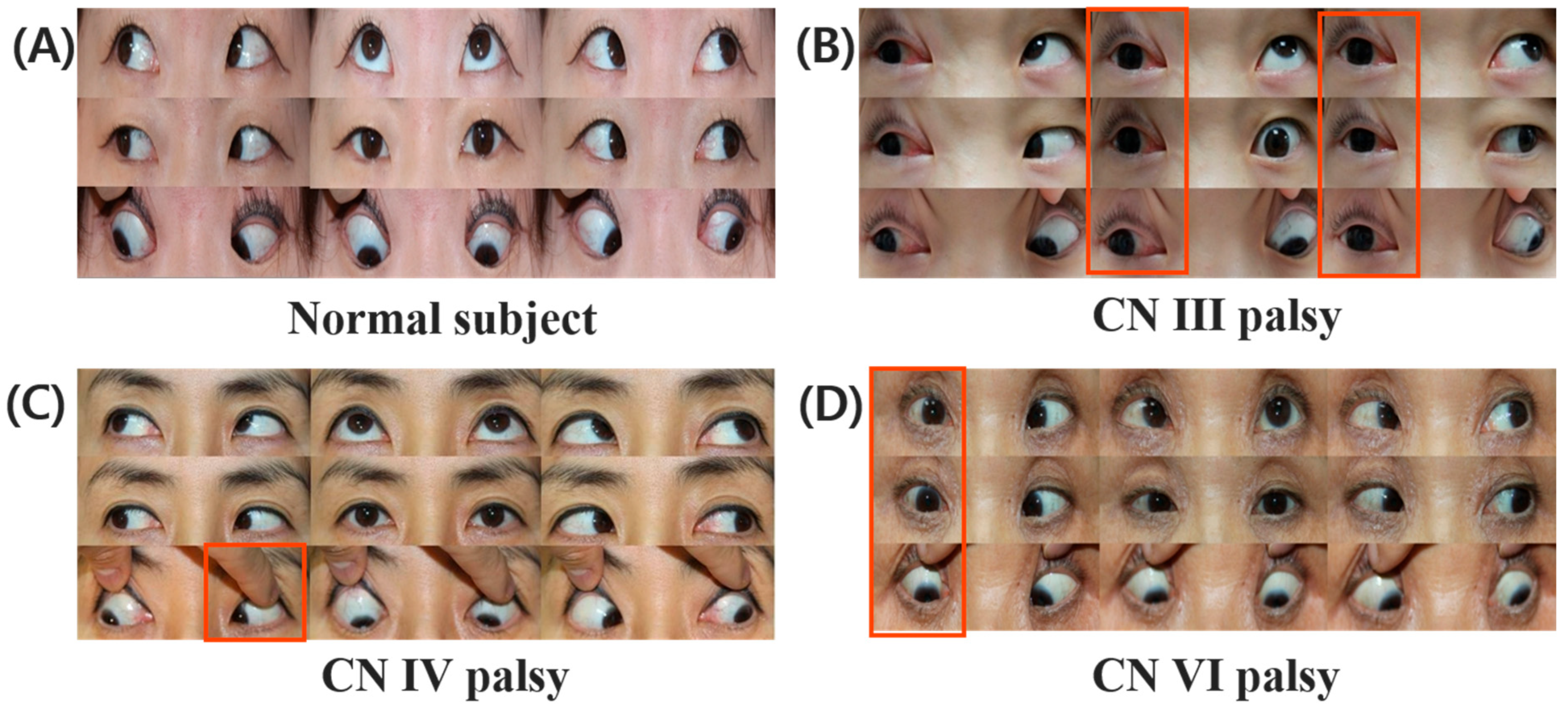
The data were processed and analyzed using MATLAB (version 2021b, MathWorks, Natick, MA, USA), which is well-established engineering and scientific data analysis software recognized for its reliability in numerous various studies [13]. To analyze abnormal nine-gaze patterns, the cardinal directions in which abnormalities primarily appear during each type of nerve paralysis were determined. For example, in the case of right CN III palsy, abnormalities were primarily observed in adduction, supraduction, and infraduction, along with ptosis. Consequently, photograph Nos. 2, 3, 5, 6, 8, and 9 exhibited these abnormalities (Figure 2B). In the case of left CN IV palsy, abnormalities were primarily observed in extrodepression, resulting in abnormalities in photograph No. 7 (Figure 2C). In the case of right CN VI palsy, abnormalities were primarily observed in extroversion, with photograph Nos. 1, 4, and 7 exhibiting these abduction abnormalities (Figure 2D). These directional criteria were then used to evaluate the nine-gaze photographs per direction, and CN palsy was classified based on the combination of abnormal directions.

2.4. Data Augmentation of the Nine-Gaze Images

In order to improve the diversity and robustness of the training dataset, offline augmentation was applied to the nine-gaze images. The nine-gaze image was first divided into nine directional components, and then each image was augmented using four image processing techniques: edge enhancement, sharpening, contrast stretching, and the addition of Gaussian noise. These augmentation techniques were selected to approximate real-world variability in clinical imaging conditions. Specifically, Gaussian noise reflects subtle degradations in image quality due to sensor noise, lighting fluctuations, or electronic interference commonly encountered in ophthalmic photography. Sharpening and edge enhancement mimic differences in focus and contrast that may result from variations in camera calibration, operator technique, or patient cooperation. Contrast stretching was applied to replicate variability in illumination levels and pupil size that influence brightness and clarity. As illustrated in Figure 3, these techniques also serve to improve the visibility of edges and enhance details in dark regions of the images, thereby simulating clinically plausible variability and improving model robustness against imaging heterogeneity encountered in real-world practice.
Furthermore, horizontal flipping was applied to augment the data for both left- and right-CN palsies. For instance, images of right CN IV palsy were horizontally flipped to serve as synthetic samples of left CN IV palsy. This approach offered a dual benefit: it not only expanded the limited dataset of 9-gaze images from CN palsy but also helped compensate for the data imbalance between left- and right-CN palsy cases. Crucially, to avoid data leakage, augmented versions of an image were never split across the training and validation sets; instead, they were treated as a single entity during the data partitioning process. This ensured that no single source image contributed to both the training and validation datasets.
However, other geometric transformations such as image rotation and vertical flipping were not applied because this can lead to misinterpretations in direction-sensitive data. Similarly, image shifting was not performed since excessive shifting would result in image cropping, and so compromise the data integrity. In this study, we did not apply weighted loss functions or explicit oversampling techniques to address the class imbalance among cranial nerve palsy groups. Instead, we attempted to mitigate this issue through data augmentation strategies such as edge enhancement, sharpening, contrast stretching, Gaussian noise addition, and horizontal flipping of nine-gaze photographs. While these approaches increased the diversity of CN IV palsy cases, the imbalance in the original dataset was not completely eliminated.

2.5. Construction of the CNN Model for CN Palsy Classification

To enhance classification accuracy despite the relatively small dataset, we developed a specialized approach that combined multiple convolutional neural networks (CNNs). Each CNN was trained as a binary classifier to differentiate between normal gaze and the presence of a specific cranial nerve (CN) palsy in one of the nine standard gaze directions. For instance, gaze position No. 1 in a normal subject (Figure 2A) was compared with the same position in a patient with right CN VI palsy (Figure 2D) in order to detect the characteristic abduction deficit associated with CN VI involvement.
After training, the outputs of these binary classifiers were aggregated. The combination of detected abnormalities across all gaze directions was then used to generate the final diagnosis, specifying both the type of palsy (CN III, IV, or VI) and its laterality (left or right eye). This stepwise approach allowed the model to capture direction-specific abnormalities while integrating the overall gaze pattern to reach a clinically meaningful classification [14].
The dataset was divided into training (80%) and validation (20%) subsets. To determine a suitable set of training conditions, a parametric study was conducted. Model performance was assessed using several key metrics, including macro precision, macro recall, macro F1 score, and accuracy as summarized in Table 2. Based on this study, the final training configuration was selected as follows. The model was trained using the Adaptive moment estimation (Adam) optimizer, starting from an initial learning rate (LR) of 5 × 10⁻⁴. LR was reduced by a factor of 0.1 every 5 epochs. The training was performed for a maximum of 25 epochs with a mini-batch size of 32 images. To prevent overfitting, validation was conducted at the end of each epoch, and an early stopping was applied with a patience of 5 epochs.
After constructing the CNN models, target nine-gaze images were divided into nine directional subimages and fed into the CNN models. The classification results were then compared with the predefined directions to recognize abnormalities among nine-gaze photographs. The procedures are illustrated in Figure 4.
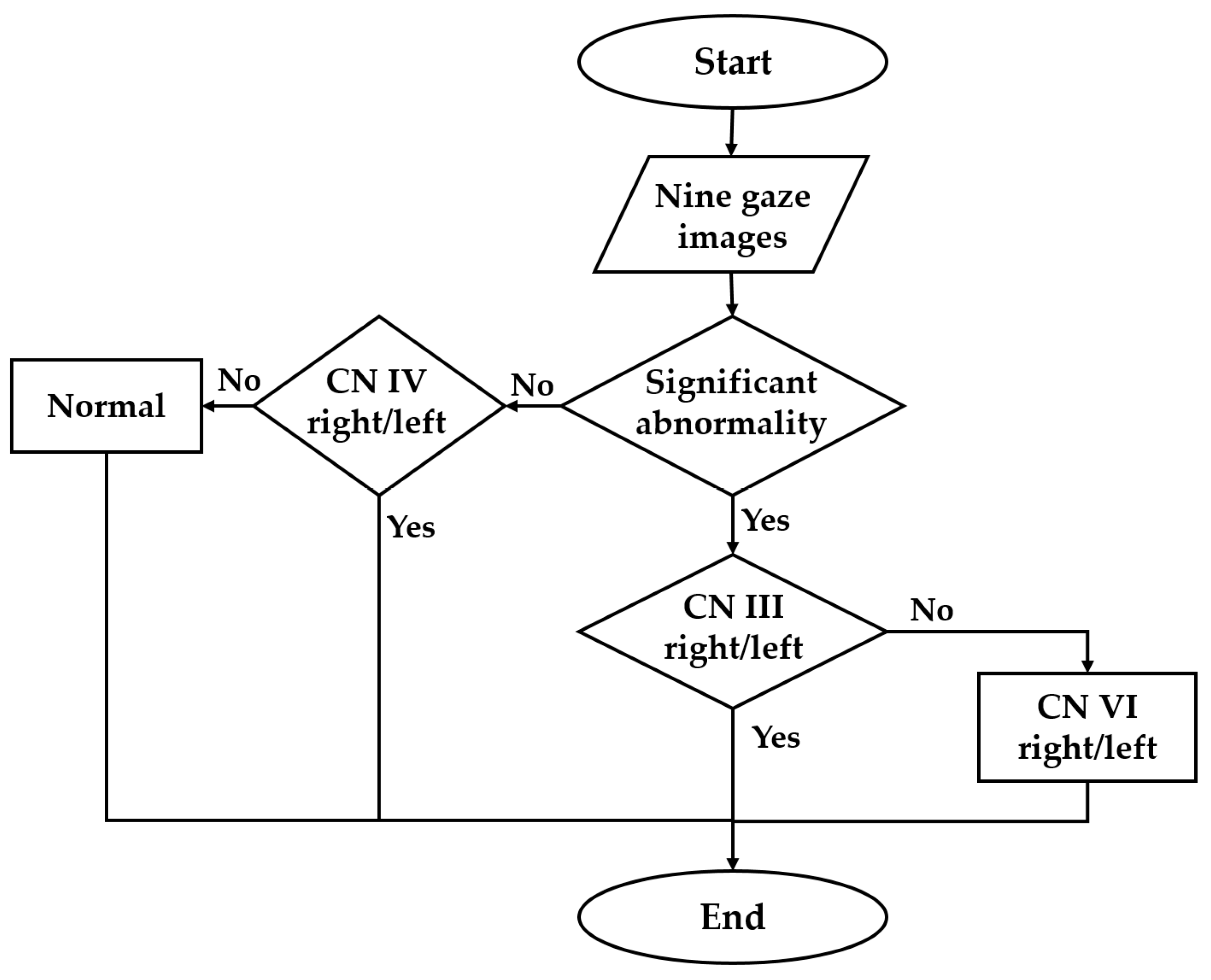
A multinetwork approach involves a combination of specialized CNNs was employed for the precise analysis of paralytic strabismus. Initially, if the CNN detected one or two minor abnormalities in the nine-gaze images, the data were referred to a network specifically trained to detect CN IV abnormalities. This network rechecked the abnormalities when the patient was looking downward and laterally, to ensure that the normal and abnormal conditions of the CN IV on both sides were accurately distinguished. In cases where more than three abnormalities were detected, the data were sent to a different network trained to distinguish between CN III and VI abnormalities. This network effectively identified whether the abnormalities were associated with CN III or VI, and determined the affected side (Figure 5).

3. Results

3.1. Abnormality Detection

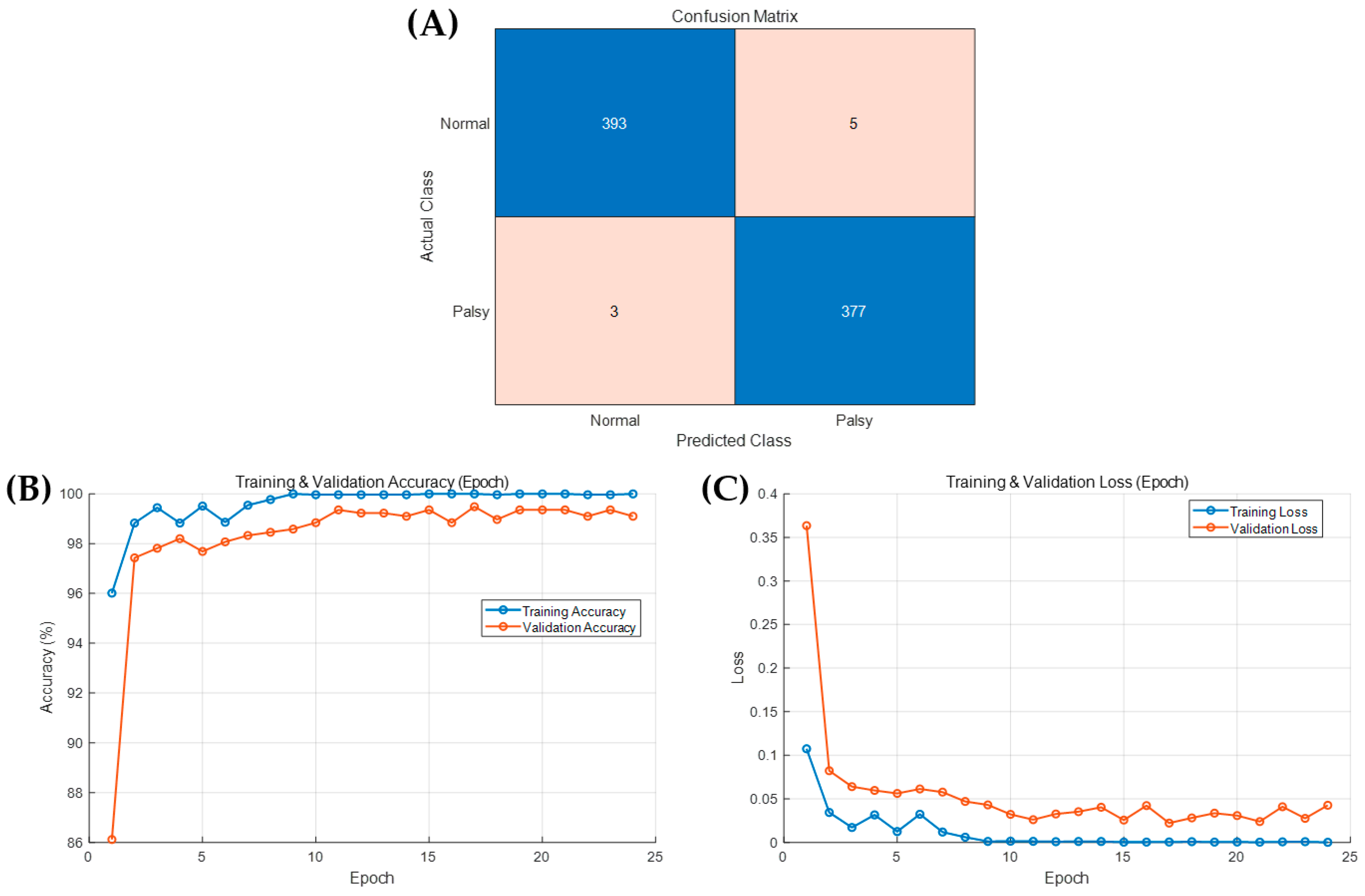
The split nine-gaze images were sent to the CNN-based abnormality detection model, which was trained to detect strabismus of CN palsy images including laterality (left or right eye), gaze direction (No. 1 to 9), and ocular version (normal vs. limited). The model achieved 98.97% accuracy in determining the occurrence of CN palsy. Figure 6 shows the achievements of CNN model in confusion matrix, accuracy, and loss during training and validation process. The F1-score, recall, and precision were summarized in Table 3.

3.2. Multinetwork CN Palsy Detection

Firstly, a single CNN model was developed to classify the 9-gaze images into one of 27 classes, each representing a specific gaze direction and CN palsy type including normal. This approach achieved an overall accuracy of 92.33%. the results were summarized in Table 4, and the confusion matrix and accuracy and loss during train and validation were illustrated in Figure 7.
To enhance the performance the CNN for CN palsies detection, the multinetwork algorithm was developed in hierarchal manner. The averaged classification accuracies for identifying CN III, IV, and VI palsies were 99.31%, 97.7%, and 98.22%, respectively. The overall diagnostic accuracy of the model increased from 92.33% to 98.77% after applying the proposed multinetwork structure, which was designed to handle different types of gaze abnormalities in a hierarchical manner. Performance metrics showed that CN III palsy detection achieved the highest precision and recall, with an F1-score of 0.99. Precision and recall for CN III palsy were 99.33% and 99.31%, respectively. For CN IV palsy, the F1-score was 0.97, with a precision of 96.54% and recall of 98.34%. For CN VI palsy, the F1-score was 0.98, with a precision of 98.29% and recall of 98.26%., indicating robust performance even in a dataset of limited size. The performance of proposed multinetwork approach was summarized in Table 5.
Stratified analysis by gaze direction revealed that the model performed most accurately in horizontal gazes (positions 4 and 6) and diagonal downgaze directions (positions 7 and 9), which are commonly affected in CN VI and CN IV palsies. Figure 8 shows occlusion sensitivity images after interference, which indicate the areas that the model relied upon heavily for judgements. In the figure, regions closer to red signify areas that the model heavily referenced; the examples show where the model correctly classified images as data showing no limitation of eye movement (Figure 8A), left infraduction limitation in CN III palsy (Figure 8B), and left adduction depression limitation in CN IV palsy (Figure 8C); regardless of sides, the inferences were correct. Most misclassifications occurred in borderline or mild cases, such as partial CN III palsy with ptosis that obscured infraduction views, or in patients whose downgaze photos were partially blocked by eyelid droop. In these cases, even manual elevation of the eyelid was insufficient to provide clear ocular visibility. However, the model rarely misclassified pathologic cases as normal, with most errors occurring between adjacent CN palsies categories.
An analysis of misclassified cases revealed that most errors occurred in borderline or atypical presentations. For example, partial CN III palsy with ptosis frequently led to incorrect classification because the drooping eyelid obscured infraduction, even when the eyelid was manually elevated. Similarly, downgaze photographs with incomplete visibility of the sclera were occasionally misclassified due to insufficient image information. In CN IV palsy, mild or compensated cases with subtle depression deficits also posed challenges for the model. These findings suggest that misclassifications were primarily caused by image obstruction or subtle motility limitations rather than gross algorithmic failure.

4. Discussion

The purpose of this study was to introduce a multinetwork CNN-based strabismus detection algorithm designed to improve the classification accuracy of CN III, IV, and VI palsies using nine-gaze photographs. A hierarchical multinetwork structure was employed to refine decision-making based on the type of abnormalities detected in eye-movement images. The CNN model first determines whether a given nine-gaze image represents normal eye movements or abnormalities: if one or two minor abnormalities are detected, the data are sent to a subnetwork specialized in CN IV palsy detection; while the presence of three or more abnormalities triggers forwarding to another subnetwork trained to differentiate between CN III and VI palsies, culminating in the CNN determining which CN is affected (III, IV, or VI) and whether the right or left eye is involved.
Several studies have attempted to investigate ocular movements using photographs of patients with strabismus. Zheng et al. [15] implemented a deep-learning algorithm to classify horizontal strabismus (e.g., esotropia and exotropia) using primary-gaze photographs of children; however, that study did not consider other types of strabismus or different gaze positions. Karaaslan et al. [16] focused solely on the Hirschberg test, but their study had limitations in comprehensively assessing all strabismus types and differentiating specific nerve palsies. Also, for strabismus diagnosis, it was constrained by its dependence on high-quality corneal light-reflection photographs, which could prevent its broad clinical application. Figueiredo et al. [17] developed a CNN-based web application that classified ocular version in nine gaze positions. Its limitations included a focus on classifying ocular version rather than nerve palsies, the use of a potentially unrepresentative sample, challenges in detecting subtle gaze abnormalities, and a lack of clarity about how to translate it into clinical strabismus management. Kang et al. [18] developed an automated strabismus measurement algorithm using deep learning to analyze gaze-position photographs. However, the effectiveness of their method was restricted by its reliance on clear visibility of the sclera and limbus, which is challenging in patients with small eyes, a peripheral cornea whitening condition such as arcus senilis, or restricted limbus exposure. These limitations highlight the need for continued research and development in AI applications for diagnosing strabismus, particularly in addressing specific nerve palsies and accounting for a wider range of patient presentations and complexities.
The multinetwork CNN-based strabismus detection algorithm developed in this study represents a significant advancement in the diagnosis of CN III, IV, and VI palsies, by effectively addressing limitations of traditional clinical examinations and previous AI models. By leveraging a sophisticated multinetwork approach, this new CNN model increased the diagnostic accuracy from 92.33% to 98.77%, surpassing previous AI-driven ophthalmological tools with specific accuracies of 99.31%, 97.7%, and 98.22%, for CN III, IV, and VI palsies, respectively. This objective and reproducible tool reduces the reliance on subjective assessments and interobserver variability, offering refined decision-making through specialized subnetworks for more-accurate analyses.
The use of nine-directional ocular photographs combined with AI enables rapid and standardized evaluation of eye movement abnormalities, potentially reducing clinician workload and fatigue, especially in busy clinics. In addition, these models can offer diagnostic support to junior physicians or in low-resource settings where trained strabismus specialists are not available. The model’s architecture, based on a tiered decision tree and multiple subnetworks, also makes it interpretable and expandable for future refinement [19,20]. From a public health perspective, this AI-assisted diagnostic tool can be integrated into screening programs at the primary care level or in rural clinics. In telemedicine environments, where eye movement videos or photographs can be transmitted, this algorithm can help triage patients and prioritize referrals to tertiary centers. This is particularly valuable for time-sensitive conditions such as aneurysm-related CN III palsy, where prompt diagnosis is essential [21,22].
However, it should be noted that while the model showed a good diagnostic performance, it does not provide insights into underlying etiologies, such as microvascular ischemia, trauma, or aneurysmal compression. Additionally, our model currently focuses solely on paralytic strabismus; incorporating other strabismic conditions such as restrictive strabismus (e.g., thyroid eye disease and orbital fractures) could expand its clinical applicability. Further improvement could involve multimodal data input, integrating orbital CT, MR imaging, or fundus photography to distinguish etiologic patterns and subtypes more accurately. Although the Bielschowsky head tilt test is highly informative for diagnosing CN IV palsy, it was not included in our AI input because head tilt photographs were not systematically collected during the study period. Our approach was intentionally limited to nine-gaze static photographs, which are routinely performed and consistently archived in clinical practice. Nevertheless, the model achieved robust performance for CN IV palsy (97.7% accuracy). Future studies may incorporate head tilt photographs to further improve diagnostic performance, particularly in subtle or borderline CN IV palsy. In particular, the relatively small number of CN IV palsy cases (n = 29) resulted in a class imbalance compared with CN III and VI palsy groups. Although augmentation techniques partially compensated for this, the limited sample size may have affected model generalization, especially in distinguishing CN IV palsy. Future studies should therefore include larger datasets and consider the application of weighted loss functions or oversampling strategies to ensure more balanced training and robust performance across all cranial nerve palsy subtypes.
Another limitation is that the proposed model was compared only with a single CNN baseline and not with other state-of-the-art architectures such as ResNet variants or Vision Transformers. The primary focus of this study was to design and validate a multinetwork framework tailored to nine-gaze photographs, and therefore extensive benchmarking across architectures was beyond the study scope. Although the proposed model achieved high accuracy, future work should incorporate comparative experiments with mainstream deep-learning architectures to more rigorously benchmark performance. In addition, statistical significance testing between models was not performed, as the relatively small dataset restricted statistical power. Finally, while the ground-truth labels in this study were established by two ophthalmologists with consensus, we did not assess diagnostic agreement with independent clinicians. Future validation studies should therefore evaluate inter-rater agreement and compare AI predictions with ophthalmologist performance to further establish clinical applicability. Prospective validation studies with blinded clinician comparison should also be conducted to evaluate performance in real clinical workflows. Moreover, longitudinal analysis to track patient outcomes after AI-supported diagnosis would strengthen the clinical utility of the model [23]. In addition, all data were collected from a single tertiary referral center, and no external validation set was used. This single-center design may limit the generalizability of the findings to other institutions or populations. Future studies should therefore incorporate multicenter datasets and independent external validation cohorts to verify the robustness and reproducibility of the proposed model across diverse clinical settings.
It should also be acknowledged that obtaining nine-gaze photographs is not always feasible in clinical practice. Some patients, particularly elderly individuals or those with poor cooperation, may not reach the full 30° gaze angle in horizontal or vertical directions, and even normal elderly adults may demonstrate limitations in elevation. These factors could restrict the applicability of the model in real-world settings. Furthermore, our model was trained and validated only on isolated single cranial nerve palsies (CN III, IV, and VI). Patients with combined cranial nerve palsies or atypical motility disorders—including restrictive or neurologic strabismus such as thyroid eye disease, orbital fractures, congenital cranial dysinnervation disorders, or congenital fibrosis of the extraocular muscles—were excluded from the dataset. This restriction limits the direct applicability of the model to more complex clinical presentations. Such conditions may produce atypical motility patterns that differ from paralytic strabismus and represent important confounders that warrant further investigation. Future work should aim to extend the algorithm to accommodate multiple concurrent cranial nerve palsies and restrictive strabismus. Incorporating such cases, along with multimodal data inputs (e.g., orbital imaging or head tilt photographs), may broaden the applicability of the model to a wider range of strabismus phenotypes encountered in real-world clinical practice. Another limitation is that misclassifications were mainly observed in borderline or atypical presentations, such as partial CN III palsy with ptosis or mild CN IV palsy. Future improvements could address these sources of misclassification. Possible approaches include the use of preprocessing methods such as eyelid segmentation to reduce obstruction artifacts, integration of head-tilt photographs to capture diagnostic information particularly relevant for CN IV palsy, and incorporation of multimodal data (e.g., fundus torsion or orbital imaging) to enhance diagnostic accuracy in borderline cases.
Another important consideration is that not all forms of ocular motility disorders can be reliably differentiated using nine-gaze photographs alone. For example, congenital CN IV palsy may develop secondary contracture of the ipsilateral superior rectus muscle over time, leading to loss of incomitance and atypical motility patterns [24]. Likewise, sagging eye syndrome can mimic superior oblique palsy on motility testing but does not produce excyclotorsion [25], and there is no evidence that nine-gaze photographs alone can distinguish between these entities. These scenarios indicate that the current algorithm is best suited for acute paralytic strabismus, where CN III, IV, or VI palsy presents with sudden-onset diplopia and requires rapid recognition, such as in cases of aneurysmal CN III palsy. For long-standing or atypical cases, detailed physician history-taking remains essential to refine differential diagnoses beyond what nine-gaze photographs can provide. Importantly, the proposed algorithm is not intended to replace a full motility assessment but rather to serve as a supportive tool for early recognition, particularly in settings where subspecialty expertise is not readily available.
Future work should also consider the collection of larger and more diverse datasets to enhance generalizability. Evaluating the algorithm’s performance across different age groups, ethnicities, and varying image quality will be essential to move toward robust clinical deployment. Moreover, integration with video-based eye-tracking technologies could enable real-time functional assessments of ocular motility [26]. Finally, regulatory considerations and explainability of AI predictions must be addressed for real-world clinical integration. By providing heatmaps and attention maps indicating the decision focus, clinicians may be able to understand and trust AI outputs more readily [27]. With sufficient external validation and software interface development, the proposed algorithm has the potential to be deployed as a user-friendly diagnostic support system in ophthalmology clinics worldwide. These advancements could improve the efficiency and accuracy of the diagnosis and management of strabismus in clinical practice.

5. Conclusions

In this study we designed a multinetwork CNN-based strabismus detection algorithm for analyzing nine-gaze photographs in strabismus patients with CN III, IV, and VI palsies, and achieved an overall diagnostic accuracy of 98.77%. We developed a reliable CNN model by gathering a relatively small dataset of labeled photographs of strabismus patients’ eyes in nine different gaze positions by designing networks specialized in identifying different aspects of the strabismus condition based on ocular version. These findings highlight the potential of the CNN approach in enhancing the eye movement evaluation process, which has traditionally relied exclusively on human interpretation. The capabilities of this new approach will make high-quality healthcare more accessible, especially in underserved and remote areas, allowing for more accurate and timely diagnoses that directly impact patient treatment outcomes. Furthermore, the proposed algorithm may assist clinicians by improving diagnostic consistency and efficiency. Its modular structure enables future enhancements, including support for additional strabismus types or video-based analysis. This AI approach complements physician expertise and meets the need for scalable, cost-effective ophthalmologic tools.

Author Contributions

Conceptualization, H.J.S.; methodology, H.K.; software, H.K. and M.S.K.; validation, H.J.S.; formal analysis, H.K. and S.J.K.; investigation, S.J.K.; data curation, H.J.S. and S.H.P.; writing—original draft preparation, H.K.; writing—review and editing, H.J.S.; visualization, S.J.K.; supervision, H.K.; project administration, H.J.S.; funding acquisition, H.K. All authors have read and agreed to the published version of the manuscript.

Funding

This paper was supported by Konkuk University in 2025.

Institutional Review Board Statement

The study was conducted in accordance with the Declaration of Helsinki and approved by the Institutional Review Board of Konkuk University Medical Center (registration number: 2022-02-014).

Informed Consent Statement

Informed consent was obtained from all subjects involved in the study.

Data Availability Statement

The datasets generated and analyzed during the current study contain patient-identifiable information and are not publicly available due to privacy and ethical restrictions. Reasonable requests for de-identified data may be considered by the corresponding author, subject to approval by the institutional ethics committee.

Conflicts of Interest

The authors declare no conflicts of interest. The funders had no role in the design of the study; in the collection, analyses, or interpretation of data; in the writing of the manuscript; or in the decision to publish the results.

References

  1. Shin, H.J.; Lee, S.J.; Oh, C.; Kang, H. Novel compact device for clinically measuring extraocular muscle (EOM) tension. J. Biomech. 2020, 26, 109955. [Google Scholar] [CrossRef]
  2. Kang, H.; Lee, S.; Oh, C.; Shin, H.J.; Lee, A.G. Quantitative measurement of passive duction force tension in intermittent exotropia and its clinical implications. Graefe’s Arch. Clin. Exp. Ophthalmol. 2021, 259, 1617–1623. [Google Scholar] [CrossRef]
  3. Shin, H.J.; Kim, S.; Kang, H.; Lee, A.G. Novel Instrument for Clinical Evaluations of Active Extraocular Muscle Tension. Appl. Sci. 2023, 13, 11431. [Google Scholar] [CrossRef]
  4. Prasad, S.; Volpe, N.J. Paralytic strabismus: Third, fourth, and sixth nerve palsy. Neurol. Clin. 2010, 28, 803–833. [Google Scholar] [CrossRef]
  5. Kim, J.; Choi, S. Third cranial nerve palsy and posterior communicating artery aneurysm. Clin. Exp. Emerg. Med. 2014, 1, 65–66. [Google Scholar] [CrossRef]
  6. Phuljhele, S.; Dhiman, R.; Sharma, M.; Kusiyaits, K.; Saxena, R.; Mahalingam, K.; Sharma, P. Acquired Ocular Motor Palsy: Current Demographic and Etiological Profile. Asia-Pac. J. Ophthalmol. 2020, 9, 25–28. [Google Scholar] [CrossRef]
  7. Lim, H.W.; Lee, D.E.; Lee, J.W.; Kang, M.H.; Seong, M.; Cho, H.Y.; Oh, J.; Oh, S.Y. Clinical measurement of the angle of ocular movements in the nine cardinal positions of gaze. Ophthalmology 2014, 121, 870–876. [Google Scholar] [CrossRef]
  8. Kang, H.; Lee, S.J.; Shin, H.J.; Lee, A.G. Measuring ocular torsion and its variations using different nonmydriatic fundus photographic methods. PLoS ONE 2020, 15, e0244230. [Google Scholar] [CrossRef]
  9. Nicoleta, A.; Catalin, L.; Bogdan, D.; Silvia, C.; Camelia, M.B.; Dorin, C.; Daniel, C.B.; Lonela, N.; Ovidiu-Dumitru, I.; Roxana, E.C. Use of Artificial Neural Networks to Predict the Progression of Glaucoma in Patients with Sleep Apnea. Appl. Sci. 2022, 12, 6061. [Google Scholar] [CrossRef]
  10. Sidhu, Z.; Mansoori, T. Artificial intelligence in glaucoma detection using color fundus photographs. Indian J. Ophthalmol. 2024, 72, 408–411. [Google Scholar] [CrossRef]
  11. Wu, L.; Fernandez-Loaiza, P.; Sauma, J.; Hernandez-Bogantes, E.; Masis, M. Classification of diabetic retinopathy and diabetic macular edema. World J. Diabetes 2013, 4, 290. [Google Scholar] [CrossRef]
  12. Zhale, R.; Hamideh, S.; Pooya, T.; Narges, B.; Mehdi, Y.; Mohadeseh, F.; Mohammad, F.; Kourosh, S. The relationship between abduction deficit and reoperation among patients with infantile esotropia. Int. J. Ophthalmol. 2018, 11, 478–483. [Google Scholar] [CrossRef]
  13. Zheng, Y.; Fu, H.; Li, R.; Lo, W.L.; Chi, Z.; Feng, D.D.; Song, Z.; Wen, D. Intelligent Evaluation of Strabismus in Videos Based on an Automated Cover Test. Appl. Sci. 2019, 9, 731. [Google Scholar] [CrossRef]
  14. Tan, M.; Le, Q.V. EfficientNet: Rethinking model scaling for convolutional neural networks. In Proceedings of the 36th International Conference on Machine Learning (ICML), PMLR, Long Beach, CA, USA, 9–15 June 2019; Volume 97, pp. 6105–6114. [Google Scholar]
  15. Zheng, C.; Yao, Q.; Lu, J.; Xie, X.; Lin, S.; Wang, Z.; Wang, S.; Fan, Z.; Qiao, T. Detection of referable horizontal strabismus in children’s primary gaze photographs using deep learning. Transl. Vis. Sci. Technol. 2021, 10, 33. [Google Scholar] [CrossRef]
  16. Karaaslan, Ş.; Kobat, S.G. A new method based on deep learning and image processing for detection of strabismus with the Hirschberg test. Photodiagnosis Photodyn Ther. 2023, 44, 103805. [Google Scholar] [CrossRef] [PubMed]
  17. de Figueiredo, L.A.; Dias, J.V.P.; Polati, M.; Carricondo, P.C.; Debert, I. Strabismus and artificial intelligence app: Optimizing diagnostic and accuracy. Transl. Vis. Sci. Technol. 2021, 10, 22. [Google Scholar] [CrossRef]
  18. Kang, Y.C.; Yang, H.K.; Kim, Y.J.; Hwang, J.; Kim, K.G. Automated Mathematical Algorithm for Quantitative Measurement of Strabismus Based on Photographs of Nine Cardinal Gaze Positions. Biomed. Res. Int. 2022, 24, 9840494. [Google Scholar] [CrossRef]
  19. Wu, D.; Li, Y.; Zeyi, Y.; Yin, T.; Chen, X.; Liu, J.; Shang, W.; Xie, B.; Yang, G.; Zhang, H.; et al. Deep Learning-Based Precision Cropping of Eye Regions in Strabismus Photographs: Algorithm Development and Validation Study for Workflow Optimization. J. Med. Internet Res. 2025, 27, e74402. [Google Scholar] [CrossRef]
  20. Yarkheir, M.; Sadeghi, M.; Azarnoush, H.; Akbari, M.R.; Pour, E.K. Automated strabismus detection and classification using deep learning analysis of facial images. Sci. Rep. 2025, 15, 3910. [Google Scholar] [CrossRef]
  21. Chia, M.A.; Turner, A.W. Benefits of Integrating Telemedicine and Artificial Intelligence into Outreach Eye Care: Stepwise Approach and Future Directions. Front. Med. 2022, 9, 835804. [Google Scholar] [CrossRef]
  22. Wu, D.; Huang, X.; Chen, L.; Hou, P.; Liu, L.; Yang, G. Integrating artificial intelligence in strabismus management: Current research landscape and future directions. Exp. Biol. Med. 2024, 249, 10320. [Google Scholar] [CrossRef]
  23. Wahab Sait, A.R. Artificial Intelligence-Driven Eye Disease Classification Model. Appl. Sci. 2023, 13, 11437. [Google Scholar] [CrossRef]
  24. Molinari, A.; Ugrin, M.C. Frequency of the superior rectus muscle overaction/contracture syndrome in unilateral fourth nerve palsy. J. AAPOS 2009, 13, 571–574. [Google Scholar] [CrossRef]
  25. Wei, Q.; Clark, R.A.; Demer, J.L. Can Binocular Alignment Distinguish Hypertropia in Sagging Eye Syndrome from Superior Oblique Palsy? Investig. Ophthalmol. Vis. Sci. 2022, 63, 13. [Google Scholar] [CrossRef]
  26. Ellahham, S. Artificial Intelligence: The Future for Diabetes Care. Am. J. Med. 2020, 133, 895–900. [Google Scholar] [CrossRef]
  27. Palaniappan, K.; Lin, E.Y.T.; Vogel, S. Global Regulatory Frameworks for the Use of Artificial Intelligence (AI) in the Healthcare Services Sector. Healthcare 2024, 12, 562. [Google Scholar] [CrossRef]
Figure 1. Splitting and numbering the nine-gaze images. The nine cardinal eye-movement photographs (left) were split and numbered (right) from 1 to 9, corresponding to the gaze directions (1: up-right, 2: up, 3: up-left, 4: right, 5: primary, 6: left, 7: down-right, 8: down, 9: down-left). These numbered images were used for data augmentation and to develop an algorithm for diagnosing cranial nerve III, IV, and VI palsies.
Applsci 15 11174 g001
Figure 2. Nine-gaze photographs of normal subjects and patients with CN III, IV, and VI palsies. (A) A normal subject exhibits full eye movements without limitations. (B) A patient with right CN III palsy displays deficits in adduction, supraduction, and infraduction, along with ptosis. The patient’s right droopy eyelid was elevated to demonstrate the abnormal eye position in primary gaze. (C) A patient with left CN IV palsy shows an adduction depression deficit in the left eye. (D) A patient with right CN VI palsy exhibits an abduction deficit in the right eye.
Applsci 15 11174 g002
Figure 3. Data augmentation using four image processing techniques: edge enhancement, sharpening, contrast stretching, and the addition of Gaussian noise in the set of nine-directional ocular photographs.
Applsci 15 11174 g003
Figure 4. Overview of the CNN model pipeline for the classification of CN III, IV, and VI palsies using nine-gaze ocular photographs. The input images are first divided into nine directional gaze subimages, which are analyzed by convolutional neural networks trained to detect patterns specific to each cranial nerve palsy. Based on the number and type of detected abnormalities, the model directs the case to one of several subnetworks specialized in either CN IV palsy or in differentiating CN III from CN VI palsies. The system then outputs the predicted diagnosis along with the affected side (L: left; R: right). This modular structure enhances diagnostic accuracy by focusing analysis on gaze directions relevant to each type of nerve dysfunction.
Applsci 15 11174 g004
Figure 5. Schematic diagram of the multinetwork algorithm for detecting CN III, IV, and VI palsies. The model first analyzes nine-directional gaze images to detect abnormalities. Cases with one or two minor abnormalities are sent to a CN IV–focused subnetwork, while cases with three or more abnormalities are routed to a subnetwork distinguishing CN III from CN VI palsies. This stepwise approach improves diagnostic accuracy by tailoring analysis to the type and severity of gaze limitation.
Applsci 15 11174 g005
Figure 6. Performance analysis of CNN model for detection of abnormality in gaze images. (A) Confusion matrix showing classification results between normal and palsy cases. (B) Training and validation accuracy curves across epochs. (C) Training and validation loss curves across epochs.
Applsci 15 11174 g006
Figure 7. Performance analysis of single CNN model for detection of CN palsy in gaze images. (A) Confusion matrix displaying classification results among multiple CN palsy classes. (B) Training and validation accuracy curves across epochs. (C) Training and validation loss curves across epochs.
Applsci 15 11174 g007
Figure 8. Occlusion sensitivity maps illustrating the performance of the proposed multinetwork CNN model for detecting cranial nerve (CN) III, IV, and VI palsies using nine-gaze photographs. (A) Normal subject showing no limitation of eye movement. (B) Patient with left CN III palsy showing infraduction limitation. (C) Patient with left CN IV palsy showing adduction-depression limitation.
Applsci 15 11174 g008
Table 1. Subjects in the CN III, IV, and VI patient groups and the control group.
GroupControlC3-LC3-RC4-LC4-RC6-LC6-R
No. of cases242234714153325
C3, C4, C5: CN III, IV, VI palsy, respectively. L, R: Left eye, right eye.
Table 2. Results of the parametric study for training parameters.
OptionOptimizerInitial LRLR Drop
Period
Mini-Batch SizeMacro
Precision
Macro
Recall
Macro
F1 Score
Accuracy
1Adam1 × 10−35320.98320.98290.98290.9848
2 *Adam5 × 10−45320.98050.98640.98290.9841
3Adam1 × 10−38320.98130.98330.98210.9832
4Sgdm a1 × 10−35320.93120.90790.91550.9229
5Rmsprop b1 × 10−35320.97770.98030.97840.9808
6Adam1 × 10−35640.97420.97810.97510.9763
7Adam1 × 10−35160.98720.9770.9810.9837
* The best performing configuration. a Stochastic Gradient Descent with momentum. b Root Mean Square propagation, respectively.
Table 3. Classification performance of abnormality detection model.
ClassPrecisionRecallF1-Score
‘Normal’0.99240.98740.9899
‘Palsy’0.98690.99210.9895
Overall accuracy: 0.9897.
Table 4. Performance of the single CNN model for detection of multi-CN palsies.
ClassPrecisionRecallF1-Score
‘C3-L-1’0.86840.750.8049
‘C3-L-2’0.84310.97720.9053
‘C3-L-4’0.89130.93180.9111
‘C3-L-7’0.85420.85420.8542
‘C3-L-8’0.88640.88640.8864
‘C3-R-2’0.85370.79550.8236
‘C3-R-3’0.85710.8750.866
‘C3-R-6’0.95740.93750.9474
‘C3-R-8’0.84090.84090.8409
‘C3-R-9’0.88640.88640.8864
‘C4-L-7’10.750.8571
‘C4-R-9’0.86360.950.9048
‘C6-L-3’0.94870.9250.9367
‘C6-L-6’0.930210.9639
‘C6-L-9’0.95560.97730.9663
‘C6-R-1’10.9750.9873
‘C6-R-4’0.86490.80.8312
‘C6-R-7’0.78850.93180.8542
‘N-1’111
‘N-2’0.97610.9877
‘N-3’0.95350.97620.9647
‘N-4’10.97830.989
‘N-5’111
‘N-6’10.95350.9762
‘N-7’111
‘N-8’0.976710.9882
‘N-9’10.91490.9556
Overall accuracy: 92.97%. C3, C4, C5: CN III, IV, VI palsy, respectively. N: Normal. L, R: Left eye, right eye. 1–9: Gaze direction number.
Table 5. Performance of the proposed multinetwork CNN models for detection of multi-CN palsies.
GroupNo. of CNNsMacro PrecisionMacro RecallMacro F1-ScoreAccuracy
C3100.9932 ± 0.00950.9931 ± 0.00940.9931 ± 0.00960.9931 ± 0.0096
C420.9654 ± 0.01530.9834 ± 0.00660.9736 ± 0.01160.9770 ± 0.0097
C660.9829 ± 0.01550.9826 ± 0.01630.9822 ± 0.01670.9822 ± 0.0167
Average 0.9867 ± 0.01470.9885 ± 0.01250.9873 ± 0.01380.9877 ± 0.0133
Overall accuracy: 98.8%. C3, C4, C6: CN III, IV, VI palsy, respectively.
Disclaimer/Publisher’s Note: The statements, opinions and data contained in all publications are solely those of the individual author(s) and contributor(s) and not of MDPI and/or the editor(s). MDPI and/or the editor(s) disclaim responsibility for any injury to people or property resulting from any ideas, methods, instructions or products referred to in the content.