Article

Silver Nanoparticle-Infused Pullulan Films for the Inhibition of Foodborne Bacteria

by
Karolina Kraśniewska
* and
Małgorzata Gniewosz
*
Department of Food Biotechnology and Microbiology, Institute of Food Sciences, Warsaw University of Life Sciences—SGGW (WULS—SGGW), Nowoursynowska Str., 159c, 02-776 Warsaw, Poland
*
Authors to whom correspondence should be addressed.
Appl. Sci. 2025, 15(20), 11297; https://doi.org/10.3390/app152011297
Submission received: 12 September 2025 / Revised: 17 October 2025 / Accepted: 18 October 2025 / Published: 21 October 2025
(This article belongs to the Special Issue Advances in Food Safety and Microbial Control)

Abstract

The aim of this research was to examine the antibacterial activity of commercially available silver nanoparticles against foodborne bacteria and to evaluate the properties of pullulan films incorporating these nanoparticles, including their antibacterial activity and selected physical properties. First, the antibacterial activity of silver nanoparticles against foodborne bacteria was investigated. The following parameters were assessed to evaluate the antibacterial activity of silver nanoparticles: minimum inhibitory concentration (MIC), minimum bactericidal concentration (MBC), percentage antibacterial activity, bacterial survival based on time–kill curves, leakage of DNA and intracellular proteins using spectrophotometric measurements, and changes in bacterial cell morphology using scanning and transmission electron microscopy (SEM and TEM). Pullulan films with silver nanoparticle content ranging from 2 to 32 µg/cm2 were obtained. The films were evaluated for antibacterial activity and physical properties, including macroscopic and microstructural (SEM) observations, thickness, light barrier, and color. Silver nanoparticles at a concentration of 25 µg/mL achieved 100% inhibition of the test bacteria, with destruction of bacterial cells after 3 or 6 h of incubation, depending on the silver nanoparticle concentration. Incorporation of silver nanoparticles into pullulan films, even in lower amounts, resulted in an antibacterial effect. All films had a compact and uniform microstructure and were shiny and flexible. Analysis of variance showed a significant (p < 0.05) effect of the addition of silver nanoparticles on the thickness, transparency, and color of the films. The obtained pullulan films containing silver nanoparticles were characterized by strong inhibitory activity against foodborne bacteria, had a brown color of varying intensity, a uniform microstructure, a smooth surface, and were barriers to UV radiation and visible light.

1. Introduction

Silver and its compounds have been a subject of interest for thousands of years due to their antiseptic properties [1]. Nowadays, silver nanoparticles (AgNPs) have received particular attention because of their unique physicochemical characteristics, including optical, electrical, thermal, and biological properties [2,3]. Silver nanoparticles possess strong antibacterial activity; therefore, they are the subject of many studies. Silver nanoparticles are highly toxic to many Gram-positive and Gram-negative bacteria, including drug-resistant pathogens [4,5,6]. The bactericidal effect of silver nanoparticles depends on many factors, including the size of the nanoparticles [7], shape [8], and concentration [5]. Because of antimicrobial activity properties, the nanoparticles were used in biomedical and industrial products. For example, AgNPs are employed in medical devices, catheters, wound dressings, dental materials, and implants to prevent microbial contamination [3,9]. Silver nanoparticles can potentially be used as a preservative in cosmetics, thus reducing the use of parabens [10]. Moreover, silver-containing materials are applied to textiles or food packaging materials [11,12].
Recently, one important direction has involved the use of biopolymers as stabilizing and dispersing agents for AgNPs to achieve a nanocomposite material with antibacterial properties. Biopolymers not only enhance nanoparticle homogeneity and structural stability but also preserve strong antimicrobial activity within nanocomposite films [13,14]. Such an approach provides new opportunities in the design of multifunctional materials that combine antimicrobial protection with biocompatibility, enabling their applications in biomedicine, healthcare, and food packaging materials [15,16]. Examples of bio-nanocomposites include gelatin/Ag nanocomposite film [17], chitosan-based nanocomposite films incorporated with AgNPs [18,19], starch-based nanocomposite films with silver [20], and cellulose film and its derivatives loaded with AgNPs [21,22,23]. Nanocomposites are characterized by improved stability, increased resistance to nanoparticle agglomeration, and high antibacterial effectiveness, which makes them promising materials for a wide range of applications.
Pullulan is a polysaccharide of microbial origin, produced by fermentation from a food-grade hydrolyzed starch by the fungus Aureobasidium pullulans. This compound is composed of repeating maltotrioses linked by α-(1,6)-glycosidic bonds. Pullulan is a non-hygroscopic, biodegradable, tasteless, and odorless biopolymer approved by the European Food Safety Authority as a food additive (E1204) and has GRAS status [24,25,26,27,28,29]. Pullulan dissolves well in water, and after drying, thin, flexible, transparent, colorless, edible and biodegradable films are obtained from aqueous solutions [30]. This feature makes pullulan a versatile material for various packaging applications, such as films, coatings, capsules, and microcapsules [24]. The use of pullulan film and capsules to deliver nutraceuticals and pharmaceuticals has been proposed [31]. Pullulan films and coatings are applied directly to food to extend the shelf life of food products. Pullulan films have favorable mechanical, optical, and barrier properties for gases, including oxygen [32,33]. In order to obtain pullulan films with antimicrobial activity, extracts, plant essential oils, and bacteriocins are included [34,35,36,37,38]. Growing interest in this biopolymer is also due to its ability to fabricate metal nanoparticles. It has been demonstrated that pullulan can serve as a reducing and stabilizing agent for the synthesis of AgNPs. Kanmani and Lim [39] demonstrated that pullulan effectively reduces silver ions and stabilizes the formed nanoparticles, preventing their aggregation. Using FTIR analysis, the authors confirmed the involvement of hydroxyl functional groups in pullulan in the reduction and stabilization processes. However, some differences were observed in the reported nanoparticle sizes. While TEM analysis showed rod, spherical, and oval-shaped AgNPs with diameters ranging from 2 to 40 nm, dynamic light scattering (DLS) measurements indicated a broader, multimodal distribution between 5.6 and 97.6 nm, suggesting a heterogeneous population of nanoparticles. Similarly, Burduniuc et al. [40] also confirmed the ability of pullulan to act as a reducing and stabilization agent. Authors showed that the hydroxyl group of pullulan reduces Ag+ to Ag0 and stabilizes particles, providing steric stabilization. Obtained AgNPs presented irregular shapes of different sizes. Moreover, pullulan-capped AgNPs maintain stability and prevent aggregation. It has been demonstrated that the hydroxyl groups of pullulan form a coating around the nanoparticles, providing steric protection against aggregation and ensuring the stability of pullulan-stabilized AgNPs for up to six months [41]. Pullulan is not only used as an agent to fabricate AgNPs, but also, in the form of pullulan film, can be used as a carrier for this compound. Lee et al. [42] showed that silver nanoparticles obtained using pullulan were then successfully incorporated into pullulan-based nanocomposite films. It was reported that the addition of AgNPs had an influence on the physical and mechanical properties of the prepared films. While color intensity, transparency, moisture content, water vapor barrier properties, hydrophobicity, thickness, and elongation at break of the films were significantly increased, tensile strength and elastic modulus of the films were decreased. Additionally, film characterization using FTIR confirmed that the hydroxyl groups of pullulan exhibit a strong affinity for Ag ions, resulting in good compatibility between AgNPs and the biopolymer. Pullulan film with silver nanoparticles also presents good antimicrobial properties. For example, a pullulan-based film with nanosilver destroyed spores and reduced the viability of the fungus Aspergillus niger [43], and also presents activity against bacteria (E. coli, Salmonella sp., Klebsiella pneumonia, P. aeruginosa, S. aureus, L. monocytogenes) [44]. In turn, Khalaf et al. [45] and Morsy et al. [46] demonstrated that incorporating AgNPs into pullulan film effectively inhibits the growth of S. aureus and L. monocytogenes, suggesting that this nanocomposite material has the potential to improve the microbial quality of meat and poultry products. Additionally, Qi et al. [47] investigated the antimicrobial properties of chitosan/pullulan composite films reinforced with AgNPs. It was demonstrated that the tested nanomaterials effectively reduce the infection of exogenous microorganisms, prevent nutrient loss, and prolong the preservation period and shelf life of litchi. Based on this data, nanocomposite pullulan film shows promising potential as a food packaging material.
Previous studies have focused on nanoparticles synthesized at the laboratory scale, which may vary in their physicochemical characteristics depending on preparation methods and conditions. Our study focuses attention on the use of commercially available silver nanoparticles. Commercial silver nanoparticles are manufactured under controlled and standardized conditions, ensuring consistency from batch to batch. In this context, this approach provides improved reproducibility in the industrial-scale production of nanomaterials. Furthermore, the results obtained for commercial nanoparticles may serve as a useful reference for validating laboratory-synthesized nanoparticles. Harun-Ur-Rashid et al. [48] reported that integrating AgNPs into polymer matrices has significant potential for use in commercial biomedical products. However, as the authors noted, optimizing the fabrication of AgNPs provides precise control over nanoparticle size, shape, dispersion, and stability, thereby ensuring scalability for industrial production. Dos Santos et al. [49] demonstrated that commercial AgNPs were more effectively absorbed and adhered to textile fibers, indicating greater stability within the material during washing cycles compared to nanoparticles synthesized specifically for their study. Shan et al. [50] also reported that the use of commercial nanoparticles allowed the formulation of a paste characterized by a low sintering temperature, high bond strength, and excellent electrical and thermal conductivity, ensuring reliable performance and batch-to-batch reproducibility. Additionally, silver pastes used as a bonding adhesive designed for devices requiring high thermal and electrical efficiency, enabled effective heat transfer from electronic components.
The aim of this research was to examine the antibacterial activity of commercially available silver nanoparticles against foodborne bacteria and to evaluate the properties of pullulan films incorporating these nanoparticles, including their antibacterial activity and selected physical properties. The MIC and MBC of silver nanoparticles, time–kill curves of the test bacteria, cellular morphological changes, and cell content leakage under the influence of AgNPs were determined. Pullulan films with silver nanoparticles at various concentrations were then prepared. The antibacterial activity of the films was determined using the disk diffusion method, and their selected physical properties were determined. The macroscopic appearance, microstructure visualized in SEM and TEM images, thickness, transparency under visible light, and color were described.

2. Materials and Methods

2.1. Materials

A commercial colloidal solution of silver nanoparticles (AgNPs), HydroSilver 1000, was supplied by Amepox (Łódź, Poland). The formulation contains AgNPs of 50–60 nm with a protective shell of polyvinyl alcohol (PVA) and silver concentration in the liquid of 1000 ppm. HydroSilver 1000 is characterized by high purity of ingredients and the following technical parameters: a translucent, opalescent liquid, viscosity 19 ± 2 mPa·s, specific gravity 1.0 ± 0.2 g/mL. Pullulan was provided by Hayashibara Biochemical Laboratories Inc. (Okayama, Japan).
Tryptic soy broth (TSB), Tryptic soy agar (TSA), Mueller-Hinton Broth (MHB), and Mueller-Hinton Agar (MHA) were purchased from BTL (Łódź, Poland). NaCl and glycerol were from Chempur (Piekary Śląskie, Poland), phosphate-buffered saline (PBS) was obtained from VWR International (Solon, OH, USA), and resazurin was from PoL-Aura (Olsztyn, Poland). A commercial disk with antibiotic azlocillin (Azlocillin 75, Mast Diagnostics, Bootle, UK) was used to determine the susceptibility of bacteria.
Nine foodborne bacteria were investigated: Gram-negative: Escherichia coli ATCC 13067, Klebsiella pneumoniae ATCC 13883, Pseudomonas aeruginosa ATCC 27853, Salmonella enterica subsp. enterica ser. Enteritidis ATCC 13076, Shigella sonnei NIPH–NIH “s”, and Gram-positive: Staphylococcus aureus ATCC 25923, Enterococcus faecalis ATCC 29212, Bacillus cereus ATCC 11778, and Listeria innocua ATCC 33090. The strain S. sonnei NIPH–NIH “s” is a clinical isolate from the National Institute of Public Health–National Institute of Hygiene (Warsaw, Poland). L. innocua is a surrogate of the pathogenic species L. monocytogenes [51].

2.2. Bacterial Culture and Inoculum Preparation

The microorganisms were stored frozen at −80 °C. They were incubated on TSB medium at 37 °C to activate the bacteria for 24 h. Afterward, the bacteria were transferred and spread onto Petri dishes containing TSA medium and cultured at 37 °C for 24 h. Then, bacteria were harvested and suspended in saline solution (0.85% NaCl). The cell suspension was standardized using a Densimat (bioMérieux, Marcy-l’Étoile, France) to 0.5 McFarland, corresponding to approximately 1 × 10⁸ cells/mL. The bacterial inoculum was diluted appropriately and used for further studies.

2.3. Determination of MIC and MBC of AgNPs

The minimum inhibitory concentration (MIC) was determined using a microdilution assay according to the CLSI (Clinical and Laboratory Standards Institute) protocol [52]. Initially, a 10% stock solution of silver nanoparticles was prepared in Mueller-Hinton Broth (MHB) medium. In the next step, 100 µL of the silver nanoparticle stock solution was added to each well in the first column of a 96-well plate, while the wells in adjacent columns were filled with 50 µL of MHB. To establish a series of double dilutions, 50 µL of the nanoparticle solution was transferred from the first column to the second column, and this dilution process was continued through the subsequent columns. Then, 50 µL of bacterial inoculum was added to each well, resulting in an inoculum concentration in each well of approximately 5 × 10⁵ cells/mL. Positive controls (bacteria inoculum in MHB medium without silver nanoparticles) and negative controls (MHB medium with silver nanoparticles) were also prepared. The plate was incubated at 37 °C for 24 h. Then, 10 µL of 0.015% (w/v) resazurin solution (color indicator to assess cell viability) was added to each well, followed by incubation at 37 °C for 2 h. Viable bacterial cells cause resazurin reduction and change color from blue to pink. The MIC was defined as the lowest concentration of silver nanoparticles at which no observable color change occurred in the indicator.
To assess the MBC (minimum bactericidal concentration), 10 μL of the content of wells showing no indicator color change was transferred onto MHA plates. These plates were then incubated at 37 °C for 24 h. The MBC was defined as the lowest concentration of AgNPs that reduced the initial inoculum by 99.9%.
Additionally, the following classification based on the MBC/MIC ratio was used to determine whether an antibacterial agent is bactericidal or bacteriostatic. An antibacterial agent is considered bactericidal if the MBC/MIC ratio is ≤4 and bacteriostatic if the MBC/MIC ratio value is >4 [53].
The percentage of inhibition was calculated using Equation (1):
P e r c e n t a g e   o f   i n h i b i t i o n   % = N u m b e r   o f   i n h i b i t e d   s t r a i n s   a t   c o n c e n t r a t i o n   c o r r e s p o n d i n g   t o   M I C T o t a l   n u m b e r   o f   s t r a i n s   t e s t e d × 100

2.4. Time–Kill Assay

The time–kill curve assay was performed using the agar plate viable count method to evaluate the inhibitory effect of silver nanoparticles (AgNPs) on bacterial growth. Bacterial suspensions were standardized to approximately 1 × 10⁵ CFU/mL in MHB, to which AgNPs were added at concentrations corresponding to ½ × MIC, 1 × MIC, and 2 × MIC. Control samples containing only MHB without AgNPs were also prepared to serve as negative controls. All samples were incubated at 37 °C for 24 h. Bacterial viability was assessed by taking samples at time intervals of 0, 1, 3, 6, and 24 h, followed by serial dilutions and plating on MHA cultivation media. The plates were incubated at 37 °C for 24 h, followed by enumeration of bacterial colonies using an automated colony counter (ProtoCol3, Synbiosis, Cambridge, UK). Bacterial counts were expressed as log CFU/mL. The time–kill curve was generated by plotting log (CFU/mL) against tested time intervals. All experiments were performed in triplicate.
The bactericidal effect of silver nanoparticles was determined as a reduction by ≥3 log CFU/mL in the viable bacterial count compared to the initial inoculum [54].

2.5. Determination of Nucleic Acid and Protein Leakage

Leakage of nucleic acid and proteins from bacterial cells exposed to AgNPs was determined as described previously with slight modification [55].
Selected bacterial strains (E. coli ATCC 25922, S. Enteritidis ATCC 13076, S. sonnei NIPH–NIH “s”, S. aureus ATCC 25923, E. faecalis ATCC 29212, L. innocua ATCC 33090) were cultivated in MHB at 37 °C for 18 h. Then, the bacteria were centrifuged at 10,000 rpm (Centrifuge 5804R, Eppendorf, Hamburg, Germany) for 10 min and washed three times with PBS solution. Next, bacteria were resuspended in PBS solution and treated with AgNPs at concentrations corresponding to ½ × MIC, 1 × MIC, and 2 × MIC. The sample without AgNPs served as a control. Cultures were incubated at 37 °C for 6 h on an orbital shaker at 200 rpm. Afterwards, samples were collected and centrifuged at 10,000 rpm for 10 min. The supernatant was examined spectrophotometrically (Metertech UV–VIS SP-8001, Metertech Inc., Taipei, Taiwan) at 260 nm and 280 nm to estimate the amounts of nucleic acids and proteins, respectively, released from the bacterial cells.

2.6. Bacterial Morphology Exposed to AgNPs Observed by SEM and TEM

Morphological changes in the selected bacterial strains (E. coli ATCC 25922, S. Enteritidis ATCC 13076, and S. aureus ATCC 25923) were examined using scanning and transmission electron microscopy (SEM and TEM). Each bacterial culture was individually suspended in PBS containing AgNPs at concentrations equivalent to 1 × MIC. Bacterial suspensions in PBS without AgNPs served as controls. After 24 h of incubation at 37 °C, the samples were centrifuged at 10,000 rpm for 10 min and subsequently prepared for SEM and TEM analysis according to the following procedures.
Sample preparation for SEM: Bacterial cells were fixed with 2.5% glutaraldehyde (Scharlau, Hamburg, Germany) at 4 °C for 24 h, and then dehydrated with a gradient ethanol concentration (25%, 50%, 70%, 90%, and 100%) and finally freeze-dried. Then, samples were coated by sputtering with gold and subjected to observation using SEM under a high vacuum with an accelerating voltage of 20 kV.
Sample preparation for TEM: Bacterial cells were treated with 2.5% glutaraldehyde (Scharlau, Hamburg, Germany) at 4 °C for 2 h, and then fixed with 1% osmium tetroxide (Merck, Darmstadt, Germany) at 4 °C for 1 h, followed by dehydration with a gradient ethanol concentration. Finally, the cells were embedded in Epon 812 resin (Spi-Chem, West Chester, PA, USA) and cut with a diamond knife on an ultramicrotome (LKB Pharmacia, Uppsala, Sweden) to obtain ultrathin sections (70 nm), which were transferred to copper grids. Microscopy was performed using a JEM 1220 TEM microscope (JEOL, Tokyo, Japan) at an electron energy of 120 keV.

2.7. Preparation of Pullulan Film with Colloidal Solution of AgNPs (HydroSilver 1000)

Solvent casting methods were used to prepare pullulan film with AgNPs. Pullulan film-forming solutions were prepared in distilled water by dissolving pullulan (10% w/v) and glycerol (2% w/v). The solution was stirred magnetically at 1000 rpm for 20 min (IKA, Warsaw, Poland) and then sterilized at 121 °C for 15 min. After cooling to room temperature, colloidal solution (HydroSilver 1000, Amepox, Łódź, Poland) was aseptically added at concentrations of 1%, 3%, 5%, 10%, 15%, and 20% (v/v) (Table 1), and the formulation was mixed using a magnetic stirrer. To maintain a constant total volume across all samples, the amount of distilled water used in the pullulan–glycerol solution was adjusted according to the volume of the colloidal AgNP suspension added. The film-forming solution of 10 mL was poured into sterile 90 mm diameter Petri dishes and dried in a laminar cabinet at ambient temperature for 24 h. Dried films were peeled off and conditioned at 22 ± 3 °C and relative humidity (RH) of 50 ± 2% for 48 h before analysis. A total of six pullulan films containing AgNPs were obtained and labeled as PFHS1, PFHS3, PFHS5, PFHS10, PFHS15, and PFHS20, corresponding to HydroSilver 1000 concentrations of 1%, 3%, 5%, 10%, 15%, and 20% (v/v), respectively. A film without silver nanoparticles (PF) was prepared and served as a control. Table 1 presents the percentage concentration of HydroSilver 1000 solution in film-forming solution, followed by calculating the AgNPs content in 1 mL of film-forming solution and 1 cm2 of dried pullulan film.

2.7.1. Antibacterial Activity of Pullulan Film with AgNPs Determined Using Disk Diffusion Methods

A bacterial inoculum prepared according to Section 2.2 was uniformly spread by swab onto the surface of MHA plates and left to dry for 15 min. From each pullulan film sample, 6 mm diameter disks were cut with a circular knife and placed on previously prepared inoculated MHA plates. Pullulan disks without AgNPs served as negative controls, and a disk with an antibiotic (azlocillin) served as a positive control. The plates were incubated at 37 °C for 24 h. After incubation, the inhibition zones around the disk were measured in millimeters using a digital caliper (Standard Digital Caliper IP54, Shahe, Wenzhou, China). All tests were performed in nine replicates, and the results were expressed as mean values ± standard deviation (SD).
The sensitivity of the bacterial strains to the tested films was classified based on the diameter of the inhibition zone (DIZ) according to the following criteria: not sensitive (DIZ < 8 mm), sensitive (DIZ = 9–14 mm), very sensitive (DIZ = 15–19 mm), and extremely sensitive (DIZ > 20 mm) [56].

2.7.2. Scanning Electron Microscopy

The surface morphology and microstructure of pullulan films were visualized using a scanning electron microscope (type FEI, Quanta 200, JEOL, Tokyo, Japan). All samples were examined under low vacuum using an LFD detector and an accelerating voltage of 30 kV.

2.7.3. Thickness

The thickness of pullulan films was determined using a thickness gauge (BYKO-Test 4500, BYK-Gardner, Geretsried, Germany). To obtain representative data, six measurements were taken per film sample at randomly chosen locations, including each film’s central and external regions.

2.7.4. Opacity and Light Transmittance

The opacity and light transmittance of pullulan films were measured using a Metertech UV–VIS SP-8001 spectrophotometer (Metertech Inc., Taipei, Taiwan). Pullulan film strips of dimensions 1 × 4 cm were placed directly into the spectrophotometer’s test chamber, and the absorbance was measured at λ = 600 nm.
The opacity of the films was calculated using Equation (2) [57]:
Opacity   =   A b s 600 T h
where Abs600 is the absorbance value at 600 nm and Th is the film thickness (mm).
Additionally, using a UV-Vis spectrophotometer, the ultraviolet (UV) barrier and visible light transmission properties of the pullulan films were evaluated by measuring transmittance at wavelengths of 280 nm and 600 nm, respectively. Transmittance values were expressed as the percentage of light transmitted through the film at each wavelength. The measurement of each film sample was performed in triplicate.

2.7.5. Color Properties of Pullulan Film

The surface color characteristics of pullulan films were assessed using a CR-400 colorimeter (Minolta, Tokyo, Japan), based on the CIE Lab* color space. Three measurements were taken at four randomly selected locations across each film sample. The color parameters L* (lightness), a* (red-green color), and b* (yellow-blue color) were recorded. The total color difference (ΔE) between the modified and unmodified films was subsequently calculated using Equation (3).
Δ E = L P F L P F H S 2 + a P F a P F H S 2 + b P F b P F H S 2
where LPF, aPF, and bPF are values for control pullulan films without AgNPs, and LPFHS, aPFHS and bPFHS are values for pullulan films with AgNPs.

2.8. Statistical Analyses

Statistical analyses were performed using Statistica 13.3 PL (TIBCO Software Inc., Palo Alto, CA, USA). A one-way ANOVA followed by Tukey’s post hoc test was used to analyze the antimicrobial and physical properties of the films. A two-way ANOVA followed by Tukey’s post hoc test was applied to evaluate the time–kill curve results with concentration and time as the two sources of variation. For all analyses, p-values < 0.05 were considered statistically significant. The heatmap was generated using Microsoft Excel, and cluster analysis was performed using Statistica 13.3 PL.

3. Results

3.1. MIC and MBC of AgNPs Against Foodborne Bacteria

The antibacterial activity of AgNPs was observed against all tested foodborne bacteria, and the results are presented in Table 2. In the case of Gram-negative bacteria, a narrow range of MIC and MBC values for AgNPs was observed, 12.5 μg/mL and 25 μg/mL, respectively. However, in the group of Gram-positive bacteria, the range of these values was wider. The highest sensitivity was demonstrated by the B. cereus strain (MIC 6.25 μg/mL, MBC 12.5 μg/mL), followed by S. aureus and E. faecalis (MIC 12.5 μg/mL, MBC 25 μg/mL). Higher resistance was found in L. innocua (MIC 25 μg/mL; MBC 50 μg/mL). The MBC/MIC values in all cases were 2, which, in relation to the classification [53] confirms that the AgNPs present in the HydroSilver 1000 solution have a bactericidal effect against all tested foodborne bacteria.
The percentage antibacterial activity of AgNPs in the tested concentration range against all test bacteria is illustrated in Figure 1. No bacteriostatic effect was observed for AgNPs at the lowest concentrations, ranging from 1.56 to 3.13 μg/mL. Partial inhibition (approximately 78%) occurred at 12.5 μg/mL. Complete inhibition of the growth of all strains (100%) occurred at a concentration of 25 μg/mL. This concentration can therefore be considered as the minimum effective concentration of AgNPs against all tested foodborne bacteria. Similar results were obtained by Singh et al. [58] in the control of bacterial biofilms. Silver nanoparticles at a concentration of 25 μg/mL showed a reduction in P. aeruginosa biofilm, and at a concentration of 50 μg/mL it was effective against E. coli biofilm formation. In a slightly lower MIC range, AgNPs inhibit the growth of multidrug-resistant enteroaggregative E. coli (MIC 8–16 μg/mL) and MBC (16–32 μg/mL) [59]. Silver nanoparticles with smaller sizes in the range of 5.0–6.0 nm stabilized with PVA showed stronger activity against clinical strains of S. aureus, E. coli, and P. aeruginosa (MIC 11.6 μg/mL), which is attributable to their larger surface available for interaction with bacteria [60]. Stabilization of silver nanoparticles using polymers prevents unwanted oxidation and agglomeration of silver nanoparticles in solution. Polyvinylpyrrolidone (PVP), polyvinyl alcohol (PVA), and polyethylene glycols (PEGs) are commonly used for this purpose. Other polymers have also been tested, e.g., polyacrylonitrile and polyacrylamide, poly(vinyl acetate-co-butyl acrylate-co-neodecanoate), and chitosan with thyme extract, with good results [60,61,62]. For example, PVA-modified silver nanoparticles are characterized by excellent dispersibility in water and physicochemical stability, which translates into better biological activity [63].

3.2. Effect of AgNPs on Bacterial Survival

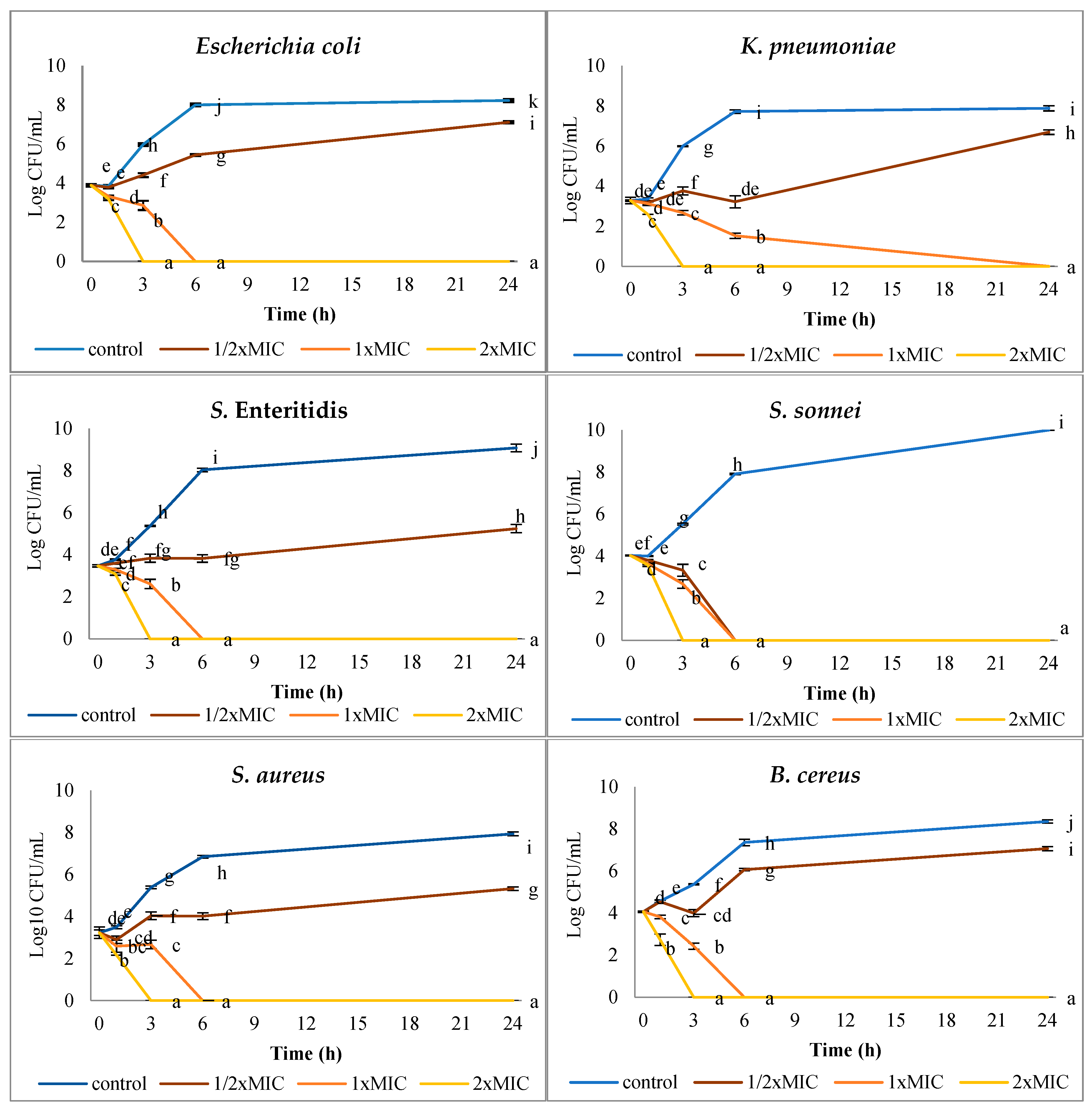
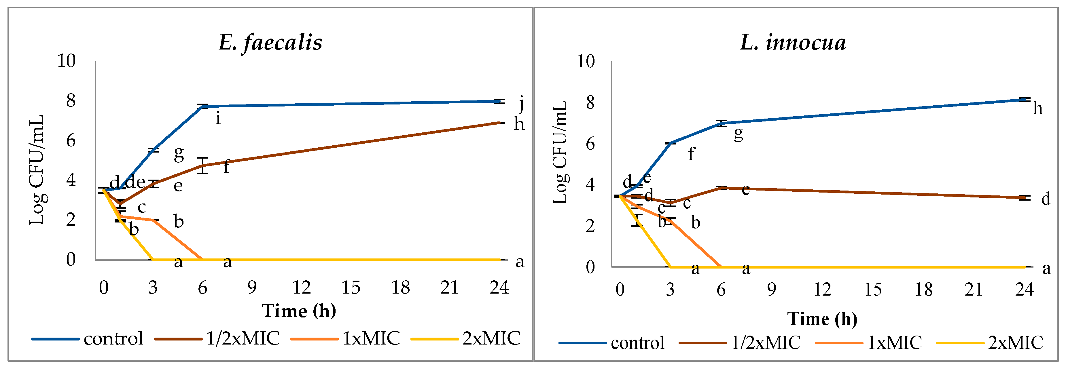
The kinetics of bacterial inactivation over a 24 h incubation period were assessed using killing curves for the tested foodborne pathogens. These curves show the rate at which AgNPs exert their antibacterial effects. Silver nanoparticles were used in the study at concentrations corresponding to three values: ½ × MIC (sublethal), 1 × MIC (minimum inhibitory concentration), and 2 × MIC (double the MIC). The results are presented in Figure 2. Additionally, the logarithmic reduction in bacterial cell counts over time at each concentration was calculated and is presented in Table 3. The number of cells of all test strains increased over 24 h only in the control samples. The presence of AgNPs in the growth medium, regardless of the concentration, statistically significantly (p < 0.05) limited the growth of strains. A reduction in cell count by 3 log CFU/mL or more differentiates the activity of compounds into bactericidal and bacteriostatic [52]. The use of AgNPs at a sublethal concentration (½ × MIC) revealed bacteriostatic activity against E. coli, S. aureus, E. faecalis, and B. cereus after 6 h of incubation and bactericidal activity against other bacterial strains. After 3 h of incubation, AgNPs at a concentration of 1 × MIC had a bactericidal effect against E. coli (3.12 log CFU/mL), P. aeruginosa (5.99 log CFU/mL), K. pneumoniae (3.33 log CFU/mL), E. faecalis (3.53 log CFU/mL), and L. innocua (3.79 log CFU/mL), and a bacteriostatic effect against the remaining strains. After 6 h of incubation, the cell count of all test strains was below the detection limit (<10 CFU/mL). Silver nanoparticles at a concentration of 2 × MIC after 3 h of incubation reduced the cell count of all strains by 3 or more logarithmic cycles. In general, it can be seen that the higher the concentration of AgNPs is, the shorter is the killing time of the test strain cells. Complete cell destruction of the tested strains occurred after 6 h of incubation at 1 × MIC and after 3 h of incubation at 2 × MIC.

3.3. Leakage of DNA and Intracellular Proteins from Cells Assessed by Spectrophotometric Measurements

Confirmation of cell destruction was carried out by assessing leakage of intracellular content during incubation of strains with AgNPs. Nucleic acid and protein leakage in the supernatant of tested bacteria were measured at 260 nm and 280 nm, respectively (Table 4). An increase in absorbance values was observed in all samples, indicating leakage of nucleic acids and proteins from the cells. Increasing the AgNPs concentration to 2 × MIC contributed to greater leakage of components from the cells, indicating greater degradation of the cell wall and cytoplasmic membrane in the cells. Our results are consistent with Lu et al. [64], who found that PVA-stabilized AgNPs copolymer disrupted the integrity of E. coli and S. aureus bacterial cell membranes, which caused leakage of cell contents, leading to disruption of the homeostasis of the intracellular environment and preventing the normal functioning of cell vital activity.

3.4. Morphological Assessment of Bacterial Cells After AgNPs Exposure

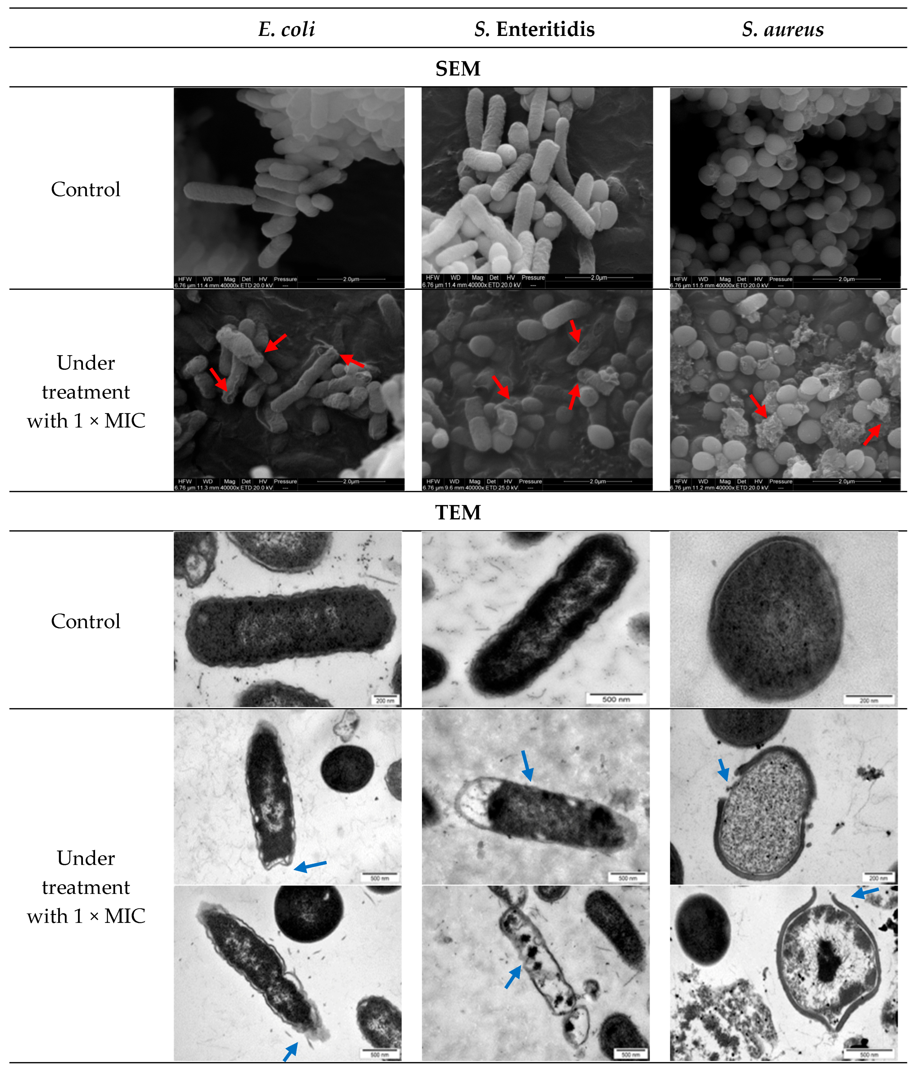
Morphological changes in the bacteria cell were examined using SEM and TEM, and the micrographs are shown in Figure 3. The micrographs illustrate the possible effects of AgNPs on the cell wall and cytoplasmic membrane. The effects of cell damage are shown in Figure 3. Formation of pores in the cell wall was observed, causing cytosol leakage outside the cell. Furthermore, AgNPs were observed to significantly deform cells, altering their shape, causing them to become folded, and shrinkage was observed. The above-mentioned features led to loss of cell integrity.
Both spectrophotometric determination and microscopic observations confirmed that AgNPs have a destructive effect on bacterial cells. Sondi and Salopek-Sondi [65] observed damage to the E. coli cell wall and the accumulation of AgNPs in the bacterial membrane, which causes a significant increase in permeability. Cell materials leak through pores in the cell membrane. The external cell structures act as a protective barrier, protecting the cell from environmental factors. These structures are the first to come into contact with AgNPs and are the first to undergo perforation and penetration [66,67]. In the cytoplasmic membrane, silver ions can bind to phosphorus and sulfur compounds present in cytoplasmic membrane proteins, disrupting their metabolic functions [68]. If damaged, AgNPs induce other effects related to changes in cell shape, resulting in multifaceted deformations. Silver accelerates the production of reactive oxygen species (ROS) inside the cell, causing oxidative stress, which is lethal to bacteria [69]. Furthermore, AgNPs can bind to DNA, which inhibits cell proliferation and leads to cell death [68,70,71].

3.5. Antibacterial Activity of Pullulan Films with AgNPs

Controlled release of AgNPs from the polymer film is crucial because it determines its effectiveness over time and determines the long-term antibacterial properties of the film [67]. The antibacterial activity of pullulan films containing AgNPs was assessed by the disk diffusion method against foodborne test bacteria. The average diameters of the growth inhibition zones of the test strains treated with pullulan films containing AgNPs are shown in Table 5. In this study, the antibiotic azlocillin was used as a positive control, and a pullulan film without AgNPs as a negative control to assess the antibacterial properties of the films. Azlocillin is an antibiotic with a broad spectrum of activity against both Gram-positive and Gram-negative bacteria.
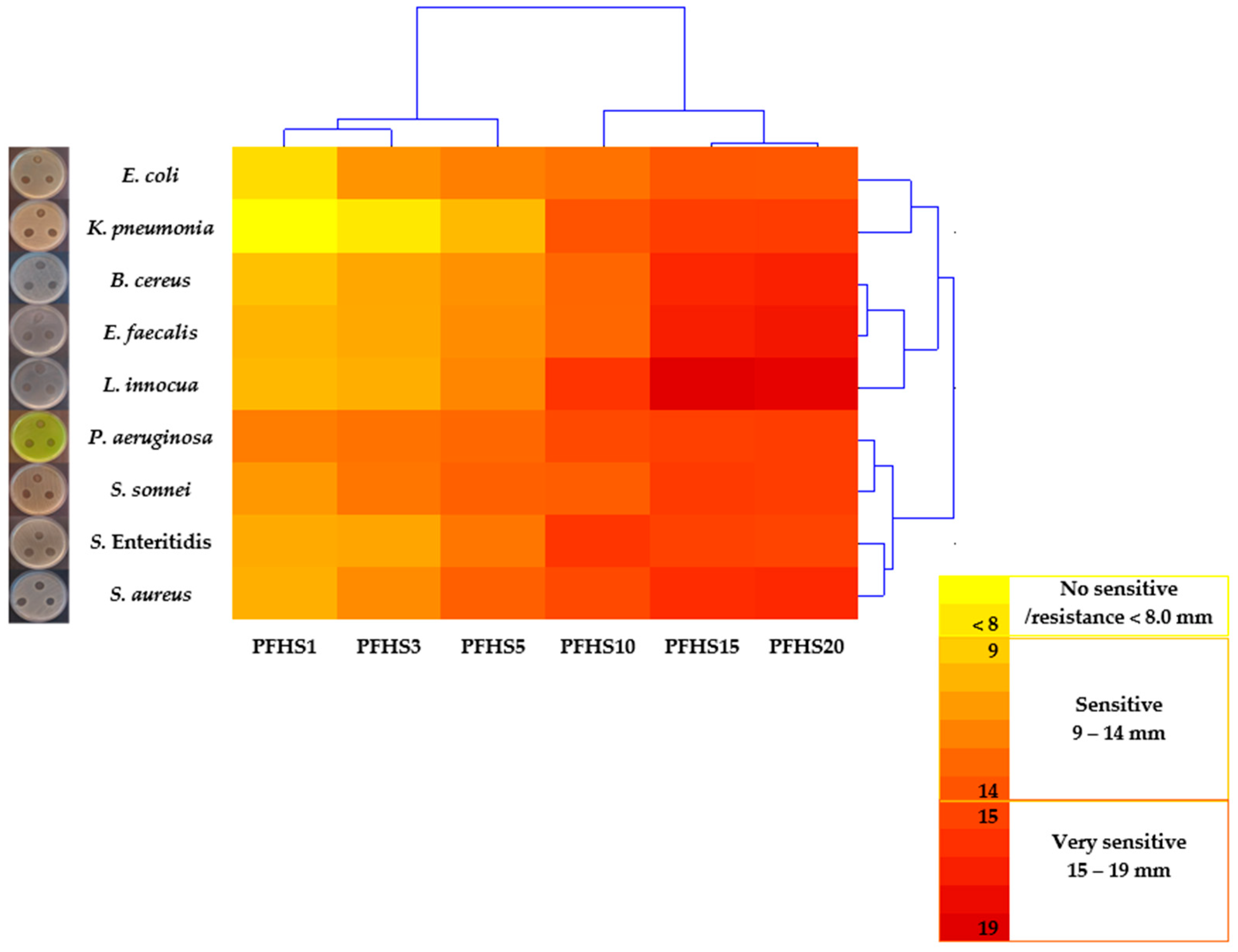
The incorporation of AgNPs, even in the lower amounts (PFHS1 and PFHS3), resulted in an antibacterial effect, except for K. pneumoniae (zones < 8 mm). Higher antibacterial potency expressed by the size of the inhibition zones of the strains’ growth [56] was correlated with a higher content of AgNPs in the pullulan film (Table 5). The heatmap revealed the division of the films into two groups in terms of antibacterial potency (Figure 4): PFHS1, PFHS3, and PFHS5 films were weaker, while PFHS10, PFHS15, and PFHS20 were stronger. Additionally, bacterial strains were divided into two groups according to their sensitivity to the films: E. coli, K. pneumoniae, B. cereus, E. faecalis, and L. innocua had weaker and more diverse sensitivity, while P. aeruginosa, S. sonnei, S. Enteritidis, and S. aureus were highly sensitive. The inhibitory effect of the films on azlocillin was varied. The PFHS20 film had an inhibitory potency against K. pneumoniae and B. cereus even greater than that of azlocillin (121% and 130%, respectively). There was no statistically significant difference (p > 0.05) between the growth inhibition zones of strains in PFHS20 and PFHS15 films. This means that the effectiveness of these films was the same. Therefore, it is more economical to use the pullulan film containing AgNPs at a lower concentration of 24 µg/cm2 (PFHS15).
The PFHS15 and PFHS20 films showed good efficacy against foodborne bacteria. The antibacterial activity is related to the release of silver ions from the pullulan film into the environment and/or contact of microorganisms with metallic silver nanoparticles [43,61]. Polymer matrices improve the stability of AgNPs and antibacterial properties [61,72]. Results presented by Khalaf et al. [45] and Morsy et al. [46] also confirmed that pullulan-based films impregnated with silver nanoparticles exhibit good efficacy against S. aureus and L. monocytogenes. The composite coating based on bacterial cellulose with montmorillonite and nanosilver shows good antibacterial activity against S. enterica, E. coli, S. aureus, and B. subtilis and is characterized by high mechanical strength, making it a promising material for food preservation applications [73]. A chitosan-gum tragacanth biomaterial containing nanosilver effectively inhibited the growth of P. aeruginosa and S. aureus [74]. Wang et al. [75] composed a film of chitosan, gelatin, and nanosilver with improved conductivity and antibacterial activity against E. coli and S. aureus, suggesting potential use in biomedicine. Pinto et al. [43] demonstrated the antifungal activity of a pullulan film with AgNPs. Increasing the AgNPs content in the pullulan film resulted in a greater reduction in fungal concentration, spore destruction, and growth inhibition of A. niger. The composite pullulan film with AgNPs has significant potential for applications in food packaging [72].

3.6. Visual Appearance and Microscopic Characterization of Pullulan Films with AgNPs

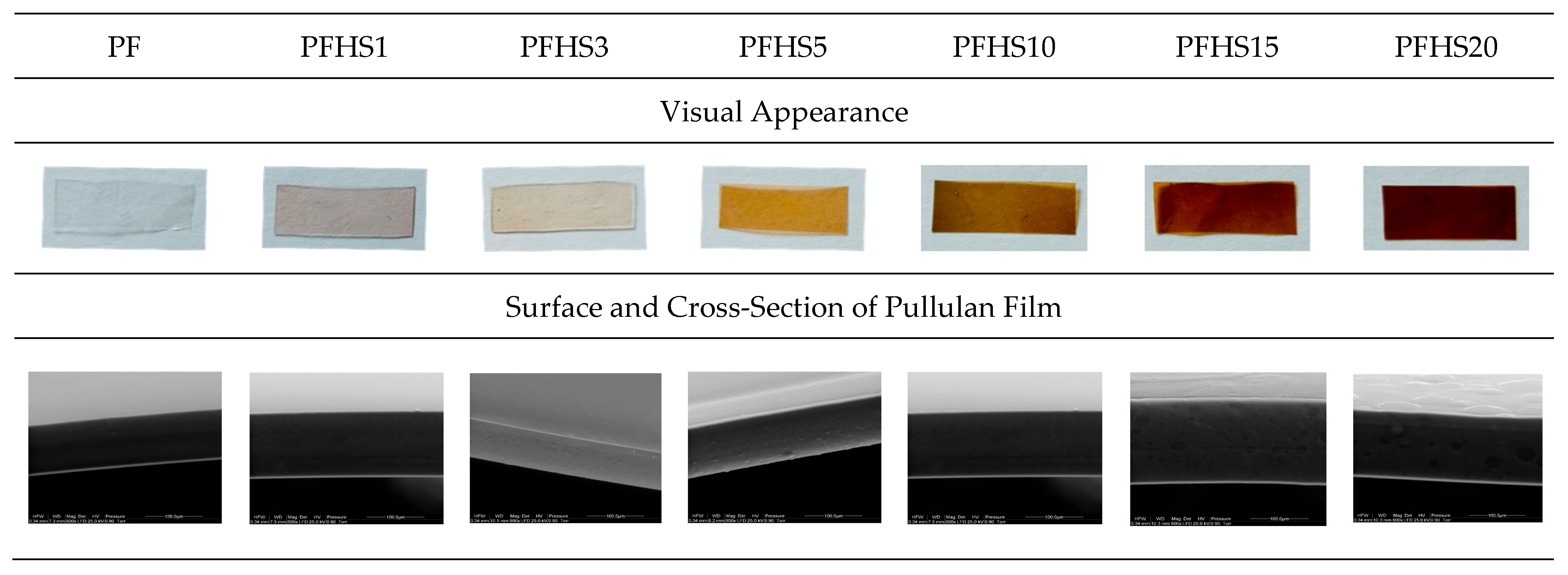
The visual appearance, along with the microscopic characterization of all tested films, is shown in Figure 5. The films were smooth to the touch, flexible, and shiny. Incorporating AgNPs into the transparent and colorless control film changed the color, with the intensity varying gradually from light yellow to dark brown, depending on the AgNPs content in the film. Very similar films were obtained by Wypij et al. [44].
In both the cross-section and on the surface, the control pullulan film (PFC) was characterized by a uniform structure and a smooth surface (Figure 5). Our observations are consistent with the pullulan film microstructure described by Mulla et al. [76]. Incorporating a 1–5% solution of silver nanoparticles (PFHS1, PFHS3, PFHS5) into the pullulan film did not change the film’s microstructure. The films exhibited a compact and uniform structure with visible individual bulges, as shown in Figure 5. This indicates good mixing of AgNPs with the polysaccharide matrix. After increasing the AgNPs addition, the films remained uniform in the cross-section in the pullulan films PFHS10, PFHS15, and PFHS20, but numerous bulges were observed on the surface (Figure 5). This proves very good dispersion of AgNPs with a diameter of 50–60 nm in the polysaccharide matrix. Similar observations of the structure of pullulan films with the addition of silver nanoparticles with a diameter of 100 nm and zinc with an average size of 110 nm at a concentration of 0.02%, v/v, were observed by Morsy et al. [46]. Islam and Yeum [77] confirmed the good embedding of Ag nanoparticles in a pullulan/PVA blend matrix from which electrospun nanofibers were prepared and found that silver nanoparticles were evenly distributed in them. Vimala et al. [78] stated that the polymers provide excellent stability of AgNPs, slow their release into the environment, and ensure prolonged antibacterial activity. These results confirm that pullulan films are a suitable material for delivering antimicrobial substances, including metal nanoparticles, to food surfaces. Good dispersion of AgNPs and deposition of small AgNPs on the composite film surface with a rough film structure, with almost uniform nanoparticle distribution, were observed in moxifloxacin-loaded chitosan-pullulan-silver-nanocomposite films [79]. Overall all tested pullulan film with AgNPs exhibited homogenous cross-section microstructure, which according to available literature it can be believed that pullulan helps in stabilization and prevent the agglomeration of AgNPs thus, maintain their good dispersion and promotes the uniform distribution of silver nanoparticles throughout the pullulan film [42].

3.7. Physical Properties of Pullulan Films with AgNPs

The physical properties of pullulan films containing AgNPs are presented in Table 6. Thickness is one of the most important parameters characterizing films. The thickness of transparent polymeric food packaging materials is typically in the range of 0.050–0.1 mm [80]. The thickness of the films increased statistically significantly (p < 0.05) from 0.080 (PFHS1) to 0.144 mm (PFHS20). The PFHS20 film was twice as thick as the PF film. Our results are consistent with those reported by Wypij et al. [44], but differ from the observations of Khalaf et al. [45], who found no association of the thickness of pullulan films with AgNPs. The authors attributed their results to the small—even negligible—molecular mass of AgNPs in relation to the mass of the polysaccharide used.
Optical transparency is an important packaging feature for consumers because it allows them to visually assess the appearance and freshness of food and make a purchasing decision [81]. On the other hand, adverse effects of light energy on food have been observed due to the degradation of nutrients and, consequently, changes in food quality, shelf life, and safety [82]. In the study of film transparency, two main parameters are considered: opacity and transmittance [80]. Analysis of variance showed a statistically significant (p < 0.05) effect of AgNPs addition on both film parameters. The opacity values of the films varied from 0.53 (PF) to 3.83 (PFHS20), and since they are less than 5, they are still transparent [80]. In turn, transmittance in the visible range did not differ significantly (p > 0.05) only in PFHS1 and PFHS3 compared to PF, and these films can be considered highly transparent (91.76–87.57% transmittance). In PFHS5–PFHS20 films, transmittance values decreased from 69.16% to 41.82%, placing these films in the category of translucent materials [80]. Similarly, transmittance in the UV range (280 nm) was significantly reduced by the AgNPs films (from 52.1 to 15.53%) compared to the PF film (79.05% transmittance), which is beneficial from the point of view of food protection against UV radiation, which damages food by causing photodegradation of nutrients [83]. A possible explanation for this phenomenon is the absorption of both UV radiation and visible light by the silver nanoparticles. UV absorption reduces food damage by delaying the formation of free radicals [84].
The color parameters of the films are expressed using the CIE L*a*b* scale. Color differences compared to the control pullulan film were also calculated. Analysis of variance showed a statistically significant (p < 0.05) effect of AgNPs on the color of the films. The increase in AgNPs content in the film caused a statistically significant (p < 0.05) darkening of the color from the light control film (L* 91.25 for PF) to a much darker one (L* 63.47 for PFHS20). The color of the films with AgNPs had a greater proportion of red (positive a* values) and yellow (positive b* values) compared to the color of the film without AgNPs. The absolute color difference (ΔE) increased with increasing AgNPs content in the film, indicating large, visible color changes for the consumer.

4. Conclusions

This study presented the results of antibacterial activity of commercially available silver nanoparticles against select 9 strains of foodborne bacteria as well as the properties of pullulan films infused with AgNPs, including their antibacterial activity and selected physical properties. The results of the first part of the study showed that silver nanoparticles exhibited strong antibacterial activity against all tested bacterial strains. Silver nanoparticles exhibited relatively uniform antibacterial activity against all Gram-negative bacteria and two Gram-positive strains (S. aureus and E. faecalis), with MIC and MBC values of 12.5 µg/mL and 25 µg/mL, respectively. In case of other tested Gram-positive bacteria, L. innocua expressed the greatest sensitivity to AgNPs, while B. cereus was the most resistant. Treatment of bacterial cells with AgNPs results in the leakage of nucleic acids and proteins from the cells. In this context, our results indicate that one of the mechanisms of antibacterial activity is connected with membrane damage, which was also confirmed by examining morphological changes in bacterial cells exposed to AgNPs using SEM and TEM. Silver nanoparticles exhibited severe structural damage on the bacteria, causing damage in the structural integrity of the cell wall and membrane, as well as broken, destroyed cells and leakage of cell contents. In the second part, our research has shown that pullulan films containing silver nanoparticles combine the beneficial properties of a biopolymer with the antibacterial activity of silver. All pullulan films with AgNPs possess antibacterial activity. Moreover, an increase in the AgNPs concentration in pullulan film resulted in a stronger inhibitory effect against the tested bacterial strains. Furthermore, among all the tested bacterial strains, L. innocua was the most sensitive. Pullulan films with the addition of AgNPs contributed to a gradual increase in thickness and opacity. In terms of optical properties, increasing concentrations of AgNPs resulted in decreased UV and visible light transmittance, along with a noticeable color change from colorless (control) to dark brown.
The promising results obtained in this study open a perspective for further research on pullulan films containing commercially available silver nanoparticles, particularly in exploring their mechanical and barrier properties. To confirm the formation and compatibility of pullulan-based films with silver nanoparticles, comprehensive analyses should be conducted, including the determination of chemical composition (FTIR), crystallinity and structural integrity (XRD), and thermal characteristics (DSC). Moreover, to evaluate their potential use as food packaging, future studies should include in vitro tests to assess the antibacterial activity of these materials directly on food products.

Author Contributions

Conceptualization, K.K., M.G., methodology, K.K.; validation, K.K., M.G.; formal analysis, K.K., M.G.; investigation, K.K., M.G.; data curation, K.K., M.G.; visualization, K.K., M.G.; writing—original draft preparation, K.K., M.G.; writing—review and editing, K.K., M.G.; supervision, M.G. All authors have read and agreed to the published version of the manuscript.

Funding

Research equipment was purchased as part of the “Food and Nutrition Centre—modernization of the WULS campus to create a Food and Nutrition Research and Development Centre (CŻiŻ)” co-financed by the European Union from the European Regional Development Fund under the Regional Operational Programme of the Mazowieckie Voivodeship for 2014–2020 (Project No. RPMA.01.01.00-14-8276/17). This work was partially financially supported by the Polish Ministry of Science and Higher Education as part of the resources allocated for young scientists. Project contract No. 505-10-092800-P00232-99.

Institutional Review Board Statement

Not applicable.

Informed Consent Statement

Not applicable.

Data Availability Statement

Data are available in the article.

Acknowledgments

The authors thank Olga Sawicka and Mateusz Szymczak for their partial assistance with the experimental work conducted during their master’s thesis.

Conflicts of Interest

The authors declared no conflict of interest.

References

  1. Slepička, P.; Kasálkova, N.; Siegel, J.; Kolská, Z.; Švorčík, V. Methods of Gold and Silver Nanoparticles Preparation. Materials 2020, 13, 1. [Google Scholar]
  2. Zhang, X.F.; Liu, Z.G.; Shen, W.; Gurunathan, S. Silver Nanoparticles: Synthesis, Characterization, Properties, Applications, and Therapeutic Approaches. Int. J. Mol. Sci. 2016, 17, 1534. [Google Scholar] [CrossRef]
  3. Kaiser, K.G.; Delattre, V.; Frost, G.W.; Buck, G.W.; Phu, J.V.; Fernandez, T.G.; Pavel, I.E. Nanosilver: An old antibacterial agent with great promise in the fight against antibiotic resistance. Antibiotics 2023, 12, 1264. [Google Scholar] [CrossRef]
  4. Lara, H.H.; Ayala-Núñez, N.V.; Ixtepan Turrent, L.D.C.; Rodríguez Padilla, C. Bactericidal effect of silver nanoparticles against multidrug-resistant bacteria. World J. Microbiol. Biotechnol. 2010, 26, 615–621. [Google Scholar] [CrossRef]
  5. Kim, J.S.; Kuk, E.; Yu, K.N.; Kim, J.H.; Park, S.J.; Lee, H.J.; Kim, S.H.; Park, Y.K.; Park, Y.H.; Hwang, C.Y.; et al. Antimicrobial effects of silver nanoparticles. Nanomed. Nanotechnol. Biol. Med. 2007, 3, 95–101. [Google Scholar] [CrossRef] [PubMed]
  6. Zhao, G.; Stevens, S.E., Jr. Multiple parameters for the comprehensive evaluation of the susceptibility of Escherichia coli to the silver ion. Biometals 1998, 11, 27–32. [Google Scholar] [PubMed]
  7. Baker, C.; Pradhan, A.; Pakstis, L.; Pochan, D.J.; Shah, S.I. Synthesis and Antibacterial Properties of Silver Nanoparticles. J. Nanosci. Nanotechnol. 2005, 5, 244–249. [Google Scholar] [CrossRef]
  8. Pal, S.; Tak, Y.K.; Song, J.M. Does the Antibacterial Activity of Silver Nanoparticles Depend on the Shape of the Nanoparticle? A Study of the Gram-Negative Bacterium Escherichia coli. Appl. Environ. Microbiol. 2007, 73, 1712–1720. [Google Scholar] [CrossRef]
  9. Chen, X.; Schluesener, H.J. Nanosilver: A nanoproduct in medical application. Toxicol Lett. 2008, 176, 1–12. [Google Scholar] [CrossRef]
  10. Kokura, S.; Handa, O.; Takagi, T.; Ishikawa, T.; Naito, Y.; Yoshikawa, T. Silver nanoparticles as a safe preservative for use in cosmetics. Nanomed. Nanotechnol. Biol. Med. 2010, 6, 570–574. [Google Scholar] [CrossRef]
  11. Goa, Y.; Cranston, R. Recent advances in antimicrobial treatments of textiles. Text. Res. J. 2008, 78, 60–72. [Google Scholar] [CrossRef]
  12. Rao, M.M.V.; Mohammad, N.; Banerjee, S.; Khanna, P.K. Synthesis and food packaging application of silver nano-particles: A review. Hybrid Adv. 2024, 6, 100230. [Google Scholar] [CrossRef]
  13. Kraśniewska, K.; Galus, S.; Gniewosz, M. Biopolymers-Based Materials Containing Silver Nanoparticles as Active Packaging for Food Applications–A Review. Int. J. Mol. Sci. 2020, 21, 698. [Google Scholar] [CrossRef] [PubMed]
  14. Bin Ahmad, M.B.; Lim, J.J.; Shameli, K.; Ibrahim, N.A.; Tay, M.E.; Chieng, B.W. Antibacterial activity of silver bionanocoposites synthesized by chemical reduction rout. Chem. Cent. J. 2012, 6, 101. [Google Scholar] [CrossRef] [PubMed]
  15. Gherasim, O.; Puiu, R.A.; Bîrcă, A.C.; Burdușel, A.-C.; Grumezescu, A.M. An Updated Review on Silver Nanoparticles in Biomedicine. Nanomaterials 2020, 10, 2318. [Google Scholar] [CrossRef]
  16. Trotta, F.; Da Silva, S.; Massironi, A.; Mirpoor, S.F.; Lignou, S.; Ghawi, S.K.; Charalampopoulos, D. Silver Bionanocomposites as Active Food Packaging: Recent Advances & Future Trends Tackling the Food Waste Crisis. Polymers 2023, 15, 4243. [Google Scholar] [CrossRef]
  17. Arfat, Y.A.; Ahmed, J.; Hiremath, N.; Auras, R.; Joseph, A. Thermo-mechanical, rheological, structural and antimicrobial properties of bionanocomposite films based on fish skin gelatin and silver-copper nanoparticles. Food Hydrocoll. 2017, 62, 191–202. [Google Scholar] [CrossRef]
  18. Youssef, A.M.; Abdel-Aziz, M.S.; El-Sayed, S.M. Chitosan nanocomposite films based on Ag-NP and Au-NP biosynthesis by Bacillus subtilis as packaging materials. Int. J. Biol. Macromol. 2014, 69, 185–191. [Google Scholar] [CrossRef]
  19. Wang, H.; Shi, T.; Ma, J.; Meng, S.; Wei, Z.; Sun, Y.; Wang, H.; Zhou, M. Chitosan-based nanocomposite films incorporated with AgNPs/porphyrinic MOFs for killing pathogenic bacteria. Int. J Biol. Macromol. 2025, 295, 139584. [Google Scholar] [CrossRef]
  20. Ortega, F.; Minnaard, J.; Arce, V.B.; García, M.A. Biodegradable starch-based nanocomposite films with laser-synthesized silver nanoparticles: A materials approach for packaging. Int. J. Biol. Macromol. 2025, 318, 144904. [Google Scholar] [CrossRef]
  21. Varsha, C.; Bajpai, S.K.; Navin, C. Investigation of water vapour permeation and anti-bacterial properties of nano silver loaded cellulose acetate film. Int. Food Res. J. 2010, 17, 623–639. [Google Scholar]
  22. De Moura, M.R.; Mattoso, L.H.C.; Zucolotto, V. Development of cellulose-based bactericidal nanocomposites containing silver nanoparticles and their use as active food packaging. J. Food Eng. 2012, 109, 520–524. [Google Scholar] [CrossRef]
  23. Maloufi, M.; Djelad, A.; Mokhtar, A.; Reguig, K.; Hasnaoui, M.A.; Kebir-Medjhouda, Z.A.; Ghamni, M.; Sassi, M. Fabrication and characterization of cellulose-based packaging film with polyethylene glycol and silver nanoparticles for enhanced antimicrobial efficacy. Int. J Biol. Macromol. 2025, 308, 142381. [Google Scholar] [CrossRef] [PubMed]
  24. Gniewosz, M.; Duszkiewicz-Reinhard, W. Comparative studies on pullulan synthesis, melanin synthesis and morphology of white mutant Aureobasidium pullulans B-1 and parent strain A.p.-3. Carbohydr. Polym. 2008, 72, 431–438. [Google Scholar] [CrossRef]
  25. European Parliament and the Council (EC). Regulation (EU) No 231/2012 of 9 March 2012 laying down specifications for food additives listed in Annexes II and III to Regulation (EC) No 1333/2008 of the European Parliament and of the Council Regulation (EU) No 231/2012 of the European Parliament and of the Council. Off. J. Eur. Union 2012, L81, 1–295. [Google Scholar]
  26. European Parliament and the Council (EC). Regulation (EC) No 1333/2008 of 16 December 2008 on food additives. Off. J. Eur. Union 2008, L354, 16–33. [Google Scholar]
  27. Food and Drug Administration. GRAS Notice No. GRN 000099. 2002. Available online: https://www.hfpappexternal.fda.gov/scripts/fdcc/index.cfm?set=grasnotices&id=99 (accessed on 20 July 2025).
  28. EFSA Panel on Food Additives and Flavourings (FAF); Castle, L.; Andreassen, M.; Aquilina, G.; Bastos, M.L.; Boon, P.; Fallico, B. Re-evaluation of pullulan (E 1204) as a food additive and new application for its extension of use. EFSA J. 2002, 23, e9267. [Google Scholar]
  29. Kraśniewska, K.; Pobiega, K.; Gniewosz, M. Pullulan—Biopolymer with potential for use as food packaging. Int. J. Food Eng. 2019, 15, 20190030. [Google Scholar] [CrossRef]
  30. Ghosh, T.; Priyadarshi, R.; Krebs de Souza, C.; Angioletti, B.L. Advances in pullulan utilization for sustainable applications in food packaging and preservation: A mini-review. Trends Food Sci. Technol. 2022, 125, 43–53. [Google Scholar] [CrossRef]
  31. Ding, Y.; Jiang, F.; Chen, L.; Lyu, W.; Chi, Z.; Liu, C.; Chi, Z. An alternative hard capsule prepared with the high molecular weight pullulan and gellan: Processing, characterization, and in vitro drug release. Carbohydr. Polym. 2020, 237, 116172. [Google Scholar] [CrossRef]
  32. Diab, T.; Biliaderis, C.G.; Gerasopoulous, D.; Sfakiotakis, E. Physicochemical properties and applications of pullulan edible films and coatings in fruit preservation. J. Sci. Food Agric. 2001, 81, 988–1000. [Google Scholar] [CrossRef]
  33. Luís, Â.; Ramos, A.; Domingues, F. Pullulan films containing rockrose essential oil for potential food packaging applications. Antibiotics 2020, 9, 681. [Google Scholar] [CrossRef]
  34. Chu, Y.; Cheng, W.; Feng, X.; Gao, C.; Wu, D.; Meng, L.; Zhang, Y.; Tang, X. Fabrication, structure, and properties of pullulan-based active films incorporated with ultrasound-assisted cinnamon essential oil nanoemulsions. Food Packag. Shelf Life 2020, 25, 100547. [Google Scholar]
  35. Kraśniewska, K.; Gniewosz, M.; Synowiec, A.; Przybył, J.L.; Bączek, K.; Węglarz, Z. The use of pullulan coating enriched with plant extracts from Satureja hortensis L. to maintain pepper and apple quality and safety. Postharvest Biol. Technol. 2014, 90, 63–72. [Google Scholar] [CrossRef]
  36. Synowiec, A.; Gniewosz, M.; Kraśniewska, K.; Przybył, J.L.; Bączek, K.; Węglarz, Z. Antimicrobial and antioxidant properties of pullulan film containing sweet basil extract and an evaluation of coating effectiveness in the prolongation of the shelf life of apples stored in refrigeration conditions. Innov. Food Sci. Emerg. Technol. 2014, 23, 171–181. [Google Scholar] [CrossRef]
  37. Gniewosz, M.; Synowiec, A.; Kraśniewska, K.; Przybył, J.L.; Bączek, K.; Węglarz, Z. The antimicrobial activity of pullulan film incorporated with meadowsweet flower extracts (Filipendula ulmariae flos) on postharvest quality of apples. Food Control 2014, 37, 351–361. [Google Scholar] [CrossRef]
  38. Trinetta, V.; Floros, J.D.; Cutter, C.N. Sakacin A-containing pullulan film: An active packaging system to control epidemic clones of Listeria monocytogenes in ready-to-eat foods. J. Food Saf. 2010, 30, 366–381. [Google Scholar] [CrossRef]
  39. Kanmani, P.; Lim, S.T. Synthesis and characterization of pullulan-mediated silver nanoparticles and its antimicrobial activities. Carbohydr. Polym. 2013, 97, 421–428. [Google Scholar] [CrossRef] [PubMed]
  40. Burduniuc, O.; Bostanaru, A.-C.; Mares, M.; Biliuta, G.; Coseri, S. Synthesis, Characterization, and Antifungal Activity of Silver Nanoparticles Embedded in Pullulan Matrices. Materials 2021, 14, 7041. [Google Scholar] [CrossRef]
  41. Ghaffarlou, M.; İlk, S.; Hammamchi, H.; Kıraç, F.; Okan, M.; Güven, O.; Barsbay, M. Green and Facile Synthesis of Pullulan-Stabilized Silver and Gold Nanoparticles for the Inhibition of Quorum Sensing. ACS Appl. Bio Mater. 2022, 5, 517–527. [Google Scholar] [CrossRef]
  42. Lee, J.H.; Jeong, D.; Kanmani, P. Study on physical and mechanical properties of the biopolymer/silver based active nanocomposite films with antimicrobial activity. Carbohydr. Polym. 2019, 224, 115159. [Google Scholar] [CrossRef]
  43. Pinto, R.J.B.; Almeida, A.; Fernandes, S.C.M.; Freire, C.S.R.; Silvestre, A.J.D.; Neto, C.P.; Trindade, T. Antifungal activity of transparent nanocomposite thin films of pullulan and silver against Aspergillus niger. Colloids Surf. B Biointerfaces 2013, 103, 143–148. [Google Scholar] [CrossRef]
  44. Wypij, M.; Rai, M.; Zemljič, L.F.; Bračič, M.; Hribernik, S.; Golińska, P. Pullulan-based films impregnated with silver nanoparticles from the Fusarium culmorum strains JTW1 for potential applications in food industry and medicine. Front. Bioeng. Biotechnol. 2023, 11, 1241739. [Google Scholar] [CrossRef]
  45. Khalaf, H.; Sharoba, A.; El-Tanahi, H.; Morsy, M. Stability of antimicrobial activity of pullulan edible films incorporated with nanoparticles and essential oils and their impact on Turkey deli meat quality. J. Food Dairy Sci. 2013, 4, 557–573. [Google Scholar] [CrossRef]
  46. Morsy, M.K.; Khalaf, H.H.; Sharoba, A.M.; El-Tanahi, H.H.; Cutter, C.N. Incorporation of essential oils and nanoparticles in pullulan films to control foodborne patho-gens on meat and poultry products. J. Food Sci. 2014, 79, M675–M684. [Google Scholar] [CrossRef]
  47. Qi, Z.; Xie, P.; Yang, C.; Xue, X.; Chen, H.; Zhou, H.; Yuan, H.; Yang, G.; Wang, C. Developing fisetin-AgNPs incorporated in reinforced chitosan/pullulan composite-film and its application of postharvest storage in litchi fruit. Food Chem. 2023, 407, 135122. [Google Scholar] [CrossRef] [PubMed]
  48. Harun-Ur-Rashid, M.; Foyez, T.; Krishna, S.B.N.; Poda, S.; Imran, A.B. Recent advances of silver nanoparticle-based polymer nanocomposites for biomedical application. RSC Adv. 2025, 15, 8480. [Google Scholar] [CrossRef] [PubMed]
  49. dos Santos, L.M.G.; Medeiros, R.J.; Maciel-Magalhães, M.; Guedes, N.C.C.; Brito, T.M.; de Souza, G.F.; Oliveira, M.L.; Pereira, R.A.; Neto, S.A.V.; Jacob, S.C.; et al. Unravelling the effects of silver nanoparticles on textiles: A comprehensive toxicological and quantitative analysis. Health Nanotechnol. 2025, 1, 6. [Google Scholar] [CrossRef]
  50. Shan, M.; Muhammad, K.F.; Muhammad, M.R.; Wong, Y.H.; Yoshida, M. Advancements in copper and silver sintering as interconnect materials in electronics applications: Review. J. Mater. Sci. Mater. Electron. 2025, 36, 1803. [Google Scholar] [CrossRef]
  51. Silva-Angulo, A.B.; Zanini, S.F.; Rosenthal, A.; Rodrigo, D.; Klein, G.; Martinez, A. Comparative study of the effects of citral on the growth and injury of Listeria innocua and Listeria monocytogenes cells. PLoS ONE 2015, 10, e0114026. [Google Scholar] [CrossRef]
  52. Wiegand, I.; Hilpert, K.; Hancock, R.E.W. Agar and broth dilution methods to determine the minimal inhibitory concentration (MIC) of antimicrobial substances. Nat. Protoc. 2008, 3, 163–175. [Google Scholar] [CrossRef]
  53. Levison, M.E. Pharmacodynamics of antimicrobial drugs. Infect. Dis. Clin. N. Am. 2004, 18, 451–465. [Google Scholar] [CrossRef]
  54. Petersen, P.J.; Jones, C.H.; Bradford, P.A. In vitro antibacterial activities of tigecycline and comparative agents by time-kill kinetic studies in fresh Mueller-Hinton broth. Diagn. Microbiol. Infect. Dis. 2007, 59, 347–349. [Google Scholar] [CrossRef]
  55. Wei, C.; Cui, P.; Liu, X. Antibacterial activity and mechanism of madecassic acid against Staphylococcus aureus. Molecules 2023, 28, 1895. [Google Scholar] [CrossRef]
  56. Ponce, A.G.; Fritz, R.; del Valle, C.; Roura, S.I. Antimicrobial activity of essential oils on the native microflora of organic Swiss chard. LWT-Food Sci. Technol. 2003, 36, 679–684. [Google Scholar] [CrossRef]
  57. Han, J.H.; Floros, J.D. Casting antimicrobial packaging films and measuring their physical properties and antimicrobial activity. J. Plast. Film Sheet. 1997, 13, 287–298. [Google Scholar] [CrossRef]
  58. Singh, P.; Pandift, P.; Mokkapati, V.R.S.S.; Garnæs, J.; Mijakovic, I.A. Sustainable approach for the green synthesis of silver nanoparticles from Solibacillus isronensis sp. and their application in biofilm inhibition. Molecules 2020, 25, 2783. [Google Scholar] [CrossRef]
  59. Ram, V.P.; Yasur, J.; Abishad, P.; Ramesh, C.; Gourkhede, D.P.; Arya, P.R.; Unni, V.; Vergis, J.; Malik, S.V.S.; Kaore, M.; et al. Enhanced therapeutic efficacy of biogenic nanosilver-conjugated thymol: In vitro and in vivo evaluation against emerging multi-drug resistant microbes. J. Drug. Deliv. Sci. Technol. 2023, 86, 104741. [Google Scholar] [CrossRef]
  60. Pencheva, D.; Bryaskova, R.; Kantardjiev, T. Polyvinyl alcohol/silver nanoparticles (PVA/AgNps) as a model for testing the biological activity of hybrid materials with included silver nanoparticles. Mater. Sci. Eng. C 2012, 32, 2048–2051. [Google Scholar] [CrossRef] [PubMed]
  61. Díaz-Barrios, A.; González, G.; Reinoso, C.; Santiana, J.; Quiroz, F.; Chango, J.I.; Costa Vera, C.; Caniglia, L.; Salazar, V.; Fernández-Delgado, M. In situ synthesis and long-term stabilization of nanosilver in poly(vinyl acetate-co-butyl acrylate-co-neodecanoate) matrix for antibacterial applications. Mater. Chem. Phys. 2020, 255, 123476. [Google Scholar] [CrossRef]
  62. Zein, M.A.; Asghar, B.H.; Almohyawi, A.M.; Alqahtani, N.F.; Alharbi, A.; Alkabli, J.; Elshaarawy, R.F.M.; Ismail, L.A. Multifunctional nanocomposites integrated green synthesized amphiphilic chitosan/thyme extract/nanosilver for antimicrobial and anti-biofilm applications. React. Funct. Polym. 2024, 194, 105791. [Google Scholar] [CrossRef]
  63. Gao, M.; Sun, L.; Wang, Z.; Zhao, Y. Controlled synthesis of Ag nanoparticles with different morphologies and their antibacterial properties. Mater. Sci. Eng. C 2013, 33, 397–404. [Google Scholar] [CrossRef] [PubMed]
  64. Lu, F.; Liu, Y.; Dai, Y.; Zhang, G.; Tong, Y. Preparation of nanosilver/polymer composites and evaluation of their antimicrobial and antitumor effect. RSC Adv. 2025, 15, 6357–6369. [Google Scholar] [CrossRef] [PubMed]
  65. Sondi, I.; Salopek-Sondi, B. Silver nanoparticles as antimicrobial agent: A case study on E. coli as a model for Gram-negative bacteria. J. Colloid Interface Sci. 2004, 275, 177–182. [Google Scholar] [CrossRef]
  66. Hashemirad, F.S.; Kavoosi, G.; Dadfar, S.M.M. Royal jelly: A novel eco-friendly biotemplate for the sustainable green synthesis of antibacterial silver, iron, copper, and zinc nanoparticles. LWT-Food Sci. Technol. 2025, 223, 117797. [Google Scholar] [CrossRef]
  67. Singh, A.; Gautam, P.K.; Verma, A.; Singh, V.; Shivapriya, P.M.; Shivalkar, S.; Sahoo, A.K.; Samanta, S.K. Green synthesis of metallic nanoparticles as effective alternatives to treat antibiotics resistant bacterial infections: A review. Biotechnol. Rep. 2020, 25, e00427. [Google Scholar] [CrossRef]
  68. Srisod, S.; Motina, K.; Inprasit, T.; Pisitsak, P. A green and facile approach to durable antimicrobial coating of cotton with silver nanoparticles, whey protein, and natural tannin. Prog. Org. Coat. 2018, 120, 123–131. [Google Scholar] [CrossRef]
  69. Yan, X.; He, B.; Liu, L.; Qu, G.; Shi, J.; Hu, L.; Jiang, G. Antibacterial mechanism of silver nanoparticles in Pseudomonas aeruginosa: Proteomics approach. Metallomics 2018, 10, 557–564. [Google Scholar] [CrossRef]
  70. Gomaa, E.Z. Silver nanoparticles as an antimicrobial agent: A case study on Staphylococcus aureus and Escherichia coli as models for Gram-positive and Gram-positive bacteria. J. Gen. Appl. Microbiol. 2017, 63, 36–43. [Google Scholar]
  71. Gevorgyan, S.; Schubert, R.; Falke, S.; Lorenzen, K.; Trchounian, K.; Betzel, C. Structural characterization and antibacterial activity of silver nanoparticles synthesized using a low-molecular-weight Royal Jelly extract. Sci. Rep. 2022, 12, 14077. [Google Scholar] [CrossRef]
  72. Guo, L.; Yuan, W.; Lu, Z.; Li, C.M. Polymer/nanosilver composite coatings for antibacterial applications. Colloids Surf. A Physicochem. Eng. Asp. 2013, 439, 69–83. [Google Scholar] [CrossRef]
  73. Zhou, B.; Yang, Y.; Yu, L.; Song, G.; Ge, J.; Du, R. Characterization of nanosilver antibacterial bacterial cellulose composite membranes coated with montmorillonite and their potential application in food packaging. Int. J. Biol. Macromol. 2025, 289, 138685. [Google Scholar] [CrossRef]
  74. Kumar, S.S.D.; Houreld, N.N.; Abrahamse, H. Nanosilver-loaded and polysaccharide-based biomaterial for antibacterial activity—In combination with photobiomodulation for effective wound healing applications. Mater. Lett. 2024, 363, 136293. [Google Scholar] [CrossRef]
  75. Wang, Y.; Guo, X.; Pan, R.; Han, D.; Chen, T.; Geng, Z.; Xion, Y.; Chen, Y. Electrodeposition of chitosan/gelatin/nanosilver: A new method for constructing biopolymer/nanoparticle composite films with conductivity and antibacterial activity. Mater. Sci. Eng. C 2015, 53, 222–228. [Google Scholar] [CrossRef]
  76. Mulla, M.Z.; Rostamabadi, H.; Habibi, N.; Falsaf, S.R. Pullulan nanocomposites: Effect of nanoparticles and essential oil reinforcement on its performance and food packaging applications. Food Humanit. 2023, 1, 887–894. [Google Scholar] [CrossRef]
  77. Islam, M.d.S.; Yeum, J.H. Electrospun pullulan/poly(vinyl alcohol)/silver hybrid nanofibers: Preparation and property characterization for antibacterial activity. Colloids Surf. A Physicochem. Eng. Asp. 2013, 436, 279–286. [Google Scholar] [CrossRef]
  78. Vimala, K.; Mohan, Y.M.; Sivudu, K.S.; Varaprasad, K.; Ravindraa, S.; Reddy, N.N.; Padma, Y.; Sreedhar, B.; MohanaRaju, K. Fabrication of porous chitosan films impregnated with silver nanoparticles: A facile approach for superior antibacterial application. Colloids Surf. B Biointerfaces 2010, 76, 248–258. [Google Scholar] [CrossRef]
  79. Shah, A.; Ashames, A.A.; Buabeid, M.A.; Murtaza, G. Synthesis, in vitro characterization and antibacterial efficacy of moxifloxacin-loaded chitosan-pullulan-silver-nanocomposite films. J. Drug Deliv. Sci. Technol. 2020, 55, 101366. [Google Scholar] [CrossRef]
  80. Guzman-Puyol, S.; Benítez, J.J.; Heredia-Guerrero, J.A. Transparency of polymeric food packaging materials. Food Res. Int. 2022, 161, 111792. [Google Scholar] [CrossRef]
  81. Eldesouky, A.; Pulido, A.F.; Mesias, F.J. The Role of Packaging and Presentation Format in Consumers’ Preferences for Food: An Application of Projective Techniques. J. Sens. Stud. 2015, 30, 360–369. [Google Scholar] [CrossRef]
  82. Duncan, S.E.; Chang, H.H. Implications of Light Energy on Food Quality and Packaging Selection. Adv. Food Nutr. Res. 2012, 67, 25–73. [Google Scholar] [PubMed]
  83. Roy, S.; Ramakrishnan, R.; Goksen, G.; Singh, S.; Łopusiewicz, Ł. Recent progress on UV-light barrier food packaging films—A systematic review. Innov. Food Sci. Emerg. Technol. 2024, 91, 103550. [Google Scholar] [CrossRef]
  84. Gore, A.H.; Prajapat, A.L. Biopolymer Nanocomposites for Sustainable UV Protective Packaging. Front. Mater. 2022, 9, 855727. [Google Scholar] [CrossRef]
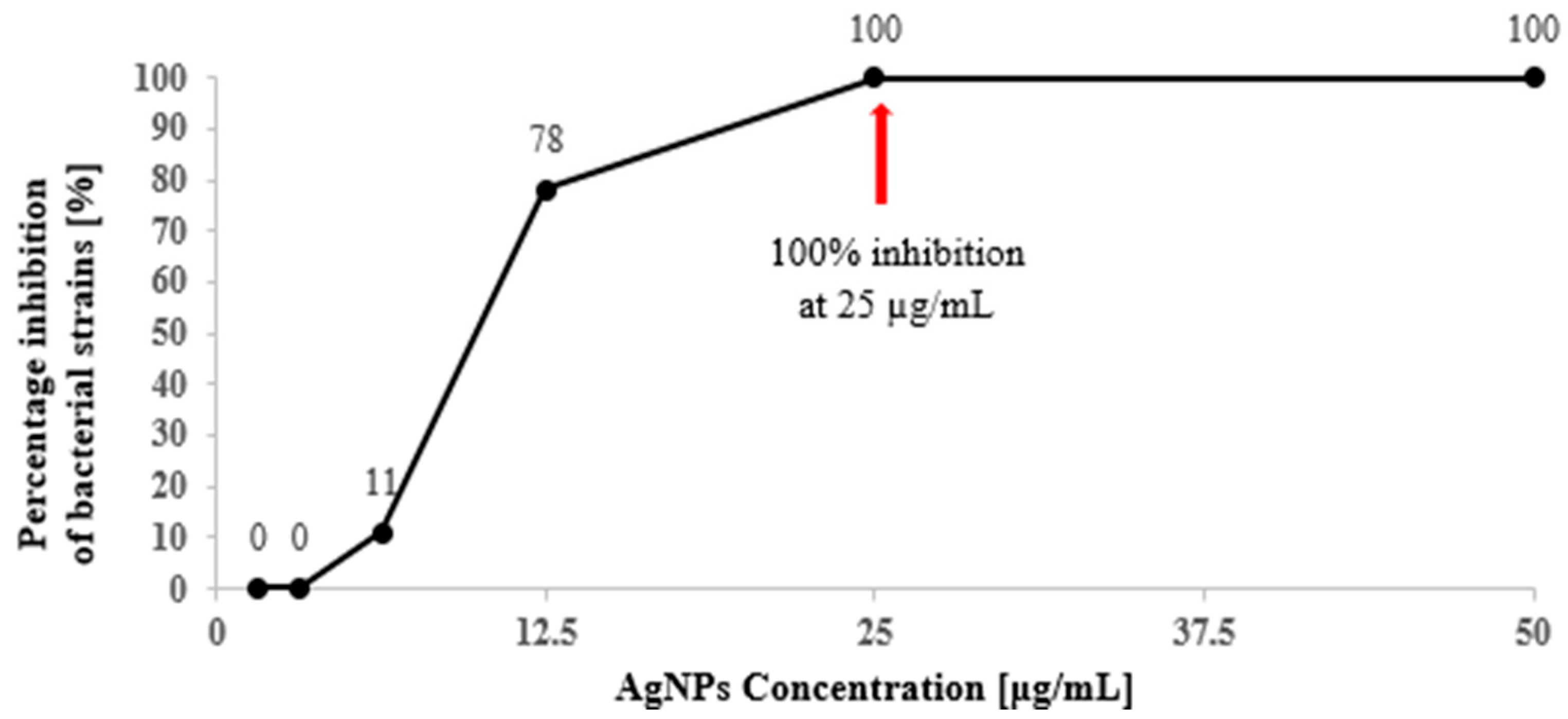
Figure 1. Percentage inhibition of bacterial strains in response to AgNPs concentration.
Applsci 15 11297 g001
Figure 2. Time–kill curves for bacteria treated with silver nanoparticles in a concentration range of ½ × MIC, 1 × MIC, and 2 × MIC; a–k—different letters indicate statistical differences at p < 0.05, as determined by a two-way ANOVA followed by Tukey’s test.
Applsci 15 11297 g002aApplsci 15 11297 g002b
Figure 3. SEM and TEM micrographs of untreated and AgNPs-treated selected bacteria strains. SEM photography: The red arrow indicates damage to bacterial cells exposed to AgNPs, including wrinkled surfaces, deformation, disruption of cell integrity, cell collapse, and leakage of cytoplasm. TEM photography: The blue arrow indicates damage to bacterial cells exposed to AgNPs, including cell membrane detachment from the cell, misshapen cells with loss of turgidity, condensed cytoplasm, interrupted continuity of the cell wall and membrane, and the leakage of cytoplasm.
Applsci 15 11297 g003
Figure 4. A heatmap with hierarchical clustering (double dendrogram) presented the distribution of bacterial sensitivity to pullulan film with silver nanoparticles. The dendrogram (blue line) shows hierarchical clustering based on the Euclidean distance and Ward’s cluster.
Applsci 15 11297 g004
Figure 5. Photographic images of pullulan film and Scanning Electron Microscopy (SEM) images, including surface and cross-section of pullulan film.
Applsci 15 11297 g005
Table 1. Concentration of HydroSilver 1000 colloid in film-forming solution and AgNPs content in 1 mL of film-forming solution and 1 cm2 of dried pullulan film.
FilmsHydroSilver 1000
[%]
AgNPs
[µg/mL]
AgNPs
[µg/cm2]
PF0--
PFHS11102
PFHS33305
PFHS55508
PFHS101010016
PFHS151515024
PFHS202020032
Table 2. Minimum Inhibitory Concentration (MIC) and Minimum Bactericidal Concentration (MBC) of AgNPs against tested strains of bacteria.
Bacteria StrainsAgNPs
MICMBCMBC/MIC *
[µg/mL]
Bacteria Gram-negative
E. coli ATCC 2592212.5252
P. aeruginosa ATCC 2785312.5252
K. pneumoniae ATCC 1388312.5252
S. Enteritidis ATCC 1307612.5252
S. sonnei NIPH-NIH “s”12.5252
Bacteria Gram-positive
S. aureus ATCC 2592312.5252
E. faecalis ATCC 2921212.5252
L. innocua ATCC 3309025502
B. cereus ATCC 117786.2512.52
* MBC/MIC ratio is ≤4, bactericidal effect; MBC/MIC ratio value is >4, bacteriostatic effect.
Table 3. Log reduction in bacterial cell counts over time by AgNPs at different MIC levels.
Bacteria Strains∆ log CFU/mL
½ × MIC1 × MIC2 × MIC
1 h3 h6 h24 h1 h3 h6 h24 h1 h3 h6 h24 h
Bacteria Gram-negative
E. coli
ATCC 25922
0.061.562.561.110.523.128.018.220.668.038.038.22
P. aeruginosa ATCC 278530.612.694.565.780.825.997.748.981.015.997.748.98
K. pneumoniae ATCC 138830.342.244.467.880.263.336.207.880.765.997.717.88
S. Enteritidis ATCC 130760.161.554.213.860.452.778.039.080.665.378.039.08
S. sonnei
NIPH–NIH “s”
0.202.207.919.990.372.857.919.990.43 5.537.919.99
Bacteria Gram-positive
S. aureus
ATCC 25923
0.581.352.832.600.892.706.847.931.335.386.847.93
E. faecalis
ATCC 29212
0.791.712.981.091.423.537.737.991.615.537.737.99
L. innocua
ATCC 33090
0.482.903.144.750.983.796.998.141.656.026.998.14
B. cereus
ATCC 11778
0.031.371.291.290.752.947.398.361.835.377.368.36
Table 4. Leakage of nucleic acid and protein from bacterial cells after treatment with AgNPs at different MIC levels.
Bacteria StrainsConcentration of AgNPsLeakage of Bacterial Cellular Material
at 260 nm and 280 nm
260 nm280 nm
6 h6 h
E. coli ATCC 25922control0.137 ± 0.025 a **0.107 ± 0.009 a
½ × MIC0.182 ± 0.008 b0.124 ± 0.006 b
1 × MIC0.210 ± 0.022 c0.157 ± 0.020 c
2 × MIC0.229 ± 0.012 d0.179 ± 0.012 d
S. Enteritidis ATCC 13076control0.181 ± 0.037 a0.122 ± 0.039 a
½ × MIC0.173 ± 0.004 a0.120 ± 0.005 a
1 × MIC0.233 ± 0.006 b0.165 ± 0.005 b
2 × MIC0.326 ± 0.003 c0.260 ± 0.003 c
S. sonnei NIPH–NIH “s”control0.196 ± 0.048 a0.155 ± 0.043 a
½ × MIC0.354 ± 0.026 b0.236 ± 0.023 b
1 × MIC0.339 ± 0.015 b0.238 ± 0.014 b
2 × MIC0.416 ± 0.014 c0.330 ± 0.012 c
S. aureus ATCC 25923control0.348 ± 0.011 a0.203 ± 0.010 a
½ × MIC0.332 ± 0.021 a0.211 ± 0.020 a
1 × MIC0.395 ± 0.002 b0.258 ± 0.002 b
2 × MIC0.513 ± 0.001 c0.369 ± 0.001 c
E. faecalis ATCC 29212control0.165 ± 0.004 a0.131 ± 0.002 a
½ × MIC0.320 ± 0.002 b0.229 ± 0.003 b
1 × MIC0.347 ± 0.007 c0.238 ± 0.006 b
2 × MIC0.469 ± 0.005 d0.391 ± 0.005 c
L. innocua ATCC 33090control0.071 ± 0.004 a0.055 ± 0.004 a
½ × MIC0.149 ± 0.008 b0.106 ± 0.006 b
1 × MIC0.236 ± 0.013 c0.157 ± 0.007 c
2 × MIC0.454 ± 0.012 d0.274 ± 0.007 d
** a–d—different letters in the column indicate statistical differences at p < 0.05 determined by a one-way ANOVA followed by Tukey’s test.
Table 5. Inhibition zone diameters of bacterial pathogens exposed to pullulan film with silver nanoparticles.
Bacteria StrainsPullulan Films
PFPFHS1PFHS3PFHS5PFHS10PFHS15PFHS20AZL75
Concentration of AgNPs in film [µg/cm2]
0258162432x
Bacteria Gram-negative
Growth inhibition zones [mm ± SD]
E. coli
ATCC 25922
0.0 a 8.4 ± 0.2 bB **11.6 ± 0.3 cC12.5 ± 0.4 dCD13.1 ± 0.3 eA14.5 ± 0.3 fA14.4 ± 0.3 fAB16.5 ± 0.4 gB
K. pneumoniae ATCC 138830.0 a6.8 ± 0.6 bA7.9 ± 0.6 cA9.9 ± 0.6 dA14.6 ± 0.5 fD15.8 ± 0.4 gBC15.9 ± 0.4 gBC13.1 ± 0.8 eA
P. aeruginosa ATCC 278530.0 a12.6 ± 0.9 bE13.0 ± 0.4 bE13.7 ± 0.4 cF15.2 ± 0.3 dE15.7 ± 0.4 dBC15.8 ± 0.2 dB26.9 ± 0.6 eF
S. Enteritidis
ATCC 13076
0.0 a10.6 ± 0.7 bCD10.9 ± 0.3 bB12.9 ± 0.4 cD16.2 ± 0.3 dF15.6 ± 0.4 dB15.4 ± 0.5 dB23.5 ± 1.2 eE
S. sonnei
NIPH–NIH “s”
0.0 a11.4 ± 1.0 bD12.5 ± 0.5 cDE13.9 ± 0.3 dF14.1 ± 0.3 dC16.0 ± 0.5 eC15.8 ± 0.8 eB18.7 ± 1.0 fC
Bacteria Gram-positive
B. cereus
ATCC 11778
0.0 a9.6 ± 0.6 bC10.8 ± 0.7 cB11.7 ± 0.4 dB13.6 ± 0.4 eBC17.0 ± 0.2 fE17.3 ± 0.2 gD13.3 ± 0.8 eA
E. faecalis
ATCC 29212
0.0 a10.2 ± 0.4 bC10.7 ± 0.3 bB12.0 ± 0.5 cBC13.5 ± 0.4 dAB17.5 ± 0.3 eF17.8 ± 0.1 eDE29.3 ± 0.6 fG
S. aureus
ATCC 25923
0.0 a10.4 ± 0.5 bCD12.0 ± 0.5 cCD14.0 ± 0.6 dF15.2 ± 1.1 eE17.0 ± 0.6 fE17.0 ± 0.4 fCD20.0 ± 0.8 gD
L. innocua
ATCC 33090
0.0 a9.9 ± 0.7 bC10.7 ± 1.1 bB12.2 ± 0.8 cBC16.3 ± 0.5 dF19.2 ± 0,4 eG18.9 ± 0.2 eE33.4 ± 0.3 fH
** a–g—different letters in the row indicate statistical differences at p < 0.05; A–H—different letters in the column indicate statistical differences at p < 0.05. PF—pullulan film, PFHS—pullulan film with HydroSilver1000 in a concentration range from 1 to 20%.
Table 6. Physical properties (thickness, opacity, transmittance and color) of pullulan film with silver nanoparticles.
Film SampleConcentration of AgNPs
in Film
[µg/cm2]
Thickness [mm]Opacity [a.u./mm]Transmittance (%)Color
280 nm600 nmL*a*b*ΔE
PF00.071 ± 0.005 a **0.53 ± 0.04 a79.05 ± 1.38 a91.76 ± 0.40 a91.25 ± 0.15 a2.08 ± 0.04 a−5.21 ± 0.03 a-
PFHS120.080 ± 0.004 ab0.59 ± 0.04 a52.10 ± 0.79 b89.63 ± 1.16 a84.22 ± 0.20 b4.71 ± 0.30 b2.86 ± 0.12 g7.9
PFHS350.095 ± 0.007 b0.61 ± 0.07 a51.25 ± 0.72 b87.57 ± 0.76 a79.14 ± 0.21 c4.38 ± 0.13 c25.78 ± 0.62 f24.0
PFHS580.098 ± 0.007 b1.64 ± 0.06 b51.17 ± 1.74 b69.16 ± 1.20 b71.11 ± 0.24 d10.16 ± 0.34 d20.89 ± 0.64 e26.7
PFHS10160.115 ± 0.011 c2.37 ± 0.31 c33.59 ± 3.20 c53.73 ± 3.86 c63.67 ± 1.13 e16.47 ± 0.70 f18.59 ± 0.26 d 33.9
PFHS15240.125 ± 0.002 c2.54 ± 0.23 c20.77 ± 1.50 d48.30 ± 3.19 c41.82 ± 2.54 f20.20 ± 0.61 g11.05 ± 0.56 c53.0
PFHS20320.144 ± 0.005 d3.83 ± 0.26 d15.53 ± 1.32 e28.24 ± 2.12 d41.45 ± 1.57 f13.50 ± 0.96 e0.01 ± 0.0 b51.4
** a–g—different letters in the column indicate statistical differences at p < 0.05 determined by a one-way ANOVA followed by Tukey’s test. PF—pullulan film, PFHS—pullulan film with HydroSilver1000 in a concentration range from 1 to 20%.
Disclaimer/Publisher’s Note: The statements, opinions and data contained in all publications are solely those of the individual author(s) and contributor(s) and not of MDPI and/or the editor(s). MDPI and/or the editor(s) disclaim responsibility for any injury to people or property resulting from any ideas, methods, instructions or products referred to in the content.