Article

Bioelectrical Impedance Versus Air-Displacement Plethysmography for Body Fat Measurements in Subjects with Abdominal Obesity: A Comparative Study

1
Department of Functional Sciences, Victor Babes University of Medicine and Pharmacy, 300041 Timisoara, Romania
2
Center for Modeling Biological Systems and Data Analysis, “Victor Babes” University of Medicine and Pharmacy, 300041 Timisoara, Romania
3
Department of Ophthalmology, Victor Babes University of Medicine and Pharmacy, 300041 Timisoara, Romania
4
“Ophthalmo-ENT” Tumor Sensory Research Center (EYE ENT), “Victor Babes” University of Medicine and Pharmacy, 300041 Timisoara, Romania
*
Author to whom correspondence should be addressed.
Appl. Sci. 2025, 15(4), 2056; https://doi.org/10.3390/app15042056
Submission received: 24 December 2024 / Revised: 7 February 2025 / Accepted: 10 February 2025 / Published: 16 February 2025

Abstract

:
Obesity is a disorder with an increasing prevalence, associated with cardiovascular and metabolic pathologies. The amount and the distribution pattern of adipose tissue must be considered when managing this disease. Abdominal obesity carries a higher risk of developing obesity-related comorbidities. Body composition methods allow an objective diagnosis and follow-up in obese patients. Although bioelectrical impedance (BIA) is a practical, affordable, and widespread technique to assess body fat percentage (%BF), its accuracy has often been questioned. Therefore, the present study aimed to determine BIA’s accuracy in subjects with abdominal obesity compared to air-displacement plethysmography (AP) as the reference method. Measurements with both body composition techniques were performed on 80 subjects (41 men and 39 women) with a large range of body mass indexes (BMIs). Abdominal obesity, diagnosed as a waist-to-height ratio (WHtR) above 0.5, was present in 28 of them. Agreement between methods was analyzed using t-tests, correlation, and Bland–Altman plots. Compared to AP, BIA underestimated %BF by 3.07 ± 5.81% (CCC = 0.82) in the entire study group. The agreement was comparable in subjects with and without central obesity (2.97 ± 6.21% and 3.26 ± 5.1%, respectively). The changes in body shape geometry due to different adipose tissue distribution patterns only marginally affected BIA’s determinations.

1. Introduction

Obesity is a multifactorial disorder stemming from genetic and environmental factors. With approximately 1.1 billion adults overweight and 312 million classified as obese, it has become a global health challenge for society [1]. Overweight and obesity, defined as abnormal or excessive fat accumulation, pose important health risks and negatively impact the quality of life and life expectancy of affected individuals. They are associated with higher morbidity and mortality due to type 2 diabetes, cardiovascular diseases, dyslipidemia, non-alcoholic fatty liver disease, stroke, obstructive sleep apnea, and osteoarticular disorders [2,3,4].
The body mass index (BMI) is the most commonly used and widely accepted method for screening obesity [5]. Originally proposed by Quetelet, it is defined as the ratio of the weight to the square of the height. A BMI higher than 25 kg/m2 classifies an individual as overweight, while one above 30 kg/m2 classifies the individual as obese, with lower cut-offs proposed for Asian populations [6]. Although the BMI is simple, cheap, and widely applicable, it has the disadvantage of not being able to differentiate between fat and lean tissue content. This aspect is of major importance in patients with edema or malnutrition, as well as in individuals with age-related muscle loss, low body fat, or higher muscle mass [7].
Cardiometabolic risk is influenced by both the amount and distribution of excess dysfunctional adipose tissue. Individuals with a higher percentage of adipose tissue within the abdominal region have a greater cardiometabolic risk. Therefore, it is important to assess both the body fat percentage (%BF) and the obesity phenotype. Based on the adipose tissue amount and its distribution, obesity is divided into two categories: metabolically healthy and metabolically unhealthy. The first one is associated with a lower inflammatory profile, better renal function, higher concentrations of adiponectin, good nutritional status, and reduced liver fat [2,8]. In contrast, metabolically unhealthy obesity is more strongly associated with cardiovascular and metabolic disorders and is characterized by a greater accumulation of adipose tissue in the visceral region compared to subcutaneous fat [9].
In order to differentiate between the two types of obesity, several anthropometric parameters can be used, such as the waist circumference (WC), hip circumference, body adiposity index, waist-to-hip ratio, waist-to-height ratio (WHtR), neck circumference, and skinfold thickness [10]. According to WHO criteria, abdominal obesity is diagnosed when the WC is equal to or greater than 102 cm in men or 88 cm in women. Different cut-offs have been proposed by the International Diabetes Federation: 90 cm for men and 80 cm for women [11]. WC, an easy and low-cost parameter, is considered to be a more sensitive and accurate method than the BMI or waist-to-hip ratio for assessing central obesity [12]. Last but not least, WHtR has an even stronger ability to predict the risk of developing cardiovascular disease, metabolic syndrome, and type 2 diabetes [13]. A WHtR above 0.5 is generally considered a diagnostic criterion for central obesity [10].
Furthermore, the “obesity paradox”, represented by an inconsistent or inverse association between obesity indicators and cardiovascular mortality in various populations, has led to the identification of several novel adiposity indexes, such as the A Body Shape Index (ABSI) and Weight-Adjusted Waist Index (WWI). The ABSI has been found to be more closely associated with cardiovascular risk [14]. The WWI, which adjusts the WC relative to weight, offers several advantages over the traditional markers of obesity. Several studies have identified it as an important predictor of diabetes, cardiovascular disease, and non-accidental mortality risk [14,15,16]. Further research aims to determine new optimal cut-off values for the existing anthropometric measures of obesity [17,18].
There are several methods for objectively evaluating the amount of body fat: density measurement methods—air-displacement plethysmography (AP) and underwater weighing—bioelectrical impedance analysis (BIA), dual X-ray absorptiometry (DEXA), and computer imaging methods, including magnetic resonance imaging (MRI) and computed tomography (CT). Each of these methods has advantages and disadvantages.
AP is a non-invasive relatively accurate method, with a %BF error margin of 1% to 2% and high repeatability [19]. It uses a two-air-filled-chamber closed system in which repeated volume and pressure variations are produced. Under the assumption of an adiabatic process, body volume and, consequently, body density are computed. Although precise, AP has the disadvantage of expensive equipment, with higher complexity and measurement time.
BIA is a safe portable method, with affordable equipment, which has become popular in recent decades. It measures body impedance and reactance. %BF is calculated using predictive equations that vary according to age, sex, ethnic group, or various clinical conditions [19,20]. BIA’s accuracy is influenced by the type of equipment (and associated algorithms) [21], as well as by environmental factors and subject characteristics. Environmental factors include humidity and temperature, while subject characteristics include body geometry that may vary between ethnic groups. Specifically, fat distribution patterns or variability in limb length can differ between ethnic groups and so may influence BIA’s accuracy [22,23].
There are several studies assessing BIA’s reliability against AP in healthy adult populations [24,25,26,27,28]. There are also studies comparing BIA with other techniques to determine body fat in overweight and obese subjects classified according to the BMI criteria [29,30,31,32,33].
Taking a closer look at BIA, as background information, it is known that bioelectrical impedance measurements depend on the assumption that the impedance of a conductor is determined by its length and configuration, as well as by its cross-sectional area. In this model, the human body is considered to be composed of cylinders with a uniform electric conductivity, corresponding to the four limbs and the trunk. The subject height is used to represent the conductive length. Abdominal obesity is defined by a larger waist circumference, hence a wider cross-sectional area of the trunk. Although it has been observed that the introduction of the waist circumference in the equations used to compute %BF improves the predictive value in the elderly [19], it has not been tested whether the higher values in subjects with abdominal obesity impact BIA’s accuracy.
The present study aimed to establish whether the body geometry changes associated with abdominal obesity influence BIA’s agreement with AP in determining %BF. In order to evaluate the magnitude of the agreement, the study group was stratified according to gender and BMI. The main analysis relied on the WHtR criterion to classify abdominal obesity. The results, corresponding to the WHO and International Diabetes Federation classifications, are included in the Supplementary Materials.

2. Materials and Methods

2.1. Participants

Eighty participants (39 women and 41 men) were enrolled in the study between the 1st of August and 30th of October 2024. The volunteers were selected via flyers and social media announcements. One of the researchers (O.M.) conducted the enrolment telephone calls. A brief description of the study, its procedures, and methods was presented to each possible volunteer. On the same telephone call, volunteers were screened for acute or chronic illness.
The study group included healthy adults aged 18 and above, who reported that their body weight was stable; further, they consented to respect the manufacturer’s recommendations for the conduct of both investigation methods. The exclusion criteria consisted of the following: (1) chronic diseases; (2) acute infections; (3) other conditions (e.g., pregnancy) (Table 1). Thus, subjects with possible hydro-electrolytic imbalances or conditions altering the thoracic gas volume were not included in the study group.
The study respected the ethical guidelines of the Declaration of Helsinki. Before enrolling in the study, each subject was informed in detail about the methods of measurement. They were assured of the non-invasiveness of the procedures and of their right to drop out for any reason. Each of the volunteers signed an informed consent form. The study (and consent form) was approved by the university’s Committee of Research Ethics (no. 20/24 July 2019 and no. 42/2 June 2022).
On study days, the subjects were instructed to abstain from eating and drinking for a minimum of 4 h before the study measurements started and not to engage in moderate or vigorous exercise for 12 h before measurements were taken. Upon their arrival in the testing facility, they were invited to use the restroom and to remove their jewelry, watch, glasses, etc.
Each of the subjects completed the testing in one day. First, the anthropometric measurements were taken, followed by the body composition determinations. To avoid a test order effect, the order of the two methods was assigned randomly in advance for each participant.

2.2. Anthropometric Measurements

Weight, height, and waist circumference were measured. Two researchers (IC and OM) collected these data; both were trained to collect the data in a standard and consistent manner.
The height was measured to the nearest 0.5 cm using a GIMA 27335 wall-mounted tape measure (GIMA, Gessate, Italy) with the subject’s Frankfort plane held horizontally. Subjects were weighed to the nearest 0.001 kg with a scale connected to the BOD POD (COSMED USA, Concord, CA, USA). The waist circumference was measured to the nearest 0.5 cm [8] in a horizontal plane at midpoint between the iliac crest and the inferior edge of the rib, at the midaxillary line, according to the WHO guidelines. The data were collected three times, and the mean value of these measurements was used in subsequent analyses. The BMI was computed as the body mass (kg) divided by the square of the subject’s height (m2).

2.3. Determination of Body Composition by AP

The body fat percentage values were measured for each subject using a BOD POD Gold Standard Body Composition Tracking System (COSMED USA, Concord, CA, USA) with software version 5.3.2. The equipment was calibrated, and a system quality check was performed at the beginning of each day of measurement.
As instructed by the manufacturer, the subjects wore minimal, form-fitting clothing (swimsuit or spandex shorts and a non-padded exercise bra), thus minimizing the artifacts caused by loose clothing and associated pockets of isothermal air.
The AP measurements were performed by trained experienced operators (IC and OM), according to the manufacturer’s instructions [34].
The body composition was determined using the Siri equation and the predicted thoracic gas volume. In order to increase the precision, we used the protocol described by Tucker [19]. For each subject, the AP was assessed three times. Each assessment consisted of two body volume measurements. If the two measurements were within 1% BF, the mean value was used in the study analysis. If the measurements differed by more than 1%BF, a third measurement was conducted, and the two measurements with the closest values were used to derive the mean value taken forward into the study analysis.

2.4. Determination of Body Composition by BIA

All measurements were performed using a multifrequency (5 kHz, 50 kHz, 100 kHz, 200 kHz) Maltron BioScan 920-2 (Maltron International Ltd., Essex, UK) by two trained experienced testers (IC and OM), according to the manufacturer’s requirements. The subjects were instructed to keep a supine position, with the hands and feet slightly abducted, in order to avoid contact between the extremities and the torso. They were required to remain still for 10 min before testing and during the measurements. The “full test” mode was selected, allowing hand to foot multifrequency body composition assessments. The subject’s skin was cleaned with 70% alcohol, and 4 adhesive electrodes were placed on the right side of the body, as follows: 2 on the hand—one directly below the third knuckle of the medius and the other on the crease of the wrist—and 2 on the foot—one directly where the second and the third toe meet the foot and one at the crease of the ankle in line with shin bone. The BF content was recorded. The measurements were performed twice for each subject, and the mean value was computed. The formula to compute the %BF is undisclosed by the manufacturer.

2.5. Statistical Analysis

An Excel database (Microsoft Corporation, Microsoft Excel 2010) was established, and data were entered using a double data entry method. The statistical analysis was performed using the R software version 4.3.3 (R Core Team 2024, Vienna, Austria). A sample size calculation was performed prior to the study initialization. For a 1% difference in %BF between the two methods, for a two-tailed test with a 0.8 power, a minimum of 34 subjects was necessary.
The results of the measurements are reported as the mean ± standard deviation (SD), followed by the range in brackets. The statistical significance was set at 0.05. The Shapiro–Wilk test was used to check data normality. Paired samples t-tests or Wilcoxon matched-pairs signed-ranks tests were performed in order to analyze the differences between the variables. The differences between the two methods were analyzed first for the entire study group. The analysis was then stratified according to the subjects’ sex, BMI category, and by the presence of abdominal obesity. In the latter case, the classification criterion was the WHtR, with the 0.5 value as the cut-off. The results of the analysis with the subjects divided in subgroups according to the central obesity criteria provided by the WHO and the International Diabetes Federation can be found in the Supplementary Materials.
The concordance between the %BF measured by the two techniques was assessed using the concordance correlation coefficient (CCC) and concordance plots. The following cut-off points were adopted for the CCC: negligible (CCC < 0.1), weak (CCC = 0.1–0.39), moderate (CCC = 0.4–0.69), strong (CCC = 0.7–0.89), and very strong (CCC = 0.9–1) [35]. Bland–Altman (BA) plots were performed in order to evaluate the agreement between the two measuring techniques. The differences di between the %BF pairs obtained with the two methods (i = 1, 2, …n, where i labels the subject and n represents the sample size) were plotted against their means. The bias was defined as the mean value of the differences, and the 95% limits of agreement were calculated as the mean ± 1.96SD, where SD is the standard deviation of the differences [36].

3. Results

The general characteristics of the study participants, such as age, height, waist circumference, body mass, and BMI, are depicted in Table 2. The values are given as the mean ± SD, with the range of the values enclosed in brackets.
A statistically significant difference was present between the %BF measured with the two body composition techniques (Table 3). BIA underestimated the %BF by 3.07 ± 5.81% in comparison to the AP. A strong concordance was observed between the two methods according to the concordance correlation coefficient analysis (CCC = 0.82) (Figure 1).
BIA tends to underestimate the %BF in subjects of both sexes. However, the difference for the male subjects is smaller and without statistical significance in comparison to the difference for females (1.28 ± 6.57% in men vs. 4.95 ± 4.23% in women). A strong concordance between the two techniques was present for both sexes (Figure 2).
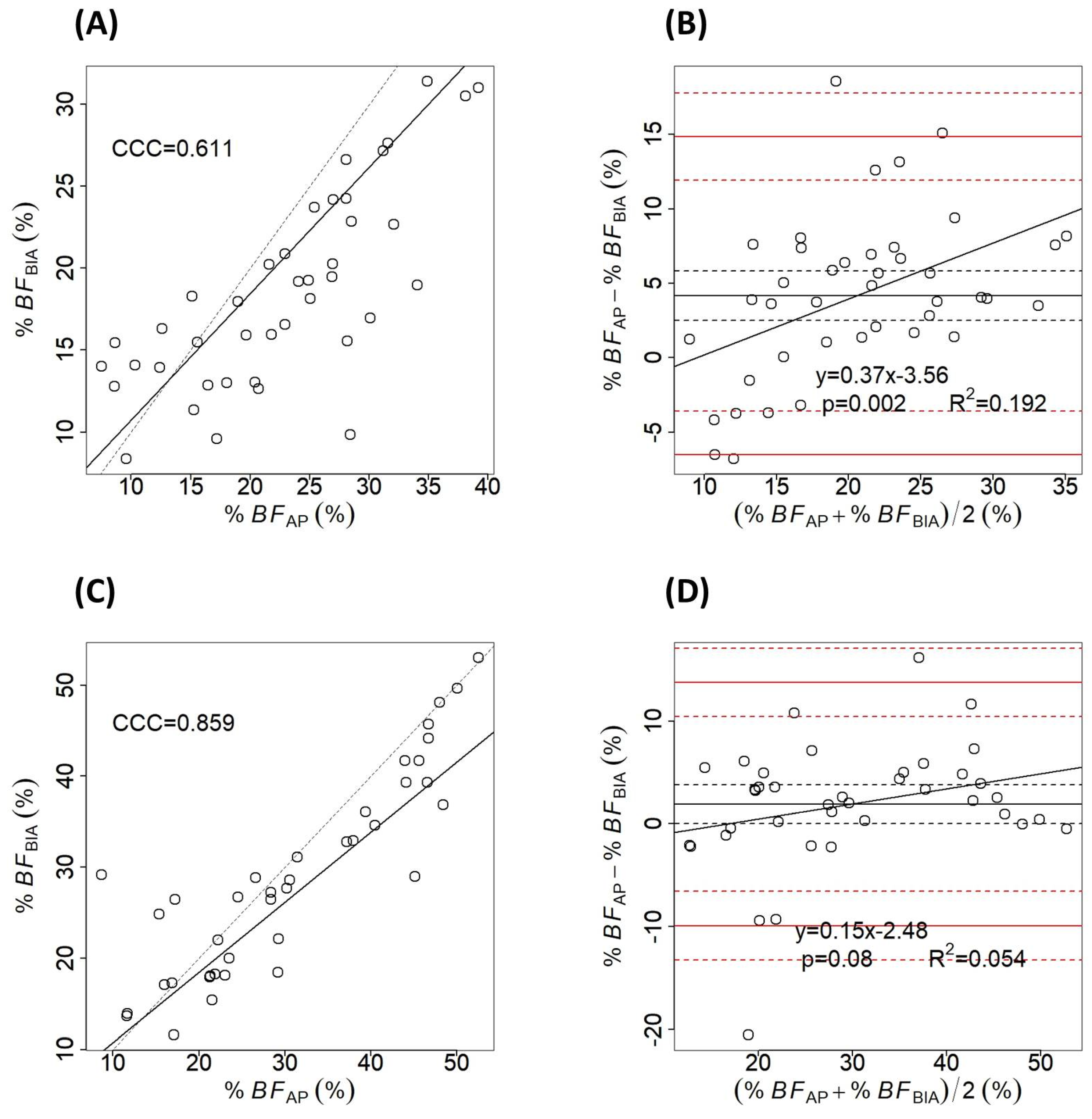
When dividing subjects by BMI, BIA underestimated the %BF mainly in underweight and normal-weight persons. In these categories, the differences between the two techniques were 5.07 ± 2.6% and 4.04 ± 5.74%, respectively; these differences reached statistical significance. The CCC showed only a moderate concordance between the two body composition methods in these subjects, and the bias significantly increased with the %BF, as indicated by the Bland–Altman plot (Figure 3). In overweight and obese participants, representing approximately half of the study group, BIA underestimated the %BF compared to the AP with only 1.68 ± 6.64% and 2.63 ± 4.02% respectively, (neither was statistically significant with p > 0.05 in both situations).
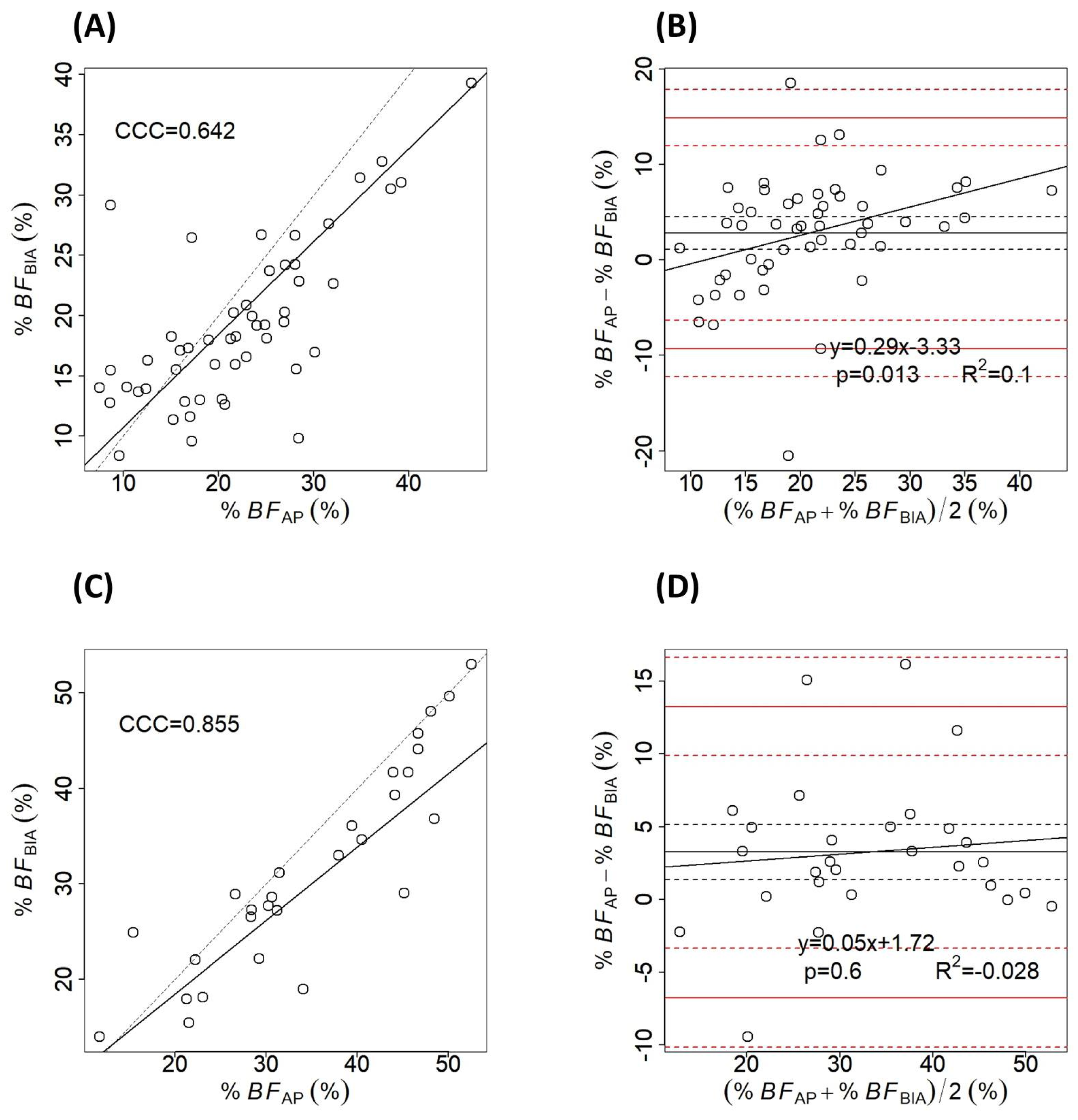
Central obesity was diagnosed according to the WHtR; in subjects with the WHtR < 0.5, the bias had a value of 2.97 ± 6.21%. BIA underestimation was higher in those with central obesity (3.26 ± 5.1%). In both groups, the differences between the two body composition techniques were statistically significant at p < 0.05 (Figure 4).

4. Discussion

An important finding of the present study is that BIA showed a strong concordance with AP across the population studied, as shown by the CCC values. A moderate concordance was present in subjects with BMI < 25 kg/m2 and in those without central obesity, as defined by the WHtR criteria. Although concordant, the two techniques had various degrees of agreement, depending on the different stratifications of the study group.
The bias (mean difference between the two methods) of 3% in %BF had large limits of agreement when computed for the entire sample. However, it reached a value to up to 4.95% in women. The high value of the bias in females, especially when compared with the bias in men (1.28%), could be partly explained by the significant difference in the anthropometric parameters between the two subgroups, particularly the body mass and height. The male subjects were significantly taller and heavier. Furthermore, previous research has shown that in female subjects, subcutaneous fat is distributed more widely than in male subjects [37]. This observation is supported by the similar bias present in underweight and normal-weight subjects according to the BMI (5.07% and 4.04%, respectively), while the differences were not significant in the overweight and obese (1.68% and 2.63%, respectively). These results confirm previous research reporting BIA’s tendency to overestimate the %BF in lean subjects and to underestimate it in obese ones [35,38].
An important consideration in the validation of the body composition estimate by BIA is the predictive equation chosen to calculate %BF. Several prediction equations are based on linear regression [15,39], and they use different reference methods. They all take account of subjects’ characteristics such as age, sex, geographical provenance, health status, and the level of physical activity [15,40]. Although the algorithms are increasingly sophisticated, BIA remains an indirect method to measure body composition, it depends on the measurement of the electrical response of the body when exposed to an electric current. The measured resistance is used to compute the fat-free mass by assuming a mean value for the resistivity and a hydration fraction, which is assumed to be equal to 0.73 for the healthy adult [41]. Therefore, the use of these estimated parameters in subjects who fall outside the “normal, healthy” ideal may contribute to prediction errors. Further errors can arise as the derivation %BF by BIA is calculated by the subtraction of the fat free mass from the body weight; thus, in lean subjects, an error in the fat free mass can have a greater impact on the fat mass prediction [21].
The main research question of the present study was to compare the two body composition methods in subjects with central obesity. To our knowledge, it is the first study to address this research question. The bioelectrical impedance (BIA) measurements are based on an empirical relationship between the impedance quotient (length2/R, where R is the resistance) and the volume of water able to conduct the electrical current due to its electrolyte content. The conductive length is approximated by the subject height. The lean body mass is then computed using the assumption that it has a 73% water content. The term height2/R is then modeled to approximate the real geometry of the human body by a coefficient, depending on a number of factors. Therefore, variations in the height-to-conductive length ratio or in the shape of the body can lead to errors in body composition measurement [15]. Further differences can be found in the estimation in subjects with central obesity, where a higher proportion of visceral fat is concentrated in the abdomen, between the internal organs and torso. In comparison, subjects without abdominal obesity have fat distributed more widely; it is located subcutaneously and is interspersed in the skeletal muscle [2]. This study explored whether the different fat distribution pattern in subjects with central obesity can affect BIA’s accuracy. We have shown that the differences between the %BF measured by the two body composition techniques are statistically significant in both subgroups—with and without central obesity. However, the values of the bias are close to each other and similar to the bias for the entire group. In addition, for the subjects with the higher WHtR (above 0.5), we confirmed the assumption that the trunk’s contribution to the whole body impedance is small, due to its relatively short length in comparison to the limbs and to its large cross-sectional area [42].
Comparable results were obtained when the study group was stratified by the obesity criteria defined by the WHO classification or to the one of the International Diabetes Federation (the results are provided in the Supplementary Materials). However, the bias reached 3.99 ± 5.1% and 4.353 ± 4.59%, respectively.
In the present study, the bias seen in the two groups, with and without abdominal obesity, was comparable, and so BIA can be regarded as an acceptable method to assess body composition changes in patients with central obesity in longitudinal studies. Since it is a widely accessible and easy to use method, it can be considered a useful tool for obesity clinicians and healthcare professionals in assessing %BF during their standard clinical practice. However, since BIA’s reliability is influenced by multiple factors, further research would be helpful to further investigate the method.
A major strength of the present study was the heterogeneity of the study group, with a wide spread of ages and a balanced gender distribution. The same applied to the anthropometric characteristics, about half of the subjects being overweight or obese. This enabled us to compare the assessment methods in both genders and to stratify the study group according to the BMI or central obesity criteria.
The study has several limitations. One of them is that the software of the device used for the BIA measurements (Maltron BioScan 920-2, Maltron International Ltd., Essex, UK) does not mention the formula it employs to transform the raw bioelectrical parameters into body mass components. This represents a more widespread drawback often associated with BIA measurement; it can be unclear which algorithms are used by each manufacturer within in each product type. Although the size of the study group was large enough to enable statistical significance, a larger sample would have allowed a more detailed stratification and more in-depth conclusions. Further research can be performed in this direction. The present results make reference only to an Eastern European Caucasian population. Applying this direction of research in other ethnic groups might be a subject of interest.
Further research is also needed to investigate the impact of the differences between the two body composition methods in longitudinal follow-up studies, especially since the highest bias was observed in subjects with a normal BMI.

5. Conclusions

BIA correlates strongly with AP in assessing %BF. The alterations in body shape associated with central obesity marginally affect BIA’s precision. However, it underestimates the %BF to a higher degree in women and lean subjects. The findings were obtained in an Eastern European population and are comparable with Caucasian populations more widely.

Supplementary Materials

The following supporting information can be downloaded at https://www.mdpi.com/article/10.3390/app15042056/s1, Figure S1: Concordance and Bland–Altman plots for the study group stratified according to the International Diabetes Federation criteria for abdominal obesity; Figure S2: Concordance and Bland–Altman plots for the study group stratified according to the WHO criteria for abdominal obesity; Table S1: Percentage body fat measured by air-displacement plethysmography and bioelectrical impedance for the subjects stratified according to the central obesity criteria defined by WHO and by the International Diabetes Federation.

Author Contributions

Conceptualization, R.H.; methodology, R.H., I.C. and O.M.; software, R.H.; validation, I.C. and O.M.; formal analysis, R.H.; investigation, O.M. and I.C.; data curation, O.M. and I.C.; writing—original draft preparation, R.H., I.C., O.M. and V.M.; writing—review and editing, R.H.; visualization, V.M.; supervision, R.H. All authors have read and agreed to the published version of the manuscript.

Funding

This research was funded by Victor Babes University of Medicine and Pharmacy of Timisoara, covering the costs of publication for this research paper.

Institutional Review Board Statement

The study was conducted in accordance with the Declaration of Helsinki and approved by the Committee of Research Ethics of the “Victor Babes” University of Medicine and Pharmacy of Timisoara (resolutions no. 20 from 24 July 2019 and no. 42 from 2 June 2022).

Informed Consent Statement

Informed consent was obtained from all subjects involved in the study.

Data Availability Statement

The data generated in the present study were anonymized and made available in the Supplementary Materials.

Acknowledgments

We would like to acknowledge Victor Babes University of Medicine and Pharmacy Timisoara for their support in covering the costs of publication for this research paper. We would also like to thank Andrew Griffiths, for language review.

Conflicts of Interest

The authors declare no conflicts of interest.

Abbreviations

The following abbreviations are used in this manuscript:
BMIBody mass index
%BFBody fat percentage
WCWaist circumference
WHtRWaist to height ratio
WHOWorld Health Organization
ABSIThe A body shape index
WWIWeight-adjusted-waist index
APAir-displacement plethysmography
BIABioelectrical impedance
DEXADual-X absorptiometry
MRIMagnetic resonance imaging
CTComputer tomography
CCCConcordance correlation coefficient
SDStandard deviation
CIConfidence interval

References

  1. Green, M.; Arora, K.; Prakash, S. Microbial Medicine: Prebiotic and Probiotic Functional Foods to Target Obesity and Metabolic Syndrome. Int. J. Mol. Sci. 2020, 21, 2890. [Google Scholar] [CrossRef] [PubMed] [PubMed Central]
  2. Salmón-Gómez, L.; Catalán, V.; Frühbeck, G.; Gómez-Ambrosi, J. Relevance of body composition in phenotyping the obesities. Rev. Endocr. Metab. Disord. 2023, 24, 809–823. [Google Scholar] [CrossRef] [PubMed] [PubMed Central]
  3. Bray, G.A.; Heisel, W.E.; Afshin, A.; Jensen, M.D.; Dietz, W.H.; Long, M.; Kushner, R.F.; Daniels, S.R.; Wadden, T.A.; Tsai, A.G.; et al. The Science of Obesity Management: An Endocrine Society Scientific Statement. Endocr. Rev. 2018, 39, 79–132. [Google Scholar] [CrossRef] [PubMed] [PubMed Central]
  4. Frühbeck, G.; Busetto, L.; Dicker, D.; Yumuk, V.; Goossens, G.H.; Hebebrand, J.; Halford, J.G.C.; Farpour-Lambert, N.J.; Blaak, E.E.; Woodward, E.; et al. The ABCD of Obesity: An EASO Position Statement on a Diagnostic Term with Clinical and Scientific Implications. Obes. Facts 2019, 12, 131–136. [Google Scholar] [CrossRef] [PubMed] [PubMed Central]
  5. Gutin, I. In BMI We Trust: Reframing the Body Mass Index as a Measure of Health. Soc. Theory Health 2018, 16, 256–271. [Google Scholar] [CrossRef] [PubMed] [PubMed Central]
  6. WHO Expert Consultation. Appropriate body-mass index for Asian populations and its implications for policy and intervention strategies. Lancet 2004, 363, 157–163. [Google Scholar] [CrossRef] [PubMed]
  7. Okorodudu, D.O.; Jumean, M.F.; Montori, V.M.; Romero-Corral, A.; Somers, V.K.; Erwin, P.J.; Lopez-Jimenez, F. Diagnostic performance of body mass index to identify obesity as defined by body adiposity: A systematic review and meta-analysis. Int. J. Obes. 2010, 34, 791–799. [Google Scholar] [CrossRef] [PubMed]
  8. Menke, A.; Muntner, P.; Wildman, R.P.; Reynolds, K.; He, J. Measures of adiposity and cardiovascular disease risk factors. Obesity 2007, 15, 785–795. [Google Scholar] [CrossRef] [PubMed]
  9. Iacobini, C.; Pugliese, G.; Blasetti Fantauzzi, C.; Federici, M.; Menini, S. Metabolically healthy versus metabolically unhealthy obesity. Metabolism. 2019, 92, 51–60. [Google Scholar] [CrossRef] [PubMed]
  10. Liu, X.; He, M.; Li, Y. Adult obesity diagnostic tool: A narrative review. Medicine 2024, 103, e37946. [Google Scholar] [CrossRef] [PubMed] [PubMed Central]
  11. Bashir, M.A.; Yahaya, A.I.; Muhammad, M.; Yusuf, A.H.; Mukhtar, I.G. Prevalence of central obesity in Nigeria: A systematic review and meta-analysis. Public Health 2022, 206, 87–93. [Google Scholar] [CrossRef] [PubMed]
  12. Bosomworth, N.J. Normal-weight central obesity: Unique hazard of the toxic waist. Can. Fam. Physician 2019, 65, 399–408. [Google Scholar] [PubMed] [PubMed Central]
  13. Tewari, A.; Kumar, G.; Maheshwari, A.; Tewari, V.; Tewari, J. Comparative Evaluation of Waist-to-Height Ratio and BMI in Predicting Adverse Cardiovascular Outcome in People with Diabetes: A Systematic Review. Cureus 2023, 15, e38801. [Google Scholar] [CrossRef] [PubMed] [PubMed Central]
  14. Park, Y.; Kim, N.H.; Kwon, T.Y.; Kim, S.G. A novel adiposity index as an integrated predictor of cardiometabolic disease morbidity and mortality. Sci. Rep. 2018, 8, 16753. [Google Scholar] [CrossRef] [PubMed] [PubMed Central]
  15. Liu, S.; Yu, J.; Wang, L.; Zhang, X.; Wang, F.; Zhu, Y. Weight-adjusted waist index as a practical predictor for diabetes, cardiovascular disease, and non-accidental mortality risk. Nutr. Metab. Cardiovasc. Dis. 2024, 34, 2498–2510. [Google Scholar] [CrossRef] [PubMed]
  16. Liu, W.; Yang, X.; Zhan, T.; Huang, M.; Tian, X.; Tian, X.; Huang, X. Weight-adjusted waist index is positively and linearly associated with all-cause and cardiovascular mortality in metabolic dysfunction-associated steatotic liver disease: Findings from NHANES 1999-2018. Front. Endocrinol. 2024, 15, 1457869. [Google Scholar] [CrossRef] [PubMed] [PubMed Central]
  17. Macek, P.; Biskup, M.; Terek-Derszniak, M.; Krol, H.; Smok-Kalwat, J.; Gozdz, S.; Zak, M. Optimal cut-off values for anthropometric measures of obesity in screening for cardiometabolic disorders in adults. Sci. Rep. 2020, 10, 11253. [Google Scholar] [CrossRef] [PubMed] [PubMed Central]
  18. Suwała, S.; Junik, R. Body Mass Index and Waist Circumference as Predictors of Above-Average Increased Cardiovascular Risk Assessed by the SCORE2 and SCORE2-OP Calculators and the Proposition of New Optimal Cut-Off Values: Cross-Sectional Single-Center Study. J. Clin. Med. 2024, 13, 1931. [Google Scholar] [CrossRef] [PubMed] [PubMed Central]
  19. Tucker, L.A.; Lecheminant, J.D.; Bailey, B.W. Test-retest reliability of the Bod Pod: The effect of multiple assessments. Percept. Mot. Ski. 2014, 118, 563–570. [Google Scholar] [CrossRef] [PubMed]
  20. Kyle, U.G.; Bosaeus, I.; De Lorenzo, A.D.; Deurenberg, P.; Elia, M.; Gómez, J.M.; Heitmann, B.L.; Kent-Smith, L.; Melchior, J.C.; Pirlich, M.; et al. Bioelectrical impedance analysis—Part I: Review of principles and methods. Clin. Nutr. 2004, 23, 1226–1243. [Google Scholar] [CrossRef] [PubMed]
  21. Ward, L.C. Bioelectrical impedance analysis for body composition assessment: Reflections on accuracy, clinical utility, and standardisation. Eur. J. Clin. Nutr. 2019, 73, 194–199. [Google Scholar] [CrossRef] [PubMed]
  22. Macias, N.; Alemán-Mateo, H.; Esparza-Romero, J.; Valencia, M.E. Body fat measurement by bioelectrical impedance and air displacement plethysmography: A cross-validation study to design bioelectrical impedance equations in Mexican adults. Nutr. J. 2007, 6, 18. [Google Scholar] [CrossRef] [PubMed] [PubMed Central]
  23. Okosun, I.S.; Tedders, S.H.; Choi, S.; Dever, G.E. Abdominal adiposity values associated with established body mass indexes in white, black and hispanic Americans. A study from the Third National Health and Nutrition Examination Survey. Int. J. Obes. Relat. Metab. Disord. 2000, 24, 1279–1285. [Google Scholar] [CrossRef] [PubMed]
  24. von Hurst, P.R.; Walsh, D.C.I.; Conlon, C.A.; Ingram, M.; Kruger, R.; Stonehouse, W. Validity and reliability of bioelectrical impedance analysis to estimate body fat percentage against air displacement plethysmography and dual-energy X-ray absorptiometry. Nutr. Diet. 2016, 73, 197–204. [Google Scholar] [CrossRef]
  25. Biaggi, R.R.; Vollman, M.W.; Nies, M.A.; Brener, C.E.; Flakoll, P.J.; Levenhagen, D.K.; Sun, M.; Karabulut, Z.; Chen, K.Y. Comparison of air-displacement plethysmography with hydrostatic weighing and bioelectrical impedance analysis for the assessment of body composition in healthy adults. Am. J. Clin. Nutr. 1999, 69, 898–903. [Google Scholar] [CrossRef] [PubMed]
  26. Vasold, K.L.; Parks, A.C.; Phelan, D.M.L.; Pontifex, M.B.; Pivarnik, J.M. Reliability and Validity of Commercially Available Low-Cost Bioelectrical Impedance Analysis. Int. J. Sport Nutr. Exerc. Metab. 2019, 29, 406–410. [Google Scholar] [CrossRef] [PubMed]
  27. Hillier, S.E.; Beck, L.; Petropoulou, A.; Clegg, M.E. A comparison of body composition measurement techniques. J. Hum. Nutr. Diet. 2014, 27, 626–631. [Google Scholar] [CrossRef] [PubMed]
  28. Benton, M.J.; Swan, P.D.; Schlairet, M.C.; Sanderson, S. Comparison of Body Composition Measurement with Whole Body Multifrequency Bioelectrical Impedance and Air Displacement Plethysmography in Healthy Middle-Aged Women. Health Care Women Int. 2011, 32, 1068–1078. [Google Scholar] [CrossRef] [PubMed]
  29. Boneva-Asiova, Z.; Boyanov, M.A. Body composition analysis by leg-to-leg bioelectrical impedance and dual-energy X-ray absorptiometry in non-obese and obese individuals. Diabetes Obes. Metab. 2008, 10, 1012–1018. [Google Scholar] [CrossRef] [PubMed]
  30. Pateyjohns, I.R.; Brinkworth, G.D.; Buckley, J.D.; Noakes, M.; Clifton, P.M. Comparison of three bioelectrical impedance methods with DXA in overweight and obese men. Obesity 2006, 14, 2064–2070. [Google Scholar] [CrossRef] [PubMed]
  31. Fakhrawi, D.H.; Beeson, L.; Libanati, C.; Feleke, D.; Kim, H.; Quansah, A.; Darnell, A.; Lammi-Keefe, C.J.; Cordero-MacIntyre, Z. Comparison of body composition by bioelectrical impedance and dual-energy x-ray absorptiometry in overweight/obese postmenopausal women. J. Clin. Densitom. 2009, 12, 238–244. [Google Scholar] [CrossRef] [PubMed]
  32. Gutiérrez-Marín, D.; Escribano, J.; Closa-Monasterolo, R.; Ferré, N.; Venables, M.; Singh, P.; Wells, J.C.; Muñoz-Hernando, J.; Zaragoza-Jordana, M.; Gispert-Llauradó, M.; et al. Validation of bioelectrical impedance analysis for body composition assessment in children with obesity aged 8–14y. Clin. Nutr. 2021, 40, 4132–4139. [Google Scholar] [CrossRef] [PubMed]
  33. Chen, F.; Wu, L.; Chen, Y.; Wang, J.; Liu, J.; Huang, G.; Hou, D.; Liao, Z.; Zhang, T.; Xie, X.; et al. A comparison of bioelectrical impedance analysis and air displacement plethysmography to assess body composition in children. Front. Public Health 2023, 11, 1164556. [Google Scholar] [CrossRef] [PubMed] [PubMed Central]
  34. COSMED. BOD POD Gold Standard Body Composition Tracking System Operator’s Manual—P/N 210-2400 Rev. M—DCO 1765; COSMED USA, Inc.: Concord, CA, USA, 2015. [Google Scholar]
  35. Silveira, E.A.; Barbosa, L.S.; Rodrigues, A.P.S.; Noll, M.; De Oliveira, C. Body fat percentage assessment by skinfold equation, bioimpedance and densitometry in older adults. Arch. Public Health 2020, 78, 65. [Google Scholar] [CrossRef] [PubMed] [PubMed Central]
  36. Bland, J.M.; Altman, D.G. Measuring agreement in method comparison studies. Stat. Methods Med. Res. 1999, 8, 135–160. [Google Scholar] [CrossRef] [PubMed]
  37. Henche, S.A.; Torres, R.R.; Pellico, L.G. An evaluation of patterns of change in total and regional body fat mass in healthy Spanish subjects using dual-energy X-ray absorptiometry (DXA). Eur. J. Clin. Nutr. 2008, 62, 1440–1448. [Google Scholar] [CrossRef] [PubMed]
  38. Ling, C.H.; de Craen, A.J.; Slagboom, P.E.; Gunn, D.A.; Stokkel, M.P.; Westendorp, R.G.; Maier, A.B. Accuracy of direct segmental multi-frequency bioimpedance analysis in the assessment of total body and segmental body composition in middle-aged adult population. Clin. Nutr. 2011, 30, 610–615. [Google Scholar] [CrossRef] [PubMed]
  39. Barbosa-Silva, M.C.; Barros, A.J. Bioelectrical impedance analysis in clinical practice: A new perspective on its use beyond body composition equations. Curr. Opin. Clin. Nutr. Metab. Care 2005, 8, 311–317. [Google Scholar] [CrossRef] [PubMed]
  40. Campa, F.; Coratella, G.; Cerullo, G.; Noriega, Z.; Francisco, R.; Charrier, D.; Irurtia, A.; Lukaski, H.; Silva, A.M.; Paoli, A. High-standard predictive equations for estimating body composition using bioelectrical impedance analysis: A systematic review. J. Transl. Med. 2024, 22, 515. [Google Scholar] [CrossRef] [PubMed] [PubMed Central]
  41. Ward, L.C.; Isenring, E.; Dyer, J.M.; Kagawa, M.; Essex, T. Resistivity coefficients for body composition analysis using bioimpedance spectroscopy: Effects of body dominance and mixture theory algorithm. Physiol. Meas. 2015, 36, 1529–1549. [Google Scholar] [CrossRef] [PubMed]
  42. Kyle, U.G.; Bosaeus, I.; De Lorenzo, A.D.; Deurenberg, P.; Elia, M.; Manuel Gómez, J.; Lilienthal Heitmann, B.; Kent-Smith, L.; Melchior, J.C.; Pirlich, M.; et al. Bioelectrical impedance analysis-part II: Utilization in clinical practice. Clin. Nutr. 2004, 23, 1430–1453. [Google Scholar] [CrossRef] [PubMed]
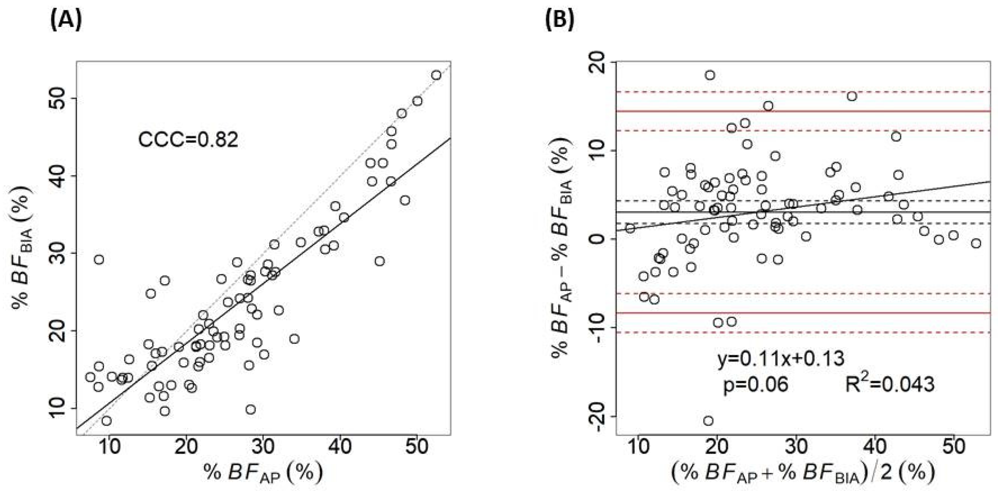
Figure 1. Concordance plots for body fat percentage (%BF) measured by air-displacement plethysmography (AP) and bioelectrical impedance (BIA) in the study group (n = 80), with the concordance correlation coefficient (CCC) (A) and Bland–Altman plot (B). The solid black line shows the bias (the mean of the differences) and the black dashed lines the 95% confidence interval (CI). The red solid lines represent the limits of agreement (bias ± 1.96SD of the differences), and the red dashed ones delimit their 95%CI. The linear regression equation between the differences and the means is given together with the p-value and the coefficient of determination (R2).
Applsci 15 02056 g001
Figure 2. Concordance plots for body fat percentage (%BF) measured by air-displacement plethysmography (AP) and bioelectrical impedance (BIA) for female (A) and male (C) subjects, with the concordance correlation coefficients (CCC). Bland–Altman plots for women (B) and men (D) show the bias as a solid black line, together with the 95% confidence, and the red dashed ones delimit their 95%CI. The linear regression equation between the differences and the means is given together with the p-value and the coefficient of determination (R2).
Applsci 15 02056 g002
Figure 3. Concordance plots with concordance correlation coefficients (CCC) for body fat percentage (%BF) in underweight and normal weight subjects (A) and in overweight and obese participants (C). Bland–Altman plots for subjects with BMI < 25 kg/m2 (B) and with BMI ≥ 25 kg/m2 (D) depict the bias as a solid black line, with the 95% confidence interval (CI) as black dashed lines. The limits of agreement (bias ± 1.96SD of the differences) are shown as solid red lines, while their CIs are represented as red dashed lines. The linear regression equation between the difference and the means with the p-value and the coefficient of determination (R2) are shown.
Applsci 15 02056 g003
Figure 4. Concordance and Bland–Altman plots for the subjects without (A,B) and with central obesity (C,D), classified according to the WHtR criterion. The concordance plots also show the concordance correlation coefficients (CCC). On the Bland–Altman plots, the bias is indicated as a black solid line and its 95% confidence interval (CI) by black dashed lines. The limits of agreement (bias ± 1.96SD of the differences) are marked by red solid lines and their CIs by red dashed lines. The linear regression equation between the differences and the means is given together with the p-value and the coefficient of determination (R2).
Applsci 15 02056 g004
Table 1. Diseases considered as exclusion criteria.
Exclusion CriteriaDisease/Condition
Chronic diseasesCardiac chronic diseases (coronary artery disease, congestive heart failure, symptomatic dysrhythmia)
Chronic obstructive pulmonary disease
Renal failure
Hepatic failure
Diabetes mellitus
Thyroid dysfunction
Chronic neurological disorders (e.g., Parkinson disease, tremor, paresis, etc.)
Cancer
Chronic infections (e.g., hepatitis, tuberculosis)
Significant mental impairment
Acute infectionsAcute respiratory infections
Gastroenteritis
Enterocolitis
Other conditionsPregnancy
Claustrophobia
Treatment with diuretics or other medications influencing body composition
Table 2. General characteristics of the subjects in the study group (mean ± standard deviation, range of values).
All (n = 80)Female (n = 39)Male (n = 41)p-Value
Age (yrs)36.31 ± 11.73
(18–71)
38.36 ± 12.74
(18–71)
34.97 ± 11.63
(18–56)
0.15
Height (cm)170.38 ± 10.3
(148–192)
162.38 ± 7.61
(148–179.5)
180 ± 5.71
(168–192)
<0.001
Waist circumference (cm)82.65 ± 14.2
(59–131)
76.46 ± 13.88
(59–118)
88.54 ± 11.94
(69.5–131)
<0.001
Body mass (kg)73.571 ± 18.457
(39.894–148.8)
62.501 ± 15.267
(39.894–114.681)
84.102 ± 14.771
(58.323–148.8)
<0.001
BMI (kg/m2)25.23 ± 5.59
(14.98–49.43)
23.84 ± 6.16
(14.98–44.15)
26.55 ± 4.7
(19.49–49.43)
0.03
Abbreviation: BMI—body mass index.
Table 3. Percentage body fat (%BF) measured by air-displacement plethysmography (AP) and bioelectrical impedance (BIA) for the entire sample (n = 80) and for the subjects divided in subgroups according to sex, body mass index (BMI), and the presence of central obesity defined according to the waist-to-height ratio (WHtR). Values are provided as mean ± standard deviation, with range between brackets.
%BFAP (%)%BFBIA (%)%BFAP − %BFBIA (%)p-Value
All (n = 80)26.62 ± 11.35
(7.5–52.55)
23.55 ± 10.18
(8.36–53.03)
3.07 ± 5.81
 
<0.001
Sex
-male (n = 41)21.73 ± 9.66
(7.5–46.7)
20.45 ± 7.57
(11.36–45.74)
1.28 ± 6.57
 
0.2
-female (n = 39)31.77 ± 10.8
(9.6–52.55)
26.81 ± 11.58
(8.36–53.03.18)
4.95 ± 4.23
 
<0.001
BMI (kg/m2)
<18.5 (n = 5)18.66 ± 5.83
(9.6–25.05)
13.59 ± 4.33
(8.36–18.13)
5.07 ± 2.6
 
0.012
18.5–24.9 (n = 36)23.21 ± 8.5
(7.5–39.2)
19.17 ± 5.86
(9.83–31.41)
4.04 ± 5.74
 
<0.001
25–29.9 (n = 29)26.66 ± 11.15
(8.65–46.6)
24.98 ± 8.79
(11.58–41.7)
1.68 ± 6.64
 
0.18
>30 (n = 10)42.79 ± 8.74
(26.6–52.55)
40.16 ± 9.08
(27.67–53.03)
2.63 ± 4.02
 
0.07
Central obesity *
Absent (n = 52)22.21 ± 8.67
(7.5–46.6)
19.25 ± 6.67
(8.36–39.3)
2.97 ± 6.21
 
<0.001
Present (n = 28)34.81 ± 11.32
(11.7–52.55)
31.54 ± 10.84
(13.93–53.03)
3.26 ± 5.1
 
<0.001
* central obesity defined as WHtR > 0.5.
Disclaimer/Publisher’s Note: The statements, opinions and data contained in all publications are solely those of the individual author(s) and contributor(s) and not of MDPI and/or the editor(s). MDPI and/or the editor(s) disclaim responsibility for any injury to people or property resulting from any ideas, methods, instructions or products referred to in the content.