Review

Antioxidants to Defend Healthy and Youthful Skin—Current Trends and Future Directions in Cosmetology

by
Anna Budzianowska
,
Katarzyna Banaś
,
Jaromir Budzianowski
and
Małgorzata Kikowska
*
Laboratory of Pharmaceutical Biology and Biotechnology, Department and Division of Practical Cosmetology and Skin Disease Prophylaxis, Collegium Pharmaceuticum, Poznan University of Medical Sciences, 3 Rokietnicka St., 60-806 Poznań, Poland
*
Author to whom correspondence should be addressed.
Appl. Sci. 2025, 15(5), 2571; https://doi.org/10.3390/app15052571
Submission received: 31 December 2024 / Revised: 21 February 2025 / Accepted: 24 February 2025 / Published: 27 February 2025
(This article belongs to the Special Issue Cosmetics Ingredients Research - 2nd Edition)

Abstract

:
Antioxidants are indispensable in protecting the skin from oxidative stress caused by environmental factors such as ultraviolet (UV) radiation, pollution, and lifestyle-related influences. This review examines the essential role of antioxidants in modern cosmetology, highlighting their dual functionality as protective agents and active components in skincare formulations. Oxidative stress, primarily driven by an imbalance between reactive oxygen species (ROS) production and the skin’s defense mechanisms, accelerates aging processes, damages cellular structures, and compromises skin integrity. Antioxidants, whether natural or synthetic, act by neutralizing ROS, reducing inflammation, and promoting cellular repair, effectively mitigating these harmful effects. This comprehensive analysis synthesizes findings from 280 studies accessed via key databases, including PubMed, Scopus, and ScienceDirect. It investigates the biochemical mechanisms of antioxidant activity, emphasizing compounds such as vitamins (C, E, A), carotenoids, polyphenols, peptides, and minerals, alongside bioactive extracts derived from algae, fungi, lichens, and plants. Carotenoids, including ꞵ-carotene, lutein, lycopene, and astaxanthin, demonstrate potent antioxidant activity, making them crucial for photoprotection and anti-aging. Phenolic compounds, such as ferulic acid, resveratrol, hesperidin, and xanthohumol, play a significant role in neutralizing oxidative stress and improving skin health. This review also highlights bioactives from algae, fungi, and lichens. Algae, particularly microalgae like Haematococcus pluvialis, known for astaxanthin production, are highlighted for their extraordinary photoprotective and anti-aging properties. Brown algae (Fucus vesiculosus) and red algae (Porphyra) provide polysaccharides and bioactive molecules that enhance hydration and barrier function. Fungi contribute a wealth of antioxidant and anti-inflammatory compounds, including polysaccharides, ꞵ-glucans, and enzymes, which support cellular repair and protect against oxidative damage. Lichens, through unique phenolic metabolites, offer potent free-radical-scavenging properties and serve as effective ingredients in formulations targeting environmental stress. Plant-derived antioxidants offer a diverse range of benefits. Plant-derived antioxidants, such as flavonoids, phenolic acids, and carotenoids, further amplify skin resilience, hydration, and repair mechanisms, aligning with the growing demand for nature-inspired solutions in cosmetics. The integration of these diverse natural sources into cosmetic formulations reflects the industry’s commitment to sustainability, innovation, and efficacy. By harnessing the synergistic potential of bioactives from algae, fungi, lichens, and plants, modern cosmetology is advancing toward multifunctional, health-conscious, and eco-friendly products. Future research directions include optimizing delivery systems for these bioactives, enhancing their stability and bioavailability, and expanding their applications to meet evolving dermatological challenges.

1. Introduction

The human body is constantly exposed to harmful environmental factors such as ultraviolet (UV) radiation, air pollution, ozone, cigarette smoke, heavy metals, and lifestyle-related stressors. Photoaging results from the cumulative impact of these stressors, which induce oxidative stress and accelerate degenerative changes in skin cells, leading to premature aging. Oxidative stress, characterized by an imbalance between the generation of reactive oxygen species (ROS) and the body’s ability to neutralize them with antioxidants, further accelerates these degenerative changes. The growing understanding of oxidative stress’s role in skin health has fueled interest in cosmetic formulations enriched with antioxidant compounds. As the field of cosmetology advances, the demand for ingredients with antioxidative properties derived from natural sources is increasing. These naturally occurring compounds are valued for their ability to neutralize ROS, thereby protecting the skin from oxidative damage. In an era of widespread environmental pollution driven by industrial expansion and urbanization, consumers increasingly prioritize natural solutions in both nutrition and skincare. Mitigating these harmful effects can be achieved through a diet rich in antioxidants, carefully selected antioxidant skincare regimens, or targeted supplementation. Cosmetology focuses on maintaining and enhancing the external appearance of the human body, including the skin, hair, and nails. This involves the use of cosmetics, skincare treatments, and customer education on proper hygiene and nutrition. Many plant-based compounds with potent antioxidant properties have been utilized in medicine and cosmetology for decades, while others have been identified only recently through ongoing research. These compounds are not only essential in preventing photoaging but also contribute to managing inflammation, enhancing hydration, and improving overall skin resilience. The exploration and application of these bioactive substances represent a significant focus in the pursuit of innovative, nature-inspired skincare and therapeutic solutions [1,2,3,4].
The history of antioxidants dates back to antiquity, with evidence suggesting that ancient civilizations, such as the Egyptians, possessed advanced technical knowledge of preservation. They used plant extracts rich in phenolic compounds for embalming, demonstrating an early understanding of oxidative processes. The scientific exploration of oxidation and reduction reactions began in the late 19th century, notably with the observation of rubber oxidation in the 1870s. By the 1940s, the mechanisms of free radical autoxidation were elucidated, leading to the identification of several chain-breaking antioxidants. By the late 1950s, research established a link between oxidative reactions, aging, and the progression of various diseases, leading to the hypothesis that antioxidants could mitigate these processes and potentially extend lifespan.
This period marked the first in vivo experiments in which antioxidants were administered to rodents, providing statistically significant evidence of lifespan extension. Consequently, extensive research efforts were undertaken to explore the sources, biological effects, and potential toxicity of antioxidants, resulting in a vast body of scientific literature—now exceeding 150,000 publications on the topic. The longstanding use of plants for nutritional and medicinal purposes is, in part, attributed to the biological activity of their secondary metabolites, many of which exhibit antioxidant properties. Key compounds such as phenolics, vitamins C and E, and carotenoids contribute to these beneficial effects, underscoring the fundamental role of natural antioxidants in health and disease prevention [5].
The figure below illustrates the effects of antioxidants on the skin, including benefits such as reducing wrinkles, enhancing skin radiance, evening out skin tone, increasing firmness, stimulating regenerative processes, protecting against harmful radiation, boosting collagen synthesis, and preventing hyperpigmentation (Figure 1).

2. Materials and Methods

A comprehensive literature review was conducted utilizing key databases, including PubMed, Google Scholar, Scopus, and ScienceDirect, as well as the recent literature. A total of 280 notable research articles were examined, with no restrictions on publication date.

3. Comprehensive Antioxidant Characteristics

Antioxidants are bioactive compounds that mitigate or delay oxidative damage to cellular structures induced by free radicals—highly reactive molecules produced through exposure to external factors such as tobacco smoke, ultraviolet (UV) radiation, and environmental pollutants. By stabilizing free radicals and reducing oxidative stress, antioxidants play a pivotal role in safeguarding the body against pathologies such as cardiovascular diseases, malignancies, and neurodegenerative disorders [6,7].

3.1. Free Radicals

Reactive oxygen species (ROS), commonly referred to as free radicals, are highly reactive molecular entities that play a dual role in biological processes, contributing to both physiological and pathological states. Oxidative stress arises when there is an imbalance between ROS production and the body’s antioxidant defense systems’ ability to neutralize them effectively.
By definition, ROS are molecular species capable of independent existence that contain an unpaired electron, a characteristic that confers high reactivity and enables them to donate or accept electrons, thereby acting as oxidizing or reducing agents [7,8]. Free radicals can carry a positive, negative, or neutral charge. For example, a neutral radical, such as nitric oxide (NO•), retains its unpaired electron without altering the charge of its parent molecule. A positively charged radical forms upon electron loss, while a negatively charged radical results from electron acquisition. These electron transfer mechanisms underpin redox reactions, wherein the electron-donating species undergoes oxidation and the electron-accepting species undergoes reduction.
Although oxygen radicals are the most widely studied, other types of radicals also exist, including those based on carbon (C), nitrogen (N), and sulfur (S). Among the oxygen-containing free radicals most implicated in pathological conditions are the hydroxyl radical (•OH), superoxide anion radical (O2), hydrogen peroxide (H2O2), singlet oxygen (1O2), hypochlorite (ClO), nitric oxide radical (NO•), and peroxynitrite (ONOO) [9]. These ROS are highly reactive and can damage critical biomolecules, including DNA, proteins, lipids, and carbohydrates, within cellular nuclei and membranes. Their activity leads to cellular dysfunction, the disruption of homeostasis, and tissue damage, which are central to the pathogenesis of numerous diseases [7].

3.2. Oxidative Stress

In a well-functioning organism, a homeostatic balance exists between the generation of reactive oxygen species (ROS) and the activity of antioxidant defense mechanisms. Excessive ROS production leads to oxidative stress, which disrupts cellular metabolism [10]. Oxygen homeostasis within the body can be disturbed by both intracellular sources of ROS, such as immune system responses and redox processes within the mitochondrial respiratory chain, as well as extracellular factors.
Lifestyle choices—including diet, alcohol consumption, stimulant use, excessive physical exertion, psychological stress, and prior viral or bacterial infections—significantly impact the efficiency of the body’s antioxidant defense mechanisms [11,12].
Oxidative stress is defined as an imbalance between the rate of ROS production and the capacity of the antioxidant defense systems to neutralize them. This imbalance is often implicated in the pathogenesis of various diseases, particularly those associated with chronic and carcinogenic effects of free radicals. The toxic byproducts of oxidative reactions can damage cellular membranes and induce cell death through apoptosis or necrosis [12,13].
The cellular redox balance is maintained by a range of antioxidant enzymes, including catalase, superoxide dismutase, and glutathione S-transferase, as well as non-enzymatic compounds such as vitamins A, C, and E and glutathione. These molecules facilitate the detoxification of excess ROS, thereby protecting cells from oxidative damage [13].

3.3. Free Radicals and Aging

It is hypothesized that free radicals and other reactive oxidants, which are metabolic byproducts whose production increases with aging, are primarily responsible for cellular degradation during the aging process. The destructive effects of reactive oxygen species (ROS) may also manifest in the accumulation of oxidatively modified cellular components within the aging organism. This accumulation is attributed to impaired cellular repair mechanisms and compromised systems responsible for degrading damaged macromolecules [12,14].
Genetic material damage occurs both through the direct action of free radicals and the incorporation of modified nucleotides during DNA replication. These alterations lead to DNA strand breaks and mutations in nitrogenous bases. Furthermore, nucleic acid damage is exacerbated by interactions with lipid peroxidation products. The highly reactive hydroxyl radical (•OH), generated within cells, is particularly responsible for breaking phosphodiester bonds, forming cross-links between genetic material and proteins, and modifying nitrogenous bases and sugar residues. Oxidative DNA modifications are a crucial factor influencing tumor proliferation and malignancy [15].
As aging progresses and pathological processes, including neurodegenerative disorders such as Parkinson’s and Alzheimer’s diseases, atherosclerosis, diabetes, and cancer, advance, there is an observable increase in the accumulation of irreversibly damaged amino acids in the body. In particular, the aging brain shows a significant decline in cellular function, with a 10–20% reduction compared to younger individuals. This decline is associated with intensified protein oxidation and enhanced neurodegenerative processes, further compounded by alterations in proteasome activity, the cellular machinery responsible for degrading damaged proteins [14].
Studies have demonstrated that the accumulation of oxidative products leads to the degradation of long-lived macromolecules, such as collagen and elastin, which are essential for tissue integrity and elasticity. Additionally, age-related changes include defects in mucopolysaccharides, the accumulation of lipofuscin (age pigment), and structural and functional alterations in mitochondria and lysosomes, all of which contribute to the aging phenotype [16].

3.4. The Positive Impact of Free Radicals on the Human Body

In the human body, reactive oxygen species (ROS) play a critical role in mediating immune responses and influencing the activation of T lymphocytes. ROS have been shown to function as signaling molecules, regulating T lymphocyte signal transduction pathways to optimize the immune defense response. The activation and functional response of lymphocytes to pathogens are modulated not only by antigen receptors and co-stimulatory molecules on the surface of lymphocytes but also by the cellular redox state, which influences immune signaling processes [17,18].
The activation of T cell functions is facilitated by ROS, particularly through alterations in the intracellular concentration of glutathione. At physiological concentrations, superoxide anion radicals (O2) and hydrogen peroxide (H2O2) enhance the production of interleukin-2 (IL-2) by T cells. Furthermore, low concentrations of hydrogen peroxide can lower the activation threshold for signal transduction cascades, which play a pivotal role in immune responses. The generation of ROS in lymphocytes is primarily mediated by enzymes such as 5-lipoxygenase (5-LO), which is involved in the biosynthesis of leukotrienes. The metabolites produced by 5-LO alter the intracellular redox balance, thereby modulating signal transduction pathways and the expression of specific genes [19].
Moreover, ROS are essential for the proper biosynthesis of DNA, RNA, and certain hormones. These processes require not only molecular oxygen but also hydrogen peroxide (H2O2) and metalloproteins, which act as cofactors in these reactions. The enzymatic activity of these metalloproteins is dependent on trace metal ions, including iron (Fe), copper (Cu), manganese (Mn), and molybdenum (Mb), which are essential for their catalytic functions [20].

3.5. Antioxidants

Antioxidants represent a diverse group of compounds, classified based on their chemical structure, solubility (either water-soluble or fat-soluble), and the kinetics of their interactions in biological processes. Fat-soluble antioxidants include α-tocopherol, β-carotene, lipoic acid, and coenzyme Q10 (ubiquinone), while water-soluble antioxidants include glutathione (GSH) and ascorbic acid [21].
Antioxidants can be categorized into enzymatic and non-enzymatic groups. Enzymatic antioxidants include superoxide dismutase (SOD)—which exists as manganese SOD (MnSOD) in the mitochondria, copper-zinc SOD (Cu/Zn SOD) in the cytoplasm, and extracellular SOD—catalase (CAT), and enzymes reliant on GSH such as glutathione peroxidase (GP×1–GP×8), glutathione transferase (GST), and glutathione reductase (GR). Non-enzymatic antioxidants are diverse and include compounds like GSH, uric acid, melatonin, metal chelators (such as transferrin and lactoferrin), lipoic acid, ubiquinone, trace metals (zinc, copper, selenium), vitamins E and C, β-carotene, and polyphenolic substances [21].
Exogenous antioxidants primarily originate from dietary sources and medicinal plants, including fruits, vegetables, cereals, mushrooms, beverages, flowers, spices, and traditional herbal medicines. Additionally, industries that process agricultural by-products represent a significant potential source of natural antioxidants. These plant-derived antioxidants predominantly consist of polyphenols (such as phenolic acids, flavonoids, anthocyanins, lignans, and stilbenes), carotenoids (including xanthophylls and carotenes), and vitamins (such as vitamins E and C) [22].

4. Natural Antioxidants and Their Role in Cosmetics

Due to the elevated reactivity of free radicals and their capacity to induce cellular and tissue damage, the human organism is endowed with endogenous antioxidant enzymes that mitigate the detrimental effects of reactive oxygen species (ROS). These enzymes serve as a crucial line of defense against oxidative stress. Additionally, exogenous antioxidants, predominantly sourced from plant-based food products, further contribute to the neutralization of ROS. These external antioxidants, when incorporated into the diet, support the body’s inherent antioxidant mechanisms, enhancing its ability to counteract oxidative damage [7,21,22].

4.1. Vitamins

4.1.1. Vitamin C (Ascorbic Acid)

Vitamin C (Figure 2), also known as ascorbic acid, is a highly versatile and biologically active vitamin with a wide range of effects on human physiology. It exhibits potent reducing properties, allowing its conversion to L-dehydroascorbic acid, the oxidized form of ascorbic acid, which retains equivalent vitamin activity. This compound plays a critical role in numerous biochemical transformations and metabolic processes, facilitating essential cellular functions [23].
Ascorbic acid acts as an excellent electron and hydrogen donor due to its structural composition, which includes adjacent hydroxyl and carbonyl groups. It can donate two electrons, making it a crucial cofactor in various enzymatic reactions. Upon oxidation, ascorbic acid is converted into the ascorbic anion and, through electron donation, forms the ascorbyl radical [7,24].
Notably, within the intracellular environment, vitamin C serves as the principal antioxidant in the cytosolic fraction. It can cross the blood–brain barrier in its oxidized form via glucose transporters, maintaining its antioxidant capacity within the central nervous system [25]. Vitamin C is particularly concentrated in the brain, where it primarily reduces reactive oxygen species and oxidized tocopherols. Dehydroascorbate is released by neurons, captured by astrocytes, and then transformed back into its reduced form before being supplied to neurons. Oxidized ascorbate is regenerated by glutathione, NADPH, GSH-dependent dehydrogenase, and omega-class GST [26].
Furthermore, ascorbic acid is essential for maintaining the reduced form of metal ions that are part of some enzymes, though this action can have harmful effects. During the two-step oxidation of ascorbate in the presence of Cu2⁺ and Fe3⁺, Cu1⁺ and Fe2⁺ are formed. These species react with hydrogen peroxide in the Fenton reaction, producing hydroxyl radicals. In addition to its antioxidant functions, vitamin C is involved in the biosynthesis of collagen, serotonin, steroid hormones, dopamine, and noradrenaline [27,28].
In cosmetics, vitamin C serves multiple essential roles, acting as an antioxidant, pH regulator, preservative, skin protectant, and ultraviolet (UV) filter with potential photoprotective effects. When applied in its free ascorbic acid form at low pH, it exhibits keratolytic effects. Additionally, vitamin C derivatives are widely used to prevent skin hyperpigmentation and stimulate collagen synthesis. It is a key ingredient in dermocosmetic products for sensitive skin and skin prone to capillary fragility and rosacea. Ascorbic acid also exhibits anti-inflammatory properties, enhancing overall skin health [29].
When combined with vitamin E, vitamin C functions as a synergistic antioxidant, amplifying protection against oxidative stress [30,31]. As a potent antioxidant, it effectively neutralizes reactive oxidants generated by environmental pollutants and UV radiation. This effect is particularly significant in the epidermis, where vitamin C concentrations are higher. Repeated UV exposure depletes ascorbic acid (AA) levels in the skin. Although AA does not absorb UVA (320–400 nm) or UVB (290–320 nm) radiation, its topical application provides photoprotective benefits due to its strong antioxidant and anti-inflammatory properties [29].
A deficiency in vitamin C can lead to various dermatological symptoms, including skin fragility, corkscrew-shaped hairs, and impaired wound healing, indicative of early-stage scurvy. While vitamin C has been shown to mitigate radiation-induced skin damage from acute UV exposure, its potential for reversing photoaging caused by chronic sun damage remains an area of ongoing research [30].
Moreover, vitamin C plays a role in the formation and maintenance of the stratum corneum, the outermost layer of the skin, helping to prevent transepidermal water loss and reduce skin roughness [29]. Due to its hydrophilic nature, difficulty penetrating the stratum corneum, and limited stability, ascorbic acid is often replaced by its derivatives. A commonly used form in cosmetics is ascorbyl glucoside. Vitamin C is widely incorporated into cosmetic formulations, with over 75% of its use in hair dyes and colors at concentrations between 0.3% and 0.6%. For other applications, concentrations range from <0.01% to 10%, depending on the intended use. Cosmetic products containing ascorbyl glucoside are typically applied to the skin at concentrations up to 5% [32].
Currently, most ascorbic acid used in cosmetics is derived from the fermentation of glucose from plant sources (e.g., corn) and is synthesized in laboratories to produce a stable, pure, and effective product. Natural cosmetics primarily utilize plant extracts rich in vitamin C, although these have milder effects [32].

4.1.2. Vitamin E (α-Tocopherol)

Vitamin E (Figure 2), primarily in the form of α-tocopherol, refers to a group of fat-soluble organic compounds, including tocopherols (T) and tocotrienols (T3). These compounds share a common structural feature: a bicyclic 6-hydroxychromane ring system attached to a side chain composed of three isoprene units. The parent compound of the vitamin E family is tocol. α-Tocopherol functions as an effective antioxidant by neutralizing superoxide free radicals (O2), converting them into hydroperoxides (ROOH). In this reaction, the tocopherol molecule is oxidized, forming a tocopheryl radical (T•). This radical can be regenerated back to its active form through reduction by other antioxidants, such as ascorbate or glutathione (GSH) [33].
Vitamin E is a potent antioxidant that plays a crucial role in mitigating oxidative damage, thereby contributing to the prevention of age-related processes. Its primary mechanism of action involves neutralizing free radicals within the body. Despite its significant biological activity, vitamin E is only partially absorbed from dietary sources and is either poorly absorbed or rapidly excreted. Malabsorption, inadequate dietary intake, or the consumption of substances such as caffeine or nicotine may impair its absorption, leading to hypovitaminosis or deficiency. A vitamin E deficiency can manifest as a range of symptoms, including reduced immune function, fatigue, muscle and joint pain, and vascular fragility [34].
Additionally, vitamin E has a protective role in cellular mitochondrial function, specifically by reducing mitochondrial superoxide levels and preventing “electron leakage” during oxidative phosphorylation. α-Tocopherol is particularly important for the maintenance of brain health [35].
Vitamin E, often referred to as the “elixir of youth”, plays a significant role in dermatological care due to its potent antioxidant properties. Tocopherols, particularly α-tocopherol, are commonly incorporated into cosmetic formulations that contain UV filters. These compounds protect epidermal lipids, collagen, and elastin fibers from oxidative damage, thereby preserving skin elasticity and structural integrity. Tocopherols prevent lipid peroxidation, which is crucial for maintaining the function and resilience of the skin barrier. Additionally, derivatives such as linoleate and tocopherol acetate are effectively integrated into the epidermal lipid matrix, providing sustained moisturizing effects and contributing to enhanced photoprotection, thereby supporting the skin’s defense against UV-induced oxidative stress [31].
Tocopherols absorb light in the mid-wavelength ultraviolet (UVB) range. Most tocopherols function in cosmetics as antioxidants or skin-conditioning agents. In contrast, tocotrienols do not function as antioxidants in cosmetics but instead serve as light stabilizers, oral care agents, or skin-conditioning agents [32].
The concentration of tocopherol in leave-on products has increased from 2% to 5.4%, while tocotrienols have a maximum reported leave-on concentration of 0.12% [32].
Tocopherols are commercially produced from vegetable oils, while tocotrienols can be isolated from various plant sources, including Vitis vinifera and Avena sativa. The most efficient extraction method is Soxhlet extraction using hexane as a solvent. Alternatively, tocotrienols can be synthesized through various synthetic pathways, predominantly involving multistep processes that introduce an alkenyl group to the chromanol core [32].

4.1.3. Vitamin A (Retinol)

Retinol (Figure 2) is a vital member of the vitamin A family, which includes several forms with distinct biological activities. These forms are categorized into three generations: Generation I: alitretinoin (9-cis-retinoic acid), isotretinoin, retinaldehyde (13-cis-retinoic acid), retinol, and tretinoin (retinoic acid); Generation II: acitretin; and Generation III: adapalene, bexarotene, and tazarotene [36].
Vitamin A occurs naturally in its active form, retinol, as well as in the form of its precursor, β-carotene. Retinol is primarily obtained from animal-based foods, whereas β-carotene, a provitamin A, is converted into its active retinoid form in the liver [37]. Both retinol and β-carotene play crucial roles in mitigating oxidative damage due to their potent antioxidant properties. These compounds contribute to cellular protection, particularly in combating oxidative stress and maintaining skin health. Vitamin A functions as an indirect antioxidant by influencing the transcription of genes involved in the body’s standard antioxidant defense mechanisms [38].
Vitamin A was the first vitamin formally approved by the Food and Drug Administration (FDA) as an anti-wrinkle agent, recognized for its ability to improve the surface appearance of the skin and exhibit significant anti-aging properties. Over the years, vitamin A, its derivatives, and β-carotene have been extensively used in cosmetic formulations due to their proven efficacy. β-carotene is a potent lipid-soluble antioxidant capable of neutralizing singlet oxygen. However, its instability in cosmetic formulations has led to the more frequent use of retinol, which is considered one of the most effective agents for reducing signs of aging [39,40].
Retinol-based products are commonly recommended for preventing skin aging, particularly after the age of 35. Studies have demonstrated that retinol enhances gene expression associated with collagen synthesis, while also inhibiting the activity of enzymes that degrade collagen, a process exacerbated by ultraviolet (UV) radiation. Topical application of retinol has been shown to stimulate the production of glycosaminoglycans (GAGs) in the skin, thereby improving hydration, strengthening the epidermal barrier, and enhancing the skin’s resilience against environmental stressors, including oxidative damage induced by free radicals. Furthermore, retinol promotes angiogenesis, contributing to improved skin tone through new blood vessel formation [41].
For visible results, consistent use of retinol-containing formulations is necessary over a period of three to six months. Clinical studies have confirmed that a 0.1% retinol concentration significantly ameliorates signs of photoaging, including increased levels of hyaluronan and procollagen type I, which are markers of improved skin hydration and collagen synthesis [42,43].
Vitamin A, particularly in the form of retinoids, is widely incorporated into topical formulations due to its significant impact on skin physiology. Topical retinoids modulate key processes in keratinocyte differentiation and keratinization, reducing cellular adhesion and promoting corneocyte exfoliation. These actions contribute to the activation of collagen biosynthesis and angiogenesis, which are essential for maintaining skin structure and function. Vitamin A enhances skin smoothness, improves elasticity, and diminishes fine wrinkles, while also reducing hyperpigmentation. It stimulates fibroblast activity, thereby increasing collagen fiber production [41,44].
By influencing the stratum corneum, vitamin A strengthens the epidermal barrier, reducing transepidermal water loss (TEWL). These properties make retinoids beneficial in treating various dermatological conditions, including atopic dermatitis, psoriasis, acne, and photoaging. In cosmetic formulations, retinoids accelerate collagen and elastin synthesis, activate the production of natural moisturizing factors, and elevate glycosaminoglycan levels. Additionally, retinoids stimulate fibrillin synthesis, a critical component of the extracellular matrix, which supports skin elasticity and structural integrity [31,45,46].
Vitamin A is essential for maintaining skin and hair health. The primary bioactive metabolites of vitamin A are retinoic acid (RA) and retinal, which play critical regulatory roles in various biological processes. Retinoic acid modulates hair follicle stem cells in a dose-dependent manner, affecting the hair cycle, wound repair, and melanocyte stem cell activity. Additionally, RA influences melanocyte differentiation and proliferation, with effects that vary depending on concentration and timing [47].
Exposure to ultraviolet (UV) radiation reduces retinoid levels in the skin, affecting their physiological functions. Retinal, a key metabolite, is essential for the phototransduction cascade, which triggers melanogenesis. In epidermal phototransduction, retinal plays a pivotal role in melanin synthesis induced by UV radiation. However, UV exposure simultaneously depletes retinoids and their nuclear receptors [47].
Retinol and retinyl palmitate, an ester of retinol and palmitic acid, are used in hair, facial makeup, and skincare products, generally at concentrations between 0.1% and 1.0% [32].
Currently, synthetic retinol dominates the cosmetics industry due to its stability, high purity, and controlled efficacy. Retinol is commonly synthesized in laboratories by processing carotenoids such as β-carotene. Numerous processes have been developed for the synthesis of retinol and its esters. Some plant oils, such as rosehip oil, sea buckthorn oil, and carrot oil, contain provitamin A (β-carotene), which the body can convert into retinol. These oils are used in natural cosmetics, although they contain milder forms of retinoids compared to pure retinol [32].

4.1.4. Vitamin B5 (Pantothenic Acid)

Pantothenic acid (Figure 2), also known as vitamin B5, is a water-soluble vitamin and a critical component of coenzyme A (CoA), which plays an essential role in the metabolic pathways of proteins, carbohydrates, and lipids. It is considered the most thermolabile of the B vitamins, exhibiting high sensitivity to heat and degrading at elevated temperatures. Approximately 85% of dietary pantothenic acid exists as CoA or phosphopantetheine, which are subsequently converted to pantothenic acid in the intestines by digestive enzymes [48].
This vitamin plays a pivotal role in maintaining optimal skin function and health. It is involved in the synthesis of acetylcholine, a neurotransmitter crucial for cellular communication, and supports wound healing by enhancing cellular regeneration. Pantothenic acid, the alcohol derivative of pantothenic acid, also exerts anti-inflammatory effects on mucosal membranes, facilitates proper growth and development, and possesses antioxidant properties that help mitigate oxidative stress. These attributes make it essential for maintaining overall skin integrity and promoting the repair of damaged tissues [48,49].
Vitamin B5 plays a critical role in regulating sebum production, contributing to its softening, reparative, moisturizing, and anti-inflammatory properties. These characteristics make it a key ingredient in dermatological and cosmetic formulations, including sun care, after-sun care, intimate hygiene products, and treatments for post-epilation and post-shaving irritation. Additionally, it is frequently incorporated into products for sensitive or atopic skin, including baby skincare formulations [31].
In hair care, pantothenic acid is indispensable due to its active involvement in protein and lipid synthesis, which are essential for healthy hair growth and maintenance. It has been shown to stimulate hair growth, influence pigmentation, and enhance hair strength and resilience. Furthermore, it is commonly used in hair care products designed to prevent split ends, improve elasticity, and impart shine and vitality. Pantothenic acid also enhances hair manageability, facilitating detangling and giving the appearance of thicker, fuller hair. These effects are primarily attributed to its ability to penetrate the hair shaft and replenish essential nutrients needed for hair structure and function [50].
Panthenol has long been utilized in hair care formulations due to its humectant properties. By enhancing water retention, it increases hydration levels within the hair shaft, thereby improving elasticity and overall hair resilience. In cosmetic applications, panthenol serves as a highly effective moisturizing agent, capable of drawing water into the stratum corneum. This action not only enhances skin hydration but also promotes a smoother and softer texture [51].
Notably, five derivatives of pantothenic acid are commonly used in cosmetic formulations: panthenol, pantothenic acid, panthenyl ethyl ether, panthenyl ethyl ether acetate, panthenyl triacetate, calcium pantothenate, and sodium pantothenate. The highest maximum use concentrations in leave-on products are reported for panthenol (5.3% in body and hand products), panthenyl ethyl ether (2% in foundation), and panthenyl triacetate (2% in lipstick and other makeup products) [32].
In recent years, there has been growing interest in biotechnological methods for producing pantothenic acid and its derivatives. Microbial fermentation using specific bacterial strains has been explored as a sustainable alternative, offering advantages such as renewable resources and reduced environmental impact compared to traditional chemical synthesis [52]. Pantothenic acid can be synthesized via the saponification of sodium β-alaninate with sodium hydroxide, followed by reaction with L-pantolactone [32].
Natural sources of pantothenic acid include eggs, meat, vegetables, and whole grains. However, extracting the vitamin from these sources for cosmetic applications is not practical due to low concentrations and high extraction costs [52].

4.1.5. Vitamin E and C Interaction

The interaction between vitamins E and C exemplifies the cooperative function of hydrophilic and hydrophobic antioxidants. This synergy is particularly evident in the regeneration of the tocopheryl radical by ascorbate. Ascorbic acid facilitates the conversion of the tocopheryl radical back to tocopherol, thereby extending its half-life. This interaction occurs at the aqueous phase boundary due to the spatial orientation of the chroman group within the tocopherol molecule, enabling efficient interaction with ascorbate [53].
Within cellular systems, vitamins C and E interact synergistically to provide robust antioxidant protection. In cellular membranes, vitamin E acts as a lipid-soluble antioxidant, becoming oxidized as it neutralizes peroxyl free radicals. The oxidized form of vitamin E is subsequently reduced and regenerated by intracellular vitamin C, owing to its lower redox potential. This regeneration process eliminates the need for continuous replacement of vitamin E within the membrane. Clinical evidence highlights the efficacy of combined supplementation with vitamins C and E in protecting against UV-induced erythema in humans. High oral doses of these vitamins provide significant photoprotection, whereas either vitamin alone proves insufficient. The topical application of L-ascorbic acid (15%) in conjunction with α-tocopherol (1%) enhances photoprotection, offering a four-fold reduction in UV-induced erythema. This combined treatment also significantly reduces sunburn cell formation and thymine dimer production in porcine skin, compared to the two-fold protection achieved by either vitamin individually. The photoprotective effects of vitamins C and E are further potentiated when used in combination with melatonin, as demonstrated in human studies. Notably, the incorporation of these hydrophilic and lipophilic antioxidants into topical formulations stabilizes their activity, resulting in enhanced efficacy and a cosmetically elegant product [54].

4.2. Carotenoids

More than 600 carotenoids have been identified to date, 60 of which are present in the daily diet. They inhibit free radical activity through reduction and oxidation mechanisms. Unlike plants, animals and humans cannot synthesize carotenoids, making dietary intake essential. Carotenoids are absorbed through the intestines into the bloodstream and transported to various tissues via lipoproteins. Carotenoids and their derivatives are widely used in cosmetics, dietary supplements, and pharmacological agents due to their potent antioxidant properties, which help inhibit excessive cellular proliferation. While the skin possesses intrinsic defense mechanisms, factors such as natural aging and prolonged exposure to ultraviolet (UV) radiation lead to an increased concentration of free radicals. This oxidative stress triggers apoptosis and necrosis in skin cells, resulting in photoaging, excessive dryness, and wrinkle formation. Carotenoids effectively absorb UV radiation, functioning as natural filters that help mitigate photoaging, reduce skin irritation, and lower the risk of skin cancer. Additionally, these compounds are used in the prevention and treatment of skin discoloration, contributing to a more even skin tone and enhanced complexion. They are commonly included in self-tanning formulations and therapies targeting hyperpigmentation. Furthermore, carotenoids exhibit anti-acne properties by normalizing sebaceous gland activity, cleansing hair follicles, and promoting epidermal exfoliation. They are also effective in reducing the appearance of freckles, further supporting their role in dermatological care [55,56].

4.2.1. β-Carotene

β-carotene (Figure 3), known for imparting vibrant color to fruits and vegetables, primarily contributes to anti-aging and disease prevention. It also exhibits prebiotic-like properties, providing protection to the intestinal mucosa [55].
Moreover, β-carotene possesses high antiradical activity and the ability to neutralize singlet oxygen. Due to these properties, it can slow down skin aging processes and prevent sun damage. Additionally, it stimulates melanogenesis while simultaneously reducing the risk of sun-induced irritation, further enhancing its anti-aging effects. In vitro studies have demonstrated that β-carotene protects liquid crystal lipid structures from UV radiation, lowers lipid oxidation levels, and inhibits UV-induced proline oxidation in collagen [55,57].
Ex vivo studies have revealed that β-carotene in the human epidermis is converted into retinal, which is subsequently reduced to produce retinol. Retinol is then esterified by fatty acids, resulting in the formation of retinyl esters, which are the final products of β-carotene bioconversion into retinoids by human skin [58]. As a natural colorant, β-carotene has wide applications in color cosmetics [57].
Currently, β-carotene used in cosmetics is primarily obtained through synthetic chemical processes or biotechnological methods. Synthetic production involves the chemical synthesis of β-carotene from simple organic compounds such as acetylene and farnesol. Natural sources are mainly utilized in organic cosmetics but face limitations related to availability and stability. Microbial fermentation, a biotechnological approach, is gaining popularity as an eco-friendly alternative to industrial synthesis. β-carotene can be produced by microorganisms such as Blakeslea trispora (a filamentous fungus), Dunaliella salina (a microalga), and genetically modified Saccharomyces cerevisiae (yeast) [59].

4.2.2. Lutein

Lutein [(3R,3′R′,6′R)-beta, epsilon-carotene-3,3′-diol] (Figure 3) is a naturally occurring, heat-sensitive oxycarotenoid. Its structure consists of a C40 isoprenoid backbone (with conjugated double bonds) and oxygen-containing rings at both ends. This structure endows lutein and its metabolite, zeaxanthin, with potent antioxidative activity. Lutein has been reported as a powerful antioxidant effective against hydroxyl radicals (HO•), peroxyl radicals (ROO•), superoxide anions (O2), and hypochlorous acid (HOCl) [60,61].
Lutein serves as a preventive measure against neoplastic changes and circulatory system diseases, while also reducing the risk of atherosclerosis, coronary heart disease, and various eye disorders [49,56].
Moreover, lutein exerts significant protective effects on ocular health, cardiovascular function, skin integrity, and pathological conditions such as cancer. Numerous studies have demonstrated lutein’s efficacy in shielding the skin and eyes from photodamage. In the retina, lutein protects retinal pigment epithelial (RPE) cells from oxidative stress by converting into the zeaxanthin radical ion. Its role as a photoprotective agent is particularly noteworthy, as it efficiently filters blue light within the visible spectrum. Chronic exposure to ultraviolet (UV) radiation accelerates skin aging and increases the risk of developing cellular carcinomas. Due to its lipophilic nature, lutein is embedded within the lipid bilayer of cell membranes, where it scavenges free radicals generated during biochemical processes. It interrupts the chain reaction of lipid peroxidation, thereby mitigating oxidative damage. Topical and systemic formulations containing lutein have demonstrated potential in repairing radiation-induced skin damage and preventing photoaging. These properties underscore lutein’s value as a key component in dermatological and photoprotective therapies [60].
Currently, lutein used in cosmetics is primarily derived from marigold extract or microbial fermentation (Chlorella zofingiensis, Muriella zofingiensis). Synthetic production is less common, as consumers increasingly prefer natural and eco-friendly ingredients. Natural cosmetics often utilize marigold-derived lutein due to its high bioavailability and potent antioxidant properties. The biotechnological production of lutein through microbial fermentation is gaining popularity, as it enables the production of a stable, pure ingredient with consistent quality, aligning with vegan and ecological trends in the cosmetics industry [62].

4.2.3. Lycopene

Another carotenoid with strong antioxidant effects is lycopene (Figure 3). It is primarily responsible for the red color of vegetables and fruits. Lycopene is best absorbed in the presence of fats, and heat treatment increases its bioavailability in food products. It belongs to the group of unsaturated hydrocarbons with a linear structure consisting of 13 double bonds, including 11 conjugated bonds. As a powerful antioxidant, lycopene neutralizes free radicals through radical addition, hydrogen detachment, or electron transfer [55].
Lycopene is used in protective creams against UV rays, as well as in cosmetics aimed at regenerating, firming, and revitalizing the skin [55]. Dietary supplements containing lycopene have been shown to significantly improve skin texture, density, and thickness. Positive effects have also been observed in wrinkle reduction [63].
Lycopene enhances the overall appearance of the skin, significantly increases the elasticity of the stratum corneum, and improves skin hydration. Preparations containing lycopene increase pigmentation index values, suggesting that it is an effective tool for preventing skin photodamage, thanks to its photoprotective effects [64].
Currently, lycopene used in cosmetics is primarily derived from tomato extracts or microbial fermentation. Synthetic production is less common, as consumers prefer natural and eco-friendly ingredients. Microbial fermentation using Blakeslea trispora (a filamentous fungus), Escherichia coli (genetically modified bacteria), and Rhodotorula glutinis (yeast) is becoming increasingly popular as a method to produce a pure, stable, and sustainable product, aligning with growing trends in anti-aging and protective cosmetics [65,66].

4.2.4. Astaxanthin

Astaxanthin (ASX, 3,3′-dihydroxy-β,β-carotene-4,4′-dione) (Figure 3) is a xanthophyll, a class of oxygen-containing carotenoids. This reddish-orange pigment naturally occurs in microalgae, yeast, and bacteria. The most important commercial source is the cryophilic algae Haematococcus pluvialis. The structure of ASX contains ketone and hydroxyl groups, which contribute to its greater stability and polarity compared to other carotenoids. Studies have shown that carotenoids with a higher number of oxygen molecules and the same number of double bonds exhibit greater photostability and enhanced antioxidant potential.
ASX functions as a potent antioxidant without exhibiting pro-oxidative properties. It effectively neutralizes reactive oxygen species (ROS) while remaining gentle on the body’s cells. Its unique structure, which includes 13 conjugated double bonds and hydroxyl groups at positions 3 and 3′, allows it to neutralize free radicals from both hydrophilic and hydrophobic regions of the cell membrane, making it a highly effective antioxidant. ASX combats free radicals/ROS through energy quenching, electron transfer, or hydrogen abstraction (scavenging) [67]. Compared to other carotenoids, it is also more resistant to high temperatures and light [68,69].
ASX is believed to be 14 times stronger than vitamin E, 54 times stronger than beta-carotene, and 65 times stronger than vitamin C as an antioxidant. It is fat-soluble, allowing it to penetrate the stratum corneum efficiently. It normalizes keratinocyte differentiation, influences protein synthesis, cell division, metabolism, and the secretion of transcription and growth factors. ASX accelerates cell renewal, strengthens the epidermal defense function, and reduces transepidermal water loss (TEWL). In the dermis, it increases the production of collagen and elastin. Studies have shown that ASX supplementation helps prevent skin damage and supports healthy skin maintenance [70].
ASX supplementation has been shown to rejuvenate the skin by reducing lipid oxidation and corneocyte desquamation in individuals over 40. Human studies indicate that ASX reduces wrinkles and age spots, improves skin elasticity, moisture content, and texture and exhibits enhanced effects when combined with topical ASX application [71]. Additionally, ASX protects the skin from solar radiation-induced damage by neutralizing singlet oxygen, increasing optical density, and facilitating the formation of all-trans retinoic acid, which is used to treat UV-induced dermatoses.
Astaxanthin is widely used in sunscreens due to its high antioxidant potential and its anti-aging, anti-inflammatory, and anti-cancer properties. It provides comprehensive protection against UV radiation-related skin problems, ranging from acute sunburn, discoloration, and redness to chronic inflammation, photoaging, and skin cancer [69]. ASX quenches peroxyl radicals (ROO•), hydroxyl radicals (OH•), and singlet oxygen (1O2). Additionally, it reduces redness and lipid oxidation. ASX is commonly used in the treatment and care of oily and combination skin and is included in mattifying preparations. It is frequently incorporated into moisturizing, nourishing, toning, and firming creams [72].
Currently, astaxanthin used in cosmetics is primarily derived from microalgae (Haematococcus pluvialis) or microbial fermentation (Phaffia rhodozyma, Paracoccus carotinifaciens). Synthetic astaxanthin is less popular due to its lower bioavailability and weaker biological activity. Biotechnological production methods are gaining increasing popularity as they ensure purity, stability, and sustainability, aligning with trends in anti-aging and protective cosmetics [73].

4.2.5. Coenzyme Q10

Coenzyme Q10 (Figure 3) is a derivative of quinone and is structurally similar to vitamins A and E. It is a factor necessary for cell life. It has oxidoreductive properties and it is an essential element of the respiratory chain—it participates in the transfer of protons and electrons. Coenzyme Q10 can occur in two forms: oxidized (ubiquinone) and reduced (ubiquinol). It should also be emphasized that only the reduced form of CoQ10—ubiquinol—has antioxidant properties. It is a fat-soluble antioxidant, the reduced form of which can constantly regenerate from the oxidized form thanks to the presence of various enzymes [74].
Coenzyme Q10 is a lipophilic antioxidant that plays a crucial role in neutralizing free radicals generated during the citric acid cycle and initiating the synthesis of endogenous antioxidants. Additionally, it regenerates other antioxidants, enhancing cellular defense mechanisms. As a widely recognized ingredient in cosmetics, coenzyme Q10 is primarily utilized in formulations targeting mature skin, particularly for its anti-aging properties. While coenzyme Q10 in topical applications does not penetrate the viable layers of the epidermis and dermis, it remains beneficial for various skin types. Its antioxidant activity helps slow the aging process in mature skin; reduces inflammation associated with conditions such as acne vulgaris, adult acne, and rosacea; and diminishes melanocyte activity to mitigate hyperpigmentation. However, the mitochondrial and ATP synthesis-enhancing effects of CoQ10 are not achieved through topical cosmetics. Such outcomes require systemic supplementation or targeted cosmetic procedures like needle mesotherapy. This method allows for the direct delivery of coenzyme Q10 into the deeper dermal layers, where it contributes to the synthesis of collagen, elastin, and glycosaminoglycan fibers. When integrated into a holistic cosmetic regimen, this approach effectively reduces wrinkles, enhances skin firmness, and improves hydration through the combined action of coenzyme Q10 and supplementary agents like hyaluronic acid [75,76]. Coenzyme Q10 (CoQ10) plays a significant role in cosmetics and dermatology, particularly in providing protection against UVA radiation. When applied to the skin, CoQ10 penetrates epidermal cells and effectively shields keratinocytes from oxidative stress. Cosmetics containing CoQ10 help smooth the skin and enhance its tone. The inclusion of CoQ10 in cosmetic formulations is well-tolerated and does not cause adverse effects such as skin burning or irritation, making it suitable for use on sensitive skin [77].
The maximum concentration for leave-on use in this ingredient group is 0.05% for ubiquinone in body and hand products [32].
Currently, coenzyme Q10 used in cosmetics is primarily obtained through microbial fermentation (Paracoccus denitrificans, Rhodobacter sphaeroides). Synthetic production is less common but still present in conventional products. Q10 can be chemically synthesized from organic precursors, such as tyrosine-based or isoprenoid-based synthesis. Extraction from natural sources has limited application due to low efficiency and high costs. Biotechnological production methods of Q10 are gaining popularity as they ensure purity, stability, and sustainability, aligning with trends in anti-aging and protective cosmetics [78,79].

4.3. Phenolic Compounds

Polyphenols in plant-based foods are categorized into four primary groups: flavonoids, stilbenes, lignans, and phenolic acids. This classification is based on their structural characteristics, the number of phenol rings, and the type of molecular linkages. Polyphenols exhibit significant antioxidant activity, contributing to various health benefits. Their antioxidant properties are demonstrated through the inhibition of oxidative processes, including the neutralization of free radicals and reactive oxygen species, which may play a protective role against oxidative stress-related conditions such as cancer and diabetes. They mainly exhibit anti-inflammatory, anticancer, anti-aging, cardioprotective, neuroprotective, immunomodulatory, antidiabetic, antiparasitic, antibacterial, and antiviral effects. Their chemical structure, and in particular the presence of hydroxyl groups, affects bioavailability and biological activity [80,81].

4.3.1. Flavonoids

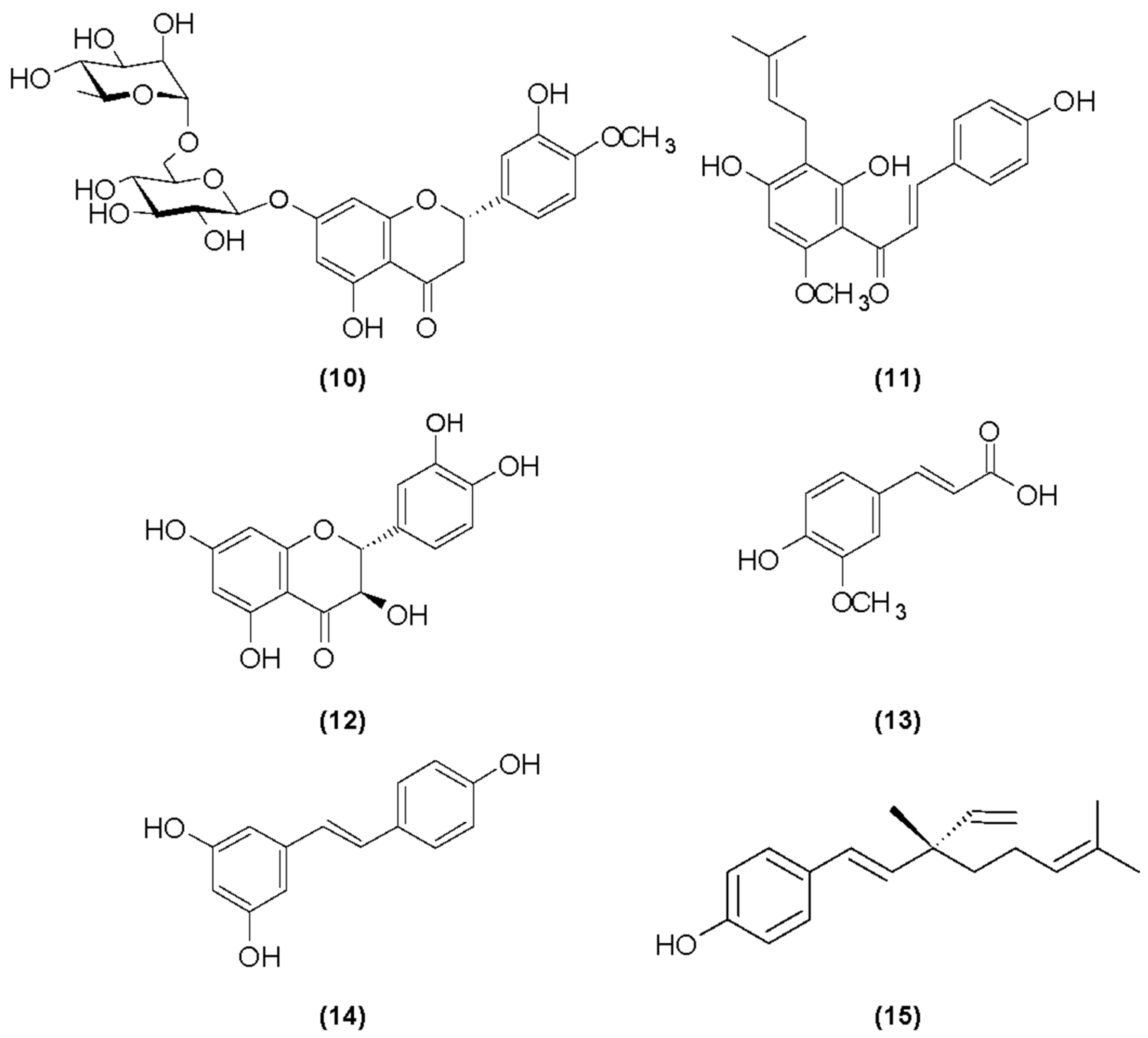
Hesperidin

Hesperidin (Figure 4) has been reported to present various biological activities with the potential to be applied in the skincare. Hesperidin acts as a scavenger against free radicals. This activity is related to the releasing of protons from this flavone, modulating the radicals’ synthesis toward the hesperidin structure. Hesperidin can inhibit the UV effects by reducing epidermal thickening, by regulating the expression of angiogenesis-related factors, by modulating apoptotic proteins induced by oxidative stress, and by regulating some immune cells and inflammatory cytokines, contributing to reducing inflammation. In addition, hydrogels formulated with nano-lipid carriers (NLCs) loaded with hesperidin showed a remarkable photoprotective effect, absorbing 99% of UVB radiation and 83% of UVA radiation. Thus, hesperidin may be a suitable ingredient in sunscreen. Hesperidin from orange peels, as an antioxidant molecule, has shown both anti-melanogenic and melanogenic activities, depending on its application. The oral intake of hesperidin helps to treat pigmented purpuric dermatosis lesions in a human case report and to treat atopic dermatitis in an animal testing model [82].
The production of hesperidin for cosmetic use encompasses various extraction (solvent extraction, microwave-assisted extraction, enzymatic extraction, and supercritical fluid extraction) and processing methods, each with its advantages and challenges. Innovations like nanocrystal formulation and biotechnological production hold promise for the more efficient and sustainable utilization of hesperidin in cosmetology. Advancements in biotechnology are exploring microbial fermentation and plant cell culture methods to produce hesperidin. These approaches aim to provide sustainable and controllable production processes, potentially increasing yield and optimizing properties through genetic engineering [83].

Xanthohumol

Xanthohumol (2,4,4-trihydroxy-6-methoxy-3-(3-methyl-but-2-en-1-yl) chalcone) (Figure 4) is a highly active flavonoid. It is characterized by a fairly broad spectrum of biological activity. It has antifungal, antiviral, antibacterial and antimalarial effects. It also prevents ischemic heart disease and reduces the risk of cancer [84].
First of all, xanthohumol is one of the strongest antioxidants that effectively protects the skin from oxidative stress. It prevents unfavorable changes in cell proteins, such as oxidation or amino acid modification, which lead to the weakening of collagen fibers contained in the skin. Studies have shown that xanthohumol reduces lipid peroxidation. It reduces their permeability, and also prevents disorders of transmembrane transport and increases its stability. These lipids are responsible for the construction of the lipid coat. In addition, studies confirm that xanthohumol is a 30 times stronger antioxidant than vitamin C. It works in the first line of antioxidant defense and does not allow hydrogen peroxide to transform into a free radical [85,86].
Xanthohumol inhibits the excessive production of IL-12 interleukins in the autoimmune response to inflammation in the body, which confirms its anti-inflammatory effect. It also inhibits the growth of Gram-positive bacteria, such as Staphylococcus aureus, Staphylococcus pyogenes, Staphylococcus epidermidis, Propionibacterium acnes, and Kocura rhizophila, which can cause purulent skin infections. High activity, which limits the growth of bacteria, can contribute to effective care of problematic skin, with changes in rosacea and acne vulgaris [86].
Metalloproteinases participate in the skin aging process, as they increase the decomposition of extracellular matrix components, especially elastin, collagen, and fibronectin. Xanthohumol has an inhibitory effect on metalloproteinases MMP-1 and MMP-8. At the same time, it protects the skin from photoaging, thus slowing down degenerative processes in the skin. Moreover, xanthohumol inhibits the secretion of GM-CSF cytokines, which activate transduction signals and then stimulate melanocytes and tyrosinase proteins, leading to the formation of discolorations. Therefore, as an active ingredient in cosmetics, xanthohumol brightens existing discolorations and also helps to inhibit the formation of new ones and reduce skin redness [86,87].
Its production for cosmetic applications involves extraction from hops and subsequent formulation processes to ensure stability and efficacy. Dried hop cones or pellets are subjected to extraction using organic solvents such as ethanol or methanol. The extract is then concentrated and purified to isolate xanthohumol. The process also utilizes supercritical carbon dioxide as a solvent to extract xanthohumol at specific temperatures and pressures. Deep eutectic solvents offer a green and sustainable alternative to traditional solvents, with the potential for the simultaneous extraction of proteins and xanthohumol [88].

Taxifolin

Taxifolin (Figure 4), also known as 3,5,7,3′,4′-pentahydroxyflavanone or dihydroquercetin (TXF), is a flavanonol with potent antioxidant properties, widely distributed in nature in both aglycone and glycoside forms. It is commonly sourced from onions, olive oils, grapes, citrus fruits, milk thistle, maritime pine bark, and Douglas fir bark. The antioxidant and free-radical-scavenging activities of TXF have been extensively validated through various in vitro bioanalytical assays. Taxifolin consists of two aromatic rings, each containing phenolic groups (–OH) positioned at the meta- and para- sites relative to each other. These phenolic groups contribute significantly to its potent antioxidant activity. The strong antioxidant properties of taxifolin are primarily attributed to the conjugated structures and the resonance stability of both phenolic rings. The presence of a C2-C3 double bond is critical for the inhibitory activity of flavonoids; however, taxifolin lacks this bond, rendering it more susceptible to inactivation through the formation of strong hydrogen bonds with macromolecules [89].
Additionally, the 3-hydroxy group (3-OH) in the C ring plays a key role in modulating the oxidative burst of neutrophils, highlighting its anti-inflammatory potential. In the B ring, the ortho arrangement of the catechol group is crucial for the modulatory effect of taxifolin against neutrophil oxidative burst. This structural configuration enhances its ability to influence cellular oxidative processes, contributing to its therapeutic efficacy in inflammatory conditions [89,90].
Scientific evidence highlights TXF’s multifunctional bioactivity, including its roles as an anti-inflammatory, depigmenting, and antitumor agent. It has been shown to mitigate oxidative DNA damage and prevent UV-induced skin carcinogenesis. TXF exhibits low toxicity and effectively protects the skin from photoaging while inhibiting melanogenesis. Furthermore, it is among the most potent flavonoids in suppressing the interferon-γ (IFNγ)-induced expression of intercellular adhesion molecule-1 (ICAM-1) protein and mRNA in human keratinocytes. Pre-treatment with TXF also significantly inhibits IFNγ-induced ICAM-1 expression in reconstructed human skin models, indicating its therapeutic potential for pathological skin conditions associated with heightened cellular adhesion and inflammation [91].
In vivo studies on experimental animal models have demonstrated TXF’s efficacy in alleviating chemically induced atopic dermatitis-like lesions and promoting recovery in chemically induced burn injuries. These findings underscore TXF’s potential as a promising candidate for the management of oxidative stress-related and inflammatory skin disorders [91].
Currently, extraction from Larix sibirica and Larix gmelinii remains the primary method of taxifolin production, but biotechnological methods (using engineered microorganisms, such as the yeast Yarrowia lipolytica) are emerging as sustainable alternatives. Stabilization techniques are also being explored to improve its effectiveness in cosmetic formulations. These advancements align with the growing demand for natural, high-performance skincare ingredients [92]. Due to its limited solubility and stability, taxifolin is often modified into derivatives (e.g., taxifolin tetraoctanoate) to enhance its performance in cosmetic formulations [93].

4.3.2. Phenolic Acids

Ferulic Acid

Ferulic acid (4-hydroxy-3-methoxycinnamic acid) (Figure 4) is an organic chemical compound and a derivative of cinnamic acid, classified as a phenol. It contains a hydroxyl and carboxyl group in its structure [94].
Due to its antioxidant properties, ferulic acid protects the skin from the harmful effects of external factors. Due to the presence of side chains with double bonds and aromatic rings, it destabilizes free radicals, reduces oxidative stress and prevents the formation of thymidine dimers, which lead to cell DNA damage and biological changes in the skin. It absorbs a beam of solar radiation by creating a phenoxy radical. It has anti-inflammatory effects by reducing the concentration of inflammatory mediators, inhibiting the expression of iNOS protein and interferon. In addition, it has antibacterial, antiviral, anti-allergic and blood vessel-sealing effects [95].
It is used in anti-acne therapy, both in rosacea and acne vulgaris, and is also recommended for the treatment of skin discolorations [96].
In cosmetic products, ferulic acid is mainly used to improve skin elasticity and firmness, maintain uniform color, inhibit skin aging processes, reduce fine wrinkles, alleviate inflammation, and strengthen the skin barrier against oxidative stress [96].
Ferulic acid helps neutralize the free radicals generated by UVA radiation, providing protective effects to endothelial cells and keratinocytes, which are typically vulnerable to this type of damage. In human fibroblasts, ferulic acid administration before UVA exposure notably reduced the harmful effects of radiation. This includes preventing UV-induced alterations in the cell cycle and protecting DNA integrity. It regulates the expression of genes involved in DNA repair, supporting cellular recovery and enhancing the skin’s resilience to UV-induced damage. UVB-induced reactive oxygen species (ROS) play a major role in the development of skin cancer by causing oxidative damage to skin cells, including DNA mutations that can lead to tumor formation. Ferulic acid, a potent antioxidant, has been shown to reduce the levels of these harmful ROS, making it a promising substance in cancer prevention. Ferulic acid has long been used in cosmetics as a stabilizer for well-known antioxidants like vitamin C and vitamin E. However, recent research highlights that ferulic acid is not just a supporting compound but also an active ingredient with its own powerful antioxidant properties. It actively contributes to the skin’s defense systems, offering multiple benefits for skin health and appearance. By strengthening antioxidant defense, it plays a protective role for key skin components, including keratinocytes, fibroblasts, collagen, and elastin. This helps to maintain the structural integrity and overall health of the skin. Ferulic acid is commonly used in skin-lightening formulations due to its ability to inhibit the activity of tyrosinase, an enzyme crucial in the process of melanogenesis (the production of melanin). By reducing tyrosinase activity, ferulic acid helps to limit melanin production, which can reduce dark spots, hyperpigmentation, and overall skin discoloration. Ferulic acid is increasingly used in various dermatological procedures, such as microneedling, non-needle mesotherapy, chemical peels, and grooming treatments, due to its potent antioxidant, anti-inflammatory, and skin-repairing properties. It is particularly effective in addressing a variety of skin concerns [96,97,98,99].
The production of ferulic acid for cosmetics predominantly relies on extraction from plant-based sources, particularly rice bran derivatives, due to their high content of ferulic acid esters. This process typically includes base-catalyzed hydrolysis of γ-oryzanol, a compound found in rice bran oil, to release ferulic acid. Biotechnological methods are emerging as promising alternatives, offering sustainable and efficient production routes. For instance, genetically modified Escherichia coli strains have been developed to produce ferulic acid from simple carbon sources. Chemical synthesis remains less prevalent, primarily due to economic and practical challenges. One such method involves the saponification and solvent extraction of plant materials rich in ferulic acid esters, followed by purification processes to obtain high-purity ferulic acid [100,101,102].

4.3.3. Resveratrol

Resveratrol (Figure 4) is an organic chemical compound, a polyphenolic derivative of stilbene. It can occur in two forms: geometric isomers cis and trans. The naturally occurring isomer is trans and only this form shows biological activity. Upon UV exposure, it can transform into its cis isomer [103]. Resveratrol acts as a powerful antioxidant, neutralizing free radicals that cause cellular damage. It is regarded as one of the most potent polyphenols, offering the strongest protection against skin aging. It is classified as a phytoestrogen, due to its ability to interact with estrogen receptors. It has a positive effect not only on the skin but also on the body, as it helps protect against circulatory system diseases, neurological diseases, e.g., Alzheimer’s disease, and has even been proven to have anti-cancer effects [104,105].
Its main advantage is its ability to reduce the effects of skin aging. It also possesses skin-repairing properties, as it acts as an activator of sirtuin—often referred to as the “enzyme of youth” (increasing sirtuin activity in the human body by 13 times). Additionally, it protects against UV-induced damage and helps reduce skin hyperpigmentation caused by sun exposure [105].
Moreover, resveratrol reduces the inflammation and redness of the skin, thanks to its soothing properties, and when used externally together with appropriate supplementation, it helps in the treatment of psoriasis and eczema; counteracts the negative effects of external factors, including free radicals; has a smoothing and elasticizing effect on the skin; has anti-aging properties; moisturizes dry skin; and evens out the skin tone and structure [106].
The production of resveratrol for cosmetic use encompasses various methods, including plant extraction, chemical synthesis, and biotechnological approaches. Resveratrol is commonly extracted from plants such as grapes, berries, and Japanese knotweed (Polygonum cuspidatum). The extraction process typically involves solvent extraction techniques to isolate resveratrol from plant materials. However, the yield can be low, and the process may be influenced by environmental factors affecting plant growth. Innovations in biotechnological production and formulation techniques continue to enhance resveratrol’s applicability and effectiveness in cosmetology. Genetically engineered microorganisms, such as Saccharomyces cerevisiae and Pichia pastoris, are utilized to biosynthesize resveratrol. To enhance resveratrol’s solubility and stability, enzymatic glycosylation can be employed. For instance, using amylosucrase from Deinococcus geothermalis, resveratrol can be converted into resveratrol-O-glucosides. These derivatives have demonstrated promising cosmetic activities, including antioxidant, anti-inflammatory, anti-aging, and skin-whitening effects [107,108].

4.3.4. Bakuchiol

Bakuchiol (Figure 4) is a plant-derived meroterpene, meaning it is partly a terpenoid due to its conjugation with a compound of a different class—a phenol, in this case. It was isolated from the seeds and leaves of Psoralea corylifolia L. (Fabaceae) [109,110].
Bakuchiol exhibits potent antioxidant properties [111,112] and retinol-like activity, as it regulates gene expression in a manner similar to retinol, stimulates the production of type I, III, and IV collagen; inhibits melanogenesis; and improves skin photoaging—all while offering better skin tolerance compared to retinol [113,114,115]. For these reasons, it is widely used in cosmeceuticals as an anti-aging, anti-inflammatory, and antibacterial agent [116,117,118].
The production of bakuchiol for cosmetic use involves traditional extraction methods, advanced biotechnological approaches, and precise formulation techniques to ensure safe and effective skincare products. Ongoing research and technological advancements continue to optimize these processes, ensuring that bakuchiol remains a valuable ingredient in the cosmetic industry [119].

4.4. Minerals

4.4.1. Selenium (Se)

In the human body, selenium plays many complex roles, including in immune system responses, preventing cognitive disorders, dementia, and cardiovascular diseases, and in oxidative defense [120]. Nuts, cereals, seafood, and meat are the richest sources of selenium in the diet [121].
The physiological effect of Se is mainly due to its incorporation in selenoproteins Selenoproteins have been implicated in many metabolic and functional pathways, such as aging. Thus, the Se-containing enzyme glutathione peroxidase can help lower free radical reactions to tolerable levels by reducing H2O2 to H2O and organic hydroperoxides (ROOH) to alcohol (ROH). The Se-dependent glutathione peroxidases (GPX1–4 and GPX6) and thioredoxin reductases (TrxR1–3) suppress oxidative stress. GPX1 is a major metabolic form of body Se against severe oxidative stress [122]
Insufficient Se levels in an organism are associated with such inflammatory skin diseases as psoriasis and atopic dermatitis [122]. The only selenium compound approved for use in cosmetic products is selenium sulfide. Usually, it occurs in anti-dandruff shampoos and preparations intended for people suffering from seborrheic dermatitis. The shampoos and rinses recommended in these cases contain 2.5 and 1% selenium sulphide, respectively. Selenium is omnipresent in cosmetics but most often its presence is not clearly marked. This element often appears as a component of various raw materials used in the cosmetic industry, such as vegetable extracts, oils, clays, milk, and honey. Thermal water from La Roche-Posay (the Poitou-Charentes region in France) contains a lot of selenium (approx. 53 μg/L). Creams, face, and body mists produced on the basis of this water effectively protect against the effects of free radicals, delay the skin aging process, improve its elasticity, and have a softening and smoothing effect. Selenium is also present in some formulations used during mesotherapy [123].
The health effects of selenium (Se) depend largely on the dose, as the difference between beneficial and harmful levels is marginal. At appropriate nutritional levels, selenium acts as a potent antioxidant, protecting cells from oxidative damage. However, when consumed in excess, it shifts to a prooxidant role, potentially causing oxidative stress and cellular damage [122].

4.4.2. Zinc (Zn)

Zinc is a trace element that is a component of over three hundred enzymes, including DNA and RNA polymerases and superoxide dismutase. It participates in the production of testosterone and insulin, stabilizes cell membranes, and affects the proper functioning of the nervous system. This element also plays a significant antioxidant role. Deficiency is often caused by malnutrition, diseases, or eating a poorly diversified diet [124]. Vitamin A helps in the absorption of zinc in the body. Animal products show greater absorption than plant products, due to the presence of phytates. High zinc content is found in oysters, fish, meat, nuts, legumes, eggs, and whole grain cereal products [125].
Topical zinc, in the form of divalent zinc ions, has been reported to provide antioxidant photoprotection for skin. Two antioxidant mechanisms have been proposed for zinc: zinc ions may replace redox active molecules, such as iron and copper, at critical sites in cell membranes and proteins; alternatively, zinc ions may induce the synthesis of metallothionein, sulfhydryl-rich proteins that protect against free radicals [126,127].
Numerous studies have shown the benefits of either topical or oral zinc in the treatment of acne. The topical application of zinc ions has been shown to induce metallothionein, which may account for its photoprotective effect [31,126]. Zinc oxide is a very effective sunscreen as well as regulating sebum secretion. Zinc salts are present in many cosmetic products—mouthwashes, toothpaste (antiseptic activity), after-shaving products, deodorants (anti-odor), and face powders. Zink pyrithione is used against dermatitis as well as dandruff. Zinc lauryl ether sulfate can be used in skin cleansing products [128,129].

4.5. Peptides, Amino Acids, Enzymes

4.5.1. Peptides

Peptides are chemical compounds made of amino acids connected by a peptide bond. Peptides are increasingly used in cosmetic preparations as a biologically active ingredient, especially in combination with copper ions Cu2+. This combination affects skin reconstruction and increases its regeneration. Low-molecular transport peptides, due to their structural and spatial structure, are able to bind other substances, increase their solubility, bioavailability, and durability. Most often, these are water-soluble tripeptides, and the best-known transporting Cu2+ ions are the tripeptide Gly-His-Lys (GHK) and Gly-Gly-His (GGH) [130].
GHK components are responsible for regenerative processes and maintaining proper skin density (Cu activates fibroblasts by increasing the production of mRNA and proteins for elastin, collagen, proteoglycans, and glycosaminoglycans). They stimulate the production of proteinases and metalloproteinases, which eliminate damaged collagen. Clinical studies have shown that copper peptides can increase collagen production by as much as 70% and reduce the number of wrinkles by 31.6% and their depth by 23.4% [131].
In addition, copper tripeptides have a number of scientifically proven properties: they support collagen synthesis; promote the production of glycosaminoglycans, which improves skin elasticity and firmness; activate the system responsible for removing damaged collagen and elastin from scar tissue and skin; smooth the skin; reduce wrinkles; lighten discolorations; support wound healing; reduce inflammation caused by free radicals; and protect skin cells against oxidative stress and the negative effects of UV radiation [132].

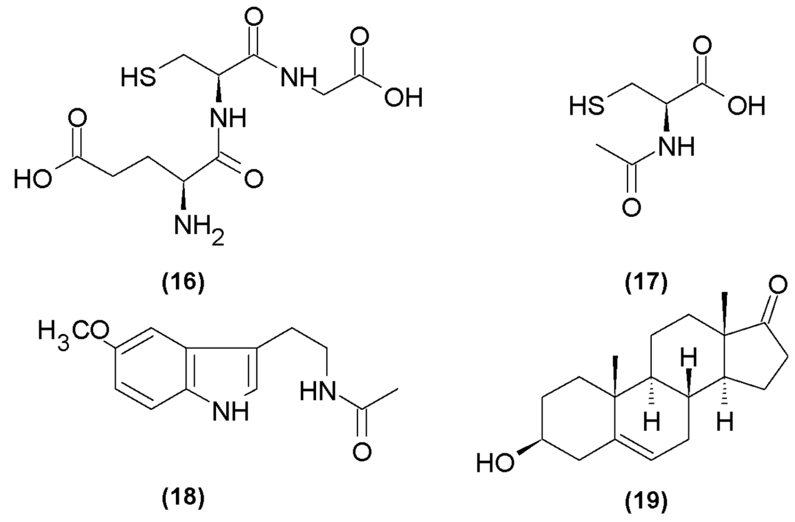
Glutathione

Glutathione (γ-glutamylcysteinylglycine) (Figure 5) is a water-soluble tripeptide containing cysteine. It is present in most plant organisms, tissues of all mammals, and microorganisms. It occurs in reduced (GSH) and oxidized (GSSG) forms. About 90% of GSH occurs in the cell cytosol, 10% in mitochondria, and a small amount in the endoplasmic reticulum. One of the most important functions of the reduced form of glutathione is the role of a storage and source of cysteine in the body, as well as maintaining the redox balance of the cell and counteracting the effects of oxidative stress. The highest concentration of GSH occurs in the liver [133]. In order to ensure an adequate level of GSH in the body, an appropriately maintained level of reducing equivalents is necessary. The most important reductant is NADPH, which is produced by the pentose pathway in the cell cytosol. The enzyme responsible for maintaining the proper concentration of NADPH is G6PDH—glucose-6-phosphate dehydrogenase [134].
Glutathione is a powerful antioxidant composed of three amino acids: glutamate, cystine, and glycine. It plays a crucial role in various biological processes, including skin lightening, through the following mechanisms: the inhibition of tyrosinase activity and shift in melanin production. Glutathione inhibits the enzyme tyrosinase, which is essential for melanin production. By blocking tyrosinase, glutathione reduces melanin synthesis, leading to lighter skin. Glutathione influences melanin types by shifting the production from eumelanin (responsible for dark brown or black pigmentation) to pheomelanin (responsible for yellow-red pigmentation) [135,136].
The choice between fermentation and enzymatic synthesis for glutathione production depends on factors such as desired purity, production scale, and cost considerations. Advancements in biotechnological methods continue to enhance the efficiency and sustainability of glutathione production for cosmetic applications. Yeasts such as Saccharomyces cerevisiae and Candida utilis are commonly used for glutathione production. Metabolic engineering and fermentation optimization in bacterial systems have been investigated to improve glutathione yields. Enzymatic synthesis involves the direct enzymatic production of glutathione using its precursor amino acids [137,138,139].

4.5.2. N-Acetyl-L-Cysteine

N-acetyl-L-cysteine (NAC) (Figure 5) is a derivative of L-cysteine, a sulfur amino acid naturally occurring in the body. It is the most absorbable form of the amino acid and plays a key role in the production of glutathione (in combination with glutamine and glycine), responsible for antioxidant processes. NAC plays an important role in the synthesis of proteins, cofactors, and enzymes and is also a component of structural proteins [140]. Studies have shown that NAC is used in autoimmune diseases and during chronic inflammation, which is accompanied by oxidative stress. This amino acid can help reduce inflammation and slow down autoimmune tissue destruction (e.g., thyroid). By reducing oxidative stress, it affects the production of proinflammatory cytokines and has antioxidant effects [141].
N-acetylcysteine (NAC) is both a thiol compound and a mucolytic agent. Upon entering cells, NAC is quickly metabolized into cysteine, which serves as a critical building block for the synthesis of glutathione (GSH). It plays a key role in maintaining the cell’s redox balance, protecting against oxidative stress. Additionally, GSH interacts with N-methyl-D-aspartate (NMDA) receptors by binding to the glutamate recognition site, allowing it to act as a natural neuromodulator and influence cellular signaling processes. This dual functionality of NAC—supporting glutathione production and influencing neuromodulation—makes it a versatile compound in therapeutic applications. Cysteine plays a dual role, not only as a precursor for glutathione (GSH) synthesis but also as an independent free radical scavenger. It effectively neutralizes reactive oxygen species (ROS), such as hydroxyl radicals (OH), hydrogen peroxide (H2O2), and superoxide anions (O2−), which can otherwise cause damage to DNA, proteins, and lipids within cells. As a result, N-acetylcysteine (NAC) is recognized as both an antioxidant and a free radical scavenger. By boosting intracellular levels of cysteine, NAC enhances GSH production, amplifying its protective effects against oxidative stress and cellular damage. N-acetylcysteine (NAC) plays a beneficial role in managing atopic dermatitis due to its ability to reduce free radical species and mitigate oxidative stress. This reduction in oxidative damage supports the restoration of cell adhesion molecules, which are essential for maintaining a healthy skin barrier. Since atopic dermatitis is partly associated with increased TEWL and decreased skin hydration, NAC appears to address these underlying issues, making it a promising therapeutic option for improving skin health in individuals with this condition [142]. N-acetylcysteine (NAC) has shown potential in protecting melanocytes from the harmful effects of UV-induced oxidative stress and damage. In in vitro studies, NAC shields melanocytes by reducing the oxidative stress caused by UV exposure, which can otherwise lead to cellular damage and dysfunction. Additionally, in vivo studies suggest that NAC may help prevent UV-induced melanoma by minimizing oxidative damage and supporting the natural defense mechanisms of melanocytes. This dual protective role highlights NAC’s potential as a therapeutic agent for skin health and as a preventive measure against UV-related skin cancers [143].
The chemical synthesis of N-acetyl-L-cysteine involves the acetylation of L-cysteine, followed by purification steps to ensure high-quality NAC suitable for cosmetic use [144].

4.5.3. Superoxide Dismutases

A group of enzymes present in humans, including zinc–copper superoxide dismutase (SOD 1), manganese superoxide dismutase (SOD 2), and extracellular superoxide dismutase (SOD 3). They are activated as defense mechanisms during the interaction of free radicals. These mechanisms include the following: preventive—the first line of defense, which consists in preventing the reaction of ROS and their derivatives with biological substances; repair—the interruption of free radical and non-radical oxidation reactions, removing the products of the ROS reaction and their derivatives, by repairing or eliminating damaged molecules [145,146].
The reaction of superoxide anion radical dismutase to molecular oxygen and hydrogen peroxide is a two-stage process. The first stage is the reduction of the metal ion with the simultaneous release of an oxygen molecule. The second step is the oxidation of the metal ion by a superoxide anion radical and hydrogen to form hydrogen peroxide [146,147].
Superoxide dismutase (SOD) has remarkable properties that make it a valuable ingredient in skincare and dermatological treatments. SOD effectively neutralizes oxygen-free radicals. It reduces inflammation, helping alleviate redness and swelling in the skin. When combined with cell-penetrating peptides, SOD can penetrate deeper into the skin and provide significant protection against UVB-induced damage. Moreover, research has consistently demonstrated that SOD is non-toxic and safe for cosmetic use. By scavenging free radicals, SOD prevents oxidative damage, stabilizes collagen and elastin, and promotes their moderate crosslinking, which helps maintain skin elasticity and smoothness. It keeps skin soft and supple. It helps mitigate UV-induced damage, making it a valuable addition to sunscreens as well as a topical agent for reducing redness and swelling associated with conditions like rosacea or sunburn. SOD’s dual role as a cosmetic and therapeutic agent underscores its versatility and significance in modern skincare formulations [148,149].
The production of superoxide dismutase for cosmetic applications encompasses several methodologies, each aiming to achieve high yield, purity, and bioactivity. SOD can be isolated from various plant sources, including marine phytoplankton, horseradish, and cantaloupe. However, this method often faces challenges such as low yield and variability in enzyme activity due to environmental factors affecting plant growth. Innovative approaches involve cultivating microalgae to produce extracts with SOD-like activity. These extracts can be utilized in cosmetic formulations to harness their antioxidant properties. Yeasts, particularly Saccharomyces cerevisiae, have been employed to produce SOD through fermentation processes. Advancements in biotechnology have enabled the production of recombinant SOD by inserting the SOD gene into host organisms such as Escherichia coli. This method allows for large-scale production with consistent quality and activity [150,151,152].

4.6. Hormones

4.6.1. Melatonin

Melatonin (N-acetyl-5-methoxytryptamine) (Figure 5) is a hormone that effectively inhibits the oxidation processes of proteins, lipids, and DNA and can also mediate the stimulation of antioxidant activity of enzymes. Studies have shown that combining this hormone with antioxidants (glutathione, vitamin C and E) significantly reduces this process. What is more, vitamin C can contribute to the regeneration of melatonin because they interact synergistically [153,154].
Melatonin has a positive effect in the fight against Alzheimer’s disease. In the aging process, the repair potential of damaged neurons in the body is reduced. Increased oxidative stress is generated by excess free radicals. With age, there is also a decrease in the secretion of melatonin, which is an effective antioxidant that plays an important role in protecting the cells of the nervous system [154].
Melatonin directly neutralizes reactive oxygen species. It has the ability to capture the hydroxyl radical, singlet oxygen, nitric oxide, peroxynitrite, hypochlorite, and peroxyl radicals. The direct role of this hormone in the body’s defense system is to eliminate ROS from the cellular environment and metabolize their inactive forms. Indirect antioxidant action is to stimulate the synthesis of prooxidant enzymes [155].
Human skin is characterized by significant levels of melatonin. The MT1 and MT2 receptors are present in fibroblasts, keratinocytes, hair follicles, eccrine glands, and blood vessels. The skin is exposed to various external factors, mainly solar radiation and environmental pollutants, which cause oxidative stress and inflammation, thus accelerating the photoaging process. Melatonin easily penetrates the skin, where it can exhibit its antioxidant and anti-inflammatory properties. As an endogenous substance, it is practically devoid of allergenic potential. Topically applied melatonin has been studied in clinical trials for its effectiveness in protecting the skin from radiation, reducing skin aging, and supporting hair growth [156].
The production of melatonin for cosmetic use encompasses traditional chemical synthesis and emerging biotechnological methods. While chemical synthesis remains the primary approach due to its established processes and scalability, biotechnological production offers a promising alternative that aligns with the growing demand for sustainable and natural ingredients in cosmetics. Ongoing research and technological advancements continue to optimize these production methods, ensuring the availability of high-quality melatonin for cosmetic applications [157].

4.6.2. DHEA (Dehydroepiandrosterone)

Dehydroepiandrosterone (Figure 5) is a metabolic intermediate in the synthesis of testosterone, estrone, and estradiol. It is a steroid secreted by the adrenal glands and brain. In both men and women, DHEA levels decrease by about 2% per year. Several clinical studies have assessed the effect of dehydroepiandrosterone treatment on oxidative damage and renal dysfunction. DHEA has been shown to protect many tissues from oxidative damage and to have beneficial effects on vascular function, atherosclerosis, and diabetic complications. DHEA interferes with early prooxidant responses and increases in TNF (tumor necrosis factor) caused by infrared radiation [158].
Research has demonstrated that DHEA treatment can increase the rate of sebum production, a positive effect particularly for menopausal individuals who often experience a decline in sebum levels due to hormonal changes. Sebum is essential for maintaining skin hydration and preventing dryness, which is common during menopause. Topical DHEA has been found to enhance the brightness of the skin, which can become dull and uneven due to hormonal aging. It helps combat the papery texture and thinning of the skin (epidermal atrophy) that often accompanies hormone-related skin aging. While there is evidence suggesting that topical DHEA could influence the skin’s aging process, especially in terms of wrinkle formation, this aspect still requires further research to confirm its effectiveness in wrinkle reduction [159].
The production of DHEA for cosmetic use involves both chemical and biotechnological methods, each offering distinct advantages. A common chemical synthesis route for DHEA starts with 16-dehydropregnenolone acetate. This compound undergoes oximation, followed by a Beckmann rearrangement, hydrolysis, and refinement to yield DHEA. Advancements in biotechnology have led to the development of methods to produce DHEA using microbial biotransformation. For instance, certain Mycobacterium species can convert phytosterols into DHEA in a vegetable oil/aqueous two-phase system, offering an efficient production method [160,161].

4.7. Butylated Hydroxytoluene (BHT)

Butylated hydroxytoluene (BHT) is a synthetic antioxidant commonly used in cosmetics and personal care products to prevent the oxidation of fats and oils, thereby extending product shelf life. It helps maintain the stability, color, odor, and texture of formulations by inhibiting oxidative degradation [162].
BHT is typically used in concentrations ranging from 0.0001% to 0.5% in various products, including lipsticks, moisturizers, and other cosmetics [32]. BHT can be absorbed through the skin, but the amount that penetrates is minimal and largely stays within the skin layers [163].

4.8. Fungi-Derived Antioxidants

In addition to plants, microorganisms and macroorganisms, including actinomycetes, bacteria, cyanobacteria, and fungi with lichens, also serve as sources of antioxidants and other bioactive compounds. This section will explore the medicinal and cosmetic potential of various mushroom species, particularly large-fruited fungi and lichens [164,165].
Mushrooms such as Ganoderma lucidum, Hericium erinaceus, Lentinus edodes, Cordyceps sinensis, Sclerotinia sclerotiorum, and Trametes versicolor are widely recognized for their health-promoting properties and are considered functional and nutraceutical foods [166].
Numerous studies indicate that fungi exhibit a broad spectrum of biological activity, including immunomodulatory, anticancer, antiviral, antibacterial, antifungal, anti-inflammatory, antioxidant, antidiabetic, neurotonic, hepatoprotective, and cosmeceutical properties. They also help regulate triglyceride and cholesterol levels in the blood and lower blood pressure, among other benefits [121,122,166,167].
The substances responsible for these versatile properties vary in chemical nature and include ergothionine, glutathione, phenols, flavonoids, β-glucans, ergosterol, and β-carotene [167]. The main nutrients found in mushroom fruiting bodies include proteins, carbohydrates, fats, vitamins, and minerals. Both wild and cultivated mushroom fruiting bodies are rich in antioxidants.
One of the most extensively studied mushroom species over the years is Ganoderma lucidum, known as lingzhi in Chinese and reishi in Japanese [166].
Among the phenolic compounds found in mushrooms, the largest percentage consists of phenolic acids, which are well known for their antioxidant activity. Species with the greatest diversity of phenolic compounds include Boletus badius, Cantharellus cibarius, and Pleurotus ostreatus [166,168,169]. Antioxidant properties are also associated with the presence of non-hallucinogenic indole compounds in mushrooms, including L-tryptophan, 5-hydroxytryptophan, serotonin, melatonin, and tryptamine [125,170]. Additionally, mushrooms are one of the few natural sources of ergosterol, a precursor of vitamin D2. The presence of other vitamins in forest mushroom fruiting bodies, such as tocopherol, β-carotene, and ascorbic acid, has also been confirmed. These compounds contribute significantly to the antioxidant potential of mushrooms [166]. Antioxidants derived from fungi (species from Basidiomycota, Ascomycota, Zygomycota), such as polysaccharides, pigments, peptides, ergothioneine, ergosterol, phenols, and alkaloids, have potential applications in the food, pharmaceutical, and cosmetic industries [171].
Active compounds extracted from mushrooms are widely used in the production of cosmetic products for hair and skin. Important bioactive ingredients derived from fungi, such as kojic acid (Figure 6), chrysophanol, cytonemin, mycosporine-like amino acids, β-glucans, triterpenoids (ganoderic acid, ergosterols), ergothioneine, ceramides, and chitosans, play a crucial role in the effectiveness of cosmetic formulations [167].
Beyond their antioxidant properties, mushrooms and their extracts exhibit anti-tyrosinase and anti-hyaluronidase activities, which help control skin discoloration and prevent the breakdown of hyaluronic acid. Additionally, they possess anti-collagenase and anti-elastase properties, which contribute to maintaining skin firmness and elasticity.
Ganoderma lucidum, Euphorbia characias, and Pistacia atlantica subsp. mutica are mushroom species with strong anti-tyrosinase activity, making them valuable ingredients in skin-whitening products. Mushroom extracts and their bioactive metabolites offer excellent antioxidant, anti-wrinkle, anti-aging, moisturizing, and skin-whitening properties, making them ideal multifunctional cosmetic ingredients.
Intensive research and potential cosmetic applications focus on mushroom species such as Lentinula, Ganoderma, Pleurotus, Cordyceps, Inonotus, Tremella, Grifola, Schizophyllum, Coriolus, Trametes, Agaricus, Phellinus, Antrodia, Euphorbia, and Pistacia [172].
The cosmetic industry’s growing interest in mushrooms is also due to their ease of cultivation on inexpensive substrates, short growth cycles, and simple purification processes. These factors provide a competitive advantage for mushroom-derived products over plant-based cosmeceuticals [167].

4.9. Lichen-Derived Antioxidants

Lichens are an important group of fungi that produce bioactive substances widely used in medicine and food. These complex organisms are composed of a fungal partner (mycobiont), primarily from Ascomycota, and a photosynthetic partner (photobiont), which may be green algae, cyanobacteria, or both. Additionally, bacteria and viruses are often present in the lichen symbiosis. Lichens are a rich source of secondary metabolites, including amino acid derivatives, sugar alcohols, aliphatic acids, minerals, macrocyclic lactones, monocyclic aromatic compounds, quinones, chromones, xanthones, dibenzofurans, depsides, depsidones, depsones, terpenoids, steroids, carotenoids, and diphenyl ethers. These organisms appear to be a promising resource for antioxidant compounds, particularly polyphenols such as depsides, depsidones, and dibenzofurans [173,174].
Atranorin, a compound with the depside structure, is one of the most common lichen secondary metabolites, and is found in various species of lichens, such as Stereocaulon caespitosum, Stereocaulon alpinum, Parmelia sulcata, Evernia prunastri, Cladina kalbii, and Lethariella canariensis. After usnic acid, atranorin (Figure 6) is the second most studied secondary metabolite with anti-inflammatory, analgesic, wound healing, antibacterial, antifungal, antitumor, cytotoxic, antioxidant, antinociceptive, antiviral, and immunomodulatory activities [175,176]. Usnic acid is the active compound present in Usnea sp. extracts, with high antibacterial and cytotoxic properties f.e. Usnea barbata is a species that has been employed in modern-day cosmetic and pharmaceutical products [177].
There are also preparations containing extracts or metabolites from various species of lichens such as Cetraria islandica, Platismatia glauca, Parmeliopsis ambigua, Punctelia subrudecta, Evernia mesomorpha, Anaptychya ciliaris, Nephroma parile, and Ochrolechia tartarea and species from the genera Cladonia, Usnea, Umbilicaria, Parmelia, Ramalina, Xanthoparmelia, and many others [164,166,177,178,179].

4.10. Algae-Derived Antioxidants

Algae, an ecological group of organisms with a highly diverse structure, belong to different kingdoms from a systematic classification perspective. They are commonly classified into four major seaweed classes: Cyanophyceae = Cyanobacteria (blue-green algae), Phaeophyceae (brown algae), Chlorophyceae (green algae), and Rhodophyceae (red algae). The broad biochemical composition of seaweeds gives them great potential as antioxidant additives suitable for commercial applications. The most promising biologically active components in their composition include sulfated polysaccharides, phlorotannins, and tyrosinase inhibitors. The main molecules with antioxidant and photoprotective properties in algae are mycosporine-like amino acids (MAAs) (Figure 6), the pigment scytonemin, and numerous phenolic compounds. Given these properties, algae have significant potential as cosmeceutical ingredients [180,181,182].
Algae contain high concentrations of polyphenols, making them excellent antioxidants. Polyphenols, chlorophyll, and carotenoids exhibit antioxidant activity, protect against the harmful effects of UV radiation, and possess anti-inflammatory properties [181,182,183,184,185,186].
Recent research has demonstrated the UV-protective effects of various algae groups, which have long been incorporated into commercial sunscreens [180,181,184,187,188,189,190].
Brown algae contain unique compounds not found in terrestrial sources and exhibit a higher antioxidant potential compared to Rhodophyceae (red algae) and Chlorophyceae (green algae) [185]. Cyanobacteria extracts have shown greater antioxidant activity than those of red macroalgae [181]. Given these properties, bioactive compounds derived from algae represent promising cosmeceutical agents for skincare applications.
A number of algae-based skin products have been marketed, such as Algenist (an anti-aging moisturizer containing microalgae oil and alguronic acid from algae); Helionori® by Gelyma (Marseille, France) and Helioguard365® (Mibelle Biochemistry, Buchs, Switzerland) (a sunscreen product containing MAAs from red seaweed Porphyra umbilicalis); Protulines® by Exsymol S.A.M., Monaco (an anti-aging agent from protein-rich extract of Arthrospira); and Dermochlorella by Codif, St. Malo, France (an anti-wrinkle agent from Chlorella vulgaris extract) [191]. Cosmetic preparations for protection from tobacco smoke included in an international index of cosmetic ingredients (INCI) are derived from marine algae—brown algae, Laminaria digitata, Sargassum muticum, and Undaria pinnatifida, and red algae, Rissoella verruculosa [192]. Interestingly, phycocyanin (usually found in red algae and cyanobacteria) is accepted as a natural color additive in food and cosmetics by the Food and Drug Administration (FDA) due to its non-toxic and biodegradable activities [191].

4.10.1. Cyanobacteria (Blue-Green Algae)

Cyanobacteria are a diverse group of photosynthetic prokaryotes that inhabit both aquatic and terrestrial environments. They produce a variety of secondary metabolites that are effective against reactive oxygen species (ROS), particularly pigments such as carotenoids and polyphenols, including phenolic acids and flavonoid compounds, which have great potential as pharmacologically active agents. The bioactive compounds isolated from various Cyanobacteria species exhibit antioxidant, photoprotective, antimicrobial, anti-inflammatory, and immunostimulant properties [181,193]. In cosmetic applications, cyanobacteria are particularly valuable as a source of substances that absorb UV radiation, making them ideal for sunscreen formulations. One of the primary mechanisms by which cyanobacteria provide photoprotection is through the production of natural sunscreens, including scytonemin (a pigment unique to cyanobacteria) and mycosporine-like amino acids (MAAs), both of which effectively absorb harmful radiation. High concentrations of scytonemin, MAAs, and polyphenols have been identified in Scytonema sp. and Lyngbya sp. Extracts from these species could serve as natural photoprotectors in UV-screen cosmetic formulations [181,183,193].
Especially Arthrospira platensis (spirulina) and Arthrospira species could be of potential value in cosmeceutical, dietary, and biomedical applications. The chemical composition of spirulina comprises proteins; polysaccharides and polyunsaturated fats (including 10–30% of γ-linolenic acid, GLA); vitamins; and minerals and pigments, including β-carotene and phycocyanin. Arthrospira species are used in many cosmetic products, such as balms and face masks which have anti-aging, moisturizing, brightening, and anti-acne properties, e.g., Protulines® by Exsymol S.A.M., Monaco—an anti-aging agent from protein-rich extract of Arthrospira [183,191,194].
The other Cyanobacteria species with active antioxidant and anti-aging compounds are Anabaena vaginicola (lycopene), Aphanizomenon sp., Nostoc sp. (MAA), and Planktothrix mougeotii, which seem promising in their applications in cosmeceuticals [191,193].

4.10.2. Phaeophyceae (Brown Algae)

Brown seaweeds are a type of eukaryotic algae widely distributed in cold-water marine environments. They are rich in seaweed polysaccharides, including alginates, laminarans, and fucoidans, as well as proteins, amino acids, polyphenols, terpenes, mannitol, fucoxanthin, hormones, and other bioactive compounds. A unique class of active polyphenolic substances extracted from Phaeophyceae are phlorotannins, a group of polymers composed of phloroglucinol. These compounds exhibit a wide range of biological properties, including antioxidant, skin-whitening, antibacterial, antiviral, anti-tumor, anti-allergic, and anti-inflammatory activities. The majority of phlorotannins and sulfated polysaccharides reported in scientific studies have been derived from Ecklonia and Eisenia species. Phlorotannins from marine algae, such as Ecklonia cava, have demonstrated the ability to protect cells from radiation-induced injury and oxidative stress. Additionally, fucoidan derived from brown algae (e.g., from Ascophyllum nodosum) has been reported to strongly inhibit UVB-induced MMP-1 expression in vitro. The further screening of brown algal species is needed to identify novel phlorotannins and polysaccharide derivatives that could be recommended as potential inhibitors of matrix metalloproteinases (MMPs), enzymes that contribute to collagen degradation and skin aging [180,195].
In cosmetology, tyrosinase inhibition is the most common approach to combat skin hyperpigmentation, and the search for effective skin-whitening agents has become an important goal in the development of cosmetic formulations. Tyrosinase inhibitors, regulators of melanogenesis (including fucoxanthin, phloroglucin) have been isolated from many marine species of brown algae, such as Ecklonia cava, E. stolonifera, Fucus vesiculosus, Hizikia fusiformis, Ishige foliacea, Laminaria japonica, Petalonia binghamiae, Sargassum siliquastrum, S. polycystum, and Undaria pinnatifida, which exhibit activity against melanogenesis and have a protective effect against photo-oxidative stress induced by UVB radiation [180,184,185,189,190,191,195].
The evidence indicated that bioactive compounds from seaweeds have great potential to be used as skin whitening and depigmentation agents [191].
Brown algae-derived phlorotannins and sulfated polysaccharides will be further explored in the cosmeceutical production development [180].

4.10.3. Chlorophyceae (Green Algae)

Chlorophytes are a division of algae rich in a variety of substances beneficial to the skin and to the whole human body. They contain large amounts of plant pigments, vitamins, and macro- and microelements possessing antioxidant properties and for this reason are often used as components of anti-aging preparations and cosmetics for mature skincare. The species most often used in cosmetic products include Ulva lactuca, Caulerpa lentillifera, and Chlorella sp. [194].
Freshwater unicellular green microalga Haematococcus pluvialis is the richest source of natural astaxanthin, the carotenoid pigment (3,3′-dihydroxy-ß-carotene-4,4′-dione), and is now cultivated at industrial scale. Astaxanthin has antioxidant, UV-light protection, anti-inflammatory, and other properties and has important applications in the nutraceuticals, cosmetics, food, and aquaculture industries. Also, the compound will likely be one of the highest-value microalgal ingredients in the future [72,196].
The single-celled algae Chlamydomonas nivalis produces phenolic compounds, free proline, astaxanthin, and provides antioxidant protection factor in response to UVA and UVC light. This species is widely used in cosmetics, known as snow algae, and possesses anti-aging properties [197].
Prasiolin (MAA) is a new UV sunscreen compound found in the terrestrial green macroalga Prasiola calophylla [188].
Some other species of green algae and their bioactive antioxidant and anti-aging compounds that are promising for applications in cosmeceuticals are Bryopsis plumose (polysaccharide), Chaetomorpha antennia (fucoxanthin), Chlamydomonas hedleyi (MAA), Chlorella (sporopollenin, MAA, lutein), Dunaliella salina (β-carotene), Entromorpha sp. (polysaccharide), Gayralia oxysperma (fucoxanthin), and Ulva sp. (polysaccharide, phenol, flavonoid, sulfated polysaccharide). A special product called Dermochlorella by Codif, St. Malo, France, it is an anti-wrinkle agent and is derived from Chlorella vulgaris extract [191].

4.10.4. Rhodophyceae (Red Algae)

Red seaweeds represent a large and diverse group of species, predominantly found in marine environments. The biodiversity of red seaweeds is greater than that of green and brown algae. Rhodophyta contain photosynthetic pigments, including chlorophyll a, phycobilins such as R-phycocyanin and R-phycoerythrin, and carotenoids such as β-carotene, lutein, and zeaxanthin. Red algae are a major source of carbohydrates and are widely used in the industrial production of hydrocolloids, such as agar and carrageenan. Other bioactive molecules found in Rhodophyta include essential fatty acids, phycobiliproteins, vitamins, minerals, and various secondary metabolites. The active compounds of red seaweeds are frequently utilized in photoprotective and anti-photoaging products [198].
Proteins, polyphenols, and polysaccharides derived from red macroalgae help prevent skin aging, regulate transepidermal water loss (TEWL), stimulate sebum production, and increase erythema and melanin production [184].
Methanol extract from marine red alga Corallina pilulifera has the ability to prevent UV-induced oxidative stress and also the expressions of MMP-2 and MMP-9 in human dermal fibroblast (HDF) cells [180].
High concentrations of MAAs were found in the red macroalgae Porphyra umbilicalis, Palmaria palmata, Gelidium corneum, and Osmundea pinnatifida [181,191]. Helionori® by Gelyma and Helioguard365® is a sunscreen product containing MAAs from the red seaweed Porphyra umbilicalis. Another algae species that possesses anti-melanogenic activity is Schizymenia dubyi. Sulfated polysaccharides with antioxidant and antiaging activity were also found in Chondrus canaliculatus, Gracilariopsis lemaneiformis, Porphyra haitanensis, Porphyridium sp., and Rhodella reticulata [191].

4.11. Selected Plant Species with Antioxidant Activity

4.11.1. Silybum marianum (L.) Gaertn

Milk thistle (Silybum marianum (L.) Gaertn), a plant belonging to the Asteraceae family, has a long history of medicinal use spanning over 2000 years. Originally native to Southern Europe and parts of Asia, it has since become widespread across the globe. Fruits are so-called achenes, which contain a complex of flavonolignans, referred to as silymarin. Silymarin has a number of protective and regenerative effects, including antioxidant, anti-inflammatory, antiviral, and detoxifying effects. In addition to flavonolignans, the raw material of the milk thistle contains quercetin, thymine, histamine, phytosterols, mucus, tannins, mineral compounds, organic acids, water, and vitamins C and K [199,200].
Moreover, milk thistle seed oil is a source of natural vitamin E, which is much better absorbed by humans than its synthetic equivalent. Vitamin E and silymarin, as a very good source of antioxidants in the milk thistle plant, improve the activity of cytochrome P450—multifunctional enzymatic proteins that catalyze the biosynthesis of endogenous compounds, mainly lipids and their derivatives. These prevent the development of liver diseases and counteract the development of cancers [201,202].
Some studies suggest that, in addition to its therapeutic effect, silymarin can also be used to prevent lipid oxidation in pharmaceuticals, maintain nutritional value, delay the formation of toxic oxidation products, and extend the shelf life of pharmaceutical products [203].
Silibinin, a primary bioactive compound derived from silymarin, has garnered attention in high-end moisturizers due to its potent ability to mitigate cutaneous oxidative damage and photoaging. Substantial scientific evidence underscores silibinin’s efficacy in protecting the skin from a variety of toxic chemicals and ultraviolet (UV) radiation, with minimal or no reported side effects. These attributes position silibinin as a promising candidate for incorporation into cosmeceutical formulations [204].
Once harvested, the seeds undergo extraction processes to isolate the active compounds, primarily silymarin, which comprises flavonolignans such as silybin and taxifolin. Common extraction techniques include solvent extraction, using ethanol or methanol to extract flavonolignans from the seeds, supercritical fluid extraction (SFE), and ultrasound-assisted extraction. An innovative approach involves the bio-fermentation of milk thistle extracts. This process utilizes microbial fermentation to enhance the bioavailability and efficacy of the active compounds. Studies have shown that both extracts and bio-ferments from defatted milk thistle seeds exhibit high antioxidant activity. Incorporating these into cosmetic formulations can enhance their antioxidant potential [205].

4.11.2. Camellia sinensis (L.) Kuntze

C. sinensis (L.) Kuntze (Theaceae) is grown mainly in Japan and China but also in Taiwan and to a lesser extent in India. Green tea is one of the oldest beverages in the world. The chemical composition of this infusion is very similar to the chemical composition of freshly picked leaves, except for the enzymatic changes that occur rapidly after the leaves are picked. Green tea leaves contain a lot of vitamins, amino acids, fiber, and minerals. Green tea also contains many active substances, such as polyphenols, flavonoids (catechins), tannins, and purine alkaloids [206]. Numerous studies have confirmed that polyphenols contained in green tea are strong antioxidants [207].
The effect of tea catechins on free radicals is multifaceted, encompassing several mechanisms: the direct scavenging of reactive oxygen species (ROS) and reactive nitrogen species (RNS); the chelation of trace elements that contribute to the generation of free radicals; the stimulation of endogenous antioxidant enzyme production, such as superoxide dismutase (SOD) and glutathione; the inhibition of enzymes involved in ROS production, including glutathione S-transferase, microsomal monooxygenase, mitochondrial succinate dehydrogenase, and NADH oxidase; and the protection and regeneration of antioxidant compounds like vitamins C and E [208].
The antioxidant properties of tea represent the primary and one of the oldest applications of C. sinensis extracts in the cosmetics industry. Given that oxidative stress is a key factor in skin aging, natural antioxidants are considered crucial in mitigating this process. Polyphenols, abundant in tea, help protect and restore vitamin C levels, an essential coenzyme in collagen synthesis. Collagen is a vital protein that contributes to skin elasticity and strength, working in conjunction with elastin and keratin to maintain the skin’s structural integrity. Various tea types, including black, white, and green, have been utilized in skincare formulations. However, green tea is most frequently incorporated due to its higher concentration of polyphenols. In addition to polyphenols, other components of the tea plant, such as vitamins, lipids, and pigments, provide additional skin benefits, including moisturizing and protective effects. Tea leaf extract absorbs ultraviolet radiation, offering protection against its harmful effects, and is commonly used in photoprotective cosmetics for daily skincare. The photoprotective properties of green tea can be enhanced when combined with other herbal extracts. One such plant, Ginkgo biloba, has been shown to significantly boost the photoprotective efficacy of green tea extract when included in cosmetic formulations [209].
Tea extracts exhibit a broad range of biological activities, making them valuable not only in pharmaceutical applications but also in the cosmetic industry. Notable among these activities are their antioxidant, photoprotective, anticellulite, slimming, and skin and hair conditioning properties, as well as their ability to enhance microcirculation [209]. Moreover, the reported functions of C. sinensis ingredients include acting as an antifungal agent, antimicrobial agent, cosmetic astringent, fragrance ingredient, light stabilizer, oral care agent, skin protectant, and skin conditioning agent—emollient, humectant, and miscellaneous [32].
Camellia Sinensis-derived ingredients are used in various cosmetic and personal care products at different concentrations. Camellia Sinensis leaf extract is reported to be used in amounts of up to 2% in leave-on products, 1% in rinse-off products, and 0.14% in ingestible oral hygiene products. Camellia Sinensis leaf powder is used at concentrations of up to 7% in body and hand products and 0.01% in rinse-off products. Camellia Sinensis leaf water is primarily found in mascara formulations, with usage levels reaching up to 30%. Camellia Sinensis seed extract is used at concentrations of up to 0.1% in leave-on products and 0.0013% in rinse-off products. These various forms of Camellia Sinensis contribute to a wide range of cosmetic applications, from skincare to hair and body care formulations [32].
The production of Camellia sinensis extracts for cosmetics involves careful cultivation, precise extraction, and rigorous standardization processes to harness the plant’s beneficial properties effectively. The extraction techniques include the following: solvent extraction—ethanol or methanol to extract polyphenols from the leaves; supercritical fluid extraction (SFE); and ultrasound-assisted extraction. Following extraction, the crude extracts may undergo purification steps to remove unwanted components and concentrate the active ingredients. Standardization ensures that the extract contains consistent levels of key bioactive compounds, such as catechins in green tea extracts, which is crucial for efficacy and safety in cosmetic formulations [209].

4.11.3. Solanum lycopersicum L.

Solanum lycopersicum L., commonly known as the tomato, is a key horticultural crop in the nightshade family (Solanaceae) and holds significant global value. This species is native to South America, particularly the regions that are now part of Peru and Ecuador. It has been an essential food source for indigenous populations in these areas since pre-Columbian times. Due to its adaptability and nutritional benefits, the tomato has been widely cultivated around the world and is now one of the most important edible plants [210].
Tomatoes and tomato products are a rich source of compounds with nutritional and antioxidant properties. They contain vitamins, phenolic compounds such as anthocyanins, phenolic acids, flavonoids, and carotenoids, including lycopene. The highest content of lycopene is found in processed and cooked products, e.g., in ketchup, purees, concentrates, soups, sauces, and juices. During heat treatment, lycopene contained in tomatoes is transformed into a form that is much better absorbed from the digestive tract. The cooking process releases some of the lycopene contained in the skin [210,211,212,213].
Important components of tomatoes are water-soluble vitamins, such as vitamin C, riboflavin, thiamine, vitamin PP, folic acid, pantothenic acid, vitamin H, and fat-soluble vitamin K [212]. Diets rich in tomatoes and tomato products are increasingly recommended in a healthy diet. Additionally, it is suggested that food manufacturers include information on the content of lycopene and its effect on product packaging. During tomato processing, lycopene is mainly obtained from by-products—skins and seeds [214,215].
The extract of S. lycopersicum demonstrated potent antioxidant activity, as evidenced by its ABTS-reducing capacity, alongside significant anti-aging effects, particularly through elastase inhibition. A linear correlation was observed between the antioxidant activity and the anti-elastase properties of the tomato extract. This extract contributes to maintaining skin health, largely due to its lycopene content, which plays a crucial role in its strong anti-aging effects [216].
Tomato extract formulations can significantly enhance skin lightening and hydration, while visibly improving skin tone through their antioxidant mechanisms [217].

4.11.4. Citrus Fruits

Citrus fruits are plants from the Rutaceae family. The most popular are lemon (Citrus limon L. (Burm)), sweet orange (Citrus sinensis (L.) Osbeck), grapefruit (Citrus x paradisi L.), tangerine (Citrus reticulata L.), and lime (Citrus aurantifolia hort ex Tanaka). Commonly known as citrus fruits, these plants have fleshy berries in colors of yellow, green, orange, or red [218]. Citrus fruits are an extremely rich source of flavonoids, with the majority found in the extract from the pulp, seeds, and white parts of grapefruit. These are mainly flavanones, flavanols, and flavones, as well as vitamin C, which naturally occurs in the fruit. They have anti-inflammatory, antioxidant and anti-allergic effects; delay the aging processes of the body and skin; and have the ability to absorb solar radiation (UVA and UVB) [219].
Hesperidin and ascorbic acid (present in C. limon) are used in antiaging cosmetics since they are antioxidant compounds. Lemon-derived products positively affect acne-prone skin that is affected by mycosis and sunburn [220].

4.11.5. Vitis vinifera L.

Vitis vinifera L. is a plant that belongs to the family Vitaceae. It comes from Asia and is cultivated predominantly in Europe, mainly for its edible fruit. The most numerous groups of active substances and the most potent antioxidants of the grape vine are proanthocyanidins, condensed tannins, and leucocyanidins, with 65% of grape polyphenols being oligomeric proanthocyanidins (OPCs). These consist mainly of 2–4 catechin or epicatechin units and a small number of oligomer units—approx. 5–7. These are substances that have strong antioxidant effects, scavenge free radicals, and are characterized by anti-inflammatory, antimutagenic, and lipid peroxidase-inhibiting effects. Active compounds that have a beneficial effect on the skin are found primarily in the seeds, skin, and stalks—these are primarily resveratrol, proanthocyanidins, and flavonols (quercetin) [221]. Resveratrol is an antioxidant that controls skin cancer and UV light-mediated skin aging. Polyphenols have skin-lightning properties which are related to their antioxidant activity able to reduce melanin biosynthesis [220].
Ingredients derived from Vitis vinifera are known for their diverse applications in cosmetic formulations. Vitis Vinifera seed extract, in particular, is reported to serve multiple functions, including acting as an anticaries agent, antidandruff agent, antifungal agent, antimicrobial agent, antioxidant, flavoring agent, light stabilizer, oral care agent, oral health care drug, and sunscreen agent. Many other V. vinifera-based ingredients primarily function as skin-conditioning agents, with some also exhibiting antioxidant properties. Additionally, five key ingredients—seed extract, fruit powder, juice, juice extract, and skin extract—are recognized for their use as flavoring agents. Of these, all except the seed extract and skin powder also function as colorants [32].
V. vinifera-derived ingredients are typically used in cosmetic formulations at relatively low concentrations. Vitis Vinifera leaf extract is incorporated at levels of up to 3% in leave-on products, such as perfumes. Vitis Vinifera fruit extract and Vitis Vinifera juice are used at concentrations of up to 2% in rinse-off skin cleansers, paste masks, and mud packs. All other V. vinifera-based ingredients are generally included at amounts of less than 1% in formulations [32].
Vitis vinifera extracts for cosmetics are primarily obtained using glycerin or alcohol-based extraction methods, with preservatives like potassium sorbate and sodium benzoate ensuring stability. Grape fruit and leaf extracts are typically glycerin-based, while grape seed extract undergoes alcohol extraction, filtration, distillation, and spray-drying, with a 133:1 plant-to-extract ratio. Grape skin extract is produced through aqueous steeping, fermentation, and vacuum evaporation and is commonly used as a color additive [32].

4.11.6. Humulus lupulus L.

Humulus lupulus L., belonging to the Cannabaceae family, has been used as medicinal plants since ancient times. It is native to temperate regions, predominantly found in deciduous forests and thickets across Europe and Western Asia. The structures produced by the female hops plant after flowering—whether fertilized or not—are commonly referred to as inflorescences, cones, or strobiles, with these terms used interchangeably. Hops offer a wide range of health benefits, thanks to their antibacterial, antifungal, cardioprotective, antioxidant, anti-inflammatory, anticancer, and antiviral properties. Hop cones are rich in active substances, including resins, essential oils, proteins, and polyphenols such as quercetin, quercitrin, kaempferol, rutin, xanthohumol, and ferulic acid [222,223].
According to the online Ingredient Dictionary, Humulus Lupulus (hops) extract serves various functions in cosmetics, including acting as an antimicrobial agent, hair conditioning agent, and a general skin-conditioning agent. Additionally, Humulus Lupulus (hops) oil is recognized for its role as a fragrance ingredient. The extract is also noted for its potential use as an antiperspirant agent [32].
In the field of cosmetics, hops are utilized in formulations such as bath lotions and shower gels. Products containing hop cone extracts are particularly valued for their ability to address oily scalp and dandruff. The inclusion of hop extract in hair care formulations is supported by its antifungal and antiseborrheic properties, which contribute to reducing hair brittleness, enhancing nourishment, improving shine, increasing strength, and minimizing hair loss [224].
The results of the concentration of use survey indicate that Humulus Lupulus (hops) extract is used at up to 0.2% in hair conditioners. The highest reported maximum concentration of use with dermal contact was reported to be 0.13% in eye lotion and in the category of other skincare preparations. Humulus Lupulus (hops) extract is reported to be used in formulations that are used around the eyes in amounts of up to 0.13% and in formulations that come in contact with mucus membranes in amounts of up to 0.084% (e.g., bath soaps and detergents, bubble baths). Humulus Lupulus (hops) extract is used in cosmetic sprays and could possibly be inhaled; for example, this ingredient is reported to be used in amounts of up to 0.0002% in hair sprays [32].
Humulus Lupulus (hops) extract for cosmetics is produced using water, propylene glycol, ethanol, or butylene glycol as solvents, followed by filtration, clarification, and concentration. Another method involves dispersing dried hops in caprylic/capric triglyceride with stirring, then filtering the solution. The extract is processed to remove sediments and decontaminated before packaging [32].

4.11.7. Aloe vera (L.) Webb.

Aloe is a drought-resistant tropical succulent belonging to the family Asphodelaceae. It is indigenous to the Mediterranean region, the Arabian Peninsula, India, China, and Eastern Africa. Wild populations of Aloe are frequently found in regions such as Cyprus, Malta, Sicily, the Canary Islands, and India. This plant is a rich source of bioactive compounds, including flavonoids, terpenoids, lectins, fatty acids, anthraquinones, mono- and polysaccharides (e.g., pectins, hemicelluloses, and glucomannan), tannins, sterols (notably campesterol and β-sitosterol), enzymes, and salicylic acid. Additionally, it contains an array of essential minerals (such as calcium, chromium, copper, iron, magnesium, manganese, potassium, phosphorus, sodium, and zinc) and vitamins (A, C, E, β-carotene, B-complex vitamins including B1, B2, B3, B6, B12, choline, and folic acid) [225,226].
The health-promoting potential of Aloe vera—encompassing its whole leaf extract, gel, and latex—has been extensively documented globally. It exhibits a wide range of biological activities, including antioxidant, anti-inflammatory, antibacterial, antifungal, antiparasitic, antiviral, immunomodulatory, anticancer, hepatoprotective, and cardioprotective effects. Scientific evidence indicates that Aloe vera gel enhances skin flexibility while reducing its fragility. Moreover, the mucopolysaccharides, amino acids, and zinc present in Aloe vera contribute to improved skin integrity by promoting moisture retention and mitigating erythema, thereby aiding in the prevention of ulcers. Its therapeutic applications extend to conditions such as erythema, genital herpes, seborrheic dermatitis, psoriasis, oral lichen planus, and UV-induced erythema [226].
Aloe vera contains aloesin, which causes a skin-lightening effect and inhibits melanogenesis. Mucopolysaccharides and the amino acids of A. vera improve water retention in the stratum corneum. Aloe gel, with its antioxidant activity, enhances the metallothionein, superoxide dismutases, and glutathione peroxidase activities in skin cells. Aloe improves elastin and collagen production by fibroblasts making the skin more elastic and reducing wrinkles [220].
Aloe polysaccharide not only has external moisturizing effects but also can promote the expression of aquaporin in epidermal keratinocytes. Aloe polysaccharide is the main active ingredient of Aloe extract and is a widely used moisturizing raw material in cosmetics. Acetylated mannan is the main component of Aloe polysaccharide, which has a good effect on healing skin wounds and locking skin moisture [227].
Plant materials derived from the Aloe used as cosmetic ingredients include Aloe Barbadensis flower extract, Aloe Barbadensis leaf, Aloe Barbadensis leaf extract, Aloe Barbadensis leaf juice, Aloe Barbadensis leaf polysaccharides, and Aloe Barbadensis leaf water. Aloe-derived ingredients are used in a wide variety of cosmetic product types at concentrations of raw material that are 0.1% or less, although can be as high as 20%. The concentration of Aloe in the raw material also may vary from 100% to a low of 0.0005% [32].
Aloe vera plants are typically harvested 7 to 8 months after planting, with leaves carefully cut to minimize juice loss, and optimal yields obtained in the second year of cultivation. Mechanical extraction methods, such as hand-filleting and cold-pressing, are commonly used to obtain the gel, while solvent extraction with ethanol or acetone ensures higher compound yields. Enzymatic extraction is an eco-friendly alternative that breaks down cell walls, facilitating the release of active compounds while preserving bioactivity [228].

4.11.8. Scutellaria baicalensis Georgi

Scutellaria baicalensis, a member of the Lamiaceae family, is widely distributed across North China, Japan, Korea, Mongolia, and Russia. The roots of S. baicalensis, commonly known in China as Huang-Qin (Scutellariae radix), are a cornerstone of traditional Chinese medicine. Phytochemical investigations have revealed that the roots are rich in bioactive compounds, including free flavonoids, flavonoid glycosides, phenylethanoid glycosides, and various other small molecules [229,230].
Extracts derived from Baikal skullcap, as well as its primary chemical constituents, have been extensively studied for their diverse pharmacological properties. These include antiviral, antitumor, antibacterial, antioxidant, anti-inflammatory, hepatoprotective, and neuroprotective activities, making this species a promising candidate for therapeutic applications.
Extracts of S. baicalensis have been extensively studied for their antioxidative properties, particularly in the context of UV-induced oxidative damage. Research indicates that these extracts effectively mitigate various pathologies, with a notable focus on skin damage resulting from solar radiation. This protective activity is primarily attributed to their potent free radical scavenging abilities as well as their capacity to inhibit lipid peroxidation [231]. In the literature, the antioxidant activity of bioactive compounds of this species—baicalin, baicalein, wogonin, and oroxylin—is often emphasized [230].
Scutellaria root extract protects human skin from UV radiation. Sunscreen cream supplemented with SR-derived extracts has higher SPF value than a control sunscreen cream without the extracts. The cream was very safe as evidenced by the lack of skin responses. SR extracts can absorb UV radiation because of a high content of flavonoids such as baicalin and balcalein. In addition, many previous studies using animal models have shown antiinflammatory effects of SR extract and its constituents. Thus, SR extract is thought to increase SPF by inhibiting inflammatory events as well as by absorbing UV radiation [232].
Surveys on concentration of use indicate that Scutellaria Baicalensis root extract is used at a maximum of 0.5% in leave-on products, such as moisturizers. Cosmetic formulations containing S. baicalensis-derived ingredients may be applied to the skin or come into incidental contact with the eyes, as seen in eye shadows containing up to 0.07% root extract. Additionally, Scutellaria Baicalensis root and sprout extracts are found in products that contact mucous membranes, with maximum concentrations of 0.0045% in lipsticks and 0.0002% in bath soaps and detergents [32].
Scutellaria Baicalensis root extract is produced using various extraction methods, including ethanolic extraction (30–90% ethanol), aqueous extraction, and butylene glycolic extraction. Ethanolic extractions involve filtration, concentration, sedimentation, and packaging, with some methods incorporating squalene before final processing. Aqueous extraction includes boiling, filtration, and spray drying or lyophilization to obtain a powdered extract stored at 4 °C. Another method involves thermal reflux extraction, filtration, and evaporation under reduced pressure to yield a concentrated extract [32].

4.11.9. Coffea arabica L.

Coffea arabica, also known as the Arabica coffee, a species of the Rubiaceae family [233]. Coffee leaves contain a diverse range of phytochemicals, including alkaloids, polyphenols, diterpenes, and various other compounds. Among alkaloids, they are rich in purine derivatives such as caffeine, theophylline, and theobromine, as well as the pyridine alkaloid trigonelline, with caffeine and trigonelline being the most prominent. Polyphenols, particularly phenolic acids, flavonoids, xanthones, and tannins, are another key class of compounds in Coffea arabica. The primary phenolic derivatives in coffee leaves are chlorogenic acids (CGAs), with 5-caffeoylquinic acid being the most abundant. Other notable phenolic acids include ferulic, p-coumaric, protocatechuic, and sinapic acids. The leaves also contain important flavonoids like quercetin, rutin, and kaempferol. Diterpenes such as cafestol and kahweol, typically found in the lipid fraction of coffee seeds, are also present in coffee leaves. Carbohydrates identified include monosaccharides like glucose, fructose, galactose, and rhamnose, along with oligosaccharides such as sucrose and maltose. Additionally, polyols like treitol, sorbitol, and galactinol, as well as starch, contribute to energy storage in the plant. Green coffee beans are composed mainly of macronutrients and bioactive compounds, with carbohydrates making up 59–61%, lipids 11–17%, proteins 10–16%, phenolic compounds 6–10%, and minerals around 4%. Other constituents include fatty acids (2%), caffeine (1–2%), trigonelline (1%), and trace amounts of free amino acids. Roasting significantly alters the composition of green coffee beans, reducing carbohydrate content to 38–42%, while protein and phenolic levels drop to 8–14% and 3–4%, respectively [234].
Chlorogenic acid (CGA), pyrocatechol (PC), and 3,4,5-tricaffeoyl quinic acid (TCQ), extracted from Coffea arabica beans, were tested for anti-wrinkle effects in UVB-stimulated mouse fibroblast cells (CCRFs). CGA was the most effective, reducing MMP-1, MMP-3, and MMP-9 expression, increasing type-I procollagen production, and inhibiting ROS generation. It also showed good SPF, DNA protection, and xanthine oxidase inhibition. These results suggest CGA may help prevent UV-induced premature skin aging [233].
The anticellulite properties of Coffea arabica have gained interest in dermato-cosmetics due to its rich bioactive profile. Extracts, particularly those high in caffeine, chlorogenic acids (CGAs), and flavonoids, have shown effectiveness in addressing cellulite-related concerns. CGAs, especially 5-caffeoylquinic acid (5-CQA), play a key role by enhancing antioxidant defenses, reducing oxidative stress, and inhibiting enzymes that break down collagen and elastin—essential for skin firmness and elasticity. Flavonoids from C. arabica further support anticellulite effects with their antioxidant and anti-inflammatory properties. Studies suggest that C. arabica-based formulations improve skin hydration and elasticity by stimulating microcirculation, increasing nutrient and oxygen delivery, reducing inflammation and edema, and promoting an even skin texture. Additionally, CGAs contribute to anti-aging benefits by neutralizing free radicals, minimizing oxidative stress, and protecting skin cells from premature aging [235].

4.11.10. Açaí Berries from Euterpe oleracea

The açaí berry, the fruit of the palm tree Euterpe oleracea (family Arecaceae), originates from the Amazon rainforest in Brazil. It has garnered significant scientific attention due to its remarkable antioxidant capacity, which is primarily attributed to its rich polyphenolic profile, with anthocyanins comprising over 90% of these compounds. Additionally, the açaí fruit is characterized by the presence of high levels of procyanidin trimers and dimers, several flavonoid acids (e.g., ferulic acid, vanillic acid, syringic acid), flavonoids (e.g., catechin and quercetin), lignans, and carotenoids (lutein, alpha-carotene, 13-cis-beta-carotene, 9-cis-beta-carotene). Both the pulp and seeds are rich in polyphenols, the majority of which are cyanidin 3-glucoside and cyanidin 3-rutinoside [236,237,238].
Euterpe oleracea, commonly known as açaí, is renowned for its potent antioxidant properties, primarily attributed to its high content of polyphenols, anthocyanins, and flavonoids. These compounds contribute to the fruit’s ability to neutralize free radicals, thereby reducing oxidative stress and potentially offering anti-inflammatory benefits. Studies have demonstrated that açaí exhibits significant antioxidant activity, which may play a role in its reported health benefits [238,239,240].
Euterpe Oleracea pulp powder is used at concentrations of up to 3% in leave-on products, such as face and neck treatments, and up to 0.6% in rinse-off formulations like paste masks. Additionally, both Euterpe Oleracea fruit extract and Euterpe Oleracea pulp powder may be incidentally ingested, with maximum concentrations of 0.025% and 0.3% in lipsticks, respectively [32].
Euterpe Oleracea juice is produced by cold pressing the thin pulp of the fruit, followed by filtration, freeze-drying, and quality control before packaging and microbiological analysis. Euterpe Oleracea pulp powder is made by freezing the fruit pulp, then spray-drying it on an industrial scale with anionic maltodextrin DE10 as a carrier agent. The final juice is reconstituted with water for use in products [32].

4.12. Plant Stem Cells

In cosmetics, plant extracts or bioactive compounds traditionally sourced from field-cultivated plants are predominantly used. However, the yield and quality of such raw materials are often suboptimal. The extraction of bioactive compounds from field-grown plants poses several challenges, including the limited availability of raw materials—particularly in the case of rare, endangered, or protected endemic species; plants from distinct climatic or geobotanical zones; or species with poor seed production or low germination rates. Additionally, field cultivation is subject to variability in plant growth, seasonal constraints, and inconsistent levels of bioactive compounds. Plant biotechnology offers a solution by enabling cultivation under strictly controlled and reproducible conditions that adhere to good manufacturing practices (GMPs) and good laboratory practices (GLPs). In vitro cultivation (conducted under sterile laboratory conditions) provides homogeneous plant material year-round, independent of seasonal or developmental cycles. Furthermore, in vitro-derived plant material is free from environmental contaminants, pathogens, and pesticides. Cultures maintained under laboratory conditions rarely contain toxic or allergenic compounds, enhancing their safety profile [241,242,243].
The production of secondary metabolites can be significantly accelerated and intensified in vitro through the application of advanced biotechnological methods. Compounds synthesized under these conditions retain the stereospecificity inherent to their natural counterparts, which is critical for preserving their biological activity. The extraction process from in vitro-derived biomass is more efficient and streamlined compared to field-sourced materials.
In vitro plant biomass typically consists of undifferentiated cell cultures, such as callus tissues or suspension cells, cultivated under fully aseptic conditions. With optimized culture parameters, these cells exhibit rapid proliferation and produce substantial quantities of bioactive compounds, making in vitro systems a robust and scalable approach for obtaining high-quality plant-derived ingredients for cosmetic applications [242,243,244].
Reports highlight the cosmetic application of whole-cell contents derived from plant cell cultures, processed into homogenates through the high-pressure disruption of cell walls and membranes. Homogenates offer a key advantage by preserving the bioactivity of cellular components, including growth factors. Moreover, the use of whole-cell contents, as opposed to isolated pure compounds, provides the benefit of synergistic interactions among the diverse cellular constituents, such as salts, acids, phenols, sugars, lipids, proteins, and other biomolecules. These components collectively play a crucial role in protecting and supporting skin stem cell health [241,243,244].
This innovative technology has been pioneered and implemented by Mibelle AG Biochemistry, which produces homogenates from Malus domestica, Vitis vinifera, Argania spinosa, Saponaria pumila, and Rosa pendulina. The process involves suspending cellular biomass in a solution containing liposomes, preservatives, and antioxidants, followed by high-pressure homogenization. During this process, lipophilic components are encapsulated within liposomes, while hydrophilic components dissolve in the aqueous phase, resulting in an amber-colored liquid. These plant cells, derived from apple, grapevine, argan, soapwort, or rose, are cultivated on a large scale using in vitro techniques that optimize explant selection and culture conditions to achieve efficient induction and proliferation. Remarkably, only minimal amounts of plant material are required to produce substantial quantities of high-quality active ingredients [241,244,245].
In a related advancement, the Institutio di Ricerche Biotecnologiche has developed a technology involving biomass homogenization, followed by extraction and concentration processes, to yield a yellow-amber powder. This powder, which contains whole-cell extracts, is subsequently encapsulated in glycerin. Their product range includes extracts derived from suspension cultures of Echinacea angustifolia, Leontopodium alpinum, Buddleja davidii, Gardenia, and Centella. Both approaches underscore the utility of in vitro culture methods in generating plant-derived raw materials, particularly those marketed under the concept of “plant stem cells”, for use in advanced cosmetic formulations [241,245].

5. Comparative Summary of Natural Antioxidants Used in Cosmetology

The data on the selected natural antioxidants used in cosmetology, and mentioned above in the Section 4, are provided in the Table 1 and Table 2.
Table 1 compiles basic data on mechanisms of action and effects of antioxidant compounds occurring as active ingredients in cosmetics.
In turn, Table 2 presents the selected species of plants, fungi, and algae, which provide the raw materials used in cosmetic preparations, concerning their active components and effects in these preparations.

6. Delivery Systems for Transporting Cosmetic Ingredients

Delivery systems are engineered technologies designed to transport active ingredients effectively. These systems enhance the permeation of active ingredients through the skin layers while modulating their concentration within the formulation and the skin. For cosmetic applications, a key objective is to confine the active ingredient to the superficial skin layers, thereby minimizing systemic absorption. Two critical characteristics of cosmetic delivery systems are burst release and sustained release. Burst release facilitates the rapid penetration of active molecules, while sustained release is crucial for mitigating irritation caused by high concentrations of active ingredients or for ensuring prolonged supply of the active ingredient to the skin. Various types of delivery systems are employed in cosmetic formulations, typically categorized into vesicular systems (e.g., liposomes, niosomes, transfersomes), emulsions (e.g., microemulsions, nanoemulsions), and particulate systems (e.g., microparticles, nanoparticles). Liposomes are spherical vesicles with a hydrophilic core encapsulated by at least one phospholipid bilayer. They can be tailored to different sizes and structures, including small unilamellar vesicles (SUV, 10–100 nm), large unilamellar vesicles (LUV, 100–3000 nm), multilamellar vesicles (MLV, >1000 nm), and multivesicular liposomes (MVL). Despite their hydrophilic core, liposomes can encapsulate hydrophobic, hydrophilic, and amphiphilic molecules. Their widespread use in research is attributed to their biocompatibility, biodegradability, low toxicity, ease of preparation, prolonged circulation time, and ability to extend product shelf life. However, the rapid release of poorly water-soluble drugs incorporated into the lipid bilayer limits their efficacy for hydrophobic molecules. Ethanol injection methods are commonly used to prepare liposomes, allowing scalability and control over vesicle size and encapsulation efficiency. Key parameters include lipid concentration, injection diameter, pressure, and aqueous phase flow rate. Niosomes are vesicles composed of nonionic surfactants, often combined with cholesterol or its derivatives. Cholesterol significantly influences entrapment efficiency, stability, storage duration, and release characteristics. Niosomes are easier to manufacture than liposomes due to the thermal and oxidative stability of surfactants. Transfersomes are deformable vesicles consisting of phospholipids and an edge activator, typically a single-chain surfactant with a high curvature radius. The edge activator reduces bilayer stiffness, enabling the vesicles to follow the hydration gradient and penetrate deeper into the skin. Ethosomes are phospholipid vesicles characterized by high ethanol content, which enhances their ability to deliver entrapped ingredients into deeper skin layers. Compared to liposomes, ethosomes exhibit smaller size, higher entrapment efficiency, and improved stability. They can enhance skin delivery under both occlusive and non-occlusive conditions, unlike transfersomes. Lipid nanoparticles, including solid lipid nanoparticles (SLNs) and nanostructured lipid carriers (NLCs), are oil-in-water (O/W) nanoemulsion systems where the liquid oil is replaced by solid lipids (SLNs) or a blend of solid and liquid lipids (NLCs). SLNs exhibit both burst and sustained release profiles, making them suitable for cosmetic applications. Their protective effects against UV radiation have led to their incorporation into sunscreen formulations. Compared to liposomes, lipid nanoparticles exhibit slower in vivo degradation, improved encapsulated molecule protection, controlled release, and cost-effective large-scale production. Nano- and microparticles are delivery systems designed to encapsulate active ingredients either within their core or distributed throughout their structural matrix. These systems can enhance the stability, bioavailability, and targeted delivery of active ingredients, providing versatility in cosmetic formulations. In summary, modern delivery systems offer sophisticated methods to optimize the delivery, stability, and efficacy of active ingredients in cosmetic products, tailoring their application to specific skincare needs [268,269,270,271,272,273,274,275,276,277,278].

7. Bioavailability and Stability of Natural Antioxidant Ingredients in Cosmetics

One of the key challenges for present and future research is the study of the bioavailability, absorption, metabolism, pharmacokinetics, and biotransformation of antioxidant compounds. The primary objective of these investigations was to determine whether antioxidants, when administered in various formulations, reached their intended target in sufficient concentrations to exert their effects [5].
Most antioxidants are inherently unstable, posing significant challenges in cosmetic formulation. To ensure their efficacy, the concentration of each antioxidant must be carefully determined, and its activity must be consistently maintained through chemical stabilization. Several structural modifications can enhance the stability of antioxidant compounds, such as the substitution with ester groups (e.g., tocopheryl acetate, ascorbyl palmitate). While these modifications improve stability, they often lead to a reduction in antioxidant activity. Emerging technologies, particularly lipid-based carriers, offer promising solutions for enhancing both the stability and skin penetration of antioxidants. Advanced delivery systems such as nanoparticle emulsions, liposomes, phytosomes, transfersomes, etosomes, niosomes, nanotopes, lipid microparticles, and lipid nanoparticles have been successfully employed in cosmetics to improve the bioavailability of various antioxidants. These include pycnogenol, quercetin, squalene, p-methoxycinnamic acid, ascorbyl palmitate, vitamin E acetate, vitamins K and A, CoQ10, and extracts from green tea and grape seeds [279].
Additionally, scientific research and patents describe innovative approaches to improving the skin penetration and stability of peptides. These strategies can be categorized into structural and physical modifications aimed at enhancing bioavailability. Structural permeability enhancers include modifications of peptide sequences, conjugation with fatty acids, triterpenoids, or myristic acid, as well as peptide cyclization. Physical methods involve changes in formulation, such as the use of hydrogels, different types of microneedles, or peptides as carriers for growth factors.
To further improve stability, modifications to peptide bonds and structures have been explored, including the incorporation of non-canonical amino acids, D-amino acids, and cyclization techniques. Additionally, conjugation with organic moieties such as polyethylene glycol (PEG), ascorbic acid, or biotin has been shown to enhance peptide stability [280].

8. Discussion

The study highlights the pivotal role of antioxidants in cosmetic formulations, emphasizing their ability to counteract the harmful effects of reactive oxygen species (ROS), which contribute significantly to skin aging, hyperpigmentation, and other oxidative stress-induced conditions. Both natural and synthetic antioxidants are extensively explored for their unique mechanisms of action and potential applications in promoting skin health and aesthetics. Natural antioxidants derived from plants, fungi, and algae are of particular interest due to their bioactive properties, biocompatibility, and low toxicity. Plant-derived antioxidants such as polyphenols (e.g., flavonoids, catechins, and tannins), carotenoids, and vitamins (C and E) are known for their ROS-scavenging properties, UV-protective effects, and ability to promote collagen synthesis. Extracts from medicinal plants such as Silybum marianum (milk thistle) or Camellia sinensis (green tea) have demonstrated significant antioxidant activity and are widely used in skincare formulations. These bioactives not only neutralize ROS but also exhibit anti-inflammatory and photoprotective properties, further enhancing their value in cosmetics. Fungi, particularly mushrooms like Ganoderma lucidum (Reishi) and Lentinula edodes (Shiitake), are increasingly recognized as valuable sources of antioxidants, including polysaccharides, polyphenols, and ergothioneine. These compounds are not only effective in reducing oxidative damage but also enhance skin hydration and barrier function. Their growing popularity in cosmetics aligns with the trend toward sustainable and natural product development. Marine algae and microalgae are emerging as potent sources of antioxidants due to their high content of carotenoids (astaxanthin, fucoxanthin), polyphenols, and sulfated polysaccharides. Algae-derived bioactives provide strong protection against UV-induced damage, enhance skin elasticity, and exhibit anti-inflammatory properties, making them ideal candidates for anti-aging and sun-care products.
The discussion also addresses key challenges in the practical application of antioxidants in cosmetics. One major hurdle is ensuring the stability and bioavailability of these compounds in formulations, as many natural antioxidants degrade upon exposure to light, oxygen, or high temperatures. Encapsulation technologies, such as liposomes and nanoparticles, are being developed to enhance their stability and delivery into deeper layers of the skin.
Moreover, the potential synergistic effects of combining different antioxidants are an exciting area for future research. Studies suggest that mixtures of antioxidants, such as vitamins C and E or polyphenols and carotenoids, may provide enhanced protection by targeting multiple pathways of oxidative stress.

9. Conclusions

Antioxidants continue to play a crucial role in the advancement of cosmetic science, offering protective, reparative, and anti-aging benefits. However, future research should focus on several key areas to enhance their effectiveness and applicability. First, improving the stability and bioavailability of natural antioxidants remains a priority, as many of these compounds degrade upon exposure to light, oxygen, and high temperatures. Advanced delivery systems, such as nano-encapsulation, liposomes, and polymer-based carriers, are promising approaches to ensure sustained efficacy in formulations.
Additionally, the further exploration of synergistic interactions between different antioxidant compounds could lead to enhanced protective effects against oxidative stress. Combining polyphenols, vitamins, and carotenoids in optimized ratios may provide superior photoprotection and anti-inflammatory benefits.
Another important direction is the discovery of novel antioxidant sources from underutilized natural resources, including marine algae, fungi, and extremophilic plants. These sources could yield bioactive compounds with unique properties suitable for next-generation skincare formulations.
Sustainability will also be a driving force in cosmetic innovation. The development of eco-friendly, biodegradable, and plant-based antioxidants aligns with the increasing consumer demand for sustainable skincare solutions. Green extraction technologies and biotechnological approaches, such as plant cell cultures, will be instrumental in ensuring a reliable and ethical supply of these potent ingredients.
Finally, the clinical validation of antioxidant efficacy in cosmetic applications should be expanded through rigorous in vivo studies and long-term dermatological assessments. This will provide stronger scientific backing for their claims and help bridge the gap between laboratory findings and real-world consumer benefits.
By addressing these challenges and opportunities, the future of antioxidant-based skincare holds great potential for more effective, sustainable, and scientifically validated solutions.

Author Contributions

Conceptualization, A.B.; methodology, A.B. and M.K.; investigation, A.B., K.B., and M.K.; writing—original draft preparation, A.B., J.B., and M.K.; writing—review and editing, M.K., A.B., and J.B.; supervision, M.K., J.B., and A.B. All authors have read and agreed to the published version of the manuscript.

Funding

This research received no external funding.

Data Availability Statement

No new data were created or analyzed in this study.

Conflicts of Interest

The authors declare no conflicts of interest.

References

  1. Guo, J.; Huang, X.; Dou, L.; Yan, M.; Shen, T.; Tang, W.; Li, J. Aging and Aging-Related Diseases: From Molecular Mechanisms to Interventions and Treatments. Signal Transduct. Target. Ther. 2022, 7, 391. [Google Scholar] [CrossRef] [PubMed]
  2. Fernandes, A.; Rodrigues, P.M.; Pintado, M.; Tavaria, F.K. A Systematic Review of Natural Products for Skin Applications: Targeting Inflammation, Wound Healing, and Photo-Aging. Phytomedicine 2023, 115, 154824. [Google Scholar] [CrossRef] [PubMed]
  3. Sharma, P.; Dhiman, T.; Negi, R.S.; OC, A.; Gupta, K.; Bhatti, J.S.; Thareja, S. A Comprehensive Review of the Molecular Mechanisms Driving Skin Photoaging and the Recent Advances in Therapeutic Interventions Involving Natural Polyphenols. S. Afr. J. Bot. 2024, 166, 466–482. [Google Scholar] [CrossRef]
  4. Debska, O.; Spiewak, R. The Meaning of the Term “Cosmetology” and the Tasks and Scope of Competences of the Cosmetologist According to Students in Cosmetology. Estetol. Med. Kosmetol. 2013, 3. [Google Scholar] [CrossRef]
  5. Ndhlala, A.R.; Moyo, M.; Van Staden, J. Natural Antioxidants: Fascinating or Mythical Biomolecules? Molecules 2010, 15, 6905–6930. [Google Scholar] [CrossRef]
  6. Lobo, V.; Patil, A.; Phatak, A.; Chandra, N. Free Radicals, Antioxidants and Functional Foods: Impact on Human Health. Pharmacogn. Rev. 2010, 4, 118–126. [Google Scholar] [CrossRef]
  7. Chaudhary, P.; Janmeda, P.; Docea, A.O.; Yeskaliyeva, B.; Abdull Razis, A.F.; Modu, B.; Calina, D.; Sharifi-Rad, J. Oxidative Stress, Free Radicals and Antioxidants: Potential Crosstalk in the Pathophysiology of Human Diseases. Front. Chem. 2023, 11, 1158198. [Google Scholar] [CrossRef]
  8. Cheeseman, K.H.; Slater, T.F. An Introduction to Free Radicals Chemistry. Br. Med. Bull. 1993, 49, 481–493. [Google Scholar] [CrossRef]
  9. Young, I.S.; Woodside, J.V. Antioxidants in Health and Disease. J. Clin. Pathol. 2001, 54, 176–186. [Google Scholar] [CrossRef]
  10. Mena, S.; Ortega, A.; Estrela, J.M. Oxidative Stress in Environmental-Induced Carcinogenesis. Mutat. Res. 2009, 674, 36–44. [Google Scholar] [CrossRef]
  11. Dryden, G.; Deaciuc, I.; Arteel, G.; McClain, C. Clinical Implications of Oxidative Stress and Antioxidant Therapy. Curr. Gastroenterol. Rep. 2005, 7, 308–316. [Google Scholar] [CrossRef] [PubMed]
  12. Snezhkina, A.V.; Kudryavtseva, A.V.; Kardymon, O.L.; Savvateeva, M.V.; Melnikova, N.V.; Krasnov, G.S.; Dmitriev, A.A. ROS Generation and Antioxidant Defense Systems in Normal and Malignant Cells. Oxid. Med. Cell. Longev. 2019, 5, 6175804. [Google Scholar] [CrossRef] [PubMed]
  13. Jena, A.B.; Samal, R.R.; Bhol, N.K.; Duttaroy, A.K. Cellular Red-Ox System in Health and Disease: The Latest Update. Biomed. Pharmacother. 2023, 162, 114606. [Google Scholar] [CrossRef] [PubMed]
  14. Afzal, S.; Abdul Manap, A.S.; Attiq, A.; Albokhadaim, I.; Kandeel, M.; Alhojaily, S.M. From Imbalance to Impairment: The Central Role of Reactive Oxygen Species in Oxidative Stress Induced Disorders and Therapeutic Exploration. Front. Pharmacol. 2023, 14, 1269581. [Google Scholar] [CrossRef]
  15. Andrés, C.M.C.; Lastra, J.M.P.d.l.; Juan, C.A.; Plou, F.J.; Pérez-Lebeña, E. Chemical Insights into Oxidative and Nitrative Modifications of DNA. Int. J. Mol. Sci. 2023, 24, 15240. [Google Scholar] [CrossRef]
  16. Rinnerthaler, M.; Bischof, J.; Streubel, M.K.; Trost, A.; Richter, K. Oxidative Stress in Aging Human Skin. Biomolecules 2015, 5, 545–589. [Google Scholar] [CrossRef]
  17. Dröge, W. Free Radicals in the Physiological Control of Cell Function. Physiol. Rev. 2002, 82, 47–95. [Google Scholar] [CrossRef]
  18. Valko, M.; Leibfritz, D.; Moncol, J.; Cronin, M.T.D.; Mazur, M.; Telser, J. Free Radicals and Antioxidants in Normal Physiological Functions and Human Disease. Int. J. Biochem. Cell Biol. 2007, 39, 44–84. [Google Scholar] [CrossRef]
  19. Gdula-Argasińska, J.; Tyszka-Czochara, M. Role of Free Radicals in Physiological Processes: I. Med. Int. Rev. 2012, 2, 41–45. Available online: http://interrev.com/mir/index.php/mir/article/view/101/75 (accessed on 14 December 2024).
  20. Jomova, K.; Alomar, S.Y.; Alwasel, S.H.; Nepovimova, E.; Kuca, K.; Valko, M. Several Lines of Antioxidant Defense against Oxidative Stress: Antioxidant Enzymes, Nanomaterials with Multiple Enzyme-Mimicking Activities, and Low-Molecular-Weight Antioxidants. Arch. Toxicol. 2024, 98, 1323–1367. [Google Scholar] [CrossRef]
  21. Michalak, M. Plant-Derived Antioxidants: Significance in Skin Health and the Ageing Process. Int. J. Mol. Sci. 2022, 23, 585. [Google Scholar] [CrossRef] [PubMed]
  22. Xu, D.-P.; Li, Y.; Meng, X.; Zhou, T.; Zhou, Y.; Zheng, J.; Zhang, J.-J.; Li, H.-B. Natural Antioxidants in Foods and Medicinal Plants: Extraction, Assessment and Resources. Int. J. Mol. Sci. 2017, 18, 96. [Google Scholar] [CrossRef] [PubMed]
  23. Doseděl, M.; Jirkovský, E.; Macáková, K.; Krčmová, L.K.; Javorská, L.; Pourová, J.; Mercolini, L.; Remião, F.; Nováková, L.; Mladěnka, P.; et al. Vitamin C—Sources, Physiological Role, Kinetics, Deficiency, Use, Toxicity, and Determination. Nutrients 2021, 13, 615. [Google Scholar] [CrossRef] [PubMed]
  24. Farbstein, D.; Kozak-Blickstein, A.; Levy, A.P. Antioxidant Vitamins and Their Use in Preventing Cardiovascular Disease. Molecules 2010, 15, 8098–8110. [Google Scholar] [CrossRef]
  25. Agus, D.B.; Gambhir, S.S.; Pardridge, W.M.; Spielholz, C.; Baselga, J.; Vera, J.C.; Golde, D.W. Vitamin C Crosses the Blood-Brain Barrier in the Oxidized Form Through the Glucose Transporters. J. Clin. Investig. 1997, 100, 2842–2848. [Google Scholar] [CrossRef]
  26. Harrison, F.E.; Bowman, G.L.; Polidori, M.C. Ascorbic Acid and the Brain: Rationale for the Use Against Cognitive Decline. Nutrients 2014, 6, 1752–1781. [Google Scholar] [CrossRef]
  27. Covarrubias-Pinto, A.; Acuña, A.I.; Beltrán, F.A.; Torres-Díaz, L.; Castro, M.A. Old Things New View: Ascorbic Acid Protects the Brain in Neurodegenerative Disorders. Int. J. Mol. Sci. 2015, 16, 28194–28217. [Google Scholar] [CrossRef]
  28. Smirnoff, N. Ascorbic Acid Metabolism and Functions: A Comparison of Plants and Mammals. Free Radic. Biol. Med. 2018, 122, 116–129. [Google Scholar] [CrossRef]
  29. Susa, F.; Pisano, R. Advances in Ascorbic Acid (Vitamin C) Manufacturing: Green Extraction Techniques from Natural Sources. Processes 2023, 11, 3167. [Google Scholar] [CrossRef]
  30. Pullar, J.M.; Carr, A.C.; Vissers, M. The Roles of Vitamin C in Skin Health. Nutrients 2017, 9, 866. [Google Scholar] [CrossRef]
  31. Wargala, E.; Sławska, M.; Zalewska, A.; Toporowska, M. Health Effects of Dyes, Minerals, and Vitamins Used in Cosmetics. Women 2021, 1, 223–237. [Google Scholar] [CrossRef]
  32. CIR—Cosmetic Ingredient Review. CIR Reports. 2025. Available online: https://cir-reports.cir-safety.org/ (accessed on 14 December 2024).
  33. Nakazawa, T.; Miyanoki, Y.; Urano, Y.; Uehara, M.; Saito, Y.; Noguchi, N. Effect of Vitamin E on 24(S)-Hydroxycholesterol-Induced Necroptosis-Like Cell Death and Apoptosis. J. Steroid Biochem. Mol. Biol. 2017, 169, 69–76. [Google Scholar] [CrossRef]
  34. Niki, E. Evidence for Beneficial Effects of Vitamin E. Korean J. Intern. Med. 2015, 30, 571–579. [Google Scholar] [CrossRef] [PubMed]
  35. Mohn, E.S.; Kuchan, M.J.; Erdman, J.W.; Neuringer, M.; Matthan, N.R.; Chen, C.O.; Johnson, E.J. The Subcellular Distribution of Alpha-Tocopherol in the Adult Primate Brain and Its Relationship with Membrane Arachidonic Acid and Its Oxidation Products. Antioxidants 2017, 6, 97. [Google Scholar] [CrossRef] [PubMed]
  36. Kong, R.; Cui, Y.; Fisher, G. A Comparative Study of the Effects of Retinol and Retinoic Acid on Histological, Molecular, and Clinical Properties of Human Skin. J. Cosmet. Dermatol. 2015, 15, 49–57. [Google Scholar] [CrossRef]
  37. Carazo, A.; Macáková, K.; Matoušová, K.; Krčmová, L.K.; Protti, M.; Mladěnka, P. Vitamin A Update: Forms, Sources, Kinetics, Detection, Function, Deficiency, Therapeutic Use and Toxicity. Nutrients 2021, 13, 1703. [Google Scholar] [CrossRef]
  38. Blaner, W.S.; Shmarakov, I.O.; Traber, M.G. Vitamin A and Vitamin E: Will the Real Antioxidant Please Stand Up? Annu. Rev. Nutr. 2021, 41, 105–131. [Google Scholar] [CrossRef]
  39. Köpcke, W.; Krutmann, J. Protection from Sunburn with -Carotene—A Meta-analysis. Photochem. Photobiol. 2008, 84, 284–288. [Google Scholar] [CrossRef]
  40. Darlenski, R.; Surber, C.; Fluhr, J.W. Topical Retinoids in the Management of Photodamaged Skin: From Theory to Evidence-Based Practical Approach. Br. J. Dermatol. 2010, 163, 1157–1165. [Google Scholar] [CrossRef]
  41. Mukherjee, S.; Date, A.; Patravale, V.; Korting, H.C.; Roeder, A.; Weindl, G. Retinoids in the Treatment of Skin Aging: An Overview of Clinical Efficacy and Safety. Clin. Interv. Aging 2006, 1, 327–348. [Google Scholar] [CrossRef]
  42. Kafi, R.; Kwak, H.; Schumacher, W. Improvement of Naturally Aged Skin with Vitamin A (Retinol). Arch. Dermatol. 2007, 143, 606–612. [Google Scholar] [CrossRef] [PubMed]
  43. Randhawa, M.; Rossetti, D.; Leyden, J.J. One-Year Topical Stabilized Retinol Treatment Improves Photodamaged Skin in a Double-Blind, Vehicle-Controlled Trial. J. Drugs Dermatol. 2015, 14, 271–280. [Google Scholar] [PubMed]
  44. Zasada, M.; Budzisz, E. Retinoids: Active Molecules Influencing Skin Structure Formation in Cosmetic and Dermatological Treatments. Postępy Dermatol. Alergol. 2019, 36, 392–397. [Google Scholar] [CrossRef] [PubMed]
  45. Bellemere, G.; Stamatas, G.N.; Bruere, V.; Bertin, C.; Issachar, N.; Oddos, T. Antiaging Action of Retinol: From Molecular to Clinical. Ski. Pharmacol. Physiol. 2009, 22, 200–209. [Google Scholar] [CrossRef]
  46. Sorg, O.; Kuenzli, S.; Kaya, G.; Saurat, J.H. Proposed Mechanisms of Action for Retinoid Derivatives in the Treatment of Skin Aging. J. Cosmet. Dermatol. 2015, 4, 237–244. [Google Scholar] [CrossRef]
  47. VanBuren, C.A.; Everts, H.B. Vitamin A in Skin and Hair: An Update. Nutrients 2022, 14, 2952. [Google Scholar] [CrossRef]
  48. Miallot, R.; Millet, V.; Galland, F.; Naquet, P. The Vitamin B5/Coenzyme A Axis: A Target for Immunomodulation? Eur. J. Immunol. 2023, 53, e2350435. [Google Scholar] [CrossRef]
  49. Sanvictores, T.; Chauhan, S. Vitamin B5 (Pantothenic Acid). In StatPearls [Internet]; StatPearls Publishing: Treasure Island, FL, USA, 2024. Available online: https://www.ncbi.nlm.nih.gov/books/NBK563233/ (accessed on 14 December 2024).
  50. Draelos, Z.D. Essentials of Hair Care often Neglected: Hair Cleansing. Int. J. Trichol. 2010, 2, 24. [Google Scholar] [CrossRef]
  51. Moncrieff, G.; Van Onselen, J.; Young, T. The Role of Emollients in Maintaining Skin Integrity. Wounds 2015, 11, 68–74. Available online: https://wounds-uk.com/journal-articles/the-role-of-emollients-in-maintaining-skin-integrity/ (accessed on 14 December 2024).
  52. Tadi, S.R.R.; Nehru, G.; Sivaprakasam, S. Microbial Production of Pantothenic Acid. In Microbial Production of Food Bioactive Compounds; Jafari, S.M., Harzevili, F.D., Eds.; Springer: Cham, Switzerland, 2022. [Google Scholar] [CrossRef]
  53. Bruno, R.S.; Leonard, S.W.; Atkinson, J.; Montine, T.J.; Ramakrishnan, R.; Bray, T.M.; Traber, M.G. Faster Plasma Vitamin E Disappearance in Smokers is Normalized by Vitamin C Supplementation. Free Radic. Biol. Med. 2006, 40, 689–697. [Google Scholar] [CrossRef]
  54. Burke, K.E. Interaction of Vitamins C and E as Better Cosmeceuticals. Dermatol. Ther. 2007, 20, 314–321. [Google Scholar] [CrossRef] [PubMed]
  55. Baran, M.; Miziak, P.; Bonio, K. Characteristics of Carotenoids and their Use in the Cosmetics Industry. J. Educ. Health Sport 2020, 10, 192–196. [Google Scholar] [CrossRef]
  56. Biskanaki, F.; Kalofiri, P.; Tertipi, N.; Sfyri, E.; Andreou, E.; Kefala, V.; Rallis, E. Carotenoids and Dermoaesthetic Benefits: Public Health Implications. Cosmetics 2023, 10, 120. [Google Scholar] [CrossRef]
  57. Arct, J.; Mieloch, M. β-Carotene in Skin Care. Pol. J. Cosmetol. 2016, 19, 206–213. Available online: https://scholar.google.com/scholar?oi=bibs&cluster=6458389035210254153&btnI=1&hl=en (accessed on 14 December 2024).
  58. Antille, C.; Tran, C.; Sorg, O.; Saurat, J.H. Topical Beta-Carotene Is Converted to Retinyl Esters in Human Skin ex Vivo and Mouse Skin in Vivo. Exp. Dermatol. 2004, 13, 558–5621. [Google Scholar] [CrossRef]
  59. Wang, J.; Ma, W.; Ma, W.; Fang, Z.; Jiang, Y.; Jiang, W.; Kong, X.; Xin, F.; Zhang, W.; Jiang, M. Strategies for the Efficient Biosynthesis of Β-Carotene through Microbial Fermentation. World J. Microbiol. Biotechnol. 2024, 40, 160. [Google Scholar] [CrossRef]
  60. Shegokar, R.; Mitri, K. Carotenoid Lutein: A Promising Candidate for Pharmaceutical and Nutraceutical Applications. J. Diet. Suppl. 2012, 9, 183–210. [Google Scholar] [CrossRef]
  61. Fuad, N.I.N.; Sekar, M.; Gan, S.H.; Lum, P.T.; Vaijanathappa, J.; Ravi, S. Lutein: A Comprehensive Review on its Chemical, Biological Activities and Therapeutic Potentials. Pharmacogn. J. 2020, 12, 1769–1778. [Google Scholar] [CrossRef]
  62. Montuori, E.; Lima, S.; Marchese, A.; Scargiali, F.; Lauritano, C. Lutein Production and Extraction from Microalgae: Recent Insights and Bioactive Potential. Int. J. Mol. Sci. 2024, 25, 2892. [Google Scholar] [CrossRef]
  63. Rescio, L.; Maio, A.; Cazzola, P. Lycopene, Photoprotection and Skin Care: The Benefits of Organic Quality. J. Plastic Dermatol. 2010, 6, 37–47. Available online: https://www.researchgate.net/publication/286888377_Lycopene_photoprotection_and_skin_care_The_benefits_of_organic_quality (accessed on 14 December 2024).
  64. Marchena, A.M.; Franco, L.; Romero, A.M.; Barriga, C.; Rodríguez, A.B. Lycopene and Melatonin: Antioxidant Compounds in Cosmetic Formulations. Ski. Pharmacol. Physiol. 2020, 33, 237–243. [Google Scholar] [CrossRef] [PubMed]
  65. Li, L.; Liu, Z.; Jiang, H.; Mao, X. Biotechnological Production of Lycopene by Microorganisms. Appl. Microbiol. Biotechnol. 2020, 104, 10307–10324. [Google Scholar] [CrossRef] [PubMed]
  66. Wang, Z.; Sun, J.; Yang, Q.; Yang, J. Metabolic Engineering Escherichia coli for the Production of Lycopene. Molecules 2020, 25, 3136. [Google Scholar] [CrossRef] [PubMed]
  67. Sztretye, M.; Dienes, B.; Gönczi, M.; Czirják, T.; Csernoch, L.; Dux, L.; Szentesi, P.; Keller-Pintér, A. Astaxanthin: A Potential Mitochondrial-Targeted Antioxidant Treatment in Diseases and with Aging. Oxid. Med. Cell Longev. 2019, 2019, 31814873. [Google Scholar] [CrossRef]
  68. Higuera-Ciapara, I.; Félix-Valenzuela, L.; Goycoolea, F.M. Astaxanthin: A Review of its Chemistry and Applications. Crit. Rev. Food Sci. Nutr. 2006, 46, 185–196. [Google Scholar] [CrossRef]
  69. Dutta, S.K.; Kumar, S.P.J.; Banerjee, R. A Comprehensive Review on Astaxanthin Sources, Structure, Biochemistry and Applications in the Cosmetic Industry. Algal Res. 2023, 74, 103168. [Google Scholar] [CrossRef]
  70. Komatsu, T.; Sasaki, S.; Manabe, Y.; Hirata, T.; Sugawara, T. Preventive Effect of Dietary Astaxanthin on UVA-Induced Skin Photoaging in Hairless Mice. PLoS ONE 2017, 12, e0171178. [Google Scholar] [CrossRef]
  71. Davinelli, S.; Nielsen, M.E.; Scapagnini, G. Astaxanthin in Skin Health, Repair, and Disease: A Comprehensive Review. Nutrients 2018, 10, 522. [Google Scholar] [CrossRef]
  72. Guerin, M.; Huntley, M.E.; Olaizola, M. Haematococcus Astaxanthin: Applications for Human Health and Nutrition. Trends Biotechnol. 2003, 21, 210–216. [Google Scholar] [CrossRef]
  73. Janiczek, M. Astaxanthin—A Carotenoid Antioxidant with Anti-Aging Properties. Zesz. Nauk. Akad. Górnośląskiej 2023, 11, 44–53. [Google Scholar]
  74. Suárez-Rivero, J.M.; Pastor-Maldonado, C.J.; Povea-Cabello, S.; Álvarez-Córdoba, M.; Villalón-García, I.; Munuera-Cabeza, M.; Suárez-Carrillo, A.; Talaverón-Rey, M.; Sánchez-Alcázar, J.A. Coenzyme Q10 Analogues: Benefits and Challenges for Therapeutics. Antioxidants 2021, 10, 236. [Google Scholar] [CrossRef] [PubMed]
  75. Zeifer, A. Coenzyme Q10. Structure, Role, Sources and Application in Cosmetology. Aesth. Cosmetol. Med. 2021, 10, 205–208. [Google Scholar]
  76. Lain, E.T.; Agrawal, N.; Ruvolo, E.; Weise, J.M.; Callender, V.D. The Role of Coenzyme Q10 in Skin Aging and Opportunities for Topical Intervention: A Review. J. Clin. Aesth. Dermatol. 2024, 17, 50–55. Available online: https://pubmed.ncbi.nlm.nih.gov/39148958/ (accessed on 14 December 2024).
  77. Potagrowicz, E.; Staniszewska, M.; Szerszenowicz, E. Coenzyme Q10—Properties and Application in Cosmetics. Pol. J. Cosmetol. 2006, 9, 2–6. Available online: http://www.kosmet.pl/pjc.php?opc=AR&lng=en&art=291 (accessed on 14 December 2024).
  78. Fan, J.; Xu, W.; Xu, X.; Wang, Y. Production of Coenzyme Q10 by Microbes: An Update. World J. Microbiol. Biotechnol. 2022, 38, 194. [Google Scholar] [CrossRef]
  79. Temova Rakuša, Ž.; Roškar, R. Quality Control of Vitamins A and E and Coenzyme Q10 in Commercial Anti-Ageing Cosmetic Products. Cosmetics 2021, 8, 61. [Google Scholar] [CrossRef]
  80. Shi, L.; Zhao, W.; Yang, Z.; Subbiah, V.; Suleria, H.A.R. Extraction and Characterization of Phenolic Compounds and their Potential Antioxidant Activities. Environ. Sci. Pollut. Res. 2022, 29, 81112–81129. [Google Scholar] [CrossRef]
  81. Foss, K.; Przybyłowicz, K.E.; Sawicki, T. Antioxidant Activity and Profile of Phenolic Compounds in Selected Herbal Plants. Plant Foods Hum. Nutr. 2022, 77, 383–389. [Google Scholar] [CrossRef]
  82. Rodrigues, C.V.; Pintado, M. Hesperidin from Orange Peel as a Promising Skincare Bioactive: An Overview. Int. J. Mol. Sci. 2024, 25, 1890. [Google Scholar] [CrossRef]
  83. Greensky Biological Tech. Co. Hesperidin Production: A Complete Guide for Consumers and Manufacturers. Available online: http://www.greenskybio.com/blog5/hesperidin-production-a-complete-guide-for-consumers-and-manufacturers.html (accessed on 14 December 2024).
  84. Girisa, S.; Saikia, Q.; Bordoloi, D.; Banik, K.; Monisha, J.; Daimary, U.D.; Verma, E.; Ahn, K.S.; Kunnumakkara, A.B. Xanthohumol from Hop: Hope For Cancer Prevention and Treatment. IUBMB Life 2021, 73, 1016–1044. [Google Scholar] [CrossRef]
  85. Żołnierczyk, A.K.; Baczyńska, D.; Potaniec, B.; Kozłowska, J.; Grabarczyk, M.; Woźniak, E.; Anioł, M. Antiproliferative and Antioxidantactivity of Xanthohumol Acyl Derivatives. Med. Chem. Res. 2017, 26, 1764–1771. [Google Scholar] [CrossRef]
  86. Kołodziejczak, A.; Dziedzic, M.; Algiert-Zielińska, B.; Mucha, P.; Rotsztejn, H. A Novel Look at Mechanisms and Applications of Xanthohumol (XN) in Dermatology and Cosmetology. Int. J. Mol. Sci. 2024, 25, 11938. [Google Scholar] [CrossRef] [PubMed]
  87. Liu, M.; Hansen, P.E.; Wang, G.; Qiu, L.; Dong, J.; Yin, H.; Qian, Z.; Yang, M.; Miao, J. Pharmacological Profile of Xanthohumol, a Prenylated Flavonoid from Kops (Humulus lupulus). Molecules 2015, 20, 754–790. [Google Scholar] [CrossRef] [PubMed]
  88. Grudniewska, A.; Pastyrczyk, N. New Insight for Spent Hops Utilization: Simultaneous Extraction of Protein and Xanthohumol Using Deep Eutectic Solvents. Biomass Conversion Biorefin. 2023, 13, 14975–14986. [Google Scholar] [CrossRef]
  89. Das, A.; Baidya, R.; Chakraborty, T.; Samanta, A.K.; Roy, S. Pharmacological Basis and New Insights of Taxifolin: A Comprehensive Review. Biomed. Pharmacother. 2021, 142, 112004. [Google Scholar] [CrossRef]
  90. Topal, F.; Nar, M.; Gocer, H.; Kalin, P.; Kocyigit, U.M.; Gülçin, İ.; Alwasel, S.H. Antioxidant Activity of Taxifolin: An Activity–Structure Relationship. J. Enzym. Inhib. Med. Chem. 2016, 31, 674–683. [Google Scholar] [CrossRef]
  91. Micek, I.; Nawrot, J.; Seraszek-Jaros, A.; Jenerowicz, D.; Schroeder, G.; Spizewski, T.; Suchan, A.; Pawlaczyk, M.; Gornowicz-Porowska, J. Taxifolin as a Promising Ingredient of Cosmetics for Adult Skin. Antioxidants 2021, 10, 1625. [Google Scholar] [CrossRef]
  92. Sui, Y.; Han, Y.; Qiu, Z.; Yan, B.; Zhao, G.-R. Heterologous Biosynthesis of Taxifolin in Yarrowia lipolytica: Metabolic Engineering and Genome-Scale Metabolic Modeling. Appl. Biochem. Biotechnol. 2024, 1–23. [Google Scholar] [CrossRef]
  93. An, H.J.; Lee, Y.; Liu, L.; Lee, S.; Lee, J.D.; Yi, Y. Physical and Chemical Stability of Formulations Loaded with Taxifolin Tetra-octanoate. Chem. Pharm. Bull. 2019, 67, 985–991. [Google Scholar] [CrossRef]
  94. Li, D.; Rui, Y.X.; Guo, S.D.; Luan, F.; Liu, R.; Zeng, N. Ferulic Acid: A Review of its Pharmacology, Pharmacokinetics and Derivatives. Life Sci. 2021, 284, 119921. [Google Scholar] [CrossRef]
  95. Lambruschini, C.; Demori, I.; El Rashed, Z.; Rovegno, L.; Canessa, E.; Cortese, K.; Grasselli, E.; Moni, L. Synthesis, Photoisomerization, Antioxidant Activity, and Lipid-Lowering Effect of Ferulic Acid and Feruloyl Amides. Molecules 2020, 26, 89. [Google Scholar] [CrossRef]
  96. Zduńska-Pęciak, K.; Dębowska, R.; Kołodziejczak, A.; Rotsztejn, H. Ferulic Acid—A Novel Topical Agent in Reducing Signs of Photoaging. Dermatol. Ther. 2022, 35, e15543. [Google Scholar] [CrossRef] [PubMed]
  97. Hahn, H.J.; Kim, K.B.; Bae, S.; Choi, B.G.; An, S.; Ahn, K.J.; Kim, S.J. Pretreatment of Ferulic Acid Protects Human Dermal Fibroblasts Against Ultraviolet a Irradiation. Ann. Dermatol. 2016, 28, 740–748. [Google Scholar] [CrossRef] [PubMed]
  98. Ambothi, K.; Nagarajan, R.P. Ferulic Acid Prevents Ultraviolet-B Radiation Induced Oxidative DNA Damage in Human Dermal Fibroblasts. Int. J. Nutr. Pharmacol. Neurol. Dis. 2014, 4, 203–213. [Google Scholar] [CrossRef]
  99. Zduńska, K.; Dana, A.; Kolodziejczak, A.; Rotsztejn, H. Ferulic Acid Properties and Application. Ski. Pharmacol. Physiol. 2018, 31, 332–336. [Google Scholar] [CrossRef] [PubMed]
  100. Truong, H.T.; Do Van, M.; Duc Huynh, L.; Thi Nguyen, L.; Do Tuan, A.; Le Xuan Thanh, T.; Duong Phuoc, H.; Takenaka, N.; Imamura, K.; Maeda, Y. A Method for Ferulic Acid Production from Rice Bran Oil Soapstock Using a Homogenous System. Appl. Sci. 2017, 7, 796. [Google Scholar] [CrossRef]
  101. Lv, H.; Zhang, Y.; Shao, J.; Liu, H.; Wang, Y. Ferulic Acid Production by Metabolically Engineered Escherichia coli. Bioresour. Bioprocess. 2021, 8, 70. [Google Scholar] [CrossRef]
  102. Blanchard, N.; Sutterlin, W.R.; Long, R.A. Methods for the Production of Ferulic Acid. US WO 2021138549A1, 31 December 2020. Available online: https://patents.google.com/patent/WO2021138549A1/en (accessed on 14 December 2024).
  103. Mlakić, M.; Fodor, L.; Odak, I.; Horváth, O.; Lovrić, M.J.; Barić, D.; Milašinović, V.; Molčanov, K.; Marinić, Ž.; Lasić, Z.; et al. Resveratrol-Maltol and Resveratrol-Thiophene Hybrids as Cholinesterase Inhibitors and Antioxidants: Synthesis, Biometal Chelating Capability and Crystal Structure. Molecules 2022, 27, 6379. [Google Scholar] [CrossRef]
  104. Farris, P.; Yatskayer, M.; Chen, N. Evaluation of Efficacy and Tolerance of a Nighttime Topical Antioxidant Containing Resveratrol, Baicalin, and Vitamin E for Treatment of Mild to Moderately Photodamaged Skin. J. Drugs Dermatol. 2014, 13, 1467–1472. Available online: https://www.semanticscholar.org/paper/Evaluation-of-efficacy-and-tolerance-of-a-nighttime-Farris-Yatskayer/c4644112a07ad3718738e6320b9d1b486699a5d2 (accessed on 14 December 2024).
  105. Leis, K.; Pisanko, K.; Jundziłł, A.; Mazur, E.; Mêcińska-Jundziłł, K.; Witmanowski, H. Resveratrol as a Factor Preventing Skin Aging and Affecting its Regeneration. Postepy Dermatol. Alergol. 2022, 39, 439–445. [Google Scholar] [CrossRef]
  106. Brinke, A.S.; Janssens-Bocker, C.; Kerscher, M. Skin Anti-Aging Benefits of a 2% Resveratrol Emulsion. JCDSA 2021, 11, 155–157. [Google Scholar] [CrossRef]
  107. Thapa, S.B.; Jeon, J.; Park, B.G.; Shim, D.; Lee, C.S.; Sohng, J.K. Production of Resveratrol Glucosides and Its Cosmetic Activities. Cosmetics 2023, 10, 98. [Google Scholar] [CrossRef]
  108. Ratz-Łyko, A.; Arct, J. Resveratrol as an Active Ingredient for Cosmetic and Dermatological Applications: A Review. J. Cosmet. Laser Ther. 2018, 21, 84–90. [Google Scholar] [CrossRef]
  109. Mehta, G.; Nayak, U.R.; Dev, S. Bakuchiol, a Novel Monoterpenoid. Tetrahedron Lett. 1966, 7, 4561–4567. [Google Scholar] [CrossRef]
  110. Labbé, C.; Faini, F.; Coll, J.; Connolly, J.D. Bakuchiol Derivatives from the Leaves of Psoralea glandulosa. Phytochemistry 1996, 42, 1299–1303. [Google Scholar] [CrossRef]
  111. Haraguchi, H.; Inoue, J.; Tamura, Y.; Mizutani, K. Antioxidative Components of Psoralea corylifolia (Leguminosae). Phytother. Res. Int. J. Devoted Pharmacol. Toxicol. Eval. Nat. Prod. Deriv. 2002, 16, 539–544. [Google Scholar] [CrossRef]
  112. Adhikari, S.; Joshi, R.; Patro, B.S.; Ghanty, T.K.; Chintalwar, G.J.; Sharma, A.; Chattopadhyay, S.; Mukherjee, T. Antioxidant Activity of Bakuchiol: Experimental Evidences and Theoretical Treatments on the Possible Involvement of the Terpenoid Chain. Chem. Res. Toxicol. 2003, 16, 1062–1069. [Google Scholar] [CrossRef]
  113. Chaudhuri, R.K.; Bojanowski, K. Bakuchiol: A Retinol-Like Functional Compound Revealed by Gene Expression Profiling and Clinically Proven to Have Anti-Aging Effects. Int. J. Cosmet. Sci. 2014, 36, 221–230. [Google Scholar] [CrossRef]
  114. Dhaliwal, S.; Rybak, I.; Ellis, S.R.; Notay, M.; Trivedi, M.; Burney, W.; Vaughn, A.R.; Nguyen, M.; Reiter, P.; Bosanac, S.; et al. Prospective, Randomized, Double-Blind Assessment of Topical Bakuchiol and Retinol for Facial Photoageing. Br. J. Dermatol. 2019, 180, 289–296. [Google Scholar] [CrossRef]
  115. Bacqueville, D.; Maret, A.; Noizet, M.; Duprat, L.; Coutanceau, C.; Georgescu, V.; Bessou-Touya, S.; Duplan, H. Efficacy of a Dermocosmetic Serum Combining Bakuchiol and Vanilla tahitensis Extract to Prevent Skin Photoaging In Vitro and to Improve Clinical Outcomes for Naturally Aged Skin. Clin. Cosmet. Investig. Dermatol. 2020, 13, 359–370. [Google Scholar] [CrossRef]
  116. Puyana, C.; Chandan, N.; Tsoukas, M. Applications of Bakuchiol in Dermatology: Systematic Review of the Literature. J. Cosmet. Dermatol. 2022, 21, 6636–6643. [Google Scholar] [CrossRef] [PubMed]
  117. Park, S.J.H. A Comprehensive Review of Topical Bakuchiol for the Treatment of Photoaging. J. Integr. Derm. 2022, 1–22. Available online: https://www.jintegrativederm.org/article/38079-a-comprehensive-review-of-topical-bakuchiol-for-the-treatment-of-photoaging (accessed on 14 December 2024).
  118. Mascarenhas-Melo, F.; Ribeiro, M.M.; Kahkesh, K.H.; Parida, S.; Pawar, K.D.; Velsankar, K.; Jha, N.K.; Damiri, F.; Costa, G.; Veiga, F.; et al. Comprehensive Review of the Skin Use of Bakuchiol: Physicochemical Properties, Sources, Bioactivities, Nanotechnology Delivery Systems, Regulatory and Toxicological Concerns. Phytochem. Rev. 2024, 23, 1377–1413. [Google Scholar] [CrossRef]
  119. Apel, A.R.; Kumar, A.; Kalbarczyk, K.; Thacker, D.F. Engineered Enzymes and Bioproduction of Bakuchiol. WO 2023168043A1, 3 March 2023. Available online: https://patents.google.com/patent/WO2023168043A1/en (accessed on 14 December 2024).
  120. Barchielli, G.; Capperucci, A.; Tanini, D. The Role of Selenium in Pathologies: An Updated Review. Antioxidants 2022, 11, 251. [Google Scholar] [CrossRef] [PubMed]
  121. Lemire, M.; Philibert, A.; Fillion, M.; Passos, C.J.S.; Guimarães, J.R.D.; Barbosa, F.; Mergler, D. No Evidence of Selenosis from a Selenium-Rich Diet in the Brazilian Amazon. Environ. Int. 2012, 40, 128–136. [Google Scholar] [CrossRef]
  122. Bjørklund, G.; Shanaida, M.; Lysiuk, R.; Antonyak, H.; Klishch, I.; Shanaida, V.; Peana, M. Selenium: An Antioxidant with a Critical Role in Anti-Aging. Molecules 2022, 27, 6613. [Google Scholar] [CrossRef]
  123. Zaporowska, H.; Szymanowski, R. Selen in Cosmetics, Nutricosmetics and Cosmetology. Pol. J. Cosmetol. 2014, 17, 197–202. Available online: http://www.kosmet.pl/pjc.php?opc=AR&lng=en&art=641 (accessed on 14 December 2024).
  124. Krzysik, M.; Biernat, J.; Grajeta, H. The influence of Chosen Nutrients on Immune System Functioning Part II. Immunomodulatory Effects of Vitamins and Trace Elements on the Human Body. Adv. Clin. Exp. Med. 2007, 16, 123–133. Available online: https://advances.umw.edu.pl/en/article/2007/16/1/123/ (accessed on 14 December 2024).
  125. Mai, W.; Wang, F.; He, S.; Wen, Y.; Yu, G.; Zhang, L.; Dong, H. Zinc Contents in Foods and Estimates of Dietary Intakes in Guangzhou, Guangdong Province, China. Front. Nutr. 2024, 11, 1364033. [Google Scholar] [CrossRef]
  126. Rostan, E.F.; DeBuys, H.V.; Madey, D.L.; Pinnell, S.R. Evidence Supporting Zinc as an Important Antioxidant for Skin. Int. J. Dermatol. 2002, 41, 606–611. [Google Scholar] [CrossRef]
  127. Marreiro, D.D.N.; Cruz, K.J.C.; Morais, J.B.S.; Beserra, J.B.; Severo, J.S.; De Oliveira, A.R.S. Zinc and Oxidative Stress: Current Mechanisms. Antioxidants 2017, 6, 24. [Google Scholar] [CrossRef] [PubMed]
  128. Frydrych, A.; Arct, J.; Kasiura, K. Zinc: A Criticai lmportance Element in Cosmetology. J. Appl. Cosmetol. 2004, 22, 1–13. Available online: https://api.semanticscholar.org/CorpusID:53871011 (accessed on 14 December 2024).
  129. Abendrot, M.; Kalinowska-Lis, U. Zinc-containing Compounds for Personal Care Applications. Int. J. Cosmet. Sci. 2018, 40, 319–327. [Google Scholar] [CrossRef]
  130. Nie, C.; Zou, Y.; Liao, S.; Gao, Q.; Li, Q. Peptides as Carriers of Active Ingredients: A Review. Curr. Res. Food Sci. 2023, 7, 100592. [Google Scholar] [CrossRef]
  131. Pickart, L.; Margolina, A. Regenerative and Protective Actions of the GHK-Cu Peptide in the Light of the New Gene Data. Int. J. Mol. Sci. 2018, 19, 1987. [Google Scholar] [CrossRef]
  132. Badenhorst, T.; Svirskis, D.; Marrilees, M.; Bolke, L.; Wu, Z. Effects of GHK-Cu on MMP and TIMP Expression, Collagen and Elastin Production, and Facial Wrinkle Parameters. J. Aging Sci. 2016, 4, 166. Available online: https://api.semanticscholar.org/CorpusID:55701310 (accessed on 14 December 2024). [CrossRef]
  133. Hussain-Łukaszewicz, A. The Role of Glutathione and Glutathione-Related Enzymes in Antioxidative Process. Med. Prac. 2003, 54, 473–479. Available online: https://pubmed.ncbi.nlm.nih.gov/14978897/ (accessed on 14 December 2024).
  134. Zhang, Z.; Apse, K.; Pang, J.; Stanton, R.C. High Glucose Inhibits Glucose-6-Phosphate Dehydrogenase via Camp in Aortic Endothelial Cells. J. Biol. Chem. 2000, 275, 40042–40047. [Google Scholar] [CrossRef]
  135. Villarama, C.D.; Maibach, H.I. Glutathione as a Depigmenting Agent: An Overview. Int. J. Cosm. Sci. 2005, 27, 147–153. [Google Scholar] [CrossRef]
  136. Mohan, S.; Mohan, L.; Sangal, R.; Singh, N. Glutathione for skin lightening for dermatologists and cosmetologists. Int. J. Res. Dermatol. 2020, 6, 284–287. [Google Scholar] [CrossRef]
  137. Li, Y.; Wei, G.; Chen, J. Glutathione: A Review on Biotechnological Production. Appl. Microbiol. Biotechnol. 2004, 66, 233–242. [Google Scholar] [CrossRef] [PubMed]
  138. Schmacht, M.; Lorenz, E.; Senz, M. Microbial Production of Glutathione. World J. Microbiol. Biotechnol. 2017, 33, 106. [Google Scholar] [CrossRef] [PubMed]
  139. Jiang, Y.; Tao, R.; Shen, Z.; Sun, L.; Zhu, F.; Yang, S. Enzymatic Production of Glutathione by Bifunctional γ-Glutamylcysteine Synthetase/Glutathione Synthetase Coupled with In Vitro Acetate Kinase-Based ATP Generation. Appl. Biochem. Biotechnol. 2016, 180, 1446–1455. [Google Scholar] [CrossRef] [PubMed]
  140. Dean, O.; Giorlando, F.; Berk, M. N-Acetylcysteine in Psychiatry: Current Therapeutic Evidence and Potential Mechanisms of Action. J. Psychiatry Neurosci. 2011, 36, 78–86. [Google Scholar] [CrossRef]
  141. Holdiness, M. Clinical Pharmokinetics of N-acetylcysteine. Clin. Pharmacokinet. 1991, 20, 123–134. [Google Scholar] [CrossRef]
  142. Janeczek, M.; Moy, L.; Riopelle, A.; Vetter, O.; Reserva, J.; Tung, R.; Swan, J. The Potential Uses of N-acetylcysteine in Dermatology: A Review. J. Clin. Aesthet. Dermatol. 2019, 12, 20–26. Available online: https://www.semanticscholar.org/paper/The-Potential-Uses-of-N-acetylcysteine-in-A-Review.-Janeczek-Moy/8fe0191240d3ff79b13509aaaa58e4c787b2aabc (accessed on 14 December 2024).
  143. Goodson, A.G.; Cotter, M.A.; Cassidy, P.; Wade, M.; Florell, S.R.; Liu, T.; Boucher, K.M.; Grossman, D. Use of oral N-acetylcysteine for protection of melanocytic nevi against UV-induced oxidative stress: Towards a novel paradigm for melanoma chemoprevention. Clin. Cancer Res. 2009, 15, 7434–7440. [Google Scholar] [CrossRef]
  144. Wuhan Grand Hoyo. Production method of N-acetyl-L-cysteine. CN 104844488A, 25 March 2015. Available online: https://patents.google.com/patent/CN104844488A/en (accessed on 14 December 2024).
  145. Gałecka, E.; Jacewicz, R.; Mrowicka, M.; Florkowski, A.; Gałecki, P. Antioxidative Enzymes—Structure, Properties, Functions. Pol. Merk. Lek. 2008, 147, 266–267. Available online: https://pubmed.ncbi.nlm.nih.gov/19112846/ (accessed on 14 December 2024).
  146. Zheng, M.; Liu, Y.; Zhang, G.; Yang, Z.; Xu, W.; Chen, Q. The Applications and Mechanisms of Superoxide Dismutase in Medicine, Food, and Cosmetics. Antioxidants 2023, 12, 1675. [Google Scholar] [CrossRef]
  147. McCord, J.M.; Edeas, M.A. SOD, Oxidative Stress and Human Pathologies: A Brief History and a Future Vision. Biomed. Pharmacother. 2005, 59, 139–142. [Google Scholar] [CrossRef]
  148. Altobelli, G.G.; Van Noorden, S.; Balato, A.; Cimini, V. Copper/Zinc Superoxide Dismutase in Human Skin: Current Knowledge. Front. Med. 2020, 7, 183. [Google Scholar] [CrossRef] [PubMed]
  149. Chen, X.; Liu, S.; Rao, P.; Bradshaw, J.; Weller, R. Topical Application of Superoxide Dismutase Mediated by HIV-TAT Peptide Attenuates UVB-Induced Damages in Human Skin. Eur. J. Pharm. Biopharm. 2016, 107, 286–294. [Google Scholar] [CrossRef] [PubMed]
  150. AQUAMER. Microalgal Culture Extract with Superoxide Dismutase-Like Activity Useful for Preparing Cosmetic, Dietetic, Pharmaceutical and Veterinary Products for Preventing or Treating Aging and Degeneration. FR 2785909A1, 16 November 1998. Available online: https://patents.google.com/patent/FR2785909A1/en (accessed on 14 December 2024).
  151. Kudari, S.; Patrana, M. Production and Characterization of Recombinant Superoxide Dismutase Protein Expressed in E.coli. Int. J. Adv. Res. Publ. 2017, 1, 81–83. Available online: https://www.ijarp.org/published-research-papers/oct2017/Production-And-Characterization-Of-Recombinant-Superoxide-Dismutase-Protein-Expressed-In-Ecoli.pdf (accessed on 14 December 2024).
  152. Suntory. Process for Production of Superoxide Dismutase. JP 0218472A2, 2 October 1985. Available online: https://patents.google.com/patent/EP0218472A2/en (accessed on 14 December 2024).
  153. Galano, A.; Tan, D.X.; Reiter, R.J. Melatonin: A Versatile Protector against Oxidative DNA Damage. Molecules 2018, 23, 530. [Google Scholar] [CrossRef] [PubMed]
  154. Ahmad, S.B.; Ali, A.; Bilal, M.; Rashid, S.M.; Wani, A.B.; Bhat, R.R.; Rehman, M.U. Melatonin and Health: Insights of Melatonin Action, Biological Functions, and Associated Disorders. Cell. Mol. Neurobiol. 2023, 43, 2437–2458. [Google Scholar] [CrossRef]
  155. Monteiro, K.K.A.C.; Shiroma, M.E.; Damous, L.L.; Simões, M.d.J.; Simões, R.d.S.; Cipolla-Neto, J.; Baracat, E.C.; Soares, J.M., Jr. Antioxidant Actions of Melatonin: A Systematic Review of Animal Studies. Antioxidants 2024, 13, 439. [Google Scholar] [CrossRef]
  156. Greco, G.; Di Lorenzo, R.; Ricci, L.; Di Serio, T.; Vardaro, E.; Laneri, S. Clinical Studies Using Topical Melatonin. Int. J. Mol. Sci. 2024, 25, 5167. [Google Scholar] [CrossRef]
  157. Yang, L.; Palsson, B.O. Biomanufacturing of Melatonin Is Now Possible. Melatonin Res. 2023, 6, 72–78. [Google Scholar] [CrossRef]
  158. Aragno, M.; Cutrin, J.C.; Mastrocola, R.; Perrelli, M.G.; Restivo, F.; Poli, G.; Danni, O.; Boccuzzi, G. Oxidative Stress and Kidney Dysfunction Due to Ischemia/Reperfusion in Rat: Attenuation by Dehydroepiandrosterone. Kidney Int. 2003, 64, 836–843. [Google Scholar] [CrossRef]
  159. Nouveau, S.; Bastien, P.; Baldo, F.; de Lacharriere, O. Effects of Topical DHEA on Aging Skin: A Pilot Study. Maturitas 2008, 59, 174–181. [Google Scholar] [CrossRef]
  160. Zhou, P.; Fang, Y.-K.; Yao, H.-K.; Li, H.; Wang, G.; Liu, Y.-P. Efficient Biotransformation of Phytosterols to Dehydroepiandrosterone by Mycobacterium sp. Appl. Biochem. Biotechnol. 2018, 186, 496–506. [Google Scholar] [CrossRef] [PubMed]
  161. Hunan Kerey Biotechnology. Synthesis Method for Dehydroepiandrosterone. CN 102212099A, 2 April 2011. Available online: https://patents.google.com/patent/CN102212099A/en (accessed on 14 December 2024).
  162. Yehye, W.A.; Rahman, N.A.; Ariffin, A.; Abd Hamid, S.B.; Alhadi, A.A.; Kadir, F.A.; Yaeghoobi, M. Understanding the Chemistry behind the Antioxidant Activities of Butylated Hydroxytoluene (BHT): A Review. Eur. J. Med. Chem. 2015, 101, 295–312. [Google Scholar] [CrossRef] [PubMed]
  163. Lanigan, R.S.; Yamarik, T.A. Final Report on the Safety Assessment of BHT(1). Int. J. Toxicol. 2002, 21, 19–94. [Google Scholar] [CrossRef]
  164. Flieger, J.; Flieger, W.; Baj, J.; Maciejewski, R. Antioxidants: Classification, Natural Sources, Activity/Capacity Measurements, and Usefulness for Synthesis of Nanoparticles. Materials 2021, 14, 4135. [Google Scholar] [CrossRef] [PubMed]
  165. Chandra, P.; Sharma, R.K.; Arora, D.S. Antioxidant Compounds from Microbial Sources: A Review. Food Res. Int. 2020, 129, 108849. [Google Scholar] [CrossRef]
  166. Ślusarczyk, J.; Adamska, E.; Czerwik-Marcinkowska, J. Fungi and Algae as Sources of Medicinal and Other Biologically Active Compounds: A Review. Nutrients 2021, 13, 3178. [Google Scholar] [CrossRef]
  167. Mukherjee, A.; Das, S.; Chakraborty, D.; Pal, N.; Das, N. Fungi’s Treasure in Cosmeceuticals—A Comprehensive Chemical Approach. S. Afr. J. Bot. 2024, 166, 311–331. [Google Scholar] [CrossRef]
  168. Muszyńska, B.; Sułkowska-Ziaja, K.; Ekiert, H. Phenolic Acids in Selected Edible Basidiomycota Species: Armillaria mellea, Boletus badius, Boletus edulis, Cantharellus cibarius, Lactarius deliciosus and Pleurotus ostreatus. Acta Sci. Pol. Hortorum Cultus 2013, 12, 107–116. Available online: https://agro.icm.edu.pl/agro/element/bwmeta1.element.agro-96d16b8e-9828-48c3-bb54-989b8cd870c2 (accessed on 11 December 2024).
  169. Reis, F.S.; Martins, A.; Barros, L.; Ferreira, I.C. Antioxidant Properties and Phenolic Profile of the Most Widely Appreciated Cultivated Mushrooms: A Comparative Study Between In Vivo and In Vitro Samples. Food Chem. Toxicol. 2012, 50, 1201–1207. [Google Scholar] [CrossRef]
  170. Muszyńska, B.; Sułkowska-Ziaja, K.; Ekiert, H. Indole Compounds in Fruiting Bodies of Some Edible Basidiomycota Species. Food Chem. 2011, 125, 1306–1308. [Google Scholar] [CrossRef]
  171. Arslan, N.P.; Dawar, P.; Albayrak, S.; Doymus, M.; Azad, F.; Esim, N.; Taskin, M. Fungi-derived Natural Antioxidants. Crit. Rev. Food Sci. Nutr. 2023, 29, 1–24. [Google Scholar] [CrossRef] [PubMed]
  172. Mago, P.; Sharma, R.; Hafeez, I.; Nawaz, I.; Joshi, M.; Mehrotra, R. Mushroom based Cosmeceuticals: An Upcoming Biotechnology Sector. Biosci. Biotechnol. Res. Asia 2023, 20. [Google Scholar] [CrossRef]
  173. Fernández-Moriano, C.; Gómez-Serranillos, M.P.; Crespo, A. Antioxidant Potential of Lichen Species and their Secondary Metabolites. A Systematic Review. Pharm. Biol. 2016, 54, 1–17. [Google Scholar] [CrossRef] [PubMed]
  174. Kosanić, M.; Ranković, B. Lichens as Possible Sources of Antioxidants. Pak. J. Pharm. Sci. 2011, 24, 165–170. Available online: https://pubmed.ncbi.nlm.nih.gov/21454165/ (accessed on 11 December 2024). [PubMed]
  175. Urbanska, N.; Simko, P.; Leskanicova, A.; Karasova, M.; Jendzelovska, Z.; Jendzelovsky, R.; Rucova, D.; Kolesarova, M.; Goga, M.; Backor, M.; et al. Atranorin, a Secondary Metabolite of Lichens, Exhibited Anxiolytic/Antidepressant Activity in Wistar Rats. Life 2022, 12, 1850. [Google Scholar] [CrossRef]
  176. Studzińska-Sroka, E.; Galanty, A.; Bylka, W. Atranorin—An Interesting Lichen Secondary Metabolite. Mini Rev. Med. Chem. 2017, 17, 1633–1645. [Google Scholar] [CrossRef]
  177. Sepahvand, A.; Studzińska-Sroka, E.; Ramak, P.; Karimian, V. Usnea sp.: Antimicrobial Potential, Bioactive Compounds, Ethnopharmacological Uses and Other Pharmacological Properties; A Review Article. J. Ethnopharmacol. 2021, 268, 113656. [Google Scholar] [CrossRef]
  178. Ranković, B.; Ranković, D.; Kosanić, M.; Marić, D. Antioxidant and Antimicrobial Properties of the Lichens Anaptychya ciliaris, Nephroma parile, Ochrolechia tartarea and Parmelia centrifuga. Open Life Sci. 2010, 5, 649–655. [Google Scholar] [CrossRef]
  179. Gulluce, M.; Aslan, A.; Sokmen, M.; Sahin, F.; Adiguzel, A.; Agar, G.; Sokmen, A. Screening the Antioxidant and Antimicrobial Properties of the Lichens Parmelia saxatilis, Platismatia glauca, Ramalina pollinaria, Ramalina polymorpha and Umbilicaria Nylanderiana. Phytomedicine 2006, 13, 515–521. [Google Scholar] [CrossRef]
  180. Thomas, N.V.; Kim, S.K. Beneficial Effects of Marine Algal Compounds in Cosmeceuticals. Mar. Drugs 2013, 11, 146–164. [Google Scholar] [CrossRef]
  181. Vega, J.; Bonomi-Barufi, J.; Gómez-Pinchetti, J.L.; Figueroa, F.L. Cyanobacteria and Red Macroalgae as Potential Sources of Anti-oxidants and UV Radiation-Absorbing Compounds for Cosmeceutical Applications. Mar. Drugs 2020, 18, 659. [Google Scholar] [CrossRef] [PubMed]
  182. Aslam, A.; Bahadar, A.; Liaqat, R.; Saleem, M.; Waqas, A.; Zwawi, M. Algae as an Attractive Source for Cosmetics to Counter Environmental Stress. Sci. Total Environ. 2021, 772, 144905. [Google Scholar] [CrossRef] [PubMed]
  183. Guehaz, K.; Boual, Z.; Abdou, I.; Telli, A.; Belkhalfa, H. Microalgae’s Polysaccharides, Are They Potent Antioxidants? Critical Review. Arch. Microbiol. 2023, 206, 14. [Google Scholar] [CrossRef] [PubMed]
  184. Hoang, H.T.; Moon, J.-Y.; Lee, Y.-C. Natural Antioxidants from Plant Extracts in Skincare Cosmetics: Recent Applications, Challenges and Perspectives. Cosmetics 2021, 8, 106. [Google Scholar] [CrossRef]
  185. Balboa, E.M.; Conde, E.; Moure, A.; Falqué, E.; Domínguez, H. In Vitro Antioxidant Properties of Crude Extracts and Compounds from Brown Algae. Food Chem. 2013, 138, 1764–1785. [Google Scholar] [CrossRef]
  186. Gupta, S.; Abu-Ghannam, N. Bioactive Potential and Possible Health Effects of Edible Brown Seaweeds. Trends Food Sci. Technol. 2011, 22, 315–320. [Google Scholar] [CrossRef]
  187. Yang, G.; Cozad, M.A.; Holland, D.A.; Zhang, Y.; Luesch, H.; Ding, Y. Photosynthetic Production of Sunscreen Shinorine Using an Engineered Cyanobacterium. ACS Synth. Biol. 2018, 7, 664–671. [Google Scholar] [CrossRef]
  188. Hartmann, A.; Holzinger, A.; Ganzera, M.; Karsten, U. Prasiolin, a New UV-Sunscreen Compound in the Terrestrial Green Macroalga Prasiola calophylla (Carmichael ex Greville) Kützing (Trebouxiophyceae, Chlorophyta). Planta 2016, 243, 161–169. [Google Scholar] [CrossRef]
  189. Heo, S.J.; Ko, S.C.; Cha, S.H.; Kang, D.H.; Park, H.S.; Choi, Y.U.; Kim, D.; Jung, W.K.; Jeon, Y.J. Effect of Phlorotannins Isolated from Ecklonia cava on Melanogenesis and their Protective Effect Against Photo-Oxidative Stress Induced by UV-B Radiation. Toxicol. Vitr. 2009, 23, 1123–1130. [Google Scholar] [CrossRef]
  190. Heo, S.J.; Jeon, Y.J. Protective Effect of Fucoxanthin Isolated from Sargassum siliquastrum on UV-B Induced Cell Damage. J. Photochem. Photobiol. B 2009, 95, 101–107. [Google Scholar] [CrossRef]
  191. Thiyagarasaiyar, K.; Goh, B.H.; Jeon, Y.J.; Yow, Y.Y. Algae Metabolites in Cosmeceutical: An Overview of Current Applications and Challenges. Mar. Drugs 2020, 18, 323. [Google Scholar] [CrossRef] [PubMed]
  192. Budzianowska, A. Protection against Tobacco Smoke—Cosmetic Preparations from Algae. Farm. Współ. 2013, 6, 1–4. Available online: https://www.akademiamedycyny.pl/wp-content/uploads/2016/05/201303_Farmacja_005.pdf (accessed on 10 December 2024).
  193. Guerreiro, A.; Andrade, M.A.; Menezes, C.; Vilarinho, F.; Dias, E. Antioxidant and Cytoprotective Properties of Cyanobacteria: Potential for Biotechnological Applications. Toxins 2020, 12, 548. [Google Scholar] [CrossRef]
  194. Posz, E.; Pinkowska, A.; Stebel, A. Usefulness of Chlorophyta in Cosmetology. Pol. J. Cosmetol. 2016, 19, 36–41. Available online: http://www.kosmet.pl/pjc.php?opc=AR&lng=pl&art=730 (accessed on 10 December 2024).
  195. Zhao, Y.; Guo, L. A Bioactive Substance Derived from Brown Seaweeds: Phlorotannins. Mar. Drugs 2022, 20, 742. [Google Scholar] [CrossRef]
  196. Shah, M.M.; Liang, Y.; Cheng, J.J.; Daroch, M. Astaxanthin-Producing Green Microalga Haematococcus pluvialis: From Single Cell to High Value Commercial Products. Front. Plant Sci. 2016, 7, 531. [Google Scholar] [CrossRef]
  197. Duval, B.; Shetty, K.; Thomas, W.H. Phenolic Compounds and Antioxidant Properties in the Snow Alga Chlamydomonas nivalis after Exposure to UV Light. J. Appl. Phycol. 1999, 11, 559–566. [Google Scholar] [CrossRef]
  198. Kasanah, N.; Ulfah, M.; Imania, O.; Hanifah, A.N.; Marjan, M.I.D. Rhodophyta as Potential Sources of Photoprotectants, Antiphotoaging Compounds, and Hydrogels for Cosmeceutical Application. Molecules 2022, 27, 7788. [Google Scholar] [CrossRef]
  199. Corchete, P. Silybum marianum (L.) Gaertn: The Source of Silymarin. In Bioactive Molecules and Medicinal Plants; Ramawat, K.G., Merillon, J.M., Eds.; Springer: Berlin/Heidelberg, Germany, 2008; pp. 123–148. [Google Scholar] [CrossRef]
  200. Sowińska, M.; Szpunar, M. Silybum marianum—Properties and Application in Medicine—A Review. Eur. J. Clin. Exp. Med. 2022, 20, 266–271. [Google Scholar] [CrossRef]
  201. Emadi, S.A.; Ghasemzadeh Rahbardar, M.; Mehri, S.; Hosseinzadeh, H. A Review of Therapeutic Potentials of Milk Thistle (Silybum marianum L.) and its Main Constituent, Silymarin, on Cancer, and Their Related Patents. Iran. J. Basic Med. Sci. 2022, 25, 1166–1176. [Google Scholar] [CrossRef]
  202. Nawaz, A.; Zaib, S.; Khan, I.; Ahmed, A.; Shahzadi, K.; Riaz, H. Silybum marianum: An Overview of its Phytochemistry and Pharmacological Activities with Emphasis on Potential Anticancer Properties. Antican. Agents Med Chem. 2023, 23, 1519–1534. [Google Scholar] [CrossRef] [PubMed]
  203. Surai, P.F. Silymarin as a Natural Antioxidant: An Overview of the Current Evidence and Perspectives. Antioxidants 2015, 4, 204–247. [Google Scholar] [CrossRef] [PubMed]
  204. Singh, R.P.; Agarwal, R. Cosmeceuticals and Silibinin. Clin. Dermatol. 2009, 27, 479–484. [Google Scholar] [CrossRef] [PubMed]
  205. Kucharska, E.; Sarpong, R.; Bobkowska, A.; Ryglewicz, J.; Nowak, A.; Kucharski, Ł.; Muzykiewicz-Szymańska, A.; Duchnik, W.; Pełech, R. Use of Silybum marianum Extract and Bio-Ferment for Biodegradable Cosmetic Formulations to Enhance Antioxidant Potential and Effect of the Type of Vehicle on the Percutaneous Absorption and Skin Retention of Silybin and Taxifolin. Appl. Sci. 2024, 14, 169. [Google Scholar] [CrossRef]
  206. Zhao, T.; Li, C.; Wang, S.; Song, X. Green Tea (Camellia sinensis): A Review of Its Phytochemistry, Pharmacology, and Toxicology. Molecules 2022, 27, 3909. [Google Scholar] [CrossRef]
  207. Lambert, J.D.; Elias, R.J. The Antioxidant and Pro-oxidant Activities of Green Tea Polyphenols: A Role in Cancer Prevention. Arch. Biochem. Biophys. 2010, 501, 65–72. [Google Scholar] [CrossRef]
  208. Zhao, C.; Li, C.; Liu, S.; Yang, L. The Galloyl Catechins Contributing to Main Antioxidant Capacity of Tea Made from Camellia sinensis in China. Sci. World J. 2014, 2014, 863984. [Google Scholar] [CrossRef]
  209. Koch, W.; Zagórska, J.; Marzec, Z.; Kukula-Koch, W. Applications of Tea (Camellia sinensis) and its Active Constituents in Cosmetics. Molecules 2019, 24, 4277. [Google Scholar] [CrossRef]
  210. Laayouni, Y.; Tlili, I.; Henane, I.; Ali, A.B.; Égei, M.; Takács, S.; Azam, M.; Siddiqui, M.W.; Daood, H.; Pék, Z.; et al. Phytochemical Profile and Antioxidant Activity of Some Open-Field Ancient-Tomato (Solanum lycopersicum L.) Genotypes and Promising Breeding Lines. Horticulturae 2023, 9, 1180. [Google Scholar] [CrossRef]
  211. Nowak, K.; Żmudzińska-Żurek, B. Tomatoes—The Best Source of Lycopene. Przem. Spoż. 2009, 6, 26–29. Available online: https://yadda.icm.edu.pl/baztech/element/bwmeta1.element.baztech-article-LOD1-0018-0026?q=12bb5e26-21be-4d91-97f6-a2997a4c36e6$4&qt=IN_PAGE (accessed on 14 December 2024).
  212. Zalewska-Korona, M.; Jabłońska–Ryś, E.; Michalak-Majewska, M. Nutritional and Health-Enhancing Value of Outdoor Tomato Fruits. Bromat. Chem. Toksykol. 2013, 2, 200–205. Available online: https://ptfarm.pl/pub/File/Bromatologia/2013/2/BR%202-2013%20-%20s.%20200-205.pdf (accessed on 14 December 2024).
  213. Čeryová, N.; Lidiková, J.; Ivanišová, E.; Bobko, M.; Bobková, A.; Ňorbová, M.; Slivková, B. Total Polyphenol Content and Antioxidant Activity of Solanum lycopersicum L. Agrobiodivers. Improv. Nutr. Health Life Qua. 2022, 6. [Google Scholar] [CrossRef]
  214. Brandt, S.; Pek, Z.; Barna, E.; Lugasi, A.; Helyes, L. Lycopene Content and Colour of Ripening Tomatoes as Affected by Environmental Conditions. J. Sci. Food Agric. 2006, 86, 568–572. [Google Scholar] [CrossRef]
  215. Skiepko, N.; Chwastowska-Siwiecka, I.; Kondratowicz, J. Properties of Lycopene and Utilizing it to Produce Functional Foods. Żywność Nauka Technologia Jakość 2015, 6, 20–32. Available online: https://journal.pttz.org/wp-content/uploads/2016/01/02_Skiepko.pdf (accessed on 14 December 2024).
  216. Henderson, A.H.; Lister, I.N.E.; Fachrial, E.; Girsang, E. Antioxidant and Anti-Elastase Activity of Ethanol Extract of Tomato (Solanum lycopersicum L.). Bul. Penelit. Tanam. Rempah Dan Obat 2020, 31, 67–74. [Google Scholar] [CrossRef]
  217. Zhang, S.; Lu, S.; Wang, Y.; Ni, J.; Xiao, G. The Efficacy of a Novel Tomato Extracts Formulation on Skin Aging and Pigmentation: A Randomized, Double-blind, Parallel-controlled Trial. J. Dermatol. Sci. Cosmet. Technol. 2024, 1, 100005. [Google Scholar] [CrossRef]
  218. Nowak, R. Nature—The Underestimated Source of Asorbic Acid. Post. Fitoter. 2004, 1, 14–18. Available online: https://www.czytelniamedyczna.pl/2528,natura-niedoceniane-rldo-kwasu-askorbinowego.html (accessed on 14 December 2024).
  219. Meiyanto, E.; Hermawan, A. Natural Products for Cancer-Targeted Therapy: Citrus Flavonoids as Potent Chemopreventive Agents. Asian Pac. J. Cancer Prev. 2012, 13, 427–436. [Google Scholar] [CrossRef]
  220. Dini, I.; Laneri, S. The New Challenge of Green Cosmetics: Natural Food Ingredients for Cosmetic Formulations. Molecules 2021, 26, 3921. [Google Scholar] [CrossRef]
  221. Sharafan, M.; Malinowska, M.A.; Ekiert, H.; Kwaśniak, B.; Sikora, E.; Szopa, A. Vitis vinifera (Vine Grape) as a Valuable Cosmetic Raw Material. Pharmaceutics 2023, 15, 1372. [Google Scholar] [CrossRef]
  222. Astray, G.; Gullón, P.; Gullón, B.; Munekata, P.E.S.; Lorenzo, J.M. Humulus lupulus L. as a Natural Source of Functional Biomolecules. Appl. Sci. 2020, 10, 5074. [Google Scholar] [CrossRef]
  223. Przybyś, M.; Skomra, U. Hops as a Source of Biologically Active Compounds. Pol. J. Agron. 2020, 43, 83–102. [Google Scholar] [CrossRef]
  224. Cieśliński, M.; Idowski, P. Application of Common Hop in Medicine and Cosmetology. Pol. J. Cosmetol. 2003, 6, 188–192. Available online: http://www.kosmet.pl/pjc.php?opc=AR&lng=pl&art=206 (accessed on 14 December 2024).
  225. Hęś, M.; Dziedzic, K.; Górecka, D.; Jędrusek-Golińska, A.; Gujska, E. Aloe vera (L.) Webb.: Natural Sources of Antioxidants—A Review. Plant Foods Hum. Nutr. 2019, 74, 255–265. [Google Scholar] [CrossRef]
  226. Catalano, A.; Ceramella, J.; Iacopetta, D.; Marra, M.; Conforti, F.; Lupi, F.R.; Gabriele, D.; Borges, F.; Sinicropi, M.S. Aloe vera—An ExtensiveCReview Focused on Recent Studies. Foods 2024, 13, 2155. [Google Scholar] [CrossRef]
  227. Xie, M.; Jiang, Z.; Lin, X.; Wei, X. Application of Plant Extracts Cosmetics in the Field of Anti-Aging. J. Dermatol. Sci. Cosmet. Technol. 2024, 1, 100014. [Google Scholar] [CrossRef]
  228. Mohite, K.; Kamble, T.; Nangare, K.; Payghan, V.; Payghan, S. A Review Article on: Aloe vera: Extraction of Gel and Extraction of Aloin From Aloe vera Gel by Ultrasonic Assisted Method. Int. J. Creat. Res. Thoughts 2021, 9. Available online: www.ijcrt.org (accessed on 14 December 2024).
  229. Wang, Z.L.; Wang, S.; Kuang, Y.; Hu, Z.M.; Qiao, X.; Ye, M. A Comprehensive Review on Phytochemistry, Pharmacology, and Flavonoid Biosynthesis of Scutellaria baicalensis. Pharm. Biol. 2018, 56, 465–484. [Google Scholar] [CrossRef]
  230. Liu, Y.; Gao, Z.; Zhao, Y.; Kong, L.; Ji, X.; Wu, J.; Gao, Z. Exploring Bioactive Constituents and Pharmacological Effects of Scutellaria baicalensis Georgi: A Review. Nat. Prod. Commun. 2024, 19, 1934578X241266692. [Google Scholar] [CrossRef]
  231. Gabrielska, J.; Oszmiańsk, J.; Żyłka, R.; Komorowska, M. Antioxidant Activity of Flavones from Scutellaria baicalensis in Lecithin Liposomes. Z. Naturforschung C J. Biosci. 1997, 52, 817–823. Available online: https://pubmed.ncbi.nlm.nih.gov/9463939/ (accessed on 14 December 2024). [CrossRef]
  232. Seok, J.K.; Kwak, J.Y.; Choi, G.W.; An, S.M.; Kwak, J.H.; Seo, H.H.; Suh, H.J.; Boo, Y.C. Scutellaria radix Extract as a Natural UV Protectant for Human Skin. Phytother. Res. 2016, 30, 374–379. [Google Scholar] [CrossRef] [PubMed]
  233. Ali, A.; Zahid, H.F.; Cottrell, J.J.; Dunshea, F.R. A Comparative Study for Nutritional and Phytochemical Profiling of Coffea arabica (C. arabica) from Different Origins and Their Antioxidant Potential and Molecular Docking. Molecules 2022, 27, 5126. [Google Scholar] [CrossRef] [PubMed]
  234. Cangeloni, L.; Bonechi, C.; Leone, G.; Consumi, M.; Andreassi, M.; Magnani, A.; Rossi, C.; Tamasi, G. Characterization of Extracts of Coffee Leaves (Coffea arabica L.) by Spectroscopic and Chromatographic/Spectrometric Techniques. Foods 2022, 11, 2495. [Google Scholar] [CrossRef]
  235. Ruse, G.; Jîjie, A.-R.; Moacă, E.-A.; Pătrașcu, D.; Ardelean, F.; Jojic, A.-A.; Ardelean, S.; Tchiakpe-Antal, D.-S. Coffea arabica: An Emerging Active Ingredient in Dermato-Cosmetic Applications. Pharmaceuticals 2025, 18, 171. [Google Scholar] [CrossRef]
  236. Censi, R.; Vargas Peregrina, D.; Lacava, G.; Agas, D.; Lupidi, G.; Sabbieti, M.G.; Di Martino, P. Cosmetic Formulation Based on an Açai Extract. Cosmetics 2018, 5, 48. [Google Scholar] [CrossRef]
  237. Chang, S.K.; Alasalvar, C.; Shahidi, F. Superfruits: Phytochemicals, Antioxidant Efficacies, and Health Effects—A Comprehensive Review. Crit. Rev. Food Sci. Nutr. 2018, 59, 1580–1604. [Google Scholar] [CrossRef]
  238. Pacheco-Palencia, L.A.; Mertens-Talcott, S.; Talcott, S.T. Chemical Composition, Antioxidant Properties, and Thermal Stability of a Phytochemical Enriched Oil from Açaí (Euterpe oleracea Mart.). J. Agric. Food Chem. 2008, 56, 4631–4636. [Google Scholar] [CrossRef]
  239. Laurindo, L.F.; Barbalho, S.M.; Araújo, A.C.; Guiguer, E.L.; Mondal, A.; Bachtel, G.; Bishayee, A. Açaí (Euterpe oleracea Mart.) in Health and Disease: A Critical Review. Nutrients 2023, 15, 989. [Google Scholar] [CrossRef]
  240. de Souza Silva, A.P.; de Camargo, A.C.; Lazarini, J.G.; Franchin, M.; Sardi, J.d.C.O.; Rosalen, P.L.; de Alencar, S.M. Phenolic Profile and the Antioxidant, Anti-Inflammatory, and Antimicrobial Properties of Açaí (Euterpe oleracea) Meal: A Prospective Study. Foods 2023, 12, 86. [Google Scholar] [CrossRef]
  241. Schürch, C.; Blum, P.; Zülli, F. Potential of Plant Cells in Culture for Cosmetic Application. Phytochem. Rev. 2008, 7, 599–605. [Google Scholar] [CrossRef]
  242. Smetanska, I. Production of Secondary Metabolites Using Plant Cell Cultures. Food Biotechnol. 2008, 111, 187–228. [Google Scholar]
  243. Barbulova, A.; Apone, F.; Colucci, G. Plant Cell Cultures as Source of Cosmetic Active Ingredients. Cosmetics 2014, 1, 94–104. [Google Scholar] [CrossRef]
  244. Hermosaningtyas, A.A.; Chanaj-Kaczmarek, J.; Kikowska, M.; Gornowicz-Porowska, J.; Budzianowska, A.; Pawlaczyk, M. Potential of Plant Stem Cells as Helpful Agents for Skin Disorders—A Narrative Review. Appl. Sci. 2024, 14, 7402. [Google Scholar] [CrossRef]
  245. Moruś, M.; Baran, M.; Rost-Roszkowska, M.; Skotnicka-Graca, U. Plant Stem Cells as Innovation in Cosmetics. Acta Pol. Pharm. Drug Res. 2014, 71, 701–707. Available online: https://pubmed.ncbi.nlm.nih.gov/25362798/ (accessed on 14 December 2024).
  246. ECHA—European Chemicals Agency. Cosmetic Products Regulation, Annex III—Restricted Substances. 2024. Available online: https://echa.europa.eu/cosmetics-restricted-substances/-/legislationlist/details/EU-COSM_PROD-ANX_III_RESTRIC-100.007.203-VSK-G54WCW (accessed on 14 December 2024).
  247. Wang, W.; Xiong, P.; Zhang, H.; Zhu, Q.; Liao, C.; Jiang, G. Analysis, Occurrence, Toxicity and Environmental Health Risks of Synthetic Phenolic Antioxidants: A Review. Environ. Res. 2021, 201, 111531. [Google Scholar] [CrossRef]
  248. Böger, B.R.; Mori, A.L.; Viegas, M.C.; Benassi, M.T. Quality attributes of roasted Arabica coffee oil extracted by pressing: Composition, antioxidant activity, sun protection factor and other physical and chemical parameters. Grasas Aceites 2021, 72, e394. [Google Scholar] [CrossRef]
  249. Phan, D.; Ha, H.; Ho, T. An Extract and Fractions from Coffea arabica Sediment on Antioxidant and Anti-Tyrosinase Activities, and on the Quality of Whiteleg Shrimp (Litopenaeus vannamei) During Refrigerated Storage. Prev. Nutr. Food Sci. 2021, 26, 346–356. [Google Scholar] [CrossRef]
  250. Shen, X.-J.; Zhou, Z.-B.; Nie, F.-Q.; Zi, C.-T.; Fan, J.-P. Progress in Phytochemical and Bioactivities of Coffea arabica L. Med. Res. 2020, 4, 200012. [Google Scholar] [CrossRef]
  251. de Mello, V.; de Mesquita, G.A., Jr.; Alvim, J.G.; Costa, J.D.; Vilela, F.M. Recent Patent Applications for Coffee and Coffee By-Products as Active Ingredients in Cosmetics. Int. J. Cosmet. Sci. 2023, 45, 267–287. [Google Scholar] [CrossRef]
  252. SpecialChem. The Material Selection Platform. Cosmetic Ingredients. Ganoderma lucidum. 2025. Available online: https://cosmetics.specialchem.com/searchsites/searchproducts?indexpage=1&q=Ganoderma%20lucidum (accessed on 14 December 2024).
  253. Prospector. Personal Care & Cosmetics. Ingredients. Ganoderma lucidum. 2025. Available online: https://www.ulprospector.com/en/na/PersonalCare/search?k=Ganoderma+lucidum&st=1 (accessed on 14 December 2024).
  254. Studzińska-Sroka, E.; Bylka, W. Iceland Moss—Active Compounds, Biological Properties. Farm. Przegląd Nauk. 2010, 7, 23–27. Available online: https://www.researchgate.net/publication/279943644_Iceland_Moss_-_active_compounds_biological_properties (accessed on 14 December 2024).
  255. Xu, M.; Heidmarsson, S.; Olafsdottir, E.S.; Buonfiglio, R.; Kogej, T.; Omarsdottir, S. Secondary Metabolites from Cetrarioid Lichens: Chemotaxonomy, Biological Activities and Pharmaceutical Potential. Phytomedicine 2016, 23, 441–459. [Google Scholar] [CrossRef] [PubMed]
  256. SpecialChem. The Material Selection Platform. Cosmetic Ingredients. Cetraria islandica. 2025. Available online: https://cosmetics.specialchem.com/searchsites/searchproducts?indexpage=1&q=Cetraria%20islandica (accessed on 14 December 2024).
  257. Prospector. Personal Care & Cosmetics. Ingredients. Cetraria islandica. 2025. Available online: https://www.ulprospector.com/en/na/PersonalCare/search?k=Cetraria+islandica&st=1 (accessed on 14 December 2024).
  258. Prateeksha, P.; Paliya, B.S.; Bajpai, R.; Jadaun, V.; Kumar, J.; Kumar, S.; Upreti, D.K.; Singh, B.R.; Nayaka, S.; Joshi, Y.; et al. The Genus Usnea: A Potent Phytomedicine with Multifarious Ethnobotany, Phytochemistry and Pharmacology. RSC Adv. 2016, 6, 21672–21696. [Google Scholar] [CrossRef]
  259. Zugić, A.; Tadić, V.; Kundaković, T.; Savić, S. Chemical Composition and Biological Activities of the Extracts and Secondary Metabolites of Lichens Belonging to the Genus Usnea, Parmeliaceae. Lek. Sirovine 2018, 38, 68–80. [Google Scholar] [CrossRef]
  260. Dash, S.; Pradhan, S.; Sahoo, B.; Rath, B. Characterization of Selected Marine Cyanobacteria as a Source of Compounds with Antioxidant Activity. Discov. Plants 2024, 1, 65. [Google Scholar] [CrossRef]
  261. Gunes, S.; Tamburaci, S.; Dalay, M.C.; Gurhan, I.D. In Vitro Evaluation of Spirulina Platensis Extract Incorporated Skin Cream with its Wound Healing and Antioxidant Activities. Pharm. Biol. 2017, 55, 1824–1832. [Google Scholar] [CrossRef]
  262. Mourelle, M.L.; Gómez, C.P.; Legido, J.L. The Potential Use of Marine Microalgae and Cyanobacteria in Cosmetics and Thalassotherapy. Cosmetics 2017, 4, 46. [Google Scholar] [CrossRef]
  263. SpecialChem. The Material Selection Platform. Cosmetic Ingredients. Laminaria digitata. 2025. Available online: https://cosmetics.specialchem.com/searchsites/searchproducts?indexpage=2&q=Laminaria+digitata+ (accessed on 14 December 2024).
  264. Prospector. Personal Care & Cosmetics. Ingredients. Laminaria digitata. 2025. Available online: https://www.ulprospector.com/en/na/PersonalCare/search?k=Laminaria+digitata&st=1 (accessed on 14 December 2024).
  265. Mendes, A.R.; Spínola, M.P.; Lordelo, M.; Prates, J.A.M. Chemical Compounds, Bioactivities, and Applications of Chlorella vulgaris in Food, Feed and Medicine. Appl. Sci. 2024, 14, 23. [Google Scholar] [CrossRef]
  266. Vega, J.; Schneider, G.; Moreira, B.R.; Herrera, C.; Bonomi-Barufi, J.; Figueroa, F.L. Mycosporine-Like Amino Acids from Red Macroalgae: UV-Photoprotectors with Potential Cosmeceutical Applications. Appl. Sci. 2021, 11, 5112. [Google Scholar] [CrossRef]
  267. Gupta, A.; Singh, A.P.; Singh, V.K.; Singh, P.R.; Jaiswal, J.; Kumari, N.; Upadhye, V.; Singh, S.C.; Sinha, R.P. Natural Sun-Screening Compounds and DNA-Repair Enzymes: Photoprotection and Photoaging. Catalysts 2023, 13, 4. [Google Scholar] [CrossRef]
  268. Patravale, V.B.; Mandawgade, S.D. Novel Cosmetic Delivery Systems: An Application Update. Int. J. Cosmet. Sci. 2008, 30, 19–33. [Google Scholar] [CrossRef]
  269. Kaur, I.P.; Kapila, M.; Agrawal, R. Role of Novel Delivery Systems in Developing Topical Antioxidants as Therapeutics to Combat Photoageing. Ageing Res. Rev. 2007, 6, 271–288. [Google Scholar] [CrossRef] [PubMed]
  270. Laouini, A.; Jaafar-Maalej, C.; Limayem-Blouza, I.; Sfar, S.; Charcosset, C.; Fessi, H. Preparation, Characterization and Applications of Liposomes: State of the Art. J. Colloid Sci. Biotechnol. 2012, 1, 147–168. [Google Scholar] [CrossRef]
  271. Wagner, A.; Vorauer-Uhl, K. Liposome Technology for Industrial Purposes. J. Drug Deliv. 2011, 591325. [Google Scholar] [CrossRef]
  272. Marianecci, C.; Di Marzio, L.; Rinaldi, F.; Celia, C.; Paolino, D.; Alhaique, F.; Esposito, S.; Carafa, M. Niosomes from 80s to Present: The State of the Art. Adv. Colloid Interface Sci. 2014, 205, 187–206. [Google Scholar] [CrossRef]
  273. Kumar, G.P.; Rajeshwarrao, P. Nonionic Surfactant Vesicular Systems for Effective Drug Delivery—An Overview. Acta Pharm. Sin. B 2011, 1, 208–219. [Google Scholar] [CrossRef]
  274. Chaudhary, H.; Kohli, K.; Kumar, V. Nano-transfersomes as a Novel Carrier for Transdermal Delivery. Int. J. Pharm. 2013, 454, 367–380. [Google Scholar] [CrossRef]
  275. Verma, P.; Pathak, K. Therapeutic and Cosmeceutical Potential of Ethosomes: An Overview. J. Adv. Pharm. Technol. Res. 2010, 1, 274. [Google Scholar] [CrossRef]
  276. Rakesh, R.; Anoop, K.R. Ethosomes for Transdermal and Topical Drug Delivery. Int. J. Pharm. Pharm. Sci. 2012, 4, 17–24. Available online: https://www.semanticscholar.org/paper/ETHOSOMES-FOR-TRANSDERMAL-AND-TOPICAL-DRUG-DELIVERY-Rakesh-Anoop/da540f1af3c85e8f400ebe3d457f9fafd403669b (accessed on 14 December 2024).
  277. Müller, R.H.; Radtke, M.; Wissing, S.A. Solid Lipid Nanoparticles (SLN) and Nanostructured Lipid Carriers (NLC) in Cosmetic and Dermatological Preparations. Adv. Drug Deliv. Rev. 2002, 54, S131–S155. [Google Scholar] [CrossRef]
  278. Müller, R.; Souto, E.; Mehnert, W. Solid Lipid Nanoparticles (SLN) and Nanostructured Lipid Carriers (NLC) for Dermal Delivery. In Percutaneous Absorption Drugs, Cosmetics, Mechanisms, Methods, 4th ed.; Bronaugh, R.L., Dragicevic, N., Maibach, H.I., Eds.; CRC Press: Boca Raton, FL, USA, 2005; pp. 719–738. [Google Scholar] [CrossRef]
  279. Kusumawati, I.; Indrayanto, G. Chapter 15—Natural Antioxidants in Cosmetics. Stud. Nat. Prod. Chem. 2013, 40, 485–505. [Google Scholar] [CrossRef]
  280. Ledwoń, P.; Errante, F.; Papini, A.M.; Rovero, P.; Latajka, R. Peptides as Active Ingredients: A Challenge for Cosmeceutical Industry. Chem. Biodiver. 2021, 1818, e2000833. [Google Scholar] [CrossRef] [PubMed]
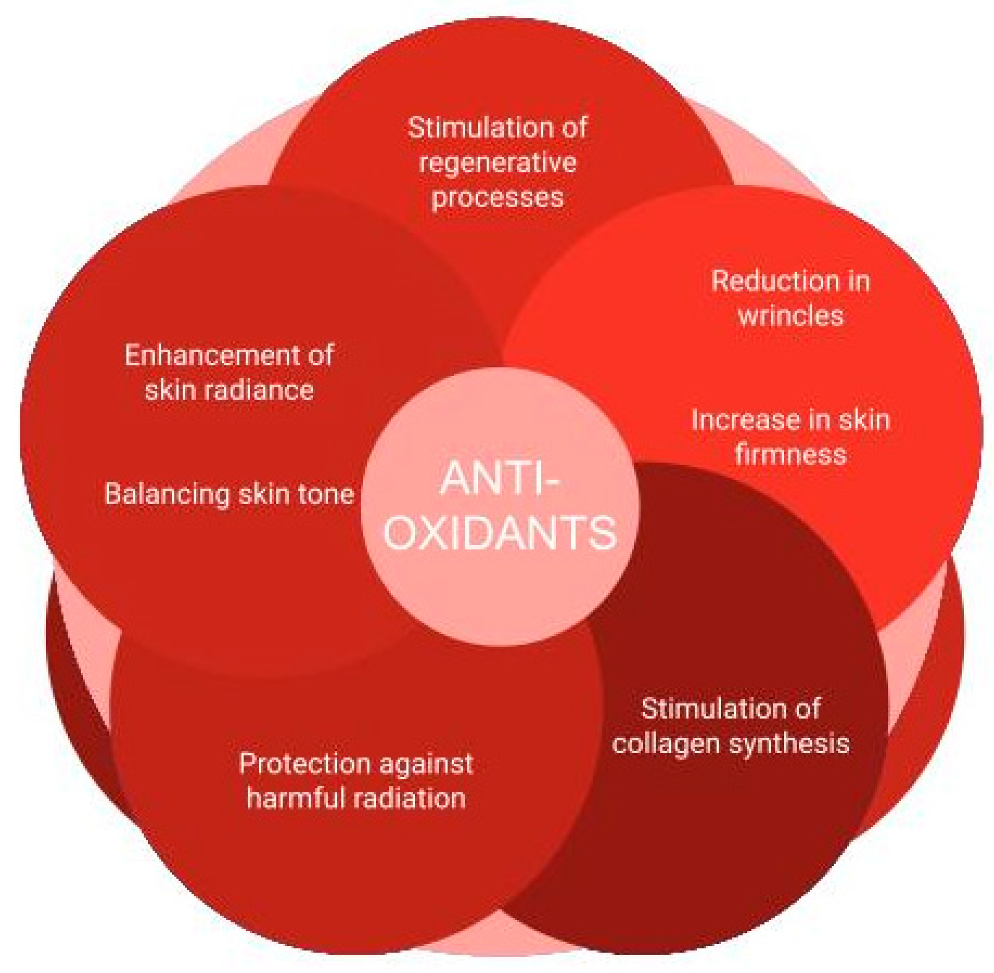
Figure 1. Effects of antioxidants on the skin.
Applsci 15 02571 g001
Figure 2. Chemical structures of antioxidant vitamins: (1) vitamin C, (2) vitamin E, (3) vitamin A, and (4) vitamin B5.
Applsci 15 02571 g002
Figure 3. Chemical structures of antioxidant carotenoids: (5) beta-carotene, (6) lutein, (7) lycopene, (8) astaxanthin, and (9) ubiquinone Q10.
Applsci 15 02571 g003
Figure 4. Chemical structures of antioxidant phenolics: (10) hesperidin, (11) xanthohumol, (12) taxifolin, (13) ferulic acid, (14) resveratrol, and (15) bakuchiol.
Applsci 15 02571 g004
Figure 5. Chemical structures of antioxidant peptides and amino acids, (16) glutathione and (17) N-acetylcysteine, and antioxidant hormones, (18) melatonin and (19) dehydroepitestosterone.
Applsci 15 02571 g005
Figure 6. Chemical structures of some antioxidant compounds specific for various organisms: (20) kojic acid (Fungi, Aspergillus sp.), (21) atranorin (Fungi, lichens), (22) mycosporine–glycine (mycosporine-like amino acid—MAA) (red algae, Rhodophyceae), (23) porphyra-334 (MAA) (red algae, Rhodophyceae), (24) epigallocatechin gallate (Plantae, Camellia sinensis), and (25) aloesin (Plantae, Aloe sp.).
Applsci 15 02571 g006
Table 1. Selected compounds that are used as antioxidants in cosmetology.
Group of IngredientsIngredientClassificationSourceMechanism of ActionEffect in CosmeticsDosageReferences
vitaminsvitamin Cnaturalexogenous
-
reduces reactive oxygen species and oxidized tocopherols
-
involved in the biosynthesis of collagen, serotonin, steroid hormones, dopamine, and noradrenaline
-
product stabilization
-
promotes keratolytic effects
-
prevents skin hyperpigmentation
-
stimulates collagen synthesis
-
exerts anti-inflammatory effects
-
enhances the overall skincare regimen
-
has photoprotective benefits against UV radiation
-
maintains of the stratum corneum
up to 10%[7,23,24,25,26,27,28,29,30,31,32]
vitamin E
-
neutralizes superoxide free radicals (O2)
-
prevents lipid peroxidation
-
reduces mitochondrial superoxide levels and prevents “electron leakage” during oxidative phosphorylation
-
protects epidermal lipids, as well as collagen and elastin fibers, from oxidative damage, thereby preserving skin elasticity and structural integrity
-
supports the skin’s defense against UV-induced oxidative stress
up to 5.4%[32,33,34,35]
vitamin A
-
mitigates oxidative damage within the body
-
enhances gene expression associated with collagen synthesis
-
improves hydration, fortifying the epidermal barrier, and enhances the skin’s resilience against external environmental stressors
-
improves skin tone
0.1–1.0%[36,37,38,39,40,41,42,43,44,45,46,47]
vitamin B5
-
helps mitigate oxidative stress
-
regulates sebum production
-
used in the treatment of conditions such as post-epilation and post-shaving irritation
-
cares of sensitive or atopic skin
-
stimulates hair growth
-
influences pigmentation
-
enhances the overall strength and resilience of hair
up to 5.3%[31,32,48,49,50,51]
carotenoidsβ-carotenenaturalexogenous
-
has antiradical activity
-
has the ability to neutralize singlet oxygen
-
slows down skin aging processes
-
prevents sun damage
-
stimulates melanogenesis
not reported[55,57,58]
lutein
-
potent antioxidant compound against hydroxyl, peroxyl, superoxide anion, and hypochlorous acid
-
used in shielding the skin and eyes from photodamage
not reported[56,60,61]
lycopene
-
neutralizes free radicals by radical addition, hydrogen detachment, or electron transfer
-
used in protective creams against UV rays
-
improves skin texture, density, and thickness
-
significantly increases the elasticity of the stratum corneum
-
improves hydration
-
reduces wrinkles
not reported[55,63,64]
astaxantin
-
counteracts free radicals/ROS by trapping energy (quenching) and through the transfer of electrons or hydrogen abstraction (scavenging)
-
normalizes keratinocyte differentiation
-
affects protein synthesis, cell division and metabolism, the secretion of transcription factors, and growth factors
-
increases the production of collagen and elastin
-
reduces wrinkles and age spots
-
improves elasticity, moisture content, and skin texture
-
provides complete protection against all UVR-related skin problems, from the acute effects of sunburn, discoloration, and redness to the chronic effects of inflammation, photoaging, and skin cancer
not reported[67,68,69,70,71,72,73]
coenzyme Q10
-
plays a crucial role in neutralizing free radicals generated during the citric acid cycle
-
initiates the synthesis of endogenous antioxidants
-
regenerates other antioxidants
-
helps slow the aging process in mature skin
-
reduces inflammation associated with conditions such as acne vulgaris, adult acne, and rosacea and diminishes melanocyte activity to mitigate hyperpigmentation
-
synthesis of collagen, elastin, and glycosaminoglycan fibers
-
reduces wrinkles, enhances skin firmness, and improves hydration
-
providing protection against UVA radiation
not reported[74,75,76,77,78,79]
phenolic compounds/flavonoidshesperidinnaturalexogenous
-
acts as a scavenger against free radicals
-
modulates apoptotic proteins induced by oxidative stress
-
has photoprotective effect absorbing UVB and UVA radiation
not reported[82,83]
xanthohumol
-
reduces lipid peroxidation
-
protects the skin from photoaging
-
brightens existing discolorations
-
reduces skin redness
not reported[84,85,86,87,88]
taxifolin
-
mitigates oxidative DNA damage
-
protects the skin from photoaging
not reported[89,90,91,92,93]
phenolic compounds/phenolic acidsferulic acidnaturalexogenous
-
neutralizes the free radicals generated by UVA radiation
-
improves skin elasticity and firmness
-
maintains uniform color of skin
-
reduces dark spots, hyperpigmentation, and overall skin discoloration
-
reduces fine wrinkles
-
a stabilizer for well-known antioxidants like vitamin C and vitamin E
not reported[94,95,96,97,98,99,100,101,102]
phenolic compoundsresveratrolnaturalexogenous
-
reduces the effects of skin aging
-
protects against damage caused by UV radiation
-
reduces skin hyperpigmentation
-
has a smoothing and elasticizing effect on the skin
not reported[104,105,106,107,108]
bakuchiolnaturalexogenous
-
exerts scavenging activity against various oxidizing radicals (Cl3CO2•, linoleic acid peroxyl radicals, LOO•, DPPH radical, •OH, and glutathiyl radicals)
-
recovers mitochondrial dysfunction from H2O2 treatment
-
protects against ROS damage in mitochondrial DNA
-
activates nuclear factor erythroid 2-related factor 2 (Nrf2), a master regulator that protects cells from oxidative and electrophilic stress
-
may reduce the oxidative stress caused by retinol
-
has retinol-like influence on gene expression but without the upregulation of RAR-β and RAR-γ (retinoic acid receptors beta-1 and gamma-1)
-
efficiently prevents and treats photodamage through its retinol-like functionality and antioxidant properties
-
shows photoprotection synergistic with other antioxidants included in the formulations
-
has anti-inflammatory and antimicrobial properties
-
inhibits melanogenesis
-
causes less irritation compared to retinol
-
very useful for individuals who have sensitive skin and who may not tolerate topical retinoids
0.25–1%[109,110,111,112,113,114,115,116,117,118]
trace elementsseleniumnaturalexogenous
-
occurs in anti-dandruff shampoos and preparations intended for people suffering from seborrheic dermatitis
not reported[120,121,122,123]
zinc
-
may replace redox active molecules
-
induce the synthesis of metallothionein and sulfhydryl-rich proteins that protect against free radicals
-
uses in the treatment of acne
-
effective sunscreen
-
regulates sebum secretion
-
uses against dermatitis as well as dandruff
-
uses in skin cleansing products
not reported[31,124,125,126,127,128,129]
peptides, amino acids, enzymespeptidesnaturalendogenous/non-enzymatic [130,131,132]
glutathione
-
maintains the redox balance of the cell
-
counteracts the effects of oxidative stress
-
skin lightening
not reported[133,134,135,136,137,138,139]
N-acetyl-L-cysteine
-
plays a key role in maintaining the cell’s redox balance
-
y neutralizes reactive oxygen species
-
a therapeutic agent for skin health
-
a preventive measure against UV-related skin cancers
not reported[140,141,142,143,144]
superoxide dismutasenaturalendogenous/enzymatic
-
prevents the reaction of ROS and their derivatives with biological substances
-
interruption of free radical and non-radical oxidation reactions
-
removes the products of the ROS reaction and their derivatives
-
alleviates redness and swelling in the skin
-
provides significant protection against UVB-induced damage
-
helps maintain skin elasticity and smoothness by stabilizing collagen and elastin
-
reduces redness and swelling associated with conditions like rosacea or sunburn
not reported[145,146,147,148,149]
hormonesmelatoninnaturalendogenous/non-enzymatic
-
neutralizes reactive oxygen species
-
eliminates ROS from the cellular environment and metabolize their inactive forms
-
stimulates the synthesis of prooxidant enzymes
-
protects the skin from radiation
-
reduces skin aging
-
supports hair growth
not reported[153,154,155,156,157]
dehydroepiandrosterone (DHEA)
-
protects many tissues from oxidative damage
-
increases the rate of sebum production
-
enhances the brightness of the skin
-
helps combat the papery texture and thinning of the skin
not reported[158,159,160,161]
fungi-derived antioxidantskojic acid naturalexogenous
-
scavenges free radicals
-
chelates pro-oxidant metal ions (e.g., iron)
-
reduces oxidative stress by regulating expression of nuclear factor erythroid-related factor 2
-
reduces ROS and lipid oxidase
-
inhibits melanogenesis (skin depigmenting activity)
-
prevents skin ageing
0.1–2%
up to 1%
[167,246]
Synthetic antioxidantst-Butylated hydroxytoluene (BHT)artificialchemical synthesis
-
quenches all kinds of ROS
-
functions as a chain-breaking antioxidant
-
protects cosmetic formulations from
-
prevents oxidative damage
up to 0.5%[32,162,163,247]
Table 2. Selected species of plants, fungi, and algae that are used as natural sources of antioxidants for cosmetology.
Plant Species/FamilyRaw Material Used/CompoundsActive CompoundsEffect in CosmeticsDosageReferences
Silybum marianum (L.) Gaertn
Asteraceae
seed extract
silibinin
flavonolignans (silymarin), quercetin, thymine, histamine, phytosterols, mucus, tannins, mineral compounds, organic acids, and vitamins C and K
-
mitigates photoaging
-
protects the skin from a variety of toxic chemicals and ultraviolet (UV) radiation
not reported[199,200,201,202,203,204]
Camellia sinensis (L.) Kuntze
Theaceae
leaf extract
seed extract
polyphenols
vitamins, amino acids, fiber and minerals, polyphenols, flavonoids (catechins-epigallocatechin 3-gallate), tannins, and purine alkaloids
-
helps protect and restore vitamin C levels
-
maintains the skin’s structural integrity
-
photoprotective properties
up to 2% (leaf extract), up to 7% (leaf powder), up to 30% (leaf water), up to 0.1% (seed extract)[32,206,207,208,209]
Solanum lycopersicum L.
Solanaceae
pulp
fruit extract
lycopene
vitamins, phenolic compounds such as anthocyanins, phenolic acids, flavonoids and carotenoids, including lycopene
-
maintains skin health
-
has strong anti-aging effects
-
enhances skin lightening and hydration
not reported[210,211,212,213,214,215,216,217]
Citrus limon (L.) Burm.
Citrus x paradisi L.
Citrus reticulata L.
Citrus aurantifolia hort. ex Tanaka
Rutaceae
fruit extract
hesperidin
flavanones, flavanols, flavones, and vitamin C
-
delays the aging processes
-
positively affects acne-prone skin
-
absorbs solar radiation (UVA and UVB)
not reported[218,219,220]
Vitis vinifera L.
Vitaceae
seed extract
fruit powder
juice
juice extract
skin extract
resveratrol
proanthocyanidins, condensed tannins, and leucocyanidins
-
controls UV light-mediated skin aging
-
has skin-lightning properties
up to 3% (leaf extract), up to 2% (fruit extract and juice)[32,220,221]
Humulus lupulus L.
Cannabaceae
hops extract
hops oil
resins, essential oils, proteins and polyphenols: quercetin, quercitrin, kaempferol, rutin, xanthohumol, and ferulic acid
-
ability to address oily scalp and dandruff
-
reduces hair brittleness, enhances nourishment, improves shine, increases strength, and minimizes hair loss
up to 0.2%[32,222,223,224]
Aloe vera (L.) Webb.
Asphodelaceae
flower extract
leaf
leaf extract
leaf juice
leaf polysaccharides
leaf water
flavonoids, terpenoids, lectins, fatty acids, anthraquinones, mono- and polysaccharides, tannins, sterols (notably campesterol and β-sitosterol), enzymes, salicylic acid, essential minerals, and vitamins
-
enhances skin flexibility while reducing its fragility
-
improves skin integrity by promoting moisture retention and mitigating erythema
-
causes a skin-lightening effect
-
improves elastin and collagen production by fibroblasts making the skin more elastic and reducing wrinkles
up to 20%[32,220,225,226,227]
Scutellaria baicalensis Georgi
Lamiaceae
root extractfree flavonoids, flavonoid glycosides, phenylethanoid glycosides, and various other small molecules
-
protects human skin from UV radiation
up to 0.5%[32,229,230,231,232]
Coffea arabica L.
Rubiaceae
leaf extract
seed extract
chlorogenic acid
alkaloids (caffeine, theophylline, theobromine, trigonelline), phenolic acids (chlorogenic acid, caffeic acid), flavonoids (quercetin, rutin, and kaempferol), xanthones, tannins, diterpenes (cafestol and kahweol), carbohydrates, organic acids, amino acids, and fatty acids
-
prevents UV-induced premature skin aging
-
has anticellulite properties
-
maintains skin firmness and elasticity
-
stimulates microcirculation, thus increasing blood flow
-
delivers nutrients and oxygen to the skin through antioxidant properties
not reported[233,248,249,250,251]
Euterpe oleracea Mart. (Açaí)
Arecaceae
pulp powder
juice
unsaturated fatty acids, anthocyanins, proanthocyanidins, other flavonoids (luteolin, quercetin, dihydrokaempferol, and chrysoerial), and carotenoids (carotene, lycopene, astaxanthin, lutein, and zeaxanthin)
-
neutralizes free radicals
up to 3%[32,236,237,238,239,240]
Ganoderma lucidum (Curtis) P. Karst, (linghzi, reishi) Ganodermataceae, Basidiomycota, Fungifruiting-body stipe
stem extract
spores extract
triterpenes (ganoderic acids), water-soluble polysaccharides, proteins, amino acids, mannitol, coumarins, sterols (ergosterol), and unsaturated fatty acids
-
has nutritive, anti-allergenic, anti-acne, anti-inflammatory, anti-photoaging, and anti-bacterial activity
-
functions as an antioxidant, calming, skin lightening, circulation booster, and healing active agent
-
prevents skin from oxidative and cellular damage, and decreases melanin concentration on dark spots
not reported[166,167,172,252,253]
Cetraria islandica (L.) Ach. Parmeliaceae, Ascomycota, Fungi (lichen)thallus extractlichen acids (protolichesterinic acid, protocetraric acid), carotenoids, and polysaccharides (lichenan, isolichenan, galactomannans)
-
acts as an emollient, moisturizing, smoothing, soothing, and cleansing agent
-
acts as an anti-aging agent
-
has anti-oxidant, anti-microbial, immunomodulating, and UV-protecting properties
not reported[254,255,256,257]
Usnea barbata (L.) Weber ex F.H. Wigg. Parmeliaceae, Ascomycota, Fungi (lichen)thallus extactlichen acids (usnic acid), polysaccharides (lichenan), and phenolic acids
-
shows activity against body odor and blemished skin
-
provides long-term protection against dandruff
-
shows excellent anti-microbial activity against Gram-positive bacteria
not reported[177,258,259]
Arthrospira platensis Gomont (spirulina) (blue-green algae)thallus powder
thallus extract
thallus hydrolysate
phycocyanin, proteins (all essential amino acids), minerals, vitamins (B, C, E), trace elements, and unsaturated fatty acids
-
protects the skin from external influences
-
protects the cosmetic product from damage caused by UV light
-
protects from exogenous and endogenous oxidative stress
-
reduces wrinkle formation and improves skin complexion
-
improves moisture balance of the skin and skin complexion
-
useful as a dye in eye shadows (phycocyanin)
not reported[191,260,261,262]
Fucus vesiculosus L. Fucaceae, Phaeophyceae (brown algae)thallus powder
thallus extract
phlorotanins, alginic acid, fucoidan, fucoxanthin, carbohydrates, and iodine
-
has moisturizing and re-mineralizing activity to the skin
-
exerts powerful antioxidant activity, making it useful for brightening and anti-ageing applications
-
has skin brightening properties, and thus significantly increases radiance and reduces the pigmentation of age spots
0.00002–5%[32,185,191,195]
Laminaria digitata (Huds.) Lamouroux, Laminariaceae, Phaeophyceae (brown algae)thallus powder
thallus extract
alginic acid, fucoidan, carbohydrates (laminarin), iodine, and γ-linolenic acid
-
acts as a moisturizing, regenerating, cutaneous-protecting, and sebum-regulating agent
-
acts as a remineralizing and anti-stress agent in hair care and skincare
0.00004–5% (extract),
40% (powder)
[32,185,195,263,264]
Haematococcus pluvialis Haematococcaceae, Chlorophyta (green algae)thalluscarotenoids (astaxanthin), lipids (polyunsaturated fatty acids), proteins, and carbohydrates
-
inhibits reactions promoted by oxygen, thus avoiding the oxidation and deterioration of ingredients
-
acts as an antioxidant and anti-aging agent
-
improves skin’s moisture content
-
counters UV-induced skin damage through the UVA and UVB protection of skin cells
not reported[191,196,262]
Chlorella vulgaris Beijer. Chlorellaceae, Chlorophyta (green algae)thallus extractMAAs, proteins, lipids, carbohydrates, vitamins, chlorophyll, and carotenoids
-
keeps skin in good condition
-
heals and repairs skin
-
prevents wrinkle formation
not reported[191,194,262,265]
Porphyra umbilicalis (L.) Kützing Bangiaceae, Rhodophyta (red algae)thallus extractmycosporine-like amino acids (MAAs, including porphyra-334), phenolic compounds (phlorotannin and taurine), vitamins (ascorbic acid), polysaccharides (porphyrans), and phycobiliproteins (phycoerythrin and phycocyanin)
-
protects cell metabolism, DNA, and cell membranes against UVA irradiation
-
prevents the formation of sunburn cells
-
useful as a dye for eye shadows (phycoerythrin)
not reported[181,191,198,266,267]
Disclaimer/Publisher’s Note: The statements, opinions and data contained in all publications are solely those of the individual author(s) and contributor(s) and not of MDPI and/or the editor(s). MDPI and/or the editor(s) disclaim responsibility for any injury to people or property resulting from any ideas, methods, instructions or products referred to in the content.