Article

Preparation, Characterization and Evaluation of the Antibacterial Activity of Ag Nanoparticles Embedded in Transparent Oxide Matrices

by
Cristina-Ștefania Gălbau
1,2,
Mihaela Idomir
1,2,
Cătălin Vițelaru
3,*,
Adrian Emil Kiss
3,
Anca Constantina Parau
3,
Lidia Ruxandra Constantin
3,
Mihaela Dinu
3,
Iulian Pana
3,
Alina Vlădescu (Dragomir)
3,
Elena Laura Gaman
4,
Marius Alexandru Moga
5,
Cătălin Mișarcă
5,
Mihai Vârciu
1,2,
Claudia Alexandrina Irimie
1 and
Mihaela Badea
1,2
1
Department of Fundamental, Prophylactic and Clinical Disciplines, Faculty of Medicine, Transilvania University of Brasov, 56 Nicolae Balcescu, 500019 Brasov, Romania
2
Research Center for Fundamental Research and Prevention Strategies in Medicine, Research and Development Institute, Transilvania University of Brasov, 10 Institutului, St., 500484 Brasov, Romania
3
National Institute for Research and Development in Optoelectronics INOE 2000, 409 Atomistilor St., 077125 Magurele, Romania
4
Department of Biochemistry, Carol Davila University of Medicine and Pharmacy, 8 Eroilor Sanitari Bdv., 020021 Bucharest, Romania
5
Department of Medical and Surgical Disciplines, Faculty of Medicine, Transilvania University of Brasov, 56 Nicolae Balcescu, 500019 Brasov, Romania
*
Author to whom correspondence should be addressed.
Appl. Sci. 2025, 15(5), 2599; https://doi.org/10.3390/app15052599
Submission received: 23 January 2025 / Revised: 24 February 2025 / Accepted: 26 February 2025 / Published: 27 February 2025
(This article belongs to the Section Nanotechnology and Applied Nanosciences)

Abstract

:
Daily exposure to contaminated environments and surfaces leads to serious health issues, increasing healthcare costs. Active materials that act against pathogens can effectively prevent their proliferation and contribute to increased protection against infections. In this contribution, nanostructured thin films containing silver are investigated, using SiO2 and TiO2 as transparent matrices to embed the Ag atoms. The thin transparent films were obtained via magnetron sputtering, using HiPIMS for Ag deposition and RF sputtering for oxides, in either an Ar or Ar/O2 environment. Atomic Force Microscopy provided information on coating topography and the thin films’ preferential growth on the textured polymer foil, X-Ray Diffraction highlighted the structural difference between different versions, Ultraviolet–Visible–Near-Infrared spectroscopy proved the thin films’ optical quality and their transparency and Energy-Dispersive X-ray Spectroscopy revealed the composition changes for different processes. The effect of O2 addition is analyzed and compared in terms of changes induced on the properties of the thin films. Following 24 h of incubation in a media containing 104 CFU/mL Escherichia coli, the TiO2+Ag sample with O2 addition showed the highest antibacterial effectiveness, as indicated by the largest inhibition zone. Experiments on selective media showed a hierarchy of efficiency, namely, TiO2+Ag+O2 > TiO2+Ag > SiO2+Ag.

1. Introduction

Biomolecules like bacteria, cells, proteins and polysaccharides are constantly adhering to device surfaces because such surfaces benefit them commensally. These biological components assemble into a biofilm on the surface of the device, shielding germs from being eliminated. Biofilm is typically 100–1000 times more resilient than planktonic microorganisms. Because patients need to take larger doses of medication to treat the illness, antibiotic resistance is now considered one of the main concerns related to human health [1]. Patients now face a significant danger of contracting severe and even fatal infections frequently due to the spread of multi-drug resistance bacteria [2] that contaminate medical supplies and equipment [3]. The bacterial structure needs to be understood to grasp the antibacterial process better. Certain bacteria contain unique features such as capsules, flagella, pili and spores [4]. The cytoplasmic membranes of bacteria, which are phospholipid bilayers containing functional proteins, are present in Gram-positive and Gram-negative bacteria. The structure of bacteria is more negatively charged than human cells [5]. The Gram-negative bacteria have extra outside membranes in addition to the cytoplasmic membrane, which is made of anionic lipopolysaccharide molecules joined by ionic bridges between the phosphate groups and divalent ions. As a result, Gram-negative bacteria have very durable outer membranes that operate as permeabilizing barriers to most big hydrophobicity molecules [6,7]. The antimicrobial effects of Ag+ ions or Ag particles also involve interactions with biological macromolecules like enzymes and DNA through mechanisms such as electron transfer or the generation of free radicals. Additionally, alterations in protein synthesis and cell wall formation, which can be observed through the accumulation of protein precursors or the disruption of the outer cellular membrane leading to ATP leakage, are also recognized as key factors contributing to the antimicrobial activity of Ag particles [8]. Making an antimicrobial coating for medical equipment is the easiest way to avoid contamination from microorganisms [9]. It is advantageous to prevent the development of drug-resistant pathogens by minimizing medicine administration, which reduces environmental pollution due to antibiotic leaching [10]. So that microbial-resistant medical devices and equipment may be offered to hospitals and patients, it is vital to produce antimicrobial materials that function as antibiotics and without drug resistance [6]. Coatings designed for use on hospital surfaces and fomites must possess several key characteristics: they should be non-toxic to both the environment and humans, affordable, readily available, effective against harmful pathogens, resistant to contamination and maintain long-term stability and durability [11].
Among the most investigated solutions as an antimicrobial agent, one can find a wide use of silver [12], a known antibacterial agent that can disrupt bacterial cell membranes. The efficiency depends greatly on the material’s properties that contain Ag and the availability of the Ag ions [13,14]. The use of nanoparticles [15] or nanomaterials that embed Ag in their structure [16] is of great interest. Various techniques have been employed for the synthesis of various materials [17], using chemical path synthesis such as CVD [18], sol–gel [19], dip coating [20], spin coating [21], etc. Among the physical methods, physical vapor deposition techniques are the most widely used, comprising methods including magnetron sputtering [22], thermal evaporation [23] and pulsed laser deposition [24]. These techniques have the advantage of providing very good control over the thickness of the coating and the materials’ properties. The silver nanoparticles can be trapped inside a matrix made of different materials (TiO2 [18], ZnO [25], SiO2 [26,27], SiO2+TiO2 mixtures [28], etc.), providing both mechanical protection and a controlled release of active elements when used as an antibacterial coating, which is important also in medical implants [29,30]. Additionally, silver can be used in conjunction with other elements, such as Ti [31], Cu [32], O [33], etc., to provide improved antibacterial effect. Different versions of magnetron sputtering, such as DC (Direct Current) sputtering [22], RF (Radio Frequency) sputtering [34] and HiPIMS [26] have been used to produce silver-containing coatings. It is known that the presence of O2 plays a crucial role in the strong antibacterial activity of the TiO2+Ag+O2 system by a synergetic contribution in both improving the photocatalytic activity of TiO2 and further enhancing antibacterial efficiency by influencing the structural state of AgNPs and triggering Ag ion release [35]. As documented by numerous studies, O2 prevents electron-hole recombination when it comes to UV/Visible light exposure of TiO2 and contributes to reactive oxygen species formation, resulting in very high oxidative stress and bacterial cell membrane disruption [36,37,38]. Additionally, O2 exposure can partially oxidize the AgNPs [39] and lead to a more stable dispersed state of AgNPs, preventing the formation of nanoclusters and triggering a strong antimicrobial effect by Interacting with bacterial cell walls, protein denaturation, blocking DNA replication, transcription processes alteration, etc. [40,41,42,43,44].
In our contribution, we focus on the deposition of Ag-containing coatings using a combination of HIPIMS and RF sputtering in a reactive gas environment. The addition of O2 to the deposition process is evaluated as a solution in terms of the enhanced antibacterial activity and overall quality of the thin film. The targeted application refers to obtaining antibacterial coatings on thin, transparent polymer foils intended to be used as protective foils for mobile devices, with particular interest in the devices and screens used in medical environments, as described in our previous work [26,27].

2. Materials and Methods

2.1. Synthesis and Characterization of Films

Magnetron sputtering was used to deposit thin films on transparent polymer foils, using a confocal configuration with 3 targets of 1-inch diameter [26]. The use of confocal geometry allows one to co-deposit 2 materials from two or more different targets and mix them at the same time to obtain nanocomposite material. For the deposition of thin films on self-adhesive polyurethane foils, we used silicon oxide, titanium oxide and silver targets. Both oxide targets were operated in RF sputtering conditions at 50W applied power. The deposition was performed in argon and argon/oxygen gas mixture at 6 mTorr pressure. The typical deposition time was 30 min., yielding 30 to 35 nm film thicknesses. From this total thickness, it is estimated that only ~10% corresponds to Ag, while the other 90% corresponds to the total thickness of the oxide. HIPIMS (high-power impulse magnetron sputtering) was used to sputter the Ag target, enabling fine-tuning of the deposition rate. The pulse characteristics selected for the deposition of Ag were the following: pulse voltage of 650 V, peak current of 1.5 A, pulse duration of 50 μs and repetition frequency of 1 Hz, with the deposition conditions being the same as in our previous work [26]. Let us note that the main parameter that controls the quantity of Ag in the thin films is the pulsed character of the Ag cathode, enabling fine control of the Ag quantity by tuning the repetition frequency. In the current study, our efforts were focused on improving the quality of the Ag+TiO2 layer as it was previously shown that the Ag+SiO2 layer has good antibacterial properties and good optical transparency [26]. According to our previous results, the Ag+TiO2 layers proved poor transparency in the visible spectral range, one possible cause being the quantity of oxygen in the layer. To compensate for this, the TiO2 layer was optimized in the current study by adding a small quantity of oxygen to the gas mixture in the deposition process. Initial tests were made with oxygen gas flows between 1 and 7.5% of the total gas flow. It was noted that the oxygen flow should be limited to only a few percent of the total, further leading to a significant decrease in the deposition rate. The most advantageous of the investigated gas flows was found to be at 2.5%, with a deposition rate of only 30% smaller compared with the Ar sputtering process. The deposition time was adjusted accordingly so that the total thickness of the layer remains comparable with the one previously used, around 30 nm. The optical, structural and morphological properties were further analyzed and compared for the different layers obtained.
The topography of thin films was analyzed based on atomic force microscope (AFM) images acquired with the AFM/STM Microscopy System (INNOVA VEECO, Berlin, Germany) working in taping mode. The elemental composition of the coatings was determined by Energy-Dispersive X-Ray Spectroscopy (EDS), performed on the scanning electron microscope (SEM, Hitachi TM3030 Plus Tabletop Microscope, Tokyo, Japan) coupled with EDS (FEI Inspects equipment). The EDS spectra were registered for a duration of at least 10 min each in 3 different areas of the samples. A Jasco V-670 double-beam spectrophotometer was utilized to record the transmission and reflectivity spectra of the TiO2+Ag and SiO2+Ag coatings. The optical characteristics of layers were examined using a Jasco V-670 spectrophotometer (Jasco, Tokyo, Japan) within the Ultraviolet–Visible–Near-Infrared Spectrum. The spectrophotometer was equipped with an ARSN-733 auxiliary module (Jasco, Tokyo, Japan) with a concave mirror and a 60 mm diameter integrating sphere. According to the manufacturer’s specifications for the integrating sphere, the potential relative errors for absolute reflectivity measurements are ±1.5% for 5° incidence angles. The device operated at a scan rate of 400 nm/min., enabling an investigation spectral range from 250 to 2000 nm. Grazing Incidence X-Ray Diffraction (GIXRD) measurements were performed using a Cu rotating 9 kW anode Smart Lab diffractometer (RIGAKU, Tokyo, Japan). To investigate the crystalline structure of coated transparent polymer foils, scans were recorded using Cu Kα radiation (λ = 0.154056 nm) from 10° to 100°, at an incidence angle of 1°, with a scanning speed of 3°/min. For extracting the grain sizes, Scherrer Equation was used by considering the measured maxima of Ag (111).

2.2. Antibacterial Efficiency Tests

The tested samples were deposited with SiO2+Ag, TiO2+Ag and TiO2+Ag+O2, and the reference samples were marked with REF. The bacterial strain used for these tests was Escherichia coli (ATCC 25922). The experiment occurred in the microbiological hood (Alpina model with laminar flow). It included two UV radiation sterilization processes: sterilization of the working environment—of the microbiological hood—and sterilization of the samples for 15 min on each side. Using a densitometer, a stock suspension of Escherichia coli of 0.5 McFarland equivalent to 1.5∙108 CFU/mL was prepared [27]. Serial dilutions of concentrations 107, 2∙106, 106, 2∙105, 105, 2∙104 and 104 CFU/mL were made from this stock suspension. One millileter of each bacterial suspension of different concentrations was added evenly over the culture medium (MC Media Pad E. coli and Coliforms, Merck, Darmstadt, Germany). Positive controls (made for each concentration of bacteria, without samples) and negative controls were inoculated with the nutrient broth used to make the bacterial suspensions [45,46,47]. Before sealing the culture medium, the foils were added with the active part in contact with the Escherichia coli suspension. The medium was sealed to prevent dehydration of the medium and the bacterium, respectively, and left for 15 min in the microbiological hood. For all experimental conditions, it worked in duplicate. After 15 min, the media were incubated at 37 °C for 24 h. The results were read after 24 h, and the diameter of bacterial growth inhibition was determined using ImageJ version 2021 software. All the following steps are summarized in Figure 1.

3. Results

3.1. Characterization of Physical Properties

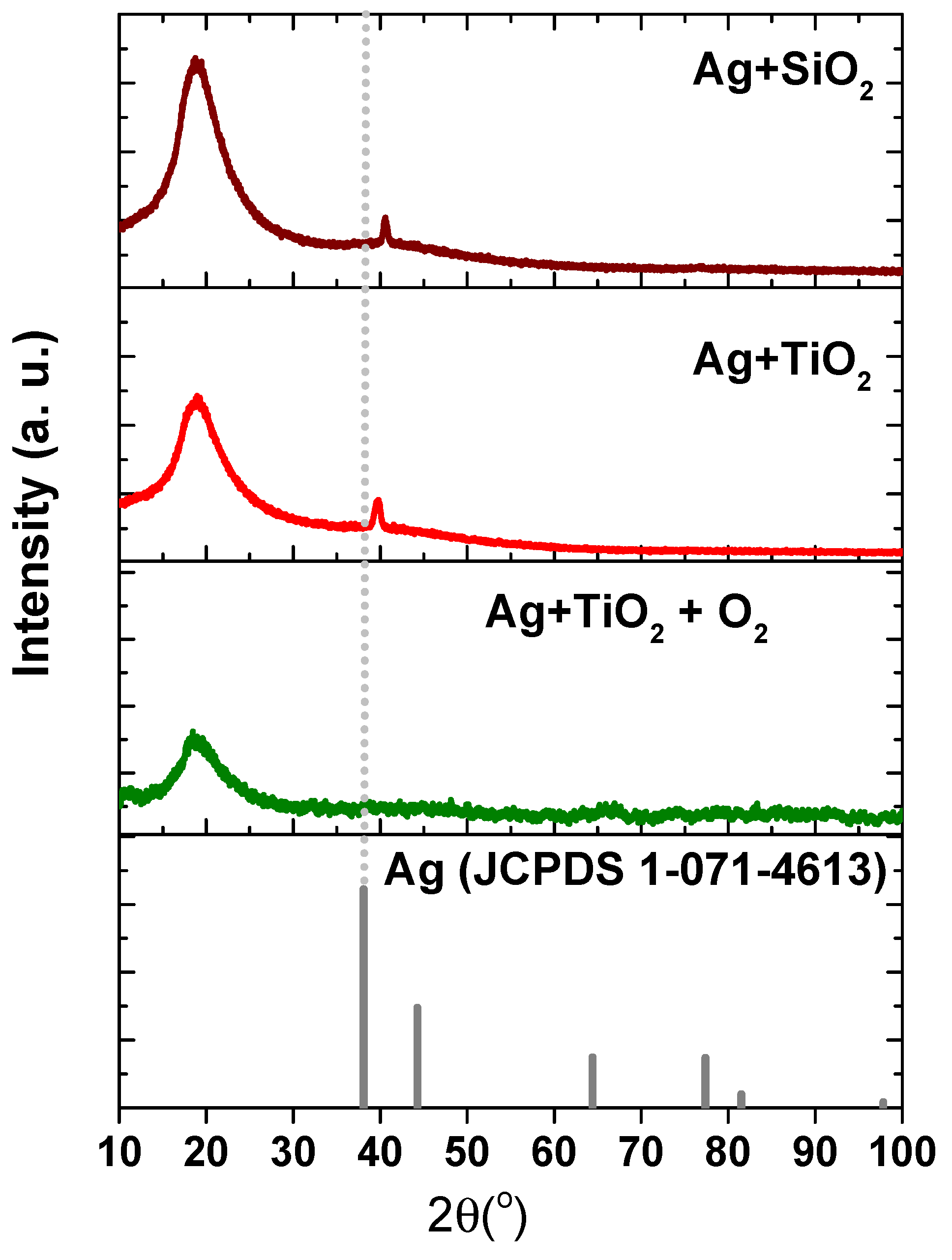
3.1.1. X-Ray Diffraction for Structural Analysis of the Films

Figure 2 shows the XRD patterns recorded for coated transparent polymer foils. As observed, in the case of Ag-based SiO2 or TiO2 matrix, respectively, there is an evidence of crystalline Ag phase formation (JCDPS No. 1-071-4613). The cubic structure exhibited a (111) preferred orientation and can be identified at the 2θ position of 40.57° in Ag+SiO2 thin film, whereas in Ag+TiO2, the silver reflection is located at 39.76°. As calculated, the grains exhibited nanometric sizes in the range of ~12–17 nm. Similar data were found [47], where they investigated magnetron-sputtered Ag:TiO2 layers. Based on TEM cross-section micrographs of as-deposited samples [47], Ag aggregates were found to be randomly distributed in the amorphous matrix and have various shapes with nanometer sizes of lateral diameters (nano-clusters). According to the mentioned standard, the position of the Ag maximum peak oriented along the (111) plane is 38.1°. Therefore, a shift towards higher angles is evident in both measured patterns, suggesting a partial oxidation due to the oxidic character of SiO2 and TiO2. The XRD analysis showed no evidence of crystallinity for any of these compounds. As previously reported, an amorphous structure can accelerate the diffusion process and facilitate the formation of larger Ag particles, the deposited layer having a heterogeneous structure [48].
Regarding the Ag+TiO2+O2-coated foils, the addition of 2.5% of oxygen in the total gas flow had a direct influence on the structural evolution, Ag being finely dispersed in the dielectric matrix. The hypothesis can be proven by the absence of the silver characteristic peak in the XRD-associated patterns, the dispersion process of the metal particles representing a consequence of the energetic impact of the oxygen ions during sputtering [49]. In this case, the only signal recorded was observed at 18.53°, being characteristic of the foil substrate.

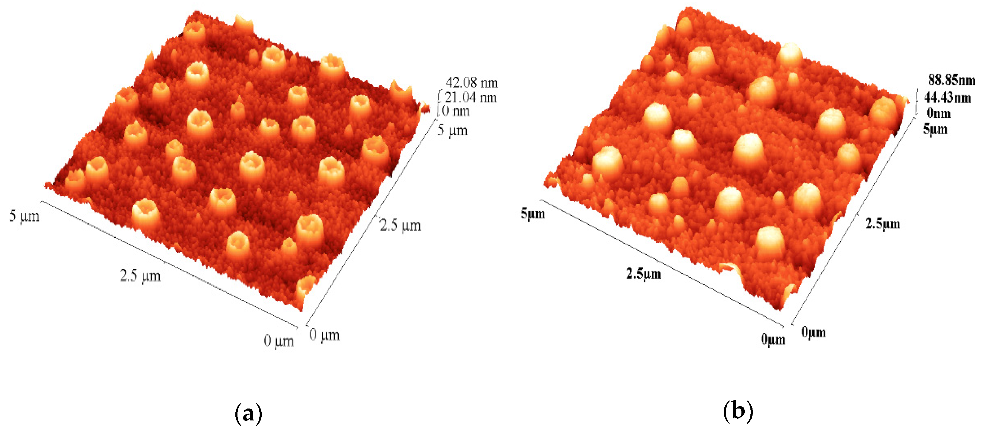
3.1.2. Atomic Force Microscopy—Surface, Topography and Roughness

Tapping scans were performed with a (INNOVA VEECO, Berlin, Germany) atomic force microscope (AFM) on a surface area of 5 × 5 µm2, respectively, with a scan rate of 0.3 Hz and a resolution of 512 pixels, using an RTESPA tip. From the surface profile images of the layers obtained from the TiO2 and Ag target, with and without oxygen addition, it can be observed that the morphology characteristics of the substrates are kept, as shown in Figure 3. Thus, surface texturing corresponding to the initial topography can be seen, forming structures with a typical size of ~500 nm.

3.1.3. Energy Dispersive X-Ray Spectroscopy, EDS and Elemental Composition

The characteristic EDS spectra for the layers obtained in different deposition conditions are represented in Figure 4. From the analysis of these spectra, it can be observed that Ag is present in all types of processes, with comparable intensities in the spectrum for processes without adding oxygen. In the process of adding oxygen, there is an increase in the intensity of Ag and a decrease in the intensity of Ti, indicating a significant change in the ratio between their concentrations in the layer composition.
The inset of Figure 4 indicates a different mode to visualize the evolution of the deposited layers composition, representing the evolution of the ratio between Ag and the main element in the oxide matrix, Si or Ti, as well as the ratio between Ag and O. From this representation, an increase in these ratios can be observed when moving from SiO2 to the TiO2 matrix, as well as when moving from the process without oxygen to the one with addition of oxygen. In the same figure, the evolution of Ag concentration is represented, being also observed that it is higher for the process in which a TiO2 target is used compared to those with a SiO2 target, respectively, for the process in which an Ar+O2 mixture is used compared to the one using only Ar. In conclusion, the layer with the largest amount of incorporated Ag, at the same total thickness, is obtained in the optimized process with the oxygen content of 2.5% of the total gas flow.

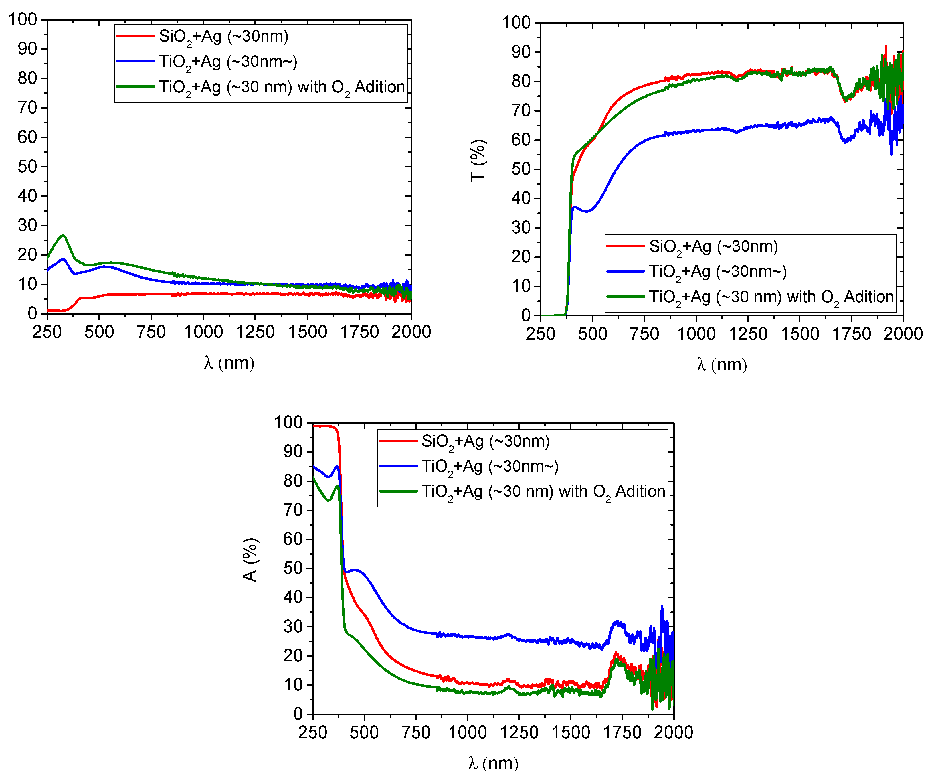
3.1.4. UV–Vis–NIR Spectrophotometry and Optical Properties

The advancement of mobile device technology demands comprehensive insight into the optical properties of protective layers, pointing to the importance of oxide selection and processing conditions in advanced display technologies. Silicon dioxide has been highlighted for its potential to maintain cell selectivity, especially regarding the stability of PEG films and suppressing cell adhesion [50]. Incorporating metals like silver into oxides to enhance their optical properties is an emerging research area [51,52,53]. When applied to protective films, Ag-doped semi-transparent layers play an important role in ensuring the optimal performance of device displays, especially regarding clarity and durability. The optical properties of TiO2+Ag and SiO2+Ag coatings were evaluated by spectrophotometric measurements, Figure 5, emphasizing their significance in designing and applying layers for mobile device displays.
Initial evaluations indicated a pronounced transparency for the SiO2 layers in the visible range, substantially higher (~20%) than the one corresponding to the TiO2 layer without O2 addition. In contrast, when the layers were synthesized using a stoichiometric TiO2 target in a reactive gas mixture of Ar and O2, TiO2 layers exhibited a comparable transparency to SiO2 layers. Further spectral analysis did not identify a unique absorption peak intrinsically linked to the surface plasmonic resonance phenomenon, attributable to the Ag nanoparticles. This suggests a fine distribution of silver within the oxide structure, preventing the formation of metallic nanoclusters. These results support our initial hypothesis drawn from X-Ray Diffraction analysis, further underlining the conclusion that silver remains finely dispersed within the oxide matrix rather than forming distinct crystalline clusters [47]. This finding also aligns with previous studies that have demonstrated the attenuation or absence of the Ag (111) diffraction peak in Ag-doped amorphous TiO2 or SiO2 matrices due to Ag incorporation at the atomic level or in sub-nanometric forms [54]. Our observations, where the absence of a characteristic Ag diffraction peak in Ag+TiO2+O2 coatings indicates that the deposition conditions mostly imply Ag dispersion rather than aggregation, provide further consistency with these previous studies.
In general, the size and shape of Ag nanoparticles also influence the optical properties, leading to an oxidation-driven stabilization of Ag species within the amorphous matrix [55]. This process can prevent Ag atoms from diffusing and clustering into larger particles, which XRD analyses would otherwise confirm. Such effects have been observed in reactive sputtering processes, where oxygen-rich conditions promote finer Ag distribution and suppress long-range crystallinity [56]. Moreover, the structural disorder in amorphous oxides enhances atomic mobility, influencing metal dispersion. Previous studies showed that the lack of distinct IR spectral features associated with silver in Ag-doped TiO2 suggests that silver atoms are incorporated within the TiO2 matrix, likely occupying interstitial sites rather than forming separate metallic clusters [57]. This is consistent with the combined results of XRD and UV–Vis–NIR spectrophotometry, where the combination of transparency, amorphization and the absence of surface plasmonic resonance features point to the effective integration of Ag within the matrix at the atomic scale rather than in clustered form.
It has to be noted also that a higher amount of Ag is still evident in the layer obtained with O2 addition, this being highlighted both by the enhanced reflection of this layer and further validated by EDS measurements, as previously shown.

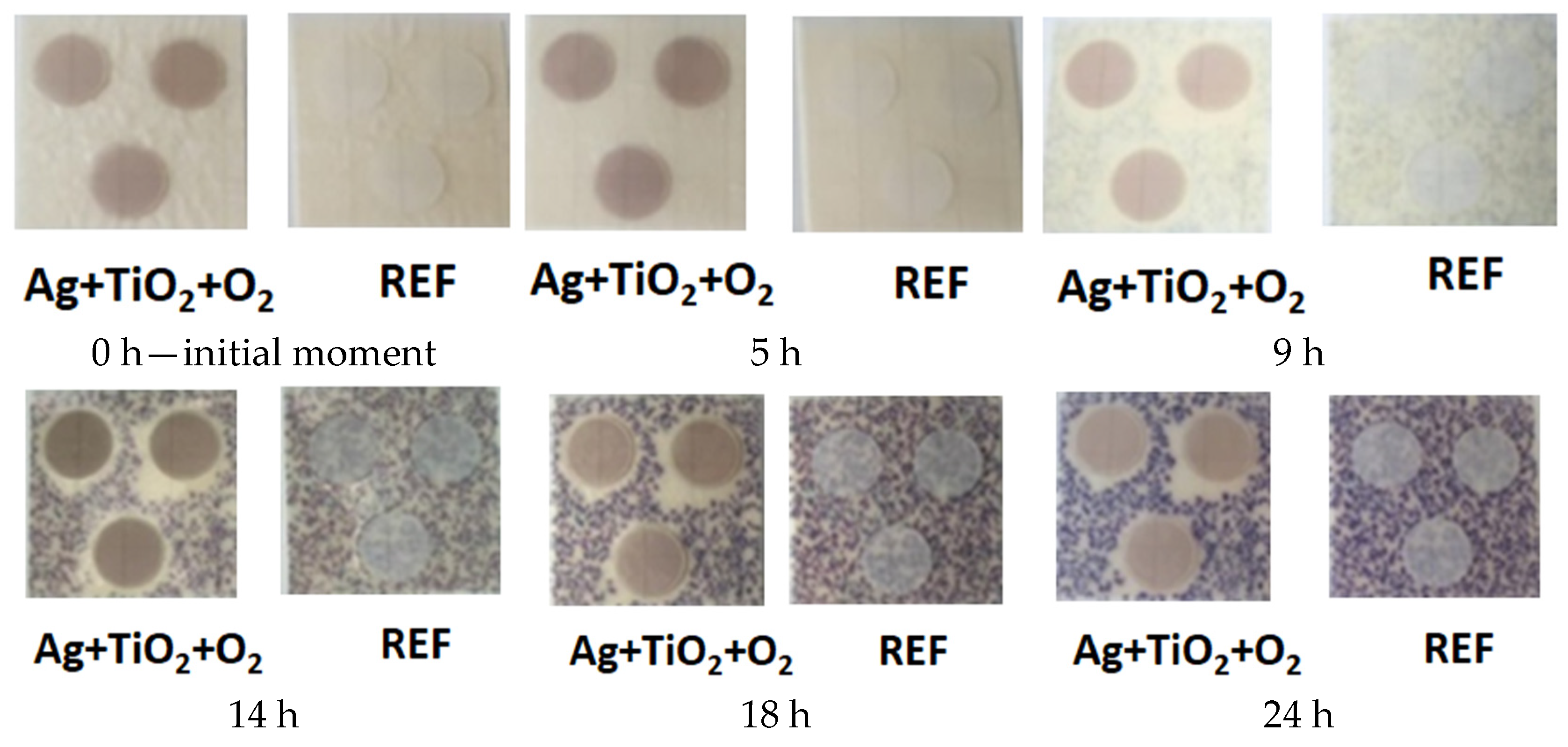
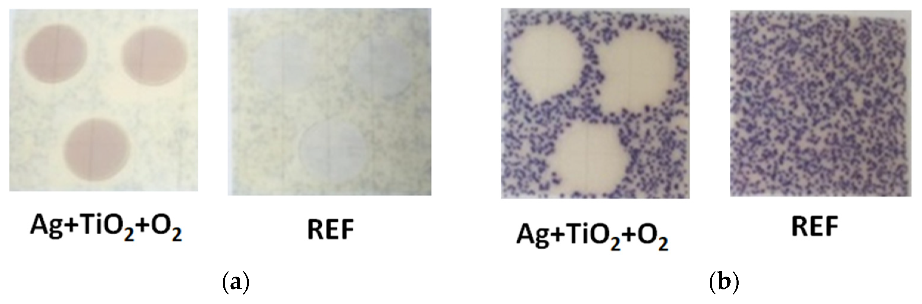
3.2. Antibacterial Activity

Because of their large active surface area, which enables them to readily pierce biofilms, these nanoparticles (NPs) are potent antibacterial agents. Certain metals, including copper (Cu) and silver (Ag), were used as antibacterial agents even before the invention of pharmacological antibiotics. A new class of “nano-metal-antibiotics” were developed due to continued progress in this area, which involves investigating different NPs for their antibacterial qualities [58]. Medical research has seen a paradigm change concerning the creation of metal-based antibacterial drugs since the 19th century when it was discovered that the evolution of illnesses and infections was directly linked [58]. Acinetobacter baumannii, Pseudomonas aeruginosa, Klebsiella pneumoniae and Escherichia coli are Gram-negative bacterial species that often cause hospital infections. Methicillin-resistant Staphylococcus aureus (MRSA) and Enterococcus species are often implicated among Gram-positive bacteria [27]. For our study, we chose to test the sample’s efficiency on Escherichia coli. For all concentrations of Escherichia coli, inoculations were carried out by the flooding method on the culture medium specific for Escherichia coli. The method of making bacterial suspensions of different concentrations was carried out by the serial dilution technique, a fact demonstrated by the differentiation of the number of colonies.
To demonstrate that the bacterium can grow according to and under the deposited material, Escherichia coli being an aerobic, facultatively anaerobic bacterium [59,60], a sample was placed with the non-deposited side in contact with the bacterial suspension.
Using ImageJ version 2021 software, the zones of the inhibition of Escherichia coli growth were determined, and the diameters in Table 1 were calculated.
From the concentration of 105 CFU/mL to the concentration of 107 CFU/mL, the colonies become uncountable. From concentrations higher than 106 CFU/mL, the high consumption of β-galactosidase (compound incorporated in the culture medium) is observed, the medium becoming a green–blue color. The following samples deposited with TiO2+Ag, SiO2+Ag were tested for the same bacteria concentrations, as well as non-deposited samples to have the original material as a reference.
In the case of concentrations higher than 106 CFU/mL, it is observed that the bacterium continues to develop under the deposited film so the composition of the films partially inhibits (only in the central area of the film) the development of Escherichia coli. From the concentration of 2 × 10⁵ CFU/mL to 104 CFU/mL, the area where the foil was highlighted—under Escherichia coli—did not develop. For both types of samples, SiO2+Ag and TiO2+Ag, the formation of a halo around the samples is observed, highlighting the antibacterial character of both samples.
In the case of non-deposited samples, bacterial growth is also observed under the sample both at concentrations of 107 CFU/mL and at concentrations of 104 CFU/mL, thus resulting in the fact that the reference sample has no bacterial activity.
For the sample with TiO2+Ag+O2, a duplicate test was performed for Escherichia coli at a concentration of 104 CFU/mL (Figure 6).
TiO2 is one of the semiconductors most often employed for antibacterial applications in modern literature because of its biocompatibility. Thus, this research focuses on a few selected metallic or oxide nanoparticles (NPs) (copper, silver and titanium dioxide (TiO2)) that are excellent and economical in offering antimicrobial qualities via functional coatings. Important synthetic, natural and biodegradable antimicrobial polymers that exhibit a fast inactivation of a wide range of bacteria and have established functional features are carefully considered [58].

3.3. Mechanism of Action of Antibacterial Properties

Using the TiO2+Ag+O2 material, tests were carried out to describe the mechanism of action of the material deposited on the foils—i.e., the inhibition of development in the respective area or antibacterial effect at a specific time in the development of Escherichia coli. Thus, the described protocol was followed, but the samples were photographed at the initial moment of the inoculation of the medium (time 0) at 1 h, 2, 3, 4, 5, 9, 10, 11, 12, 13, 14, 15, 17, 18 and 24 h of incubation at 37 °C (Figure 7).
By preventing or regulating the microbial biofilm architecture of surfaces, antifouling coatings inhibit biofilm deposition on surfaces [9]. Antimicrobial coatings, on the other hand, only function by means of bacteriostatic or bactericidal action [9]. The latter exploits the method of contact killing or releases the antibacterial chemicals or inorganic metal ions that induce cell death, while the former is based on the mechanism of steric repulsion or nanoscale-based topographies. The polymers are classified as either active (killing) or passive (repelling) materials based on their antibacterial activity and associated mechanism [61]. This test shows that bacterial development between 0 and 5 h does not occur, but bacterial cultures appear between 5 h and 9 h of incubation. However, from the first 9 h of incubation at 37 °C, bacterial cultures do not develop around and under the film deposited with TiO2+Ag+O2 (Figure 8a). This trend is maintained during the 24 h of incubation at 37 °C—no Escherichia coli colonies appeared around and under the TiO2+Ag+O2 film (Figure 8b).
The diameter of the zone of inhibition after the 24 h of incubation of the media at 37 °C was 1.841 ± 0.142 for the sample deposited with TiO2+Ag+O2, while the non-deposited sample did not register bacterial activity. The low surface energy of the matrix and hydrophilic/hydrophobic and electrostatic repulsions are the primary sources of the “repelling”-based process. Electrostatic and biocidal interactions are the foundation of the “killing”-based process [61]. Gram-negative bacteria have a thick layer of lipopolysaccharide in their cell walls, with a relatively thin layer of peptidoglycan; this thick peptidoglycan structure of the cell wall can limit the penetration of AgNPs into the cells. The internalization of AgNPs is vital for their antibacterial activity for this type of bacteria [43].

4. Conclusions

Three experimental models of the layers with antimicrobial potential were obtained and characterized: two reproducing the properties obtained in the previous research and one improved, with high transparency and higher silver content. The layers’ thickness was 30–35 nm, with an Ag content equivalent to a thickness of 3 nm, and approximately 10% of the total and the topographic characteristics of the foil surface are preserved after the deposition of thin layers. Analyzing X-ray diffractometry spectra and spectrophotometric measurements indicates the presence of metallic Ag nanoclusters embedded in the polymer matrix for deposition processes without oxygen and the absence of such structures for the optimized process with the addition of oxygen. The presence of silver within the layer was also proved by EDX measurements, highlighting an increase in its concentration when a sputtering process with added oxygen is used. Using the ready-to-use MC Media Pad E. coli and Coliform culture media is effective for evaluating the antibacterial activity of the layers deposited on the foils and evaluating the antibacterial properties of the samples; the maximum concentration for possible applications (countable colonies) for the PAD media is 104 CFU/mL. The highest antibacterial efficiency (expressed in cm—zone of inhibition of development) was recorded for the film deposited with TiO2+Ag+O2. Comparing the three types of foils, an efficiency of TiO2+Ag+O2 > TiO2+Ag > SiO2+Ag was recorded for the tests performed on the PAD media.

Author Contributions

Conceptualization, C.-Ș.G., M.B. and C.V.; methodology, C.-Ș.G., M.B., M.I., A.C.P., A.E.K. and M.V.; software, C.-Ș.G., M.B., C.A.I., C.M. and I.P.; validation, C.-Ș.G., M.B., M.I, L.R.C. and M.D.; formal analysis, C.-Ș.G., M.B., M.V., C.V., C.A.I. and C.M.; investigation, C.-Ș.G., M.B., M.I., M.D., I.P., A.C.P. and C.M.; resources, C.-Ș.G., M.B. and A.V.; data curation, C.-Ș.G., M.B., A.C.P. and C.A.I.; writing—original draft preparation, C.-Ș.G., M.B., C.V., C.M. and C.A.I.; writing—review and editing, C.-Ș.G., M.B., E.L.G., M.A.M., A.V. and M.V.; visualization, all authors; supervision, M.B. and C.V.; project administration, C.-Ș.G., M.B., C.V. and L.R.C.; funding acquisition, C.-Ș.G., M.B., M.A.M. and L.R.C. All authors have read and agreed to the published version of the manuscript.

Funding

This research was funded by the Romanian Ministry of Research, Innovation and Digitization, CCCDI-UEFISCDI, project number PN-III-P2-2.1-PED-2019-4966, within PNCDI III, Core Program of the National Institute for R&D in Optoelectronics INOE 2000 Project No. PN 23 05 (PN11N-03–01–2023). The Transilvania University of Brasov partially supported the cost for publication of this paper.

Institutional Review Board Statement

Not applicable.

Informed Consent Statement

Not applicable.

Data Availability Statement

The raw data supporting the conclusions of this article will be made available by the authors on request.

Conflicts of Interest

The authors declare no conflict of interest.

References

  1. Mishra, S.K.; Basukala, P.; Basukala, O.; Parajuli, K.; Pokhrel, B.M.; Rijal, B.P. Detection of Biofilm Production and Antibiotic Resistance Pattern in Clinical Isolates from Indwelling Medical Devices. Curr. Microbiol. 2015, 70, 128–134. [Google Scholar] [CrossRef] [PubMed]
  2. Bereanu, A.-S.; Bereanu, R.; Mohor, C.; Vintilă, B.I.; Codru, I.R.; Olteanu, C.; Sava, M. Prevalence of Infections and Antimicrobial Resistance of ESKAPE Group Bacteria Isolated from Patients Admitted to the Intensive Care Unit of a County Emergency Hospital in Romania. Antibiotics 2024, 13, 400. [Google Scholar] [CrossRef]
  3. Orapiriyakul, W.; Young, P.S.; Damiati, L.; Tsimbouri, P.M. Antibacterial Surface Modification of Titanium Implants in Orthopaedics. J. Tissue Eng. 2018, 9, 1–16. [Google Scholar] [CrossRef]
  4. Silhavy, T.J.; Kahne, D.; Walker, S. The Bacterial Cell Envelope. Cold Spring Harb. Perspect. Biol. 2010, 2, a000414. [Google Scholar] [CrossRef]
  5. Li, P.; Li, X.; Saravanan, R.; Li, C.M.; Leong, S.S.J. Antimicrobial Macromolecules: Synthesis Methods and Future Applications. RSC Adv. 2012, 2, 4031–4044. [Google Scholar] [CrossRef]
  6. Qiu, H.; Si, Z.; Luo, Y.; Feng, P.; Wu, X.; Hou, W.; Zhu, Y.; Chan-Park, M.B.; Xu, L.; Huang, D. The Mechanisms and the Applications of Antibacterial Polymers in Surface Modification on Medical Devices. Front. Bioeng. Biotechnol. 2020, 8, 910. [Google Scholar] [CrossRef]
  7. Negut, I.; Floroian, L.; Ristoscu, C.; Mihailescu, C.N.; Rosca, J.C.M.; Tozar, T.; Badea, M.; Grumezescu, V.; Hapenciuc, C.; Mihailescu, I.N. Functional Bioglass—Biopolymer Double Nanostructure for Natural Antimicrobial Drug Extracts Delivery. Nanomaterials 2020, 10, 385. [Google Scholar] [CrossRef]
  8. Park, J.; Lim, D.H.; Lim, H.J.; Kwon, T.; Choi, J.S.; Jeong, S.; Choi, I.H.; Cheon, J. Size Dependent Macrophage Responses and Toxicological Effects of Ag Nanoparticles. Chem. Commun. 2011, 47, 4382–4384. [Google Scholar] [CrossRef]
  9. Francolini, I.; Vuotto, C.; Piozzi, A.; Donelli, G. Antifouling and Antimicrobial Biomaterials: An Overview. APMIS 2017, 125, 392–417. [Google Scholar] [CrossRef]
  10. Riga, E.K.; Vöhringer, M.; Widyaya, V.T.; Lienkamp, K. Polymer-Based Surfaces Designed to Reduce Biofilm Formation: From Antimicrobial Polymers to Strategies for Long-Term Applications. Macromol. Rapid Commun. 2017, 38, 1700216. [Google Scholar] [CrossRef]
  11. Jose, A.; Gizdavic-Nikolaidis, M.; Swift, S. Antimicrobial Coatings: Reviewing Options for Healthcare Applications. Appl. Microbiol. 2023, 3, 145–174. [Google Scholar] [CrossRef]
  12. Le Ouay, B.; Stellacci, F. Antibacterial Activity of Silver Nanoparticles: A Surface Science Insight. Nano Today 2015, 10, 339–354. [Google Scholar] [CrossRef]
  13. Tang, S.; Zheng, J. Antibacterial Activity of Silver Nanoparticles: Structural Effects. Adv. Healthc. Mater. 2018, 7, 1701503. [Google Scholar] [CrossRef]
  14. Menichetti, A.; Mavridi-Printezi, A.; Mordini, D.; Montalti, M. Effect of Size, Shape and Surface Functionalization on the Antibacterial Activity of Silver Nanoparticles. J. Funct. Biomater. 2023, 14, 244. [Google Scholar] [CrossRef] [PubMed]
  15. Guzman, M.; Dille, J.; Godet, S. Synthesis and Antibacterial Activity of Silver Nanoparticles against Gram-Positive and Gram-Negative Bacteria. Nanomed. Nanotechnol. Biol. Med. 2012, 8, 37–45. [Google Scholar] [CrossRef]
  16. Azizi-Lalabadi, M.; Garavand, F.; Jafari, S.M. Incorporation of Silver Nanoparticles into Active Antimicrobial Nanocomposites: Release Behavior, Analyzing Techniques, Applications and Safety Issues. Adv. Colloid Interface Sci. 2021, 293, 102440. [Google Scholar] [CrossRef]
  17. Jeevanandam, J.; Krishnan, S.; Hii, Y.S.; Pan, S.; Chan, Y.S.; Acquah, C.; Danquah, M.K.; Rodrigues, J. Synthesis Approach-Dependent Antiviral Properties of Silver Nanoparticles and Nanocomposites. J. Nanostruct. Chem. 2022, 12, 809–831. [Google Scholar] [CrossRef]
  18. Mungkalasiri, J.; Bedel, L.; Emieux, F.; Doré, J.; Renaud, F.N.R.; Sarantopoulos, C.; Maury, F. CVD Elaboration of Nanostructured TiO2-Ag Thin Films with Efficient Antibacterial Properties. Chem. Vap. Depos. 2010, 16, 35–41. [Google Scholar] [CrossRef]
  19. Park, M.S.; Kang, M. The Preparation of the Anatase and Rutile Forms of Ag-TiO2 and Hydrogen Production from Methanol/Water Decomposition. Mater. Lett. 2008, 62, 183–187. [Google Scholar] [CrossRef]
  20. Herrmann, J.M.; Tahiri, H.; Ait-Ichou, Y.; Lassaletta, G.; González-Elipe, A.R.; Fernandez, A. Characterization and Photocatalytic Activity in Aqueous Medium of TiO2 and Ag-TiO2 Coatings on Quartz. Appl. Catal. B Environ. 1997, 13, 219–228. [Google Scholar] [CrossRef]
  21. Chang, C.C.; Lin, C.K.; Chan, C.C.; Hsu, C.S.; Chen, C.Y. Photocatalytic Properties of Nanocrystalline TiO2 Thin Film with Ag Additions. Thin Solid Film. 2006, 494, 274–278. [Google Scholar] [CrossRef]
  22. Chakravadhanula, V.S.K.; Hrkac, T.; Zaporojtchenko, V.; Podschun, R.; Kotnur, V.G.; Kulkarni, A.; Strunskus, T.; Kienle, L.; Faupel, F. Nanostructural and Functional Properties of Ag-TiO2 Coatings Prepared by Co-Sputtering Deposition Technique. J. Nanosci. Nanotechnol. 2011, 11, 4893–4899. [Google Scholar] [CrossRef] [PubMed]
  23. Fischer, D. Influence of Substrate Temperature and Silver-Doping on the Structural and Optical Properties of TiO2 Films. Thin Solid Films 2016, 598, 204–213. [Google Scholar] [CrossRef]
  24. Sauthier, G.; Pérez Del Pino, A.; Figueras, A.; György, E. Synthesis and Characterization of Ag Nanoparticles and Ag-Loaded TiO2 Photocatalysts. J. Am. Ceram. Soc. 2011, 94, 3780–3786. [Google Scholar] [CrossRef]
  25. Hou, Y.; Feng, J.; Wang, Y.; Li, L. Enhanced Antibacterial Activity of Ag-Doped ZnO/Polyaniline Nanocomposites. J. Mater. Sci. Mater. Electron. 2016, 27, 6615–6622. [Google Scholar] [CrossRef]
  26. Vitelaru, C.; Parau, A.C.; Kiss, A.E.; Pana, I.; Dinu, M.; Constantin, L.R.; Vladescu, A.; Tonofrei, L.E.; Adochite, C.S.; Costinas, S.; et al. Silver-Containing Thin Films on Transparent Polymer Foils for Antimicrobial Applications. Coatings 2022, 12, 170. [Google Scholar] [CrossRef]
  27. Adochițe, C.; Vițelaru, C.; Parau, A.C.; Kiss, A.E.; Pană, I.; Vlădescu, A.; Costinaș, S.; Moga, M.; Muntean, R.; Badea, M.; et al. Synthesis and Investigation of Antibacterial Activity of Thin Films Based on TiO2-Ag and SiO2-Ag with Potential Applications in Medical Environment. Nanomaterials 2022, 12, 902. [Google Scholar] [CrossRef]
  28. Vladkova, T.; Angelov, O.; Stoyanova, D.; Gospodinova, D.; Gomes, L.; Soares, A.; Mergulhao, F.; Ivanova, I. Magnetron Co-Sputtered TiO2/SiO2/Ag Nanocomposite Thin Coatings Inhibiting Bacterial Adhesion and Biofilm Formation. Surf. Coatings Technol. 2020, 384, 125322. [Google Scholar] [CrossRef]
  29. Floroian, L.; Ristoscu, C.; Mihailescu, N.; Negut, I.; Badea, M.; Ursutiu, D.; Chifiriuc, M.C.; Urzica, I.; Dyia, H.M.; Bleotu, C.; et al. Functionalized Antimicrobial Composite Thin Films Printing for Stainless Steel Implant Coatings. Molecules 2016, 21, 740. [Google Scholar] [CrossRef]
  30. Floroian, L.; Ristoscu, C.; Candiani, G.; Pastori, N.; Moscatelli, M.; Mihailescu, N.; Negut, I.; Badea, M.; Gilca, M.; Chiesa, R.; et al. Antimicrobial Thin Films Based on Ayurvedic Plants Extracts Embedded in a Bioactive Glass Matrix. Appl. Surf. Sci. 2017, 417, 224–233. [Google Scholar] [CrossRef]
  31. Bai, L.; Hang, R.; Gao, A.; Zhang, X.; Huang, X.; Wang, Y.; Tang, B.; Zhao, L.; Chu, P.K. Nanostructured Titanium-Silver Coatings with Good Antibacterial Activity and Cytocompatibility Fabricated by One-Step Magnetron Sputtering. Appl. Surf. Sci. 2015, 355, 32–44. [Google Scholar] [CrossRef]
  32. Fan, X.; Yahia, L.; Sacher, E. Antimicrobial Properties of the Ag, Cu Nanoparticle System. Biology 2021, 10, 137. [Google Scholar] [CrossRef] [PubMed]
  33. Rebelo, R.; Calderon, S.V.; Fangueiro, R.; Henriques, M.; Carvalho, S. Influence of Oxygen Content on the Antibacterial Effect of Ag-O Coatings Deposited by Magnetron Sputtering. Surf. Coatings Technol. 2016, 305, 1–10. [Google Scholar] [CrossRef]
  34. Zuo, J.; Keil, P.; Grundmeier, G. Synthesis and Characterization of Photochromic Ag-Embedded TiO2 Nanocomposite Thin Films by Non-Reactive RF-Magnetron Sputter Deposition. Appl. Surf. Sci. 2012, 258, 7231–7237. [Google Scholar] [CrossRef]
  35. Chakhtouna, H.; Benzeid, H.; Zari, N.; Qaiss, A.e.k.; Bouhfid, R. Recent Progress on Ag/TiO2 Photocatalysts: Photocatalytic and Bactericidal Behaviors. Environ. Sci. Pollut. Res. 2021, 28, 44638–44666. [Google Scholar] [CrossRef]
  36. Yong, S.S.; Lee, J.I.; Kang, D.H. TiO2-Based Photocatalyst Generated Reactive Oxygen Species Cause Cell Membrane Disruption of Staphylococcus Aureus and Escherichia Coli O157:H7. Food Microbiol. 2022, 109, 104119. [Google Scholar] [CrossRef]
  37. Liao, C.; Li, Y.; Tjong, S.C. Visible-Light Active Titanium Dioxide Nanomaterials with Bactericidal Properties. Nanomaterials 2020, 10, 124. [Google Scholar] [CrossRef]
  38. Ouyang, B.; Wei, D.; Wu, B.; Yan, L.; Gang, H.; Cao, Y.; Chen, P.; Zhang, T.; Wang, H. In the View of Electrons Transfer and Energy Conversion: The Antimicrobial Activity and Cytotoxicity of Metal-Based Nanomaterials and Their Applications. Small 2024, 20, e2303153. [Google Scholar] [CrossRef]
  39. Lok, C.N.; Ho, C.M.; Chen, R.; He, Q.Y.; Yu, W.Y.; Sun, H.; Tam, P.K.H.; Chiu, J.F.; Che, C.M. Silver Nanoparticles: Partial Oxidation and Antibacterial Activities. J. Biol. Inorg. Chem. 2007, 12, 527–534. [Google Scholar] [CrossRef]
  40. Rodrigues, A.S.; Batista, J.G.S.; Rodrigues, M.Á.V.; Thipe, V.C.; Minarini, L.A.R.; Lopes, P.S.; Lugão, A.B. Advances in Silver Nanoparticles: A Comprehensive Review on Their Potential as Antimicrobial Agents and Their Mechanisms of Action Elucidated by Proteomics. Front. Microbiol. 2024, 15, 1440065. [Google Scholar] [CrossRef]
  41. Anees Ahmad, S.; Sachi Das, S.; Khatoon, A.; Tahir Ansari, M.; Afzal, M.; Saquib Hasnain, M.; Kumar Nayak, A. Bactericidal Activity of Silver Nanoparticles: A Mechanistic Review. Mater. Sci. Energy Technol. 2020, 3, 756–769. [Google Scholar] [CrossRef]
  42. Bruna, T.; Maldonado-Bravo, F.; Jara, P.; Caro, N. Silver Nanoparticles and Their Antibacterial Applications. Int. J. Mol. Sci. 2021, 22, 7202. [Google Scholar] [CrossRef] [PubMed]
  43. More, P.R.; Pandit, S.; Filippis, A.D.; Franci, G.; Mijakovic, I.; Galdiero, M. Silver Nanoparticles: Bactericidal and Mechanistic Approach against Drug Resistant Pathogens. Microorganisms 2023, 11, 369. [Google Scholar] [CrossRef] [PubMed]
  44. Mikhailova, E.O. Green Silver Nanoparticles: An Antibacterial Mechanism. Antibiotics 2025, 14, 5. [Google Scholar] [CrossRef]
  45. Teramura, H.; Ogura, A.; Everis, L.; Betts, G. MC-Media Pad EC for Enumeration of Escherichia Coli and Coliforms in a Variety of Foods. J. AOAC Int. 2019, 102, 1502–1515. [Google Scholar] [CrossRef]
  46. Kang, J.Y.; Lee, S.H.; Jo, A.H.; Park, E.J.; Bak, Y.S.; Kim, J.B. Improving the Accuracy of Coliform Detection in Meat Products Using Modified Dry Rehydratable Film Method. Food Sci. Biotechnol. 2020, 29, 1289–1294. [Google Scholar] [CrossRef]
  47. Adochite, R.C.; Munteanu, D.; Torrell, M.; Cunha, L.; Alves, E.; Barradas, N.P.; Cavaleiro, A.; Riviere, J.P.; Le Bourhis, E.; Eyidi, D.; et al. The Influence of Annealing Treatments on the Properties of Ag:TiO2 Nanocomposite Films Prepared by Magnetron Sputtering. Appl. Surf. Sci. 2012, 258, 4028–4034. [Google Scholar] [CrossRef]
  48. Duhan, S.; Devi, S.; Srivastava, M. Characterization of Nanocrystalline Ag/SiO2 Nanocomposites and Synthesis by Wet Chemical Method. Indian J. Pure Appl. Phys. 2010, 48, 271. [Google Scholar]
  49. Okumu, J.; Dahmen, C.; Sprafke, A.N.; Luysberg, M.; Von Plessen, G.; Wuttig, M. Photochromic Silver Nanoparticles Fabricated by Sputter Deposition. J. Appl. Phys. 2005, 97, 094305. [Google Scholar] [CrossRef]
  50. Veiseh, M.; Zhang, M. Effect of Silicon Oxidation on Long-Term Cell Selectivity of Cell-Patterned Au/SiO2 Platforms. J. Am. Chem. Soc. 2006, 128, 1197–1203. [Google Scholar] [CrossRef]
  51. Chen, Z.; Li, J.; Wang, S.; Zhao, J.; Liu, J.; Shen, J.; Qi, C.; Yang, P. Structure-Property-Performance Relationship of Transition Metal Doped WO3 Mixed Oxides for Catalytic Degradation of Organic Pollutants. Chemosphere 2023, 316, 137797. [Google Scholar] [CrossRef] [PubMed]
  52. Farooq, U.; Ahmad, T.; Naaz, F.; Islam, S. Review on Metals and Metal Oxides in Sustainable Energy Production: Progress and Perspectives. Energy Fuels 2023, 37, 1577–1632. [Google Scholar] [CrossRef]
  53. Ishfaq, M.; Kousar, T.; Hussain, M.; Somaily, H.; Mubeen, S.; Potrich, E.; Panduro-Tenazoa, N.M.; Salam, M.A.; Ejaz, S.R.; Aadil, M. Synergistic Effect of Binary Metal Doping and Nanotechnology to Boost the Light-Harvesting Properties of Rare Earth Metal Oxide. Ceram. Int. 2023, 49, 745. [Google Scholar] [CrossRef]
  54. Zarzzeka, C.; Goldoni, J.; de Paula de Oliveira, J.d.R.; Lenzi, G.G.; Bagatini, M.D.; Colpini, L.M.S. Photocatalytic Action of Ag/TiO2 Nanoparticles to Emerging Pollutants Degradation: A Comprehensive Review. Sustain. Chem. Environ. 2024, 8, 100177. [Google Scholar] [CrossRef]
  55. Martínez-Castañón, G.; Martínez, J.R.; Zarzosa, G.O.; Ruiz, F.; Sánchez-Loredo, M.G. Optical Absorption of Ag Particles Dispersed in a SiO2 Amorphous Matrix. J. Sol-Gel Sci. Technol. 2005, 36, 137–145. [Google Scholar] [CrossRef]
  56. Mosquera, A.A.; Albella, J.M.; Navarro, V.; Bhattacharyya, D.; Endrino, J.L. Effect of Silver on the Phase Transition and Wettability of Titanium Oxide Films. Sci. Rep. 2016, 6, 32171. [Google Scholar] [CrossRef]
  57. Usha, K.; Kumbhakar, P.; Mondal, B. Effect of Ag-Doped TiO2 Thin Film Passive Layers on the Performance of Photo-Anodes for Dye-Sensitized Solar Cells. Mater. Sci. Semicond. Process. 2016, 43, 17–24. [Google Scholar] [CrossRef]
  58. Balasubramaniam, B.; Prateek; Ranjan, S.; Saraf, M.; Kar, P.; Singh, S.P.; Thakur, V.K.; Singh, A.; Gupta, R.K. Antibacterial and Antiviral Functional Materials: Chemistry and Biological Activity toward Tackling COVID-19-like Pandemics. ACS Pharmacol. Transl. Sci. 2021, 4, 8–54. [Google Scholar] [CrossRef]
  59. Mueller, M.; Tainter, C.R. Escherichia Coli Infection; StatPearls Publishing LLC: Treasure Island, FL, USA, 2025. [Google Scholar]
  60. Cattaruzza, E.; Gonella, F.; Ali, S.; Sada, C.; Quaranta, A. Silver and Gold Doping of SiO2 Glass by Solid-State Field-Assisted Diffusion. J. Non. Cryst. Solids 2009, 355, 1136–1139. [Google Scholar] [CrossRef]
  61. Huang, K.S.; Yang, C.H.; Huang, S.L.; Chen, C.Y.; Lu, Y.Y.; Lin, Y.S. Recent Advances in Antimicrobial Polymers: A Mini-Review. Int. J. Mol. Sci. 2016, 17, 1578. [Google Scholar] [CrossRef]
Figure 1. Flow chart of working steps for testing the antibacterial activity of SiO2+Ag, TiO2+Ag and TiO2+Ag+O2-deposited films using MC Media Pad E. coli and Coliform media as culture medium.
Applsci 15 02599 g001
Figure 2. XRD patterns of foils coated with three versions of thin films: Ag+SiO2, Ag+TiO2 and Ag+TiO2 +O2 (with 2.5% O2 content in total gas flow).
Applsci 15 02599 g002
Figure 3. AFM images of the surface topography of the samples deposited on the transparent polymer foils with TiO2 and Ag in a sputtering process with Ar (a) and Ar+O2 (b) mixture.
Applsci 15 02599 g003
Figure 4. (a) EDX spectra of the coatings deposited on polymer support in 3 different processes: SiO2 and Ag targets in Ar atmosphere, TiO2 and Ag targets in Ar atmosphere and TiO2 and Ag targets in Ar+O2 atmosphere, with a concentration of 2.5% O2 from the total gas flow, and (b) the variation of the ratios Ag to Si(Ti) and Ag to O, respectively, and of Ag concentration.
Applsci 15 02599 g004
Figure 5. Spectrophotometric curves (R, T and A) for layers obtained on flexible polymer support for 3 different experimental conditions: SiO2 and Ag targets in an Ar atmosphere, TiO2 and Ag targets in an Ar atmosphere (SiO2+Ag, TiO2+Ag) and TiO2 and Ag targets in an Ar+O2 atmosphere, with a content of 2.5% O2 from the total gas flow (TiO2+Ag+O2).
Applsci 15 02599 g005
Figure 6. The appearance of the development of Escherichia coli with a concentration of 104 CFU/mL under the conditions of using the foils deposited with SiO2+Ag, TiO2+Ag, TiO2+Ag+O2 and the non-deposited sample (reference sample—REF).
Applsci 15 02599 g006
Figure 7. The aspect of the development of Escherichia coli with a concentration of 104 CFU/mL under the conditions of using the films deposited with TiO2+Ag+O2 and the film not deposited at different incubation intervals.
Applsci 15 02599 g007
Figure 8. Aspect of the development of Escherichia coli of concentration 104 CFU/mL under the conditions of using the films deposited with TiO2+Ag+O2 and the non-deposited sample after 9 h (a) and after 24 h (b) of incubation (with the samples removed).
Applsci 15 02599 g008
Table 1. Escherichia coli growth inhibition diameters under the conditions of different concentrations 107–104 CFU/mL, at 37 °C for 24 h, using films deposited with TiO2+Ag and SiO2+Ag.
Concentration of Escherichia coli (CFU/mL)SampleBacterial Growth Inhibition Diameter (cm)
(Average ± stdev)
107TiO2+Ag1.21 ± 0.032
SiO2+Ag0.90 ± 0.037
2 × 10⁶TiO2+Ag1.39 ± 0.020
SiO2+Ag1.08 ± 0.111
106TiO2+Ag1.26 ± 0.0110
SiO2+Ag0.97 ± 0.025
2 × 10⁵TiO2+Ag1.55 ± 0.004
SiO2+Ag1.59 ± 0.013
105TiO2+Ag1.54 ± 0.020
SiO2+Ag1.51 ± 0.104
2 × 10⁴TiO2+Ag1.71 ± 0.033
SiO2+Ag1.60 ± 0.092
104TiO2+Ag1.79 ± 0.101
SiO2+Ag1.84 ± 0.004
Disclaimer/Publisher’s Note: The statements, opinions and data contained in all publications are solely those of the individual author(s) and contributor(s) and not of MDPI and/or the editor(s). MDPI and/or the editor(s) disclaim responsibility for any injury to people or property resulting from any ideas, methods, instructions or products referred to in the content.

