Review

Scanning Electron Microscopy Techniques in the Analysis of Gunshot Residues: A Literature Review

by
Matteo Antonio Sacco
1,
Saverio Gualtieri
1,
Agostinho Santos
2,3,
Bárbara Mendes
2,
Roberto Raffaele
1,
Alessandro Pasquale Tarallo
1,
Maria Cristina Verrina
1,
Francesco Ranno
1,
Maria Daniela Monterossi
1,
Pietrantonio Ricci
1 and
Isabella Aquila
1,*
1
Institute of Legal Medicine, Department of Medical and Surgical Sciences, University “Magna Graecia” of Catanzaro, Viale Europa, Loc. Germaneto, 88100 Catanzaro, Italy
2
Instituto Nacional de Medicina Legal e Ciencias Forenses, Jardim Carrilho Videira, 4050-167 Porto, Portugal
3
School of Medicine and Biomedical Sciences, University of Porto, 4169-007 Porto, Portugal
*
Author to whom correspondence should be addressed.
Appl. Sci. 2025, 15(5), 2634; https://doi.org/10.3390/app15052634
Submission received: 11 February 2025 / Revised: 23 February 2025 / Accepted: 27 February 2025 / Published: 28 February 2025

Abstract

:
The analysis of gunshot residues (GSRs) is a critical component of criminal investigations, linking suspects to firearms or shooting incidents. Among the various analytical techniques employed, scanning electron microscopy (SEM) has emerged as a valuable tool due to its ability to provide high-resolution imaging and detailed elemental composition analysis of GSR particles. Recent technological advancements have significantly enhanced the effectiveness of SEM in GSR analysis, incorporating improved detectors and software that facilitate the more accurate detection and characterization of GSR particles. To ensure the reliability of SEM-based GSR analysis, it is essential to adhere to established methodologies for sample collection and preparation, as well as to implement best practices in data interpretation within the forensic context. Through a narrative review, this paper aims to explore the application of SEM techniques for GSR analysis, elucidate the methodological approaches that underpin effective forensic investigations, and highlight the advantages and limitations of SEM, thereby addressing the ongoing challenges and opportunities in the field.

1. Introduction

1.1. Definition and Significance of Gunshot Residues (GSRs)

Gunshot residues (GSRs) consist of microscopic particles ejected from a firearm upon discharge [1]. These residues are composed of a mixture of inorganic and organic components, including metallic elements such as lead (Pb), barium (Ba), and antimony (Sb), which originate from the primer of the ammunition. Inorganic GSRs (iGSRs) have traditionally been the focus of forensic investigations due to their characteristic elemental composition, while organic GSRs (oGSRs), derived from propellant and stabilizers, are gaining forensic interest [1]. The forensic relevance of GSR analysis lies in its ability to establish a connection between an individual and a firearm discharge event, aiding in criminal investigations and judicial proceedings. The persistence and transferability of GSRs, however, introduce challenges in interpreting forensic findings, necessitating robust analytical techniques such as scanning electron microscopy (SEM) for accurate identification.

1.2. Historical Evolution and Technological Advances in SEM for GSR Analysis

The application of scanning electron microscopy (SEM) in forensic science represents a significant advancement in analytical methodology, particularly in the detection and characterization of gunshot residues (GSRs). The historical development of SEM for forensic purposes can be traced back to the late 1960s and early 1970s, a period marked by the need for more reliable, objective, and reproducible techniques to identify microscopic evidence. Initial approaches in GSR detection heavily relied on chemical tests such as the Modified Griess Test and sodium rhodizonate staining, which provided limited information due to their susceptibility to false positives and a lack of specificity in complex forensic scenarios.
The introduction of SEM into forensic laboratories revolutionized the analytical workflow for GSR detection, allowing for the direct observation of individual particles at high magnification and resolution. SEM’s unique capability to perform simultaneous morphological and elemental analysis, particularly when coupled with energy-dispersive X-ray spectroscopy (EDS), enabled forensic experts to move beyond simple presumptive tests toward the conclusive identification of GSRs. This combination of imaging and chemical profiling has since become the gold standard for GSR analysis, providing highly reliable results that are widely accepted in judicial settings. Over the years, technological advances—such as improved electron optics, the development of field-emission electron guns (FEGs), and the integration of automated particle recognition systems—have further enhanced the resolution, sensitivity, and throughput of SEM-based GSR analysis.

1.3. Analytical Workflow and Methodological Considerations in SEM-Based GSR Detection

Scanning electron microscopy (SEM) is an advanced technique that enables the acquisition of detailed images of a sample’s surface using an electron beam. The operating principle of SEM involves scanning the sample’s surface with an electron beam and collecting the emitted signals to form high-resolution images. SEM is a fundamental tool in various fields of scientific and technological research due to its ability to obtain detailed morphological and compositional data.
SEM begins with the emission of electrons from a source, typically a tungsten filament or a cold field-emission gun (FEG). These electrons are then accelerated and focused into a fine beam through the use of electromagnetic lenses. This focused electron beam allows for high-resolution imaging [1]. The electron beam scans the sample in a zigzag pattern, covering its entire surface. During this process, interactions between the electron beam and the sample generate various signals [2]. When the beam strikes the sample, multiple interactions occur, including the emission of secondary electrons (SEs), which provide detailed images of surface topography and are highly sensitive to morphological variations [3]. Backscattered electrons (BSEs), on the other hand, are reflected from the sample and provide information about its composition and structure [4]. Additionally, characteristic X-rays are emitted during electron–sample interactions, facilitating spectroscopic analysis to determine the chemical composition [5].
The signals generated (SEs, BSEs, and X-rays) are collected by detectors within the SEM. These detectors convert the signals into electrical impulses, which are processed to create an image of the sample’s surface. The resulting image represents the surface morphology and compositional characteristics of the sample [6]. To obtain high-quality images, non-conductive samples must be coated with a thin layer of conductive material to prevent the accumulation of surface charges, which could distort the image. This preparation step is essential to ensure the accuracy and quality of the images obtained [7].
The forensic applications of SEM have gained considerable traction due to their ability to provide conclusive evidence in criminal cases. SEM plays a crucial role in GSR analysis, where it is used to characterize the morphology and elemental composition of particles originating from firearm discharge. The integration of advanced spectral techniques, such as wavelength-dispersive X-ray spectroscopy (WDS) and X-ray photoelectron spectroscopy (XPS), has further broadened its scope, enabling forensic experts to distinguish between authentic GSRs and environmental contaminants. By leveraging these enhanced analytical capabilities, forensic scientists can provide more reliable interpretations of crime scene evidence, strengthening the role of SEM in judicial proceedings. With ongoing research and technological improvements, SEM is expected to expand its forensic applications, facilitating more accurate and efficient criminal investigations.

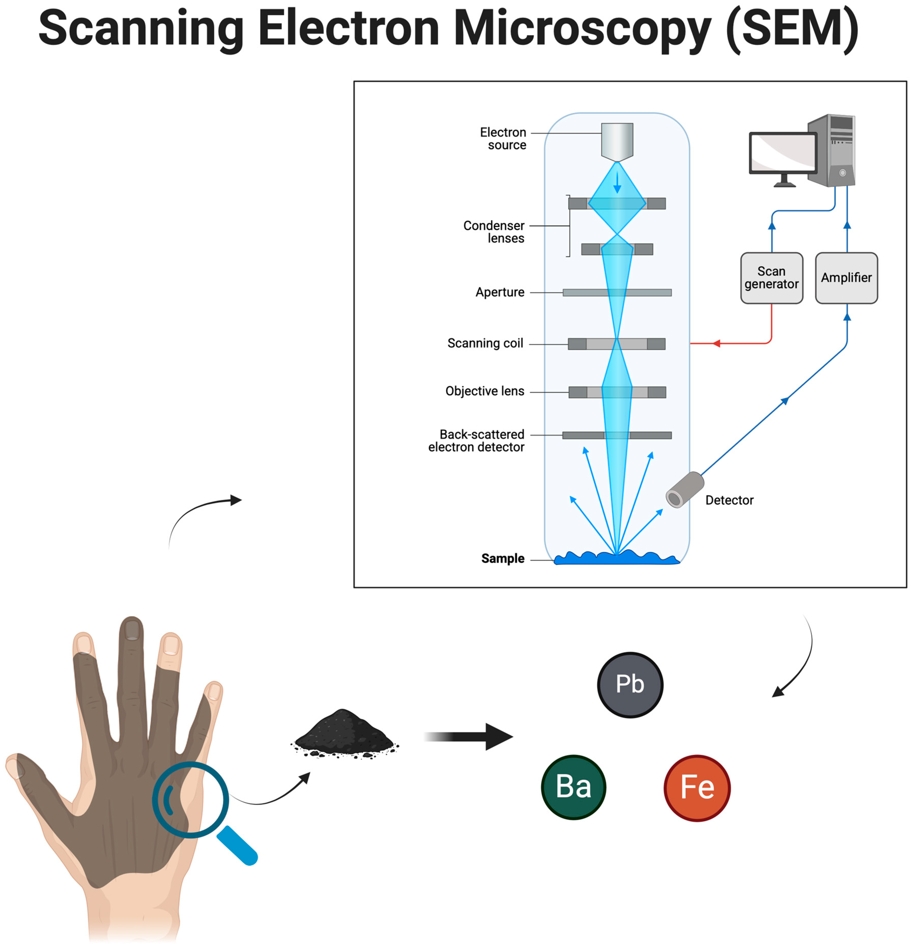
1.4. SEM Detectors and Imaging Techniques in GSR Analysis

SEM employs different types of detectors to capture various signals generated when the electron beam interacts with the sample.
The secondary electron (SE) detector captures low-energy electrons emitted from the sample surface when bombarded by the electron beam [1,2,3,4,5,6,7]. These electrons originate from the top layers of the material, providing high-resolution images with excellent surface detail. This type of imaging is particularly useful for examining the morphology of GSR particles, allowing forensic scientists to identify characteristic spherical shapes that result from the high-temperature vaporization and condensation of metals upon firearm discharge [1,2,3,4,5,6,7]. Since secondary electrons are highly sensitive to topographical variations, they are effective in distinguishing the fine surface features of GSRs from environmental contaminants.
The backscattered electron (BSE) detector operates by detecting high-energy electrons that are elastically scattered from the sample. Unlike secondary electrons, backscattered electrons originate deeper within the material, and their signal intensity is directly related to the atomic number of the elements present. This characteristic makes BSE imaging particularly valuable in forensic applications, as it enables the differentiation of GSR particles based on their elemental composition. Heavier elements, such as lead (Pb), barium (Ba), and antimony (Sb), appear brighter in BSE images, while lighter elements, commonly found in non-GSR particles, appear darker. This contrast allows forensic scientists to quickly assess the presence of GSR particles and distinguish them from naturally occurring environmental debris [1,2,3,4,5,6,7].
In addition to electron-based imaging, SEM is frequently combined with energy-dispersive X-ray spectroscopy (EDS) to perform elemental analysis of GSR particles. When the electron beam interacts with the sample, it excites atoms within the material, causing them to emit characteristic X-rays that correspond to specific elements. By analyzing the emitted X-ray spectrum, EDS allows for the precise identification of the chemical composition of the particles. This technique is crucial in forensic investigations, as it confirms the presence of elements commonly associated with firearm discharge, such as lead, barium, and antimony. Combining SEM imaging with EDS analysis enhances the reliability of GSR identification, reducing the risk of misclassification due to environmental contamination [1,2,3,4,5,6,7].
Different imaging techniques can be employed in SEM to optimize the visualization of GSR particles. Low-voltage imaging improves resolution and reduces sample charging effects, which is particularly beneficial for analyzing non-conductive GSR particles without requiring a conductive coating. Environmental SEM (ESEM) allows imaging under low-vacuum or variable-pressure conditions, enabling the analysis of uncoated forensic samples while preserving their original state. Additionally, the integration of automated SEM-EDS particle recognition software has significantly enhanced the efficiency of GSR analysis by streamlining particle detection, classification, and chemical composition assessment.
This article explores, through a narrative review, the application of SEM techniques in GSR analysis to clarify the methodological approaches supporting effective forensic investigations, highlight the advantages and limitations of SEM, and enhance the accuracy of GSR particle identification. Understanding the morphology and composition of GSRs provides valuable information about various aspects of the crime scene and the firearm used. Moreover, this study addresses analytical and methodological challenges to reduce artifacts, improve the sensitivity and specificity of SEM-based GSR analysis, and develop and validate new forensic techniques based on cutting-edge technologies. Ultimately, SEM supports forensic investigations and judicial processes by providing concrete and detailed evidence to corroborate or refute witness statements and event reconstructions.

2. Materials and Methods

To conduct this narrative review, a structured search was performed using Google Scholar and PubMed NCBI. The search terms included “Scanning Electron Microscopy”, “SEM and Forensic Science”, “SEM and Gunshot Residues”, and “SEM and Ballistics Analysis”. Articles published between 2009 and 2024 were considered, focusing on the role of SEM in forensic investigations. The selection criteria included studies that explicitly addressed SEM’s application in GSR detection, the effectiveness of SEM-EDS (energy-dispersive spectroscopy) in forensic casework, and comparative analyses between SEM and other microscopic techniques. Studies primarily discussing environmental contamination, without forensic implications, were excluded. A total of 65 articles were included, ensuring a comprehensive overview of current methodologies and advancements in SEM-based GSR analysis. The reference lists of selected studies were also reviewed to identify the additional relevant literature.

3. Results

3.1. Imaging Detectors and Techniques

SEM uses a variety of detectors and imaging techniques to obtain detailed information on the surface of samples. The main detectors include those for secondary electrons, backscattered electrons, and X-rays, while imaging techniques may involve the use of low voltage, non-vacuum environments, and combined methods to provide a comprehensive understanding of sample characteristics. Among the available detectors, secondary electron (SE) detectors are used to capture detailed images of the sample’s surface topography. Secondary electrons are emitted from the surface when the primary electron beam interacts with it, making these detectors particularly useful for obtaining high-resolution images with topographic contrast [8]. Backscattered electron (BSE) detectors, on the other hand, collect electrons that have been reflected back from the sample. These detectors provide information on the composition and structure of the sample, as the BSE contrast depends on the atomic number and density of the material [9]. Finally, X-ray detectors, such as energy-dispersive spectroscopy (EDS) detectors, analyze the chemical composition of the sample by generating an energy spectrum of the emitted X-rays [10]. Regarding the imaging techniques for obtaining surface details, low-voltage imaging is used to improve image quality and reduce sample damage, particularly for sensitive samples such as biological specimens. By lowering the voltage of the electron beam, more detailed images can be acquired without excessive penetration [11]. Another technique involves imaging in non-vacuum environments, such as humid or gaseous atmospheres, using environmental SEM (ESEM). This technique is beneficial for analyzing samples that cannot be prepared in a vacuum [12]. Finally, imaging techniques can be combined with other methods, such as energy-dispersive spectroscopy (EDS) and wavelength-dispersive X-ray spectroscopy (WDS), to obtain detailed information on both the topography and the chemical composition of the sample [13] (Figure 1).

3.2. Applications of SEM

Scanning electron microscopy is a versatile and powerful technique used in multiple scientific and industrial fields to analyze and characterize materials and structures at the microscopic level. SEM has applications in nanotechnology, biology, the electronics industry, and beyond. Its ability to provide high-resolution, detailed images of sample surfaces makes it indispensable for a wide range of research and industrial applications. In nanotechnology, SEM is essential for the analysis and characterization of nanomaterials and nanostructures. Its high resolution enables the observation of details at the nanometric level, providing critical information on the morphology, size, and distribution of nanoparticles [14]. Another major application of SEM is in materials science and metallurgy, where it is used to analyze material microstructures, identify defects, and study failure mechanisms. It is particularly useful for examining the surfaces of metals, alloys, and composites to evaluate their quality and performance [15]. In the electronics industry, SEM is used to examine the structure and quality of electronic components and integrated circuits. It allows for the identification of manufacturing defects, observation of material distribution, and analysis of the interfaces between different components [16]. In geological and natural materials sciences, SEM is used to analyze minerals, rocks, and other natural materials, providing insights into their structure, composition, and the geological processes that influenced their formation [17]. In nanoscience, SEM plays a crucial role in developing and characterizing nanostructures and nanoscopic devices. It enables the visualization of nanotubes, nanoparticles, and nanowires and is essential for the research and development of emerging technologies [18]. In biology and medicine, SEM is widely used to study the morphology of cells, tissues, and organs. It allows for the observation of the fine details of cellular structures and interactions between cells and pathogens, making it particularly useful for analyzing biological samples that require high resolution [19].
SEM offers a wide range of applications in medicine, from the detailed characterization of cell and tissue structures to the analysis of pathogens and biomaterials. Its ability to provide high-resolution, detailed images is critical for medical research and diagnostics. In cell morphology studies, SEM enables the acquisition of detailed images of cell surfaces, revealing structures such as microvilli, cilia, and other cellular extensions that are not visible with optical microscopy. This capability is fundamental for studying cellular alterations due to diseases or treatments [20]. In tissue and organ analysis, SEM is used to examine the structure and composition of tissues and organs at a microscopic level. This technique is particularly useful for identifying pathological changes, such as modifications in adipose tissue structure or cell membranes during diseases [21]. Another rapidly growing field is pathogen analysis, where SEM is used for observing and analyzing bacteria, viruses, and other pathogens. It enables the visualization of pathogen morphology with high resolution, contributing to the understanding of their characteristics and the development of therapeutic interventions [22]. SEM is also used to study biomaterials and medical implants, such as prosthetics and suture materials. The technique helps evaluate the quality and durability of materials, cell adhesion, and interactions between biomaterials and tissues [23]. In cancer research, SEM is used to examine cellular alterations associated with diseases such as cancer. It enables the observation of morphological changes in tumor cells and their interactions with surrounding tissues [24]. Another critical application is investigating cellular and tissue responses to pharmacological treatments. SEM images can reveal side effects or structural changes caused by drugs, aiding in the design of new treatments [25].

3.3. Applications of SEM in the Forensic Field

SEM is an essential technique in forensic science for the detailed analysis of physical and biological evidence. Its forensic applications include determining the provenance and composition of materials and documenting microscopic details that can be crucial in criminal investigations. SEM is used for analyzing various types of evidence, including fibers, powders, explosives, and drug residues. Its ability to provide high-resolution, detailed images makes it invaluable for forensic analysis. One of the primary applications of SEM in forensic science is the analysis of textile fibers and dust residues collected at crime scenes. SEM’s high resolution enables the identification of fiber morphology and composition, which can be crucial for comparison and correlation with specific sources [26]. SEM is also essential for analyzing dust particles and bullet residue. It allows for the identification of the chemical composition and morphology of these particles, providing clues about the specific types of ammunition and materials used in violent crimes [27]. The identification of damage to materials and fabrics, such as those caused by tears or abrasions, can also be assessed using SEM. High-resolution images can reveal the microscopic details of damage, which can help reconstruct the events and movements during a crime. SEM is also employed in the analysis of explosive residues found at crime scenes. It allows for the identification of the chemical components and microscopic structures of explosives, helping to link residues to specific types of explosive devices [28]. Another key forensic application of SEM is the analysis of seized pharmaceuticals, drugs, and chemicals. The technique provides details on the morphology and structure of drug crystals, which can be used to identify substances and determine their purity. Finally, the analysis of biological and blood residues remains an important forensic application of SEM, as it enables the examination of blood cell structure and tissue morphology. This application is useful for identifying and characterizing biological samples collected at crime scenes [29].

3.4. Identification and Composition of Bullet Residue Particles with SEM

SEM provides high-resolution imaging and chemical composition analysis of particles, making it a valuable tool for solving complex forensic cases and obtaining detailed evidence. In the analysis of bullet particles, SEM is used to identify such residues at crime scenes. This technique allows for the examination of bullet residue particles to determine their chemical composition and morphology. This helps forensic experts identify the type of ammunition used and correlate the residues with specific weapons. These particles may contain metals such as lead, copper, or antimony, which are key elements for forensic identification [30,31].
SEM is also crucial for the analysis of the morphology and size of bullet residue particles. By examining the shape and size of these particles, forensic investigators can differentiate between different types of ammunition and determine shooting distances. The morphology of the particles can vary depending on the type of weapon and projectile used [32].
In explosive residue analysis, SEM plays a critical role in identifying the chemical components of explosives through their morphology and composition. This technique provides valuable insights into the type of explosive used and its production method, contributing to investigations into explosions and terrorist attacks [33]. Additionally, SEM allows forensic experts to study the damage caused by explosives on materials and surfaces. By analyzing the explosive residues and structural damage on affected surfaces, it is possible to gather information about the strength and nature of the explosion and the manner in which the explosive was used [34].
Regarding metal analysis, SEM combined with EDS spectroscopy is widely used to identify the metals present in bullet residues. Common elements include lead (Pb), copper (Cu), antimony (Sb), and barium (Ba). The presence and concentration of these elements can provide information on the manufacturing process of the projectile and help compare the residues with known samples [35].
SEM also plays a significant role in the study of the crystalline structures of residue particles. Certain metals and compounds form characteristic crystalline structures that can be used to determine the ammunition type and firing conditions. For instance, lead may appear in different crystalline forms depending on its origin and the treatment it underwent [17].
SEM offers several advantages in the analysis of bullet residues. Its very high resolution allows for the visualization of the minute details of residue particles, which is particularly beneficial when analyzing small amounts of residue that may not be visible using other techniques [36]. Furthermore, SEM provides both morphological and chemical information in a single analysis. The ability to combine high-resolution imaging with detailed chemical analysis makes SEM a powerful tool for identifying and characterizing bullet residues [37].

3.5. Gunshot Residue Analysis: Detection, Persistence, and Emerging Forensic Solutions

Gunshot residue (GSR) analysis represents a crucial component of forensic investigations, allowing the reconstruction of shooting events and the establishment of a possible link between suspects, victims, and firearms. GSR particles, composed of both inorganic (iGSRs) and organic components (OGSRs), are traditionally analyzed using scanning electron microscopy coupled with energy-dispersive X-ray spectroscopy (SEM/EDS) to detect characteristic elements such as lead (Pb), barium (Ba), and antimony (Sb), which form during the rapid cooling of the molten metals expelled from the firearm discharge. Recent studies have expanded our understanding of the distribution, persistence, and secondary transfer dynamics of these residues, highlighting the complexity of their forensic interpretation. For example, a study conducted on police vehicles in Zagreb County revealed a significant prevalence of iGSR contamination on multiple vehicle surfaces, notably on the driver and back seats. The research demonstrated that 63.1% of the sampled vehicles contained characteristic GSR particles, with logistic regression identifying the transport of individuals involved in shooting incidents as a statistically significant risk factor for GSR contamination within police vehicles [36]. This finding underscores the necessity of implementing contamination control protocols during forensic evidence collection to preserve its integrity.
In addition to contamination risks, the dynamics of GSR secondary transfer were comprehensively evaluated through experimental studies involving various common surfaces, including ceramic, glass, metal, paper, and plastic. The results revealed that paper retained the highest quantity of transferred particles, followed by plastic and ceramic, while washing hands with water and soap significantly reduced particle persistence. This highlights the importance of accounting for surface material and post-discharge activities when interpreting secondary transfer evidence [37]. Moreover, research on the persistence and spatial distribution of GSRs on different regions of the human body emphasized the variability in residue retention based on sampling location. While the highest concentrations of both OGSRs and iGSRs were found on the hands, alternative collection sites, such as the forearms and face, were identified as reliable sampling options when hand-washing occurred. Interestingly, sampling from the nostrils showed minimal residue retention, making it less suitable as an alternative site [38]. The ear, however, emerged as an innovative sampling site due to its relative protection from environmental exposure and cleaning. Sampling the concha and external auditory canal proved effective in capturing GSRs following shotgun discharge, particularly the right ear, which was most affected due to body posture during firing [39].
Technological advancements have greatly enhanced the precision and efficiency of GSR analysis in recent years. Portable SEM devices have revolutionized on-site forensic investigations by enabling the real-time detection of GSR particles directly at crime scenes, significantly reducing the delay between evidence collection and analysis while maintaining a high level of accuracy [40]. Furthermore, combined analytical approaches integrating SEM/EDS with ultra-high-performance liquid chromatography–tandem mass spectrometry (UHPLC-MS/MS) now allow the simultaneous detection of both inorganic and organic GSRs from a single sample. This methodological innovation increases the probative value of GSR evidence by offering a more comprehensive chemical profile. The sequential analysis protocol using SEM/EDS for iGSR detection followed by UHPLC-MS/MS for OGSRs demonstrated excellent recovery rates for both types of residues without significant particle loss, making it a valuable tool for complex forensic investigations [41].
Despite these technological advancements, challenges persist in ensuring the reliability and accuracy of GSR analysis. One of the most pressing concerns is the differentiation of genuine GSR particles from environmental contaminants that may share similar elemental profiles. Non-firearm sources such as fireworks, brake linings, and certain industrial processes can produce particles that resemble GSRs in both morphology and composition. Addressing this issue requires strict particle classification criteria and the continuous refinement of detection algorithms. The application of machine learning techniques for automated particle classification has shown promise in reducing false positives and improving detection efficiency, thereby enhancing the overall reliability of forensic interpretations [42]. However, the variability in GSR classification practices across different forensic laboratories remains a significant obstacle to achieving standardized protocols. Studies have highlighted inconsistencies in particle classification, particularly regarding borderline particles containing combinations of barium, lead, and antimony. This variability underscores the need for rigorous inter-laboratory validation and the adoption of universally accepted standards to ensure consistency in forensic reporting [43]. Such efforts are essential not only for the scientific robustness of GSR analysis but also for maintaining its credibility in judicial proceedings, where even minor analytical discrepancies can have profound legal implications.

3.6. Judicial Implications and the Role of Expert Testimony

In forensic investigations, the findings from SEM-EDS analysis carry significant weight in judicial proceedings. The proper interpretation and presentation of these results are essential to avoid the misrepresentation or miscommunication of scientific evidence. The forensic expert’s role extends beyond the laboratory to the courtroom, where they must explain complex analytical processes and their conclusions in a manner that is both scientifically accurate and comprehensible to non-specialist audiences, such as judges and jurors.
Judicial scrutiny of forensic evidence has intensified in recent years, with courts demanding greater transparency, validation, and standardization of forensic methods. While SEM-EDS is widely regarded as a reliable method for GSR detection, the forensic expert must clearly articulate the limitations of the analysis, particularly regarding the potential for false positives and the influence of environmental factors. Furthermore, the expert should be prepared to address alternative hypotheses and demonstrate how the findings support or refute these scenarios. In cases where GSR evidence plays a pivotal role, the expert’s testimony can influence the outcome of the trial, making it imperative to present balanced, well-supported conclusions based on validated methodologies.

3.7. Future Prospects of SEM in the Forensic Field

The adoption of AI and automation, the miniaturization of instruments, and innovations in imaging and spectroscopy techniques could significantly enhance SEM applications, making forensic investigations faster, more accurate, and more efficient. Future developments in SEM-based forensic investigations are characterized by technological innovations and the integration of new techniques, which promise to improve analytical capabilities and investigation efficiency.
One of the main advancements in SEM involves the integration of artificial intelligence (AI) and machine learning algorithms. AI can revolutionize the interpretation of SEM images by automating the identification and classification of particles and structures, reducing analysis time and increasing accuracy [38,39]. Automation in sample preparation, including automated fixation and embedding, could further minimize human errors and improve the reproducibility of results. Standardized preparation processes would ensure higher consistency and quality in forensic sample analyses [40,41].
Another promising direction for SEM involves miniaturization and portability. The development of portable SEM instruments could allow forensic investigators to analyze samples directly at crime scenes or in emergency situations. These portable instruments could be designed to be lighter, more compact, and easy to transport, while maintaining an adequate resolution and sensitivity for forensic applications [42,43]. The miniaturization of SEM components could also lead to the creation of smaller and more versatile microscopes, with applications in forensic and clinical situations that require high-resolution instruments in limited spaces [44,45].
Advancements in imaging techniques are also shaping the future of SEM. The use of electron tomography and other 3D imaging techniques is becoming increasingly common in SEM. These innovations provide a three-dimensional view of samples, improving the understanding of their structure and composition [46,47]. Furthermore, integrating SEM with advanced spectroscopic techniques, such as X-ray photoelectron spectroscopy (XPS) and mass spectrometry, can enhance the compositional analysis of samples, providing more detailed chemical and structural information [36,48].

3.8. Comparative Analysis of SEM with Other Microscopic Techniques

While SEM-EDS remains the gold standard for gunshot residue (GSR) analysis due to its ability to provide simultaneous morphological and elemental characterization, other complementary techniques, such as Raman spectroscopy, time-of-flight secondary ion mass spectrometry (TOF-SIMS), and X-ray photoelectron spectroscopy (XPS), offer additional advantages in forensic investigations [10,11,12,13,14,15,16,17,18,19,20].
Raman spectroscopy is a non-destructive vibrational spectroscopy technique that provides molecular information about a sample based on its interaction with laser light. Unlike SEM-EDS, which primarily focuses on the elemental composition, Raman spectroscopy detects organic components in GSRs, such as stabilizers, plasticizers, and unburnt gunpowder residues. This capability is particularly valuable in distinguishing gunshot residues from other environmental contaminants that share similar elemental compositions. However, Raman spectroscopy has significant limitations, including fluorescence interference from certain materials, which can obscure spectral signals, and relatively low sensitivity to metallic GSR particles.
TOF-SIMS is a surface-sensitive analytical technique that detects trace amounts of organic and inorganic species present in GSR particles. By using a focused ion beam to sputter material from the surface, TOF-SIMS provides high-resolution chemical mapping of individual particles, making it effective for differentiating between GSRs originating from different types of ammunition. Compared to SEM-EDS, TOF-SIMS offers higher chemical specificity, allowing for the detection of characteristic organic compounds that may not be visible through SEM imaging. However, TOF-SIMS is limited by its destructive nature, as the analysis process removes material from the sample, which may not be ideal for forensic cases requiring evidence preservation. Additionally, TOF-SIMS instruments are expensive and require specialized expertise, limiting their widespread use in routine forensic investigations [10,11,12,13,14,15,16,17,18,19,20].
X-ray photoelectron spectroscopy (XPS) is another powerful technique used for surface chemical analysis, providing information on the oxidation states and chemical bonding of elements within GSR particles. Unlike SEM-EDS, which detects bulk elemental composition, XPS is capable of differentiating between chemical states of elements, offering deeper insights into the oxidation and environmental degradation of GSRs. This can be particularly useful in forensic cases where residue persistence over time is a factor. However, XPS has limitations, including longer analysis times and the requirement for ultra-high vacuum conditions, which may not always be practical in high-throughput forensic laboratories. Furthermore, the technique has a relatively shallow depth of analysis, meaning that only the outermost atomic layers of a particle are examined, which might not provide a complete compositional profile.
Despite these alternatives, SEM remains the gold standard due to its combined morphological and compositional analysis capabilities, essential for forensic casework. Future research should focus on integrating multiple techniques to enhance GSR characterization and reduce misclassification errors.

3.9. SEM Applications Beyond Forensic Science

While the primary focus of this study is the application of scanning electron microscopy (SEM) in forensic science, particularly in gunshot residue (GSR) analysis, SEM is widely used across various scientific disciplines [1,2,3,4,5,6,7,8,9,10,11,12,13].
In materials science, SEM is an essential tool for characterizing the microstructure, composition, and surface properties of a wide range of materials, including metals, ceramics, polymers, and composites. High-resolution SEM imaging allows researchers to analyze the fracture surfaces, phase distribution, and grain boundaries in metallic alloys, providing crucial insights into material failure mechanisms. Additionally, SEM-EDS is extensively used to study corrosion processes, enabling the identification of elemental composition changes in degraded materials [1,2,3,4,5,6,7,8,9,10,11,12,13]. The ability to perform in situ environmental SEM (ESEM) also allows researchers to observe real-time structural changes in materials under different environmental conditions, such as high humidity or elevated temperatures.
In nanotechnology, SEM plays a critical role in imaging and characterizing nanomaterials, nanoparticles, and nanostructures [1,2,3,4,5,6,7,8,9,10,11,12,13,14,15]. Due to its high-magnification capabilities, SEM is used to examine carbon nanotubes, graphene, quantum dots, and nanocomposites, providing detailed morphological and compositional information. The use of field-emission scanning electron microscopy (FE-SEM) enhances resolution even further, enabling the visualization of structures at the sub-nanometer scale. Additionally, SEM is widely used in semiconductor research, where it facilitates failure analysis, contamination detection, and quality control of microelectronic components.
Another significant application of SEM is in biological and medical research, where it is used to study cellular structures, bacteria, viruses, and biofilms. SEM provides detailed three-dimensional images of biological specimens, revealing intricate surface morphologies that are not visible using traditional optical microscopy. In medical device development, SEM is instrumental in evaluating the surface properties of biomaterials, implants, and tissue-engineered scaffolds, ensuring their compatibility with biological systems.

4. Discussion

4.1. Gunshot Residue Composition and Formation

Gunshot residues (GSRs) primarily consist of a complex mix of chemical components resulting from the discharge of a firearm. These components include particles of lead (Pb), barium (Ba), and antimony (Sb), which are commonly found in the primer of ammunition. The chemical composition of GSRs can be further analyzed using advanced techniques such as scanning electron microscopy coupled with energy-dispersive X-ray analysis (SEM-EDX) [49]. This method allows for detailed examination of both the elemental and morphological characteristics of GSR particles. The formation and deposition processes of GSRs are highly dynamic and occur in milliseconds during the firing of a gun. When a firearm is discharged, the primer ignites the gunpowder, generating a high-pressure explosion that expels the bullet from the barrel. This explosion produces a cloud of microscopic particles, which are then deposited on the hands, clothing, and nearby surfaces of the shooter. Several factors influence the distribution of GSR particles, including the type of firearm and ammunition used, environmental conditions, and the shooter’s actions immediately following the discharge. For instance, semi-automatic firearms tend to produce a higher quantity of GSRs compared to revolvers due to differences in their firing mechanisms [27]. Additionally, environmental factors such as wind and humidity can affect the dispersion and adhesion of GSR particles.

4.2. SEM Application on Gunshot Residue Identification

Recent technological advancements have significantly enhanced the effectiveness of scanning electron microscopy (SEM) in gunshot residue (GSR) analysis, particularly through innovations in energy-dispersive spectroscopy (EDS) detectors. The development of new types of EDS detectors has greatly improved the sensitivity and accuracy of particle identification [50]. This improvement is complemented by the introduction of sophisticated algorithms, such as the binary tree-based classification algorithm proposed by Mandel et al. which optimizes the initial classification step and minimizes errors during the automatic run of GSR analysis [50,51]. These algorithms are designed to reduce the incidence of false positives and false negatives, thus enhancing the reliability of the results [52]. Moreover, advancements in SEM/EDS technology have led to a substantial reduction in both the overall analysis time and the time required for reviewing particles of interest, thereby streamlining the entire GSR analysis process [53]. This time efficiency is further bolstered by automation, which not only speeds up the process but also ensures consistency and accuracy in particle detection and classification [54]. Collectively, these technological improvements have made SEM a more effective and reliable tool for forensic scientists, enabling quicker and more accurate GSR analyses while preserving the integrity of the evidence.

4.3. Collection Phase

The collection phase often involves the use of adhesive stubs or swabs to gather trace evidence, such as GSRs, from surfaces or individuals involved in forensic investigations. This step is critical, as improper collection can lead to the contamination or loss of vital particles. Once collected, samples undergo a preparation phase that frequently involves mounting the samples on conductive tapes and coating them with a thin layer of conductive material, such as gold or carbon, to prevent charging under the electron beam. This preparation is essential to enhance the visibility and detection of particles under SEM [50,51,52,53,54]. Furthermore, advancements in automation for SEM-based methods, including SEM-EDX, have streamlined the process, enabling more efficient and accurate identification of trace elements like GSRs and glass fragments [27,48,49,50,51,52,53]. In the context of SEM-based GSR analysis, two distinct methods have been introduced to enhance the preparation process. These methods involve either direct analysis of collected particles or their extraction and concentration before SEM examination. Both approaches aim to optimize the detection of GSRs from various surfaces, including nasal hairs, which serve as a novel sampling site for forensic purposes [54]. Also, several best practices are recommended. First, conducting a reliability and validity assessment of the measurement model is crucial. This initial step ensures that the constructs being measured are both consistent and accurate, laying a solid foundation for subsequent analysis. Following this, a structural model assessment should be performed as part of a two-step analysis process. This step involves evaluating the relationships between variables, allowing for a comprehensive understanding of the underlying structure of the data. Additionally, utilizing multiple fit indices, such as the Comparative Fit Index (CFI), Tucker–Lewis Index (TLI), Root Mean Square Error of Approximation (RMSEA), and Standardized Root Mean Square Residual (SRMR), is a best practice for evaluating the model’s goodness of fit. These indices provide a multifaceted view of how well the model represents the data, enhancing the robustness of the findings. Ensuring consistency between results obtained from different methods, such as the Technique for Order of Preference by Similarity to Ideal Solution (TOPSIS) and the Analytic Hierarchy Process (AHP), is also important for validating findings. This cross-method validation helps to confirm the reliability of the results and mitigate any method-specific biases [55].

4.4. Analytical Procedures and Protocols

Once the samples are collected, they are mounted onto an SEM stub and placed into the scanning electron microscope (SEM) for examination. Automated software is utilized to screen the sample for candidate GSR particles. The SEM provides high-resolution images, which are then analyzed using energy-dispersive X-ray spectroscopy (EDX) to determine the elemental composition of the particles, offering both morphological and compositional data [27,50,51,52,53,54,55]. The calibration and standardization of SEM-EDX instruments are critical to ensure the accuracy and reproducibility of the analysis. This process involves performing a series of tests when the instrument is first commissioned or whenever significant modifications are made [27,50,51,52,53,54,55]. Calibration ensures that the EDX detector is accurately measuring the energy peaks corresponding to different elements, thereby providing precise compositional data. Standardization involves using known reference materials to establish baseline measurements, which helps in comparing and interpreting GSR data [27,48,49,50,51,52,53]. Quality control and validation of analytical results in SEM-EDX analysis are paramount to ensure the credibility of the findings. Instrument validation is performed to characterize the automated GSR instrumentation, ensuring it operates within the specified parameters [27,50,51,52,53]. Quality control measures, such as the use of blank samples and reference standards, are implemented to monitor the performance of the SEM-EDX system continuously. These checks help in detecting any anomalies or inconsistencies in the data, thereby safeguarding the integrity of the analysis. Validation studies also include comparing the SEM-EDX results with those of other established methods to confirm their accuracy and reliability [53,54,55] (Figure 2).

4.5. Interpretation and Reporting of Results

Criteria for identifying gunshot residues using SEM-EDX are based on both morphological and elemental analysis. SEM-EDX provides detailed information about the shape and size of particles as well as their elemental composition [27,48,49,50,51,52,53]. Morphologically, GSRs exhibit distinct characteristics such as spherical shapes or irregular particles with specific surface textures, which are identifiable under a scanning electron microscope [27,47,48,49]. This dual approach ensures that the identification of GSRs is both accurate and reliable, as it eliminates false positives that may arise from analyzing morphology or elemental composition alone [27,48,49,50,51,52,53]. Interpreting elemental composition data from SEM-EDX analysis involves comparing the detected elements against the known standards for GSRs. The characteristic elements—typically lead, barium, and antimony—are analyzed quantitatively to confirm their presence and proportions [27,48,49,50,51,52,53]. For instance, photomicrographs from SEM/EDX can reveal the indefinite morphology of GSRs from clean-range ammunition, which contrasts with conventional GSRs [53,54,55]. This distinction is crucial for forensic experts who need to differentiate between genuine GSR particles and other environmental contaminants. The interpretation is guided by established forensic protocols that emphasize both the presence of these specific elements and their relative abundances within the sample [48]. Reporting findings from SEM-EDX analysis in forensic investigations involves presenting both the morphological and elemental data in a clear, concise manner. The technique offers visual identification through detailed photomicrographs and spectral data that show the chemical composition of GSR particles [27,48,49,50,51,52,53]. These findings are then compiled into comprehensive reports that include interpretations of the data, potential sources of the residues, and their implications for the investigation. For instance, the presence of significant amounts of GSRs on a suspect can suggest recent discharge of a firearm, thus providing crucial evidence in criminal cases [27,48,49,50,51,52,53]. The specificity and accuracy of SEM-EDX make it a useful tool in forensic science, helping to establish connections between suspects and crime scenes [27,48,49,50,51,52,53,54,55]. Notable forensic cases involving SEM and gunshot residue (GSR) analysis underscore the critical role of this technology in criminal investigations. One such case is the landmark investigation of the assassination of President John F. Kennedy, where SEM was crucial in examining GSR particles found on various surfaces and clothing items to reconstruct the sequence of events [27,48,49,50,51,52,53,54,55]. These residues, specifically lead (Pb), barium (Ba), and antimony (Sb), were analyzed using SEM coupled with energy-dispersive X-ray spectroscopy (EDS), providing conclusive evidence that linked the firearm discharge to specific locations and individuals [27,48,49,50,51,52,53]. Another significant case involved the analysis of GSRs in a high-profile murder trial, where SEM/EDS technology was instrumental in confirming the presence of primer residues on the suspect’s hands, thereby corroborating eyewitness testimony and strengthening the prosecution’s case [27,48,49,50,51]. In the judicial context, we must evaluate various factors including the chain of custody, the precision of the SEM technique, and the expertise of the analyst in determining the admissibility and weight of the evidence. For instance, in a notable criminal case, the defense challenged the SEM results by questioning the possibility of contamination and the accuracy of particle identification [27,48,49,50,51,52,53]. However, the meticulous documentation and adherence to standardized protocols ensured the acceptance of the SEM results, ultimately leading to a conviction. This case exemplifies the critical importance of rigorous scientific procedures and expert testimony in leveraging SEM evidence in the judicial process. The advantages of using SEM in forensic investigations are manifold. One of the primary benefits is the ability to provide a non-destructive analysis of the GSR samples, preserving them for possible re-examination or additional testing [27,48,49,50,51,52,53]. SEM offers high-resolution imaging and precise elemental analysis, which can distinguish GSR particles from other similar-looking substances, thereby increasing the reliability of forensic conclusions [27,47,48,49]. Furthermore, the use of SEM in GSR analysis improves the efficiency of forensic investigations by delivering rapid and accurate results, which are crucial in time-sensitive criminal cases [27,48,49,50,51]. The detailed data obtained from SEM analysis can also be used to establish a strong link between the suspect and the crime scene, thereby supporting the overall investigative process [27,48,49,50,51].

4.6. Technical Limitations of SEM and False Positives/Negatives in GSR Analysis

One of the primary concerns in SEM-based GSR detection is the potential for false positives and false negatives. False positives can occur when environmental particles share morphological and elemental characteristics with GSRs, such as residues from brake linings, fireworks, or welding operations. Studies have reported that environmental contaminants containing Pb, Ba, and Sb can be misidentified as GSRs unless additional criteria, such as morphology and particle size distribution, are considered [1,2,3,4,5,6,7,8,9,10]. False negatives, on the other hand, arise due to the loss of GSR particles over time. Research indicates that GSR particles can degrade or be transferred away from an individual’s hands within hours, reducing the probability of detection. The accuracy of SEM in GSR identification varies, with reported false negative rates ranging between 10 and 30%, depending on the collection method and environmental factors [1,2,3,4,5,6,7,8,9,10]. To improve reliability, automated particle classification algorithms and standardized analysis protocols are being developed to enhance detection efficiency and reduce human error in forensic evaluations.

4.7. Portable SEM Devices: Revolutionizing On-Site Forensic Analysis

The development of portable SEM devices represents a groundbreaking innovation in forensic science, offering the possibility of performing high-resolution microscopy and elemental analysis directly at crime scenes. Traditional SEM instruments are large, stationary, and require highly controlled environments to operate effectively. Portable SEM systems, by contrast, are compact and designed for field use, bringing the power of electron microscopy to forensic investigators in real time.
Portable SEM devices can be used to quickly screen suspects, vehicles, and other surfaces for GSRs, providing immediate preliminary results that can guide the direction of the investigation. These systems retain many of the core capabilities of laboratory-based SEMs, including high-resolution imaging and EDS analysis, although with some limitations in resolution and analytical depth due to their smaller size and simplified design [52,53,54,55].
The main challenges associated with portable SEM systems are related to sample preparation and environmental control. Maintaining vacuum conditions and ensuring accurate calibration in field settings can be complex. Nevertheless, ongoing improvements in design and software are addressing these limitations, making portable SEM an increasingly viable option for rapid, in situ forensic analysis.

4.8. Complementary Analytical Techniques in GSR Identification

While SEM-EDS remains the cornerstone of GSR analysis, the integration of complementary analytical techniques can provide additional layers of information, improving both sensitivity and specificity. Among the most promising techniques is Raman spectroscopy, which offers molecular-level information about the chemical structure of individual particles. Raman spectroscopy is particularly useful for distinguishing GSR particles from similar environmental particles that share the same elemental composition but differ in molecular characteristics [56,57,58,59,60,61,62].
Another valuable technique is TOF-SIMS, which allows for high-resolution surface analysis and the detection of trace elements and molecular fragments. TOF-SIMS has been used to identify unique chemical markers in GSRs that are not detectable with traditional SEM-EDS, providing a more comprehensive chemical profile. X-ray photoelectron spectroscopy (XPS) is another powerful tool for surface analysis, capable of providing detailed information about the oxidation states of elements in GSR particles, which may offer insights into the type of ammunition used.
The combination of these techniques with SEM-EDS forms a robust multi-modal analytical approach, enabling forensic scientists to build a more complete picture of GSR evidence. This multi-technique strategy is particularly important in complex cases where traditional SEM-EDS analysis alone may be inconclusive.
Although SEM-EDS is the most widely used technique for GSR detection, Electron Backscatter Diffraction (EBSD) has been explored as a complementary method. EBSD provides crystallographic information about particles, allowing forensic experts to distinguish authentic GSRs from environmental contaminants that may exhibit similar elemental compositions. While EBSD is not yet standard in forensic GSR analysis, its application in other forensic material studies suggests that it could be useful for characterizing the structural properties of GSR particles. Future research should investigate the feasibility of integrating EBSD with SEM-EDS to improve classification accuracy.

4.9. Challenges and Limitations in SEM-Based GSR Analysis: False Positives and Environmental Contaminants

While SEM-EDS remains the most reliable method for GSR detection, it is not without limitations. One of the most significant challenges in forensic GSR analysis is the potential for false positives due to environmental contamination. Numerous materials found in the environment share morphological and elemental characteristics with GSRs, leading to potential misinterpretations of results. For example, particles from brake linings, fireworks, certain construction materials, and welding processes can mimic GSR particles in both shape and elemental composition. These particles can often contain combinations of lead (Pb), barium (Ba), and antimony (Sb), making their differentiation from authentic GSR particles challenging without additional contextual information.
Another critical limitation is the degradation or loss of GSR particles over time. GSR particles are highly susceptible to environmental factors such as friction, washing, or prolonged exposure to wind and rain, which can significantly reduce their detectability. Studies have shown that the number of GSR particles on a suspect’s hands can decrease dramatically within hours after firearm discharge, complicating sample recovery and interpretation. This issue underscores the importance of timely sample collection and the development of more sensitive detection protocols.
Addressing these challenges requires a multi-faceted approach, including the refinement of particle classification protocols, the integration of complementary spectroscopic techniques such as Raman spectroscopy, and the development of probabilistic models for interpreting SEM data. Furthermore, the standardization of SEM-EDS protocols across forensic laboratories is critical to ensure consistency and reliability in the results presented in court.
Moreover, the reliance on multiple fit indices for model evaluation, while a best practice, necessitates a careful approach to ensure that the relationships between variables are accurately captured. The methodologies still require meticulous validation to ensure the reliability of the findings, as any error in data interpretation can have significant legal implications [27,48,49,50,51,52,53,54,55]. Therefore, continuous improvements and the establishment of Standard Reference Materials (SRMs) are essential to maintain the accuracy and trustworthiness of SEM-based forensic analyses [27,48,49,50,51,52,53].

4.10. Standardization and Future Prospects of SEM in Forensic Science

One of the major challenges in forensic SEM analysis is the lack of standardized protocols across laboratories. Differences in sample collection methods, particle classification criteria, and reporting standards contribute to variability in forensic findings. To address this issue, forensic organizations are working towards developing universal guidelines for SEM-EDS analysis in GSR detection. Additionally, technological advancements, such as the integration of artificial intelligence and machine learning, are being explored to automate the classification of GSR particles and improve accuracy. Portable SEM devices are another promising development, allowing forensic investigators to perform preliminary GSR analysis directly at crime scenes. Although current portable SEM systems have limitations in resolution and analytical depth, ongoing improvements in hardware and software are expected to enhance their forensic applicability. In the future, the combination of SEM with complementary spectroscopic techniques will likely provide more comprehensive forensic evidence, strengthening the role of SEM in judicial processes [60,61,62].

4.11. Emerging Technologies and Future Perspectives in SEM for Forensic Science

The future of SEM in forensic science is promising, with several emerging technologies poised to enhance the accuracy, efficiency, and scope of GSR analysis. One of the most exciting developments is the advent of portable SEM devices, which offer the potential for in-field forensic investigations. Unlike conventional laboratory-based SEM systems, portable SEM instruments are compact and can be deployed directly at crime scenes, enabling rapid preliminary screening for GSRs and other trace evidence. This capability could significantly reduce the time required for forensic analysis, allowing investigators to make more informed decisions in real time. Another major trend is the integration of artificial intelligence (AI) and machine learning algorithms into SEM systems. These algorithms can be trained to recognize and classify GSR particles with high accuracy, reducing human error and improving the overall reliability of the analysis. AI-driven SEM systems can also process large datasets more efficiently, facilitating the detection of patterns that might otherwise be overlooked in manual analyses [57,58,59,60,61,62,63].
Additionally, research into alternative elemental markers for GSR detection is expanding. With the increasing use of lead-free ammunition, traditional elemental markers such as Pb, Ba, and Sb may no longer be sufficient for comprehensive GSR detection. New analytical strategies, including the use of advanced spectroscopic techniques such as X-ray photoelectron spectroscopy (XPS) and time-of-flight secondary ion mass spectrometry (TOF-SIMS), offer a broader chemical perspective that could enhance GSR identification. The combination of these emerging technologies with traditional SEM-EDS analysis promises to revolutionize forensic science, providing investigators with more powerful tools to solve crimes and deliver justice.
This study advocates for continued innovation in SEM methodologies, particularly in refining sample collection and preparation techniques, to bolster the reliability of forensic conclusions. Future research should also explore the integration of SEM with other analytical techniques, which could provide a more comprehensive framework for GSR analysis and enhance the interpretative power of SEM results.

4.12. Study Limitations

The literature retrieval for this study was conducted for the period 2009 to 2024, focusing on the most recent advancements in SEM for GSR analysis. However, it is acknowledged that this time range may not fully encompass all relevant research, particularly foundational studies conducted before 2009 that could provide valuable insights into the early development of SEM techniques in forensic science. Another critical consideration is the geographical distribution of research studies. The current review primarily focuses on research findings from specific regions where SEM-based forensic analysis is well established. However, the application of SEM in forensic science varies globally due to differences in law enforcement protocols, technological accessibility, and forensic methodologies. In addition, this study acknowledges the variability in data quality and consistency among different research sources. The detection and classification of GSR particles using SEM-EDS depend on various instrumental parameters, sample collection protocols, and analytical methodologies, which may differ across studies. Factors such as beam energy, detector sensitivity, and particle size classification criteria can influence the reported results, leading to potential discrepancies in the identification and quantification of GSR components.
Future reviews could implement meta-analysis techniques, where quantitative comparisons of multiple studies are performed to assess the consistency of findings. Additionally, inter-laboratory validation studies should be highlighted, as these collaborative efforts ensure that SEM-based forensic methodologies remain scientifically rigorous and legally admissible (Table 1).

Author Contributions

Conceptualization, I.A. and M.A.S.; formal analysis, M.A.S., S.G., M.C.V., A.P.T. and I.A.; data curation, S.G., B.M., R.R., F.R., M.D.M. and A.S.; writing—original draft preparation, I.A., M.A.S. and S.G.; supervision, P.R. and I.A. All authors have read and agreed to the published version of the manuscript.

Funding

This research received no external funding.

Institutional Review Board Statement

Not applicable.

Informed Consent Statement

Not applicable.

Data Availability Statement

Not applicable to this article as no datasets were generated.

Conflicts of Interest

The authors declare no conflicts of interest.

References

  1. Goldstein, J.; Newbury, D.E.; Joy, D.C.; Lyman, C.E.; Echlin, P.; Lifshin, E.; Sawyer, L.; Michael, J.R. Scanning Electron Microscopy and X-Ray Microanalysis; Springer: Berlin/Heidelberg, Germany, 2017. [Google Scholar]
  2. Kröner, A.; Hirsch, T. Current Trends in the Optical Characterization of Two-Dimensional Carbon Nanomaterials. Front. Chem. 2020, 7, 927. [Google Scholar] [CrossRef] [PubMed]
  3. Reimer, L.; Kohl, H. Transmission Electron Microscopy: Physics of Image Formation; Springer: Berlin/Heidelberg, Germany, 2013. [Google Scholar]
  4. Williams, D.B.; Carter, C.B. Transmission Electron Microscopy: A Textbook for Materials Science; Springer: Berlin/Heidelberg, Germany, 2009. [Google Scholar]
  5. Brinza, C.A.; Salceanu, M.; Melian, A.; Taraboanta, I.; Cimpoesu, R.; Giuroiu, C.L.; Sandu, I.; Andrian, S. Evaluation by Scanning Electron Microscopy and Energy Dispersive X-Ray Spectroscopy of the Effects of Root Canal Instrumentation on the Radicular Dentine. Microsc. Res. Tech. 2025. [Google Scholar] [CrossRef] [PubMed]
  6. Croce, A.; Bertolotti, M.; Crivellari, S.; Amisano, M.; Nada, E.; Grosso, F.; Cagna, L.; Rinaudo, C.; Gatti, G.; Maconi, A. Scanning Electron Microscopy Coupled with Energy Dispersive Spectroscopy Applied to the Analysis of Fibers and Particles in Tissues from Colon Adenocarcinomas. Work. Pap. Public Health 2023, 11, 9586. [Google Scholar] [CrossRef]
  7. Din, M.S.U.; Naveed, M.; Aziz, T.; Makhdoom, S.I.; Waseem, M.; Khan, A.A.; Al-Harbi, M.; Alasmari, A.F. Comprehensive Characterization and Bioactivity Assessment of Titanium Dioxide Nanoparticles Synthesized Using Chrysanthemum indicum Leaf Extract. Chem. Biodivers. 2025. [Google Scholar] [CrossRef]
  8. Zhao, J.; Yang, H.; Sui, Y. Computer Vision-Based Automatic Evaluation Method of Y2O3 Steel Coating Performance with SEM Image. Sci. Rep. 2025, 15, 1722. [Google Scholar] [CrossRef]
  9. Chiappe, C.; Pomelli, C.S.; Sartini, S. Combined Use of Scanning Electron Microscopy-Energy-Dispersive X-ray Spectroscopy and Fourier Transform Infrared Imaging Coupled with Principal Component Analysis in the Study of Ancient Egyptian Papyri. ACS Omega 2019, 4, 22041–22047. [Google Scholar] [CrossRef]
  10. Kang, J.K.; Yoon, S.J.; Park, H.; Lee, S.J.; Baek, J.; Jeon, I.Y.; Gwak, S.J. Characterization of Antimicrobial Properties of Copper-Doped Graphitic Nanoplatelets. Int. J. Mol. Sci. 2024, 25, 12414. [Google Scholar] [CrossRef]
  11. Bijelić, L.; Ruiz-Zepeda, F.; Hodnik, N. The Role of High-Resolution Transmission Electron Microscopy and Aberration-Corrected Scanning Transmission Electron Microscopy in Unraveling the Structure-Property Relationships of Pt-Based Fuel Cells Electrocatalysts. Inorg. Chem. Front. 2023, 11, 323–341. [Google Scholar] [CrossRef]
  12. Winterroth, F.; Wang, J.; Wink, O.; Carelsen, B.; Dahl, J.; Thakor, A.S. A Theoretical Approach in Applying High-Frequency Acoustic and Elasticity Microscopy to Assess Cells and Tissues. Annu. Rev. Biomed. 2025, 27. [Google Scholar] [CrossRef]
  13. Muangkaew, W.; Thanomsridetchai, N.; Tangwattanachuleeporn, M.; Ampawong, S.; Sukphopetch, P. Unveiling Lodderomyces elongisporus as an Emerging Yeast Pathogen: A Holistic Approach to Microbiological Diagnostic Strategies. Mycopathologia 2024, 189, 94. [Google Scholar] [CrossRef]
  14. Miao, S.; Zhu, W.; Castro, N.J.; Leng, J.; Zhang, L.G. Four-Dimensional Printing Hierarchy Scaffolds with Highly Biocompatible Smart Polymers for Tissue Engineering Applications. Tissue Eng. Part C Methods 2016, 22, 952–963. [Google Scholar] [CrossRef] [PubMed]
  15. Beil, M.; Braxmeier, H.; Fleischer, F.; Schmidt, V.; Walther, P. Quantitative Analysis of Keratin Filament Networks in Scanning Electron Microscopy Images of Cancer Cells. J. Microsc. 2005, 220, 84–95. [Google Scholar] [CrossRef]
  16. Tambuzzi, S.; Gentile, G.; Primavera, R.; Muccino, E.; Zoja, R. Pilot Application of SEM/EDX Analysis on Suspected Cigarette Burns in a Forensic Autopsy Case of Child Abuse. Am. J. Forensic Med. Pathol. 2024, 45, 135–143. [Google Scholar] [CrossRef] [PubMed]
  17. Senješ, H.; Kuzmić, S.; Jerković, I. Police Vehicle Contamination by Inorganic Gunshot Residue (iGSR) in Zagreb County Police Administration (Croatia): Analysis of Characteristic and Indicative Particles Across Different Vehicle Parts and Contributory Risk Factors. J. Forensic Sci. 2024, 69, 2230–2239. [Google Scholar] [CrossRef] [PubMed]
  18. Hietpas, J.; Buscaglia, J.; Richard, A.H.; Shaw, S.; Castillo, H.S.; Donfack, J. Microscopical Characterization of Known Postmortem Root Bands Using Light and Scanning Electron Microscopy. Forensic Sci. Int. 2016, 267, 7–15. [Google Scholar] [CrossRef]
  19. Tambuzzi, S.; Gentile, G.; Boracchi, M.; Andreola, S.; Zoja, R. Colorimetric Barium Detection of Gunshot Residues on Cadaveric Human Skin: A Pilot Application for Forensic Purposes. Acad. Forensic Pathol. 2024, 14, 62–73. [Google Scholar] [CrossRef]
  20. Arouca, A.M.; Lucena, M.A.M.; Rossiter, R.J.; Talhavini, M.; Weber, I.T. Use of Luminescent Gunshot Residues Markers in Forensic Context-Part II. Forensic Sci. Int. 2017, 281, 161–170. [Google Scholar] [CrossRef]
  21. Romolo, F.S.; Margot, P. Identification of Gunshot Residue: A Critical Review. Forensic Sci. Int. 2001, 119, 195–211. [Google Scholar] [CrossRef]
  22. Wolten, G.; Nesbitt, R.; Calloway, A.; Loper, G.; Jones, P. Particle Analysis for the Detection of Gunshot Residue. I: Scanning Electron Microscopy/Energy Dispersive X-Ray Characterization of Hand Deposits from Firing. J. Forensic Sci. 1979, 24, 409–422. [Google Scholar] [CrossRef]
  23. Niewoehner, L.; Wenz, H.W.; Andrasko, J.; Beijer, R.; Gunaratnam, L. ENFSI Proficiency Test Program on Identification of GSR by SEM/EDX. J. Forensic Sci. 2003, 48, 786–793. [Google Scholar] [CrossRef] [PubMed]
  24. Salucci, S.; Traversari, M.; Valentini, L.; Versari, I.; Ventura, L.; Giampalma, E.; Righi, E.; Petrella, E.; Gobbi, P.; Pasquinelli, G.; et al. The Role of 3D Virtual Anatomy and Scanning Environmental Electron Microscopy in Understanding Morphology and Pathology of Ancient Bodies. Tomography 2025, 11, 5. [Google Scholar] [CrossRef]
  25. Eshun, J.; Lamar, N.C.; Aksoy, S.G.; Akers, S.; Garcia, B.; Cunningham, H.; Chin, G., Jr.; Bilbrey, J.A. Identifying Sample Provenance from SEM/EDS Automated Particle Analysis via Few-Shot Learning Coupled with Similarity Graph Clustering. Microsc. Microanal. 2024, 30, 741–750. [Google Scholar] [CrossRef] [PubMed]
  26. Krishna, S.; Ahuja, P. A Study on the Correlation of the Physico-Chemical Properties of Gunshot Residue (GSR) Particles with Distance Using a 9×19 mm Indian Ammunition. Forensic Sci. Med. Pathol. 2024, 20, 1371–1387. [Google Scholar] [CrossRef]
  27. Serol, M.; Ahmad, S.M.; Quintas, A.; Família, C. Chemical Analysis of Gunpowder and Gunshot Residues. Molecules 2023, 28, 5550. [Google Scholar] [CrossRef] [PubMed]
  28. Nešić, N.; Heiligenstein, X.; Zopf, L.; Blüml, V.; Keuenhof, K.S.; Wagner, M.; Höög, J.L.; Qi, H.; Li, Z.; Tsaramirsis, G.; et al. Automated Segmentation of Cell Organelles in Volume Electron Microscopy Using Deep Learning. Microsc. Res. Tech. 2024, 87, 1718–1732. [Google Scholar] [CrossRef]
  29. Kalinin, S.V.; Ziatdinov, M.; Hinkle, J.; Jesse, S.; Ghosh, A.; Kelley, K.P.; Lupini, A.R.; Sumpter, B.G.; Vasudevan, R.K. Automated and Autonomous Experiments in Electron and Scanning Probe Microscopy. ACS Nano 2021, 15, 12604–12627. [Google Scholar] [CrossRef] [PubMed]
  30. Kapun, G.; Majorovits, E.; Šturm, S.; Marinšek, M.; Skalar, T. The Microstructural Reconstruction of Variously Sintered Ni-SDC Cermets Using Focused Ion Beam Scanning Electron Microscopy Nanotomography. Materials 2024, 17, 3068. [Google Scholar] [CrossRef]
  31. Stempinski, E.S.; Pagano, L.; Riesterer, J.L.; Adamou, S.K.; Thibault, G.; Song, X.; Chang, Y.H.; López, C.S. Automated Large Volume Sample Preparation for vEM. Methods Cell Biol. 2023, 177, 1–32. [Google Scholar] [CrossRef]
  32. Batsungnoen, K.; Riediker, M.; Suárez, G.; Hopf, N.B. From Nano to Micrometer Size Particles—A Characterization of Airborne Cement Particles During Construction Activities. J. Hazard. Mater. 2020, 398, 122838. [Google Scholar] [CrossRef]
  33. Zappa, D. The Influence of Nb on the Synthesis of WO3 Nanowires and the Effects on Hydrogen Sensing Performance. Sensors 2019, 19, 2332. [Google Scholar] [CrossRef]
  34. Yao, Y.; Liu, J.; Wang, Z.; Yao, S.; Du, F. Compact Vacuum Transfer Devices for Highly Air-Sensitive Materials in Scanning Electron Microscopy. Micron 2024, 187, 103720. [Google Scholar] [CrossRef] [PubMed]
  35. Kamel, A.H.; Al-Sabbagh, N.H.A.; Moussa, I.; Obaida, M.; Abd-Rabboh, H.S.M.; Boraie, W.E. A New Compact Potentiometric Electrode for pH Monitoring Built Upon a Glass Substrate with a Ce-Doped SnO2 Layer. Anal. Methods 2024, 17, 92–103. [Google Scholar] [CrossRef]
  36. Demircioğlu, D.; Yildirim, M.Ş.; Erkan, E.; Çapaci, M.; Tümer, A.R.; Akçan, R. Beyond the shot: Exploring secondary transfer of gunshot residue on common surfaces and the impact of hand cleaning methods. Forensic Sci. Int. 2024, 361, 112135. [Google Scholar] [CrossRef]
  37. Minzière, V.R.; Weyermann, C. Organic and inorganic gunshot residues on the hands, forearms, face, and nostrils of shooters 30 min after a discharge. Sci. Justice 2024, 64, 557–571. [Google Scholar] [CrossRef] [PubMed]
  38. Akçan, R.; Demircioglu, D.; Aydogan, H.C.; Cavlak, M.; Erkan, E.; Demiray, E.; Mercan, M.; Rıza Tümer, A. Ear as an alternative sampling site for GSR analysis following shotgun discharge. J. Forensic Sci. 2021, 66, 1267–1275. [Google Scholar] [CrossRef] [PubMed]
  39. Doña-Fernández, A.; de Andres-Gimeno, I.; Santiago-Toribio, P.; Valtuille-Fernández, E.; Aller-Sanchez, F.; Heras-González, A. Real-time detection of GSR particles from crime scene: A comparative study of SEM/EDX and portable LIBS system. Forensic Sci. Int. 2018, 292, 167–175. [Google Scholar] [CrossRef]
  40. Redouté Minzière, V.; Werner, D.; Schneider, D.; Manganelli, M.; Jung, B.; Weyermann, C.; Gassner, A.L. Combined collection and analysis of inorganic and organic gunshot residues. J. Forensic Sci. 2020, 65, 1102–1113. [Google Scholar] [CrossRef]
  41. Mandel, M.; Israelsohn Azulay, O.; Zidon, Y.; Tsach, T.; Cohen, Y. Classification Improvements in Automated Gunshot Residue (GSR) Scans. J. Forensic Sci. 2018, 63, 1269–1274. [Google Scholar] [CrossRef]
  42. Charles, S.; Dodier, T.; Kaindl, M.; Kastéropoulos, A.; Knijnenberg, A.; Larsson, M.; Lauper, S.; Merat, N.; Niewoehner, L.; Scholz, T.; et al. Conduction of a round-robin test on a real sample for the identification of gunshot residues by SEM/EDX. Forensic Sci. Int. 2020, 309, 110183. [Google Scholar] [CrossRef]
  43. Zhao, J.; Yu, X.; Shentu, X.; Li, D. The Application and Development of Electron Microscopy for Three-Dimensional Reconstruction in Life Science: A Review. Cell Tissue Res. 2024, 396, 1–18. [Google Scholar] [CrossRef]
  44. Ogura, T.; Okada, T. Observation of Biological and Emulsion Samples by Newly Developed Three-Dimensional Impedance Scanning Electron Microscopy. Comput. Struct. Biotechnol. J. 2024, 23, 4064–4076. [Google Scholar] [CrossRef] [PubMed]
  45. Pratiush, U.; Houston, A.; Kalinin, S.V.; Duscher, G. Realizing Smart Scanning Transmission Electron Microscopy Using High Performance Computing. Rev. Sci. Instrum. 2024, 95, 103701. [Google Scholar] [CrossRef] [PubMed]
  46. Fresnais, M.; Richardin, P.; Gimat, A.; Sepúlveda, M.; Leize-Wagner, E.; Charrié, A. Recent Advances in the Characterization of Hair of Mummies from the Chilean Andean Coast. Forensic Sci. Int. 2015, 249, 25–34. [Google Scholar] [CrossRef]
  47. Brożek-Mucha, Z. Scanning electron microscopy and X-ray microanalysis for chemical and morphological characterisation of the inorganic component of gunshot residue: Selected problems. Biomed. Res. Int. 2014, 2014, 428038. [Google Scholar] [CrossRef] [PubMed] [PubMed Central]
  48. Charles, S.; Geusens, N.; Vergalito, E.; Nys, B. Interpol review of gunshot residue 2016–2019. Forensic Sci. Int. Synerg. 2020, 2, 416–428. [Google Scholar] [CrossRef] [PubMed] [PubMed Central]
  49. Bonizzoni, L.; Mazzarelli, D.; Franceschetti, L.; Vitali, C.; Amadasi, A.; Cattaneo, C. Investigating gunshot wounds in charred bone with XRF spectroscopy: A technical note. Int. J. Legal Med. 2024, 138, 2587–2593. [Google Scholar] [CrossRef] [PubMed]
  50. Onetto, M.A.; Carignano, E.; Pregliasco, R.G. False-negative probability in the SEM/EDS automated discovery of iGSR particles: A Bayesian approach. J. Forensic Sci. 2023, 68, 1792–1799. [Google Scholar] [CrossRef] [PubMed]
  51. Ristova, M.; Skenderovska, M.; Skulic, Z.; Brożek-Mucha, Z. A study of dispersion of gunshot residue from a frequently used Serbian ammunition cal. 7.65 mm to support selected aspects of casework in North Macedonia. Sci. Justice 2023, 63, 396–405. [Google Scholar] [CrossRef] [PubMed]
  52. Ansari, N.; Dasgupta, J.; Umre, S.; Rajput, P. Harnessing Electron Microscope for Trace Evidence Analysis; IntechOpen: Rijeka, Croatia, 2024. [Google Scholar] [CrossRef]
  53. Redouté Minzière, V.; Robyr, O.; Weyermann, C. Should inorganic or organic gunshot residues be analysed first? Forensic Sci. Int. 2023, 348, 111600. [Google Scholar] [CrossRef] [PubMed]
  54. Osnat, I.A.; Hila, R.; Amit, C.; Yigal, Z.; Zohar, P. A likelihood-ratio framework for evaluating results of forensic gunshot-residue analysis. Forensic Sci. Int. 2022, 336, 111339. [Google Scholar] [CrossRef] [PubMed]
  55. Maitre, M.; Horder, M.; Kirkbride, K.P.; Gassner, A.L.; Weyermann, C.; Gupta, A.; Beavis, A.; Roux, C. An application example of the likelihood ratio approach to the evaluation of organic gunshot residues using a fictional scenario and recently published data. Forensic Sci. Int. 2022, 335, 111267. [Google Scholar] [CrossRef] [PubMed]
  56. Ritchie, N.W.M.; DeGaetano, D.; Edwards, D.; Niewoehner, L.; Platek, F.; Wyatt, J.M. Proposed practices for validating the performance of instruments used for automated inorganic gunshot residue analysis. Forensic Chem. 2020, 20, 100252. [Google Scholar] [CrossRef]
  57. Tahirukaj, M.; Olluri, B.; Surleva, A. A study of the effect of working parameters and validation of SEM/EDS method for determination of elemental composition of commonly encountered GSR samples in shooting events in Kosovo. J. Forensic Sci. 2021, 66, 2393–2404. [Google Scholar] [CrossRef] [PubMed]
  58. Cecchetto, G.; Giraudo, C.; Amagliani, A.; Viel, G.; Fais, P.; Cavarzeran, F.; Feltrin, G.; Ferrara, S.D.; Montisci, M. Estimation of the firing distance through micro-CT analysis of gunshot wounds. Int. J. Leg. Med. 2011, 125, 245–251. [Google Scholar] [CrossRef] [PubMed]
  59. Molina, D.K.; Martinez, M.; Garcia, J.; DiMaio, V.J. Gunshot residue testing in suicides: Part I: Analysis by scanning electron microscopy with energy-dispersive X-ray. Am. J. Forensic Med. Pathol. 2007, 28, 187–190. [Google Scholar] [CrossRef] [PubMed]
  60. Pagano, F.; Vincenti, F.; Montesano, C.; Fanti, F.; Gregori, A.; Curini, R.; Sergi, M. Oral fluid as a new investigative matrix for the determination of organic gunshot residue exposure. J. Chromatogr. B Anal. Technol. Biomed. Life Sci. 2022, 1210, 123477. [Google Scholar] [CrossRef] [PubMed]
  61. Xu, S.; Wang, T.; Liu, G.; Cao, Z.; Frank, L.A.; Jiang, S.; Zhang, C.; Li, Z.; Krasitskaya, V.V.; Li, Q.; et al. Analysis of interactions between proteins and small-molecule drugs by a biosensor based on a graphene field-effect transistor. Sensors 2021, 326, 128991. [Google Scholar] [CrossRef]
  62. Liu, G.; Wei, J.; Li, X.; Tian, M.; Wang, Z.; Shen, C.; Sun, W.; Li, C.; Li, X.; Lv, E.; et al. Near-Infrared-Responded High Sensitivity Nanoprobe for Steady and Visualized Detection of Albumin in Hepatic Organoids and Mouse Liver. Adv. Sci. 2022, 9, e2202505. [Google Scholar] [CrossRef] [PubMed] [PubMed Central]
  63. Tian, M.; Wei, J.; Lv, E.; Li, C.; Liu, G.; Sun, Y.; Yang, W.; Wang, Q.; Shen, C.; Zhang, C.; et al. Drug evaluation platform based on non-destructive and real-time in situ organoid fate state monitoring by graphene field-effect transistor. Chem. Eng. J. 2024, 498, 155355. [Google Scholar] [CrossRef]
Figure 1. Time bar from PubMed illustrating the trend of publications over time about “gunshot residues analysis”. This graph shows the number of scientific papers published on the selected topic between 2000 and 2025, highlighting the evolution of research interest in the field.
Applsci 15 02634 g001
Figure 2. Analysis of GSRs using SEM. The process involves collecting samples from the suspect’s hands; analyzing the residue under SEM; and detecting characteristic elements such as lead (Pb), barium (Ba), and iron (Fe) to confirm the presence of GSRs.
Applsci 15 02634 g002
Table 1. Summary for SEM applications.
SectionKey Points
Applications of SEMSEM is applied in nanotechnology, materials science, geology, electronics, and forensic science, offering high-resolution analysis.
Applications of SEM in Forensic ScienceSEM aids in identifying textile fibers, bullet residues, explosives, and drug samples, playing a vital role in forensic investigations.
Identification and Composition of Bullet Residue ParticlesSEM provides high-resolution imaging and chemical analysis of bullet residues, identifying key elements like Pb, Ba, and Sb.
Future Prospects of SEM in Forensic ScienceAdvances in AI, automation, miniaturization, and 3D imaging are improving SEM capabilities for forensic investigations.
DiscussionGSR formation and dispersion depend on firearm type and environmental factors. Recent SEM/EDS advancements enhance GSR identification and classification.
Collection PhaseAdhesive stubs or swabs are used to collect GSR samples, which are then prepared for SEM analysis using conductive coatings.
Analytical Procedures and ProtocolsSEM-EDX is used for automated particle screening, calibration, and standardization to ensure accuracy and reproducibility.
Interpretation and Reporting of ResultsMorphological and elemental analysis criteria help distinguish GSR particles from environmental contaminants, ensuring forensic reliability.
Disclaimer/Publisher’s Note: The statements, opinions and data contained in all publications are solely those of the individual author(s) and contributor(s) and not of MDPI and/or the editor(s). MDPI and/or the editor(s) disclaim responsibility for any injury to people or property resulting from any ideas, methods, instructions or products referred to in the content.

