Article

Evaluating the Link Between Cardiovascular Risk and Alzheimer’s Disease: A Comprehensive Case-Control Study in Castilla y León, Spain

by
Laura Bello-Corral
1,2,
Jesús Seco-Calvo
3,4,*,†,
Marta Celorrio San Miguel
5,
Evelina Garrosa
6,
Diego Fernández-Lázaro
7,8,*,† and
Leticia Sánchez-Valdeón
1,2,†
1
Health Research Nursing Group (GREIS), University of Leon, 24071 Leon, Spain
2
Department of Nursing and Physiotherapy, University of Leon, 24071 Leon, Spain
3
Institute of Biomedicine, University of Leon, 24071 Leon, Spain
4
Department of Physiology, University of the Basque Country, 48940 Leioa, Spain
5
Emergency Department, Línea de la Concepción Hospital, C. Gabriel Miró, 108, 11300 La Línea de la Concepción, Spain
6
Faculty of Psychology, University of Salamanca, 37007 Salamanca, Spain
7
Department of Cellular Biology, Genetics, Histology and Pharmacology, Faculty of Health Sciences, University of Valladolid, Campus of Soria, 42004 Soria, Spain
8
Neurobiology Research Group, Faculty of Medicine, University of Valladolid, 37007 Valladolid, Spain
*
Authors to whom correspondence should be addressed.
These authors share senior authorship.
Appl. Sci. 2025, 15(6), 3409; https://doi.org/10.3390/app15063409
Submission received: 5 February 2025 / Revised: 14 March 2025 / Accepted: 17 March 2025 / Published: 20 March 2025

Abstract

:
Alzheimer’s disease (AD) represents a growing public health challenge due to its increasing prevalence, projected to reach 150 million cases by 2050. Characterised by neuropathological changes such as the accumulation of beta-amyloid peptide and hyperphosphorylated Tau protein, the disease is related to genetic and environmental factors. The main objective of this research has been to analyse the possible relationship between some cardiovascular factors and AD. This analytical observational case-control study carried out in Castilla y León (Spain), comprised a total of 511 individuals between 60 and 90 years of age, of whom 260 had a diagnosis of AD and the rest were healthy individuals. The results showed that the group with AD were predominantly women, widowed and with primary education, who showed a higher prevalence of family history of the disease. It was also observed that hypertension, cardiac pathology and diabetes mellitus were three cardiovascular risk factors that showed significant increased differences in the group of AD patients compared to the group of control individuals. Although the precise mechanisms require further research, these results underline the importance of addressing complex interactions between genetic and environmental factors in the prevention of AD.

1. Introduction

Addressing dementia is a crucial public health issue, as well as a major health and social problem. It has now become the silent epidemic of the 21st century and is one of the greatest challenges to the sustainability of health and social care systems [1]. The current prevalence of dementia worldwide is estimated at 50 million people over 65 years of age and it is expected that these figures will triple, reaching about 150 million by 2050 [2]. This mainly as a consequence of the progressive increase in aging given the greater life expectancy of the population worldwide, as well as the increase in other risk factors such as metabolic, environmental and personal factors [3].
Among the dementias, Alzheimer’s disease (AD) stands out. AD is a neurodegenerative disease specifically affecting certain brain areas such as the neocortex or the hippocampus [4]. AD is characterised by both microscopic and macroscopic neuropathological changes. At the microscopic level, accumulation of beta-amyloid (Aβ) peptide and hyperphosphorylated Tau (τ) protein results in extracellular senile plaques and intracellular neurofibrillary tangles, as well as early and steady synaptic loss [5]. At the macroscopic level, significant brain atrophy is observed due to loss of neural and synaptic tissue, correlated with disease progression and cognitive decline [4]. Also, neuroinflammation, oxidative stress and damage to cholinergic neurons contribute to the pathological process of AD [6].
The risk factors for AD are the result of a complex interaction between an individual’s genetic predisposition and exposure to modifiable risk factors, called environmental factors [7]. Both types, genetic and environmental factors, could serve as prognostic biomarkers and early diagnostic tools for AD [8]. On the one hand, genetic factors are non-modifiable risk factors inherent to the person, such as the ε4 allele polymorphism of the Apo E gene, as the most prevalent [9]. Other non-modifiable risk factors are gender, race or ethnicity and family history [10]. On the other hand, modifiable risk factors are influenced by a person’s decisions and actions, which means that they can be acted upon [10,11]. In fact, modifiable risk factors contribute substantially to the development of AD [11]. However, the actions and management of these factors would significantly reduce the risk of suffering from AD [10]. In this sense, the study conducted by Barnes et al. [12] estimated that if modifiable risk factors could be reduced by 10% to 25%, 3 million cases of AD could be prevented worldwide. The modifiable risk factors most associated with AD are low educational level [12], diet [13], sedentary lifestyle [14] and cardiovascular risk factors [15]. In this way, Wanleenuwat et al. [15] have studied ApoE, cholesterol metabolism and diabetes mellitus (DM), suggesting a convergence of these factors with cardiovascular disease and AD [15]. In addition, metabolic disorders such high blood pressure (HBP), hypercholesterolemia or obesity increase the predisposition to AD (23). Previous findings [16,17] would support the idea that there could be a link between AD and cardiovascular disease. AD and cardiovascular disease share common risk factors and similarities in the triggering molecular mechanisms, shown in Figure 1. In addition, it should be noted that the ε4Apo E gene polymorphism affects the cardiovascular and the nervous systems [9].
Therefore, our study aims to evaluate the possible association between key cardiovascular factors and the development of AD. This observational case-control study will be carried out in “probable AD patients” (pAD), without the visualization of plaques and tangles within the brain by autopsy or neuroimaging, in a region of Spain, Castilla y León. This study would allow personalizing future interventions on modifiable risk factors in day centres for pAD.

2. Material and Methods

2.1. Study Design

The design of this research work was an observational case-control study, which was carried out at the “Messengers of Peace” daycare centre and at the “Associations of Relatives of Alzheimer’s Patients” (ARAP) of Castilla y León (Spain), using the Strengthening the Reporting of Observational Studies in Epidemiology (STROBE) rules [18].
The sample size was determined using G*Power 3.1 software [19]. A power analysis (1-β error probability) determined that a sample size of 18 participants was sufficient to detect a difference of at least 1% in DM. The power was set at 0.95, and the effect size was estimated to be 0.8 [20].

2.2. Ethical Considerations

This research was conducted in full compliance with the ethical standards outlined in the Declaration of Helsinki and the update of Fortaleza (2013) (World Medical Association, 2013) [21]. Approval was secured from the Ethics Committee of the University of León (approval code: ETICA-ULE-021-2022) prior to the study’s commencement, and the trial was registered with ClinicalTrials.gov under the code NCT06275243.

2.3. Cohort Study

The study population comprised a total of 511 individuals, of whom 260 had a diagnosis of pAD and formed the case group, while the remaining 251 were healthy individuals who formed the control group (CG). The population sample consisted of people aged between 60 and 90 years, both men and women. All potential subjects underwent a medical examination before being accepted into the study. The participants were recruited consecutively from October 2022 to May 2024. The recruitment process involved presentations by researchers from the University of León and University of Valladolid at the ARAP and at the “Messengers of Peace” daycare centre, where pAD patients were invited to participate as part of the case group. In addition, individuals in the AD group were selected and recruited in the outpatient memory department by neurology specialists from University Hospital of León. Similarly, CG subjects were recruited through talks delivered at the ARAP, the University of Experience in León, Ponferrada and Soria, and the Maintenance Gymnastics program organized by the León City Council. During these presentations, a member of the research team provided detailed information about the study, including potential risks and benefits. Participants were then given an information sheet, and after addressing any questions and confirming that the inclusion and exclusion criteria were met, all participants provided signed informed consent.

2.4. Inclusion and Exclusion Criteria

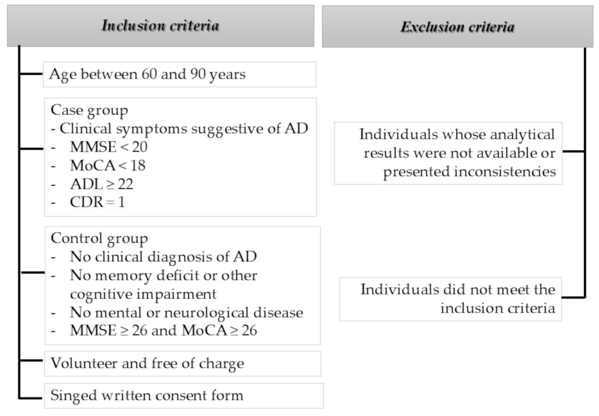
A set of inclusion, exclusion and withdrawal criteria were established and had to be strictly adhered to (Figure 2). The inclusion criteria were (i) age between 60 and 90 years; (ii) clinical symptoms suggestive of AD by the National Institute on Aging—Alzheimer’s Association workgroups on the diagnostic guidelines for AD [22] (in the case group, Mental State Examination (MMSE) < 20 points and Montreal Cognitive Assessment (MoCA) < 18 points; Activity of Daily Living (ADL) Scale was impaired (ADL ≥ 22 points); and Clinical Dementia Rating (CDR) = 1 point; except for changes in AD, there were no other abnormalities); (iii) normal elderly people with no clinical diagnosis of AD in the CG, no memory deficit or other cognitive impairment and no mental or neurological disease (MMSE ≥ 26 and MoCA ≥ 26); (iv) individuals who voluntarily consented to participate free of charge; and (v) singed written consent form. The exclusion criteria were (i) individuals whose analytical results were not available or presented inconsistencies that prevented their inclusion in the study; and (ii) individuals who did not meet the inclusion criteria.

2.5. Socio-Demographic Data and Clinical Data

An individual interview was conducted with case group participants to collect personal, clinical and socio-demographic data. Personal data included first name, surname, date of birth, age and gender. For clinical data, the diagnosis of pAD, date of diagnosis, stage of disease, medical speciality in charge of follow-up and other pathologies such as hypercholesterolemia, DM, HBP and cardiac pathologies were recorded. Sociodemographic data collected included level of education, marital status, employment status, profession, municipality of birth and residence, living with whom, number of people in the living unit, main caregiver and relationship and family history of AD or other dementias.
Participants in the CG underwent a similar interview, with slight variations in the questions related to clinical and socio-demographic data. The clinical data collected included the diseases they suffered from and the medical specialist in charge of their follow-up, as well as other pathologies such as hypercholesterolemia, DM, HBP and cardiac pathologies. The sociodemographic data collected were like those of the case group, with the addition of the question of whether they were primary caregivers of a patient with AD.
We use the term cardiac pathology to refer to a set of pathologies and diseases according to the International Classification of Diseases (ICD), 11th revision (2019) of the major groups of circulatory system diseases of the World Health Organization (WHO) [23]. They include coronary heart disease, a disease of the blood vessels supplying the heart muscle; cerebrovascular disease, a disease of the blood vessels supplying the brain; peripheral arterial disease, a disease of blood vessels supplying the arms and legs; rheumatic heart disease, damage to the heart muscle and heart valves from rheumatic fever, caused by streptococcal bacteria; congenital heart disease, birth defects that affect the normal development and functioning of the heart caused by malformations of the heart structure from birth; and deep vein thrombosis and pulmonary embolism, blood clots in the leg veins, which can dislodge and move to the heart and lungs.

2.6. Statistical Analysis

To analyse the information collected, firstly, a descriptive study was carried out by tabulating the data from the corresponding absolute (number of individuals) and relative (percentage) frequency distributions. This numerical description was completed with the graphical representation of the information, which allows us to draw descriptive conclusions in a more visual way.
As a complement to the descriptive study, an inferential analysis was applied in order to test certain hypotheses. Specifically, to test the hypothesis of equality of means in the case of quantitative variables, we applied the t-test for difference in means for independent samples in normal populations, after testing for equality of variances using Levene’s test. In the case of qualitative variables, the z-test for difference in proportions was applied to test the hypothesis of equality of percentages, or, when the modalities of the variable were more than two, the chi-2 test for independence.
The statistical significance of the results was assessed based on the general criterion of a p-value of less than 0.05, although cases where significance reached a p-value of less than the 1% significance level were also discussed. All procedures were carried out with the statistical software IBM SPSS Statistics for Windows, version 26.0 (IBM Corp., Armonk, NY, USA).

3. Results

The study population consisted of 511 individuals, including 260 pAD and 251 controls. AD patients were recruited from ARAP located in Soria, Salamanca, León and Ponferrada, as well as from “Messengers of Peace” daycare centre in Mansilla de las Mulas (León) and La Bañeza (León).

3.1. Sociodemographic Data

Table 1 presents the socio-demographic characteristics of the study population, with a clear distinction between individuals with AD and those in the CG.

3.1.1. Age and Sex

The average age of all participants for whom data were available for this variable (n = 487) was 76.95 years, with a deviation of ±9.72 years, the median being the same age (77 years). Participants in the case group diagnosed with AD (n = 238) were significantly older in age (83.94 ± 7.55 years) than those in the CG (n = 249; 70.28 ± 6.26 years), applying a t-hypothesis test for the difference in means (p-value < 0.0001).
Regarding to gender, the majority were female, both in the case group (76.15%) and in the CG (83.67%), although the proportion of females was significantly higher in the CG at the 5% level (p-value = 0.0343; z-test for the difference in proportions).
Figure 3 shows the distribution of individuals by sex in each of the groups, with both groups comprised of mostly females.

3.1.2. Marital Status

Regarding marital status, it was observed that in the group of cases diagnosed with AD, widowhood predominated (52.30%), in contrast to the CG where the majority were married (63.56%). Using a chi-2 independence test, significant differences were observed at a level of less than 1% (p-value < 0.0001).
A summary of the distribution of cases and controls according to marital status is shown in Figure 4.

3.1.3. Educational Level

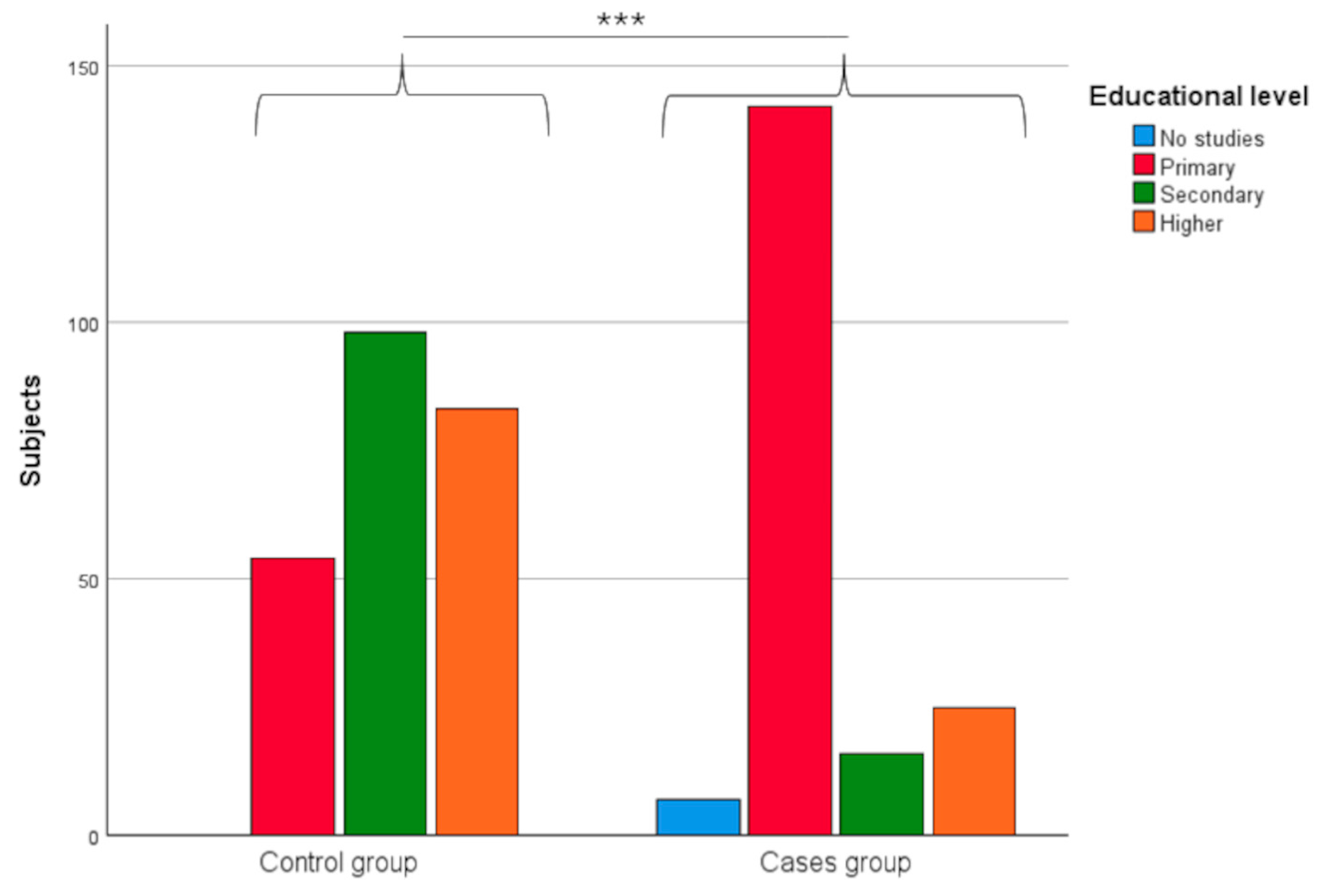
Data were collected on the educational level of the study participants and classified into categories including no education, primary education, secondary education and higher education. The group of cases diagnosed with AD showed a primary educational characterisation (74.74%), compared to the CG, where the majority had a higher secondary and higher educational characterisation (77.02%). This situation implied the existence of significant differences between the two groups, according to the chi-2 independence statistic (p-value < 0.0001).
These differences are shown in Figure 5, which shows the number of individuals in each group according to their level of education.

3.1.4. Residential Area

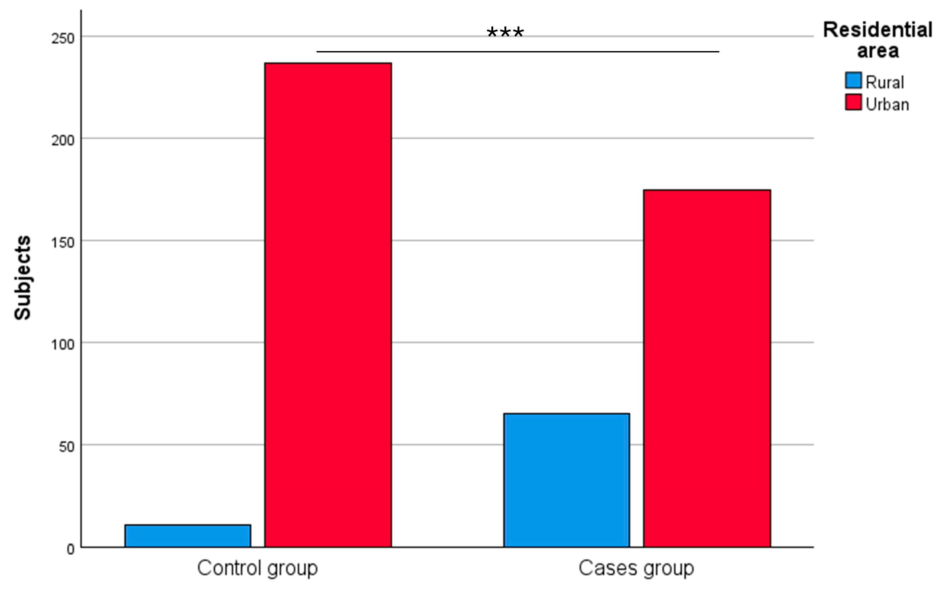
In terms of the residence area, there was a predominance of urban area in both groups (72.92% in the case group and 95.56% in the CG), as shown in Figure 6. However, the percentage of urban residents in the CG was significantly higher than in the group of cases diagnosed with AD (p-value < 0.0001; z-test for difference in proportions).

3.1.5. Family History of Probable Alzheimer’s Disease

The analysis of family history of AD is presented in Figure 7, where the following results are observed. Approximately 2% of individuals in each group were unaware of any family history. As for the rest, 45.60% of cases diagnosed with AD indicated that they had a family history of the disease, while in the CG, the existence of a family history was found in 26.75% of individuals. These percentages revealed the existence of differences in the proportion of family history in each group, which were significant at a level of less than 1% (p-value = 0.0002, chi-2 test of independence).

3.2. Clinical Data

In addition to differences in sociodemographic data, relevant clinical factors in the study participants were analysed. The following section presents the main findings related to medical conditions such as HBP, hypercholesterolaemia, cardiac pathology (according to the ICD, 11th revision of the major groups of circulatory system diseases of the WHO [23]) and DM, as well as their possible association with AD.

3.2.1. High Blood Pressure

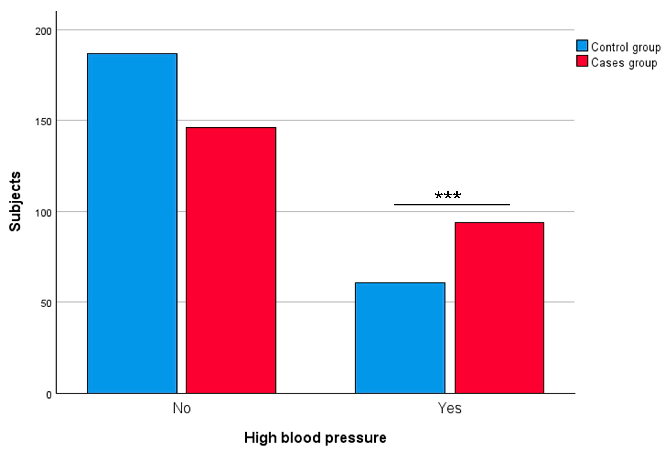
In the group of individuals with AD, 39.17% of them had HBP, a percentage that decreased to 24.60% in the CG. The difference between both percentages was significant at a level below 1% (p-value = 0.0005, z-test for difference in proportions).
Figure 8 shows the status of pathology in the two groups.

3.2.2. Hypercholesterolaemia

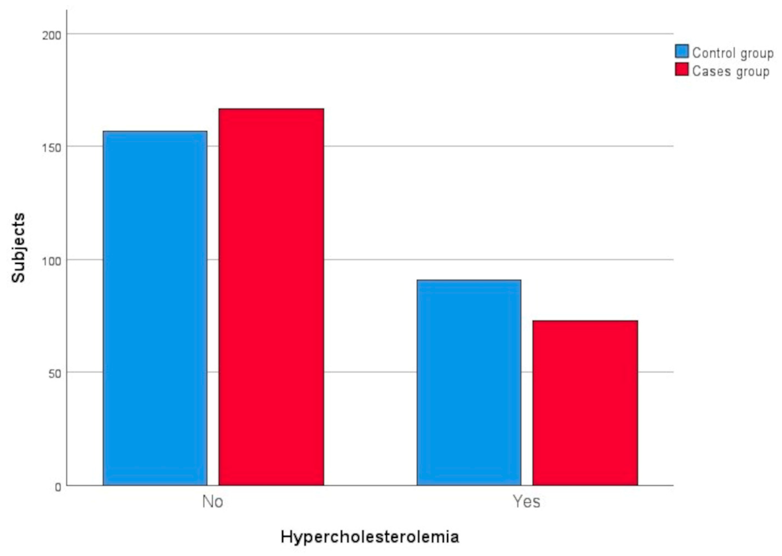
In the case of hypercholesterolaemia, 30.42% of individuals in the group of subjects with AD and 36.69% in the CG suffered from this pathology. In this case, the differences were not significant (p-value = 0.1422; z-test for the difference in proportions).
This situation can be seen in Figure 9, which shows the distribution of individuals according to their pathology by group.

3.2.3. Cardiac Pathology

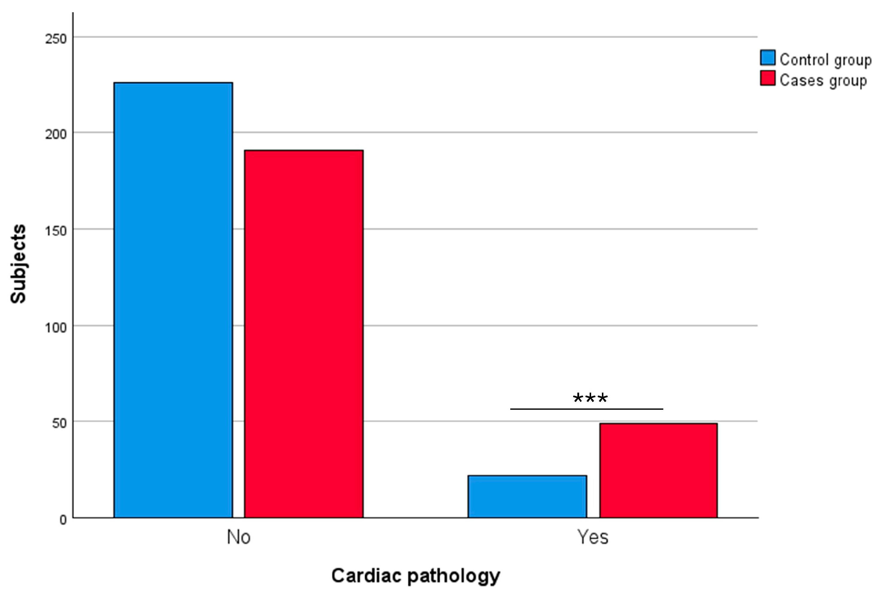
With regard to cardiac pathology, the situation is shown in Figure 10, where it can be observed that the percentage of individuals suffering from this pathology was higher in the group of cases with AD (Supplementary Materials).
Specifically, in this group, the percentage of individuals with cardiac pathology was 20.42%, while in the CG, the percentage was reduced to 8.87%. The difference between the two percentages was statistically significant, applying a z-hypothesis test for the difference in proportions (p-value = 0.0003).

3.2.4. Diabetes Mellitus

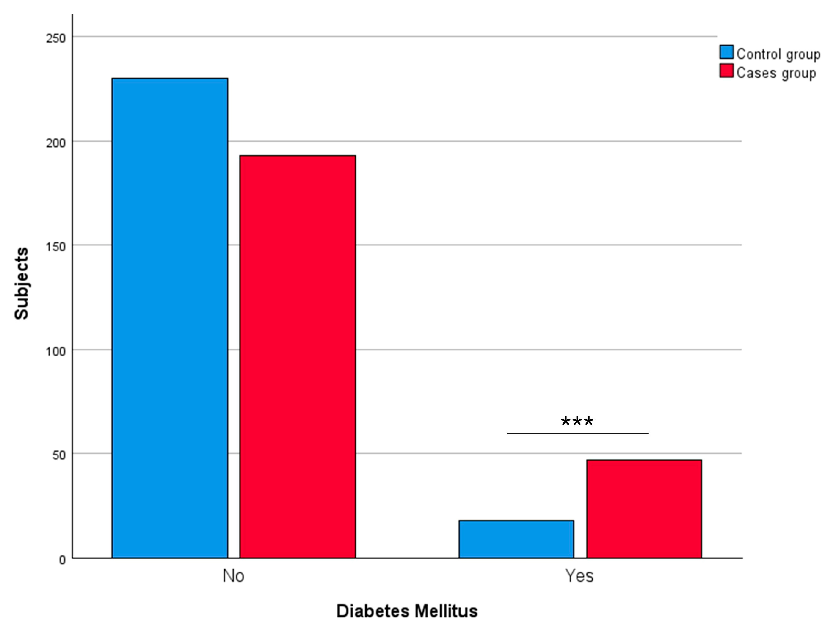
With regard to individuals with DM, 19.58% of the subjects with AD had this pathology, while in the CG, the percentage of individuals with DM was 7.26%. In this line, the percentage of individuals with this pathology was significantly higher in the case group (p-value < 0.0001; z-test for difference in proportions).
Figure 11 shows the situation of DM pathology in the two groups.
The difference in this pathology between cases with AD and healthy individuals is also observed when analysing the glucose level. In the first group, the mean glucose level was 107.67 mg/dL ± 36.52, while in the CG, the mean glucose level was 95.70 mg/dL ± 22.08. A t-contrast was applied for the difference in population means with unknown and unequal variances (p-value < 0.0001; Levene test for equality of variances) and concluded that the mean glucose level was significantly higher in the group of individuals with AD (p-value < 0.0001).
A summary of the clinical data for the two groups of participants in the present study is presented in Table 2.

4. Discussion

The overall aim of this research was to analyse the relationship between some cardiovascular risk factors and AD. The results of this study would provide information that can be used for the design of more effective strategies in personalising future interventions (management and treatment) on AD and cardiovascular diseases in day centres for pAD.

4.1. Socio-Demographic Data of Participants

It is observed from the results that the mean age of the participants was 76.95 years, with participants diagnosed with pAD being significantly older in age than control individuals. The observation that participants in the case group are significantly older than those in the CG reinforces the well-established notion that age remains the most important risk factor for the development of AD [24]. In Spain, the life expectancy in 2022 was 80.4 years for men and 85.7 years for women, highlighting the relevance of aging as a key factor in AD development [25]. Such results are in line with the extensive evidence suggesting that ageing is a major factor driving the development of the AD [24,26,27], which would support therapeutic interventions on the biological mechanisms of aging as precursors of nicotinamide adenine dinucleotide (NAD+), inducers of mitophagy and inhibitors of cellular senescence and stimulation of muscle satellite cells [28,29].
The results of this study revealed a significant prevalence of female gender in both groups, with a statistical association (p = 0.0343), although there were more women in the CG (83.75%) compared to the case group (76.15%). Recent findings [30,31,32] have pointed out that women exhibit a more pronounced development, progression and clinical presentation of AD compared to men. Advanced age is one of the most important risk factors for AD. In this regard, women have a higher prevalence and risk of suffering from AD throughout life than men, mainly due to their longer life expectancy. Hormones and sex chromosomes interact differentially with several AD triggering mechanisms during the aging process such as neuroinflammation, metabolism and autophagy, the microbiome and genetic polymorphisms, among others [31,32]. In this way, the critical factor that contributes to more women suffering from AD than men is due to sex differences in cognition and brain function caused by steroid sex hormones. Estrogen, progesterone and androgen increase substantially the resistance of neurons to AD by preventing amyloidosis, tauopathy and the regulation of gliosis. Based on the high incidence of AD after menopause, exposure to estrogens during a shorter life span may be associated with an increased risk of AD, because it makes the female brain more physiologically vulnerable to the pathophysiology of AD [33], especially of metabolic and inflammatory pathways, increasing vulnerability to AD [6]. Specifically, estrogen appears to play a protective role by reducing Aβ accumulation through the regulation of amyloid precursor protein (APP) processing, while its loss may contribute to increased Aβ burden. In contrast, the gradual decline of testosterone in men may mitigate these effects [34]. Additionally, sex chromosome-linked genetic expression differences may further modulate AD susceptibility. Additionally, women carry two X chromosomes, one of which undergoes incomplete inactivation, leading to a higher expression of certain X-linked genes involved in immunity and neurodegeneration. Some of these genes, such as Usp11, may contribute to increased τ pathology in women by promoting τ acetylation, which impairs its degradation [32]. Of relevance seems to be ApoE4, where those women carrying the ApoE4 genotype had more advanced brain aging than their male counterparts, highlighting the differential influence of AD risk factors in both genders [35,36]. Buckley et al. [37] indicated that, although the prevalence of ApoE4 and brain Aβ peptide burden did not differ by sex, they did observe that women with a high Aβ peptide burden had more pronounced cognitive impairment. This could indicate the involvement of the triggering mechanisms already discussed [31,32]. These interactions would provide differential features in AD progression between men and women. Given all these factors, it would be necessary to consider sex in order to effectively address AD [30].
Additionally, the results of this study also reveal that 74.74% of the group of cases diagnosed with AD have higher primary education compared to the CG, where 77% have secondary and higher education. Regarding education levels in the Spanish population over 65 years old, in 2023, 71.2% have preschool, primary and lower secondary education, 12.2% have upper secondary and post-secondary non-tertiary education, and 16.7% have bachelor’s, master’s and doctoral degrees [38]. The results indicate a significant association between educational level and AD prevalence (p < 0.0001). These results are accordance with those of previous studies [39,40], suggesting that education protects neurocognitive function in preclinical stages of AD [39], and indicating a protective causal effect of educational level on AD [40]. Seyedsalehi et al. [40] showed a significant inverse association between education and AD, noting that a one-standard-deviation increase in years of schooling was associated with a 30% reduction in the likelihood of AD. This is probably due to cognitive reserve, i.e., the ability to acquire alternative brain networks or use structures or neural networks that are not normally used to compensate for brain aging [41]. Perhaps education, which helps improve brain circuits, could increase the reserve of brain networks capable of continuing to function. However, in the study conducted by Hu et al. [42], strong cognitive performance was associated with a 26% decreased likelihood of developing AD, irrespective of the person’s educational status or intelligence. Thus, it is important to note that there is controversy in the scientific literature about the exact role of education as a modifiable risk factor. These discrepancies highlight the need for further detailed research to investigate the relationship between education and AD.

4.2. Alzheimer’s Disease and Cardiovascular Risk Factors

The research reveals a significant relationship between AD and cardiovascular disease, as both entities share not only common risk factors, but also similar molecular mechanisms [17]. A longitudinal study by Lee et al. [43] examined 330 patients with AD, revealing that the most frequent risk factors were HBP (65.8%), DM (46.7%) and physical inactivity (36.7%). Their results indicated that individuals with AD who had more than three cardiovascular risk factors experienced a more rapid cognitive decline over the 3-year follow-up period compared to subjects with no cardiovascular risk factors or fewer than three factors. In a similar vein, Vemuri et al. [44] aimed to investigate the relationships between age, vascular health and imaging biomarkers of AD in a sample of 430 individuals over 60 years of age. These researchers observed that neurodegeneration biomarkers, positron emission tomography (PET) and magnetic resonance imaging (MRI), were significantly more unfavourable (p < 0.05) in the presence of five vascular health markers such as hyperlipidaemia, cardiac arrhythmias, coronary heart disease, congestive heart failure and DM. Thus, their findings indicated that poor vascular health does not significantly affect brain amyloid deposition but has a significant direct and indirect effect on AD neurodegeneration [44]. Probably, these results would highlight the importance of analysing the connections between AD and various cardiovascular risk factors.
The present study focused on an in-depth analysis of crucial factors such as HBP, hypercholesterolaemia, DM and cardiac pathology to better understand their possible involvement in the development and progression of AD. Our analysis may be particularly relevant given the growing evidence suggesting that cardiovascular risk factors play a role in vascular health and may also influence the pathogenesis of AD.

4.2.1. High Blood Pressure and Alzheimer’s Disease

HBP has been identified as a factor affecting the vascular integrity of the blood–brain barrier, resulting in an impaired blood–brain barrier, leading to brain cell damage with increased Aβ-peptide deposition, hypoperfusion and neuroinflammation, and leading to impaired memory abilities [45]. In the epidemiological study conducted by Wu et al. [46], the relationship between HBP and AD was observed, while blood pressure variations in individuals diagnosed with AD before and after the onset of the disease were analysed. The results revealed a significant association between HBP and AD prevalence, finding a higher prevalence of AD in those participants with HBP, especially in the group with systolic blood pressure greater than or equal to 170 mmHg (p < 0.0002). In addition, HBP contributed 28.3% to the incidence of new AD cases, suggesting that approximately 1/3 of new AD cases could be related to elevated blood pressure. They compared blood pressure values between cases diagnosed with AD and individuals without a diagnosis of dementia, finding that the blood pressure values of the AD group were significantly higher than those of the CG (without dementia). The odds ratio (OR) for the group with systolic blood pressure greater than or equal to 140 mmHg and less than 170 mmHg was 1.70, while for the other group with systolic blood pressure greater than 170 mmHg, the OR was 2.52. The results of this study were consistent with the findings obtained in previous studies [45], suggesting a positive association between HBP and AD. In the present investigation, it is observed that the percentage of individuals with HBP in the group of cases with AD (39.19%) is significantly higher compared to the group of healthy individuals (24.60%). This difference in percentages shows a statistically significant association, suggesting that the presence of HBP could be related to AD in the sample studied [46].
Arendonk et al. [47] conducted a recent study focusing on the impact of vascular risk factors and the severity of Aβ-peptide plaques in the brain, measured in vivo in 635 participants of the Rotterdam study. Vascular risk factors were assessed 7 and 13 years prior to brain scanning via PET. Their results showed an association between vascular risk factors and increased levels of brain Aβ-peptide, especially in ApoE4 gene carriers.
Two meta-analyses support the association between high blood pressure and AD [48,49]. Xu et al. [48] found grade I evidence suggesting an association between medical history, such as HBP, hypotension and type 2 DM, and a risk of developing AD. In 2020, the study carried by Ou et al. [49] identified significant associations between HBP in middle-aged individuals and cognitive function. Systolic blood pressure above 130 mmHg was associated with a relative risk (RR) of 1.19 and 1.55 for cognitive impairment. Regarding dementia, moderate evidence showed a significantly elevated risk (RR: 1.20) associated with HBP in these individuals, also with elevated systolic blood pressure (RR: 1.50) and excessive change in diastolic blood pressure (RR: 1.65). The authors indicated that a systolic blood pressure greater than or equal to 140 mmHg, a diastolic blood pressure greater than or equal to 80 mmHg and a change in diastolic blood pressure of more than 5 mmHg were associated with a 37% to 52% increased risk of AD. If the relationship between AD and HBP is accepted, lowering blood pressure levels should have a protective effect. Therefore, Vicario et al. [50] reported that antihypertensive treatments have a beneficial effect on AD, delaying the onset of cognitive decline. Also, Ou et al. [49] collected studies indicating that antihypertensive drugs were associated with a 21% reduction in the risk of dementia.
HBP is a risk factor for developing AD in old age due to an increase in the deposition of Aβ-peptide and neurofibrillary tangles in the cerebral blood vessels [51]. This abnormal deposition produces cerebral hypoperfusion and damages the cerebral blood vessels, deteriorating the blood–brain barrier and glial function early and chronically [52]. In addition, it enhances the ApoE 4 gene, a genetic risk factor for the development of AD [51]. In this sense, antihypertensive drugs are a protective factor against the development of AD thanks to their ability to maintain blood pressure levels within values that do not harm the cerebral microvasculature. For this reason, it would be advisable to have good control and adherence to hypertension treatment to reduce the risk of suffering from AD, unlike untreated or poorly controlled arterial hypertension, which increases the risk due to the accelerated deposition of Aβ-peptide and a greater decrease in cerebral blood flow [53]. However, more clinical studies are needed to confirm this hypothesis with certainty.

4.2.2. Hypercholesterolaemia and Alzheimer’s Disease

Elevated cholesterol is a well-established risk factor for cardiovascular disease. However, the relationship between cholesterol levels and AD is complex and challenging to fully elucidate [54]. Notably, our results obtained did not demonstrate a statistically significant association between AD and a clinical diagnosis of hypercholesterolaemia in the analysed sample.
However, when further exploring blood levels of total cholesterol in relation to AD, significant associations were observed, showing higher TG levels in the case group. In our study, hypercholesterolemia was defined based on medical history and individual interviews, which may have limited the accuracy of participant classification. It is possible that some individuals with elevated cholesterol levels had not been previously diagnosed or were undergoing statin treatment, which could have attenuated the association with AD. Furthermore, previous studies have indicated that the relationship between cholesterol and AD is more evident when evaluating levels in midlife, whereas in older ages, the influence of cholesterol may be modulated by other factors, such as the presence of comorbidities or the use of lipid-lowering medication. In this context, the finding of significantly higher TC levels in the CG suggests that other lipid parameters may play a more relevant role in the pathophysiology of AD.
It should be noted that the association of elevated cholesterol has been observed especially with vascular dementia, not with AD [55]. However, over the decades, from 1990 to the present day, several studies exploring the possible association between hypercholesterolaemia and AD have been carried out with divergent results. Notkola et al. [56] conducted one of the pioneering longitudinal studies on total serum cholesterol and AD in 1998. In this study, they used a sample of 444 participants aged 70–89 years, whose total cholesterol was measured over several decades. The results revealed that participants who developed AD showed elevated cholesterol concentrations in early measurements compared to participants who did not have dementia (p < 0.0008). These findings suggest that individuals with a history of high cholesterol levels in middle age have an increased risk of developing AD [56]. Years later, authors Xu et al. [57] investigated the association of hypercholesterolaemia and AD neuropathology in 3508 subjects with both diseases. Their results showed that hypercholesterolaemia was significantly associated with a higher burden of diffuse neuritic plaques (OR: 1.32) as well as more severe cerebral amyloid angiopathy (OR: 1.26), thus suggesting that hypercholesterolaemia was associated with a higher severity of AD pathology. Following this line, two prospective studies indicated that hypercholesterolaemia is significantly associated with long-term AD incidence [58,59]. In this way, Golstein et al. [59] specified that hypercholesterolaemia and HBP were associated with deficits in visual and memory abilities, verbal reasoning and visuospatial skills. Recently, Pappolla et al. [60] have described a possible pathway between Proprotein convertase subtilisin/kexin type 9 (PCSK9) and the development of AD, because PCSK9 is involved in controlling hypercholesterolemia by modulating levels of low-density lipoprotein (LDL), an established risk factor for the onset of the AD [54]. In addition, PCSK9 causes inflammation and oxidative stress, which alters the histology of the blood-brain barrier and induces the onset of AD. This would imply that PCSK9 stimulates neurodegeneration, because PCSK9 has been found to be expressed in AD by neurons and glia cells [61].

4.2.3. Cardiac Pathology and Alzheimer’s Disease

We have reported that a significant association between cardiac pathology (according to the ICD, 11th revision of the major groups of circulatory system diseases of the WHO [23]) and AD was observed, manifested by a higher percentage of individuals with cardiac pathology in the group diagnosed with AD (29.42%) compared to the CG (8.87%). Our results could suggest a connection between heart disease and AD [62]. Two studies [63,64] have shown that heart failure, in addition to coronary heart disease, may contribute to an increased risk of AD. Mechanisms such as impaired cerebral blood flow, systemic inflammation and amyloid-β accumulation in the brain support this link [63,64].
Additionally, studies have explored the relationship between cardiac functional parameters and AD. In a recent study by Zheng et al. [65], the left ventricular ejection fraction (LVEF), a key indicator of cardiac dysfunction, was associated with cerebrospinal fluid (CSF) biomarkers of AD in cognitively normal individuals. Participants with lower LVEF had significantly higher levels of CSF total τ and an increased τ/Aβ42 ratio, suggesting a link between cardiac function and AD pathology. These associations were particularly evident in women and individuals younger than 65 years old, reinforcing the hypothesis that maintaining cardiac function may be relevant for AD prevention. Furthermore, an experimental study by Murphy et al. [66] using the 5XFAD mouse model of AD demonstrated that these mice exhibited a significant reduction in cardiac fractional shortening and ejection fraction compared to wild-type controls. Additionally, 5XFAD mice showed altered electrical activity, decreased calcium influx in cardiomyocytes, and increased activation of AMP-activated protein kinase (AMPK), indicative of energy metabolism dysfunction.
Liang et al. [67] have investigated various cardiac diseases associated with cognitive impairment. In that study, they conducted a meta-analysis assessing the association of coronary heart disease and cognitive impairment. Their results [67] showed a positive association with an RR of 1.27 between coronary heart disease and the risk of cognitive impairment, as well as an association with an RR of 1.49 between myocardial infarction and the risk of cognitive impairment. However, between angina pectoris and the risk of cognitive impairment, there was no significant association (RR: 0.99). Nevertheless, these authors showed that individuals with coronary heart disease have an increased risk of vascular dementia, but not of AD [67]. Consistently, Starmans et al. [68] examined the relationship of heart disease, including atrial fibrillation, coronary artery disease, heart failure and carotid occlusive disease, with brain Aβ burden. Their findings, through a meta-analysis study, revealed the absence of significant differences in amyloid burden between the affected individuals and the CG. This evidence suggests that, as yet, there are no current studies to support such a relationship [68]. However, Papanastasiou et al. [69] included 43 studies in their meta-analysis, in which they aimed to study the association between atrial fibrillation and cognitive impairment in various dementias. Among their results, they showed that atrial fibrillation was significantly associated with an increased risk of vascular dementia (OR: 1.7) and Alzheimer’s dementia (OR: 1.4). These researchers [69] concluded that atrial fibrillation increases the risk of cognitive impairment, vascular dementia and AD. Also, a recent case-control study [70] investigated core blood biomarkers of AD, specifically, τ protein and Aβ-peptide, in individuals with severe brain injury following cardiac arrest. The study reported elevated levels of Aβ-peptide 40 and Aβ-peptide 42 in the blood 48–72 h post-arrest. The authors [70] concluded that the increase in Aβ peptides may indicate amyloid activation as a response to acute hypoxia-ischemia. Although the study does not establish a direct link between cardiac pathology and AD, it underscores the significance of AD biomarkers, which exhibit distinct dynamic changes after cardiac arrest [70].
The link between AD and cardiac pathologies has emerged as a crucial area of research in the comprehensive understanding of neurodegenerative diseases. The heart, a central organ in the regulation of blood flow and tissue oxygenation, plays a critical role in brain health. In this context, the present section focuses on exploring the interrelationships between heart disease and AD, as recent evidence suggests a connection between them [62]. Therefore, several pathophysiological processes contribute to the increased risk of dementia in people with heart failure. (i) Reduced blood flow: Decreased blood flow to the brain can lead to damage to brain tissue. (ii) Chronic cerebral hypoxia: Reduced cardiac output can lead to prolonged oxygen deprivation to the brain. (iii) Systemic inflammation: Heart failure can lead to widespread inflammation and disruption of small blood vessels in the brain. (iv) Vascular autoregulation: Heart failure can affect the brain’s ability to regulate its own blood supply [71,72].

4.2.4. Diabetes Mellitus and Alzheimer’s Disease

In the early 1990s, the Rotterdam study [73] marked a crucial starting point by establishing the initial link between AD and DM. This research, which included 6330 participants aged 55–59 years, revealed significant data, such as that of the 265 participants diagnosed with dementia, 59 also had DM (22.3%). Multiple regression analysis performed in the research conclusively confirmed a positive association between DM and the incidence of dementia (OR: 1.3). Although the relationship was more evident for vascular dementia, a significant association with AD was also identified [73]. In addition, a subsequent study [74], which included a total of 6370 participants, showed remarkable results. During follow-up, 126 participants developed dementia, of whom 89 had AD. This study revealed that DM almost doubled the risk of dementia and AD, suggesting that DM may play a role in the pathogenesis of AD in a considerable proportion of all patients with dementia [74]. In line with the previously reported studies [73,74], the results of the present investigation reflected similar findings. Of the 240 participants with available data and diagnosed with AD, 47 also had DM, representing a percentage of 19.58%. In comparison, in the group of healthy individuals, the percentage of DM was 7.26% (18/248). This contrast significantly reveals a higher prevalence of DM in the case group compared to the CG.
Arendonk et al. [47] conducted a more recent study focusing on the impact of DM on the severity of Aβ plaques measured in vivo in the brains of Rotterdam study participants [73]. The research group of Arendonk et al. [47] assessed DM in 635 participants without dementia, a few years before performing the PET test. Their findings revealed that DM, 7 years before neuroimaging assessment, was associated with an increased risk of positive amyloid burden. Accordingly, they concluded that DM appears to contribute to the pathophysiology of AD in a general population without dementia. Similarly, Mejía-Arango et al. [75] showed in their study that subjects with DM have an increased risk of developing dementia.
In this investigation, it was observed that participants in the case group had a higher mean glucose level (107.67 mg/dL ± 36.52) compared to the mean glucose level of the CG (95.70 mg/dL ± 22.08). Based on these findings, Arendonk et al. [47] also noted in their results that higher glucose levels, measured in blood 7 years prior to PET, were associated with increased accumulation of brain Aβ-peptide. Specifically, the strongest effects were seen in the posterior cingulate and praecuneus.
Another study [76] assessed cognitive impairment in individuals diagnosed with AD and type 2 DM. They divided 104 patients with AD and DM into two groups according to the anti-diabetic drug therapy administered: group A, whose participants were treated with oral anti-diabetic drugs, and group B, whose participants were treated with insulin combined with other oral anti-diabetic drugs. Cognitive functions were assessed via the Mini Mental Scale Examination (MMSE) and showed that there was a significant worsening in 56.7% of participants in group A compared to 23.2% of participants in group B, compared to baseline MMSE scores. These results suggest that insulin therapy may be effective in slowing cognitive decline in diabetic patients with AD [76].
DM is a risk factor for the development of cognitive impairment and, when there is already some impairment, it is associated with a higher risk of worsening to the point of dementia or of progressing to AD [73,74]. The main mechanisms underlying DM related dementia are cerebral resistance to insulin (causing an alteration in the transmission of this hormone) and amyloidogenesis (accumulation of Aβ-peptide deposits in the brain), without excluding the appearance of other processes related to neuroinflammation or oxidative stress [77]. Chronic hyperglycemia in DM leads to the formation of advanced glycation end products (AGEs), which interact with their receptor (RAGE) and trigger a series of pathological events in the brain. This AGE–RAGE interaction induces oxidative stress and activates inflammatory pathways, promoting the accumulation of Aβ and the τ protein, both hallmark features of AD. Additionally, AGEs have been shown to interfere with neuronal function by increasing neuroinflammation and affecting the integrity of the blood–brain barrier, facilitating Aβ entry into the central nervous system. Together, these processes contribute to the neurodegeneration and cognitive decline observed in patients with both diabetes and AD [78]. As a result of these pathological changes, oxidative damage, chronic inflammation and impaired clearance mechanisms lead to Aβ aggregation and τ, ultimately triggering neurodegeneration and cognitive decline. In short, it has been observed that people with high blood glucose levels also have higher levels of Aβ-peptide, which is related to AD. Also, insulin has a protective role by (i) suppressing Aβ toxicity, (ii) reducing Aβ oligomer formation and (iii) regulating extracellular Aβ degradation through the modulation of insulin-degrading enzyme (IDE) activity. Furthermore, brain insulin regulates the balance between amyloidogenic (APP expression/degradation) and non-amyloidogenic pathways by controlling PI3K/AKT/GSK-3β signalling. However, decreased insulin signalling impairs these protective mechanisms, potentially accelerating AD progression and contributing significantly to cognitive decline and neurodegenerative processes [79].
In short, it has been observed that people with high blood glucose levels also have higher levels of Aβ-peptide, which is related to AD. This could be because people with diabetes have a lower capacity of body tissues, including the nervous system, to use glucose (sugar) and respond to insulin.

5. Conclusions

In this study, participants with AD had a mean age of 83.94 years, with a higher percentage being female (76.15%). The majority were widowed (52.30%) and had a primary level of education (74.74%). In terms of place of residence, there was a predominance of individuals living in urban areas, with 72.94% of the participants living in this environment. In addition, 45% had a history of AD.
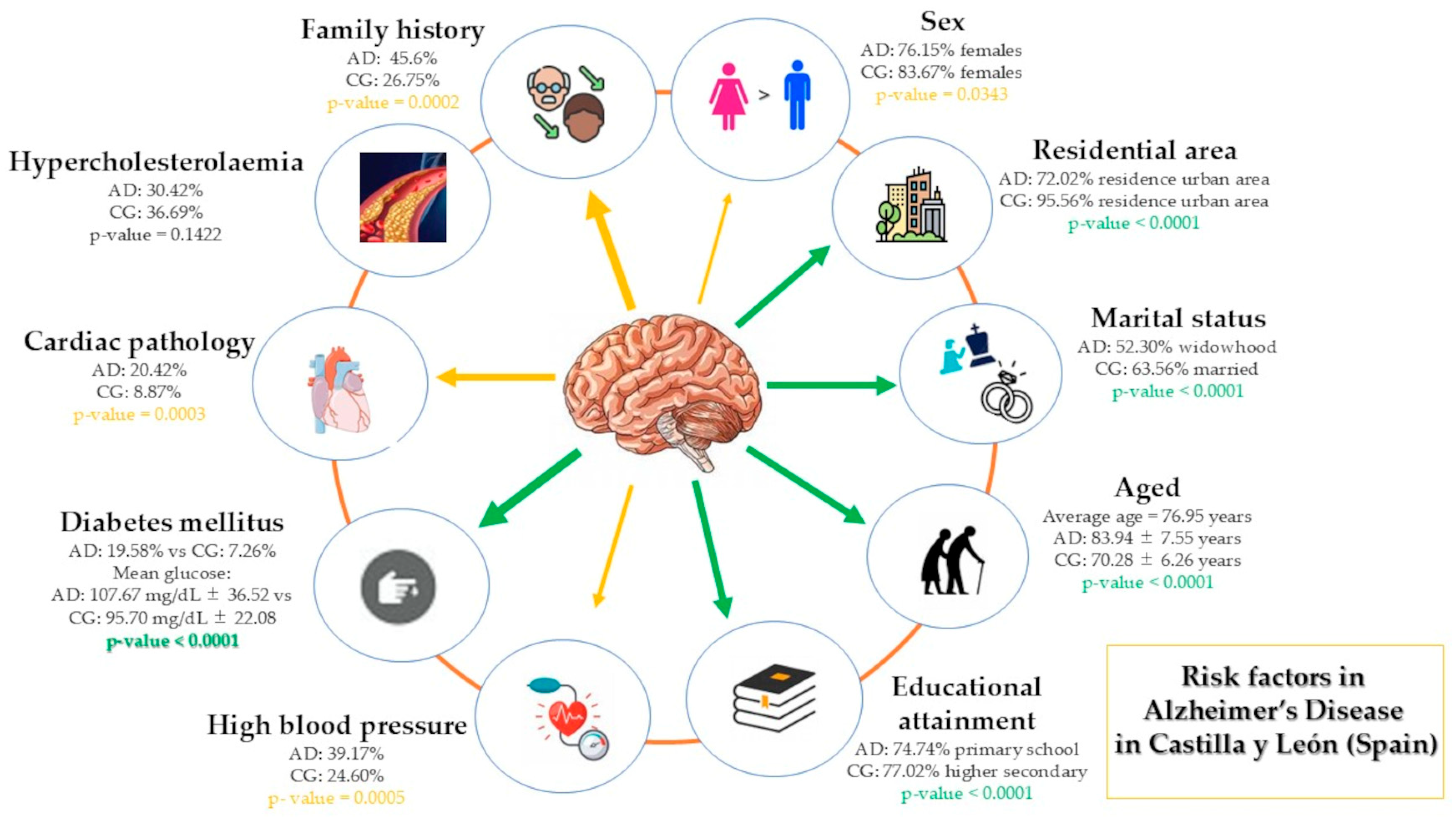
HBP, DM and cardiac pathology are three cardiovascular risk factors that showed significant increased differences in the group of AD cases compared to the group of healthy individuals, suggesting a relationship with AD (Figure 12). However, since this study is an observational case-control study, these results reflect associations and do not establish a causal relationship. Longitudinal and experimental studies are needed to confirm the direction and underlying mechanisms of these associations. Although the underlying mechanisms remain unknown, these cardiovascular factors emerge as relevant in AD.

Supplementary Materials

The following supporting information can be downloaded at: https://www.mdpi.com/article/10.3390/app15063409/s1.

Author Contributions

Conceptualization, J.S.-C., D.F.-L. and L.S.-V.; Data curation, L.B.-C., M.C.S.M. and D.F.-L.; Formal analysis, L.B.-C.; Funding acquisition, L.S.-V., J.S.-C. and D.F.-L.; Investigation, L.B.-C. and D.F.-L.; Methodology, L.B.-C., D.F.-L., M.C.S.M. and E.G.; Project administration, L.S.-V., J.S.-C. and E.G.; Resources, L.B.-C. and L.S.-V.; Software, L.B.-C.; Supervision, J.S.-C., D.F.-L. and L.S.-V.; Validation, J.S.-C. and D.F.-L.; Visualization, J.S.-C. and D.F.-L.; Writing—original draft, D.F.-L. and L.S.-V.; Writing—review and editing, J.S.-C., D.F.-L., M.C.S.M., E.G. and L.S.-V.; Figures/Tables: D.F.-L., M.C.S.M., E.G. and L.S.-V. All authors have read and agreed to the published version of the manuscript.

Funding

This research was funded by the Department of Education of the Junta de Castilla & León and the European Regional Development Fund (ERDF) by TCUE Plan 2024–2028 (grant no. 067/230003). The principal investigator was Prof. Dr. Diego Fernández-Lázaro. In addition, his work was supported by the “Consejo General de Enfermería” (grant ID: PNI_CGE11).

Institutional Review Board Statement

The study was conducted in accordance with the Declaration of Helsinki, and approved by the Ethics Committee of the University of Leon (Spain) (Ref: “ETICA-ULE-021-2022” and date of approval was 5 May 2022). This study was registered in ClinicalTrials.gov (NCT06275243).

Informed Consent Statement

Informed consent was obtained from all subjects involved in the study.

Data Availability Statement

The original contributions presented in the study are included in the article or in Supplementary Materials, further inquiries can be directed to the corresponding author.

Acknowledgments

We would like to thank the AFAs of Castilla y León and in the residences of “Mensajeros de la Paz” for their support, in the recruitment process of the individuals. The authors would like to thank the Neurobiology Research Group of the University of Valladolid (Spain) for their collaboration.

Conflicts of Interest

The authors declare no conflicts of interest.

References

  1. Olivari, B.S.; French, M.E.; Mcguire, L.C. The Public Health Road Map to Respond to the Growing Dementia Crisis. Innov. Aging 2020, 4, igz043. [Google Scholar] [CrossRef] [PubMed]
  2. Sánchez, G.; Rátiva, N.; González, J.; Ramos, T.; Miranda, C.; León, T. La historia olvidada del alzhéimer: Recordando a Oskar Fischer. Rev. Chil. Neuropsiquiatr. 2024, 62, 82–88. [Google Scholar] [CrossRef]
  3. Orgeta, V.; Mukadam, N.; Sommerlad, A.; Livingston, G. The Lancet Commission on Dementia Prevention, Intervention, and Care: A call for action. Ir. J. Psychol. Med. 2019, 36, 85–88. [Google Scholar] [CrossRef] [PubMed]
  4. Deture, M.A.; Dickson, D.W. The neuropathological diagnosis of Alzheimer’s disease. Mol. Neurodegener. 2019, 14, 32. [Google Scholar] [CrossRef] [PubMed]
  5. Rahman, M.M.; Lendel, C. Extracellular protein components of amyloid plaques and their roles in Alzheimer’s disease pathology. Mol. Neurodegener. 2021, 16, 59. [Google Scholar] [CrossRef]
  6. Teleanu, D.M.; Niculescu, A.G.; Lungu, I.I.; Radu, C.I.; Vladâcenco, O.; Roza, E.; Costăchescu, B.; Grumezescu, A.M.; Teleanu, R.I. An Overview of Oxidative Stress, Neuroinflammation, and Neurodegenerative Diseases. Int. J. Mol. Sci. 2022, 23, 5938. [Google Scholar] [CrossRef]
  7. Migliore, L.; Coppedè, F. Gene–environment interactions in Alzheimer disease: The emerging role of epigenetics. Nat. Rev. Neurol. 2022, 18, 643–660. [Google Scholar] [CrossRef]
  8. Behl, T.; Kyada, A.; Roopashree, R.; Nathiya, D.; Arya, R.; Kumar, M.R.; Khalid, M.; Gulati, M.; Sachdeva, M.; Fareed, M.; et al. Epigenetic biomarkers in Alzheimer’s disease: Diagnostic and prognostic relevance. Ageing Res. Rev. 2024, 102, 102556. [Google Scholar] [CrossRef]
  9. Bello-Corral, L.; Seco-Calvo, J.; Molina Fresno, A.; González, A.I.; Llorente, A.; Fernández-Lázaro, D.; Sánchez-Valdeón, L. Prevalence of ApoE Alleles in a Spanish Population of Patients with a Clinical Diagnosis of Alzheimer’s Disease: An Observational Case-Control Study. Medicina 2024, 60, 1941. [Google Scholar] [CrossRef]
  10. Loeffler, D.A. Modifiable, Non-Modifiable, and Clinical Factors Associated with Progression of Alzheimer’s Disease. J. Alzheimers Dis. 2021, 80, 1–27. [Google Scholar] [CrossRef]
  11. Litke, R.; Garcharna, L.C.; Jiwani, S.; Neugroschl, J. Modifiable Risk Factors in Alzheimer Disease and Related Dementias: A Review. Clin. Ther. 2021, 43, 953. [Google Scholar] [CrossRef] [PubMed]
  12. Barnes, D.E.; Yaffe, K. The projected effect of risk factor reduction on Alzheimer’s disease prevalence. Lancet Neurol. 2011, 10, 819–828. [Google Scholar] [CrossRef] [PubMed]
  13. Śliwińska, S.; Jeziorek, M. The role of nutrition in Alzheimer’s disease. Rocz. Panstw. Zakl. Hig. 2021, 72, 29–39. [Google Scholar] [CrossRef] [PubMed]
  14. Valenzuela, P.L.; Castillo-García, A.; Morales, J.S.; de la Villa, P.; Hampel, H.; Emanuele, E.; Lista, S.; Lucia, A. Exercise benefits on Alzheimer’s disease: State-of-the-science. Ageing Res. Rev. 2020, 62, 101108. [Google Scholar] [CrossRef]
  15. Wanleenuwat, P.; Iwanowski, P.; Kozubski, W. Alzheimer’s dementia: Pathogenesis and impact of cardiovascular risk factors on cognitive decline. Postgrad. Med. 2019, 131, 415–422. [Google Scholar] [CrossRef]
  16. Waigi, E.W.; Webb, R.C.; Moss, M.A.; Uline, M.J.; McCarthy, C.G.; Wenceslau, C.F. Soluble and insoluble protein aggregates, endoplasmic reticulum stress, and vascular dysfunction in Alzheimer’s disease and cardiovascular diseases. GeroScience 2023, 45, 1411–1438. [Google Scholar] [CrossRef]
  17. Leszek, J.; Mikhaylenko, E.V.; Belousov, D.M.; Koutsouraki, E.; Szczechowiak, K.; Kobusiak-Prokopowicz, M.; Mysiak, A.; Diniz, B.S.; Somasundaram, S.G.; Kirkland, C.E.; et al. The Links between Cardiovascular Diseases and Alzheimer’s Disease. Curr. Neuropharmacol. 2021, 19, 152–169. [Google Scholar] [CrossRef]
  18. Von Elm, E.; Altman, D.G.; Egger, M.; Pocock, S.J.; Gøtzsche, P.C.; Vandenbroucke, J.P. The Strengthening the Reporting of Observational Studies in Epidemiology (STROBE) statement: Guidelines for reporting observational studies. Ann. Intern. Med. 2007, 147, 573–577. [Google Scholar] [CrossRef]
  19. Faul, F.; Erdfelder, E.; Lang, A.G.; Buchner, A. G*Power 3: A flexible statistical power analysis program for the social, behavioral, and biomedical sciences. Behav. Res. Methods 2007, 39, 175–191. [Google Scholar] [CrossRef]
  20. Serdar, C.C.; Cihan, M.; Yücel, D.; Serdar, M.A. Sample size, power and effect size revisited: Simplified and practical approaches in pre-clinical, clinical and laboratory studies. Biochem. Medica 2021, 31, 1–27. [Google Scholar] [CrossRef]
  21. World Medical Association World Medical Association Declaration of Helsinki: Ethical principles for medical research involving human subjects. JAMA 2013, 310, 2191–2194. [CrossRef] [PubMed]
  22. Jack, C.R.; Albert, M.S.; Knopman, D.S.; McKhann, G.M.; Sperling, R.A.; Carrillo, M.C.; Thies, B.; Phelps, C.H. Introduction to the recommendations from the National Institute on Aging-Alzheimer’s Association workgroups on diagnostic guidelines for Alzheimer’s disease. Alzheimers Dement. 2011, 7, 257–262. [Google Scholar] [CrossRef] [PubMed]
  23. Santos, M.; Almeida, C.; Ferreira De Sousa, L.; Ii, F.; Moreira, P.; Iii, R.; Marques, B.; Iv, S. International Classification of Diseases—11th revision: From design to implementation. Rev. Saude Publica 2020, 54, 104. [Google Scholar] [CrossRef]
  24. Zhao, J.; Huai, J. Role of primary aging hallmarks in Alzheimer’s disease. Theranostics 2023, 13, 197–230. [Google Scholar] [CrossRef]
  25. José, M.; Fructuoso, P.; García Revilla, R.; Moure, O.M.; Moure, R.C. Analysis of Aging in Spain: Contemporary Sociological and Demographic Implications. Societies 2025, 15, 46. [Google Scholar] [CrossRef]
  26. Riedel, B.C.; Thompson, P.M.; Brinton, R.D. Age, APOE and Sex: Triad of Risk of Alzheimer’s Disease. J. Steroid Biochem. Mol. Biol. 2016, 160, 134. [Google Scholar] [CrossRef]
  27. Xia, X.; Jiang, Q.; McDermott, J.; Han, J.D.J. Aging and Alzheimer’s disease: Comparison and associations from molecular to system level. Aging Cell 2018, 17, e12802. [Google Scholar] [CrossRef]
  28. Azam, S.; Haque, M.E.; Balakrishnan, R.; Kim, I.S.; Choi, D.K. The Ageing Brain: Molecular and Cellular Basis of Neurodegeneration. Front. Cell Dev. Biol. 2021, 9, 683459. [Google Scholar] [CrossRef]
  29. Fernández-Lázaro, D.; Garrosa, E.; Seco-Calvo, J.; Garrosa, M. Potential Satellite Cell-Linked Biomarkers in Aging Skeletal Muscle Tissue: Proteomics and Proteogenomics to Monitor Sarcopenia. Proteomes 2022, 10, 29. [Google Scholar] [CrossRef]
  30. Miramontes, S.; Pereda Serras, C.; Woldemariam, S.R.; Khan, U.; Li, Y.; Tang, A.S.; Tsoy, E.; Oskotsky, T.T.; Sirota, M. Alzheimer’s disease as a women’s health challenge: A call for action on integrative precision medicine approaches. NPJ Women’s Health 2024, 2, 17. [Google Scholar] [CrossRef]
  31. Cui, S.S.; Jiang, Q.W.; Chen, S. Di Sex difference in biological change and mechanism of Alzheimer’s disease: From macro- to micro-landscape. Ageing Res. Rev. 2023, 87, 101918. [Google Scholar] [CrossRef] [PubMed]
  32. Lopez-Lee, C.; Torres, E.R.S.; Carling, G.; Gan, L. Mechanisms of sex differences in Alzheimer’s disease. Neuron 2024, 112, 1208–1221. [Google Scholar] [CrossRef] [PubMed]
  33. Zhu, D.; Montagne, A.; Zhao, Z. Alzheimer’s pathogenic mechanisms and underlying sex difference. Cell. Mol. Life Sci. 2021, 78, 4907–4920. [Google Scholar] [CrossRef] [PubMed]
  34. Calvo, N.; Einstein, G. Steroid hormones: Risk and resilience in women’s Alzheimer disease. Front. Aging Neurosci. 2023, 15, 1159435. [Google Scholar] [CrossRef]
  35. Subramaniapillai, S.; Rajagopal, S.; Snytte, J.; Otto, A.R.; Einstein, G.; Rajah, M.N. Sex differences in brain aging among adults with family history of Alzheimer’s disease and APOE4 genetic risk. NeuroImage Clin. 2021, 30, 102620. [Google Scholar] [CrossRef]
  36. Barnes, L.L.; Wilson, R.S.; Bienias, J.L.; Schneider, J.A.; Evans, D.A.; Bennett, D.A. Sex differences in the clinical manifestations of Alzheimer disease pathology. Arch. Gen. Psychiatry 2005, 62, 685–691. [Google Scholar] [CrossRef]
  37. Buckley, R.F.; Mormino, E.C.; Amariglio, R.E.; Properzi, M.J.; Rabin, J.S.; Lim, Y.Y.; Papp, K.V.; Jacobs, H.I.L.; Burnham, S.; Hanseeuw, B.J.; et al. Sex, amyloid, and APOE ε4 and risk of cognitive decline in preclinical Alzheimer’s disease: Findings from three well-characterized cohorts. Alzheimers Dement. 2018, 14, 1193–1203. [Google Scholar] [CrossRef]
  38. Valdés, M.T.; Requena, M. The effect of the age at school entry on educational attainment and field of study: An analysis using the Spanish census. High. Educ. 2024, 87, 1061–1083. [Google Scholar] [CrossRef]
  39. Zhu, W.; Gao, Z.; Li, H.; Huang, Z.; Li, X.; Wang, H.; Wu, X.; Tian, Y.; Zhou, S.; Li, X.; et al. Education reduces cognitive dysfunction in Alzheimer’s disease by changing regional cerebral perfusion: An in-vivo arterial spin labeling study. Neurol. Sci. 2023, 44, 2349–2361. [Google Scholar] [CrossRef]
  40. Seyedsalehi, A.; Warrier, V.; Bethlehem, R.A.I.; Perry, B.I.; Burgess, S.; Murray, G.K. Educational attainment, structural brain reserve and Alzheimer’s disease: A Mendelian randomization analysis. Brain 2023, 146, 2059–2074. [Google Scholar] [CrossRef]
  41. Larsson, S.C.; Traylor, M.; Malik, R.; Dichgans, M.; Burgess, S.; Markus, H.S. Modifiable pathways in Alzheimer’s disease: Mendelian randomisation analysis. BMJ 2017, 359, j5375. [Google Scholar] [CrossRef] [PubMed]
  42. Hu, Y.; Zhang, Y.; Zhang, H.; Gao, S.; Wang, L.; Wang, T.; Han, Z.; Sun, B.L.; Liu, G. Cognitive performance protects against Alzheimer’s disease independently of educational attainment and intelligence. Mol. Psychiatry 2022, 27, 4297–4306. [Google Scholar] [CrossRef]
  43. Lee, W.J.; Liao, Y.C.; Wang, Y.F.; Lin, Y.S.; Wang, S.J.; Fuh, J.L. Summative Effects of Vascular Risk Factors on the Progression of Alzheimer Disease. J. Am. Geriatr. Soc. 2020, 68, 129–136. [Google Scholar] [CrossRef] [PubMed]
  44. Vemuri, P.; Lesnick, T.G.; Przybelski, S.A.; Knopman, D.S.; Lowe, V.J.; Graff-Radford, J.; Roberts, R.O.; Mielke, M.M.; Machulda, M.M.; Petersen, R.C.; et al. Age, Vascular Health, and Alzheimer’s disease Biomarkers in an Elderly Sample. Ann. Neurol. 2017, 82, 706. [Google Scholar] [CrossRef] [PubMed]
  45. Carnevale, D.; Mascio, G.; D’Andrea, I.; Fardella, V.; Bell, R.D.; Branchi, I.; Pallante, F.; Zlokovic, B.; Yan, S.S.; Lembo, G. Hypertension induces brain β-amyloid accumulation, cognitive impairment, and memory deterioration through activation of receptor for advanced glycation end products in brain vasculature. Hypertension 2012, 60, 188–197. [Google Scholar] [CrossRef]
  46. Wu, C.; Zhou, D.; Wen, C.; Zhang, L.; Como, P.; Qiao, Y. Relationship between blood pressure and Alzheimer’s disease in Linxian County, China. Life Sci. 2003, 72, 1125–1133. [Google Scholar] [CrossRef]
  47. van Arendonk, J.; Neitzel, J.; Steketee, R.M.E.; van Assema, D.M.E.; Vrooman, H.A.; Segbers, M.; Ikram, M.A.; Vernooij, M.W. Diabetes and hypertension are related to amyloid-beta burden in the population-based Rotterdam Study. Brain 2023, 146, 337–348. [Google Scholar] [CrossRef]
  48. Xu, W.; Tan, L.; Wang, H.F.; Jiang, T.; Tan, M.S.; Tan, L.; Zhao, Q.F.; Li, J.Q.; Wang, J.; Yu, J.T. Meta-analysis of modifiable risk factors for Alzheimer’s disease. J. Neurol. Neurosurg. Psychiatry 2015, 86, 1299–1306. [Google Scholar] [CrossRef]
  49. Ou, Y.N.; Tan, C.C.; Shen, X.N.; Xu, W.; Hou, X.H.; Dong, Q.; Tan, L.; Yu, J.T. Blood Pressure and Risks of Cognitive Impairment and Dementia: A Systematic Review and Meta-Analysis of 209 Prospective Studies. Hypertension 2020, 76, 217–225. [Google Scholar] [CrossRef]
  50. Vicario, A.; Cerezo, G.H.; Coca, A.; Gaseck, D.; Zaninelli, A.; Lovic, D.; Manios, E.; Hering, D.; Sierra, C.; Cunha, P. Efectos del tratamiento y las drogas anti-hipertensivas sobre la función cognitiva: Deterioro cognitivo, demencia y enfermedad de Alzheimer. Rev. Fed. Argentina Cardiol. 2019, 48, 98–106. [Google Scholar]
  51. Rodrigue, K.M.; Rieck, J.R.; Kennedy, K.M.; Devous, M.D.; Diaz-Arrastia, R.; Park, D.C. Risk factors for β-amyloid deposition in healthy aging: Vascular and genetic effects. JAMA Neurol. 2013, 70, 600–606. [Google Scholar] [CrossRef] [PubMed]
  52. Muller, M.; Van Der Graaf, Y.; Visseren, F.L.; Mali, W.P.T.M.; Geerlings, M.I. Hypertension and longitudinal changes in cerebral blood flow: The SMART-MR study. Ann. Neurol. 2012, 71, 825–833. [Google Scholar] [CrossRef] [PubMed]
  53. Wharton, W.; Goldstein, F.C.; Tansey, M.G.; Brown, A.L.; Tharwani, S.D.; Verble, D.D.; Cintron, A.; Kehoe, P.G. Rationale and Design of the Mechanistic Potential of Antihypertensives in Preclinical Alzheimer’s (HEART) Trial. J. Alzheimers Dis. 2018, 61, 815–824. [Google Scholar] [CrossRef] [PubMed]
  54. Testa, G.; Giannelli, S.; Staurenghi, E.; Cecci, R.; Floro, L.; Gamba, P.; Sottero, B.; Leonarduzzi, G. The Emerging Role of PCSK9 in the Pathogenesis of Alzheimer’s Disease: A Possible Target for the Disease Treatment. Int. J. Mol. Sci. 2024, 25, 13637. [Google Scholar] [CrossRef]
  55. Kalmijn, S.; Foley, D.; White, L.; Burchfiel, C.M.; Curb, J.D.; Petrovitch, H.; Ross, G.W.; Havlik, R.J.; Launer, L.J. Metabolic cardiovascular syndrome and risk of dementia in Japanese-American elderly men. The Honolulu-Asia aging study. Arterioscler. Thromb. Vasc. Biol. 2000, 20, 2255–2260. [Google Scholar] [CrossRef]
  56. Notkola, I.L.; Sulkava, R.; Pekkanen, J.; Erkinjuntti, T.; Ehnholm, C.; Kivinen, P.; Tuomilehto, J.; Nissinen, A. Serum total cholesterol, apolipoprotein E epsilon 4 allele, and Alzheimer’s disease. Neuroepidemiology 1998, 17, 14–20. [Google Scholar] [CrossRef]
  57. Xu, C.; Apostolova, L.G.; Oblak, A.L.; Gao, S. Association of Hypercholesterolemia with Alzheimer’s Disease Pathology and Cerebral Amyloid Angiopathy. J. Alzheimers Dis. 2020, 73, 1305. [Google Scholar] [CrossRef]
  58. Kivipelto, M.; Helkala, E.L.; Laakso, M.P.; Hänninen, T.; Hallikainen, M.; Alhainen, K.; Iivonen, S.; Mannermaa, A.; Tuomilehto, J.; Nissinen, A.; et al. Apolipoprotein E epsilon4 allele, elevated midlife total cholesterol level, and high midlife systolic blood pressure are independent risk factors for late-life Alzheimer disease. Ann. Intern. Med. 2002, 137, 149–155. [Google Scholar] [CrossRef]
  59. Goldstein, F.C.; Ashley, A.V.; Endeshaw, Y.W.; Hanfelt, J.; Lah, J.J.; Levey, A.I. Effects of hypertension and hypercholesterolemia on cognitive functioning in patients with alzheimer disease. Alzheimer Dis. Assoc. Disord. 2008, 22, 336–342. [Google Scholar] [CrossRef]
  60. Pappolla, M.A.; Refolo, L.; Sambamurti, K.; Zambon, D.; Duff, K. Hypercholesterolemia and Alzheimer’s Disease: Unraveling the Connection and Assessing the Efficacy of Lipid-Lowering Therapies. J. Alzheimers Dis. 2024, 101, S371–S393. [Google Scholar] [CrossRef]
  61. O’Connell, E.M.; Lohoff, F.W. Proprotein Convertase Subtilisin/Kexin Type 9 (PCSK9) in the Brain and Relevance for Neuropsychiatric Disorders. Front. Neurosci. 2020, 14, 548891. [Google Scholar] [CrossRef]
  62. Cermakova, P.; Eriksdotter, M.; Lund, L.H.; Winblad, B.; Religa, P.; Religa, D. Heart failure and Alzheimer’s disease. J. Intern. Med. 2014, 277, 406. [Google Scholar] [CrossRef]
  63. Al-Kuraishy, H.M.; Sulaiman, G.M.; Mohammed, H.A.; Mohammed, S.G.; Al-Gareeb, A.I.; Albuhadily, A.K.; Dawood, R.A.; Al Ali, A.; Abu-Alghayth, M.H. Amyloid-β and heart failure in Alzheimer’s disease: The new vistas. Front. Med. 2025, 12, 1494101. [Google Scholar] [CrossRef]
  64. Arega, Y.; Shao, Y. Heart failure and late-onset Alzheimer’s disease: A Mendelian randomization study. Front. Genet. 2022, 13, 1015674. [Google Scholar] [CrossRef]
  65. Zheng, Y.M.; Zhao, Y.Y.; Zhang, T.; Hou, X.H.; Bi, Y.L.; Ma, Y.H.; Xu, W.; Shen, X.N.; Dong, Q.; Tan, L.; et al. Left Ventricular Ejection Fraction and Cerebrospinal Fluid Biomarkers of Alzheimer’s Disease Pathology in Cognitively Normal Older Adults: The CABLE Study. J. Alzheimers Dis. 2021, 81, 743–750. [Google Scholar] [CrossRef]
  66. Murphy, J.; Le, T.N.V.; Fedorova, J.; Yang, Y.; Krause-Hauch, M.; Davitt, K.; Zoungrana, L.I.; Fatmi, M.K.; Lesnefsky, E.J.; Li, J.; et al. The Cardiac Dysfunction Caused by Metabolic Alterations in Alzheimer’s Disease. Front. Cardiovasc. Med. 2022, 9, 850538. [Google Scholar] [CrossRef]
  67. Liang, X.; Huang, Y.; Han, X. Associations between coronary heart disease and risk of cognitive impairment: A meta-analysis. Brain Behav. 2021, 11, e02108. [Google Scholar] [CrossRef]
  68. Starmans, N.L.P.; Leeuwis, A.E.; Biessels, G.J.; Kappelle, L.J.; van der Flier, W.M.; Tolboom, N. Cerebral amyloid-β deposition in patients with heart disease or carotid occlusive disease: A systematic review and meta-analysis. J. Neurol. Sci. 2023, 445, 120551. [Google Scholar] [CrossRef]
  69. Papanastasiou, C.A.; Theochari, C.A.; Zareifopoulos, N.; Arfaras-Melainis, A.; Giannakoulas, G.; Karamitsos, T.D.; Palaiodimos, L.; Ntaios, G.; Avgerinos, K.I.; Kapogiannis, D.; et al. Atrial Fibrillation Is Associated with Cognitive Impairment, All-Cause Dementia, Vascular Dementia, and Alzheimer’s Disease: A Systematic Review and Meta-Analysis. J. Gen. Intern. Med. 2021, 36, 3122–3135. [Google Scholar] [CrossRef]
  70. Ashton, N.J.; Moseby-Knappe, M.; Benedet, A.L.; Grötschel, L.; Lantero-Rodriguez, J.; Karikari, T.K.; Hassager, C.; Wise, M.P.; Stammet, P.; Kjaergaard, J.; et al. Alzheimer Disease Blood Biomarkers in Patients With Out-of-Hospital Cardiac Arrest. JAMA Neurol. 2023, 80, 388–396. [Google Scholar] [CrossRef]
  71. Agrawal, S.; Schneider, J.A. Vascular pathology and pathogenesis of cognitive impairment and dementia in older adults. Cereb. Circ.-Cogn. Behav. 2022, 3, 100148. [Google Scholar] [CrossRef] [PubMed]
  72. Saeed, A.; Lopez, O.; Cohen, A.; Reis, S.E. Cardiovascular Disease and Alzheimer’s Disease: The Heart–Brain Axis. J. Am. Heart Assoc. 2023, 12, 30780. [Google Scholar] [CrossRef]
  73. Ott, A.; Stolk, R.P.; Hofman, A.; Van Harskamp, F.; Grobbee, D.E.; Breteler, M.M.B. Association of diabetes mellitus and dementia: The Rotterdam Study. Diabetologia 1996, 39, 1392–1397. [Google Scholar] [CrossRef] [PubMed]
  74. Ott, A.; Stolk, R.P.; Van Harskamp, F.; Pols, H.A.P.; Hofman, A.; Breteler, M.M.B. Diabetes mellitus and the risk of dementia: The Rotterdam Study. Neurology 1999, 53, 1937–1942. [Google Scholar] [CrossRef]
  75. Mejía-Arango, S.; Zúñiga-Gil, C. Diabetes mellitus as a risk factor for dementia in the Mexican elder population. Rev. Neurol. 2011, 53, 397–405. [Google Scholar] [CrossRef]
  76. Plastino, M.; Fava, A.; Pirritano, D.; Cotronei, P.; Sacco, N.; Sperlì, T.; Spanò, A.; Gallo, D.; Mungari, P.; Consoli, D.; et al. Effects of insulinic therapy on cognitive impairment in patients with Alzheimer disease and diabetes mellitus type-2. J. Neurol. Sci. 2010, 288, 112–116. [Google Scholar] [CrossRef]
  77. Singh, D.D.; Shati, A.A.; Alfaifi, M.Y.; Elbehairi, S.E.I.; Han, I.; Choi, E.-H.; Yadav, D.K.; Singh, D.D.; Shati, A.A.; Alfaifi, M.Y.; et al. Development of Dementia in Type 2 Diabetes Patients: Mechanisms of Insulin Resistance and Antidiabetic Drug Development. Cells 2022, 11, 3767. [Google Scholar] [CrossRef]
  78. Kong, Y.; Wang, F.; Wang, J.; Liu, C.; Zhou, Y.; Xu, Z.; Zhang, C.; Sun, B.; Guan, Y. Pathological Mechanisms Linking Diabetes Mellitus and Alzheimer’s Disease: The Receptor for Advanced Glycation End Products (RAGE). Front. Aging Neurosci. 2020, 12, 217. [Google Scholar] [CrossRef]
  79. Kciuk, M.; Kruczkowska, W.; Gałęziewska, J.; Wanke, K.; Kałuzińska-Kołat, Ż.; Aleksandrowicz, M.; Kontek, R. Alzheimer’s Disease as Type 3 Diabetes: Understanding the Link and Implications. Int. J. Mol. Sci. 2024, 25, 11955. [Google Scholar] [CrossRef]
Figure 1. Pathophysiological connections between Alzheimer’s disease and cardiometabolic comorbidities.
Applsci 15 03409 g001
Figure 2. Flow chart: inclusion and exclusion criteria. Abbreviations = MMSE: Mental State Examination; MoCA: Montreal Cognitive Assessment; ADL: Activity of Daily Living; CDR: Clinical Dementia Rating (CDR); AD: Alzheimer’s disease.
Applsci 15 03409 g002
Figure 3. Distribution of individuals by sex according to group. A significantly higher proportion of females was observed in the control group compared to the cases group. * p < 0.05.
Applsci 15 03409 g003
Figure 4. Distribution of individuals by marital status according to group. Significant differences were observed between the control group and the cases group. *** p-value < 0.0001.
Applsci 15 03409 g004
Figure 5. Distribution of individuals by educational level according to group. Significant differences were observed between the control group and the cases group. *** p-value < 0.0001.
Applsci 15 03409 g005
Figure 6. Distribution of individuals by municipality of residence according to group. The proportion of urban residents was significantly higher in the control group compared to the cases group *** p < 0.0001.
Applsci 15 03409 g006
Figure 7. History of Alzheimer’s disease by group. Significant differences were observed between the control group and the cases group *** p < 0.001.
Applsci 15 03409 g007
Figure 8. High blood pressure according to group. Significant differences were observed between the control group and the cases group, with a higher prevalence of HBP in the cases group. *** p < 0.001.
Applsci 15 03409 g008
Figure 9. Hypercholesterolemia according to group.
Applsci 15 03409 g009
Figure 10. Cardiac pathology according to group. Significant differences were observed between the control group and the cases group, with a higher prevalence of cardiac pathology in the cases group. *** p < 0.001.
Applsci 15 03409 g010
Figure 11. Diabetes mellitus according to group. Significant differences were observed between the control group and the cases group, with a higher prevalence of diabetes mellitus in the cases group. *** p < 0.0001.
Applsci 15 03409 g011
Figure 12. Link between cardiovascular risk and Alzheimer’s disease in Castilla y León, Spain. Abbreviations = AD: Alzheimer’s disease; CG: control group as healthy individuals.
Applsci 15 03409 g012
Table 1. Socio-demographic information of the study participants per group.
CharacteristicspAD Case GroupControl Groupp-Value
Frequency%Frequency%
GenderMen6223.854116.330.0343 *
Women19876.1521083.67
Civil statusSingle125.02228.91<0.0001 ***
Married10041.8415763.56
Widowed12552.305020.24
Divorced10.42135.26
Separated10.4252.02
Not available214
Educational levelWithout studies73.6800<0.0001 ***
Primary14274.745422.98
Secondary168.429841.70
Higher2513.168335.32
Not available7016
Residential areaRural6527.58114.44<0.0001 ***
Urban17572.9223795.56
Not available203
Family history of ADYes8845.606526.750.0002 ***
No10152.3017471.60
Unknown42.1041.65
Not available88
FrequencyMean (SD)FrequencyMean (SD)
Age23883.94 (7.55)24970.28 (6.26)<0.0001 ***
Abbreviation: pAD: probable Alzheimer’s disease; SD: standard deviation; (–) no data. Values are expressed as the frequency (percentage) for categorical variables, while mean and standard deviation for quantitative variables. z-test for variables of gender and residential area; chi-2 test for variables of civil status, educational level and family history of AD; t-test for independent samples with unequal variances for variable of age. Differences were statistically significant at * p < 0.05; *** p < 0.001.
Table 2. Clinical information of the study participants per group.
Medical Diseases and ConditionspAD GroupControl Groupp-Value
Frequency%Frequency%
High blood pressureYes9439.176124.600.0005 ***
No14660.8318775.40
Not available203
HypercholesterolemiaYes7330.429136.690.1422
No16769.5815763.31
Not available203
Cardiac pathologyYes4920.42228.870.0003 ***
No19179.5822691.13
Not available203
Diabetes mellitusYes4719.58187.26<0.0001 ***
No19380.4223092.74
Not available203
Abbreviation: pAD: probable Alzheimer’s disease; enumeration data were subjected to the Chi-square test and described as percentages. Differences were statistically significant at *** p < 0.001.
Disclaimer/Publisher’s Note: The statements, opinions and data contained in all publications are solely those of the individual author(s) and contributor(s) and not of MDPI and/or the editor(s). MDPI and/or the editor(s) disclaim responsibility for any injury to people or property resulting from any ideas, methods, instructions or products referred to in the content.