Article

Shielding Efficacy of Tungsten Oxide-Reinforced Polyisoprene in Attenuating Technetium-99m Gamma Radiation: An Alternative Shielding Solution for Occupational Safety in Nuclear Medicine

by
Suphalak Khamruang Marshall
*,
Jarasrawee Chuaymuang
,
Poochit Kwandee
and
Nueafa Songphum
Department of Radiology, Faculty of Medicine, Prince of Songkla University, Songkhla 90110, Thailand
*
Author to whom correspondence should be addressed.
Appl. Sci. 2025, 15(7), 3892; https://doi.org/10.3390/app15073892
Submission received: 3 March 2025 / Revised: 24 March 2025 / Accepted: 28 March 2025 / Published: 2 April 2025
(This article belongs to the Section Materials Science and Engineering)

Abstract

:
Tungsten oxide (WO3) is a high-density material with exceptional radiation attenuation properties, making it a strong candidate for advanced shielding applications. This study explores the structural, mechanical, and shielding performance of WO3-reinforced polyisoprene composites. Morphological analysis reveals a plate-like structure, indicating robust interfacial interactions that enhance mechanical integrity and thermal stability. X-ray diffraction confirms the crystalline nature of WO3, while Fourier transform infrared spectroscopy detects distinct W–O bond absorption bands, validating uniform dispersion. Computational analysis using XCOM demonstrates remarkable improvements in attenuation properties, particularly at intermediate- and high-photon energies. While PbO2 outperforms at lower energies due to the photoelectric effect, Phy-X/PSD analysis confirms that composites with ≥75% WO3 offer strong shielding capabilities. Variations in effective atomic number, linear attenuation coefficient, and mass attenuation coefficient establish WO3-reinforced NR as a compelling lead-free alternative, especially for Tc-99m applications. Experimental findings further reveal that increasing WO3 content significantly reduces Tc-99m gamma radiation dose equivalents Hp(0.07), Hp(3), and Hp(10), emphasizing the potential of WO3-reinforced composites for next-generation radiation shielding solutions.

1. Introduction

Medical imaging is critical in healthcare since it provides significant benefits for the diagnosis and treatment of patients. As a result, spurred by its many applications and advances in medical technology, the worldwide usage of ionizing radiation in healthcare has grown rapidly [1]. Technetium-99m (Tc-99m) is a medical radioisotope created from Molybdenum-99 (Mo-99), and it is the most commonly utilized medical radioisotope in nuclear medicine [2]. Tc-99m offers various advantages, including a half-life of around six hours, which limits patient exposure, and it is swiftly eliminated from the patient’s body, reducing any potential harm [3]. There is also a monoenergetic gamma-ray emission photopeak of 140.5 keV, an external photon output of 90%, and images recorded using a SPECT, PET, or PET/CT scan. The Organization for Economic Co-operation and Development (OECD) stated in 2019 that worldwide Tc-99m is applied in 85% of diagnostic scans, equating to approximately 30 million patient scans annually [4]. At the 1000 bed Songklanagarind Hospital, Thailand, approximately 5000 scans are performed annually, utilizing Tc-99m mainly for bone scintigraphy [5].
Consequently, as the number of ionizing radiation scans increases, there is growing concern regarding increased exposure levels’ impact on medical staff and patients. This increase is a critical component that may result in elevated cumulative radiation exposure for medical personnel [6,7]. Between 1965 and 1994, a dosimetry database of 109,019 medical workers has established that medical radiation professionals had an elevated risk of lung cancer, with a greater prevalence in males compared to women [8]. Furthermore, in 2004, research published in The Lancet sought to estimate the probability of cancer from X-rays. The findings placed the probability between 0.6% and 1.8%, and in Japan, greater than 3% [9]. However, more recently, some 20 years later, an update by Picano, Eugenio, and Eliseo Vano stated that the average radiation exposure from diagnostic and interventional procedures has now risen significantly, from 0.50 mSv to 2.29 mSv per capita annually [10]. Of concern is that this updated evidence indicates that the cancer risk has doubled, with current exposure increasing the risk by 4.1%, and 8.2% when applying the latest risk estimates. Including cardiovascular effects, the total additional risk now exceeds 10%. Primary data for radiation protection standards derive from studies of individuals exposed to high doses of ionizing radiation. However, a recent study of nuclear workers in France, the UK, and the US reveals a linear increase in cancer rates with radiation exposure, with some evidence suggesting a steeper dose–response relationship at lower doses [11,12]. In addition, an epidemiological study on populations living near nuclear medicine facilities highlights an increased risk from ionizing radiation, including higher incidences of leukemia, thyroid cancer, genetic disorders, and other health anomalies [13].
According to the linear no-threshold (LNT) model, all exposure to ionizing radiation raises cancer risk proportionally without a predetermined safe threshold, no matter how minor. This concept has proven useful in radiation protection methods. While the LNT model offers a precautionary framework supported by high-dose epidemiological data, its extrapolation to low-dose exposures remain contentious. There is increasing evidence indicating a direct correlation between radiation exposures and rising cancer incidence. The intricacies of biological reactions to low-dose ionizing radiation (LDIR) and epidemiological ambiguities highlight the need for more studies to create more precise risk assessment models [14,15].
Despite the concerns regarding the potential risks associated with LDIR, there is still a lack of safety awareness within the medical community. For example, a survey of 384 healthcare workers revealed that a high proportion have an inadequate understanding of radiation exposure safety. Furthermore, the majority of the participants in the survey exhibited insufficient adherence to radiation safety protocols [16]. Similarly, Shubayr reported nurses exhibiting poorer radiation safety knowledge and compliance but higher risk perceptions than other healthcare professionals, thereby requiring interventions that link threat perception to safety behavior [17]. In addition, a global survey of 258 interventional echocardiographers revealed significant variability in radioprotection measures, with respondents underusing protective devices, receiving limited training, and being poorly aware of radiation doses [18]. As emphasized by the call for action from international scientific societies, improved radioprotection training, dose assessment, and shielding are urgently needed.
Consequently, radiation shielding is increasingly an essential component of contemporary radiation protection strategies since it mitigates the risks associated with ionizing radiation. This encompasses radiation source shielding supplied by the equipment manufacturer and structural shielding for rooms containing diagnostic or radiation therapy apparatuses. Moreover, medical operations often use lead-based materials, including lead aprons, portable lead shields, lead spectacles, and fixed lead barriers, to facilitate radiation attenuation. The ALARA (As Low As Reasonably Achievable) principle was established to maintain radiation exposure at minimal levels to reduce dangers while fulfilling diagnostic or therapeutic objectives. It is based on the guidelines of the International Commission on Radiological Protection (ICRP) Publication 9 from 1966, which underscored the need of maintaining radiation doses to be as low and as feasible in practice to mitigate hazards while still fulfilling diagnostic or therapeutic objectives [19]. It is predicated on the linear no-threshold concept (LNT), which posits that even slight radiation exposure has some danger and should be mitigated. As a result, there is greater appeal in developing novel radiation shielding materials, with a significant focus on the structure–property relationships of composites infused with metal oxides. This research provides valuable insights into the development and performance of metal oxide-infused composites for radiation shielding purposes [20,21,22,23].
Polyisoprene (Natural Rubber, NR) is primarily composed of polyisoprene, itself comprising repeat isoprene (C5H8) units. As the main elements of polyisoprene are carbon, with an atomic number of 6, and hydrogen, with an atomic number of 1, it is ineffective as a gamma radiation shield. Consequently, studies have shown that the addition of high-Z and high-density compounds increase the gamma attenuation of NR [24,25,26].
This research thoroughly examines the formulation and assessment of NR augmented with tungsten oxide (WO3) for their potential as radiation-shielding materials, specifically for Tc-99m applications. Moreover, this study is unique as it combines sophisticated computational techniques, including XCOM: photon cross section database (version 1.5) and Phy-X/PSD software available at https://www.researchgate.net/publication/335993372_Phy-X_PSD_Development_of_a_user_friendly_online_software_for_calculation_of_parameters_relevant_to_radiation_shielding_and_dosimetry (accessed date 25 November 2024), with additional practical simulations using anthropomorphic phantoms to symbolize the human anatomy. This combined approach enables a more precise dose assessment and provides a robust framework for evaluating the shielding performance of the natural rubber composites.

2. Materials and Methods

2.1. Materials

NR was manufactured by Chalong Latex Industry Co., (Songkhla, Thailand). WO3, sulfur powder, paraffin wax, zinc oxide, stearic acid, and mercaptobenzothiazoles were obtained from Thermo Fisher Scientific (Waltham, MA, USA). Direct-Q3 water purification system was used to produce deionized water. All solvents employed in this study were procured from Millipore Sigma (St. Louis, MO, USA) and Thermo Fisher Scientific (Waltham, MA, USA).

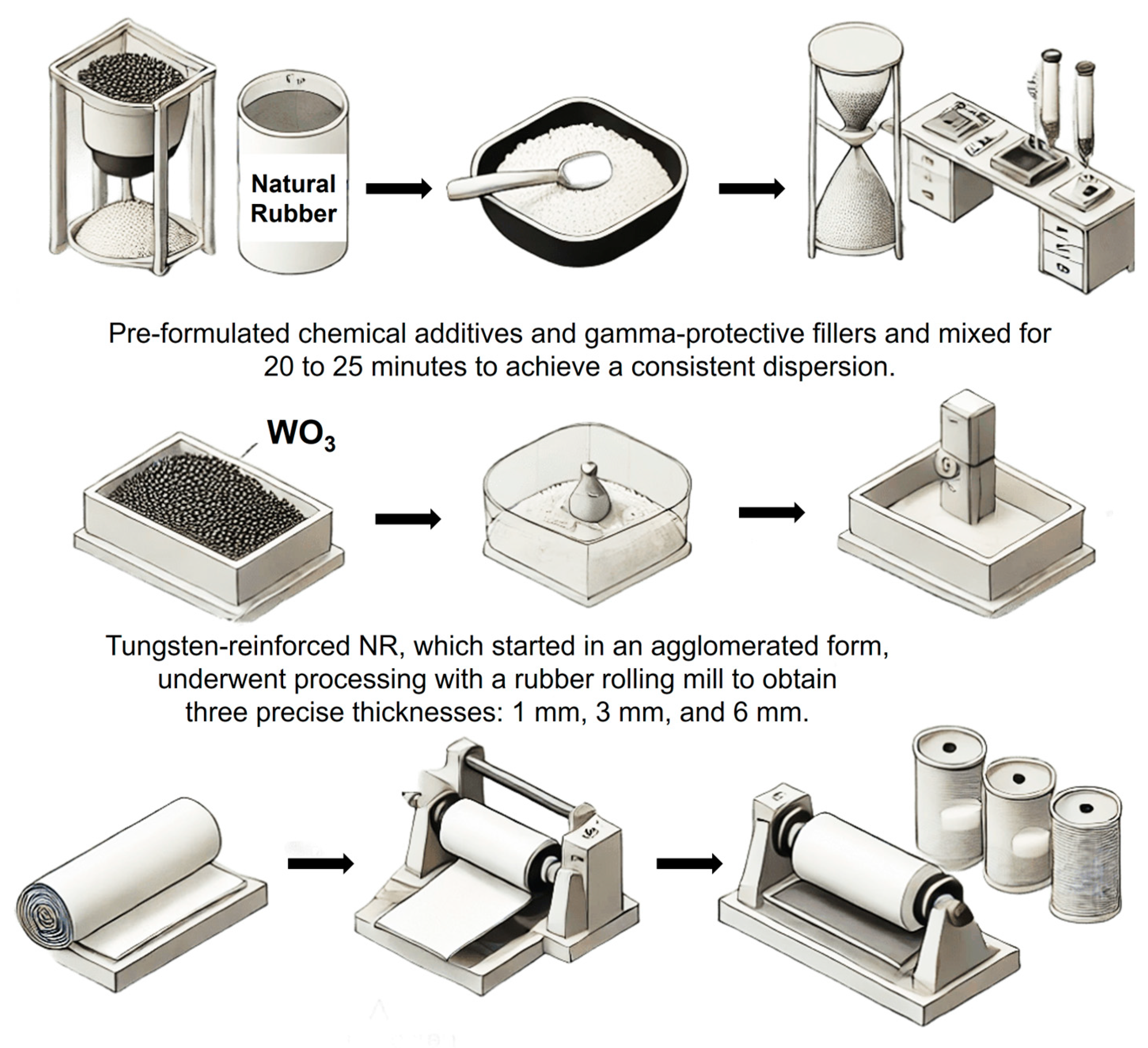
2.2. Preparation of Tungsten-Reinforced Polyisoprene

NR was the primary matrix in this investigation. Table 1 provides a comprehensive overview of the vulcanization formulation, encompassing all chemical components and their respective roles in developing tungsten-reinforced polyisoprene. The preparation of natural rubber composites involved a two-step process that included mastication followed by compounding. The NR underwent processing in a two-roll mill for a duration of 5 min during the mastication stage to improve its workability. Following this, the processed NR was blended with pre-formulated chemical additives and gamma-protective fillers and mixed for an additional 20 to 25 min to achieve a consistent dispersion [27]. Additionally, the tungsten-reinforced NR, which started in an agglomerated form, underwent processing with a rubber rolling mill to obtain three precise thicknesses: 1 mm, 3 mm, and 6 mm. The material was meticulously dried before proceeding with additional analysis (Figure 1).

2.3. Structural and Morphological Characterization of Tungsten-Reinforced Polyisoprene

2.3.1. Material Characterization via Scanning Electron Microscopy (SEM) and Energy-Dispersive X-Ray Spectroscopy (EDX): Morphological and Compositional Analysis

To compare the structural characteristics and infer potential enhancements in the mechanical and thermal properties due to WO3 reinforcement, the tungsten-reinforced polyisoprene was characterized by SEM (FE-SEM Apreo, FEI Technologies Inc., Hillsboro, OR, USA). A Scanning Electron Microscopy (SEM) (FE-SEM Apreo, FEI Technologies Inc., Hillsboro, OR, USA) was utilized to produce high-resolution enlarged images. Additionally, further characterization to determine the elemental composition was caried out using Energy-Dispersive X-ray (EDX) (Oxford Instruments NanoAnalysis, High Wycombe, UK). Prior to characterization, the tungsten-reinforced polyisoprene was comprehensively cleaned to achieve good picture quality and analysis. To eliminate contaminants, the tungsten-reinforced polyisoprene was washed with ethanol, followed by 10–15 min of sonication. After cleaning, the tungsten-reinforced polyisoprene samples were washed with deionized water. After washing, the samples were put in a vacuum desiccator to eliminate any remaining moisture. Moisture must be eliminated since it may distort pictures and cause charging characteristics [28]. To address the moisture issue, all samples were sputter-coated with a 20 nm thin coating of gold, a conductive substance [29]. Following the completion of the gold sputter coating, all samples were placed in a vacuum desiccator for 15 min to eliminate any remaining moisture before SEM.

2.3.2. Structural Characterization of Tungsten-Reinforced Polyisoprene Using X-Ray Diffraction (XRD)

To identify the crystalline phases of the NR and tungsten-reinforced polyisoprene, an X-ray diffraction (XRD) examination was used. The diffraction patterns offer information about the existence of distinct crystalline formations. This work analyzes the crystallographic composition of NR and WO3-reinforced polyisoprene to understand better the structural changes caused by the addition of tungsten oxide. Tungsten-reinforced polyisoprene was pulverized using an agate mortar and pestle to provide solid samples for XRD examination with particle sizes smaller than 10 µm. The samples were then crushed for two minutes using a hydraulic press with a capacity of five tons to generate flat pellets. To increase the diffraction quality, the pellets must have a smooth, uniform surface. The tungsten-reinforced polyisoprene samples were then placed in the XRD sample container for analysis, with XRD patterns produced with Cu-Kα radiation (λ = 1.5406 Å) at a scan range of 10–90° 2θ [30]. The resultant peak positions and intensities of the crystalline phases were matched with existing databases [31].

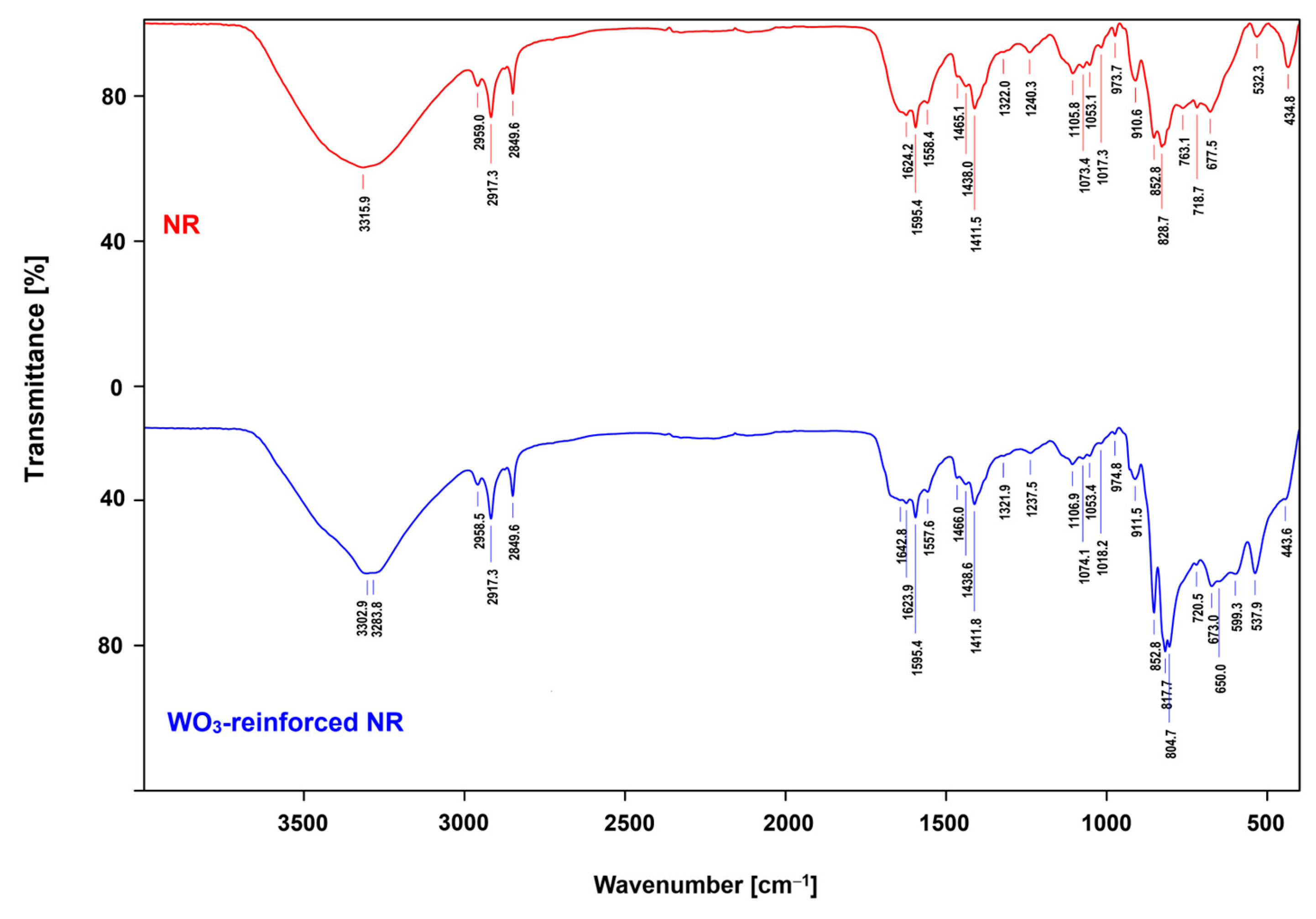
2.3.3. Characterization of Molecular Structures and Chemical Bonding in Tungsten-Reinforced Polyisoprene Using Fourier Transform Infrared Spectroscopy (FTIR)

To investigate the materials’ molecular structure and chemical bonds, we conducted a Fourier transform infrared spectroscopy (FTIR) analysis (VERTEX 70, Bruker Daltonics GmbH & Co. KG, Bremen, Germany) using the attenuated total reflection (ATR) approach [32], with a spectra range from 4000 cm−1 to 400 cm−1, with 64 scans for each measurement. Furthermore, to concentrate on detecting particular chemical bonds, all measurements were conducted using a deuterated l-alanine-doped triglycine sulfate (DLATGS) detector (Leonardo Electronics Us Inc., McLean, VA, USA) and Potassium Bromide (KBr) precision windows. Furthermore, the FTIR analysis of tungsten-reinforced polyisoprene gave valuable insights by detecting changes in its chemical structure and the emergence or absence of certain bonds.

2.3.4. Structural Deformation in Tungsten-Reinforced Polyisoprene: An ANSYS-Based Bending Analysis

This work synthesized a tungsten-reinforced polyisoprene system to explore its deformation behavior under different applied forces. ANSYS Workbench 2025 R1 (version 25.1) (Ansys, Inc., Canonsburg, PA, USA) was used in a bending deformation study to replicate the structural response of the composite under many force loads. By including exact material parameters and reasonable boundary limitations, the computational model was intended to reproduce experimental settings. Modeled as a three-dimensional (3D) structure inside ANSYS DesignModeler, the composite sheet high-precision in stress–strain distribution was obtained by building a structured mesh with an element size of 50 × 60 × 6 mm. Experimental data and the literature values guided assignments of material attributes including elastic modulus, Poisson’s ratio, and density [33,34,35,36].
The composite sheet’s bottom edge was entirely limited to replicating a fixed support condition, hence simulating bending deformation. Applied near the middle of the sheet along the y-axis, a point force matched the main loading direction. To evaluate the deformation behavior under many loading circumstances, the force magnitudes were methodically changed at 10 N, 25 N, 50 N, and 100 N. Using a nonlinear solution under quasi-static circumstances, the study was conducted to capture significant deformation effects, therefore guaranteeing the simulation’s correctness. Recording the greatest displacement at the location of force application for every loading condition allowed one to analyze the deformation response.

2.4. Radiation-Shielding Performance of Tungsten-Reinforced Polyisoprene: Simulation and Experimental Assessment

2.4.1. Utilizing XCOM Simulations for the Design and Evaluation of Radiation-Shielding Materials

The XCOM algorithm was used to determine shielding characteristics such as coherent scattering, incoherent scattering, photoelectric absorption, pair production in the nuclear field, pair production in the electron field, total attenuation with coherent scattering, and total attenuation without coherent scattering, and the results were compared to EPICS2017 values [37,38]. XCOM calculates total cross sections, attenuation coefficients, and partial cross sections using a typical energy grid (logarithmically spaced), a user-selected grid, or a combination of both grids. A windows version of XCOM provides a user interface that expedites detecting and specifying compounds [39].

2.4.2. Computational Analysis of Radiation Shielding in Tungsten-Reinforced Polyisoprene Using Phy-X/PSD Simulations

The attenuation coefficient was computed using the Phy-X/PSD program in this investigation [40]. The mass attenuation coefficient (MAC) is a critical parameter employed to evaluate the capacity of tungsten-reinforced Polyisoprene to attenuate Tc-99m gamma radiation per unit mass. It provides a density-independent measure of shielding efficacy. Furthermore, determining the linear attenuation coefficient (LAC), the half-value layer (HVL), tenth-value layer (TVL), mean free path (MFP), effective electron density (Neff), effective conductivity (Ceff), atomic cross section (ACS), electronic cross section (ECS), effective Atomic Number (Zeff), and the equivalent atomic number (Zeq).

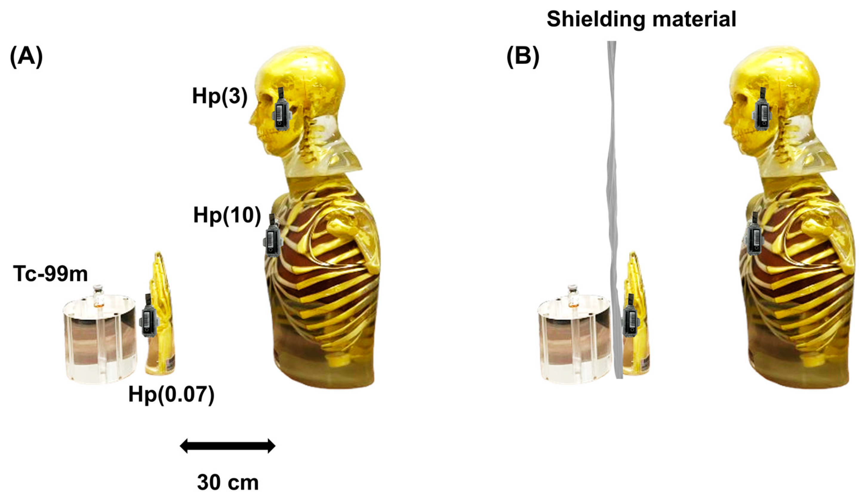
2.4.3. Experimental Investigation of Materials for Gamma Radiation Shielding of Tc-99m

This investigation used Quixel optically stimulated luminescence (OSL) badges (Landauer Inc., Glenwood, IL, USA) and Tc-99m radiopharmaceuticals supplied by the Thailand Institute of Nuclear Technology (Public Organization), Ongkarak, Nakornnayok, Thailand. Anthropomorphic phantoms were used to simulate the radiation dose to nuclear medical staff anatomy. For Hp(3) eye lens dosimetry at 3 mm tissue depth, an RSD anthropomorphic head phantom with a complete cervical spine (C1–C7) was utilized. For Hp(10), the whole-body personal dose equivalent, a 10 mm deep RSD anthropomorphic thorax phantom was used. Additionally, an anthropomorphic hand phantom (Universal Medical Inc., Oldsmar, FL, USA) was used to assess shallow doses (Hp(0.07)) for the skin, hands, and feet at 0.07 mm (Figure 2). Experimental uncertainties were addressed to guarantee dosage measurement reliability. The InLight dosimeter in this research has a minimum reporting level of 50 µSv for gamma and X-ray radiation, indicating greater uncertainty for data below this threshold. Statistical fluctuations in dosimeter response result in an estimated uncertainty of ±10%. The NCRP is methods to integrate dose uncertainties into dose–response models [41]. According to ISO 4037 standards for radiation shielding tests, discrepancies in positioning and material thickness create ±2% uncertainty [42].

2.5. Statistical Analysis

Each experiment was carried out three to ten times independently in this research, with the mean ± standard deviation applied. By examining residuals, the normality of the data was assessed, and the homogeneity of variance across groups was confirmed. The Student’s t-test and one- or two-way analysis of variance (ANOVA), followed by the Student–Newman–Keuls post hoc test, were used to determine statistical significance. The significance thresholds were set: * for p < 0.05 and ns for p > 0.05. GraphPad Prism version 10.0 (GraphPad Software Inc., Boston, MA, USA) was applied for all statistical analyses.

3. Results

3.1. Characterization of the Structural and Morphological Properties of Tungsten-Reinforced Polyisoprene

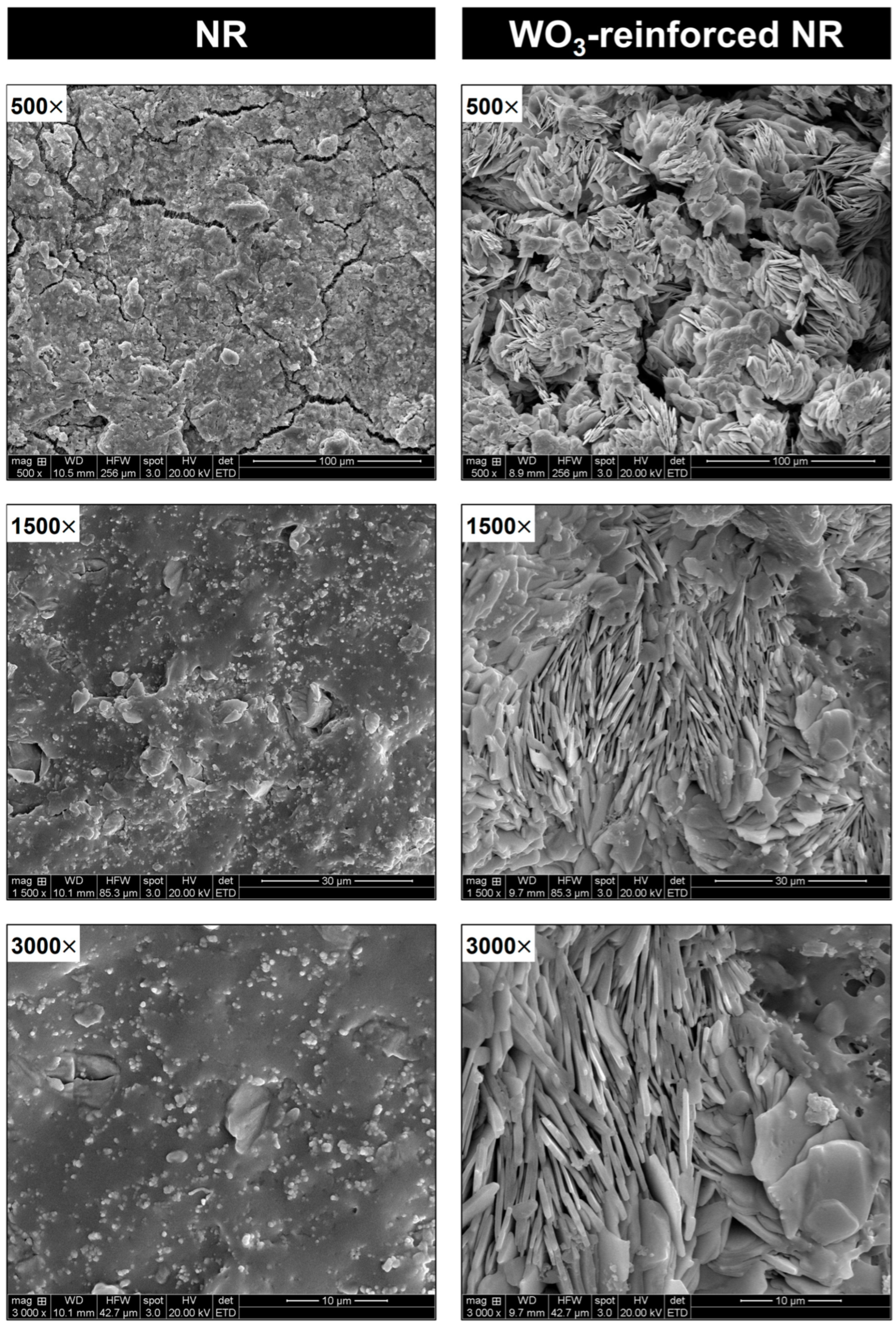
3.1.1. Material Characterization of Tungsten-Reinforced Polyisoprene: SEM and EDX for Microstructural and Elemental Analysis

The SEM images (Figure 3) compare NR and WO3-reinforced polyisoprene at 500×, 1500×, and 3000× magnifications. The NR picture at 500× mostly shows a smooth surface but also some cracks and small granular structures. The 500× scan of WO3-reinforced polyisoprene reveals a unique morphology, with clusters of elongated and plate-like structures. Showing the polyisoprene and WO3 particles interact strongly within these structures, which makes the structures more stable.
The 1500× reveals greater detail of the NR showing surface-scattered granules, indicating phase separation or minor inhomogeneities in the matrix. The 1500× scan of the WO3-reinforced polyisoprene shows more prominent well-aligned plate-like structures. Suggesting a higher degree of reinforcement, which could contribute to improvements in mechanical properties, such as stiffness and tensile strength.
The higher 3000× magnification of NR reveals fine particles and an increase in surface roughness, as well as highlighting small-scale heterogeneities within the polymer structure. The 3000× magnification image of the WO3-reinforced polyisoprene shows the particles being well-dispersed, to form a highly ordered and interconnected structure. This highly ordered alignment of the needle-like WO3-reinforced polyisoprene nanostructures and organization suggest strong interfacial adhesion between the polymer and the WO3 filler, enhancing its thermal stability and mechanical performance.
To confirm the distribution of WO3 within the polymer matrix, the EDX elemental analysis results are illustrated in Figure 4. The SEM image of NR displays a relatively uniform morphology with dispersed particles. The EDX analysis revealed a high carbon content (68.3 wt%), characteristic of organic rubber matrices. Oxygen (6.5 wt%), sodium (3.6 wt%), zinc (16.9 wt%), and sulfur (4.7 wt%) were also detected. The elemental mapping indicates a uniform homogeneous distribution of carbon, while zinc and sulfur appear as dispersed clusters, potentially linked to the vulcanization process. The SEM image of the WO3-reinforced sample exhibits a more heterogeneous and rougher appearance, probably due to the presence of WO3 particles. The carbon level decreased significantly (14.4 wt%), suggesting partial replacement of the polymer matrix with inorganic filler. The presence of tungsten (52.0 wt%) indicates that WO3 has been successfully incorporated into the material. Increased oxygen (21.4 wt%) and sodium (10.1 wt%) concentrations imply more interactions between the polymer and the oxide filler. Zinc (2.1 wt%) is present, albeit at a lower quantity than NR, which might be owing to the filler impacting additive dispersion. The EDX elemental analysis indicates that adding WO3 considerably modifies the elemental composition of polyisoprene. Decreased carbon concentration and substantial tungsten presence highlight the successful polymer reinforcement. The EDX elemental analysis shows that adding WO3 decreased carbon content, and the high tungsten content indicates polymer reinforcing.

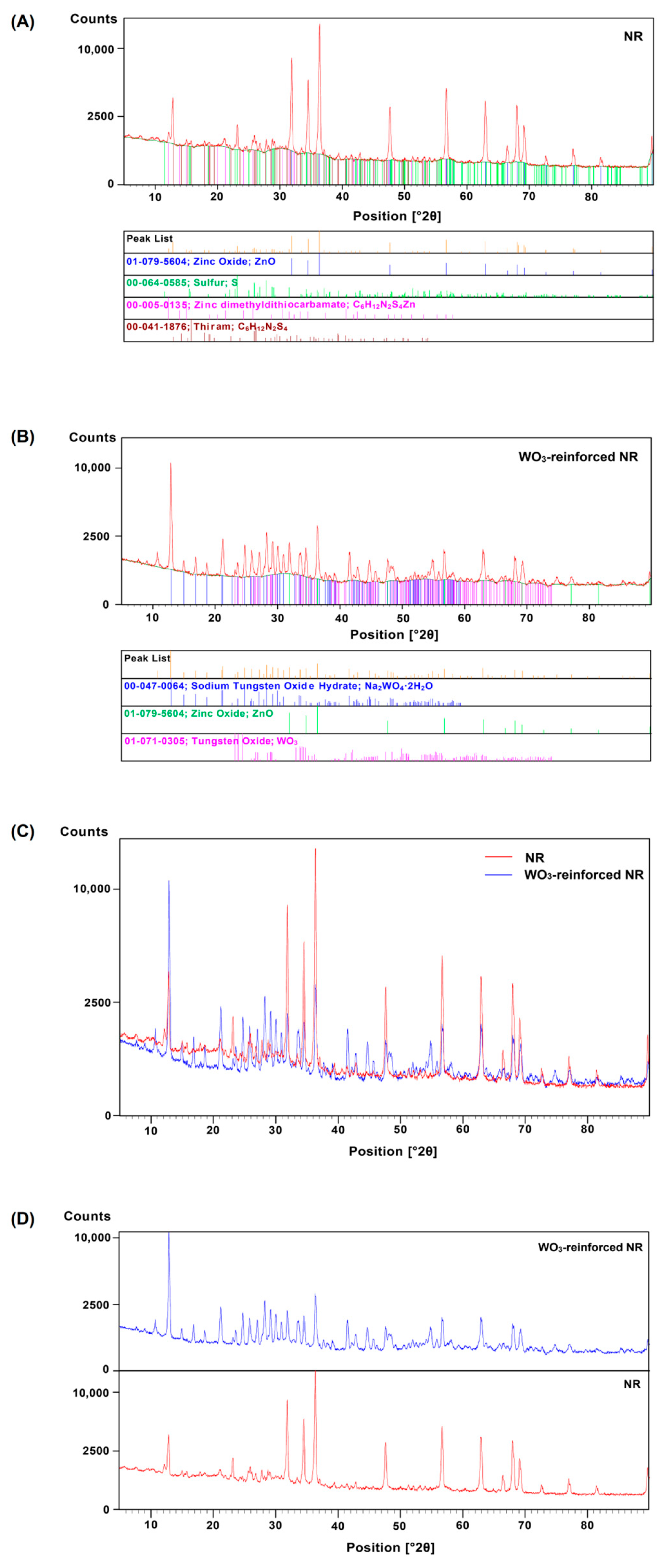
3.1.2. Structural Properties of Tungsten-Reinforced Polyisoprene Characterized by X-Ray Diffraction

The results of the XRD analysis (Figure 5) emphasize the structural differences between NR and tungsten-reinforced polyisoprene. Figure 5A depicts the XRD pattern of NR, which reveals the crystalline phases contained in the sample. The diffraction peaks correspond to the main crystallographic components. ZnO (matching well with the standard of Joint Committee on Powder Diffraction Standards (JCPDS) number 01-079-5604) is a regularly used activator in the vulcanization process [43]. Sulfur (S) (matching well with the standard JCPDS number 00-064-0585) is a vulcanizing agent that promotes crosslinking in the polymer matrix [44,45,46]. Zinc dimethyldithiocarbamate (matching well with the standard JCPDS number 00-005-0315) acts as an accelerator during the vulcanization process [44,47,48]. Thiram (C6H12N2S4) (matching well with the standard JCPDS number 00-041-1876) is a vulcanizing accelerator. The patterns corroborate the nature of the NR formulation, which includes vulcanizing chemicals and fillers [49].
Figure 5B shows the XRD pattern for polyisoprene reinforced with WO3. The diffraction peaks reveal the existence of crystallographic phases. Sodium tungsten oxide hydrate (Na2WO4·2H2O) (matching well with the standard JCPDS number 00-047-0064) may result from interactions between tungsten compounds and moisture during processing [50]. ZnO is a crucial component in the composite. WO3 (matching well with the standard JCPDS number 01-071-0305) was found in the main reinforcement phase, indicating its effective integration into the polymer matrix [51]. The inclusion of WO3 into the polyisoprene matrix creates extra diffraction peaks attributable to tungsten-based compounds. This indicates a significant shift in the crystalline structure of the composite, which will most likely affect its mechanical and physicochemical characteristics.
Figure 5C presents a comparative XRD analysis of NR (red) and WO3-reinforced polyisoprene (blue), highlighting key differences in their diffraction patterns. Both NR and WO3-reinforced polyisoprene exhibit characteristic diffraction peaks across the 2θ range, indicating the presence of crystalline phases in both materials. However, the peak intensities vary significantly between the two samples. Crystalline phase identification reveals that NR (red) primarily contains peaks associated with ZnO, sulfur (S), and vulcanization accelerators such as zinc dimethyldithiocarbamate and thiram. In contrast, WO3-reinforced polyisoprene exhibits additional diffraction peaks corresponding to WO3 and Na2WO4·2H2O, confirming successful incorporation of the reinforcing agent.
The addition of WO3 increases the number of diffraction peaks, suggesting improved crystallinity. WO3-reinforced polyisoprene exhibited sharper and more intense peaks, indicating enhanced structural ordering within the composite. In addition, minor peak shifts and broadening may be caused by strain, lattice distortions, or interactions between WO3 and the polymer matrix. These structural alterations may potentially have an influence on the material’s mechanical and thermal characteristics.
The XRD patterns for NR and WO3-reinforced polyisoprene are displayed in Figure 5D. The NR XRD pattern shows broad, low-intensity peaks, suggesting it is primarily amorphous with little crystallinity. Sharp peaks imply the possibility of crystalline domains, although they are not predominant. The 2θ range of around 10–50° has the most significant diffraction peaks, with reflections at 30°, 40°, and 50°. These peaks could be caused by the existence of residual structural order in the polymer matrix.
The addition of WO3 to the NR matrix significantly influences the diffraction pattern. The XRD spectrum of WO3-reinforced polyisoprene shows more distinct peaks than NR alone, suggesting higher crystallinity. A wide 2θ range of peaks is found, with noticeable reflections about 10–80°. Crystalline WO3 phases exhibit significant peaks at 10°, 23°, 30°, 35°, 50°, and 60°. Enhanced peak intensity indicates effective reinforcement of NR by WO3 nanoparticles, resulting in improved structural order.

3.1.3. Characterization of Chemical Bonding and Molecular Structures in Tungsten-Reinforced Polyisoprene Using FTIR

The FTIR analysis of tungsten-reinforced polyisoprene gave valuable insights by detecting changes in its chemical structure and the emergence or absence of certain bonds. The FTIR spectra of NR (red curve) and WO3-reinforced polyisoprene (blue curve) are plotted with transmittance (%) on the y-axis and wavenumber (cm−1) on the x-axis (Figure 6). The characteristic absorption bands indicate the presence of different functional groups in each sample. The NR spectrum broad absorption band at 3343 cm−1 is attributed to O–H stretching, suggesting the presence of hydroxyl groups. The asymmetrical and symmetrical C–H bonds stretching in the methylene (–CH2–) groups are indicated by the peaks at 2917 cm−1 and 2848 cm−1, a common feature in polyisoprene. Additionally, the absorption bands at 1654 cm−1, 1448 cm−1, and 1376 cm−1 can be related to the stretching and bending vibrations of both the C=C and C–H bonds in the rubber matrix. The peaks at 1243 cm−1, 1073 cm−1, and 1013 cm−1 suggest the presence of C–O stretching vibrations from possible oxidation of rubber. Additional peaks in the region 900–600 cm−1 correspond to out-of-plane bending of C–H bonds, confirming the structural integrity of NR.

3.1.4. Characterization of Shape Deformation in Tungsten-Reinforced Polyisoprene Under Bending: An ANSYS Simulation Study

The deformation behavior of natural rubber-based shielding material in response to varying applied forces (10 N, 25 N, 50 N, and 100 N) is illustrated in Figure 7. The color gradient representing the displacement distribution indicates that the regions undergoing the most deformation are highlighted in red, while those with minimal displacement are displayed in blue. The increased deformation observed with greater applied forces demonstrates the material’s mechanical characteristics and flexibility. Comprehending this behavior is crucial for assessing the viability of natural rubber composites as gamma radiation shielding materials, especially in structural applications that necessitate the maintenance of mechanical integrity.
The deformation characteristics of the natural rubber composite under different applied forces demonstrate a nonlinear correlation between force and displacement. At a modest force of 10 N, slight deformation (55.14 mm) occurs, signifying structural stiffness. As the force increases to 25 N, the displacement attains 137.86 mm, demonstrating improved flexibility while sustaining support. A force of 50 N induces considerable bending (275.72 mm), emphasizing flexibility to uneven surfaces. At 100 N, significant deformation (580.87 mm) indicates the nearing of mechanical limitations, which could affect shielding efficacy. Excessive stress may modify thickness and homogeneity, influencing radiation attenuation. Reinforcement techniques such as WO3 are essential to improve mechanical stability while maintaining flexibility for gamma radiation-shielding applications.

3.2. Evaluating the Radiation-Shielding Effectiveness of Tungsten-Reinforced Polyisoprene via Simulation and Experimental Validation

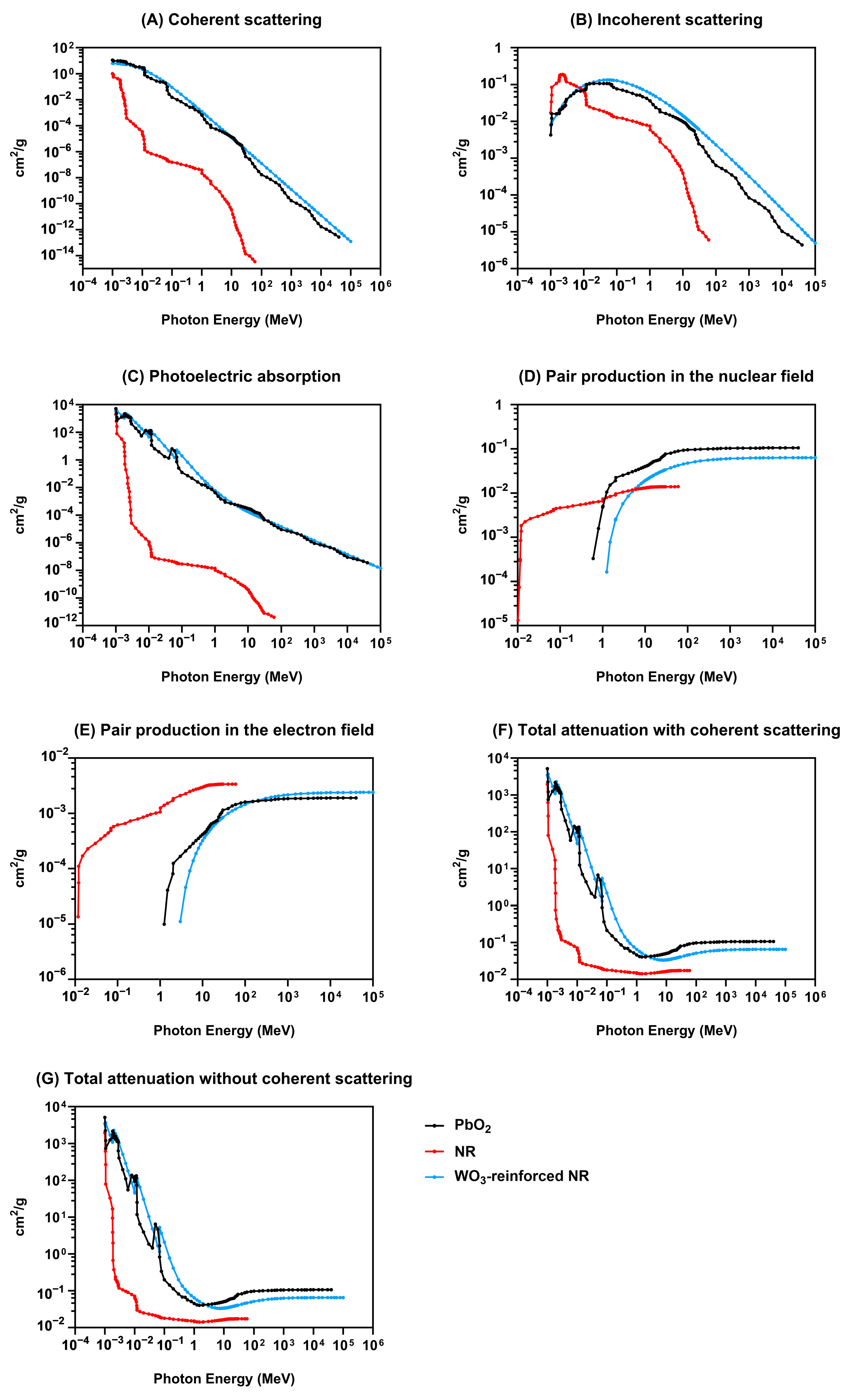
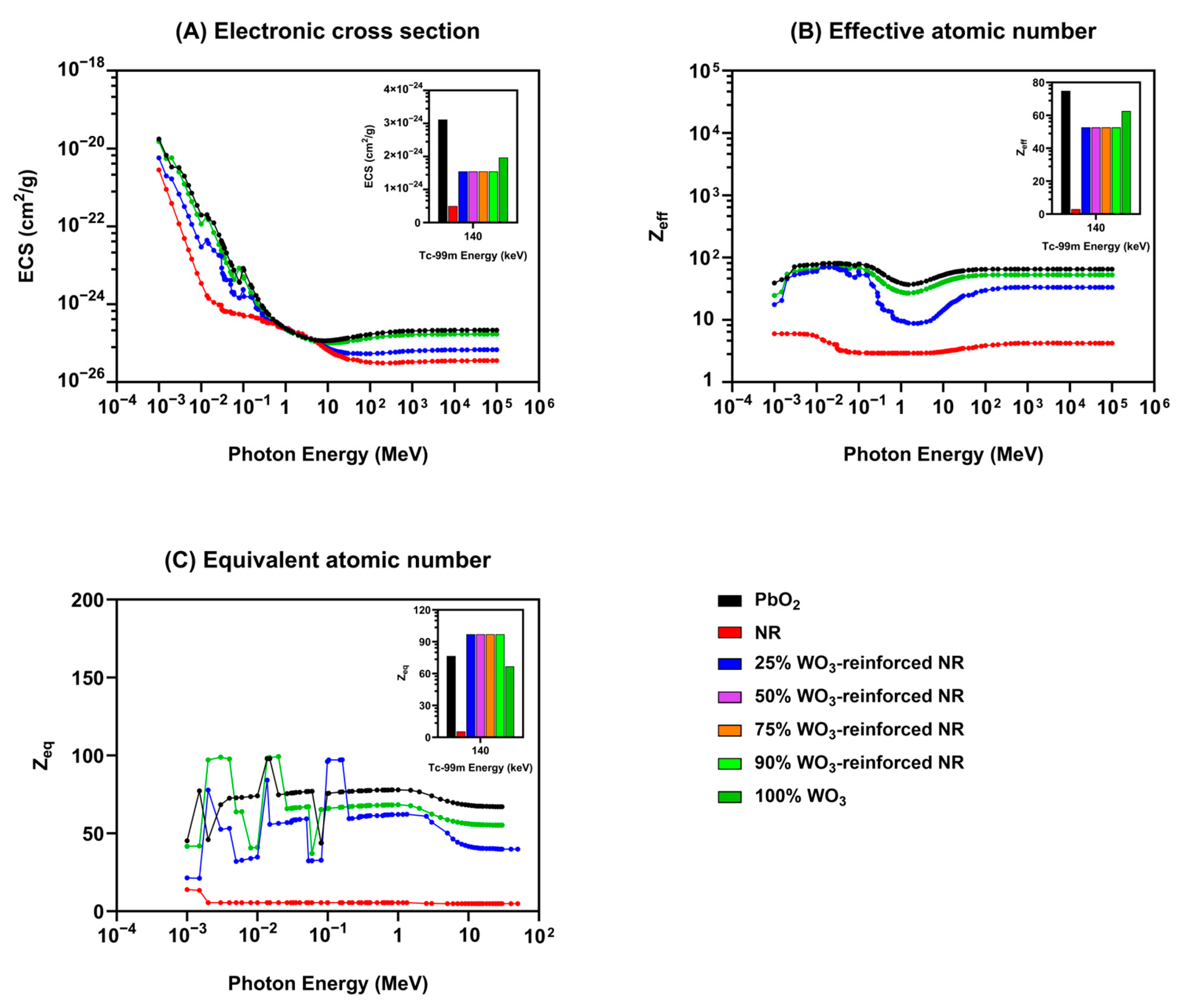
3.2.1. Computational Analysis of Tungsten-Reinforced Polyisoprene as a Radiation Shielding Material Using XCOM Simulations

The mass attenuation coefficients (MAC) of PbO2, NR, and WO3-reinforced NR were calculated using XCOM, a photon cross section database developed by the National Institute of Standards and Technology (NIST), at various photon energies [38]. The simulation ranged in photon energy from 10−4 to 106 MeV. Figure 8A illustrates the MAC (cm2/g) for various photon interactions as a function of photon energy (MeV). As photon energy rises, the MAC of NR (red) decreases dramatically. NR has a considerably larger coherent scattering probability at low photon energies (~10−3 MeV) than at higher energies. Beyond 0.1 MeV, the attenuation coefficient decreases substantially and becomes insignificant beyond ~1 MeV. The PbO2 across all photon energies exhibits a higher MAC than NR, demonstrating its greater capacity for coherent scattering at low photon energies. The rate of decline is more gradual than NR, showing better shielding performance over a broader energy range. Tungsten-reinforced polyisoprene (blue curve) follows a similar trend to PbO2 but with slightly lower attenuation coefficients at low energies and greater attenuation than lead at higher energies. The graph indicates WO3-reinforced NR effectiveness in coherent scattering and a viable alternative to conventional PBO2 shielding.
The incoherent scattering behavior of PbO2, NR, and WO3-reinforced NR are illustrated in Figure 8B. In the intermediate energy range (0.1–a few MeV), incoherent (Compton) scattering predominates photon interaction. It occurs when incoming photons transfer energy to bound or free electrons, scattering low-energy photons. The efficiency of Compton scattering depends on electron density and is determined by its atomic number (Z) and composition. NR exhibits the lowest attenuation, particularly at higher photon energies, primarily comprising low-Z elements (carbon and hydrogen) with lower electron densities and interaction probabilities. PbO2 displays significantly higher incoherent scattering than NR, attributed to its high-Z lead atoms, which increase electron density and enhance Compton interactions.
Figure 8C shows the MAC for photoelectric absorption as a function of photon energy. PbO2 (black curve) has the maximum photoelectric absorption throughout the energy spectrum, corresponding with lead’s high atomic number (Z = 82), which increases interaction likelihood. Due to its organic makeup, NR exhibits a much lower attenuation. Photoelectric absorption strongly depends on Z in the low-energy region (10−4–1 MeV). PbO2 has the maximum attenuation, whereas WO3-reinforced NR outperforms pure NR. In the Intermediate Energy Region (1–100 MeV), photoelectric absorption decreases for all materials owing to Compton scattering competition. Photoelectric interactions are less important in the High-Energy Region (>100 MeV) as pair formation and Compton scattering take over.
Figure 8D shows how MAC for pair creation vary with photon energy ranging from 10−2 MeV to 105 MeV. PbO2 has the most significant mass attenuation coefficient. A rapid spike at about 1 MeV, indicating the threshold for pair creation, continues to grow until it plateaus at high photon energies. WO3-reinforced NR exhibits superior attenuation compared to NR, although inferior to PbO2. The curve exhibits a similar tendency to PbO2, characterized by an increase in attenuation at the threshold energy and a subsequent gradual climb thereafter.
Pair Production in the electron field (Figure 8E) indicates the MAC as a function of photon energy. The MAC for pair creation is insignificant below 1 MeV, indicating that the process occurs only at elevated photon energies. At 10 MeV, a notable increase in attenuation is seen, especially for PbO2 and WO3-reinforced NR, indicating their enhanced shielding efficacy. Beyond 100 MeV, the attenuation coefficients for PbO2 and WO3-reinforced NR begin to converge, showing that tungsten reinforcement enables WO3-reinforced NR to resemble PbO2 shielding performance closely. The results show that PbO2 remains the most effective; however, it can be seen that WO3-reinforced NR approaches its performance at high photon energies.
Analysis of total attenuation with coherent scattering (Figure 8F) shows that PbO2 is more effective at intermediate energies, although WO3-reinforced NR approaches PbO2’s performance in the high-energy zone. The findings of the XCOM computational analysis support the use of WO3-reinforced NR as a lead-free substitute for radiation shielding.
Figure 8G illustrates the variation in the MAC of PbO2 (black), NR (red), and WO3-reinforced NR (blue). In the High-Energy Region (10–106 MeV), all materials show a decrease in attenuation. PbO2 has a slightly greater attenuation coefficient than WO3-reinforced NR, indicating a higher atomic number and density. A clear distinction emerges between WO3-reinforced NR and NR, at the intermediate-energy region (0.1–10 MeV), where it can be observed the inclusion of tungsten in the WO3-reinforced NR significantly enhances the attenuation. At the low-energy region (10−4–0.1 MeV), PbO2 and WO3-reinforced NR display high attenuation values due to their strong photoelectric absorption. The observed peaks correspond to the characteristic absorption edges of tungsten and lead, further validating the computational model. The results of the XCOM-based computational analysis confirm that WO3-reinforced NR exhibits good radiation attenuation that approaches the performance of PbO2.

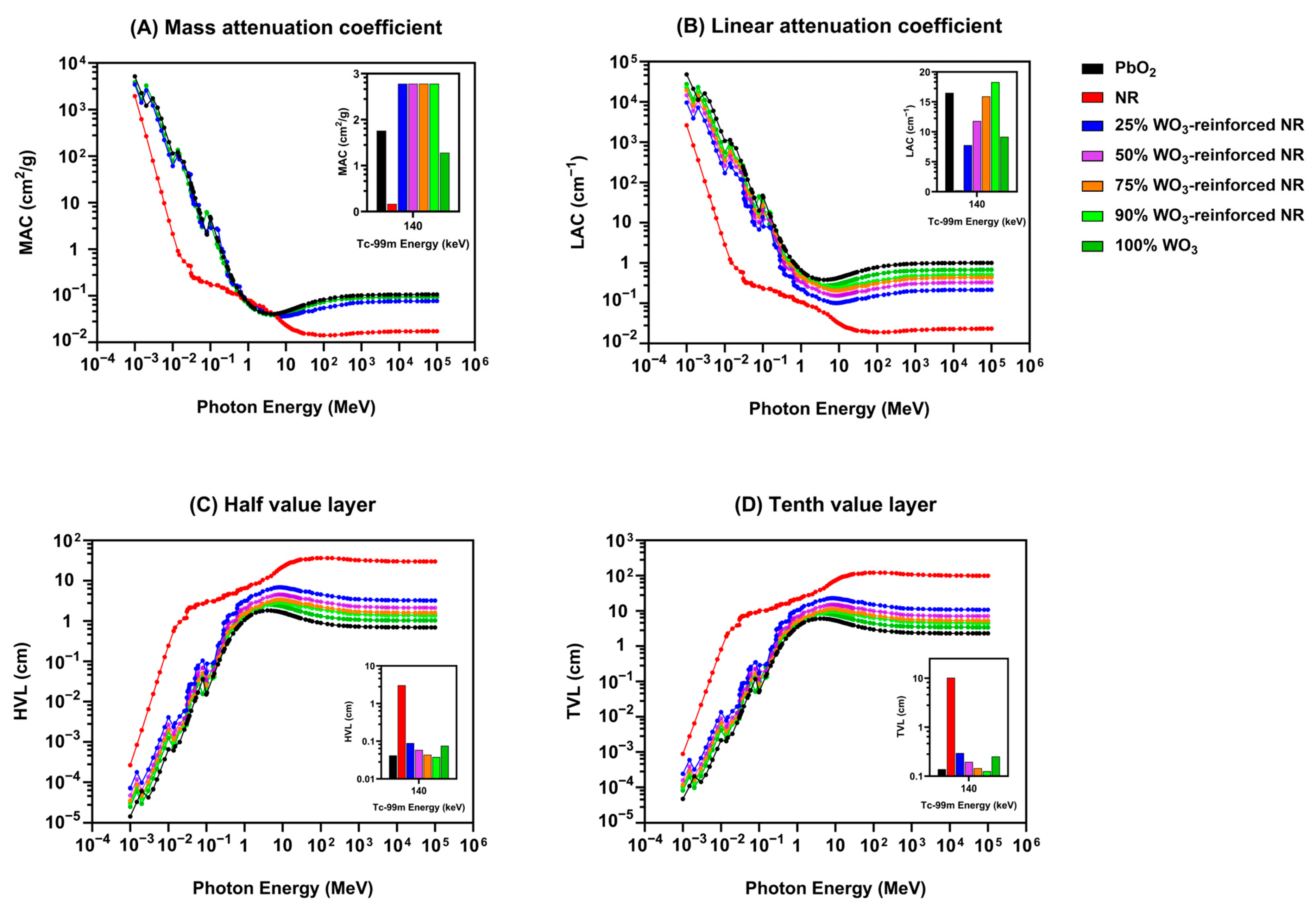
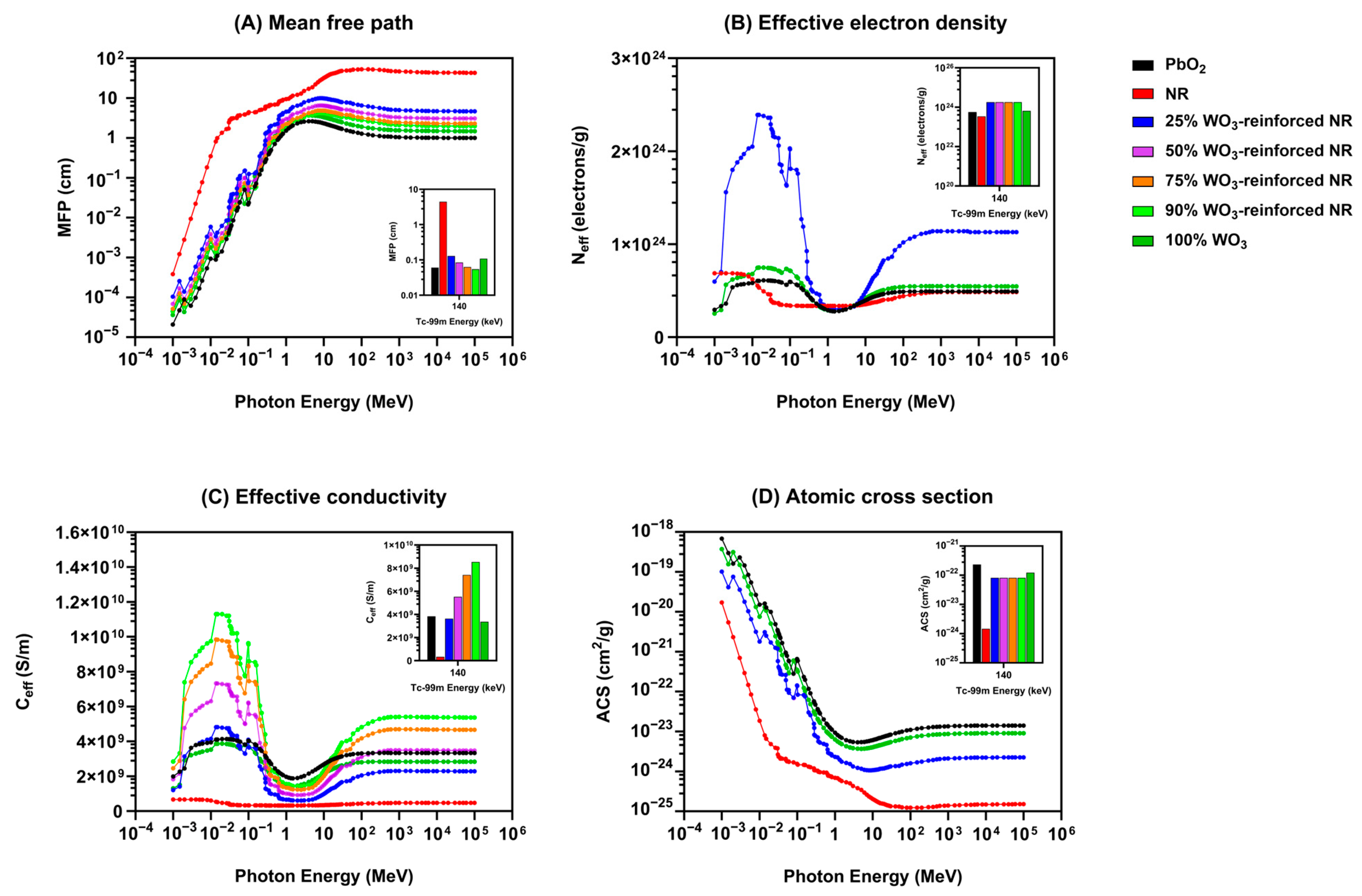
3.2.2. Computational Assessment of Tungsten-Reinforced Polyisoprene for Radiation Shielding Applications Using Phy-X/PSD Simulations

The Phy-X/PSD software shielding values are presented in Figure 9, at 140 keV, corresponding to the primary photon energy of Tc-99m [40]. The mass attenuation coefficient (MAC) is an essential metric that explains how photons interact with matter, specifically the potential of photon absorption or scattering per unit mass. MAC is a key component in many applications, including radiation shielding. Figure 9A shows that the low-energy region of 10−4 to 1 MeV presents a significant variation because of the photoelectric effect dominance, which is dependent on the atomic number (Z) of the material, PbO2 high atomic number (Z = 82), exhibiting the highest MAC, followed by the 100% WO3 composite, which contains tungsten (Z = 74). The fluctuations at low-energy photon energies, for high-Z materials, correspond to the K-edge absorption of WO3 and PbO2. The intermediate-energy area (10−2 to 10 MeV) shows a significant drop in MAC, indicating a shift from photoelectric absorption to Compton scattering as the primary interaction mechanism. Although the attenuation behavior of various materials converges, PbO2 and high-WO3 compositions still provide better shielding capacities. At the high-energy region, the MAC values stabilize at a lower level, suggesting dominance of pair production at high energies. PbO2 is the most effective material for photon attenuation and the WO3-based composites exhibit promising shielding properties, particularly at high concentrations (≥75% WO3).
Figure 9B indicates that increasing photon energy results in a declining trend in the linear attenuation coefficient (LAC) values. This result is compatible with the typical photon interaction models, in which pair formation at extremely high energies (>1 MeV) dominates, photoelectric absorption dominates at lower energies, and Compton scattering becomes prominent at intermediate levels. The sharp peaks at lower photon energies (<10−2 MeV) correspond to the K-edge binding energy of K-shell electrons, beyond which incident X-ray photons have enough energy to eject them from the atom of the high-Z elements (lead 88.0 keV, tungsten 69.5 keV), where photoelectric absorption dominates. Given Tc-99m gamma emission energy of 140 keV, the most suitable shielding material should have a high LAC value in this energy range. The inset graph shows that PbO2 and high-percentage WO3 composites (≥75% WO3) have better attenuation at 140 keV. The LAC data confirms that tungsten-reinforced NR composites display increased radiation shielding performance with increasing WO3 concentration.
The half-value layer (HVL) refers to the thickness of a material needed to reduce incoming radiation intensity to 50% of its initial level. Figure 9C presents HVL values as a function of photon energy in the range of 10−4 MeV to 106 MeV. The NR shows significantly higher HVL values. PbO2 and WO3-reinforced composites show significantly lower HVL values than NR, highlighting their superior shielding performance. Increasing WO3 content results in a systematic reduction in HVL. The PbO2 and WO3-rich composites (≥75%) exhibit the lowest HVL, making them good candidates for Tc-99m radiation shielding.
The graph (Figure 9D) illustrates the variation of tenth-value layer (TVL) as a function of photon energy in the range of 10−4 MeV to 106 MeV. Indicating that at lower photon energies (below 10 keV), the TVL values rise noticeably. Thereafter, they show a more constant trend in the middle energy range (10 keV to 10 MeV). For high-energy photons (>103 MeV), a rapid increase in TVL indicates a decrease in shielding efficacy resulting from the predominance of Compton scattering and pair creation. The inset graph illustrates the TVL values at the characteristic energy of 140 keV for Tc-99m. PbO2 and high-percentage WO3 composites (90% and 100%) exhibit the lowest TVL values, thereby confirming their effectiveness for Tc-99m shielding.
The mean distance a photon travels within a material before absorption or scattering is called the mean free path (MFP). Due to more photon interactions with the material, a lower MFP improves shielding. The MFP graph (Figure 10A) shows that in the low-energy region (10−4 to 10−2 MeV), all materials have very short mean free path values, indicating intense photon interactions attributable to the predominance of the photoelectric effect. In the intermediate-energy range (10−2 to 10 MeV), the MFP rises as the interaction mechanism shifts from photoelectric absorption to Compton scattering. In the high-energy range (>10 MeV), a plateau occurs as MFP values increase, as pair creation emerges as the predominant interaction mechanism.
Figure 10B shows the Neff as a function of photon energy, emphasizing its energy-dependent variability. Neff shows considerable variances for all materials along the photon energy spectrum, particularly in the low-energy band (<10−2 MeV). NR has the lowest effective electron density among all energies, suggesting limited radiation interaction capabilities. At higher photon energies (>100 MeV), the effective electron density stabilizes, indicating there is less of a reliance on the composition of the material. For Tc-99m shielding the inset graph indicates that PbO2 and high-WO3 composites (90% and 100%) display the highest effective electron densities, making them optimal choices for the Tc-99m shielding.
Effective conductivity is a crucial parameter in radiation shielding, reflecting the material’s ability to conduct charge under radiation exposure. Figure 10C presents the results of the Ceff analysis, illustrating the relationship between Ceff and photon energy, covering a wide range of values from 10−4 MeV to 106 MeV. The overall pattern noted in all materials indicates a preliminary rise in Ceff at lower photon energies, reaching a peak around 0.01 to 0.1 MeV. After that, there is a decrease followed by a stabilization at elevated energies. PbO2 has the greatest Ceff values at all photon energies, implying good charge transfer and making it a good shielding material. NR has the lowest Ceff, suggesting limited charge transport capability and poor radiation shielding. Furthermore, by increasing addition of WO3 concentration to WO3-reinforced NR it was shown to improve shielding performance.
The atomic cross section (ACS) values for each material as a function of photon energy are shown in Figure 10D. The findings show how effectively WO3-enhanced polyisoprene composites shield Tc-99m radiation. PbO2 and 100% WO3 achieve the most effective shielding performance. The high-Z elements and greater density of the PbO2 and WO3 enhance the likelihood of photon interaction through photoelectric absorption and Compton scattering.
Additionally, Figure 11A is a graphical representation of electronic cross section (ECS) as a function of photon energy, providing an insight into the shielding materials radiation interaction capabilities across a broad photon energy range (10−4 MeV to 106 MeV). PbO2 has the most significant ECS values for low-energy photon interactions (10–4 to 0.1 MeV), suggesting better interaction probability with low-energy photons that fit the high-Z of lead. The ECS values of WO3-reinforced composites show a gradual rise with increasing WO3 content in the intermediate photon energy range (0.1 to 10 MeV), thereby approaching the shielding performance of PbO2.
The graph Figure 11B illustrates Zeff as a function of photon energy across a large spectrum (10−4 to 106 MeV). The graph inset emphasizes Tc-99m photon energy (140 keV), enabling a direct comparison of shielding effectiveness. Consistent with its high-Z, PbO2 has the best Zeff in the low-energy region (E = 0.1 MeV). The low-Z of NR’s composition (mostly carbon, hydrogen, and oxygen) is shown to have the lowest Zeff suggesting poor shielding capabilities. In contrast, 100% WO3 approaches PbO2 levels, increasing WO3 concentration is shown to improve Zeff. The Zeff values for all materials converge in the intermediate energy range (0.1 to 10 MeV), demonstrating the predominance of Compton scattering being less reliant on atomic numbers. A clear decrease in Zeff for the 25% and 50% WO3 composites, suggest that lower concentrations of WO3 are less effective in shielding against mid-range photon energy.
Figure 11C demonstrates that Zeq varies significantly with photon energy; high atomic number of PbO2 results in it having the highest Zeq values across most of the energy range. The increasing trend in Zeq with greater WO3 concentration indicates that tungsten oxide improves radiation-shielding capabilities in WO3-reinforced NR composites. The energy-dependent behavior of Zeq for the WO3-reinforced composites creating sharp oscillations in the low-energy zone (<10−1 MeV) are probably caused by photoelectric absorption edges of tungsten (K-edge at ~69.5 keV). The characteristic photoelectric absorption edges correspond to absorption edges of lead and tungsten, where photon interactions increase sharply. For high-WO3 composites, Zeq stabilizes in the intermediate energy range (10−2 to 100 MeV), therefore suggesting a shift from photoelectric absorption dominance to Compton scattering. Though the variance closes as energy rises, PbO2 maintains higher Zeq values than WO3-reinforced composites. NR shows negligible changes in Zeq, indicating it provides negligible shielding. In the high-energy region (>100 MeV), the Zeq values for all materials converge, indicating that at elevated photon energies, Compton scattering prevails, diminishing the influence of atomic number on attenuation. The PbO2 and WO3-reinforced NR composites demonstrate similar shielding performance, while NR continues to be less effective.
In summary, the Phy-X/PSD shielding analysis indicates that PbO2 demonstrates exceptional shielding capabilities, with WO3-reinforced NR composites following closely, contingent upon their WO3 content. Natural rubber, in isolation, proves inadequate as a means of radiation shielding. At 140 keV (Tc-99m energy), WO3-reinforced NR composites exhibit improved shielding capabilities, positioning them as promising substitutes for lead-based materials.

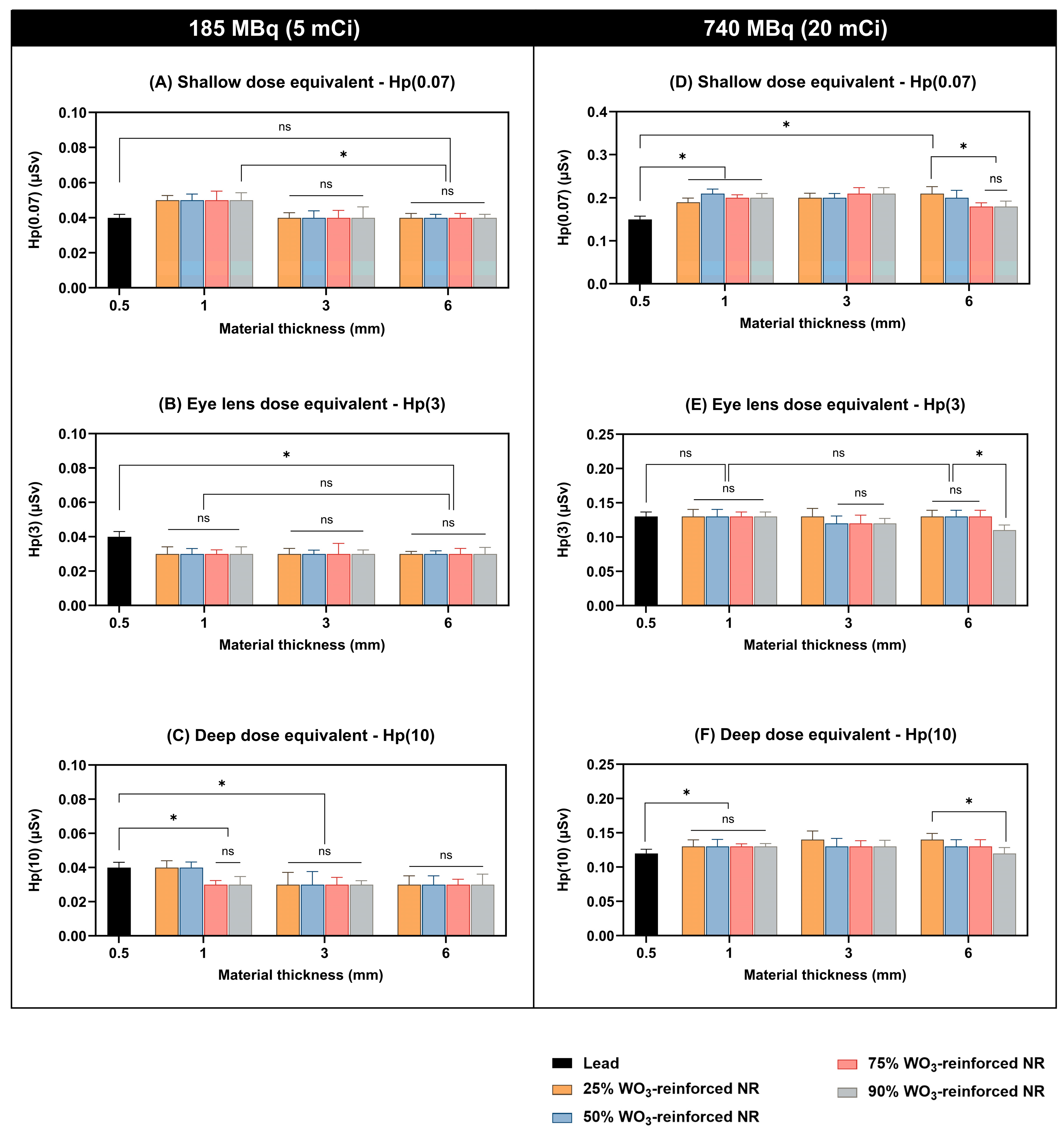
3.2.3. Experimental Evaluation of the Gamma Radiation-Shielding Efficiency of Tungsten-Reinforced Polyisoprene for Tc-99m

The experimental data presented in Figure 12 compares the shallow-dose equivalent (Hp(0.07)) for different shielding materials at two activity levels: 185 MBq (5 mCi) and 740 MBq (20 mCi). The shielding materials include WO3-reinforced NR composites with varying tungsten content (25%, 50%, 75%, 90%). In addition, a lead shield of 0.5 mm thickness is commonly used as a reference standard in gamma radiation shielding, providing a baseline for comparing the attenuation effectiveness of alternative materials under equivalent radiation conditions. The shallow-dose equivalent results (Figure 12A,D) indicate overall material thicknesses, with the lead (0.5 mm) routinely exhibiting the lowest Hp(0.07). At lower activity, the shielding performance of WO3-reinforced NR composites is comparable, with no significant advantage of higher tungsten concentrations. Lead outperforms polymer-based composites at 1 mm thickness, but differences diminish at 3 mm and 6 mm. Increasing material thickness (1 mm → 3 mm → 6 mm) lowers the dose equivalents. Lead shield and 25% WO3-reinforced NR at 1 mm thickness exhibit a notable difference (p < 0.05) according to statistical analysis. There are no statistically significant variations (ns) across compositions of WO3-reinforced NR.
The eye lens dose equivalent (185 MBq, 5 mCi) in Figure 12B indicates a declining trend with increasing material thickness across all compositions. At a thickness of 1 mm, NR-WO3 compositions provide somewhat reduced dose equivalents compared to lead, while statistical significance is noted only for certain compositions (p < 0.05). At a thickness of 3 mm, the same trends are seen, with the shielding efficacy of WO3-reinforced NR nearing that of lead. At a thickness of 6 mm, all WO3-reinforced NR compositions provide equivalent shielding, exhibiting no statistically significant differences from lead. The eye lens dose equivalent at high activity (Figure 12E) is generally elevated under these conditions. At a thickness of 1 mm, WO3-reinforced NR compositions exhibit comparable shielding efficacy to lead, with no statistically significant differences. At 3 mm thickness, a marginal decrease in dose is noted with increasing WO3-reinforced NR concentration, though statistical differences are minimal. At 6 mm thickness, the 90% WO3-reinforced NR composition shows a significantly lower dose compared to other materials (p < 0.05), indicating enhanced shielding efficiency at elevated concentrations.
According to the deep-dose results in Figure 12C, the Hp(10) values range from approximately 0.02 to 0.06 μSv, depending on the shielding material and its thickness. At a thickness of 1 mm, lead (0.5 mm Pb) demonstrates a markedly superior dose reduction compared to 25% WO3-reinforced NR (p < 0.05). However, beyond 25%, further increases in WO3 concentration do not yield statistically significant differences (ns). A similar pattern is evident at a thickness of 3 mm, with 0.5 mm Pb surpassing WO3-reinforced NR composites, yet no significant differences are observed among the 25%, 50%, 75%, and 90% WO3-reinforced NR formulations. At 6 mm thickness, no significant differences in Hp(10) are noted across all shielding materials, suggesting that increased thickness improves shielding only up to a saturation point, beyond which compositional differences become inconsequential. Figure 12F illustrates the deep-dose equivalent at an elevated dose (740 MBq). As observed, the Hp(10) values exceed those of the low-dose, ranging from roughly 0.10 to 0.20 μSv. At a thickness of 1 mm, 0.5 mm Pb exhibits much superior shielding compared to 25% WO3-reinforced NR (p < 0.05), although no significant differences are seen among the WO3-reinforced NR compositions (ns). No notable differences at a thickness of 3 mm among the WO3-reinforced NR composites indicate that greater thickness diminishes composition-dependent variations. At a thickness of 6 mm, Hp(10) values are statistically comparable across all materials, corroborating the finding that thicker materials provide adequate attenuation irrespective of tungsten concentration.
In summary, the experimental results indicate that tungsten-reinforced polyisoprene effectively reduces gamma radiation dose equivalents (Hp(0.07), Hp(3), and Hp(10)) for Tc-99m, with increasing material thickness generally leading to lower radiation exposure. At both low (185 MBq) and high (740 MBq) activity levels, lead (0.5 mm) demonstrated superior shielding performance compared to tungsten oxide (WO3) composites, though higher WO3 concentrations (75% and 90%) exhibited improved attenuation. Statistical analysis revealed significant reductions (p < 0.05) in dose equivalents with increased shielding material thickness, while some variations between compositions were not statistically significant.

4. Discussion

Shielding design is vital for radiation protection, ensuring barriers protect workers, patients, the public, and the environment from X-ray machine emissions. The NCRP 147 method, widely used by radiation protection experts (RPEs) for structural shielding in medical X-ray facilities, relies on 1996 data. With significant advancements in interventional radiology, evaluating the need to update workload data across specialties is essential [52].
WO3 is known for excellent radiation attenuation owing to its high atomic number and density. Tungsten’s high atomic number (Z = 74) increases photoelectric absorption and Compton scattering, the central gamma and X-ray attenuation processes. Also, the substantial density of WO3 (WO3 = 7.16 g/cm3, lead(II) oxide = 9.5 g/cm3, tungsten (W) = 19.25 g/cm3, and lead = 11.3 g/cm3) makes it suitable for absorbing ionizing radiation. This work established WO3-reinforced polyisoprene as having a highly structured, plate-like morphology, suggesting improved mechanical and thermal stability due to strong interfacial interactions between the polymer and the WO3 particles. Indicating enhanced reinforcement, potentially making this composite suitable for applications such as radiation shielding (Figure 4). Additionally, XRD shows that the NR composite shows significant diffraction peaks after WO3 reinforcement, indicating higher crystallinity and crystalline WO3 phases (Figure 5). These structural changes may increase mechanical strength and thermal stability. This increased crystallinity restricts the mobility of the polymer chains, resulting in a stiffer and stronger material. Jayasinghe et al. reported 92% improvement in tensile strength of natural rubber composites reinforced with titanium carbide nanocrystals [53]. Furthermore, the FTIR analysis of tungsten-reinforced polyisoprene (Figure 6) demonstrates the incorporation of WO3 into polyisoprene leads to new absorption bands related to W–O bonds, indicating successful dispersion of tungsten oxide within the polymer matrix.
XCOM-based computational analysis shows that WO3-reinforced NR, a tungsten-reinforced NR, exhibits significantly enhanced attenuation properties. Although PbO2 is still a widely used shielding material, WO3-reinforced NR greatly improves attenuation performance, showing great promise as a lead-free substitute, especially at intermediate and high photon energies. These results are consistent with other studies on tungsten-based shielding composites [54,55,56,57].
The Phy-X/PSD shielding analysis confirms that PbO2 exhibits superior attenuation properties, particularly at low photon energies where the photoelectric effect dominates, aligning with previous studies on high-Z materials for radiation shielding. The enhanced shielding performance of WO3-reinforced NR composites, particularly at WO3 concentrations ≥ 75%, suggests their potential as effective alternatives to lead-based shielding, consistent with findings that tungsten-based materials provide efficient gamma-ray attenuation [58,59]. The variation in MAC, LAC, and Zeff values across the energy spectrum indicates that while PbO2 remains the most effective, higher concentration of WO3 composites offer promising shielding capabilities for Tc-99m applications, as previously demonstrated in polymer-based radiation shields [60,61].
The experimental findings align with prior research indicating that while lead remains the most effective shielding material for gamma radiation, high-density polymer composites incorporating tungsten oxide (WO3) can provide comparable attenuation at increased thicknesses [62,63]. The observed dose reductions across Hp(0.07), Hp(3), and Hp(10) metrics demonstrate that material composition influences shielding performance primarily at lower thicknesses, whereas increasing thickness minimizes compositional differences, supporting previous studies on polymer-based radiation shielding [64]. Furthermore, the statistically significant reductions in dose equivalents (p < 0.05) with increasing thickness suggest that WO3-reinforced NR composites exhibit dose attenuation characteristics that are dependent on both composition and geometry, which concurs with findings from gamma shielding studies on tungsten-infused elastomers. Notably, the diminishing differences in shielding efficiency at 6 mm thickness indicate a saturation effect, where additional WO3 content does not yield proportionally greater attenuation, consistent with nonlinear attenuation behaviors reported for high-Z polymer matrices [65,66]. Moreover, the superior shielding efficacy of high WO3 content (≥75%) at higher radiation activity (740 MBq) suggests that the material benefits from enhanced photon absorption due to increased effective atomic number (Zeff), reinforcing conclusions from prior work on tungsten-based shielding composites. However, the absence of statistically significant differences among WO3-reinforced NR formulations at greater thicknesses implies that material optimization should focus not only on composition but also on overall density and structural integrity to maximize shielding efficiency [67].

5. Conclusions

In this study, we have comprehensively investigated the formulation and evaluation of natural rubber (NR) enhanced with tungsten oxide (WO3) as a potential alternative radiation shielding materials, specifically focusing on their applicability for Tc-99m shielding. The radiation attenuation characteristics of WO3-reinforced polyisoprene composites make them an appropriate replacement for traditional lead-based shielding. XRD, FTIR, structural analysis, and computational investigations demonstrate increased reinforcing and crystallinity, boosting shielding effectiveness. Although PbO2 is still better in attenuation, WO3-reinforced NR composites with ≥75% WO3 content show equivalent shielding performance, especially at higher photon energy, supporting their possible use in medical and nuclear radiation protection. Future studies should focus on maximizing the density and composition to raise shielding performance even more.

Author Contributions

Conceptualization, S.K.M.; methodology, S.K.M., J.C., P.K. and N.S.; software, S.K.M., J.C., P.K. and N.S.; validation, S.K.M., J.C., P.K. and N.S.; formal analysis, S.K.M., J.C., P.K. and N.S.; investigation, S.K.M., J.C., P.K. and N.S.; resources, S.K.M.; data curation, S.K.M., J.C., P.K. and N.S.; writing—original draft preparation, S.K.M.; writing—review and editing, S.K.M.; visualization, S.K.M., J.C., P.K. and N.S.; supervision, S.K.M.; project administration, S.K.M.; funding acquisition, S.K.M. All authors have read and agreed to the published version of the manuscript.

Funding

This work was supported by the Research and Development Office (RDO), Prince of Songkla University. The research was financially supported by the Faculty of Medicine, Prince of Songkla University (MR PSU-670722-234) [Grant number 67-059-1, 7 June 2024].

Institutional Review Board Statement

The research complied with the ethical standards established in the Declaration of Helsinki and adhered to the International Conference on Harmonization (ICH) recommendations for Good Clinical Practice (GCP). The Human Research Ethics Committee, Faculty of Medicine, Prince of Songkla University, Thailand, issued ethical permission (REC. 67-250-7-2, 24 May 2024).

Informed Consent Statement

Informed consent was obtained from all subjects involved in the study.

Data Availability Statement

The original contributions presented in this study are included in the article material. Further inquiries can be directed to the corresponding author.

Acknowledgments

The authors sincerely acknowledge the invaluable support of all staff members and extend their deep appreciation to the radiological technology personnel in the Department of Radiology, Faculty of Medicine, Prince of Songkla University, for their essential role in conducting the technical procedures.

Conflicts of Interest

The authors declare no conflicts of interest.

References

  1. Brambilla, M.; Vassileva, J.; Kuchcinska, A.; Rehani, M.M. Multinational Data on Cumulative Radiation Exposure of Patients from Recurrent Radiological Procedures: Call for Action. Eur. Radiol. 2020, 30, 2493–2501. [Google Scholar] [PubMed]
  2. Papagiannopoulou, D. Technetium-99m Radiochemistry for Pharmaceutical Applications. J. Label. Compd. Radiopharm. 2017, 60, 502–520. [Google Scholar] [CrossRef]
  3. Marshall, S.K.; Prom-On, P.; Sangkue, S.; Thiangsook, W. Assessment of Radiation Exposure in a Nuclear Medicine Department during 99mTc-MDP Bone Scintigraphy. Toxics 2023, 11, 814. [Google Scholar] [CrossRef] [PubMed]
  4. Wenzl, M.; Colombo, F.; Guanais, F.; Lopert, R.; Paris, V.; Pearson, M.; Charlton, K.; Demeter, S.J.; Bilbao y Leon, S.; Pike, C. The Supply of Medical Isotopes: An Economic Diagnosis and Possible Solutions; Organisation for Economic Co-Operation and Development: Paris, France, 2019. [Google Scholar]
  5. Marshall, S.K.; Hayeeabdunromae, A.; Noomad, N.; Durawee, W.; Sirieak, N.; Karnkorn, P.; Keawtong, V. Evaluation of Environmental Radiation Exposure and Algorithms for Determining the Occupational Effective Dose During 99mTc-MDP Bone Scintigraphy: A Comprehensive Analysis. Appl. Sci. 2024, 14, 11211. [Google Scholar] [CrossRef]
  6. Lee, W.J.; Choi, Y.; Ko, S.; Cha, E.S.; Kim, J.; Kim, Y.M.; Kong, K.A.; Seo, S.; Bang, Y.J.; Ha, Y.W. Projected Lifetime Cancer Risks from Occupational Radiation Exposure among Diagnostic Medical Radiation Workers in South Korea. BMC Cancer 2018, 18, 1206. [Google Scholar] [CrossRef]
  7. Güngördü, N.; Kurtul, S.; Özdil, A.; Erdoğan, M.S. Does Occupational Ionizing Radiation Exposure in Healthcare Workers Affect Their Hematological Parameters? Arch. Environ. Occup. Health 2023, 78, 80–87. [Google Scholar] [CrossRef]
  8. Boice, J.D.; Cohen, S.S.; Mumma, M.T.; Howard, S.C.; Yoder, R.C.; Dauer, L.T. Mortality among Medical Radiation Workers in the United States, 1965–2016. In The Million Person Study of Low-Dose Radiation Health Effects; CRC Press: Boca Raton, FL, USA, 2024; pp. 255–279. [Google Scholar]
  9. de Gonzalez, A.B.; Darby, S. Risk of Cancer from Diagnostic X-Rays: Estimates for the UK and 14 Other Countries. Lancet 2004, 363, 345–351. [Google Scholar] [CrossRef]
  10. Picano, E.; Vano, E. Updated Estimates of Radiation Risk for Cancer and Cardiovascular Disease: Implications for Cardiology Practice. J. Clin. Med. 2024, 13, 2066. [Google Scholar] [CrossRef]
  11. Richardson, D.B.; Leuraud, K.; Laurier, D.; Gillies, M.; Haylock, R.; Kelly-Reif, K.; Bertke, S.; Daniels, R.D.; Thierry-Chef, I.; Moissonnier, M. Cancer Mortality after Low Dose Exposure to Ionising Radiation in Workers in France, the United Kingdom, and the United States (INWORKS): Cohort Study. BMJ 2023, 382, e074520. [Google Scholar]
  12. Rühm, W.; Laurier, D.; Wakeford, R. Cancer Risk Following Low Doses of Ionising Radiation–Current Epidemiological Evidence and Implications for Radiological Protection. Mutat. Res. Toxicol. Environ. Mutagen. 2022, 873, 503436. [Google Scholar]
  13. Adelodun, M.O.; Anyanwu, E.C. Health Effects of Radiation: An Epidemiological Study on Populations near Nuclear Medicine Facilities. Health 2024, 13, 228–239. [Google Scholar]
  14. Calabrese, E.J. Linear Non-Threshold (LNT) Fails Numerous Toxicological Stress Tests: Implications for Continued Policy Use. Chem. Biol. Interact. 2022, 365, 110064. [Google Scholar]
  15. Calabrese, E.J.; Selby, P.B. Muller Mistakes: The Linear No-Threshold (LNT) Dose Response and US EPA’s Cancer Risk Assessment Policies and Practices. Chem. Biol. Interact. 2023, 383, 110653. [Google Scholar] [CrossRef] [PubMed]
  16. Allam, S.M.E.; Algany, M.M.A.; Khider, Y.I.A. Radiation Safety Compliance Awareness among Healthcare Workers Exposed to Ionizing Radiation. BMC Nurs. 2024, 23, 208. [Google Scholar] [CrossRef]
  17. Shubayr, N. Operating Room Radiation Safety Measures: Awareness, Compliance, and Perceived Risks among Nurses and Other Healthcare Workers. Int. Nurs. Rev. 2024; ahead of print. [Google Scholar] [CrossRef]
  18. Galli, E.; Soliman-Aboumarie, H.; Gargani, L.; Szymański, P.; Gimelli, A.; Petersen, S.E.; Sade, L.E.; Stankovic, I.; Donal, E.; Cosyns, B. EACVI Survey on Radiation Exposure in Interventional Echocardiography. Eur. Heart J.-Cardiovasc. Imaging 2024, 25, 727–734. [Google Scholar]
  19. Clarke, R.; Valentin, J. A History of the International Commission on Radiological Protection. Health Phys. 2005, 88, 407–422. [Google Scholar]
  20. Wu, Y.; Wang, Z. Progress in Ionizing Radiation Shielding Materials. Adv. Eng. Mater. 2024, 26, 2400855. [Google Scholar] [CrossRef]
  21. More, C.V.; Alsayed, Z.; Badawi, M.S.; Thabet, A.A.; Pawar, P.P. Polymeric Composite Materials for Radiation Shielding: A Review. Environ. Chem. Lett. 2021, 19, 2057–2090. [Google Scholar] [CrossRef]
  22. Prabhu, S.; Bubbly, S.G.; Gudennavar, S.B. X-Ray and γ-Ray Shielding Efficiency of Polymer Composites: Choice of Fillers, Effect of Loading and Filler Size, Photon Energy and Multifunctionality. Polym. Rev. 2023, 63, 246–288. [Google Scholar] [CrossRef]
  23. Marshall, S.K.; Boonpeng, K.; Buapud, N.; Chimhashat, S.; Chuaymuang, J.; Kwandee, P.; Songphum, N. Bismuth Oxide Nanoparticle-Enhanced Poly (Methyl Methacrylate) Composites for I-131 Radiation Shielding: A Combined Simulation and Experimental Investigation. Polymers 2025, 17, 590. [Google Scholar] [CrossRef]
  24. Intom, S.; Kalkornsurapranee, E.; Johns, J.; Kaewjaeng, S.; Kothan, S.; Hongtong, W.; Chaiphaksa, W.; Kaewkhao, J. Mechanical and Radiation Shielding Properties of Flexible Material Based on Natural Rubber/Bi2O3 Composites. Radiat. Phys. Chem. 2020, 172, 108772. [Google Scholar]
  25. Yasmin, S.; Kamislioglu, M.; Sayyed, M.I. Assessment of Radiation Shielding Performance of Li2O-BaO-Bi2O3-P2O5 Glass Systems within the Energy Range from 0.081 MeV to 1.332 MeV via MCNP6 Code. Optik 2023, 274, 170529. [Google Scholar]
  26. Gamal, R.; Salama, E.; Elshimy, H.; El-Nashar, D.E.; Bakry, A.; Ehab, M. Gamma Attenuation and Mechanical Characteristics of a Lead/NBR/SBR Rubber Composite with Black Nanocarbon Reinforcement. Sustainability 2023, 15, 2165. [Google Scholar] [CrossRef]
  27. Toyen, D.; Rittirong, A.; Poltabtim, W.; Saenboonruang, K. Flexible, Lead-Free, Gamma-Shielding Materials Based on Natural Rubber/Metal Oxide Composites. Iran. Polym. J. 2018, 27, 33–41. [Google Scholar] [CrossRef]
  28. Ali, A.; Zhang, N.; Santos, R.M. Mineral Characterization Using Scanning Electron Microscopy (SEM): A Review of the Fundamentals, Advancements, and Research Directions. Appl. Sci. 2023, 13, 12600. [Google Scholar] [CrossRef]
  29. Belz, G.T.; Auchterlonie, G.J. An Investigation of the Use of Chromium, Platinum and Gold Coating for Scanning Electron Microscopy of Casts of Lymphoid Tissues. Micron 1995, 26, 141–144. [Google Scholar]
  30. Holder, C.F.; Schaak, R.E. Tutorial on Powder X-Ray Diffraction for Characterizing Nanoscale Materials. ACS Nano 2019, 13, 7359–7365. [Google Scholar] [CrossRef]
  31. Fawcett, T.G.; Kabekkodu, S.N.; Blanton, J.R.; Blanton, T.N. Chemical Analysis by Diffraction: The Powder Diffraction FileTM. Powder Diffr. 2017, 32, 63–71. [Google Scholar]
  32. Chan, K.A.; Kazarian, S.G. Attenuated Total Reflection Fourier-Transform Infrared (ATR-FTIR) Imaging of Tissues and Live Cells. Chem. Soc. Rev. 2016, 45, 1850–1864. [Google Scholar]
  33. Smith, T.L. Ultimate Tensile Properties of Elastomers. II. Comparison of Failure Envelopes for Unfilled Vulcanizates. Rubber Chem. Technol. 1964, 37, 792–807. [Google Scholar]
  34. Wood, L.A. Uniaxial Extension and Compression in Stress-Strain Relations of Rubber. J. Res. Natl. Bur. Stand. 1977, 82, 57. [Google Scholar]
  35. Bokobza, L.; Rapoport, O. Reinforcement of Natural Rubber. J. Appl. Polym. Sci. 2002, 85, 2301–2316. [Google Scholar] [CrossRef]
  36. James, H.M.; Guth, E. Theory of the Elastic Properties of Rubber. J. Chem. Phys. 1943, 11, 455–481. [Google Scholar]
  37. Brown, D.A.; Chadwick, M.B.; Capote, R.; Kahler, A.C.; Trkov, A.; Herman, M.W.; Sonzogni, A.A.; Danon, Y.; Carlson, A.D.; Dunn, M. ENDF/B-VIII.0: The 8th Major Release of the Nuclear Reaction Data Library with CIELO-Project Cross Sections, New Standards and Thermal Scattering Data. Nucl. Data Sheets 2018, 148, 1–142. [Google Scholar]
  38. Berger, M.J.; Hubbell, J.H. XCOM: Photon Cross Sections on a Personal Computer; U.S. Department of Commerce, National Bureau of Standards: Gaithersburg, MD, USA, 1987. [Google Scholar]
  39. Gerward, L.; Guilbert, N.; Jensen, K.B.; Levring, H. X-Ray Absorption in Matter. Reengineering XCOM. Radiat. Phys. Chem. 2001, 60, 23–24. [Google Scholar]
  40. Şakar, E.; Özpolat, Ö.F.; Alım, B.; Sayyed, M.I.; Kurudirek, M. Phy-X/PSD: Development of a User Friendly Online Software for Calculation of Parameters Relevant to Radiation Shielding and Dosimetry. Radiat. Phys. Chem. 2020, 166, 108496. [Google Scholar]
  41. Bellamy, M.B.; Bernstein, J.L.; Cullings, H.M.; French, B.; Grogan, H.A.; Held, K.D.; Little, M.P.; Tekwe, C.D. Recommendations on Statistical Approaches to Account for Dose Uncertainties in Radiation Epidemiologic Risk Models. Int. J. Radiat. Biol. 2024, 100, 1393–1404. [Google Scholar] [CrossRef]
  42. ISO 4037-3-2019; Radiological Protection—X and Gamma Reference Radiation for Calibrating Dosemeters and Dose Rate Meters and for Determining Their Response as a Function of Photon Energy—Part 3: Calibration of Area and Personal Dosemeters and the Measurement of Their Response as a Function of Energy and Angle of Incidence. ISO: Geneva, Switzerland, 2019.
  43. Singhal, R.K.; Samariya, A.; Xing, Y.-T.; Kumar, S.; Dolia, S.N.; Deshpande, U.P.; Shripathi, T.; Saitovitch, E.B. Electronic and magnetic properties of Co-doped ZnO diluted magnetic semi-conductor. J. Alloys Compd. 2010, 496, 324. [Google Scholar]
  44. McMurdie, H.F.; Morris, M.C.; Evans, E.H.; Paretzkin, B.; Wong-Ng, W.; Ettlinger, L.; Hubbard, C.R. Standard X-ray diffraction powder patterns from the JCPDS research associateship. Powder Diffr. 1986, 1, 64–77. [Google Scholar]
  45. Han, W.; Gao, M. Investigations on iron sulfide nanosheets prepared via a single-source precursor approach. Cryst. Growth Des. 2008, 8, 1023–1030. [Google Scholar]
  46. Ahn, W.; Kim, K.B.; Jung, K.N.; Shin, K.H.; Jin, C.S. Synthesis and electrochemical properties of a sulfur–multi-walled carbon nanotubes composite as a cathode material for lithium–sulfur batteries. J. Power Sources 2012, 202, 394–399. [Google Scholar] [CrossRef]
  47. Zhang, Y.C.; Wang, G.Y.; Hu, X.Y.; Shi, Q.F.; Qiao, T.; Yang, Y. Phase-controlled synthesis of ZnS nanocrystallites by mild solvothermal decomposition of an air-stable single-source molecular precursor. J. Cryst. Growth 2005, 284, 554–560. [Google Scholar] [CrossRef]
  48. Onwudiwe, D.C.; Ajibade, P.A. Synthesis, characterization and thermal studies of Zn(II), Cd(II), and Hg(II) complexes of N-methyl-N-phenyldithiocarbamate: The single crystal structure of [(C6H5)(CH3)NCS2]4Hg2. Int. J. Mol. Sciences 2011, 12, 1964–1978. [Google Scholar] [CrossRef] [PubMed]
  49. DiMarco, J. ICDD Grant-in-Aid 1990; JCPDS code 00-041-1876; Physics Department, The Polytechnic University: Brooklyn, NY, USA, 1990. [Google Scholar]
  50. Parks, J.; McCarthy, G. ICDD Grant-in-Aid 1993; JCPDS code 00-047-0064; North Dakota State University: Fargo, ND, USA, 1993. [Google Scholar]
  51. Diehl, R.; Brandt, G.; Salje, E. The crystal structure of triclinic WO3. Acta Crystallogr., Sect. B Struct. Crystallogr. Cryst. Chem. 1978, 34, 1105. [Google Scholar] [CrossRef]
  52. Biondi, M.; Bortoli, E.; Avitabile, R.; Bartoli, A.; Busatti, E.; Tozzi, A.; Cimmino, M.C.; Pini, A.; Guasti, A. Radiation Shielding Calculation for Interventional Radiology: An Updated Workload Survey Using a Dose Monitoring Software. Phys. Med. 2024, 125, 104509. [Google Scholar] [CrossRef]
  53. Jayasinghe, J.; De Silva, R.T.; de Silva, K.N.; de Silva, R.M.; Silva, V.A. Titanium Carbide Ceramic Nanocrystals to Enhance the Physicochemical Properties of Natural Rubber Composites. RSC Adv. 2020, 10, 19290–19299. [Google Scholar] [CrossRef]
  54. AbuAlRoos, N.J.; Azman, M.N.; Amin, N.A.B.; Zainon, R. Tungsten-Based Material as Promising New Lead-Free Gamma Radiation Shielding Material in Nuclear Medicine. Phys. Med. 2020, 78, 48–57. [Google Scholar] [CrossRef]
  55. Kim, S.-C. Evaluation of Shielding Performance of Gamma Ray Shielding Tungsten Polymer Composite with LBL-Type Layered Structure. Coatings 2023, 14, 36. [Google Scholar] [CrossRef]
  56. Tishkevich, D.I.; Rotkovich, A.A.; German, S.A.; Zhaludkevich, A.L.; Vershinina, T.N.; Bondaruk, A.A.; Razanau, I.U.; Dong, M.; Sayyed, M.I.; Leonchik, S.V. Heavy Alloy Based on Tungsten and Bismuth: Fabrication, Crystal Structure, Morphology, and Shielding Efficiency against Gamma-Radiation. RSC Adv. 2023, 13, 24491–24498. [Google Scholar] [CrossRef]
  57. Taniguchi, Y.; Wakabayashi, H.; Yoneyama, H.; Chen, Z.; Morino, K.; Otosaki, A.; Yamada, M.; Inaki, A.; Kayano, D.; Kinuya, S. Application of a Tungsten Apron for Occupational Radiation Exposure in Nursing Care of Children with Neuroblastoma during 131I-Meta-Iodo-Benzyl-Guanidine Therapy. Sci. Rep. 2022, 12, 47. [Google Scholar] [CrossRef]
  58. El-Khatib, A.M.; Alabsy, M.T.; El-Khatib, A.Y.; Dib, M.F.; Abbas, M.I. Superiority of Micro/Nano Tungsten Carbide Reinforced Poly-Methyl Methacrylate Composites in Shielding Gamma Radiation. Nucl. Eng. Technol. 2024, 56, 4103–4114. [Google Scholar]
  59. Kassim, H.; Aldawood, S.; Prasad, S.; Asemi, N.N.; Aziz, A.A.; AlSalhi, M.S. Advanced Polymeric Matrix Utilizing Nanostructured Bismuth and Tungsten Oxides for Gamma Rays Shielding. Heliyon 2024, 10, e37289. [Google Scholar] [PubMed]
  60. Karabul, Y.; İçelli, O. The Assessment of Usage of Epoxy Based Micro and Nano-Structured Composites Enriched with Bi2O3 and WO3 Particles for Radiation Shielding. Results Phys. 2021, 26, 104423. [Google Scholar]
  61. Eyssa, H.M.; Sadek, R.F.; Mohamed, W.S.; Ramadan, W. Structure-Property Behavior of Polyethylene Nanocomposites Containing Bi2O3 and WO3 as an Eco-Friendly Additive for Radiation Shielding. Ceram. Int. 2023, 49, 18442–18454. [Google Scholar]
  62. Kim, D.H.; Cho, Y.I. Evaluation of Shielding Performance of Polymer-Tungsten Composites in High-Energy Electron Beam Therapy. J. Korean Soc. Radiol. 2024, 18, 783–791. [Google Scholar]
  63. Kim, S.-C. Tungsten-Based Hybrid Composite Shield for Medical Radioisotope Defense. Materials 2022, 15, 1338. [Google Scholar] [CrossRef]
  64. Al Hassan, M.; Liu, W.B.; Wang, J.; Ali, M.M.M.; Rawashdeh, A. Monte Carlo Simulations of Gamma-Rays Shielding with Phthalonitrile-Tungsten Borides Composites. Int. J. Radiat. Res. 2022, 20, 621–626. [Google Scholar]
  65. Gilys, L.; Griškonis, E.; Griškevičius, P.; Adlienė, D. Lead Free Multilayered Polymer Composites for Radiation Shielding. Polymers 2022, 14, 1696. [Google Scholar] [CrossRef]
  66. Bijanu, A.; Arya, R.; Agrawal, V.; Tomar, A.S.; Gowri, V.S.; Sanghi, S.K.; Mishra, D.; Salammal, S.T. Metal-Polymer Composites for Radiation Protection: A Review. J. Polym. Res. 2021, 28, 392. [Google Scholar] [CrossRef]
  67. Li, Z.; Zhou, W.; Zhang, X.; Gao, Y.; Guo, S. High-Efficiency, Flexibility and Lead-Free X-Ray Shielding Multilayered Polymer Composites: Layered Structure Design and Shielding Mechanism. Sci. Rep. 2021, 11, 4384. [Google Scholar] [CrossRef]
Figure 1. Preparation of tungsten-reinforced polyisoprene (WO3-reinforced NR) composite. The schematic illustrates the preparation process of WO3-reinforced NR used in this study. The complete vulcanization formulation, including all components and their functional roles in the composite, is detailed in Table 1.
Applsci 15 03892 g001
Figure 2. The experimental setup for evaluating the shielding efficacy of materials against Tc-99m radiation using an RSD anthropomorphic phantom. The phantom simulates human tissue and is equipped with radiation dosimeters Hp(3), Hp(10), and Hp(0.07), placed at the eye lens, thoracic, and abdominal regions, respectively, to measure dose equivalents. Two configurations were assessed: (A) without shielding material and (B) with shielding material placed between the Tc-99m radiation source and the phantom. In both configurations, the Tc-99m source was positioned 30 cm in front of the phantom.
Applsci 15 03892 g002
Figure 3. Scanning electron microscopy (SEM) images of polyisoprene (natural rubber, NR) and tungsten oxide (WO3)-reinforced polyisoprene at different magnifications (500×, 1500×, and 3000×).
Applsci 15 03892 g003
Figure 4. Energy-dispersive X-ray spectroscopy (EDX) and scanning electron microscopy (SEM) analysis of polyisoprene (natural rubber, NR) and tungsten oxide (WO3)-reinforced polyisoprene. Elemental mapping images illustrate the spatial distribution of key elements, including carbon (C), oxygen (O), sodium (Na), zinc (Zn), sulfur (S), and tungsten (W). The overlay images present a composite visualization of elemental dispersion within each sample.
Applsci 15 03892 g004
Figure 5. X-ray diffraction (XRD) analysis of polyisoprene (natural rubber, NR) and tungsten oxide (WO3)-reinforced polyisoprene. (A) XRD pattern of NR, with corresponding phase identification for major crystalline components, including zinc oxide (ZnO), sulfur (S), zinc dimethyldithiocarbamate, and thiram. (B) XRD pattern of WO3-reinforced polyisoprene, showing characteristic peaks of sodium tungsten oxide hydrate (Na2WO4·2H2O), tungsten oxide (WO3), and ZnO. (C) Comparative XRD patterns of NR and WO3-reinforced polyisoprene, highlighting differences in crystallinity and phase composition. (D) Stacked XRD patterns of NR and WO3-reinforced polyisoprene for direct visualization of peak variations.
Applsci 15 03892 g005
Figure 6. Fourier transform infrared (FTIR) spectroscopy spectra of polyisoprene (natural rubber, NR) and tungsten oxide (WO3)-reinforced polyisoprene. The transmittance spectra display characteristic absorption bands corresponding to different functional groups present in each material.
Applsci 15 03892 g006
Figure 7. Characterization of shape deformation in tungsten-reinforced polyisoprene under bending, simulated using ANSYS. The deformation is presented for applied forces of (A) 10 N, (B) 25 N, (C) 50 N, and (D) 100 N. The color gradient represents the displacement magnitude (in mm), with red indicating the maximum deformation and blue indicating the minimum deformation.
Applsci 15 03892 g007
Figure 8. Computational analysis of tungsten-reinforced polyisoprene as a radiation-shielding material using XCOM simulations. Comparison of mass attenuation coefficients (cm2/g) for different photon interaction processes as a function of photon energy (MeV) for PbO2 (black), natural rubber (NR) (red), and tungsten-reinforced polyisoprene (WO3-reinforced NR) (blue). (A) Coherent scattering, (B) incoherent scattering, (C) photoelectric absorption, (D) pair production in the nuclear field, (E) pair production in the electron field, (F) total attenuation with coherent scattering, and (G) total attenuation without coherent scattering.
Applsci 15 03892 g008
Figure 9. Phy-X/PSD-based simulation of radiation shielding materials for Tc-99m applications. The graphs depict key radiation-shielding parameters as a function of photon energy for various shielding materials, including lead oxide (PbO2), natural rubber (NR), and tungsten-reinforced polyisoprene (WO3-reinforced NR) composites with 25%, 50%, 75%, 90%, and 100% tungsten oxide (WO3). The evaluated shielding parameters include (A) mass attenuation coefficient (MAC), (B) linear attenuation coefficient (LAC), (C) half-value layer (HVL), and (D) tenth-value layer (TVL). The insets in each plot provide parameter values at 140 keV, corresponding to the primary photon energy of Tc-99m, facilitating a comparative assessment of shielding efficiency across the materials.
Applsci 15 03892 g009
Figure 10. Phy-X/PSD-based simulation of radiation-shielding materials for Tc-99m applications. The graphs depict key radiation-shielding parameters as a function of photon energy for various shielding materials, including lead oxide (PbO2), natural rubber (NR), and tungsten-reinforced polyisoprene (WO3-reinforced NR) composites with 25%, 50%, 75%, 90%, and 100% tungsten oxide (WO3). The evaluated shielding parameters include (A) mean free path (MFP), (B) effective electron density (Neff), (C) effective conductivity (Ceff), and (D) atomic cross section (ACS). The insets in each plot provide parameter values at 140 keV, corresponding to the primary photon energy of Tc-99m, facilitating a comparative assessment of shielding efficiency across the materials.
Applsci 15 03892 g010
Figure 11. Phy-X/PSD-based simulation of radiation-shielding materials for Tc-99m applications. The graphs depict key radiation-shielding parameters as a function of photon energy for various shielding materials, including lead oxide (PbO2), natural rubber (NR), and tungsten-reinforced polyisoprene (WO3-reinforced NR) composites with 25%, 50%, 75%, 90%, and 100% tungsten oxide (WO3). The evaluated shielding parameters include (A) electronic cross section (ECS), (B) effective atomic number (Zeff), and (C) equivalent atomic number (Zeq). The insets in each plot provide parameter values at 140 keV, corresponding to the primary photon energy of Tc-99m, facilitating a comparative assessment of shielding efficiency across the materials.
Applsci 15 03892 g011
Figure 12. Experimental evaluation of the gamma radiation-shielding efficiency of tungsten-reinforced polyisoprene for Tc-99m. Radiation dose equivalents were measured at low and high activity levels for various shielding materials and compositions. (A) Shallow-dose equivalent (Hp(0.07)), (B) eye lens dose equivalent (Hp(3)), and (C) deep-dose equivalent (Hp(10)) were evaluated at a low dose (185 MBq, 5 mCi). (D) Shallow-dose equivalent (Hp(0.07)), (E) eye lens dose equivalent (Hp(3)), and (F) deep-dose equivalent (Hp(10)) were analyzed at a high dose (740 MBq, 20 mCi). The shielding materials tested include lead (0.5 mm) and tungsten oxide (WO3) at varying concentrations (25%, 50%, 75%, 90%) with thicknesses of 1, 3, and 6 mm. Data are presented as mean ± standard deviation (n = 3), where n = 3 represents the number of independent experimental replicates per condition. Statistical significance is indicated by * (p < 0.05), while “ns” denotes no significant difference.
Applsci 15 03892 g012
Table 1. The composition of the tungsten-reinforced polyisoprene (natural rubber, NR).
IngredientContent (phr) *Chemical RoleRole in Composite
Natural rubber (NR)100 (part)ElastomerPrimary matrix providing flexibility, elasticity, and mechanical strength.
Zinc Oxide (ZnO)10Vulcanization
activator
Activator for sulfur vulcanization, enhances cross-linking efficiency.
Stearic acid2Vulcanization
co-activator
Processing aid and activator, improves dispersion of fillers.
Tetramethylthiuram
disulfide (TMTD)
2Secondary accelerator and
sulfur donor
Accelerator, speeds up vulcanization, improves cross-linking.
Sulfur (S)5Vulcanizing agentCross-linking agent, enhances elasticity and strength.
WO30, 25, 50,
75, 90 wt%
Reinforcing filler and radiation-shielding additiveReinforcing filler, provides radiation-shielding, and increases density.
* phr: parts per hundred parts of rubber by weight.
Disclaimer/Publisher’s Note: The statements, opinions and data contained in all publications are solely those of the individual author(s) and contributor(s) and not of MDPI and/or the editor(s). MDPI and/or the editor(s) disclaim responsibility for any injury to people or property resulting from any ideas, methods, instructions or products referred to in the content.

