Article

Physicochemical, Functional, and Antibacterial Properties of Inulin-Type Fructans Isolated from Dandelion (Taraxacum officinale) Roots by “Green” Extraction Techniques

by
Nadezhda Petkova
1,*,
Ivanka Hambarliyska
1,
Ivan Ivanov
1,
Manol Ognyanov
2,
Krastena Nikolova
3,
Sevginar Ibryamova
4 and
Tsveteslava Ignatova-Ivanova
4
1
Department of Organic Chemistry and Inorganic Chemistry, Technological Faculty, University of Food Technologies, 26 Maritza Blvd., 4002 Plovdiv, Bulgaria
2
Laboratory of Biologically Active Substances, Institute of Organic Chemistry with Centre of Phytochemistry, Bulgarian Academy of Sciences, 139 Ruski Blvd., 4000 Plovdiv, Bulgaria
3
Department of Physics and Biophysics, Medical University “Prof. Dr. Paraskev Stoyanov”, 84 Tsar Osvoboditel Blvd., 9000 Varna, Bulgaria
4
Department of Biology, Shumen University “Konstantin Preslavski”, 115 Universitetska Str., 9700 Shumen, Bulgaria
*
Author to whom correspondence should be addressed.
Appl. Sci. 2025, 15(8), 4091; https://doi.org/10.3390/app15084091
Submission received: 6 March 2025 / Revised: 2 April 2025 / Accepted: 4 April 2025 / Published: 8 April 2025
(This article belongs to the Special Issue Novel Extraction Methods and Applications)

Abstract

:
The current study aims for the isolation and physicochemical characterization of inulin from defatted dandelion roots using green extraction techniques, including microwave extraction (MAE) and ultrasound-assisted extraction (UAE). The structure and degree of polymerization of inulin were elucidated by chromatographic techniques, as well as by FTIR and NMR spectroscopies. The color characteristics, water- and oil-holding capacity, solubility, swelling properties, wettability, angle of repose, flowability, and cohesiveness of dandelion inulin were evaluated. Moreover, the antioxidant and antibacterial potential of dandelion inulin were revealed. The results were compared with the conventional extraction and inulin from chicory. Dandelion inulin was evaluated as a powder substance with a degree of polymerization (DP) of 17–24. The highest yield (20%) was obtained by classical extraction; however, UAE and MAE demonstrated the highest purity. FT-IR and NMR spectra revealed that dandelion inulin is glucofructan with a molecular weight of 2.7–3.2 kDa that consists mainly of fructosyl units β-(2→1) linked to one α-D-glucose unit UAE was evaluated as the most perspective technique for the simultaneous extraction of inulin from dandelion roots, with the highest average DP 24 and high purity (82%), molecular mass, total fructose content, swelling index, and oil-holding capacity. Dandelion inulin exhibited intermediate cohesiveness, fair flowability, and moderate antimicrobial activity against Listeria monocytogenes 863 and Bacillus subtilis 6633. The physicochemical and functional properties of dandelion inulin reveal its future potential as an additive in food, cosmetic, and pharmaceutics formulations as a texture modifier, a fat replacer, and a drug carrier.

1. Introduction

Dandelion (Taraxacum sect. Taraxacum F.H. Wigg., syn. Taraxacum officinale) is a medicinal and edible plant that belongs to the genus Taraxacum, subfamily Cichorioideae, Asteraceae family, and occurs in New Zealand, Australia, Eurasia, Africa, and North and South America. It is widespread in subarctic and Northern temperate regions as a common perennial weed growing in roadsides, fallow fields, meadows, and lawns [1,2,3]. Because of its medicinal properties, it is predominantly produced as a cultivated plant in Bulgaria, Romania, Hungary, and Poland [4,5]. Dandelion roots and their herb extracts have been “generally recognized as safe” according to the Food and Drug Administration, and they are applied for dietary supplements, medicinal use, and food purposes [1,4]. Dandelion is traditionally used for bacterial infections, kidney diseases, diuretic diabetes, liver, and spleen disorders. The root can be utilized as an anti-inflammatory agent, and also for the treatment of hepatitis, dyspepsia, heartburn, liver complaints, spleen, anorexia, and for the prevention of renal gravel and loss of appetite [1,2,3,4,5,6,7]. The mentioned effects are due to the numerous bioactive substances in its roots [1,2,3]. Among them, dandelion polysaccharides deserve special attention. Taraxacum officinale roots contain polysaccharides [3], mainly inulin-type fructans [1,2,5]. Inulin and fructooligosaccharides are fructans that consist mainly of β-(2→1)-fructosyl fructose units (Fm), with one α-glucopyranosyl unit (1→2) (GFn) usually, but not always [2,8]. Inulin possesses many bioactive properties, such as prebiotic and immunomodulation activity, improves mineral absorption and glucolipid metabolism, lowers blood sugar, and represses obesity, osteoporosis, and cancer. Its healthy effect is connected mainly to the action of short-chain fatty acids that result after inulin fermentation in the large intestine (colon) by bifidobacteria [2,4,5,6]. Inulin in dandelion roots is situated in parenchyma cells in colorless lumps and clumps [9]. Its content in dandelion roots varies from 2 to 40% depending on the season and location [4,5,10,11,12], and as usual for Bulgarian representatives total fructans do not exceed 20% dw, while inulin content was found to be 11% [5].
Many studies have demonstrated that dandelion polysaccharides exhibit anti-inflammatory, anti-complement, antioxidant, antibacterial, antitumor, hypoglycemic, anticoagulant, and other effects [3,4,6,7]. It was shown that the two polysaccharides from dandelion roots could protect the liver from hepatic injury, while the fructan fraction showed good antioxidant activities in DPPH, hydroxyl free radical-scavenging abilities, and good hypoglycemic activities [6].
Many different extraction approaches have been performed for the extraction of inulin-type fructans from different plants: classical extraction, enzyme-assisted extraction, pressure liquid extraction, accelerated solvent extraction, microwave-assisted extraction, and ultrasound-assisted extraction [6,13,14,15,16,17]. It was reported that dandelion polysaccharides, especially inulin, were obtained mainly by classical extraction [5,10,11,12,18], and only one study mentioned ultrasound-assisted extraction [6]. In most cases, the classical extraction of dandelion inulin is connected mainly to its content determination, structure elucidation, or physicochemical evaluation, but its functional properties were not listed at all. Moreover, no studies have been conducted on the isolation of inulin from dandelion roots by microwave-assisted extraction. Therefore, an investigation into the impact of ultrasound-assisted and microwave extraction on the physicochemical properties of dandelion inulin remains a challenge. It was reported earlier that the physicochemical properties of polysaccharides (mainly chemical composition and molecular weight distribution) were strongly influenced by the used extraction processes [19]. Extraction with hot water is not considered a cost-effective method and has a negative environmental impact because of the high temperature and possibility of altering the polysaccharide structure [3]. The application of green extraction methods, in particular, ultrasound-assisted extraction and microwave irradiation, possesses many advantages, such as short extraction time, low energy consumption, low extraction temperature, high yield, high efficiency, and good quality of extracted compounds compared to conventional methods [15,16,17,20]. Moreover, microwaves accelerate the extraction process through the action of high-frequency magnetic waves, which penetrate the plant material in depth and protect target bioactive substances, especially polysaccharides [3]. A lack of studies about microwave and ultrasonic extraction of inulin from Taraxacum sect. Taraxacum F.H. Wigg roots motivated the current study. Therefore, investigating the quality and structure of isolated dandelion inulin obtained by using the cavitation process of ultrasonic waves and high-energy microwaves remains a challenge. Additionally, structural elucidation, as well as an evaluation of physicochemical and functional properties and antimicrobial potential of dandelion inulin, have not been investigated in detail.
To the best of our knowledge, microwave and ultrasonic irradiation techniques have not been implemented in inulin extraction from dandelion (Taraxacum sect. Taraxacum F.H. Wigg) roots. In addition, the antimicrobial potential of dandelion inulin has not been studied at all. Therefore, the aim of the current study is connected mainly to the isolation and investigation of the physiochemical, functional, and antibacterial properties of inulin obtained from dandelion roots using green extraction techniques.

2. Materials and Methods

2.1. Materials

Dry chopped dandelion (Radix Taraxaci) roots with Lot number 60/06.2022 supplied by ALIN Company, (Alino, Sofia, Bulgaria) were used in the current study. The roots had an initial moisture content of 10.79 ± 0.22%. The sample (100 g roots) was finely ground in a laboratory homogenizer and then it was sieved through a 0.5 mm filter. All other chemicals were of analytical grade. Chicory inulin Raftiline HPX (Beneo, Orafti, Belgium) with a declared average DP ≥ 23 was used as a reference in all analyses in the current research.

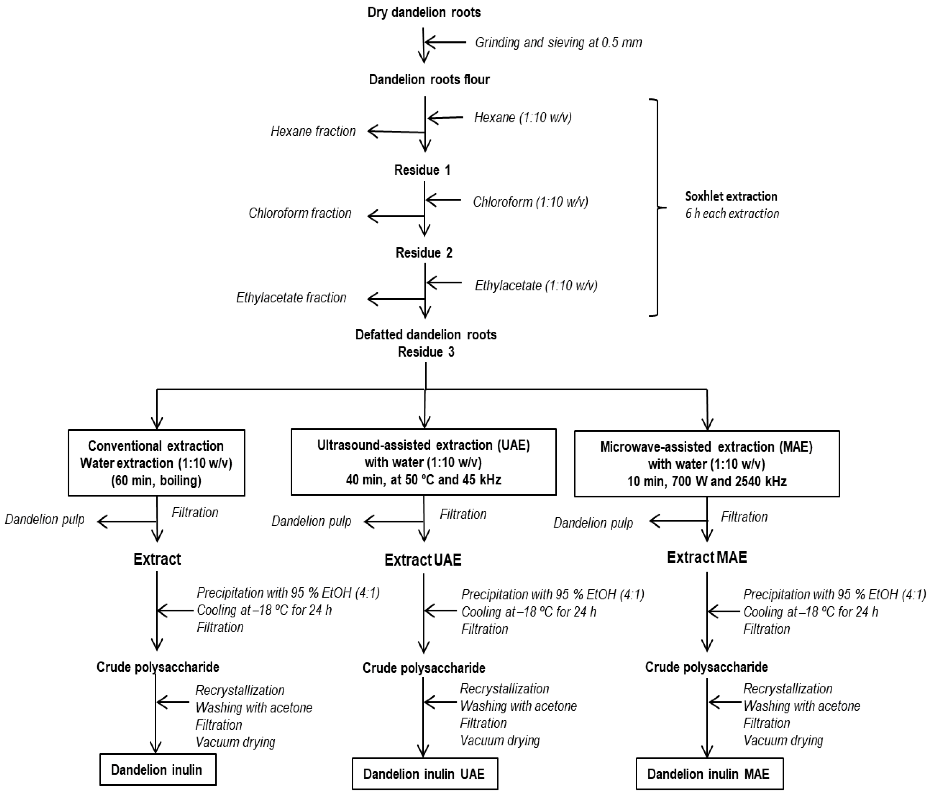
2.2. Fractional Extraction of Dandelion Roots

The procedure of inulin isolation from dandelion roots is illustrated in Figure 1. Firstly, the ground dandelion roots (100 g) were successively extracted in a Soxhlet apparatus with n-hexane, CHCl3, and ethyl acetate in a solid-to-solvent ratio of 1:10 (w/v). The solvents were removed after each extraction step through vacuum concentration. The defatted residues from dandelion roots were air-dried. A preliminary study of the inulin content in the initial plant material was performed before its further extraction. The obtained defatted roots (5 g) were extracted with water as previously described and the fructan, inulin, and sugar content in water extracts were determined as described previously [5,17].
Inulin was extracted from the defatted dandelion roots using distilled water as a solvent (1:10 w/v) in three different techniques: conventional, ultrasound-assisted, and microwave-assisted extractions.

2.3. Isolation of Inulin from Dandelion Roots

Conventional or classical extraction of inulin from the defatted dandelion roots (listed as Residue 3 in Figure 1) was conducted with distilled water in a solvent-to-solid ratio of (1:10 w/v) under reflux at 100 °C for 60 min, with constant stirring. Ultrasound-assisted extraction (UAE) was performed in an ultrasonic bath VWR USC 100 TH (Malaysia) employing an ultrasonic frequency of 45 kHz and 30 W power at a temperature of 50 °C for 20 min. Microwave-assisted extraction (MAE) was performed in a microwave device (CROWN, Norwich, Norfolk, The United Kingdom) microwave output power 700 W and 2450 MHz frequency) for 5 min. All water extractions were performed in duplicate with distilled water as a solvent. The water extracts were filtered by vacuum filtration using a Buchner funnel. The combined extracts were precipitated by the addition of four volumes of ethanol (95%), and then cooled down at −18 °C for 24 h and a filtration step was performed. The crude polysaccharide was dried, redissolved in hot water, reprecipitated, and washed with acetone. The resulting precipitate was vacuum-dried, kept in tightly sealed plastic containers, and used for further analysis.

2.4. Characterization of Dandelion Inulin

2.4.1. Yield, Moisture, and Ash Content

The yield of dandelion inulin was calculated on the basis of dry and defatted dandelion roots and expressed in percent [19]. The moisture content of dandelion inulin was determined by the AOAC method [21]. The weight loss percentage of inulin was calculated (%), after drying at 105 °C until constant weight, and the moisture content was expressed on the dry weight basis. The ash content in inulin was evaluated according to the AOAC method by assessing the ignition of the sample in a muffle furnace [21].

2.4.2. pH Values

The pH values of inulin powders were determined using the AOAC method 14.022 [21]. Inulin solutions of 10% (w/v) were prepared using Milli-Q water and pH was measured at 25 °C using a pH meter (model HI 9025, Hanna Instruments, Keysborough, Australia).

2.4.3. Melting Point and Angle of Optical Rotation

The melting point of the isolated dandelion inulin was measured on a Kofler melting point apparatus (Carl Zeiss, Jena, Germany) coupled with a digital camera and thermometer. The angle of optical rotation of 10 mL dandelion inulin–water solutions with a concentration of 2% was measured on an automated polarimeter Polamat A (Carl Zeiss, Jena, Germany) in a tube 1 dm long using a sodium lamp as a light source.

2.4.4. Protein Content

Protein content was assessed according to the method of Bradford, employing bovine serum albumin as a standard [22].

2.4.5. Reducing Groups and Total Fructose Content

The reducing groups were quantified by using the PAHBAH method at 410 nm [14]. The calibration curve was constructed using D-glucose as a reference in a concentration range of 5 to 100 μg/mL (Y = 0.0143x + 0.0174; R2 = 0.999). The total fructose content in dandelion inulin was estimated spectrophotometrically by resorcinol-thiourea reagent [23].

2.4.6. Total Phenolic Content and Antioxidant Potential

Total phenolic contents were measured using Folin–Ciocalteu reagent, with some modifications [24]. The absorbance was measured at 765 nm against a blank. The results were expressed as an mg equivalent of gallic acid (GAE)/g. The ferric reducing antioxidant power (FRAP) assay and ABTS (2,2′-azino-bis(3-ethylbenzothiazoline-6-sulfonic acid) assay were performed according to the described procedure [17], and the results were expressed as mM Trolox® equivalents (TE)/g dw.

2.5. High-Performance Liquid Chromatography Analysis of Dandelion Inulin

Dandelion and chicory inulin (3 mg) were mixed with 1 mL distilled water and heated up to 70 °C for 5 min until dissolved. The samples were filtered through a 0.45 μm filter PTFE45/25 mm (ISOLAB GmbH, Eschau, Germany). The purity of inulin was analyzed by a HPLC instrument Elite LaChrome Hitachi with a Shodex® Sugar SP0810 (300 × 8.0 mm i.d.), with Pb2+ and a guard column Shodex SP-G (5 μm, 6 × 50 mm) at 85 °C, with a refractive index detector (VWR Hitachi Chromaster, 5450, Chiyoda, Japan). According to a validated HPLC-RID method, the mobile phase was distilled water and the samples (20 μL) were eluted at a flow rate of 1.0 mL/min [25]. The linearity was in the inulin concentration range of 0.1–10 mg/mL (Y = 769,884x + 24,074, R2 > 0.997), while the limit of detection and the limit of quantification for inulin were 0.07 and 0.23 mg/mL, respectively.

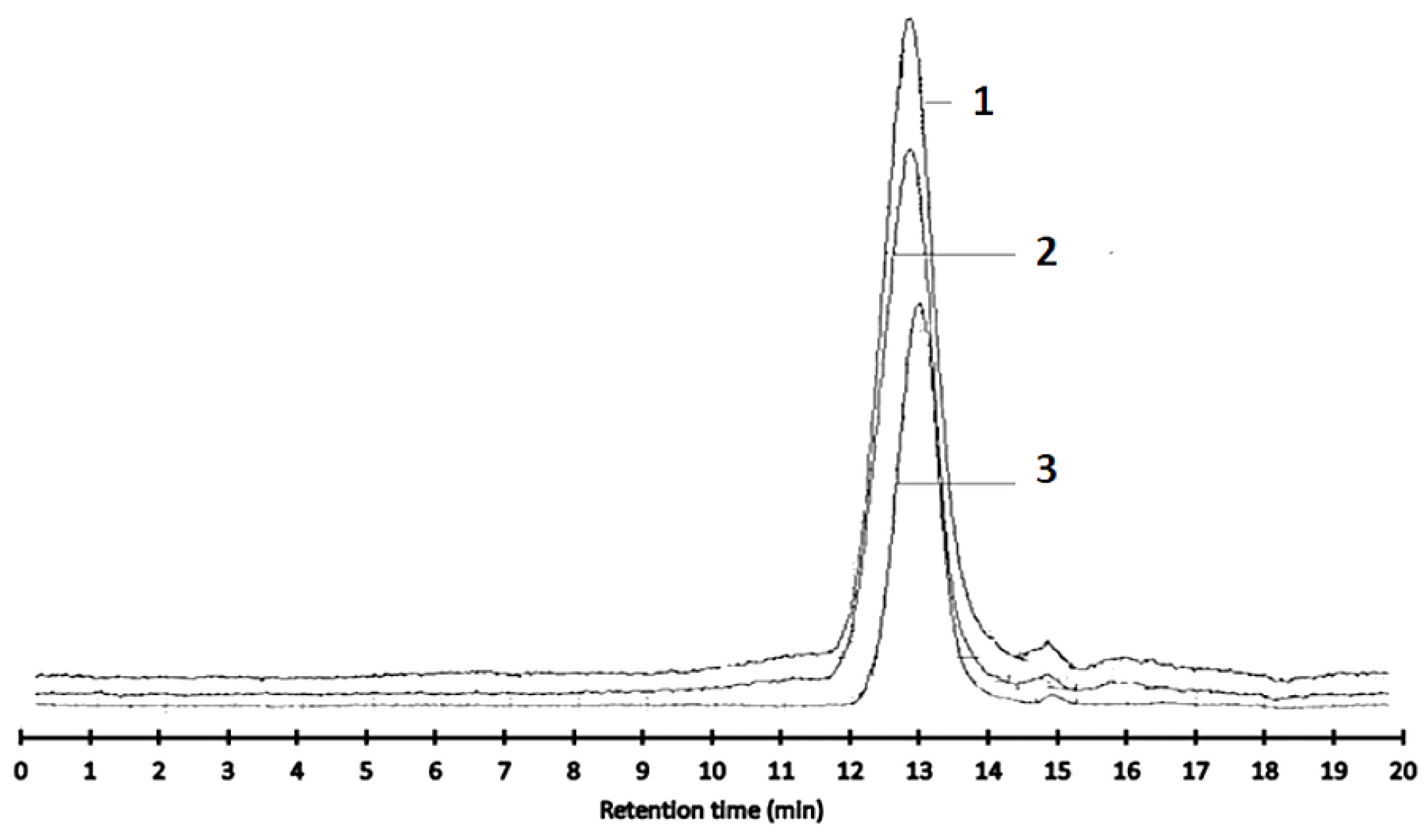
2.6. Molecular Weight Distribution Analysis

High-performance size-exclusion chromatography (HPLC-SEC) analysis was carried out using a HPLC chromatograph ELITE LaChrome (VWR Hitachi, Chiyoda, Japan) with a Shodex OH-pack 806 M (300 × 8.0 mm i.d.), (Shodex Co., Tokyo, Japan) column at 30 °C and an RI detector (VWR Hitachi Chromaster, 5450, Chiyoda Japan). Dandelion inulin (3 mg/mL) was dissolved in 0.1 M NaNO3. It was subsequently heated (75 °C) for 5 min in an ultrasonic bath to dissolve. The samples were filtered through a 0.45 μm filter, PTFE45/25 mm (ISOLAB GmbH), and 20 μL were injected. The mobile phase consisted of 0.1 M NaNO3 solution and a flow rate of 0.8 mL/min was used. A standard curve was built with different pullulan standards (Shodex 159 standard P-82 kit, Showa Denko, Kawasaki, Japan). The polydispersity index of the dandelion inulin was calculated as the ratio of the weight molecular mass to the number average molecular weight (Mw/Mn). DP was also calculated using the molecular mass from HPLC-SEC [23].

2.7. Spectroscopic Characterization of Dandelion Inulin

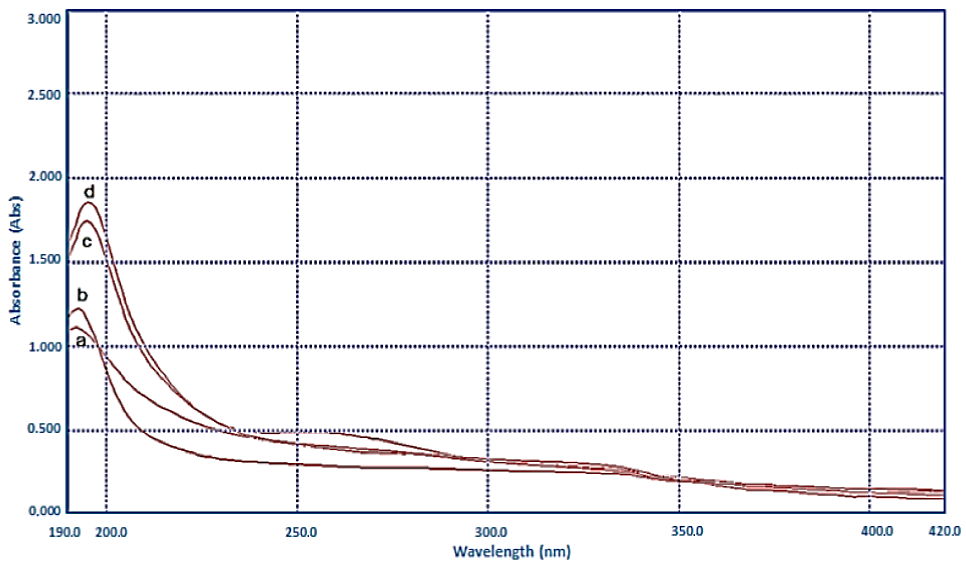
2.7.1. UV Spectrometric Analysis

Dandelion and chicory inulin were dissolved in distilled water to a final concentration of 0.3% and then analyzed using a UV-Vis spectrophotometer UV-Vis LLGSpec 2 (LLG Labware, Lab Logistics Group GmbH, Meckenheim, Germany). The UV absorption spectra of the samples were recorded against distilled water in the 190–420 nm wavelength range.

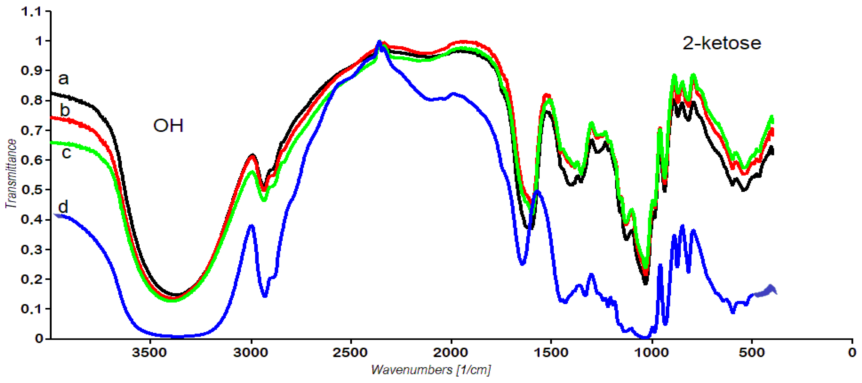
2.7.2. Fourier Transformation Infrared Spectroscopy

For the characterization of inulin isolated from dandelion roots, Fourier transformation infrared spectroscopy (FTIR) was used. The polysaccharide sample (2 mg) was pressed into pellets of KBr (200 mg). The spectra were recorded on a Nicolet FT-IR Avatar Nicolet Termo Science spectrometer in the range 4000–400 cm−1, with a resolution of 4 cm−1 and 132 scans; absorption was reported in wavenumbers (cm−1). Spectwin 32 software was used for FTIR spectrum analysis.

2.7.3. NMR Spectroscopy

The structure of inulin isolated from dandelion roots was identified by 1H and 13C NMR spectroscopy using a Bruker AVIII 500 M spectrometer (Bruker Nano GmbH Berlin, Germany) operating at a frequency of 500 MHz and 125 MHz, respectively. Dandelion inulin (25 mg) was dissolved in 0.6 mL 99.95% D2O. The degree of polymerization (DP) of dandelion inulin was also estimated from the 1H NMR spectrum by taking the ratio of the peak integral values of carbons in fructose units (X) to the peak integration values of the corresponding carbons in the glucose unit, as previously described [26,27]. MestReNova 14 software (Mestrelab, Santiago de Compostela, Spain) was used for NMR spectra analysis.

2.8. Functional Characterization of Dandelion Inulin

2.8.1. Color

Color measurement of the inulin powders was carried out using a portable colorimeter Model WR-10QC D 65 lighting, a 10° standard observer angle, and an 8 mm aperture in the measuring head colorimeter, according to the CIELAB (L*, a*, b*) system, where L* indicates lightness (0 = black and 100 = white), and a* and b* are coordinates for green (−a*)/red (+a*) and blue (−b*)/yellow (+b*). The instrument was calibrated using chicory inulin standard Raftiline HPX (Beneo, Orafti, Oreye, Belgium). The visual color appearance (hue angle, h*), color intensity (chroma, C*), browning index (BI), and yellowness index (YI) were calculated using the following Equations (1)–(3) [28]:
h * = t a n 1 ( b * / a * ) ,
C * a b = a * 2 + b * 2
Y I = 142.86 × b * C *

2.8.2. Swelling Properties, Solubility, Water- and Oil-Holding Capacity

The swelling properties of dandelion inulin were evaluated according to Robertson et al. [29]. The inulin sample (100 mg) was hydrated with 10 mL distilled water in a calibrated cylinder (1.5 cm diameter) at room temperature. After equilibration (18 h), the bed volume was recorded and expressed as volume/g original substrate dry weight [14]. The solubility of inulin was conducted in a water bath, as previously described [30], at a temperature of 80 °C. Inulin (0.1 g) was weighed, and then 10 mL of distilled water was slowly added to a plastic tube. The mixture was vigorously stirred. The solubility was evaluated gravimetrically [30] and expressed in %. The water-holding and oil-holding capacities of the isolated dandelion inulin were evaluated [31,32]. Dandelion inulin (100 mg) was put into the pre-weighed 50 mL polypropylene centrifuge tubes and 10 mL deionized water or sunflower oil (Biser Oliva, Sofia, Bulgaria) was added, respectively. After storage for 24 h at 20 °C, the samples were centrifuged at 3500 rpm for 15 min and the excess water or sunflower oil was discarded. The tubes were incubated for 1 h at 40 °C and then weighed and dried at 105 °C to constant weight.

2.8.3. Angle of Repose

Dandelion inulin (5 g) was carefully introduced into a funnel clamped to a stand with its tip 10 cm away from a plain black paper surface. The powder was allowed to flow freely onto the surface. The angle of repose was calculated using the height of the cone (H), formed after complete flow, and the radius of the cone (R) [31].

2.8.4. True, Bulk, Tapped Densities, Bulkiness and Porosity

Dandelion inulin (10 g) was added to a 50 mL measuring cylinder, and the volume occupied by samples was measured. After that, the measuring cylinder was tapped 500 times, and the final volume of the sample was recorded. The bulk and tapped density were calculated as the ratio of weight to volume [31]. Bulkiness was calculated as the number 1 divided by the value of the bulk density [33]. The true density was evaluated by the liquid displacement method using a measuring cylinder (10 mL) filled with 5 mL 95% ethanol and calculated as previously described [31,33]. Porosity was calculated as previously described by Jinapong et al. [32].

2.8.5. Flowability and Cohesiveness

The flowability and cohesiveness of the powder were determined by Carr’s index (CI) and the Hausner ratio (HR) using the bulk and tapped density [32].

2.8.6. Wettability

The time(s) needed for a mass of dandelion inulin powder (50 mg) placed on the surface of a fixed distilled water volume (20 mL) to completely submerge without agitation was evaluated [34].

2.8.7. Hygroscopicity

The hygroscopicity was determined using a 0.5 g inulin sample weighed in a small Petri dish and placed in a desiccator previously conditioned with sodium chloride (75% relative humidity at a temperature of 25 °C). The inulin sample was stored for seven days until a constant weight was reached [35]. The percentage of moisture absorbed was calculated [31].

2.8.8. Taste and Sweetness

The taste and sweetness of dandelion inulin were evaluated organoleptically through a descriptive test [14]. Chicory inulin served as a reference. For the evaluation of inulin taste, four basic tastes were evaluated using standard reference solutions: sour taste—0.025% lactic acid, basic sweet taste—1% sucrose, salty strength of basic salty taste—0.15% NaCl, and bitter taste–caffeine solution. A sucrose solution (10%) was used as a reference equivalent to 100 units for the sweetness evaluation.

2.9. Antimicrobial Activity

Gram-positive bacteria (Bacillus subtilis ATCC 6633, Listeria monocytogenes 863, Staphylococcus aureus 745) and Gram-negative bacteria (Escherichia coli ATCC 3398, Salmonella typhy 745) were selected to be used in the antimicrobial assay. The strains were obtained from the Collection of the Department of General and Applied Microbiology, Sofia University. The concentration of the viable cells and spores in the suspensions for inoculation was adjusted to 1.0 × 10⁹ cfu/mL for bacterial cells. The tested concentration of inulin was 1 mg/mL. The antibacterial activity was assayed by using the well diffusion method with a digital caliper. The disk diameter was 6 mm. The used growth medium was Nutrient Agar (Biolife 272-20128, Milano, Italia). The antimicrobial assay was performed as previously described [14].

2.10. Statistical Analysis

All determinations were performed in triplicate (n = 3) and the data were expressed as mean ± standard deviation (SD). Statistical analysis was performed using MS Excel 2015. ANOVA, with Tukey’s test of difference, was considered statistically significant when p < 0.05.

3. Results

3.1. Fructans and Sugar Composition of Defatted Dandelion Roots

The results of the content analysis of the carbohydrates in defatted dandelion roots are summarized in Table 1. As can be seen, inulin is the dominating compound with 16.10 g/100 g dry weight, which occupied 63% of the total fructan content. Prebiotic short-chained oligofructoses, 2-kestose, and nystose were also found in the roots, as well as sucrose and two monosaccharides (glucose and fructose). The level of total fructans in defatted dandelion roots reached 25.35 g/100 g dry weight. Therefore, these roots are promising and reliable sources for further inulin extraction. The reported carbohydrate composition of dandelion roots in the current study is consistent with previous reports [5,10]. Moreover, the total fructans in this study were higher than detected in the commercial dry raw material of T. officinale roots (13.16–17.95%) [10], and those from wild-growing dandelion roots (17–19% dw) [5].

3.2. Physicochemical Characterization of Dandelion Inulin

The physicochemical characteristics of the inulin-type fructans from dandelion roots obtained by different extraction techniques are listed in Table 2. The characteristics of dandelion inulin were compared with commercial chicory inulin Raftiline HPX. The highest yield was obtained during classical water extraction—20%, while the lowest was found for dandelion inulin after microwave-assisted extraction—11%. The highest purity of inulin was detected in MAE dandelion inulin—89%, followed by UAE—82%. However, the highest total fructose content 74% was found in inulin extracted by UAE. The ultrasound-assisted extraction led to the isolation of inulin from the dandelion with the highest molecular weights, the highest degree of polymerization (DP = 22), and a polydispersity index of 1.05. As regards the polydispersity index, it is known that the higher values of the index correspond to a greater spread of the molecular weight. The polydispersity index impacts the physical properties (melting point, crystallization, and dissolution) and also affects the tensile strength, viscosity, and thermal characteristics of polysaccharides. Moreover, the main effect of the index is related to polymer aggregation. For example, a polymer with a low index value (≈1) will have a narrow molecular weight distribution, causing more uniform behavior in solution and thus more predictable aggregation patterns presumably. On the other hand, a higher value leads to a more complex network structure, as longer chains can generate junctions with other chains, potentially generating stronger gels or network formation. All dandelion inulin showed a negative angle of optical rotation −25 and pH values 6–7, as well as a high melting point of 178–180 °C, comparable with chicory inulin Raftiline HPX. In general, dandelion inulin obtained by classical extraction was characterized by the lowest purity 71%, fructose content 72%, and the highest ash and protein content: 3.31% and 1.8%, respectively.
The dandelion inulin isolated in our study contained a small total phenolic content (0.82–0.60 mg GAE/g). We did not detect any phenolic content in the chicory inulin Raftiline HPX (Beneo, Orafti). In our case, the antioxidant potential of dandelion inulin samples was evaluated by FRAP and ABST methods. It was found that the inulin samples demonstrated a very low antioxidant potential, and better radical-scavenging properties evaluated by the ABTS method than metal reducing (FRAP assay), based on electron transfer (Table 1). These properties could be explained by the presence of a small amount of protein and total phenols in samples. Amongst all the samples, the highest antioxidant activity by both methods was demonstrated by dandelion inulin isolated by classical extraction, also showing a higher total phenols content.
From the above-mentioned results, it is obvious that the long time and high temperature employed during conventional extraction led to a decrease in the quality of the dandelion inulin, as well as a lowering in the degree of polymerization to 17 (HPLC-SEC) and of molecular masses 2885 and 2762 Da. The application of green extraction methods such as ultrasonic and microwave methods led to a short extraction time, to inulin with a better purity of over 80%, and to a high degree of polymerization comparable with chicory inulin as a reference. In general, UAE resulted in the highest fructose content, degree of polymerization, and melting point for dandelion inulin, as well as a good yield.

3.3. Spectral Analysis of Dandelion Inulin

3.3.1. UV-VIS Analysis

The ultraviolet-visible (UV-vis) spectrum of dandelion and chicory inulin is shown in Figure 2. In general, all inulin in this study showed no absorption at 260 and 280 nm, indicating the absence of higher amounts of UV-absorbing constituents (protein, etc.), except dandelion inulin after classical extraction (Figure 2 line d). These findings are consistent with the results of the Bradford method listed in Table 1. A similar tendency in UV-Vis inulin spectra with a decreasing trend from 200 to 800 nm was reported previously [6,36]. Moreover, it was also reported that some acidic polysaccharides from dandelion roots isolated by MAE did not show an absorption peak at 260 nm [3].

3.3.2. Molecular Weight Distribution Analysis of Dandelion Inulin

The molecular weight distribution pattern of dandelion inulin is shown in Figure 2.
The elution profile of ultrasound-assisted extracted inulin (Figure 3) coincided with that of the inulin obtained by microwave-assisted water extraction. The molecular weights of UAE dandelion inulin were Mw 3809 Da and Mn 3624, and they were close to the results for the MAE dandelion inulin (Mw 3345 Da and Mn 3192, Table 2). The isolated dandelion inulin comprised high-molecular-weight populations, with a molecular weight near that of the chicory inulin standard (Raftiline HPX with DP 25). The peaks were single, homogeneous, and eluted between 9 and 13 min without a pronounced tailing. Considering the purity data (Table 2) and the retention time, the corresponding peaks can be ascribed to inulin fractions.

3.3.3. FT-IR Spectra of Dandelion Inulin

The obtained FT-IR spectra of the isolated dandelion inulin after classical, ultrasound-assisted, and microwave-assisted extraction are shown in Figure 4.
It was noticed that all the spectra contain main bands characteristic of inulin-type fructan. In general, the FT-IR spectra show a broad band at about 3370 cm−1 typical for the stretching vibrations of OH groups and inter- and intra-molecular hydrogen bonds. The band at 2935 cm−1 was assigned to the asymmetric stretching vibrations of CH2 groups (νC–Has(CH2)), and the bands at 2893 cm−1 to the symmetric stretching vibrations νC–Hs(CH2). The 1647 cm−1 band is due to the absorption of water, while the bands at 1330 cm−1 and 1255–1240 cm−1 are typical for βO–H (OH). The band at 1143 cm−1 is assigned to C–O–C ring stretching vibrations from glycoside linkage. The bands at 1033 cm−1 and 989 cm−1 are assigned to C–O stretching vibrations in the furanose ring, and the band at around 937 cm−1 indicates the presence of β-(2→1) linkage in inulin. The typical bands for fructans were noticed in the fingerprint region: 869 cm−1 due to β-anomer bendings C1-H, ring vibration of 2-ketofuranose, and 820–819 cm−1 due to 2-ketose in the furanose ring. Moreover, the bands of dandelion inulin were overlaid with chicory inulin Raftiline for comparison (Figure 3 line d). It was obvious that the bands coincided, which additionally confirmed the inulin structure of isolated polysaccharides. The applied ultrasonic and microwave irradiation did not cause changes in inulin bands.

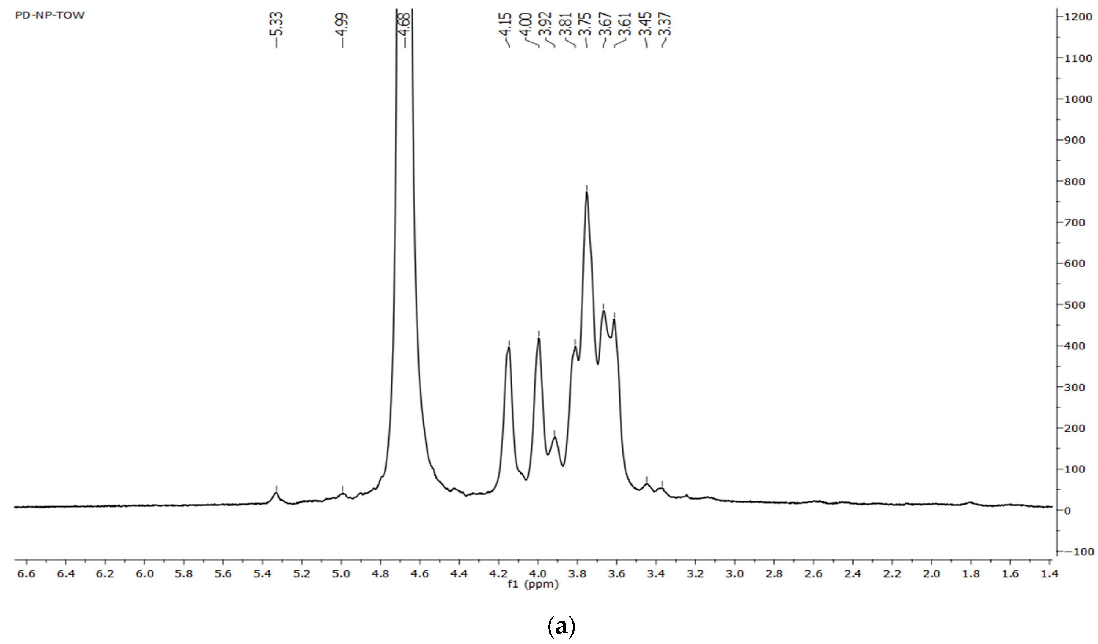
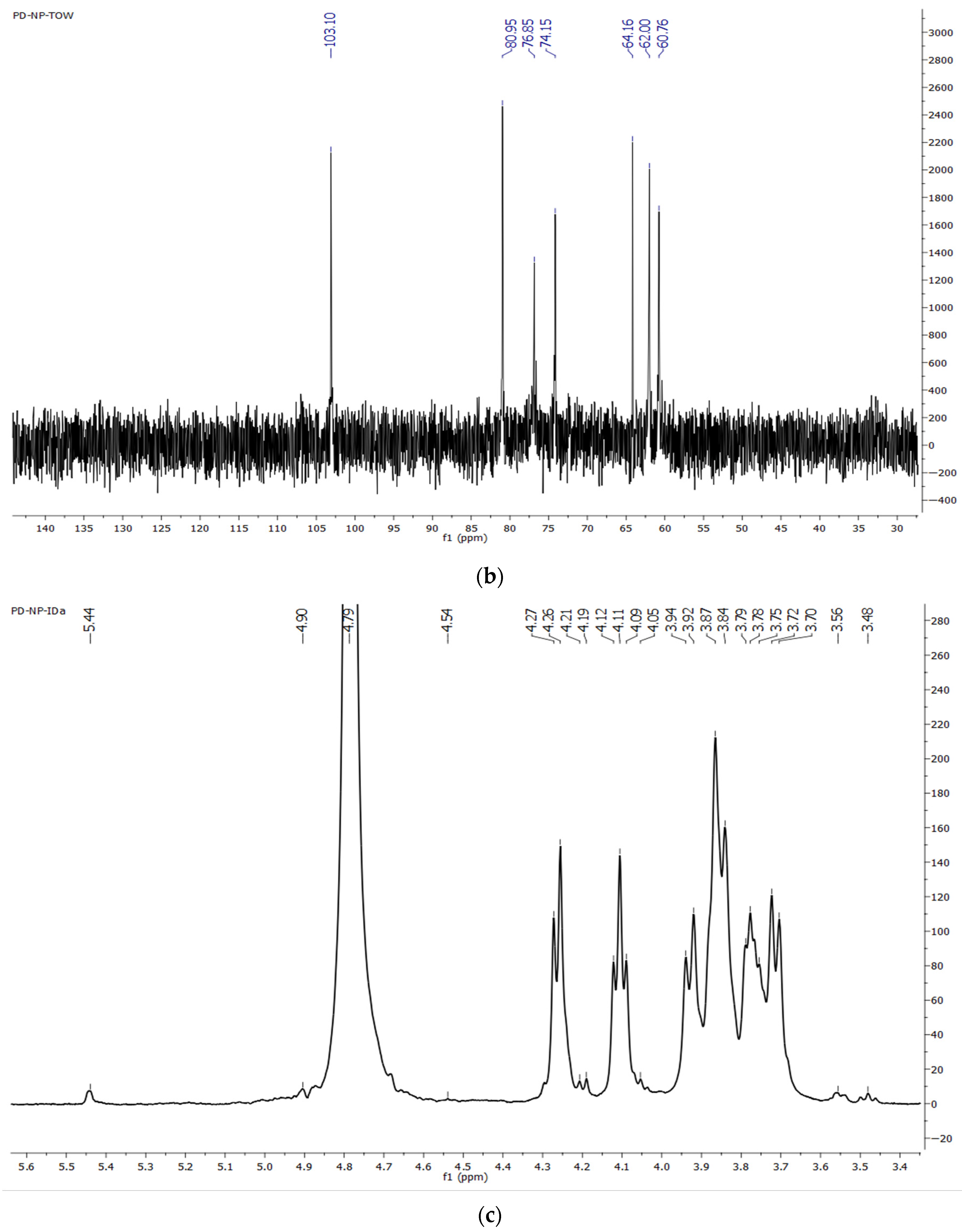
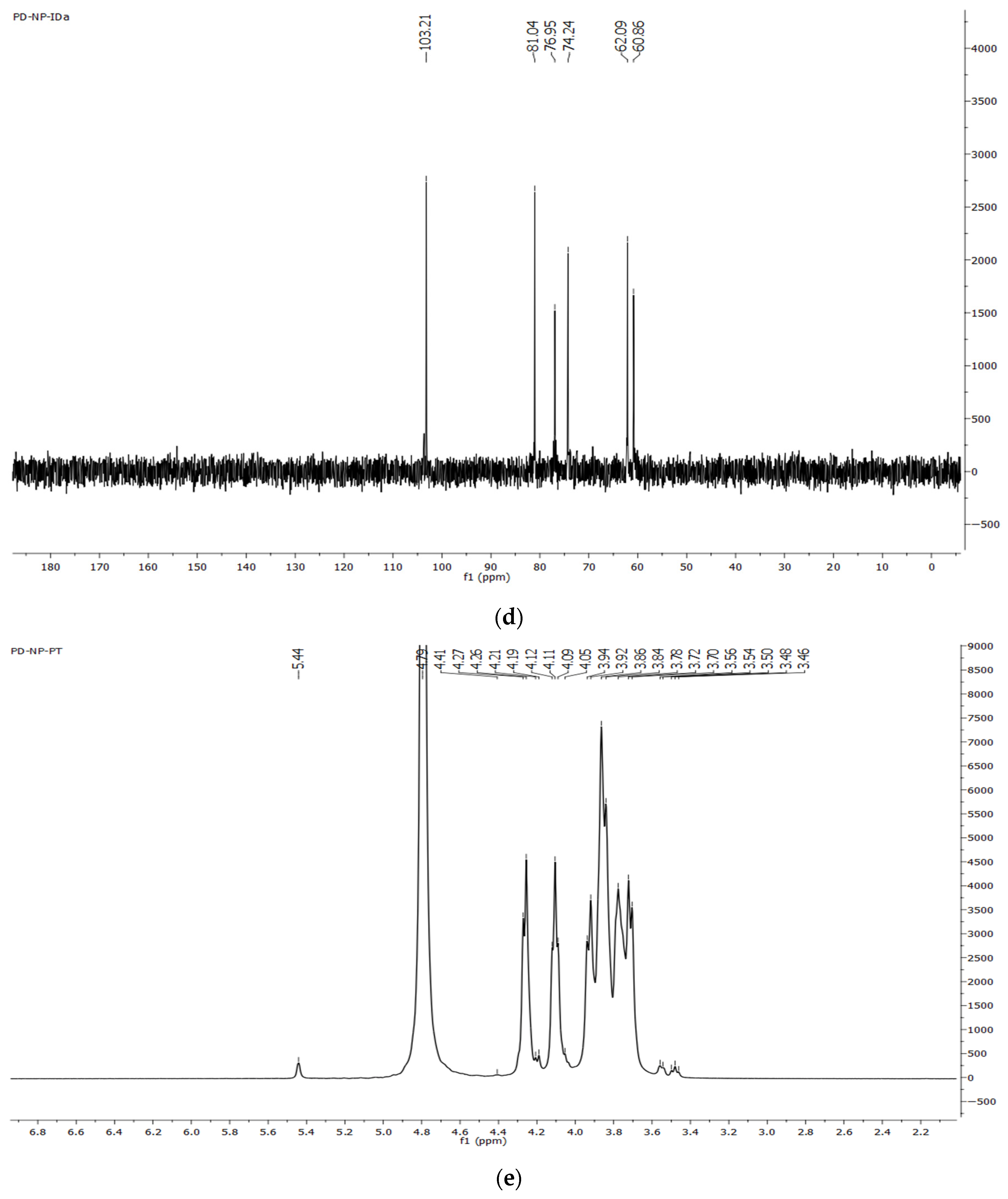
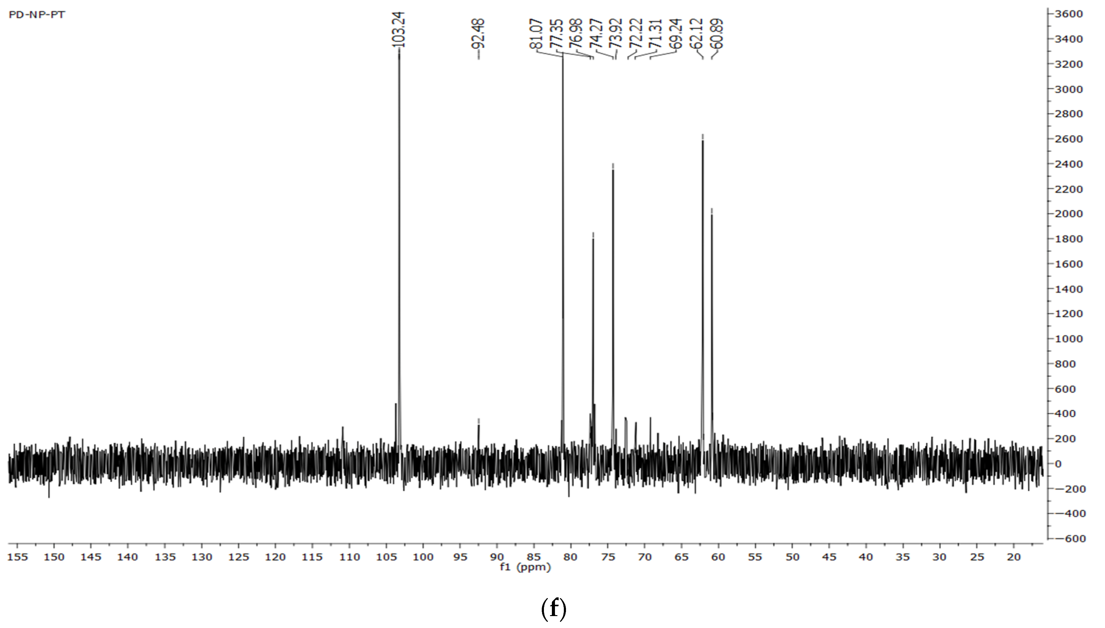
3.3.4. NMR Spectra of Dandelion Inulin

1H and 13C NMR spectra of dandelion inulin isolated by MAE and UAE are presented in Figure 5. NMR spectra of chicory inulin were used as a reference (Figure 5e,f).
1H NMR (500 MHz, D2O) δ 5.33, 4.99, 4.68, 4.15, 4.00, 3.92, 3.81, 3.75, 3.67, 3.61, 3.45, 3.37 (MAE).
The 1H-NMR spectrum (Figure 5a) of dandelion inulin demonstrated characteristic shifts corresponding to the skeleton protons of a fructose ring at δ 4.15 (H3f) and δ 4.00 (H4f), in the region from 3.37 to 3.92 (H1f, H5f and H6f), and one isolated resonance for the single anomeric α-glucose proton (H1g) at 5.33 (5.44) ppm. The integration ratio between 3.3 ppm and 4.2 ppm of fructofuranose and H-1 of glucopyranose could be used to calculate the degree of polymerization of inulin-type fructans as 20, while UAE gave DP 25 (Table 2). In the 13C NMR spectra of dandelion inulin (Figure 5b,d), chemical shifts typical of only fructose units were observed. The 13C NMR spectra of the dandelion inulin obtained after microwave-assisted extraction are presented in Figure 4b): 13C NMR (126 MHz, D2O) δ 103.10 (Cf2), 80.95 (Cf5), 76.85 (Cf3), 74.15 (Cf4), 64.16, 62.00 (Cf6), 60.74 (Cf1) ppm, while the UAE spectra are presented in Figure 5d): δ 103.21 (Cf2), 81.04 (Cf5), 76.95 (Cf3), 74.24 (Cf4), 62.09 (Cf6), 60.86 (Cf1) ppm. These spectra contain prominent shifts for the C1–C6 carbons of fructosyl residue due to fructose repeated units. The 13C shifts from glucose were not observed (Figure 2b,d) due to its superposition. A similar observation was reported for inulin from other plants [14,27]. However, in the 13C NMR of chicory inulin, glucose units were observed together with fructose units (Figure 5f): δ 103.24 (Cf2), 81.07 (Cf5), 76.96 (Cf3), 74.24 (Cf4), 62.12 (Cf6), 60.86 (Cf1) ppm and carbon from glucose units: 92.48 (C1g), 73.92 (C2g), 74.00 (C3g) 72.22 (C5g), 71.31 (C4g), 69.24 (C6g) ppm. In all the 13C NMR spectra (Figure 5), only one shift at 103.10–103.2 ppm was observed, corresponding to the C-2 carbon involved in β-(2→1)-D-fructofuranosyl–fructose bonds. The data from the 1H and 13C NMR spectra confirm the chemical structure of dandelion roots composed mainly of fructose units linked with β-(2→1) bonds and of only one terminal-linked glucose unit α-(1→2).

3.4. Functional Properties of Dandelion Inulin

The results of the functional properties (swelling properties, solubility, oil- and water-holding capacity) of dandelion inulin are shown in Table 3. The results obtained were compared with chicory inulin and some of the literature data on inulin from different species of dandelion.
The swelling properties of dandelion inulin varied from 4.40 to 5.60 mL water/g, and solubility was in the range of 21 to 30%. Dandelion inulin showed twice-higher swelling properties than chicory inulin Raftiline HPX (DP 25). All dandelion and chicory inulin demonstrated a better oil-holding capacity than water-holding capacity. OHC influences the functional properties of a hydrocolloid, representing the oil absorption capacity. The OHC value of dandelion inulin was in the range of 3.03 to 3.92 g oil/g sample. In general, UAE dandelion inulin possessed the highest swelling properties 5.60 mL water/g, WHC 2.29 g water/g, and OHC 3.92 g oil/g, followed by MAE inulin. Classical dandelion inulin showed the highest solubility, but the lowest WHC and OHC.
WHC values evaluate the stability, texture, and sensory of the sample [37]. The WHC of dandelion inulin represents the amount of water held and absorbed by the hydrated inulin sample. The oil-holding capacity of dandelion inulin reveals its potential to be used as a flavor and texture enhancer, especially in the mouthfeel of foods.
To the best of our knowledge, until now, many of the functional properties of dandelion inulin had not been evaluated, especially some color characteristics, the angle of repose, wettability, hygroscopicity, different densities, and Carr’s index, as well as the Hausner ratio. These parameters are important for the packaging, transportation, food, and pharmaceutical applications of powder compounds. The influence of the type of extraction on the functional characteristics of dandelion inulin is shown in Table 4.
The values of L*, which show the whiteness of inulin, vary between 82.81 and 96.25, demonstrating the high lightness of dandelion inulin, especially of inulin obtained by microwave-assisted extraction. In general, the long period of extraction and intense thermal treatment cause a decrease in the L* value of dandelion inulin. The inulin obtained by classical extraction possessed the highest values for L, a, b, hue, chroma, BI, and YI. The L*, a*, and b* values were used for the calculation of the yellowness index (YI) and browning index (BI), indicating the purity of the brown color. The lowest values for BI and YI were found for the dandelion inulin obtained by microwave-assisted extraction. These characteristics were close to those for the control chicory inulin. In general, all inulin demonstrated high whiteness with an L above 82, low yellowness, and low browning index. It was observed that YI was higher than BI in all the inulin samples.
Wettability is one of the most important physical properties related to the reconstitution of powders in aqueous solutions. In the current research, the time required for the dandelion inulin powders to be completely wet ranged from 119 to 367 s (Table 3).
Bulk properties are important characteristics for handling and packaging powders. Powders with a high bulk density occupy less volume per mass unit and can be packaged in smaller containers [38]. The bulk density of dandelion inulin was in the range of 0.30 and 0.47 g/mL, while the tapped density ranged from 0.51 to 0.61 g/mL, respectively. Dandelion inulin isolated by microwave-assisted extraction showed bulk, tapped, and true density values close to the chicory inulin used as a reference. The bulk density of porous starch inulin-loaded quercetin microcapsules demonstrated a lower bulk density of 0.22 to 0.27 g/mL, for comparison [39].
The bulk and tapped densities participate in the calculation of bulkiness, Carr’s index, and the Hausner ratio. Carr’s index defines the flowability of powders, while the Hausner ratio gives information about their cohesiveness. All the dandelion inulin samples demonstrated Carr’s indexes above 20% (24–26%), which correspond to a fair flowability of powders. The Hausner ratio was between 1.2 and 1.4, which defines the dandelion inulin powders as having intermediate cohesiveness. On the other hand, the chicory inulin was classified as a powder with good flowability and intermediate cohesiveness. Therefore, the dandelion inulin samples were evaluated as being close to chicory inulin in terms of cohesiveness and flowability properties. Moreover, the dandelion inulin demonstrated better cohesiveness and flowability than soymilk and soymilk concentrates [32].
The bulkiness values of the dandelion and chicory inulin were in the range of 2–3 mL/g, and the values of dandelion inulin obtained by classical and microwave extractions were closer to the chicory inulin Raftiline. Its values were comparable with the bulkiness of almond gum, with values of 2.52, indicating that the powder is ‘light’ in nature [33].

3.5. Antimicrobial Activity of Fructan Isolated from Dandelion

To the best of our knowledge, the reports about the antimicrobial potential of fructans, especially inulin, are insufficient, and their mechanisms against pathogens have still not been elucidated in depth. It is well known that the soluble oligosaccharides mimic the sugar chains situated in the glycolipids and glycoproteins of the gut epithelial cells, and in this way they prevent pathogen adhesion [40]. This study aimed to perform a preliminary antimicrobial study of dandelion inulin because little is known about the direct antimicrobial effect of fructans. The results obtained from the antibacterial activity of isolated dandelion and commercial chicory inulin are summarized in Table 5.
As a result, chicory and UAE dandelion inulin demonstrated moderate antimicrobial activity against Listeria monocytogenes. Moreover, dandelion inulin mainly demonstrated low-to-moderate antimicrobial activity against Salmonella thyphy 745 and Bacillus subtilis 6633 at a concentration of 1 mg/mL, while chicory inulin was inactive at this concentration. Dandelion inulin demonstrated an antibacterial potential comparable to that of Biseptol (400 μg/mL) used as a positive control. However, none of the tested inulin samples were active against St. aureus 745, nor against Gram-negative bacteria E. coli 3398. To the best of our knowledge, this is the first study to evaluate the antimicrobial potential of dandelion inulin.

4. Discussion

4.1. Physicochemical Characterization of Dandelion Inulin

The employed chromatographic and spectral analyses demonstrate that the isolated polysaccharides were characterized as typical inulin-type fructans. The extraction approaches had a serious impact on the yield and purity of the isolated dandelion inulin. Most of the studies discussed report the characteristics of inulin from dandelion obtained by classic hot water extraction [41,42,43,44]. In this research, the classical extraction procedure gave the highest yield of 20%; however, the isolated inulin was of lower purity, containing other impurities such as protein and minerals (Table 2). The high extraction temperature led to a lower molecular weight and lower degree of polymerization in comparison to MAE and UAE. In general, the inulin yield from dandelion reveals its future potential as a promising source of inulin isolation. The dandelion inulin yield is comparable to other industrial sources such as Helianthius tuberosus and Chicory intybus, where the yield varies from 16 to 20% [44].
In an earlier report, Nuridullaeva et al. [41] developed an industrial technology for inulin production from dandelion roots using water extraction at 80 °C, circulation of the extractant at a rate of 90 L/h, concentration of the extract to 70% dry residue, the precipitation of inulin by adding 95% EtOH (1:4) in a concentrate, and sedimentation for 8 h. This dandelion inulin was produced at 30.0% yield of the raw material mass, with a purity of at least 80%, ash content ≤2%, and inulin content ≥80.0%. Mudannayake et al. [18] obtained inulin (yield 6.5%) with a purity of 43% dw, protein content of 4.18%, moisture content of 2.92%, and ash content of 3.88% from the roots of Taraxacum javanicum by using classical hot water extraction at 80 °C, without employing precipitation with organic solvent, using only concentration and drying. Olennikov et al. [10] reported the isolation of two fractions of glucofructans (TGf-1) of 5.7 kDa and TGf-2 with 2.6 kDa. The yield of glucofructan was 17.93%, with a fructose content of 85.65% and a negative angle of rotation. Hahn et al. [30] optimized and scaled-up inulin extraction from Taraxacum kok-saghyz Rodin using water extraction at 85 °C and reported that the inulin had an average degree of polymerization (DP = 15.5), 77% purity, and yield of 35% dw. The current research findings on ash content and molecular mass are close to those found with the classical isolation of dandelion inulin [10,18]. Moreover, the data of the reducing groups, melting point, and total fructose content of dandelion inulin are in complete agreement with those of inulin isolated from other plant sources [10,14,27,42]. The negative angle of optical rotation is typical for fructans, especially inulin [10,26]. The current data are consistent with previous reports for inulin-type fructans [10,26].
The molecular masses reported for dandelion inulin in our study (Table 2) are lower than those reported by Olennikov et al. [10] (5.7 to 5.9 kDa fractions) and Zhang et al. [7]. The inulin obtained by classical extraction and coincided with the previously reported fraction of dandelion extracted by hot had the lowest molecular weight water [10]. In this study, the polydispersity index of the dandelion inulin was in the range of 1.04–1.05, and this is confirmation that the molecular weight distribution was narrow. These values are close to those for the chicory inulin used as a reference, as well as to the polydispersity index of inulin found in T. mongolicum (1.03) [7].
The data on the total phenols in the current study are in accordance with another study where an absence of phenolic compounds was reported for another chicory inulin (Orafti GR) [18]. However, Mudannayake et al. [18] found 4.37 mg GAE/g total phenolics in inulin isolated from the roots of Taraxacum javanicum, with an antioxidant activity of 833 mM TE/g (ABTS method), while Sharma et al. [31] reported 0.46 mg GAE/100 g total phenols in purified Prunus domestica gum with an antioxidant potential of 207.49 µM TE/g (ABTS method).
Moreover, the efficiency of the ultrasonic extraction confirmed it as an appropriate method for the isolation of dandelion inulin, resulting in the highest purity, fructose content, and degree of polymerization. The reduced time of extraction, together with the improved efficiency of the extraction process, is a clear advantage of ultrasound-assisted extraction over classical extraction. The advantages of ultrasonic irradiation have been previously demonstrated for the extraction of polysaccharides, in particular inulin-type fructans [6,13,14,16,23]. The efficiency of ultrasound extraction, together with the cavitation process that occurred and the pressure of collapse, additionally disrupts the cell wall material alongside the high temperature. The mechanical effect of the ultrasonic power led to the release of inulin from the plant cells by disrupting cell walls, enhancing the mass transfer and solvent access to the cell content [16].
High-intensity microwave heating leads to a rapid increase in temperature in the whole volume, promotes the rotation of water molecular dipoles, and causes expansion with subsequent rupture of the cell walls, thus accelerating the extraction of inulin [16].

4.2. FTIR and NMR Spectra of Dandelion Inulin

The observed bands in the FT-IR spectra of the dandelion inulin are typical for inulin. The bands at 817 cm−1 in both spectra prove the presence of β-D-fructose residues linked to 1→2 glycoside bonds. The IR spectra of the dandelion inulin exhibit absorption bands characteristic of inulin-type glucofructans and are consistent with a report by Olennikov et al. [10], who identified the band at 937 cm−1 as characteristic of a furanose ring. The absorption bands at about 874 cm−1 and 819 cm−1 are characteristic of 2-ketoses in furanose form and confirm the CH2 ring vibration of β-anomer and the presence of 2-ketofuranose [10,14,25,42,43]. Moreover, Mudannayake et al. [18] also reported that 934 cm−1 is typical for fructofuranosyl group, while Du et al. [3] demonstrated that a band at 874 cm−1 is typical for β-glycosidic bonds. In addition, the bands observed in the dandelion inulin spectra coincide with the chicory inulin Raftiline used as a reference and are consistent with FT-IR data for inulin-type fructan [10,14,15,16,44]. The bands present in the IR spectrum are close to the reported 818, 874, and 937 cm−1 for inulin, indicating the presence of a β-(2→1) glycosidic bond [42,43,44].
Moreover, the NMR spectra additionally confirm the linear structure of inulin-type fructan, and the reported chemical shifts are consistent with previous reports for dandelion inulin [6,7,10]. Additionally, the NMR spectra of dandelion were compared with those of the chicory inulin Raftiline HPX used as a reference (Figure 5e,f). The results confirm the structure of inulin. In the 1H NMR spectra of dandelion inulin, only one anomeric shift was found at 5.3–5.4 ppm due to the 1H of a glucose unit with low intensity, in comparison with the protons from the fructose unit with high-intensity shifts. However, in the 13C NMR spectra of dandelion inulin, only shifts typical for carbons from the fructose unit were found. The chemical shift at 103 ppm in the 13C spectra was due to the C-2 of →1)-β-D-Fruf-(2→ ). In addition, the chemical shifts for glucose carbons were not observed in dandelion inulin, while they were found in the 13C NMR spectra of the chicory inulin used as a reference (Figure 5f). The absence of signals for a glucose unit in the 13C NMR spectra has also been reported for inulin-type fructans isolated from stevia, agave, and echinacea, due to its very small amount in the sample [14,36,44]. This observation has been reported in all studies with inulin with a low degree of polymerization [44]. The reported chemical shifts for the fructose unit are consistent with other data for inulin-type fructans [3,10,14,26,44]. The results presented from the NMR studies confirm the linear chemical structure of inulin isolated from dandelion roots, composed of one terminal glucose unit and fructofuranosyl units linked by β2→1. The type of extraction did not cause a significant effect on the structure of inulin, and only the molecular mass and degree of polymerization decreased when a high temperature and long extraction time were applied, especially during classical extraction.

4.3. Functional Properties

To the best of our knowledge, this is the first detailed report about the functional properties of inulin from dandelion roots. Considering the color characteristics, our data for the lightness of dandelion inulin are comparable with some of the data for other inulin samples isolated from different plant sources such as echinacea, chicory, and globe artichoke [14,45], which had higher values than dandelion inulin [18]. It was previously reported that YI is related to product deterioration by light exposure and processing. The increased YI and BI values could be explained by pigment release [28]. The difference in a*, b*, L*, YI, and BI could also be explained by the small presence of polyphenols in the dandelion inulin samples, as presented in Table 2. In addition, Mudannayake et al. [18] reported lower results for lightness and a and b values.
It was found that isolated dandelion inulin demonstrated a better oil-holding capacity than water-holding capacity. Similar observations were reported earlier by our team for echinacea and burdock inulin [14,30]. The obtained values for the water- and oil-holding properties of dandelion inulin were close to those for the commercial chicory inulin Raftiline HP (DP 25) used as a reference.
The oil-holding capacity of dandelion inulin (3.03–3.93 g oil/g sample) was higher than the reported values for Taraxacum javancium inulin (72.50 g corn oil/100 g dw inulin) [18], chicory inulin, and globe artichoke inulin (1.37 and 1.38 g oil/g sample, respectively) [46], lower than for burdock by-products [16], and in good agreement with dandelion polysaccharide [6] and echinacea inulin [14]. To the best of our knowledge, this research presents the first report of dandelion inulin flow properties—tapped and bulk density, angle of repose, Carr’s index, and Hausner ratio. All the dandelion inulin samples demonstrated a fair flowability and intermediate cohesiveness based on Nandi’s classification [47]. The bulk density values of the dandelion inulin are comparable to those found for microcapsules containing cashew gum, inulin, and ginger oil and varied between 0.28 g/mL and 0.31 g/mL, and for tapped density between 0.50 g/mL and 0.53 g/mL [38]. A poor-flowing material (e.g., a fine powder) should be characterized by the existence of larger interparticle interactions, and thus a greater difference between bulk and tapped densities [14]. In general, the UAE dandelion inulin demonstrated a lower wetting time and lower porosity than the other inulin samples. The observed short wetting times and inulin solubility could be explained by a high number of hydrophilic groups in the molecule and the highest degree of polymerization [14].
Therefore, due to its better solubility, neutral taste, and oil-holding properties, dandelion inulin can be used as a functional ingredient to improve taste and other sensory properties. Additionally, its bulkiness, intermediate cohesiveness, and fair flowability reveal its potential as a bulk agent in pharmaceutical and food nutritional formulas.

4.4. Antibacterial Activity

Little is known about the direct antimicrobial potential of fructans. Martínez-Ortega et al. [48] reported that fructan isolated from agave (Agave tequilana Weber var. azul) demonstrated antimicrobial activity against Salmonella typhimurium. The minimum inhibitory concentration of fructans against Salmonella under culture conditions was 3%, with the greatest effect observed at a concentration of 25%; thus, they have an antibacterial effect against this strain at 50% concentrations. It was reported that water-soluble polysaccharide dandelion showed an antibacterial effect against E. coli, Bacillus subtilis, and St. aureus at concentrations of 100 mg/mL, with zones of 12.47 mm, 11.02 mm, and 15.26 mm, respectively [49]. Other authors have demonstrated the antibacterial activity of commercially available dandelion polysaccharides against L. monocytogenes, and their TEM analysis showed the potential of this polysaccharide to destroy cell membranes and the cell integrity of bacteria [50]. The agar diffusion method was also applied for the evaluation of the antibacterial potential of dandelion leaf polysaccharide at a concentration of 2.0 mg/mL. The authors reported inhibitory diameters against E. coli, Salmonella, S. aureus, and L.monocytogenes of 9.4 ± 0.2 mm, 11.6 ± 0.6 mm, 11.8 ± 0.7 mm, and 15.4 ± 0.5 mm, respectively [51]. In our study, the tested dandelion inulin mainly inhibited the growth of Gram-positive bacteria, except for the growth of St. aureus at a concentration of 1 mg/mL. Our findings suggest that dandelion and chicory inulin were inactive against the Gram-negative bacteria E.coli. This statement is consistent with previous report on chicory inulin (DP 2-60) which revealed that a concentration of 0.1% was inactive against Escherichia coli ATCC 3398 and Staphylococcus aureus [52]. To our knowledge, the current study is the first to evaluate the antibacterial potential of dandelion inulin. The possible explanation for its activity could be due to its adsorption to the cell membrane, as was suggested in earlier studies [50,51,52]. It is well known that polysaccharides may cause the destruction of the inner cell membrane to increase permeability and accelerate electrolyte release. This action has led to bacterial cell death [51]. With these data, we suggest that dandelion inulin could be useful in the prevention of infections caused by Listeria monocytogenes or Salmonella. However, to elucidate the mechanisms involved in this action, further future studies are needed.

5. Conclusions

A detailed study on the structural characterization, as well as the physicochemical and functional properties, of inulin isolated by green extraction methods from dandelion roots was performed. It was demonstrated that ultrasound-assisted water extraction resulted in the highest inulin yield and degree of polymerization. The structure of all the dandelion inulin samples was confirmed by NMR spectroscopy. It consisted of fructofuranosyl units linked with β-(2→1) bonds and a terminal glucose unit linked with α-(1→2). The molecular weight properties were comparable with those of chicory inulin. All dandelion inulin samples possessed good solubility, better oil-holding capacities than water-holding properties, intermediate cohesiveness comparable with chicory inulin, and fair flowability. In addition, the moderate antibacterial activity of dandelion inulin against Listeria monocytogenes highlights its biological activity. The current results reveal the future potential of dandelion inulin’s application as a functional ingredient in taste and texture modifiers or as a drug carrier and bio-based material. However, additional research is needed on its future perspective application in pharmacy and food formulations.

Author Contributions

Conceptualization, N.P. and I.I.; methodology, N.P., M.O. and I.H.; software, N.P.; validation, M.O.; formal analysis, N.P.; investigation, N.P, M.O., I.H., S.I., T.I.-I. and I.I.; resources, K.N.; data curation, N.P.; writing—original draft preparation, N.P. and M.O.; writing—review and editing, I.I.; visualization, N.P.; supervision, N.P. and I.I.; project administration, K.N.; funding acquisition, K.N. All authors have read and agreed to the published version of the manuscript.

Funding

This research was funded by the European Union NextGenerationEU through the National Recovery and Resilience Plan of the Republic of Bulgaria, project No BG-RRP-2.004-0009-C02.

Institutional Review Board Statement

Not applicable.

Informed Consent Statement

Not applicable.

Data Availability Statement

Data are contained within the article.

Acknowledgments

The authors are thankful for the technical support of Nevena Petkova and Nikola Burdjiev from the University “St. Klimet Ohridski”, Sofia, for recording NMR spectra of dandelion inulin.

Conflicts of Interest

The authors declare no conflicts of interest. The funders had no role in the design of the study; in the collection, analyses, or interpretation of data; in the writing of the manuscript; or in the decision to publish the results.

Abbreviations

MAE Microwave-assisted extraction
UAE Ultrasound-assisted extraction
DPDegree of polymerization
NMRNuclear magnetic resonance
YIYelowness index
BIBrowning index
OHCOil-holding capacity
WHCWater-holding capacity

References

  1. Kania-Dobrowolska, M.; Baraniak, J. Dandelion (Taraxacum officinale L.) as a source of biologically active compounds supporting the therapy of co-existing diseases in metabolic syndrome. Foods 2022, 11, 2858. [Google Scholar] [CrossRef] [PubMed]
  2. Schütz, K.; Muks, E.; Carle, R.; Schieber, A. Separation and quantification of inulin in selected artichoke (Cynara scolymus L.) cultivars and dandelion (Taraxacum officinale WEB. ex WIGG.) roots by high-performance anion exchange chromatography with pulsed amperometric detection. Biomed. Chromatogr. 2006, 20, 1295–1303. [Google Scholar] [PubMed]
  3. Du, G.; Liu, Y.; Zhang, J.; Fang, S.; Wang, C. Microwave-assisted extraction of dandelion root polysaccharides: Extraction process optimization, purification, structural characterization, and analysis of antioxidant activity. Int. J. Biol. Macromol. 2025, 299, 139732. [Google Scholar] [CrossRef]
  4. Lis, B.; Olas, B. Pro-health activity of dandelion (Taraxacum officinale L.) and its food products—History and present. J. Funct. Foods 2019, 59, 40–48. [Google Scholar] [CrossRef]
  5. Petkova, N.; Ivanov, I.; Topchieva, S.; Denev, P.; Pavlov, A. Biologically active substances and in vitro antioxidant activity of different extracts from dandelion (Taraxacum officinale) roots. Sci. Bull. F Biotechnol. 2015, 19, 190–197. [Google Scholar]
  6. Guo, H.; Zhang, W.; Jiang, Y.; Wang, H.; Chen, G.; Guo, M. Physicochemical, structural, and biological properties of polysaccharides from dandelion. Molecules 2019, 24, 1485. [Google Scholar] [CrossRef]
  7. Zhang, S.; Song, Z.; Shi, L.; Zhou, L.; Zhang, J.; Cui, J.; Li, Y.; Jin, D.-Q.; Ohizumi, Y.; Xu, J.; et al. A dandelion polysaccharide and its selenium nanoparticles: Structure features and evaluation of anti-tumor activity in zebrafish models. Carbohydr. Polym. 2021, 270, 118365. [Google Scholar] [CrossRef]
  8. Van den Ende, W.; Michiels, A.; Van Wonterghem, D.; Vergauwen, R.; Van Laere, A. Cloning, developmental, and tissue-specific expression of sucrose: Sucrose 1-fructosyl transferase from Taraxacum officinale. Fructan localization in roots. Plant Physiol. 2000, 123, 71–80. [Google Scholar] [CrossRef]
  9. Bokov, D.O.; Karabeshkin, D.I.; Samylina, I.A.; Potanina, O.G.; Krasnyuk, I.I.; Malinkin, A.D.; Sergunova, E.V.; Kovaleva, T.Y.; Bobkova, N.V.; Antsyshkina, A.M.; et al. Pharmacopoeial analysis of inulin-containing medicinal plant raw materials and drugs. Pharmacog. J. 2020, 12, 415–421. [Google Scholar] [CrossRef]
  10. Olennikov, D.N.; Tankhaeva, L.M.; Rokhin, A.V. Glucofructans from Taraxacum officinale roots. Chem. Nat. Compd. 2009, 45, 141–144. [Google Scholar] [CrossRef]
  11. Savych, A.; Bilyk, O.; Vaschuk, V.; Humeniuk, I. Analysis of inulin and fructans in Taraxacum officinale L. roots as the main inulin-containing component of antidiabetic herbal mixture. Pharmacia 2021, 68, 527. [Google Scholar] [CrossRef]
  12. Bagaoutdinova, R.; Fedoseyeva, G.; Okoneshnikova, T. Fructose-containing carbohydrates in plants of different families localization and content. Chem. Comput. Simul. Butlerov Commun. 2001, 2, 13–16. [Google Scholar]
  13. Zhu, Z.; He, J.; Liu, G.; Barba, F.J.; Koubaa, M.; Ding, L.; Bals, O.; Grimi, N.; Vorobiev, E. Recent insights for the green recovery of inulin from plant food materials using non-conventional extraction technologies: A review. Innov. Food Sci. Emerg. Technol. 2016, 33, 1–9. [Google Scholar] [CrossRef]
  14. Petkova, N.; Petrova, A.; Ivanov, I.; Hambarlyiska, I.; Tumbarski, Y.; Dincheva, I.; Ognyanov, M.; Denev, P. Chemical composition of different extracts from Echinacea purpurea (L.) Moench roots and evaluation of their antimicrobial activity. ChemEngineering 2023, 7, 94. [Google Scholar] [CrossRef]
  15. López-Molina, D.; Navarro-Martínez, M.D.; Rojas-Melgarejo, F.; Hiner, A.N.P.; Chazarra, S.; Rodríguez-López, J.N. Molecular properties and prebiotic effect of inulin obtained from artichoke (Cynara scolymus L.). Phytochemistry 2005, 66, 1476–1484. [Google Scholar] [CrossRef]
  16. Lou, Z.; Wang, H.; Wang, D.; Zhang, Y. Preparation of inulin and phenols-rich dietary fiber powder from burdock roots. Carbohydr. Polym. 2009, 78, 666–671. [Google Scholar] [CrossRef]
  17. Petkova, N.; Ivanov, I.; Vrancheva, R.; Denev, P.; Pavlov, A. Ultrasound and microwave-assisted extraction of elecampane (Inula helenium) roots. Nat. Prod. Commun. 2017, 12, 171–174. [Google Scholar] [CrossRef]
  18. Mudannayake, D.C.; Wimalasiri, K.M.; Silva, K.F.; Ajlouni, S. Comparison of properties of new sources of partially purified inulin to those of commercially pure chicory inulin. J. Food Sci. 2015, 80, C950–C960. [Google Scholar] [CrossRef]
  19. Shang, H.; Zhao, J.; Guo, Y.; Zhang, H.; Duan, M.; Wu, H. Extraction, purification, emulsifying property, hypoglycemic activity, and antioxidant activity of polysaccharides from comfrey. Ind. Crops Prod. 2020, 146, 112183. [Google Scholar] [CrossRef]
  20. Zhou, P.; Xiao, W.; Wang, X.; Wu, Y.; Zhao, R.; Wang, Y. A comparison study on polysaccharides extracted from Atractylodes chinensis (DC.) Koidz. using different methods: Structural characterization and anti-SGC-7901 effect of combination with apatinib. Molecules 2022, 27, 4727. [Google Scholar] [CrossRef]
  21. Association of Official Analytical Chemists—AOAC. Official Methods of Analysis, 17th ed.; AOAC: Rockville, MD, USA, 2007. [Google Scholar]
  22. Bradford, M.M. A rapid and sensitive method for the quantitation of microgram quantities of protein utilizing the principle of protein-dye binding. Anal. Biochem. 1976, 72, 248–254. [Google Scholar] [CrossRef] [PubMed]
  23. Petkova, N.T.; Sherova, G.; Denev, P.P. Characterization of inulin from dahlia tubers isolated by microwave and ultrasound-assisted extractions. Int. Food Res. J. 2018, 25, 1876–1884. [Google Scholar]
  24. Stintzing, F.C.; Herbach, K.M.; Mosshammer, M.R.; Carle, R.; Yi, W.; Sellappan, S.; Akoh, C.C.; Bunch, R.; Felker, P. Color, betalain pattern, and antioxidant properties of cactus pear (Opuntia spp.) clones. J. Agric. Food Chem. 2005, 53, 442–451. [Google Scholar] [CrossRef]
  25. Petkova, N.; Vrancheva, R.; Denev, P.; Ivanov, I.; Pavlov, A. HPLC-RID method for determination of inulin and fructooligosacharides. ASN 2014, 1, 99–107. [Google Scholar]
  26. Caleffi, E.R.; Krausova, G.; Hyrslova, I.; Paredes, L.L.; dos Santos, M.M.; Sassaki, G.L.; de Oliveira, A.J. Isolation and prebiotic activity of inulin-type fructan extracted from Pfaffia glomerata (Spreng) Pedersen roots. Int. J. Biol. Macromol. 2015, 80, 392–399. [Google Scholar] [CrossRef]
  27. Barclay, T.; Ginic-Markovic, M.; Johnston, M.; Cooper, P.; Petrovsky, N. Analysis of the hydrolysis of inulin using real-time 1H NMR spectroscopy. Carbohydr. Res. 2012, 352, 117–125. [Google Scholar] [CrossRef]
  28. Nistor, O.V.; Mocanu, G.D.; Andronoiu, D.G.; Barbu, V.V.; Ceclu, L. A complex characterization of pumpkin and quince purees obtained by a combination of freezing and conventional cooking. Foods 2022, 11, 2038. [Google Scholar] [CrossRef]
  29. Robertson, F.; de Monredon, D.; Dysseler, P.; Guillon, F.; Amado, R.; Thibault, J.-F. Hydration properties of dietary fibre and resistant starch: A European collaborative study. LWT—Food Sci. Technol. 2000, 33, 72–79. [Google Scholar] [CrossRef]
  30. Hahnm, T.; Klemmm, A.; Ziesse, P.; Harms, K.; Wach, W.; Rupp, S.; Hirth, T.; Zibek, S. Optimization and scale-up of inulin extraction from Taraxacum kok-saghyz roots. Nat. Prod. Commun. 2016, 11, 689–692. [Google Scholar]
  31. Sharma, A.; Bhushette, P.R.; Annapure, U.S. Purification and physicochemical characterization of Prunus domestica exudate gum polysaccharide. Carbohydr. Polym. Technol. Appl. 2020, 1, 100003. [Google Scholar] [CrossRef]
  32. Jinapong, N.; Suphantharika, M.; Jamnong, P. Production of instant soymilk powders by ultrafiltration, spray drying and fluidized bed agglomeration. J. Food Eng. 2008, 84, 194–205. [Google Scholar] [CrossRef]
  33. Farooq, U.; Sharma, P.K.; Malviya, R. Extraction and characterization of almond (Prunus sulcis) gum as pharmaceutical excipient. Am. Eurasian J. Agric. Environ. Sci. 2014, 14, 269–274. [Google Scholar]
  34. A-sun, K.; Thumthanaruk, B.; Lekhavat, S.; Jumnongpon, R. Effect of spray drying conditions on physical characteristics of coconut sugar powder. Int. Food Res. J. 2016, 23, 1315–1319. [Google Scholar]
  35. Kuhn, F.; de Azevedo, E.S.; Noreña, C.P.Z. Behavior of inulin, polydextrose, and egg albumin as carriers of Bougainvillea glabra bracts extract: Rheological performance and powder characterization. J. Food Process. Preserv. 2020, 44, e14834. [Google Scholar] [CrossRef]
  36. Apolinario, A.C.; de Carvalho, E.M.; de Lima Damasceno, B.P.G.; da Silva, P.C.D.; Converti, A.; Pessoa, A., Jr.; da Silva, J.A. Extraction, isolation and characterization of inulin from Agave sisalana boles. Ind. Crops Prod. 2017, 108, 355–362. [Google Scholar] [CrossRef]
  37. Bayar, N.; Kriaa, M.; Kammoun, R. Extraction and characterization of three polysaccharides extracted from Opuntia ficus indica cladodes. Int. J. Biol. Macromol. 2016, 92, 441–450. [Google Scholar] [CrossRef]
  38. de Barros Fernandes, R.V.; Botrel, D.A.; Silva, E.K.; Borges, S.V.; de Oliveira, C.R.; Yoshida, M.I.; de Andrade Feitosa, J.P.; de Paula, R.C.M. Cashew gum and inulin: New alternative for ginger essential oil microencapsulation. Carbohydr. Polym. 2016, 153, 133–142. [Google Scholar] [CrossRef]
  39. Davoudi, Z.; Azizi, M.H.; Barzegar, M.; Bernkop-Schnürch, A. Porous starch-inulin loaded quercetin microcapsules: Characterization, antioxidant activity, in-vitro release, and storage stability. J. Pharm. Sci. 2024, 113, 1228–1238. [Google Scholar] [CrossRef]
  40. Dai, D.; Nanthkumar, N.N.; Newburg, D.S.; Walker, W.A. Role of oligosaccharides and glycoconjugates in intestinal host defense. J. Pediatr. Gastroenterol. Nutr. 2000, 30, S23–S33. [Google Scholar] [CrossRef]
  41. Nuridullaeva, K.N.; Karieva, E.S.; Khalilov, R.M. Development of industrial technology of inulin production from dandelion roots (Taraxacum officinale Wigg.). Pharm. Chem. J. 2023, 57, 1298–1303. [Google Scholar] [CrossRef]
  42. Grube, M.; Bekers, M.; Upite, D.; Kaminska, E. Infrared spectra of some fructans. Spectroscopy 2002, 16, 289–296. [Google Scholar]
  43. Tipson, R.S. Infrared Spectroscopy of Carbohydrates: A Review of the Literature; National Bureau of Standards: Washington, DC, USA, 1968; p. 11. [Google Scholar]
  44. Lopes, S.M.; Krausová, G.; Rada, V.; Gonçalves, J.E.; Gonçalves, R.A.; de Oliveira, A.J. Isolation and characterization of inulin with a high degree of polymerization from roots of Stevia rebaudiana (Bert.) Bertoni. Carbohydr. Res. 2015, 411, 15–21. [Google Scholar] [CrossRef] [PubMed]
  45. El-Kholy, W.; Bisar, G.; Aamer, R. Impact of inulin extracted, purified from (chicory and globe artichoke) roots and the combination with maltodextrin as prebiotic dietary fiber on the functional properties of stirred bio-yogurt. Food Nutr. Sci. 2023, 14, 70–89. [Google Scholar] [CrossRef]
  46. Bouaziz, M.A.; Rassaoui, R.; Besbes, S. Chemical composition, functional properties, and effect of inulin from Tunisian Agave americana L. leaves on textural qualities of pectin gel. J. Chem. 2014, 2014, 758697. [Google Scholar] [CrossRef]
  47. Nandi, K.; Sen, D.J.; Patra, F.; Nandy, B.; Bera, K.; Mahanti, B. Angle of repose walks on its two legs: Carr index and Hausner ratio. World J. Pharm. Pharm. Sci. 2020, 9, 1565–1579. [Google Scholar]
  48. Martínez-Ortega, E.A.; López-Briones, J.S.; Rodríguez-Hernández, G.; Ramírez-Orozco, R.E.; Franco-Robles, E. Antibacterial activity of agave fructans against Salmonella typhimurium. Nat. Prod. Res. 2020, 34, 2639–2641. [Google Scholar] [CrossRef]
  49. Wang, H.-B. Cellulase-assisted extraction and antibacterial activity of polysaccharides from the dandelion Taraxacum officinale. Carbohydr. Polym. 2014, 103, 140–142. [Google Scholar] [CrossRef]
  50. Lin, L.; Zhu, Y.; Li, C.; Liu, L.; Surendhiran, D.; Cui, H. Antibacterial activity of PEO nanofibers incorporating polysaccharide from dandelion and its derivative. Carbohydr. Polym. 2018, 198, 225–232. [Google Scholar] [CrossRef]
  51. Li, M.; Zhang, H.; Hu, X.; Liu, Y.; Liu, Y.; Song, M.; Wu, R.; Wu, J. Isolation of a new polysaccharide from dandelion leaves and evaluation of its antioxidant, antibacterial, and anticancer activities. Molecules 2022, 27, 7641. [Google Scholar] [CrossRef]
  52. Xu, W.; Huang, K.; Jin, W.; Luo, D.; Liu, H.; Li, Y.; Liu, X. Catalytic and anti-bacterial properties of biosynthesized silver nanoparticles using native inulin. RSC Adv. 2018, 8, 28746–28752. [Google Scholar] [CrossRef]
Figure 1. Scheme for the different extraction procedures of inulin from dandelion roots.
Applsci 15 04091 g001
Figure 2. UV-Vis spectra of dandelion inulin: (a) reference chicory Raftiline HPX (DP = 25), (b) UAE dandelion inulin, (c) MAE dandelion inulin, and (d) dandelion inulin after classical extraction.
Applsci 15 04091 g002
Figure 3. HPSEC-RID elution profile of dandelion and chicory inulin, where 1: UAE dandelion inulin, 2: MAE dandelion inulin, and 3: reference chicory inulin Raftiline HPX (DP = 25).
Applsci 15 04091 g003
Figure 4. FTIR spectra of dandelion inulin obtained by different extraction techniques, where (a) classical extraction, (b) UAE, (c) MAE, and (d) chicory inulin Raftilne HPX.
Applsci 15 04091 g004
Figure 5. NMR spectra of dandelion and chicory inulin, where (a) 1H NMR of MAE dandelion inulin, (b) 13C NMR of MAE dandelion inulin, (c) 1H NMR of UAE dandelion inulin, (d) 13C NMR of UAE dandelion inulin, (e) 1H NMR, and (f) 13C NMR reference chicory inulin Raftiline HPX.
Applsci 15 04091 g005aApplsci 15 04091 g005bApplsci 15 04091 g005cApplsci 15 04091 g005d
Table 1. Fructans and sugar composition of defatted dandelion roots.
Carbohydrate Content, g/100 g Dry weightDandelion Roots
Total fructans 25.35 ± 0.14
Inulin16.10 ± 0.01
Nystose3.10 ± 0.02
1-Kestose1.62 ± 0.80
Sucrose3.36 ± 0.50
Glucose0.48 ± 0.23
Fructose1.43 ± 0.20
Table 2. Physicochemical characteristics of inulin-type fructans from dandelion roots obtained by different extraction techniques.
CharacteristicsClassical ExtractionUltrasound-Assisted ExtractionMicrowave-Assisted ExtractionChicory Inulin
Raftiline HPX (DP = 25)
Yield, %20.0 ± 1.1 a15.2 ± 0.5 b11.1 ± 0.6 c-
Purity, %71.0 ± 0.9 c82.0 ± 0.5 b89.0 ± 0.1 a85.0 ± 0.1 b
Moisture, %10.8 ± 0.5 ns11.0 ± 0.1 ns11.3 ± 0.2 a11.3 ± 0.1 a
Ash content, %3.3 ± 0.2 a1.6 ± 0.1 b1.7 ± 0.1 b0.1 ± 0.1 c
Protein content, %1.8 ± 0.1 a0.6 ± 0.1 b0.6 ± 0.1 b0.2 ± 0.1 c
Total fructose content, %72.0 ± 0.5 b74.0 ± 0.2 a62.0 ± 0.1 c75.0 ± 0.5 a,b
Reducing groups, %3.8 ± 0.9 b4.9 ± 0.5 a2.2 ± 0.5 c2.3 ± 0.2 c
pH6–76–76–76–7
Melting point, °C178–178.5179–180177–179175–179
Angle of rotation, °−24−25−25−25
Molecular weight
Mw, Da
Mn, Da
2885380933454020
2762362431922890
Polydispersity index1.041.051.041.03
Degree of polymerization (HPLC-SEC)17221925
Degree of polymerization spectrophotometric20162933
Degree of polymerization by NMR16252031
Total phenolic content, mg GAE/g dry weight0.82 ± 0.06 a0.71 ± 0.05 a,b0.60 ± 0.06 b-
Antioxidant activity -
FRAP assay, mM/TE g1.54 ± 0.15 a1.45 ± 0.11 a,b1.34 ± 0.16 b
ABTS75.02 ± 0.56 70.55 ± 0.45 b62.79 ± 0.25 c
Notes: ns—not significant, Values are presented as a mean ± standard deviation of three replications. Different letters in each column show significant differences according to Tukey’s test at p < 0.05.
Table 3. Swelling, solubility, WHO, and OHC of inulin-type fructans from dandelion roots compared with chicory inulin Raftiline HPX and other dandelion species sources of inulin.
SamplesSwelling Properties, mL Water/g Sample Solubility, %WHC g
Water/g Sample
OHC
g Oil/g Sample
Dandelion inulin, classical extraction 4.40 ± 0.11 b30.02 ± 1.20 a1.42 ± 0.30 b3.03 ± 0.10 c
Dandelion inulin, UAE5.60 ± 0.12 a25.25 ± 0.51 b2.29 ± 0.30 a3.92 ± 0.15 a
Dandelion inulin, MAE4.71 ± 0.11 b21.12 ± 0.32 c2.58 ± 0.17 a3.18 ± 0.10 a,b
Chicory inulin Raftiline HPX (DP 25)2.01 ± 0.12 c25.05 ± 0.26 b1.81 ± 0.12 b3.32 ± 0.15 b
Notes: Values are presented as a mean ± standard deviation of three replications. Different letters in each column show significant differences according to Tukey’s test at p < 0.05.
Table 4. Functional characteristics of inulin-type fructans from dandelion roots.
Functional CharacteristicsClassical ExtractionUltrasound-Assisted ExtractionMicrowave-Assisted ExtractionChicory Inulin
L88.16 ± 1.84 b82.81 ± 2.10 c96.25 ± 0.64 a97.82 ± 0.37 a
a5.97 ± 0.46 a4.81 ± 0.53 a1.97 ± 0.65 b0.18 ± 0.13 c
b15.46 ± 0.23 a10.61 ± 0.78 b4.29 ± 0.74 c0.75 ±0.48 d
C16.58 ± 0.38 a11.65 ± 0.92 b4.73 ± 0.70 c0.79 ± 0.45 d
68.80 ± 1.28 a,b65.65 ± 1.07 b66.82 ± 1.79 a,b72.14 ± 0.53 a
ΔE22.17 ± 0.97 a19.04 ± 0.25 b4.65 ± 1.00 c-
YI25.0518.306.371.10
BI23.5317.345.630.59
AppearanceFaint brown powderWhite powderWhite powderWhite powder
Angle of repose (°)27.4728.3727.4242.61
Wettability, s367 ± 12 a270 ± 15 b119 ± 11 c9 ± 1 d
Hygroscopicity, %7.06.75.33.7
True density (g/mL)1.44 ± 0.01 a1.02 ± 0.02 c1.14 ± 0.03 b,c1.25 ± 0.05 b
Bulk density (g/mL)0.38 ± 0.02 b0.30 ± 0.03 c0.47 ± 0.04 a,b0.50 ± 0.02 a
Tapped density (g/mL)0.51 ± 0.04 b0.55 ± 0.05 a,b0.61 ± 0.02 a0.61 ± 0.03 a
Bulkiness (mL/g)2.63 b3.33 a2.13 c2.00 c
Porosity, %64.58 a46.07 c46.49 c51.20 b
Carr’s index, %25262418
Hausner ratio1.331.401.371.21
FlowabilityFairFairFairGood
CohesivenessIntermediateIntermediateIntermediateIntermediate
TasteNeutralNeutralNeutralNeutral
SweetnessNoneNoneNoneNone
Notes: Values are presented as a mean ± standard deviation of three replications. Different letters in each column show significant differences according to Tukey’s test at p < 0.05.
Table 5. Antibacterial activity assays of dandelion and chicory inulin at 1 mg/mL concentration, expressed as the diameter of zones of inhibition in mm * (ddisc = 6 mm).
Test MicroorganismClassical Extraction Dandelion Inulin UAE Dandelion Inulin MAE Dandelion Inulin
Chicory
Inulin
DP 25
Positive Control Biseptol,
400 μg/mL
Listeria monocytogenes 86312.0 ± 0.1 c14.9 ± 0.1 b11.1 ± 0.1 d15.6 ± 0.2 a12.0 ± 0.2 c
Salmonella thyphy 74513.0 ± 0.2 a11.0 ± 0.2 b11.0 ± 0.1 b,ns-12.3 ± 0.1 a,b
Bacillus subtilis 663314.6 ± 0.1 a12.3 ± 0.1 c12.2 ± 0.2 c-13.0 ± 0.1 b
E. coli ATCC 3398----16.2 ± 0.1
St. aureus 745----10.1 ± 0.2
* Legend: low antimicrobial activity, up to 12 mm; moderate antimicrobial activity (12–18 mm); strong antimicrobial effect (>18 mm); “-” no inhibition. Notes: Values are presented as a mean ± standard deviation of three replications. Different letters in each column show significant differences according to Tukey’s test at p < 0.05.
Disclaimer/Publisher’s Note: The statements, opinions and data contained in all publications are solely those of the individual author(s) and contributor(s) and not of MDPI and/or the editor(s). MDPI and/or the editor(s) disclaim responsibility for any injury to people or property resulting from any ideas, methods, instructions or products referred to in the content.

