Featured Application
This review examines microplastics (MPs) in aquatic environments, focusing on their sources, risks, and classification by size, shape, and color. Key extraction and analytical methods are highlighted, assessing their advantages and limitations. By addressing advances and challenges in MPs analysis, this study provides valuable insights for researchers tackling environmental microplastic pollution.
Abstract
This study is a review of current knowledge on microplastics (MPs) in aquatic environments. In addition to identifying the sources of contamination by MPs in water and the hazards of their presence, an attempt is made to systematize the terminology of polymeric microparticles according to their size and to describe other parameters characteristic of MPs, i.e., shape and color. Special focus was placed on a review of the most important methods used to extract MPs from environmental matrices, as well as the latest and most effective analytical methods, highlighting their advantages and disadvantages. The value of the paper is in pointing out important developments in MPs analytics, identifying existing inaccuracies and limitations in the field and providing practical guidance. Thanks to its comprehensive approach, this article is a valuable resource for researchers concerned with the problem of environmental MPs pollution.
1. Introduction
At the beginning of the 20th century (1907–1909), Leo Hendrik Baekeland made a breakthrough discovery, that is, he developed technology to produce a material based on phenol-formaldehyde resin (so-called bakielite) [1]. Since the invention of Bakelite (probably one of the first polymeric materials to be produced on a large scale), plastics have become a common and integral part of human life [2]. Convenient and consumptive human lifestyles account for a significant share of the increasing amount of plastic waste in the environment. Examples of this phenomenon include the widespread use of single-use plastic products [3]. The COVID-19 pandemic also played a significant role in the global scale of this problem, during which the use of disposable personal protective equipment (masks, gloves, etc.) increased significantly [4]. Unfortunately, despite many years that have passed, the issues related to mass plastic waste, which still ends up in the environment in large amounts, have not been effectively resolved.
Plastics exposed to atmospheric conditions undergo mechanical, physical, and chemical degradation. The natural environment gradually transforms plastic waste into microplastics (MPs) through a series of factors [5]. Examples include solar radiation (photodegradation), sand abrasion, mechanical damage from contact with transportation, and decomposition by microorganisms [6]. This degradation process leads to the formation of microplastics, which are defined as plastic particles smaller than 5 mm. Due to their small size and persistence, MPs exhibit unique environmental behaviors and impacts compared to larger plastic debris [7]. The continuously increasing amount of microplastics in the environment poses one of the main threats both to the natural environment and human health. Once formed, MPs are highly mobile and can be transported over long distances by wind, water, and biological vectors. Their small size and hydrophobic nature make them prone to adsorb harmful pollutants, such as heavy metals and persistent organic pollutants, which can further exacerbate their environmental and health risks [8,9]. Consequently, as early as the 1970s, attention was drawn to polymeric micropollutants [10]. Over time, evidence has emerged that microplastics accumulate in soil [11] and bottom sediments [12] and are readily bioaccumulated (including in the tissues of plants and animals) [13]. This bioaccumulation can lead to trophic transfer, where MPs and their associated pollutants move through the food chain, ultimately posing risks to human health through the consumption of contaminated food and water [14].
Despite extensive research on MPs, inconsistencies in their definitions and size classifications persist, complicating comparative studies and regulatory frameworks. Establishing standardized terminology and size ranges is crucial to ensure consistency in MP research and to facilitate more reliable assessments of its environmental impact. Therefore, this study aims to address these discrepancies by reviewing existing classifications and proposing a more unified framework. Given the fundamental role of water in human life, this review focuses on the presence of MPs in water resources, examining their sources, associated risks, and the analytical challenges in their detection. By addressing these knowledge gaps, this study contributes the ongoing efforts to standardize MP definitions and size classifications, ultimately supporting the development of more accurate and comparable research methodologies.
2. Microplastics in the Aquatic Environment
2.1. Sources and Transport Pathways of Microplastics to Aquatic Ecosystems
Aquatic ecosystems, such as rivers, lakes, seas, and oceans, have become sites of intense accumulation of MPs [15]. Until recently, the vast majority of global research focused on the issue of MPs pollution in marine environments, as it was believed that rivers merely served as transport routes for MPs from terrestrial areas to seas and oceans. However, MPs have been shown to also be retained in riverine and lacustrine sediments, with concentrations as high as those in marine environments. There are many factors that make freshwater bodies particularly susceptible to this type of pollution. Inland water areas and adjacent land remain under strong anthropogenic influence, including residential, industrial, agricultural, transportation, and tourism activities. As a result, MPs can be introduced into aquatic environments from various sources, such as industrial emissions, discharge of municipal and industrial wastewater, and runoff from urban and agricultural areas by rainwater [16,17]. Figure 1 schematically presents the sources and transport pathways of MPs to aquatic environments.
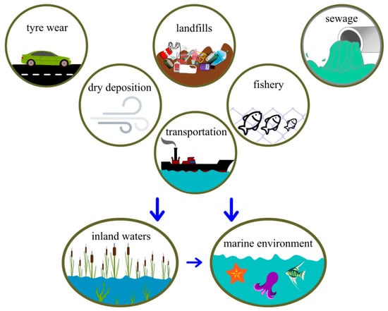
Figure 1.
Main sources and transport pathways of microplastics to the aquatic environment.
MPs migrate in the environment, and their movement results from various transport mechanisms. One of these is atmospheric transport. MPs, as components of suspended dust, can be carried by wind over long distances. Airborne MPs settle on the surfaces of soils and waters, contaminating them even in locations far from direct emission sources [18]. It has been shown that the efficiency of transporting MPs through the air is influenced by their size and shape. An interesting and unexpected finding of the analysis conducted by Fox et al. [19] is the increased proportion of blue-colored MPs at significant distances from areas under anthropogenic influence compared to other MPs. According to Brahney et al. [20], up to 84% of plastics suspended in the air are MPs lifted from roads due to vehicle movement. Tire abrasion generates MPs in the form of both fragments and fibers, predominantly in the size range of 2.5–350 µm [21]. The amount of tire rubber particles in suspended dust near roads is reported to range from 5% to 28% for particles smaller than 10 µm. Analyzing these estimates, the emission of tire particles and road dust as a source of microplastic particles in the air is significant [22]. Another important mechanism for understanding the distribution of MPs in the environment is river transport. Plastic particles, both large and microscopic, can be carried by river currents for long distances. Consequently, this leads to the contamination of their outlets: lakes, reservoirs, and even seas and oceans [12,23,24]. For example, Li et al. [25] demonstrated using the Yangtze River, which flows into the East China Sea, that the load of MPs carried by rivers can be high and significantly contribute to marine pollution. The annual amount of MPs transported by this river to the sea was estimated at 7000 tons. Oceanic currents also play a key role in the spread of MPs. Plastic from various land areas can reach the oceans, where it becomes part of the global marine transport pathways for MPs [26]. In 2014, the distribution of floating plastic debris around the Kuroshio Current was studied, which transports plastics from coastal waters of Asian countries to the subtropical gyre of the North Pacific. The average abundance and mass of plastic debris on the sea surface were 100,376 particles/km2 and 446.16 g/km2, respectively. The debris was predominantly composed of small (≤1 mm) fragmented white particles (1 mm) of polyethylene and polypropylene. This evidence points to the global reach of MPs pollution. Ocean currents are extensive, allowing polymer pollution to be transported over intercontinental distances [27]. The discussion of the accumulation of MPs in the oceans cannot overlook the Great Pacific Garbage Patch. It consists of floating waste on the water surface, stretching from Hawaii to the California coast [28]. The total amount of floating polymer waste in this patch is estimated to be between 45,000 and 129,000 tons. Interestingly, MPs may constitute 8% of their mass but they account for 94% of all particles by count [29].
2.2. Threats Resulting from the Presence of MPs in Aquatic Ecosystems
MPs introduce chemicals and organic compounds into water, which can negatively impact water quality [30]. A particular type of MPs in this regard are PVC particles. To improve the processing and utility properties of this material, additives are added including phthalates in amounts ranging from 10% to 70% [31]. According to research from the PlastChem report, more than 16,000 chemicals are known to be present in plastic materials and products, with less than 6% of them currently subject to global regulations. For nearly 4000 plastic chemicals, their annual production volumes exceed 1000 tons. Numerous studies have demonstrated the potential for these substances to enter the environment. The content of plasticizers is primarily significant, but so is the shape and structure of MPs. A larger surface area promotes leaching, which is highest in the outer layer of plastic particles [32]. According to Gulizia et al. [33], for PVC particles smaller than 42 μm, the release of plasticizers occurs almost instantly upon contact with water, indicating that contamination by fine micro- and nanoplastics in the environment can quickly affect water quality. The rate of this process is directly correlated with the particle size, and even small changes in size have a significant impact on the duration and characteristics of the leaching process. Since larger microplastics (e.g., >136 μm) are transported and dispersed worldwide [34], and can release plasticizers for up to 60 days, their harmful reach is greater than that of small particles. Gulizia et al. [33] also point out the acceleration of the process at higher temperatures, which, in correlation with rising sea and ocean temperatures [35], creates an environmentally hazardous phenomenon. The leaching of phthalates has potential consequences for the water resources used for drinking, agricultural, and industrial purposes. Once in the environment, phthalates persist in every component through biogeochemical processes and can accumulate in the food chain, subsequently causing serious health effects in mammals [36]. The impact may be related to its ability to act as so-called endocrine-disrupting substances, which means that they can disrupt hormonal functions in the body. Phthalates are suspected to affect the hormonal system, especially by mimicking sex hormones [37,38]. Furthermore, laboratory studies in animals suggest that phthalates may have a negative impact on the development of the reproductive system and may be associated with certain diseases such as diabetes, cancer, obesity, and immune system problems [39,40].
MPs have a wide capacity to absorb toxic chemicals. There are reports of heavy metal adsorption, pharmaceuticals, and other organic substances in plastic microparticles. In laboratory studies, Moura et al. [41] demonstrated the absorption of pharmaceuticals in the presence of MPs. The particles in the undegraded plastic had a much lower absorption capacity than those subjected to aging. The absorption capacity also varied depending on the particle size. Smaller MPs particles (below 35 µm) absorbed more substances than larger particles (59–157 µm). Among the plastics, the highest absorption occurred in PP, PA, and PVC. In a study by Zong et al. (2021) [42], the adsorptive properties of heavy metals in polystyrene (PS) MPs were examined, as well as the bioavailability and toxicity of MPs and heavy metals in an experiment with hydroponic wheat seedlings. The results showed that PS microplastics (0.5 µm, 100 mg/L) had no significant impact on the growth of wheat seedlings, photosynthesis, and the content of reactive oxygen species (ROS). However, PS microplastics could adsorb copper and cadmium, mainly through chemisorption. The accumulation of copper and cadmium in wheat seedlings decreased in the presence of PS microparticles, indicating that the toxic effects of heavy metals could be mitigated. The ability of MPs to absorb heavy metals means their possible high concentrations in microplastics present in environments contaminated simultaneously with heavy metals and MPs [43]. As a result, when ingested by aquatic organisms, these substances can be transferred along the food chain, posing a threat to the health of entire aquatic ecosystems [44,45,46].
2.3. MPs in Drinking Water Sources
As discussed above, MPs enter surface waters through various pathways, including wet and dry atmospheric deposition, runoff from urban [47] and agricultural areas [48], as well as through wastewater [49]. Surface water contamination presents an obstacle to using these waters as sources for domestic purposes [50,51]. The presence of MPs in water has increased interest in more detailed issues, such as the presence of these contaminants in drinking water treatment processes and the effectiveness of water treatment plants in removing them [52]. The presence of MPs in drinking water sources poses a significant challenge to water treatment processes, increasing costs and technical requirements, which can hinder the provision of clean water to communities. It has been observed that the removal efficiency of MPs in a simple treatment plant (including coagulation, flocculation, and sand filtration) is lower (about 70% MP removal efficiency) than in more advanced drinking water treatment stations (about 81%), which include coagulation and flocculation, sand filtration, and granular-activated carbon filtration [53]. Studies on the efficiency of MP removal in a pilot water treatment plant in Switzerland indicated that the coagulant does not play a significant role in the elimination of microplastic particles (the efficiency of MP removal after sand filtration was 95% without the coagulant and 92% with the coagulant). The coagulant played a more significant role in the removal of MPs in the form of synthetic fibers. The removal efficiency was 69% and 96%, respectively, without and with the coagulant [52].
In light of the threat posed by the presence of MPs in the water supplied by networks using surface waters, an alternative could be the use of groundwater. However, even groundwater is not free of the risk of MP contamination [54]. Small micro and nanoplastics have a particular ability to migrate vertically in soil. Due to their size, microplastics easily penetrate microorganisms and move with them within the soil profile, eventually leading to their entry into groundwater [55]. In shallow soil layers, agricultural practices, soil erosion, and soil fauna activities, such as those of earthworms, play important roles [56].
The presence of MPs in groundwater has been repeatedly confirmed. For example, Gong et al. [57] verified the presence of MPs in groundwater on Dawanshan Island. The collected water samples contained microplastics in amounts ranging from 34 to 64 particles per liter, with polyester fibers accounting for a significant 80% of the total. According to the authors, the presence of MPs in the groundwater of the island is due to their infiltration with surface water and wells contaminated with microplastics. Dey et al. [58] highlighted the still insufficient knowledge on MP contamination in groundwater. They pointed out potential factors that contribute to groundwater contamination with MPs, such as groundwater recharge from standing water bodies used for agricultural purposes and watercourses near waste disposal sites. Scientists emphasize the need for comprehensive solutions to protect groundwater, as it is a crucial and strategic resource for the development of civilization [59]. Reports from researchers in the Czech Republic indicate the presence of MPs in quantities ranging from 2 to 20 particles per liter in water from deep wells and inactive mines. This finding is particularly surprising given that the water samples were collected from depths of about 700 m [60]. Mechanisms of transport or water contamination pathways at such great depths remain unknown. Given the surprising presence of MPs so far from potential sources, there are concerns among scientists about errors in groundwater and soil studies. It was found that in most studies, blank samples were not used, MPs were incorrectly classified by mixing shapes and forms, and filters with too large pore sizes or materials that could be potential sources of MPs were used. According to Chia et al. [61], these suboptimal procedures have led to inflated results in many studies.
Thus, many aspects related to the presence of MPs in groundwater remain unclear, particularly the mechanisms of transport and distribution of MPs in groundwater at significant depths. To address these knowledge gaps, future research should prioritize understanding the primary mechanisms of vertical MP transport in soil and aquifers, as well as the long-term impacts of MPs on groundwater quality and aquifer systems.
3. Characterization MPs
3.1. Size and Nomenclature
An important challenge that remains unresolved is the development of a consistent and precise nomenclature. Relatively recently, just a few decades ago, Thompson (2004) [62] were the first to use the term “microplastic” in their work. It was introduced as an evolution of the term “microscopic plastic fragments”. However, the work did not attempt to clearly define this term, assuming that it was intuitively understood. It was not until around 2009 that the definition of “microplastic” was proposed as a polymer fragment less than 5 mm in diameter [6].
In recent years, there has been significant interest in the detection and identification of microplastics in the environment. The information contained in scientific papers produced in a short period has led to discrepancies in the terminology used and the proper meanings of the concepts. Separate terms have also emerged for microplastics resulting from the breakdown of larger plastic fragments (secondary microplastic) and particles intentionally manufactured in small sizes (primary microplastic) [63]. Moreover, new methods for identifying microplastics have allowed researchers to detect particles on the nanometer scale [64]. Consequently, in recent years, a new term, “nanoplastic”, has increasingly appeared in the literature [65,66]. Like microplastics, there is no consistent definition for nanoplastics. Gigault et al. [67] proposed the following definition for nanoplastics: “particles resulting from the degradation of plastic waste, possessing colloidal properties, and ranging in size from 1 nm to 1 µm”. This description is also referenced by other researchers [68,69], indicating that the definition has been accepted by the scientific community.
In light of the above, it is crucial to exercise great caution when comparing the obtained research results with those from other studies. Comparing numerical values alone carries the risk of error. Referencing results to other studies requires a thorough study and understanding of the research methodology, as there is a lack of consistency in the use of microplastic terminology depending on particle sizes (Table 1), descriptions of their shapes, and potential origins [6]. Unambiguously assigning a defined range of particle sizes to the appropriate name would significantly facilitate the comparison of research results from different research centers worldwide. Therefore, there is an urgent need to develop uniform terms and definitions for micro- and nanoplastics.

Table 1.
Nomenclature used for plastic particles depending on their size.
3.2. Shape and Color
Microplastic particles can vary not only in size but also in shape, surface area, material, and color [6,79] because the industry offers a wide range of plastics. According to current knowledge, any of these materials can potentially enter the environment as waste. This requires specifying additional parameters (i.e., shape and structure) to differentiate microplastics into several other groups: fibers, rubbers, sponges, fragments, etc. [80,81]. Although shape and structure are relatively straightforward to determine using microscopic methods, identifying color is a much greater challenge. The perception of color is always subjective and depends on many factors, including the method of observation [6].
Research indicates that the color and shape of polymer microfragments are significant. It turns out that some animals ingest microplastics along with their food nonrandomly. Marine benthic organisms have been found to preferentially consume microplastics in fiber form. MPs shaped like rods and fibers are also the most toxic to these organisms [82]. In a study on fish conducted by Xiong et al. [83], a higher intake of microplastics was demonstrated in colors similar to those of their typical food. The size of microplastic particles also affects their ingestion, as they must be small enough to be swallowed by the fish. In the same study, plastic films were also observed to be ingested more frequently by fish than fragments and fibers. Unfortunately, because of differences in translations, experiences, and the context of various researchers’ work, the same shapes or forms of microplastics may be referred to differently. Although defining the range of dimensions seems relatively straightforward, developing a universal key to describe microplastics is challenging, but worth the effort. The International Organization for Standardization (ISO) has prepared a document summarizing the current state of knowledge about the presence of microplastics in the environment and living organisms. It also lists methods for detecting MPs in various environmental matrices and sample preparation. In its current form, this document is merely a set of recommendations and, therefore, does not have the status of a universally recommended ISO standard. The ISO website also contains information on documents standardizing the quantitative and qualitative detection of microplastics from sources related to textile production (ISO) [84].
4. Methods for Separation and Extraction of MPs from Environmental Matrices
4.1. The Importance of MP Separation for the Reliability of the Study
One of the greatest challenges in the study of microplastics (MPs) is the separation of them from other components of an environmental sample before the identification stage. Under magnification, microplastic particles resemble plant fibers, zooplankton, or sand grains. These elements are still present in the samples even after prior chemical digestion or filtration [85]. Researchers continually refine methods for separating MPs from samples taken from natural environments because samples are rarely suitable for direct analysis. This step is critical for subsequent quantitative analysis. The efficiency of the extraction method applied for MPs limits the results later. Figure 2 schematically presents commonly used practices in preparing environmental samples for MP analysis. Usually, a multistep sample preparation is required, employing mechanical, chemical, and other methods that exploit the physical property differences between plastics and other sample components [86,87]. Regardless of the subsequent identification method, the extraction stage of plastics from environmental samples is crucial to the accuracy and reliability of the results obtained. Plastics are distinguished by several parameters that separate them from natural materials. They have relatively low density, are mechanically durable, highly impact-resistant, and importantly, chemically resistant to acids, bases, and oxidizing agents [88]. This latter feature allows for the chemical digestion of samples to dissolve as much of the sample matrix as possible into simple substances and smaller fragments. The most commonly used methods for this purpose include Fenton reactions, enzymes, hydrogen peroxide, and acid or base solutions [87]. Hydrogen peroxide is particularly frequently used, both in combination with heating and in reactions catalyzed by Fe2+ ions. The variety of materials studied, e.g., water, soil, sediment, plant, and animal tissues, necessitates specifying methods and their specific applications.
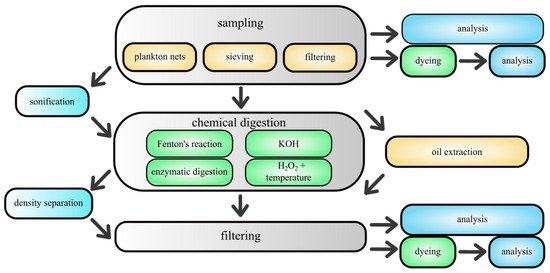
Figure 2.
Flow chart for handling environmental samples in MPs analyses.
4.2. Sieving
The simplest method of preparing samples involves sieving the material through screens and meshes, which also defines the upper and lower size limits of the analyzed microplastic particles. Stainless steel sieves are ideal for this purpose, as they do not contaminate the samples [6,89]. This method is used for sediment, soil and organic matter fragments and can be used both dry and wet. Sieving can be performed directly at the time of sample collection [90], MPs traps largely consist of screens and meshes mounted on structures that allow them to be installed at research sites [82,91]. This method is mainly used to collect samples from surface waters. Various types of plankton nets, which have found new applications with increasing interest in microplastics, are popular for this purpose [92]. Sieves are also used during the chemical processing of environmental samples. This minimizes errors during multi-step digestion and facilitates transferring samples between solutions. Additionally, using a specific mesh size allows the exclusion of particles that fall outside the lower measurement range for some identification methods [93].
4.3. Flotation
Another method practiced for separating MPs from environmental samples is flotation of froth. Generally, materials are classified as hydrophilic or hydrophobic [94]. Most artificial polymer materials have hydrophobic surfaces, so adding a surfactant to the solution causes its molecules to accumulate on the surfaces of MPs [95]. This phenomenon is utilized in the froth flotation process. With appropriate aeration, gas bubbles lift MPs particles that are physically attached to the surfactant. The lower the density of the plastic and the higher the surface-to-mass ratio, the more efficient the process. Meanwhile, other hydrophilic substances present in the system sink to the bottom [96]. Froth flotation shows promise for samples containing large amounts of hydrophilic substances, such as various matrices containing sand. However, scientists are cautious about making statements regarding the efficiency of this process, often defining it as relatively low. In their studies, Imhof et al. [97] estimated the overall efficiency of the flotation process in separating microplastics was approximately 55%. This value was highly dependent on the type and density of the polymer. The size and shape of MPs particles are also significant for the overall efficiency of this process. Surface properties can even vary for the same type of plastic, as different additives used during production can alter these properties. Additionally, the aging process changes the surface properties and density of the plastic, which in turn affects the flotation process [98]. A drawback of froth flotation is the duration of the process (the longer it is run, the greater the recovery of MPs) [99]. Moreover, the separating fluid must be replenished continuously [100]. Froth flotation also does not guarantee the separation of all types of MPs and can essentially only be used as one of the sample preparation processes. Large fragments of high-density plastics (e.g., PVC) are difficult to separate using this method. In a broader context, one of the key advantages of flotation of froth is its continuous nature. This process also allows for the permanent removal of MPs. Furthermore, the relatively low technological complexity and operational costs of froth flotation could argue for its wider application. This process does not require complex equipment or advanced infrastructure, which could make it an accessible means of removing microplastics, for example, in wastewater treatment plants [101].
4.4. Density Separation
Currently, the most common and widely developed method for separating MPs from environmental samples is density separation. High-density solutions are prepared from filtered water in which large amounts of highly soluble salts are dissolved [102]. As a result, plastic fragments with a density lower than that of the separating solution can float to the surface, while heavier particles remain at the bottom. MPs can then be extracted by filtering the separation medium, followed by analysis and characterization. Since the density of most plastics ranges from 0.8 g/cm3 (silicone) to around 1.5 g/cm3 (polyethylene terephthalate (PET), polyvinyl chloride (PVC), and polyoxymethylene), the denser the solution, the more types of polymers can be extracted from sediments [103]. However, it is important to note that the density of polymers like PVC can vary significantly due to the presence of plasticizers, which are commonly added to increase flexibility. For example, pure PVC has a density of ~1.4 g/cm3, making it difficult to separate using standard density solutions, as it often sinks even in high-density media. On the contrary, plasticized PVC can have a reduced density of ~1.2 g/cm3, making it easier to float. This variability complicates the separation process and highlights a key limitation of density-based methods for certain polymers [104]. In reality, polymers such as PVC and polytetrafluoroethylene (PTFE) have densities higher than those of others; hence, they are more challenging to extract via density separation. This method is most effective for samples containing a high proportion of mineral components [105]. Soil and sediment samples contain a significant amount of organic matter, which has a density similar to polymers, approximately in the range of 1 to 1.4 g/cm3 [106]. Consequently, not only polymer fragments but also all organic particles with similar densities float to the surface of the separating liquid [107]. Current devices used for density separation, including beakers and separators shaped as inverted funnels with valves, have drawbacks in terms of recovery speed and operational complexity [108]. There is still a need to refine this method and objectively consider it as just one of the sample preparation processes [109]. Figure 2 presents the most widely used separating salts along with the density of their saturated solutions. All plastics with a density higher than the given salt density can be separated using it. The densities of the plastics are approximate, as they can vary depending on the filler and additive content. As seen in Figure 3, the most effective salts for microplastic density separation are K2CO3, ZnCl2, NaI and ZnBr2. These salts differ in not only density but also other characteristics. NaBr, NaI, ZnCl2, and ZnBr2 are toxic salts with a high potential environmental impact and are also costly [103]. Therefore, the most optimal salts in terms of cost and safety are K2CO3 and CaCl2. These salts are characterized by low toxicity and have even been approved as food additives, making them safer alternatives for laboratory use.
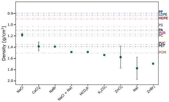
Figure 3.
The applicability of saturated salt solutions in the process of density separation of microparticles of individual plastics. Plastics with a density lower than the density of the salt flow to its surface [103]. PP—polypropylene, LDPE—low-density polypropylene, HDPE—high-density polyethylene, PS—polystyrene, PA—polyamide, PUR—polyurethane, PC—polycarbonate, PVC—polyvinyl chloride, PET—polyethylene terephthalate, POM—polyoxymethylene.
To minimize contamination risks, it is crucial to implement blank controls and filter all salt solutions before use. This step is particularly important because chemical reagents are often packaged in plastic containers and may come into contact with plastics during production, potentially introducing additional MPs into the samples. Proper filtering and the use of blank controls ensure the reliability of the separation process and reduce the risk of false positives.
4.5. Oil Extraction
Another separation process that utilizes the hydrophobic properties of polymers is oil phase extraction. This method is effective for samples with a large number of mineral substances [99]. In this method, dried sediments are subjected to the action of a two-phase mixture of water and oil [110]. Due to the organophilic nature of their surfaces, MPs interact more strongly with the oil phase and are retained in it [94]. Research indicates that the mass-to-surface ratio of MP particles and the density of the oil phase play a significant role in this process. The more developed the surface, the more easily the particle is absorbed into the organic layer [111]. However, additional sample processing steps may be necessary before applying this method in many cases. An excessive amount of organic matter can accumulate as a contaminant in the oil, which is an undesirable effect. Due to its simplicity and low cost, this method is used by researchers. Efficiency results in favorable systems are reported to be as high as 90–97% [112]. The use of an oil layer is a promising idea in the microplastic separation process, and new variants of this method, including multi-stage processes, combinations of several methods, and changes in the state of the oil due to temperature, are appearing in the literature [112,113].
4.6. Chemical and Enzymatic Digestion
The general good chemical resistance of plastics allows the application of chemical digestion methods to the sample. This procedure involves exposing the sample to an aggressive environment to break down organic substances and disintegrate agglomerates of mineral substances [114]. Depending on the type of sample, different environments are used. Soil rich in plant matter, animal tissue, sewage, or mineral suspensions may require different protocols. Most often, samples are treated with concentrated potassium hydroxide and hydrogen peroxide combined with iron ions (Fe2+). Other substances used include nitric acid, citric acid, acetone, and ethanol [87]. The main reason for the use of chemical preparation is to release MPs particles trapped in the sample matrix [87]. This facilitates subsequent steps of their extraction. Many researchers often use chemical digestion of the sample as a preliminary step before density separation [12]. The unpredictable effect is the influence of chemicals on the MPs themselves [115]. Due to their small size and often developed surfaces, they may be more susceptible to erosion or even more than the larger fragments on which reference studies are usually conducted. This can generate errors in particle size determination. Fragmentation of MPs during chemical preparation leads to an increase in their total quantity. In the study by Alfonso et al. (2021) [116], it was demonstrated that PET (polyethylene terephthalate) is the polymer most susceptible to chemical damage. During alkaline digestion, PET lost 3.1% of its mass, while oxidative digestion resulted in a loss of mass of 0.7%. In contrast, other polymers such as LDPE (low-density polyethylene), HDPE (high-density polyethylene), PP (polypropylene), PS (polystyrene), PVC (polyvinyl chloride) and PC (polycarbonate) showed greater resistance to chemical degradation [116]. In the study by Tuuri et al., PA (polyamide), PE (polyethylene), PET (polyethylene terephthalate), PP (polypropylene), and PS (polystyrene) were exposed to acid solutions, alkaline solutions, enzymatic digestion, and oxidation, with changes in particle size being observed. The most significant changes were observed in polyamide (PA), which almost completely decomposed in HNO3 (nitric acid), while in other solutions, its size decreased by 0.5–3%. An interesting observation is that every polymer type tested, regardless of the chemical digestion method, showed a change in size of at least 0.1% [117].
An alternative and complement to oxidative methods based on H2O2 are enzymatic methods. They seem to be a “gentle” and optimal way to preserve the integrity of MPs extracted from the sample. However, these methods are time-consuming and costly. Therefore, efforts should be made to simplify them as much as possible and combine them with other methods. Good results are obtained by using enzymes followed by digestion with H2O2. Both reagents complement each other [118,119,120].
5. Methods for Determining Microplastics
5.1. Trends in MPs Analytics
In recent years, the topic of MPs has been rapidly developing due to its relevance and importance. Not only is the number of scientific reports on this subject increasing, but new analytical methods are also emerging. A similar phenomenon is the adaptation of long-known analytical methods, such as IR spectroscopy or the use of photoluminescence, specifically for MPs analysis. In Figure 4, the number of publications retrieved from the ScienceDirect portal using keywords indicating the applied analytical method in the context of MPs is presented. The analysis identifies the FTIR method as the most popular, and the LDIR method is emerging as a new approach. Regardless of the identification method, the total number of studies is increasing year by year [121].
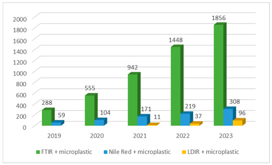
Figure 4.
Number of scientific publications searched by the ScienceDirect portal tool using the keywords FTIR + microplastic, Nile Red + microplastic, LDIR + microplastic by year [121].
5.2. Optical Microscopy Method
Optical microscopes and stereomicroscopes are effective tools in microplastics analysis because of their ability to provide high-resolution images and the capability to employ various imaging techniques. The advantages of this method include the rapid identification and assessment of the morphology of microplastics, which is crucial to understanding their source, distribution, and environmental impact [122,123]. Optical microscopy is useful for a quick preliminary assessment of samples for the presence of microplastics. The low complexity of the method allows multiple samples to be evaluated without the need for special preparation. For example, in the case of water samples, simply filtering the sample and examining the filter under a microscope is often sufficient. However, there are some disadvantages associated with this method. Optical microscopy is not sufficient for identifying the chemical composition of microplastics, which is essential to understanding the potential hazards to organisms and the environment. A comprehensive analysis of the size distribution and type of material helps to find correlations between these characteristics and the impact of MPs on the environment [118]. In addition, identifying microplastics of very small sizes, especially those smaller than 20 μm, can be challenging. Much depends on the skills and experience of the researcher conducting the examination [124]. Human error is a significant limitation of optical microscopy, particularly when distinguishing microplastics from organic or mineral particles. Studies, such as Lenz et al. (2015) [125], have demonstrated that misidentification rates can range from 10% to 60%, depending on the sample type and the researcher’s expertise. These error rates were determined by comparing the results of optical microscopy with those of Raman spectroscopy, which is a more accurate and reliable method for identifying microplastics [125]. Therefore, human error cannot be completely eliminated. Under microscopic magnification, microplastic particles can exhibit various morphological features, making their identification difficult. These microscopic particles often assume various shapes and structures, making them similar to other small organic or mineral fragments. In a microscopic image, microplastics often resemble tiny grains of sand, fibers, or plant fragments. They can also be similar to microorganisms, such as phytoplankton [126]. Due to their small size, microplastics can be difficult to distinguish from natural environmental components, further complicating the identification process [127]. As a result, optical microscopy is often used as part of more advanced analyses, such as FTIR or Raman spectroscopy (RS), which allow for the identification of the type of plastic material [128]. Therefore, detecting MPs visually using a microscope can be prone to errors, leading to quantitative overestimation or underestimation [125]. Despite these limitations, optical microscopy remains a widely used method in microplastic research because of its simplicity, speed, and effectiveness in identifying larger particles. When used in conjunction with other advanced analytical techniques, this tool enables a more comprehensive analysis of the composition and characteristics of microplastics.
5.3. FT IR and RS
Fourier transform infrared (FTIR) spectroscopy is a widely used chemical analysis method that is widely applied in the identification and characterization of microplastics. To apply this technique to microplastics analysis, proper sample preparation is essential [129]. The cleaning and extraction processes for the samples do not differ significantly from procedures used in other analytical methods and include chemical digestion, density separation, filtration, etc. [130]. Subsequently, microplastics are placed on an appropriate slide in a thin layer or in a KBr pellet to obtain a clear IR spectrum [131]. The FTIR spectrometer emits infrared radiation at various wavelengths, which passes through the sample. The microplastic molecules absorb radiation at frequencies characteristic of their chemical bonds, causing changes in the FTIR spectrum. The transmitted radiation passes through an interferometer, and the resulting spectrum is recorded. The outcome is an FTIR spectrum that represents light absorption as a function of wavenumber (cm−1) [132]. Characteristic bands in the FTIR spectrum correspond to different chemical bonds in the plastic molecules. Analyzing these bands allows for the identification of the type of plastic and additional components, such as additives or contaminants. The obtained FTIR spectrum can be compared with a reference database of microplastic spectra, allowing precise identification of the plastic type [133]. FTIR also allows for the assessment of the presence of chemical additives in the sample, which is crucial to determining the environmental risk [134]. Using the FTIR method to study microplastics enables precise identification of the plastic type, which is key to understanding and monitoring the presence of microplastics in various environments. However, as with any method, FTIR has drawbacks. FTIR may encounter difficulties in analyzing microplastics present in complex mixtures, especially when other components, such as organic substances or mineral compounds, are present. In addition, other substances present in the sample, such as dyes, can introduce interference in the FTIR analysis, making it difficult to precisely determine the type of microplastic. Although characteristic bands are identified, FTIR may not always unequivocally determine the type of microplastic, especially when they are modified or contain additives [135,136]. Proper sample preparation is the key to obtaining accurate results. Improper sample preparation, such as improper distribution of microplastics on the backing, can lead to erroneous results [137]. Also, in the case of very small microplastic particles, FTIR may have difficulty detecting and identifying them [138]. In such cases, alternative techniques such as Raman spectroscopy can be more effective, particularly when analyzing small or fine microplastics. Raman spectroscopy analyses molecular vibrations through light scattering, detecting unique vibrational signatures specific to molecules. It is highly sensitive to nonpolar groups, less affected by water, and is effective for analyzing wet samples with minimal preparation. RS offers better spatial resolution than IR techniques, making it suitable for identifying small microplastics [139]. Araujo et al. (2018) noted that Raman spectroscopy naturally has better spatial resolution than IR-based techniques and is therefore the preferred method for identifying small microplastics (<20 μm) [132]. Interesting insights regarding FTIR and Raman methods were provided by Käppler et al. in their study. They showed that Raman spectroscopy enables the identification of microplastic particles as small as 5 μm, with the potential to detect particles down to 1 μm, which is considered the lower size limit for microplastics. However, compared to FTIR imaging, Raman spectroscopy requires longer measurement times, which can be reduced at the cost of spectral quality and detection sensitivity. Additionally, both techniques require optimization of sample preparation to minimize fluorescence (in the case of Raman) and improve spectral quality. The authors also highlight that Raman and FTIR differ in their sensitivity to specific polymer types; for example, Raman is more effective in detecting PVC, while FTIR performs better in analyzing polyesters [140].
5.4. Fluorescence Microscopy
Spectroscopic methods such as Raman and FTIR are commonly used for identifying types of polymer materials. However, these spectroscopic methods are expensive and require labor-intensive sample purification, leading to parallel interest in alternative detection methods such as cytometry [141], thermal degradation [142], the hot needle method [143], and fluorescence microscopy [144]. Among these methods, fluorescence microscopy has the potential for rapid detection because it involves a simple fluorescent labeling step with the solvatochromic dye Nile Red. Nile Red is an organic fluorescent dye that exhibits fluorescence in the presence of hydrophobic substances, particularly fats and lipids. Its fluorescent properties make it a useful tool in lipid studies, cellular research, and more recently in microplastics analysis [145]. The ability of Nile Red as an indicator to distinguish plastic samples based on their polarity has been investigated using existing protocols that employ fluorescence microscopy [146]. In these published protocols, fluorescence micrographs of Nile Red-stained primary microplastics were sufficient to successfully classify plastics based on their polarity (polar/less polar) [147].
However, most studies that use Nile Red dye do not specify the type of plastics found in the microplastics. Previous studies only considered the emission color in fluorescence images. In the work of Idehara et al. (2025) [148], spectral emission was additionally examined using photoluminescence (PL) spectroscopy by studying the effects of staining parameters: dye concentration, solvent type, staining temperature, and staining time. As a result of these studies, an optimized staining protocol was developed, but it was not possible to completely eliminate the confusion of MPs with natural materials such as cellulose or protein fragments of animal origin [148]. In their study, Sturm et al. demonstrated that fluorescence microscopy images of microplastics and natural particles dyed with Nile Red (NR) and its derivative (NR3) reveal that selectivity depends on the emission wavelength. Using green fluorescence filters (excitation: 455–495 nm, emission: 505–555 nm), nonpolar plastics such as PE and PP show strong signals, while polar plastics such as PVC are barely detectable. Natural materials such as wood and chalk show minimal green fluorescence, but chitin exhibits strong signals, leading to false positives. This can be mitigated with treatment with hydrogen peroxide or chitinase. Orange (excitation: 533–558 nm, emission: 570–640 nm) and red fluorescence filters (excitation: 575–635 nm, emission: 660–710 nm) further complicate differentiation, as polar plastics, chitin, and wood all show strong signals, making it harder to distinguish them from non-polar plastics [149].
5.5. LDIR
Since knowledge of not only the type of polymer but also its characteristics (size, shape, surface, etc.) is very important and useful information that can affect the proper interpretation of research results, the positive aspect of growing interest in the issue of MPs is the simultaneous development of new techniques and methods for identifying MPs. Currently, in contrast to commonly used classical methods for microscopic identification of MPs, modern and advanced techniques that allow for both quantitative and qualitative descriptions of polymer particles are extremely valuable and desirable. The latest technique for identifying MPs (enabling both qualitative and quantitative determination) is laser direct infrared imaging (LDIR). The use of this technique combined with appropriate software significantly expands the current possibilities for analyzing and identifying MPs [150].
The LDIR method has recently been introduced as a new technique for analyzing polymers and MPs in environmental samples. Compared to methods such as FTIR, it has the advantage of scanning the sample for the presence of microplastics before actual imaging and ultimately only analyzing areas with detected particles [151]. This shortens the analysis time because spectra are obtained when particles are detected during the initial scan. Therefore, samples with small amounts of MPs are measured much faster. The only drawback is that if two particles are adjacent to each other, they may be recognized by the device as a single particle because only one spectrum is recorded per particle [152]. However, automation of measurement and particle recognition reduces the risk of error, which is very likely when identifying MPs using optical microscopy methods. Direct determination of samples taken from the environment using the LDIR method is mostly impossible to achieve.
Lopez-Rosales et al. [153] write about the challenge of proper sample preparation and its quantitative transfer to a reflective microscope slide, a requirement for the LDIR method. They applied this method for the first time to identify MPs in fish tissues. The researchers noted difficulties in preparing sufficiently clean samples. In another study, the same research group focused on developing a rapid and effective method for characterizing airborne microplastics [154]. They used an optimized LDIR method, achieving over 90% efficiency in differentiating fibers from particles. Quantitative recovery of particles was 82–90%, and for fibers, 62–73%. They evaluated the quantity and type of MPs deposited in northwestern Spain. The most common were polyethylene, polypropylene, and polyethylene terephthalate. The deposition rate was 98–1220 MPs/m2/day, constituting about 1.7% of the collected particles. The sizes of more than half of the deposited microplastic particles ranged from 20 to 50 μm, and the fibers ranged from 50 to 500 μm. Hansen et al. [155] used the LDIR method to determine microplastics collected in the northeastern Atlantic region. The analysis was performed on microplastics less than 300 μm. The authors noted the relatively short analysis time and the automatic mode of operation. The device identified up to 20 different types of polymers. Thus, the ability to quickly recognize the type of plastic is an advantage of the LDIR method compared to other methods, such as the often-used Nile Red method [156]. In studies conducted in the Rzeszów reservoir, the usefulness of the LDIR system for determining microplastics extracted from bottom sediments was demonstrated. The precision of the method allowed for the identification of particles ranging in size from 30 to 1818 μm. It should be noted that the high-quality optical magnification allowed for the determination of particle morphology, which is often impossible with other methods, especially for the smallest fragments. The results of the presented studies were preceded by a detailed description of the chemical sample preparation process before analysis using LDIR [12].
The LDIR method requires the introduction of a sample into the device on a surface that reflects IR radiation well. Therefore, filters made of nonreflective materials such as glass fiber and cellulose cannot be used. Water samples and microplastics suspended in solutions require filtration. Currently available options include membranes coated with gold and aluminum. Because of the metallic coating, they reflect IR radiation well. Gold-coated filters have some advantages over aluminum-coated ones. Unlike aluminum, gold is very resistant to chemical agents, so the components of solutions do not damage the surface. For practical use in LDIR, gold-coated filters must have a gold layer thickness of at least 20–40 µm, which is a standard specification for such filters. However, their high cost can pose a financial barrier for some laboratories, potentially limiting the accessibility and practicality of the LDIR method, particularly for routine or large-scale analyses. In the case of aluminum, salts, acids, and bases can damage the thin layer of metal, depriving the membrane of its reflective properties. The device works with filters with a diameter of 25 mm. The small size of the filter prevents the examination of samples with a high content of suspensions. It is required that a monolayer of particles be present in the filter after filtration. Otherwise, the analysis will not be performed correctly. Moreover, the filtration setup should be assembled so that the edges of the filter (about 3 mm from the edge) are not covered with the sample. The filter holder bends the filter in these areas and the sample material will be lost in this part. The filters are disposable because there is currently no effective method for cleaning them and because polycarbonate is under the metallic coating, they cannot be calcined. Due to the novelty of the LDIR system, there is still little literature on sample preparation protocols for MP determination using this device. One possibility recommended by the device manufacturer is to filter samples through metallic membranes and then sonicate them for 20 min in ethanol. The resulting suspension of particles in ethanol can be transferred onto a slide. After the ethanol evaporates, it is possible to analyze the particles deposited on the slide [157].
6. Summary
The development of plastic production has led to a significant increase in plastic waste, which is now a global environmental issue. The presence of microplastics (MPs) has been confirmed in all environmental components. They can accumulate in soil, sediment, and within plant, animal, and human organisms, posing serious threats. MPs travel through the environment via air, rivers, and ocean currents, contaminating even remote areas far from emission sources. Their presence in freshwater and marine environments can lead to the release of many hazardous chemicals, which can threaten the health of aquatic organisms and humans. Additionally, MPs have the ability to adsorb toxic substances, facilitating their transport along the food chain and bioaccumulation. The presence of MPs in surface and groundwater also poses significant challenges for drinking water treatment processes.
In recent years, there has been a significant increase in interest in MPs in the environment, resulting in the dynamic development of analytical methods. These methods include optical microscopy, FTIR, fluorescence microscopy, and the relatively new LDIR method. Each of these methods has certain advantages and limitations. Optical microscopy is quick and simple, but has limitations in identifying the chemical composition of MPs and can lead to errors. FTIR allows for precise identification of the types of plastic and chemical additives but requires complicated sample preparation and has difficulties analyzing very small particles. Fluorescence microscopy is a quick and less expensive method, but it has difficulties in unequivocally identifying the plastic and may confuse MPs with other materials. LDIR is a modern technique that offers fast and automated quantitative and qualitative analyses with a low risk of errors, although it requires complicated sample preparation, the use of relatively expensive filters, and may have difficulty identifying adjacent particles. These limitations highlight the need for a more integrated approach to MP analysis, where the choice of method is tailored to the specific characteristics of the MPs in question. For example, combining LDIR with advanced image processing algorithms could mitigate the adjacency issue, while coupling FTIR with complementary techniques such as Raman spectroscopy could improve the detection of small particles.
Obtaining reliable results with each of the aforementioned methods is mainly dependent on proper sample preparation for analysis. MPs are often difficult to distinguish from other microparticles, such as plant fibers or zooplankton, present in environmental samples, making proper separation and extraction of MPs from samples crucial. Mechanical, chemical, and physical methods such as sieving, density separation, foam flotation, or oil phase extraction are used for this purpose. Chemical and enzymatic methods are also used to dissolve organic matter. Each of these methods has its applications and limitations, and the appropriate choice depends on the type of sample and the specific analytical requirements.
In conclusion, researchers dealing with MPs still face many challenges. There is still a need to develop effective methods for identifying MPs, particularly to address the interplay between MP characteristics and analytical limitations. Future research should focus on developing standardized protocols that account for the diversity of MP sizes and shapes, as well as integrating multiple analytical techniques to overcome individual method limitations. Consistent terminology and methods for describing detected micro- and nanoplastics also need to be established. Therefore, effectively managing the microplastics problem in the environment requires a comprehensive approach based on interdisciplinary scientific collaboration and practical actions at local, national, and international levels. For example, integration of hydrology and polymer science could provide a more comprehensive understanding of MP transport and degradation in aquatic environments, while toxicology can help assess the ecological and health impacts of MPs. At the local level, monitoring programs for MPs in rivers and lakes should be implemented. Nationally, regulations are needed that limit the use of primary MPs and promote innovations in waste management.
Author Contributions
Conceptualization, W.S., R.G.-R. and M.C.; writing—original draft, W.S., R.G.-R. and M.C. All authors have read and agreed to the published version of the manuscript.
Funding
The research was financed by: Minister of Science and Higher Education, Republic of Poland, within the program “Regional Excellence Initiative”.
Data Availability Statement
Not applicable.
Conflicts of Interest
The authors declare no conflicts of interest.
References
- Baekeland, L.H. Address of Acceptance: The Chemical Constitution of Resinous Phenolic Condensation Products. J. Ind. Eng. Chem. 1913, 5, 506–511. [Google Scholar] [CrossRef][Green Version]
- Crespy, D.; Bozonnet, M.; Meier, M. 100 Years of Bakelite, the Material of a 1000 Uses. Angew. Chem. Int. Ed. 2008, 47, 3322–3328. [Google Scholar] [CrossRef] [PubMed]
- Thompson, R.C.; Moore, C.J.; vom Saal, F.S.; Swan, S.H. Plastics, the Environment and Human Health: Current Consensus and Future Trends. Philos. Trans. R. Soc. B Biol. Sci. 2009, 364, 2153–2166. [Google Scholar] [CrossRef]
- Shams, M.; Alam, I.; Mahbub, M.S. Plastic Pollution during COVID-19: Plastic Waste Directives and Its Long-Term Impact on the Environment. Environ. Adv. 2021, 5, 100119. [Google Scholar] [CrossRef] [PubMed]
- Julienne, F.; Delorme, N.; Lagarde, F. From Macroplastics to Microplastics: Role of Water in the Fragmentation of Polyethylene. Chemosphere 2019, 236, 124409. [Google Scholar] [CrossRef] [PubMed]
- Frias, J.P.G.L.; Nash, R. Microplastics: Finding a Consensus on the Definition. Mar. Pollut. Bull. 2019, 138, 145–147. [Google Scholar] [CrossRef] [PubMed]
- da Costa, J.P.; Santos, P.S.M.; Duarte, A.C.; Rocha-Santos, T. (Nano)Plastics in the Environment—Sources, Fates and Effects. Sci. Total Environ. 2016, 566–567, 15–26. [Google Scholar] [CrossRef]
- Ziccardi, L.M.; Edgington, A.; Hentz, K.; Kulacki, K.J.; Kane Driscoll, S. Microplastics as Vectors for Bioaccumulation of Hydrophobic Organic Chemicals in the Marine Environment: A State-of-the-Science Review. Environ. Toxicol. Chem. 2016, 35, 1667–1676. [Google Scholar] [CrossRef]
- Eerkes-Medrano, D.; Thompson, R.C.; Aldridge, D.C. Microplastics in Freshwater Systems: A Review of the Emerging Threats, Identification of Knowledge Gaps and Prioritisation of Research Needs. Water Res. 2015, 75, 63–82. [Google Scholar] [CrossRef]
- Carpenter, E.J.; Smith, K.L. Plastics on the Sargasso Sea Surface. Science (1979) 1972, 175, 1240–1241. [Google Scholar] [CrossRef]
- Yan, Y.C.; Yang, Z.F. Sources, Distribution, Behavior, and Detection Techniques of Microplastics in Soil: A Review. China Geol. 2023, 6, 695–715. [Google Scholar]
- Strojny, W.; Gruca-Rokosz, R.; Cieśla, M. Preliminary Study of the Occurrence of Microplastics in the Sediments of the Rzeszów Reservoir Using the Laser Direct Infrared (LDIR) Method. Sustainability 2023, 15, 16653. [Google Scholar] [CrossRef]
- Rosati, L.; Carraturo, F.; Capozzi, F.; Chianese, T.; La Pietra, A.; Salamone, M.; Spagnuolo, V.; Ferrandino, I.; Giordano, S. Microplastics’ Impact on the Environment and the Challenging Selection of Reliable Key Biomonitors. Water 2024, 16, 2637. [Google Scholar] [CrossRef]
- Huang, W.; Song, B.; Liang, J.; Niu, Q.; Zeng, G.; Shen, M.; Deng, J.; Luo, Y.; Wen, X.; Zhang, Y. Microplastics and Associated Contaminants in the Aquatic Environment: A Review on Their Ecotoxicological Effects, Trophic Transfer, and Potential Impacts to Human Health. J. Hazard Mater. 2021, 405, 124187. [Google Scholar] [CrossRef] [PubMed]
- Asensio-Montesinos, F.; Blaya-Valencia, G.; Corbí, H.; Beltrán-Sanahuja, A.; Sanz-Lázaro, C. Microplastic Accumulation Dynamics in Two Mediterranean Beaches with Contrasting Inputs. J. Sea Res. 2022, 188, 102269. [Google Scholar] [CrossRef]
- Pilechi, A.; Mohammadian, A.; Murphy, E. A Numerical Framework for Modeling Fate and Transport of Microplastics in Inland and Coastal Waters. Mar. Pollut. Bull. 2022, 184, 114119. [Google Scholar] [CrossRef]
- Mutshekwa, T.; Munyai, L.F.; Mugwedi, L.; Cuthbert, R.N.; Dondofema, F.; Dalu, T. Seasonal Occurrence of Microplastics in Sediment of Two South African Recreational Reservoirs. Water Biol. Secur. 2023, 2, 100185. [Google Scholar] [CrossRef]
- Hee, Y.Y.; Hanif, N.M.; Weston, K.; Latif, M.T.; Suratman, S.; Rusli, M.U.; Mayes, A.G. Atmospheric Microplastic Transport and Deposition to Urban and Pristine Tropical Locations in Southeast Asia. Sci. Total Environ. 2023, 902, 166153. [Google Scholar] [CrossRef]
- Fox, S.; Stefánsson, H.; Peternell, M.; Zlotskiy, E.; Ásbjörnsson, E.J.; Sturkell, E.; Wanner, P.; Konrad-Schmolke, M. Physical Characteristics of Microplastic Particles and Potential for Global Atmospheric Transport: A Meta-Analysis. Environ. Pollut. 2024, 342, 122938. [Google Scholar] [CrossRef]
- Brahney, J.; Mahowald, N.; Prank, M.; Cornwell, G.; Klimont, Z.; Matsui, H.; Prather, K.A. Constraining the Atmospheric Limb of the Plastic Cycle. Proc. Natl. Acad. Sci. USA 2021, 118, e2020719118. [Google Scholar] [CrossRef]
- Vogelsang, C.; Lusher, A.L.; Dadkhah, M.E.; Sundvor, I.; Umar, M.; Ranneklev, S.B.; Eidsvoll, D.; Meland, S. Environmental Contaminants-Freshwater Distribution; Norsk institutt for vannforskning: Oslo, Norway, 2018; ISBN 9788257769666. [Google Scholar]
- O’Brien, S.; Rauert, C.; Ribeiro, F.; Okoffo, E.D.; Burrows, S.D.; O’Brien, J.W.; Wang, X.; Wright, S.L.; Thomas, K.V. There’s Something in the Air: A Review of Sources, Prevalence and Behaviour of Microplastics in the Atmosphere. Sci. Total Environ. 2023, 874, 162193. [Google Scholar] [CrossRef]
- Wu, Y.; Wang, S.; Wu, L.; Yang, Y.; Yu, X.; Liu, Q.; Liu, X.; Li, Y.; Wang, X. Vertical Distribution and River-Sea Transport of Microplastics with Tidal Fluctuation in a Subtropical Estuary, China. Sci. Total Environ. 2022, 822, 153603. [Google Scholar] [CrossRef] [PubMed]
- Idowu, G.A.; Oriji, A.Y.; Olorunfemi, K.O.; Sunday, M.O.; Sogbanmu, T.O.; Bodunwa, O.K.; Shokunbi, O.S.; Aiyesanmi, A.F. Why Nigeria Should Ban Single-Use Plastics: Excessive Microplastic Pollution of the Water, Sediments and Fish Species in Osun River, Nigeria. J. Hazard. Mater. Adv. 2024, 13, 100409. [Google Scholar] [CrossRef]
- Li, S.; Wang, H.; Liang, D.; Li, Y.; Shen, Z. How the Yangtze River Transports Microplastic to the East China Sea. Chemosphere 2022, 307, 136112. [Google Scholar] [CrossRef] [PubMed]
- Yuan, D.; Corvianawatie, C.; Cordova, M.R.; Surinati, D.; Li, Y.; Wang, Z.; Li, X.; Li, R.; Wang, J.; He, L.; et al. Microplastics in the Tropical Northwestern Pacific Ocean and the Indonesian Seas. J. Sea Res. 2023, 194, 102406. [Google Scholar] [CrossRef]
- Thushari, G.G.N.; Miyazono, K.; Sato, T.; Yamashita, R.; Takasuka, A.; Watai, M.; Yasuda, T.; Kuroda, H.; Takahashi, K. Floating Plastic Accumulation and Distribution around Kuroshio Current, Western North Pacific. Mar. Pollut. Bull. 2023, 188, 114604. [Google Scholar] [CrossRef] [PubMed]
- Dautel, S.L. Transoceanic Trash: International and United States Strategies For the Great Pacific Garbage Patch. Gold. Gate Univ. Environ. Law J. 2009, 3, 181. [Google Scholar]
- Lebreton, L.; Slat, B.; Ferrari, F.; Sainte-Rose, B.; Aitken, J.; Marthouse, R.; Hajbane, S.; Cunsolo, S.; Schwarz, A.; Levivier, A.; et al. Evidence That the Great Pacific Garbage Patch Is Rapidly Accumulating Plastic. Sci. Rep. 2018, 8, 4666. [Google Scholar] [CrossRef]
- Li, Y.; Liu, C.; Yang, H.; He, W.; Li, B.; Zhu, X.; Liu, S.; Jia, S.; Li, R.; Tang, K.H.D. Leaching of Chemicals from Microplastics: A Review of Chemical Types, Leaching Mechanisms and Influencing Factors. Sci. Total Environ. 2024, 906, 167666. [Google Scholar] [CrossRef]
- Yan, Y.; Zhu, F.; Zhu, C.; Chen, Z.; Liu, S.; Wang, C.; Gu, C. Dibutyl Phthalate Release from Polyvinyl Chloride Microplastics: Influence of Plastic Properties and Environmental Factors. Water Res. 2021, 204, 117597. [Google Scholar] [CrossRef]
- Kida, M.; Pochwat, K.; Ziembowicz, S. Assessment of Machine Learning-Based Methods Predictive Suitability for Migration Pollutants from Microplastics Degradation. J. Hazard Mater. 2024, 461, 132565. [Google Scholar] [CrossRef]
- Gulizia, A.M.; Philippa, B.; Zacharuk, J.; Motti, C.A.; Vamvounis, G. Plasticiser Leaching from Polyvinyl Chloride Microplastics and the Implications for Environmental Risk Assessment. Mar. Pollut. Bull. 2023, 195, 115392. [Google Scholar] [CrossRef]
- Andrady, A.L. The Plastic in Microplastics: A Review. Mar. Pollut. Bull. 2017, 119, 12–22. [Google Scholar] [CrossRef]
- Cheng, Y.; Zhang, M.; Song, Z.; Wang, G.; Zhao, C.; Shu, Q.; Zhang, Y.; Qiao, F. A Quantitative Analysis of Marine Heatwaves in Response to Rising Sea Surface Temperature. Sci. Total Environ. 2023, 881, 163396. [Google Scholar] [CrossRef] [PubMed]
- Naveen, K.V.; Saravanakumar, K.; Zhang, X.; Sathiyaseelan, A.; Wang, M.H. Impact of Environmental Phthalate on Human Health and Their Bioremediation Strategies Using Fungal Cell Factory—A Review. Environ. Res. 2022, 214, 113781. [Google Scholar] [CrossRef]
- Neuvonen, R.; Huovinen, M.; Dorman, D.C.; Laitinen, H.; Sahlman, H. Phthalates and Polycystic Ovary Syndrome—Systematic Literature Review. Reprod. Toxicol. 2023, 121, 108473. [Google Scholar] [CrossRef] [PubMed]
- Mérida, D.M.; Moreno-Franco, B.; Marquès, M.; León-Latre, M.; Laclaustra, M.; Guallar-Castillón, P. Phthalate Exposure and the Metabolic Syndrome: A Systematic Review and Meta-Analysis. Environ. Pollut. 2023, 333, 121957. [Google Scholar] [CrossRef] [PubMed]
- Chi, Z.; Lin, H.; Wang, X.; Meng, X.; Zhou, J.; Xiang, L.; Cao, G.; Wu, P.; Cai, Z.; Zhao, X. Dimethyl Phthalate Induces Blood Immunotoxicity through Oxidative Damage and Caspase-Dependent Apoptosis. Sci. Total Environ. 2022, 838, 156047. [Google Scholar] [CrossRef]
- Martin, L.; Zhang, Y.; First, O.; Mustieles, V.; Dodson, R.; Rosa, G.; Coburn-Sanderson, A.; Adams, C.D.; Messerlian, C. Lifestyle Interventions to Reduce Endocrine-Disrupting Phthalate and Phenol Exposures among Reproductive Age Men and Women: A Review and Future Steps. Environ. Int. 2022, 170, 107576. [Google Scholar] [CrossRef]
- Moura, D.S.; Pestana, C.J.; Moffat, C.F.; Gkoulemani, N.; Hui, J.; Irvine, J.T.S.; Lawton, L.A. Aging Microplastics Enhances the Adsorption of Pharmaceuticals in Freshwater. Sci. Total Environ. 2024, 912, 169467. [Google Scholar] [CrossRef]
- Zong, X.; Zhang, J.; Zhu, J.; Zhang, L.; Jiang, L.; Yin, Y.; Guo, H. Effects of Polystyrene Microplastic on Uptake and Toxicity of Copper and Cadmium in Hydroponic Wheat Seedlings (Triticum aestivum L.). Ecotoxicol. Environ. Saf. 2021, 217, 112217. [Google Scholar] [CrossRef] [PubMed]
- Meng, Z.; Wu, J.; Huang, S.; Xin, L.; Zhao, Q. Competitive Adsorption Behaviors and Mechanisms of Cd, Ni, and Cu by Biochar When Coexisting with Microplastics under Single, Binary, and Ternary Systems. Sci. Total Environ. 2024, 913, 169524. [Google Scholar] [CrossRef]
- Kim, D.; Kim, S.A.; Nam, S.H.; Kwak, J.I.; Kim, L.; Lee, T.Y.; Kim, H.; An, S.; An, Y.J. Microplastic Ingestion in Aquatic and Soil Biota: A Comprehensive Review of Laboratory Studies on Edible Size and Intake Pattern. Mar. Pollut. Bull. 2024, 200, 116056. [Google Scholar] [CrossRef] [PubMed]
- Lim, K.P.; Ding, J.; Loh, K.H.; Sun, C.; Yusoff, S.; Chanthran, S.S.D.; Lim, P.E. First Evidence of Microplastic Ingestion by Crescent Perch (Terapon jarbua) in Malaysia. Reg. Stud. Mar. Sci. 2023, 67, 103202. [Google Scholar] [CrossRef]
- Rose, P.K.; Yadav, S.; Kataria, N.; Khoo, K.S. Microplastics and Nanoplastics in the Terrestrial Food Chain: Uptake, Translocation, Trophic Transfer, Ecotoxicology, and Human Health Risk. TrAC Trends Anal. Chem. 2023, 167, 117249. [Google Scholar] [CrossRef]
- Ross, M.S.; Loutan, A.; Groeneveld, T.; Molenaar, D.; Kroetch, K.; Bujaczek, T.; Kolter, S.; Moon, S.; Huynh, A.; Khayam, R.; et al. Estimated Discharge of Microplastics via Urban Stormwater during Individual Rain Events. Front. Environ. Sci. 2023, 11, 1090267. [Google Scholar] [CrossRef]
- Hooge, A.; Hauggaard-Nielsen, H.; Heinze, W.M.; Lyngsie, G.; Ramos, T.M.; Sandgaard, M.H.; Vollertsen, J.; Syberg, K. Fate of Microplastics in Sewage Sludge and in Agricultural Soils. TrAC Trends Anal. Chem. 2023, 166, 117184. [Google Scholar] [CrossRef]
- Qaiser, Z.; Aqeel, M.; Sarfraz, W.; Fatima Rizvi, Z.; Noman, A.; Naeem, S.; Khalid, N. Microplastics in Wastewaters and Their Potential Effects on Aquatic and Terrestrial Biota. Case Stud. Chem. Environ. Eng. 2023, 8, 100536. [Google Scholar] [CrossRef]
- Kernchen, S.; Löder, M.G.J.; Fischer, F.; Fischer, D.; Moses, S.R.; Georgi, C.; Nölscher, A.C.; Held, A.; Laforsch, C. Airborne Microplastic Concentrations and Deposition across the Weser River Catchment. Sci. Total Environ. 2022, 818, 151812. [Google Scholar] [CrossRef]
- la Cecilia, D.; Philipp, M.; Kaegi, R.; Schirmer, M.; Moeck, C. Microplastics Attenuation from Surface Water to Drinking Water: Impact of Treatment and Managed Aquifer Recharge—And Identification Uncertainties. Sci. Total Environ. 2024, 908, 168378. [Google Scholar] [CrossRef]
- Negrete Velasco, A.; Ramseier Gentile, S.; Zimmermann, S.; Le Coustumer, P.; Stoll, S. Contamination and Removal Efficiency of Microplastics and Synthetic Fibres in a Conventional Drinking Water Treatment Plant in Geneva, Switzerland. Sci. Total Environ. 2023, 880, 163270. [Google Scholar] [CrossRef]
- Pivokonsky, M.; Cermakova, L.; Novotna, K.; Peer, P.; Cajthaml, T.; Janda, V. Occurrence of Microplastics in Raw and Treated Drinking Water. Sci. Total Environ. 2018, 643, 1644–1651. [Google Scholar] [CrossRef] [PubMed]
- Sangkham, S.; Aminul Islam, M.; Adhikari, S.; Kumar, R.; Sharma, P.; Sakunkoo, P.; Bhattacharya, P.; Tiwari, A. Evidence of Microplastics in Groundwater: A Growing Risk for Human Health. Groundw. Sustain. Dev. 2023, 23, 100981. [Google Scholar] [CrossRef]
- Haque, F.; Fan, C. Fate of Microplastics under the Influence of Climate Change. iScience 2023, 26, 107649. [Google Scholar] [CrossRef]
- Ren, Z.; Gui, X.; Xu, X.; Zhao, L.; Qiu, H.; Cao, X. Microplastics in the Soil-Groundwater Environment: Aging, Migration, and Co-Transport of Contaminants—A Critical Review. J. Hazard. Mater. 2021, 419, 126455. [Google Scholar] [CrossRef] [PubMed]
- Gong, X.; Tian, L.; Wang, P.; Wang, Z.; Zeng, L.; Hu, J. Microplastic Pollution in the Groundwater under a Bedrock Island in the South China Sea. Environ. Res 2023, 239, 117277. [Google Scholar] [CrossRef]
- Dey, U.; Raj, D.; Mondal, M.; Roy, P.; Mukherjee, A.; Mondal, N.K.; Das, K. Microplastics in Groundwater: An Overview of Source, Distribution, Mobility Constraints and Potential Health Impacts during the Anthropocene. Groundw. Sustain. Dev. 2023, 23, 101036. [Google Scholar] [CrossRef]
- Viaroli, S.; Lancia, M.; Re, V. Microplastics Contamination of Groundwater: Current Evidence and Future Perspectives. A Review. Sci. Total Environ. 2022, 824, 153851. [Google Scholar] [CrossRef]
- Brožová, K.; Halfar, J.; Čabanová, K.; Motyka, O.; Drabinová, S.; Hanus, P.; Heviánková, S. The First Evidence of Microplastic Occurrence in Mine Water: The Largest Black Coal Mining Area in the Czech Republic. Water Res. 2023, 244, 120538. [Google Scholar] [CrossRef]
- Chia, R.W.; Lee, J.Y.; Jang, J.; Cha, J. Errors and Recommended Practices That Should Be Identified to Reduce Suspected Concentrations of Microplastics in Soil and Groundwater: A Review. Environ. Technol. Innov. 2022, 28, 102933. [Google Scholar] [CrossRef]
- Thompson, R.C.; Olsen, Y.; Mitchell, R.P.; Davis, A.; Rowland, S.J.; John, A.W.G.; McGonigle, D.; Russell, A.E. Lost at Sea: Where Is All the Plastic? Science (1979) 2004, 304, 838. [Google Scholar] [CrossRef] [PubMed]
- Li, C.; Gan, Y.; Zhang, C.; He, H.; Fang, J.; Wang, L.; Wang, Y.; Liu, J. “Microplastic Communities” in Different Environments: Differences, Links, and Role of Diversity Index in Source Analysis. Water Res. 2021, 188, 116574. [Google Scholar] [CrossRef]
- Li, Y.; Wang, Z.; Guan, B. Separation and Identification of Nanoplastics in Tap Water. Environ. Res. 2022, 204, 112134. [Google Scholar] [CrossRef] [PubMed]
- Yin, L.; Wen, X.; Huang, D.; Du, C.; Deng, R.; Zhou, Z.; Tao, J.; Li, R.; Zhou, W.; Wang, Z.; et al. Interactions between Microplastics/Nanoplastics and Vascular Plants. Environ. Pollut. 2021, 290, 117999. [Google Scholar] [CrossRef]
- Enfrin, M.; Dumée, L.F.; Lee, J. Nano/Microplastics in Water and Wastewater Treatment Processes—Origin, Impact and Potential Solutions. Water Res. 2019, 161, 621–638. [Google Scholar] [CrossRef] [PubMed]
- Gigault, J.; ter Halle, A.; Baudrimont, M.; Pascal, P.Y.; Gauffre, F.; Phi, T.L.; El Hadri, H.; Grassl, B.; Reynaud, S. Current Opinion: What Is a Nanoplastic? Environ. Pollut. 2018, 235, 1030–1034. [Google Scholar] [CrossRef]
- Zhang, B.; Chao, J.; Chen, L.; Liu, L.; Yang, X.; Wang, Q. Research Progress of Nanoplastics in Freshwater. Sci. Total Environ. 2021, 757, 143791. [Google Scholar] [CrossRef]
- Sun, M.; Ding, R.; Ma, Y.; Sun, Q.; Ren, X.; Sun, Z.; Duan, J. Cardiovascular Toxicity Assessment of Polyethylene Nanoplastics on Developing Zebrafish Embryos. Chemosphere 2021, 282, 131124. [Google Scholar] [CrossRef]
- Kershaw, P.J.; Rochman, C.M. Sources, Fate and Effects of Microplastics in the Marine Environment: Part 2 of a Global Assessment; GESAMP; International Maritime Organization: London, UK, 2015. [Google Scholar]
- Barboza, L.G.A.; Cózar, A.; Gimenez, B.C.G.; Barros, T.L.; Kershaw, P.J.; Guilhermino, L. Macroplastics Pollution in the Marine Environment. In World Seas: An Environmental Evaluation Volume III: Ecological Issues and Environmental Impacts; Academic Press: Cambridge, MA, USA, 2019; pp. 305–328. [Google Scholar] [CrossRef]
- Barnes, D.K.A.; Galgani, F.; Thompson, R.C.; Barlaz, M. Accumulation and Fragmentation of Plastic Debris in Global Environments. Philos. Trans. R. Soc. B Biol. Sci. 2009, 364, 1985–1998. [Google Scholar] [CrossRef]
- Hanvey, J.S.; Lewis, P.J.; Lavers, J.L.; Crosbie, N.D.; Pozo, K.; Clarke, B.O. A Review of Analytical Techniques for Quantifying Microplastics in Sediments. Anal. Methods 2017, 9, 1369–1383. [Google Scholar] [CrossRef]
- Eriksen, M.; Lebreton, L.C.M.; Carson, H.S.; Thiel, M.; Moore, C.J.; Borerro, J.C.; Galgani, F.; Ryan, P.G.; Reisser, J. Plastic Pollution in the World’s Oceans: More than 5 Trillion Plastic Pieces Weighing over 250,000 Tons Afloat at Sea. PLoS ONE 2014, 9, e111913. [Google Scholar] [CrossRef]
- Alimi, O.S.; Farner Budarz, J.; Hernandez, L.M.; Tufenkji, N. Microplastics and Nanoplastics in Aquatic Environments: Aggregation, Deposition, and Enhanced Contaminant Transport. Environ. Sci. Technol. 2018, 52, 1704–1724. [Google Scholar] [CrossRef] [PubMed]
- Galgani, F.; Hanke, G.; Werner, S.; De Vrees, L. Marine Litter within the European Marine Strategy Framework Directive. ICES J. Mar. Sci. 2013, 70, 1055–1064. [Google Scholar] [CrossRef]
- Lambert, S.; Sinclair, C.; Boxall, A. Occurrence, Degradation, and Effect of Polymer-Based Materials in the Environment. In Reviews of Environmental Contamination and Toxicology; Springer: Cham, Switzerland, 2014; pp. 1–53. [Google Scholar]
- Koelmans, A.A.; Besseling, E.; Shim, W.J. Nanoplastics in the Aquatic Environment. Critical Review. In Marine Anthropogenic Litter; Springer International Publishing: Cham, Switzerland, 2015; pp. 325–340. [Google Scholar]
- Jung, J.W.; Park, J.W.; Eo, S.; Choi, J.; Song, Y.K.; Cho, Y.; Hong, S.H.; Shim, W.J. Ecological Risk Assessment of Microplastics in Coastal, Shelf, and Deep Sea Waters with a Consideration of Environmentally Relevant Size and Shape. Environ. Pollut. 2021, 270, 116217. [Google Scholar] [CrossRef] [PubMed]
- Qiao, R.; Deng, Y.; Zhang, S.; Wolosker, M.B.; Zhu, Q.; Ren, H.; Zhang, Y. Accumulation of Different Shapes of Microplastics Initiates Intestinal Injury and Gut Microbiota Dysbiosis in the Gut of Zebrafish. Chemosphere 2019, 236, 124334. [Google Scholar] [CrossRef] [PubMed]
- Hidalgo-Ruz, V.; Gutow, L.; Thompson, R.C.; Thiel, M. Microplastics in the Marine Environment: A Review of the Methods Used for Identification and Quantification. Environ. Sci. Technol. 2012, 46, 3060–3075. [Google Scholar] [CrossRef]
- Wright, S.L.; Thompson, R.C.; Galloway, T.S. The Physical Impacts of Microplastics on Marine Organisms: A Review. Environ. Pollut. 2013, 178, 483–492. [Google Scholar] [CrossRef]
- Xiong, X.; Tu, Y.; Chen, X.; Jiang, X.; Shi, H.; Wu, C.; Elser, J.J. Ingestion and Egestion of Polyethylene Microplastics by Goldfish (Carassius Auratus): Influence of Color and Morphological Features. Heliyon 2019, 5, e03063. [Google Scholar] [CrossRef]
- ISO Search. Available online: https://www.iso.org/search.html?prod_isoorg_en%5bquery%5d=microplastic (accessed on 24 February 2025).
- Reineccius, J.; Bresien, J.; Waniek, J.J. Separation of Microplastics from Mass-Limited Samples by an Effective Adsorption Technique. Sci. Total Environ. 2021, 788, 147881. [Google Scholar] [CrossRef]
- Mári, Á.; Bordós, G.; Gergely, S.; Büki, M.; Háhn, J.; Palotai, Z.; Besenyő, G.; Szabó, É.; Salgó, A.; Kriszt, B.; et al. Validation of Microplastic Sample Preparation Method for Freshwater Samples. Water Res. 2021, 202, 117409. [Google Scholar] [CrossRef]
- Prata, J.C.; Sequeira, I.F.; Monteiro, S.S.; Silva, A.L.P.; da Costa, J.P.; Dias-Pereira, P.; Fernandes, A.J.S.; da Costa, F.M.; Duarte, A.C.; Rocha-Santos, T. Preparation of Biological Samples for Microplastic Identification by Nile Red. Sci. Total Environ. 2021, 783, 147065. [Google Scholar] [CrossRef] [PubMed]
- Wang, F.; Wu, H.; Wu, W.; Wang, L.; Liu, J.; An, L.; Xu, Q. Microplastic Characteristics in Organisms of Different Trophic Levels from Liaohe Estuary, China. Sci. Total Environ. 2021, 789, 148027. [Google Scholar] [CrossRef]
- Tirkey, A.; Upadhyay, L.S.B. Microplastics: An Overview on Separation, Identification and Characterization of Microplastics. Mar. Pollut. Bull. 2021, 170, 112604. [Google Scholar] [CrossRef]
- Schönlau, C.; Karlsson, T.M.; Rotander, A.; Nilsson, H.; Engwall, M.; van Bavel, B.; Kärrman, A. Microplastics in Sea-Surface Waters Surrounding Sweden Sampled by Manta Trawl and in-Situ Pump. Mar. Pollut. Bull. 2020, 153, 111019. [Google Scholar] [CrossRef]
- Tokai, T.; Uchida, K.; Kuroda, M.; Isobe, A. Mesh Selectivity of Neuston Nets for Microplastics. Mar. Pollut. Bull. 2021, 165, 112111. [Google Scholar] [CrossRef] [PubMed]
- Zheng, Y.; Li, J.; Sun, C.; Cao, W.; Wang, M.; Jiang, F.; Ju, P. Comparative Study of Three Sampling Methods for Microplastics Analysis in Seawater. Sci. Total Environ. 2021, 765, 144495. [Google Scholar] [CrossRef]
- Prume, J.A.; Gorka, F.; Löder, M.G.J. From Sieve to Microscope: An Efficient Technique for Sample Transfer in the Process of Microplastics’ Quantification. MethodsX 2021, 8, 101341. [Google Scholar] [CrossRef]
- Jiang, H.; Zhang, Y.; Wang, C.; Wang, H. A Clean and Efficient Flotation towards Recovery of Hazardous Polyvinyl Chloride and Polycarbonate Microplastics through Selective Aluminum Coating: Process, Mechanism, and Optimization. J. Environ. Manag. 2021, 299, 113626. [Google Scholar] [CrossRef] [PubMed]
- Zhang, M.; Yang, J.; Kang, Z.; Wu, X.; Tang, L.; Qiang, Z.; Zhang, D.; Pan, X. Removal of Micron-Scale Microplastic Particles from Different Waters with Efficient Tool of Surface-Functionalized Microbubbles. J. Hazard. Mater. 2021, 404, 124095. [Google Scholar] [CrossRef]
- Zhang, Y.; Li, C.; Wang, L.; Wang, H. Application of Froth Flotation in the Separation of Polyvinyl Chloride and Polycarbonate for Recycling of Waste Plastic Based on a Novel Surface Modification. Waste Manag. 2020, 110, 43–52. [Google Scholar] [CrossRef]
- Imhof, H.K.; Schmid, J.; Niessner, R.; Ivleva, N.P.; Laforsch, C. A Novel, Highly Efficient Method for the Separation and Quantification of Plastic Particles in Sediments of Aquatic Environments. Limnol. Ocean. Methods 2012, 10, 524–537. [Google Scholar] [CrossRef]
- Morét-Ferguson, S.; Law, K.L.; Proskurowski, G.; Murphy, E.K.; Peacock, E.E.; Reddy, C.M. The Size, Mass, and Composition of Plastic Debris in the Western North Atlantic Ocean. Mar. Pollut. Bull. 2010, 60, 1873–1878. [Google Scholar] [CrossRef]
- Honaker, R.Q.; Saracoglu, M.; Huang, Q. Application of Hydrophobic and Magnetic Plastic Particles for Enhanced Flotation Recovery. Min. Eng. 2016, 98, 223–231. [Google Scholar] [CrossRef]
- Sun, B.; Yang, W.; He, M.; Wang, X. An Integrated Multi-Mode Model of Froth Flotation Cell Based on Fusion of Flotation Kinetics and Froth Image Features. Min. Eng. 2021, 172, 107169. [Google Scholar] [CrossRef]
- Zhang, Y.; Jiang, H.; Bian, K.; Wang, H.; Wang, C. Is Froth Flotation a Potential Scheme for Microplastics Removal? Analysis on Flotation Kinetics and Surface Characteristics. Sci. Total Environ. 2021, 792, 148345. [Google Scholar] [CrossRef]
- Cutroneo, L.; Reboa, A.; Geneselli, I.; Capello, M. Considerations on Salts Used for Density Separation in the Extraction of Microplastics from Sediments. Mar. Pollut. Bull. 2021, 166, 112216. [Google Scholar] [CrossRef]
- Gohla, J.; Bračun, S.; Gretschel, G.; Koblmüller, S.; Wagner, M.; Pacher, C. Potassium Carbonate (K2CO3)—A Cheap, Non-Toxic and High-Density Floating Solution for Microplastic Isolation from Beach Sediments. Mar. Pollut. Bull. 2021, 170, 112618. [Google Scholar] [CrossRef]
- Kendall, M.J.; Siviour, C.R. Rate Dependence of Poly(Vinyl Chloride), the Effects of Plasticizer and Time–Temperature Superposition. Proc. R. Soc. A Math. Phys. Eng. Sci. 2014, 470, 20140012. [Google Scholar] [CrossRef]
- Jahan, S.; Strezov, V.; Weldekidan, H.; Kumar, R.; Kan, T.; Sarkodie, S.A.; He, J.; Dastjerdi, B.; Wilson, S.P. Interrelationship of Microplastic Pollution in Sediments and Oysters in a Seaport Environment of the Eastern Coast of Australia. Sci. Total Environ. 2019, 695, 133924. [Google Scholar] [CrossRef]
- Bläsing, M.; Amelung, W. Plastics in Soil: Analytical Methods and Possible Sources. Sci. Total Environ. 2018, 612, 422–435. [Google Scholar] [CrossRef]
- Möller, J.N.; Löder, M.G.J.; Laforsch, C. Finding Microplastics in Soils: A Review of Analytical Methods. Environ. Sci. Technol. 2020, 54, 2078–2090. [Google Scholar] [CrossRef] [PubMed]
- Knutsen, H.; Cyvin, J.B.; Totland, C.; Lilleeng, Ø.; Wade, E.J.; Castro, V.; Pettersen, A.; Laugesen, J.; Møskeland, T.; Arp, H.P.H. Microplastic Accumulation by Tube-Dwelling, Suspension Feeding Polychaetes from the Sediment Surface: A Case Study from the Norwegian Continental Shelf. Mar. Environ. Res 2020, 161, 105073. [Google Scholar] [CrossRef]
- Nakajima, R.; Tsuchiya, M.; Lindsay, D.J.; Kitahashi, T.; Fujikura, K.; Fukushima, T. A New Small Device Made of Glass for Separating Microplastics from Marine and Freshwater Sediments. PeerJ 2019, 7, e7915. [Google Scholar] [CrossRef]
- Crichton, E.M.; Noël, M.; Gies, E.A.; Ross, P.S. A Novel, Density-Independent and FTIR-Compatible Approach for the Rapid Extraction of Microplastics from Aquatic Sediments. Anal. Methods 2017, 9, 1419–1428. [Google Scholar] [CrossRef]
- Yang, M.; Chen, B.; Xin, X.; Song, X.; Liu, J.; Dong, G.; Lee, K.; Zhang, B. Interactions between Microplastics and Oil Dispersion in the Marine Environment. J. Hazard. Mater. 2021, 403, 123944. [Google Scholar] [CrossRef]
- Scopetani, C.; Chelazzi, D.; Mikola, J.; Leiniö, V.; Heikkinen, R.; Cincinelli, A.; Pellinen, J. Olive Oil-Based Method for the Extraction, Quantification and Identification of Microplastics in Soil and Compost Samples. Sci. Total Environ. 2020, 733, 139338. [Google Scholar] [CrossRef]
- Constant, M.; Billon, G.; Breton, N.; Alary, C. Extraction of Microplastics from Sediment Matrices: Experimental Comparative Analysis. J. Hazard. Mater. 2021, 420, 126571. [Google Scholar] [CrossRef]
- Pagter, E.; Frias, J.; Nash, R.; O’Connor, I. Standardised Protocol for Monitoring Microplastics in Sediments. 2018. Available online: https://repository.oceanbestpractices.org/handle/11329/1206 (accessed on 24 February 2025).
- Chand, R.; Rasmussen, L.A.; Tumlin, S.; Vollertsen, J. The Occurrence and Fate of Microplastics in a Mesophilic Anaerobic Digester Receiving Sewage Sludge, Grease, and Fatty Slurries. Sci. Total Environ. 2021, 798, 149287. [Google Scholar] [CrossRef]
- Alfonso, M.B.; Takashima, K.; Yamaguchi, S.; Tanaka, M.; Isobe, A. Microplastics on Plankton Samples: Multiple Digestion Techniques Assessment Based on Weight, Size, and FTIR Spectroscopy Analyses. Mar. Pollut. Bull. 2021, 173, 113027. [Google Scholar] [CrossRef]
- Tuuri, E.M.; Gascooke, J.R.; Leterme, S.C. Efficacy of Chemical Digestion Methods to Reveal Undamaged Microplastics from Planktonic Samples. Sci. Total Environ. 2024, 947, 174279. [Google Scholar] [CrossRef]
- López-Rosales, A.; Andrade, J.M.; López-Mahía, P.; Muniategui-Lorenzo, S. Development of an Analytical Procedure to Analyze Microplastics in Edible Macroalgae Using an Enzymatic-Oxidative Digestion. Mar. Pollut. Bull. 2022, 183, 114061. [Google Scholar] [CrossRef] [PubMed]
- Löder, M.G.J.; Imhof, H.K.; Ladehoff, M.; Löschel, L.A.; Lorenz, C.; Mintenig, S.; Piehl, S.; Primpke, S.; Schrank, I.; Laforsch, C.; et al. Enzymatic Purification of Microplastics in Environmental Samples. Environ. Sci Technol 2017, 51, 14283–14292. [Google Scholar] [CrossRef]
- Lusher, A.L.; Munno, K.; Hermabessiere, L.; Carr, S. Isolation and Extraction of Microplastics from Environmental Samples: An Evaluation of Practical Approaches and Recommendations for Further Harmonization. Appl. Spectrosc. 2020, 74, 1049–1065. [Google Scholar] [CrossRef]
- ScienceDirect Search. Available online: https://www.sciencedirect.com/ (accessed on 24 February 2025).
- Rozman, U.; Turk, T.; Skalar, T.; Zupančič, M.; Čelan Korošin, N.; Marinšek, M.; Olivero-Verbel, J.; Kalčíková, G. An Extensive Characterization of Various Environmentally Relevant Microplastics—Material Properties, Leaching and Ecotoxicity Testing. Sci. Total Environ. 2021, 773, 145576. [Google Scholar] [CrossRef] [PubMed]
- Tasnim, J.; Ahmed, M.K.; Hossain, K.B.; Islam, M.S. Spatiotemporal Distribution of Microplastic Debris in the Surface Beach Sediment of the Southeastern Coast of Bangladesh. Heliyon 2023, 9, e21864. [Google Scholar] [CrossRef]
- Claessens, M.; De Meester, S.; Van Landuyt, L.; De Clerck, K.; Janssen, C.R. Occurrence and Distribution of Microplastics in Marine Sediments along the Belgian Coast. Mar. Pollut. Bull. 2011, 62, 2199–2204. [Google Scholar] [CrossRef]
- Lenz, R.; Enders, K.; Stedmon, C.A.; MacKenzie, D.M.A.; Nielsen, T.G. A Critical Assessment of Visual Identification of Marine Microplastic Using Raman Spectroscopy for Analysis Improvement. Mar. Pollut. Bull. 2015, 100, 82–91. [Google Scholar] [CrossRef]
- Chen, G.; Fu, Z.; Yang, H.; Wang, J. An Overview of Analytical Methods for Detecting Microplastics in the Atmosphere. TrAC Trends Anal. Chem. 2020, 130, 115981. [Google Scholar] [CrossRef]
- Wang, W.; Wang, J. Investigation of Microplastics in Aquatic Environments: An Overview of the Methods Used, from Field Sampling to Laboratory Analysis. TrAC Trends Anal. Chem. 2018, 108, 195–202. [Google Scholar] [CrossRef]
- Chen, Y.; Wen, D.; Pei, J.; Fei, Y.; Ouyang, D.; Zhang, H.; Luo, Y. Identification and Quantification of Microplastics Using Fourier-Transform Infrared Spectroscopy: Current Status and Future Prospects. Curr. Opin. Environ. Sci. Health 2020, 18, 14–19. [Google Scholar] [CrossRef]
- Xu, J.L.; Thomas, K.V.; Luo, Z.; Gowen, A.A. FTIR and Raman Imaging for Microplastics Analysis: State of the Art, Challenges and Prospects. TrAC Trends Anal. Chem. 2019, 119, 115629. [Google Scholar] [CrossRef]
- Tagg, A.S.; Sapp, M.; Harrison, J.P.; Ojeda, J.J. Identification and Quantification of Microplastics in Wastewater Using Focal Plane Array-Based Reflectance Micro-FT-IR Imaging. Anal. Chem. 2015, 87, 6032–6040. [Google Scholar] [CrossRef]
- Tan, A.; Zhao, J.; Zhao, Y.; Li, X.; Su, H. Determination of Microplastics by FTIR Spectroscopy Based on Quaternion Parallel Feature Fusion and Support Vector Machine. Chemom. Intell. Lab. Syst. 2023, 243, 105018. [Google Scholar] [CrossRef]
- Araujo, C.F.; Nolasco, M.M.; Ribeiro, A.M.P.; Ribeiro-Claro, P.J.A. Identification of Microplastics Using Raman Spectroscopy: Latest Developments and Future Prospects. Water Res. 2018, 142, 426–440. [Google Scholar] [CrossRef]
- Yan, X.; Cao, Z.; Murphy, A.; Qiao, Y. An Ensemble Machine Learning Method for Microplastics Identification with FTIR Spectrum. J. Environ. Chem. Eng. 2022, 10, 108130. [Google Scholar] [CrossRef]
- Corami, F.; Rosso, B.; Morabito, E.; Rensi, V.; Gambaro, A.; Barbante, C. Small Microplastics (<100 Μm), Plasticizers and Additives in Seawater and Sediments: Oleo-Extraction, Purification, Quantification, and Polymer Characterization Using Micro-FTIR. Sci. Total Environ. 2021, 797, 148937. [Google Scholar] [CrossRef]
- Hahn, A.; Gerdts, G.; Völker, C.; Niebühr, V. Using FTIRS as Pre-Screening Method for Detection of Microplastic in Bulk Sediment Samples. Sci. Total Environ. 2019, 689, 341–346. [Google Scholar] [CrossRef]
- Bridson, J.H.; Abbel, R.; Smith, D.A.; Northcott, G.L.; Gaw, S. Release of Additives and Non-Intentionally Added Substances from Microplastics under Environmentally Relevant Conditions. Environ. Adv. 2023, 12, 100359. [Google Scholar] [CrossRef]
- Uurasjärvi, E.; Sainio, E.; Setälä, O.; Lehtiniemi, M.; Koistinen, A. Validation of an Imaging FTIR Spectroscopic Method for Analyzing Microplastics Ingestion by Finnish Lake Fish (Perca fluviatilis and Coregonus albula). Environ. Pollut. 2021, 288, 117780. [Google Scholar] [CrossRef]
- Andoh, C.N.; Attiogbe, F.; Bonsu Ackerson, N.O.; Antwi, M.; Adu-Boahen, K. Fourier Transform Infrared Spectroscopy: An Analytical Technique for Microplastic Identification and Quantification. Infrared. Phys. Technol. 2024, 136, 105070. [Google Scholar] [CrossRef]
- Konings, M.C.; Zada, L.; Schmidt, R.W.; Ariese, F. Optimization of Sample Preparation, Fluorescence- and Raman Techniques for Environmental Microplastics. Spectrochim. Acta A Mol. Biomol. Spectrosc. 2024, 319, 124537. [Google Scholar] [CrossRef]
- Käppler, A.; Fischer, D.; Oberbeckmann, S.; Schernewski, G.; Labrenz, M.; Eichhorn, K.-J.; Voit, B. Analysis of Environmental Microplastics by Vibrational Microspectroscopy: FTIR, Raman or Both? Anal. Bioanal. Chem. 2016, 408, 8377–8391. [Google Scholar] [CrossRef] [PubMed]
- Li, C.; Campos, L.C.; Busquets, R. A Novel High-Throughput Analytical Method to Quantify Microplastics in Water by Flow Cytometry. Green Anal. Chem. 2023, 5, 100057. [Google Scholar] [CrossRef]
- Dümichen, E.; Eisentraut, P.; Bannick, C.G.; Barthel, A.-K.; Senz, R.; Braun, U. Fast Identification of Microplastics in Complex Environmental Samples by a Thermal Degradation Method. Chemosphere 2017, 174, 572–584. [Google Scholar] [CrossRef]
- Postma, J.V. An Inexpensive Atmospheric Microplastic Collector for Use in Remote Areas. Atmos. Pollut. Res. 2022, 13, 101550. [Google Scholar] [CrossRef]
- Lv, L.; Qu, J.; Yu, Z.; Chen, D.; Zhou, C.; Hong, P.; Sun, S.; Li, C. A Simple Method for Detecting and Quantifying Microplastics Utilizing Fluorescent Dyes—Safranine T, Fluorescein Isophosphate, Nile Red Based on Thermal Expansion and Contraction Property. Environ. Pollut. 2019, 255, 113283. [Google Scholar] [CrossRef]
- Kang, H.; Park, S.; Lee, B.; Ahn, J.; Kim, S. Modification of a Nile Red Staining Method for Microplastics Analysis: A Nile Red Plate Method. Water 2020, 12, 3251. [Google Scholar] [CrossRef]
- Wang, C.; Jiang, L.; Liu, R.; He, M.; Cui, X.; Wang, C. Comprehensive Assessment of Factors Influencing Nile Red Staining: Eliciting Solutions for Efficient Microplastics Analysis. Mar. Pollut. Bull. 2021, 171, 112698. [Google Scholar] [CrossRef]
- Cole, M. A Novel Method for Preparing Microplastic Fibers. Sci. Rep. 2016, 6, 34519. [Google Scholar] [CrossRef]
- Idehara, W.; Haga, Y.; Tsujino, H.; Ikuno, Y.; Manabe, S.; Hokaku, M.; Asahara, H.; Higashisaka, K.; Tsutsumi, Y. Exploring Nile Red Staining as an Analytical Tool for Surface-Oxidized Microplastics. Environ. Res. 2025, 269, 120934. [Google Scholar] [CrossRef]
- Sturm, M.T.; Horn, H.; Schuhen, K. The Potential of Fluorescent Dyes—Comparative Study of Nile Red and Three Derivatives for the Detection of Microplastics. Anal. Bioanal. Chem. 2021, 413, 1059–1071. [Google Scholar] [CrossRef]
- Li, Q.; Zeng, A.; Jiang, X.; Gu, X. Are Microplastics Correlated to Phthalates in Facility Agriculture Soil? J. Hazard. Mater. 2021, 412, 125164. [Google Scholar] [CrossRef] [PubMed]
- Dong, M.; She, Z.; Xiong, X.; Ouyang, G.; Luo, Z. Automated Analysis of Microplastics Based on Vibrational Spectroscopy: Are We Measuring the Same Metrics? Anal. Bioanal. Chem. 2022, 414, 3359–3372. [Google Scholar] [CrossRef]
- Tian, X.; Beén, F.; Bäuerlein, P.S. Quantum Cascade Laser Imaging (LDIR) and Machine Learning for the Identification of Environmentally Exposed Microplastics and Polymers. Environ. Res. 2022, 212, 113569. [Google Scholar] [CrossRef]
- López-Rosales, A.; Andrade, J.; Fernández-González, V.; López-Mahía, P.; Muniategui-Lorenzo, S. A Reliable Method for the Isolation and Characterization of Microplastics in Fish Gastrointestinal Tracts Using an Infrared Tunable Quantum Cascade Laser System. Mar. Pollut. Bull. 2022, 178, 113591. [Google Scholar] [CrossRef] [PubMed]
- López-Rosales, A.; Ferreiro, B.; Andrade, J.; Fernández-Amado, M.; González-Pleiter, M.; López-Mahía, P.; Rosal, R.; Muniategui-Lorenzo, S. A Reliable Method to Determine Airborne Microplastics Using Quantum Cascade Laser Infrared Spectrometry. Sci. Total Environ. 2024, 913, 169678. [Google Scholar] [CrossRef]
- Hansen, J.; Hildebrandt, L.; Zimmermann, T.; El Gareb, F.; Fischer, E.K.; Pröfrock, D. Quantification and Characterization of Microplastics in Surface Water Samples from the Northeast Atlantic Ocean Using Laser Direct Infrared Imaging. Mar. Pollut. Bull. 2023, 190, 114880. [Google Scholar] [CrossRef]
- Nalbone, L.; Panebianco, A.; Giarratana, F.; Russell, M. Nile Red Staining for Detecting Microplastics in Biota: Preliminary Evidence. Mar. Pollut. Bull. 2021, 172, 112888. [Google Scholar] [CrossRef]
- Agilent 2019. Microplastics Workflow Frequendly Asked Question (FAQ). Available online: https://www.alphachrom.hr/cms_files/downloads/najcesca-pitanja-o-analizi-mikroplastike-i-ldir-8700-instrumentu-faq-download_news-1588066879.pdf (accessed on 24 February 2025).


Disclaimer/Publisher’s Note: The statements, opinions and data contained in all publications are solely those of the individual author(s) and contributor(s) and not of MDPI and/or the editor(s). MDPI and/or the editor(s) disclaim responsibility for any injury to people or property resulting from any ideas, methods, instructions or products referred to in the content. |
© 2025 by the authors. Licensee MDPI, Basel, Switzerland. This article is an open access article distributed under the terms and conditions of the Creative Commons Attribution (CC BY) license (https://creativecommons.org/licenses/by/4.0/).