Magnetic Nanoparticles in the Imaging of Tumor Angiogenesis

Abstract
:1. Introduction

2. Steady State Imaging Using Magnetic Nanoparticles

3. Translational Studies
3.1. Are Magnetic Nanoparticles Truly Intravascular Agents?
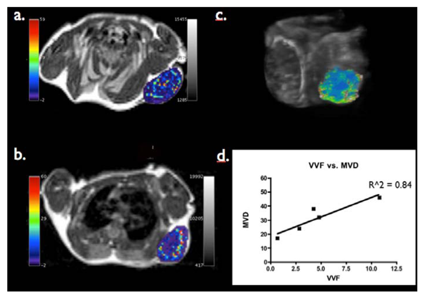
3.2. Can Steady State MRI Characterize Tumor Microvasculature?

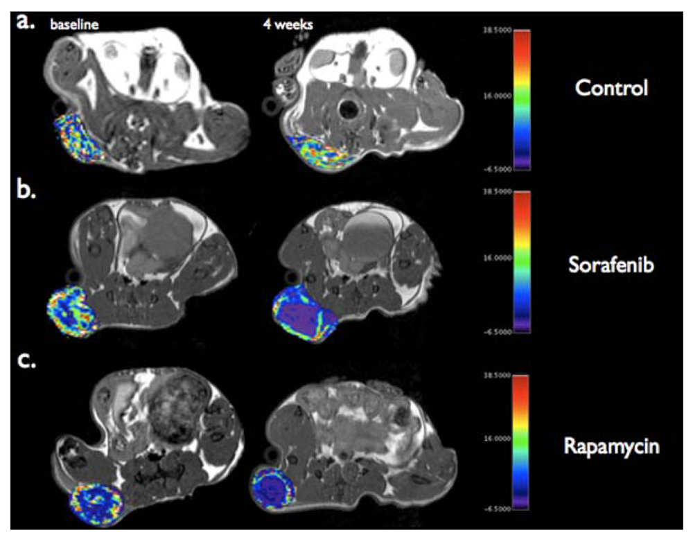
3.3. Can Steady State MRI Monitor Response to Anti-Angiogenic Therapy?

3.4. Can targeted Nanoparticles Bind to Markers of Angiogenesis?
4. Conclusions
References
- Folkman, J. Angiogenesis research: From laboratory to clinic. Forum (Genova) 1999, 9, 59–62. [Google Scholar]
- Bergers, G.; Benjamin, L.E. Tumorigenesis and the angiogenic switch. Nat. Rev. Cancer. 2003, 3, 401–410. [Google Scholar] [CrossRef]
- Hanahan, D.; Folkman, J. Patterns and emerging mechanisms of the angiogenic switch during tumorigenesis. Cell 1996, 86, 353–364. [Google Scholar] [CrossRef]
- Folkman, J. Role of angiogenesis in tumor growth and metastasis. Semin Oncol. 2002, 29, 15–18. [Google Scholar]
- Jain, R.K. Normalization of tumor vasculature: An emerging concept in antiangiogenic therapy. Science 2005, 307, 58–62. [Google Scholar] [CrossRef]
- Tong, R.T.; Boucher, Y.; Kozin, S.V.; Winkler, F.; Hicklin, D.J.; Jain, R.K. Vascular normalization by vascular endothelial growth factor receptor 2 blockade induces a pressure gradient across the vasculature and improves drug penetration in tumors. Cancer Res. 2004, 64, 3731–3736. [Google Scholar]
- Stohrer, M.; Boucher, Y.; Stangassinger, M.; Jain, R.K. Oncotic pressure in solid tumors is elevated. Cancer Res. 2000, 60, 4251–4255. [Google Scholar]
- Padera, T.P.; Stoll, B.R.; Tooredman, J.B.; Capen, D.; di Tomaso, E.; Jain, R.K. Pathology: Cancer cells compress intratumour vessels. Nature 2004, 427, 695. [Google Scholar]
- Helmlinger, G.; Yuan, F.; Dellian, M.; Jain, R.K. Interstitial pH and pO2 gradients in solid tumors in vivo: High-resolution measurements reveal a lack of correlation. Nat. Med. 1997, 3, 177–182. [Google Scholar] [CrossRef]
- Teicher, B.A. A systems approach to cancer therapy. (Antioncogenics + standard cytotoxics → mechanism(s) of interaction). Cancer Metastasis Rev. 1996, 15, 247–272. [Google Scholar] [CrossRef]
- Wouters, B.G.; Brown, J.M. Cells at intermediate oxygen levels can be more important than the “hypoxic fraction” in determining tumor response to fractionated radiotherapy. Radiat Res. 1997, 147, 541–550. [Google Scholar] [CrossRef]
- Jain, R.K. Delivery of novel therapeutic agents in tumors: physiological barriers and strategies. J Natl. Cancer Inst. 1989, 81, 570–576. [Google Scholar] [CrossRef]
- Jain, R.K.; Duda, D.G.; Clark, J.W.; Loeffler, J.S. Lessons from phase III clinical trials on anti-VEGF therapy for cancer. Nat. Clin. Pract. Oncol. 2006, 3, 24–40. [Google Scholar]
- Sandler, A.; Gray, R.; Perry, M.C.; Brahmer, J.; Schiller, J.H.; Dowlati, A.; Lilenbaum, R.; Johnson, D.H. Paclitaxel-carboplatin alone or with bevacizumab for non-small-cell lung cancer. N. Engl. J. Med. 2006, 355, 2542–2550. [Google Scholar] [CrossRef]
- Saltz, L.B.; Clarke, S.; Diaz-Rubio, E.; Scheithauer, W.; Figer, A.; Wong, R.; Koski, S.; Lichinitser, M.; Yang, T.S.; Rivera, F.; et al. Bevacizumab in combination with oxaliplatin-based chemotherapy as first-line therapy in metastatic colorectal cancer: a randomized phase III study. J. Clin. Oncol. 2008, 26, 2013–2019. [Google Scholar]
- Jain, R.K. Normalizing tumor vasculature with anti-angiogenic therapy: A new paradigm for combination therapy. Nat. Med. 2001, 7, 987–989. [Google Scholar] [CrossRef]
- Takei, H.; Bhattacharjee, M.B.; Rivera, A.; Dancer, Y.; Powell, S.Z. New immunohistochemical markers in the evaluation of central nervous system tumors: A review of 7 selected adult and pediatric brain tumors. Arch. Pathol. Lab. Med. 2007, 131, 234–241. [Google Scholar]
- Carmeliet, P.; Jain, R.K. Angiogenesis in cancer and other diseases. Nature 2000, 407, 249–257. [Google Scholar] [CrossRef]
- Brown, E.B.; Campbell, R.B.; Tsuzuki, Y.; Xu, L.; Carmeliet, P.; Fukumura, D.; Jain, R.K. In vivo measurement of gene expression, angiogenesis and physiological function in tumors using multiphoton laser scanning microscopy. Nat. Med. 2001, 7, 864–868. [Google Scholar]
- Corot, C.; Robert, P.; Idee, J.M.; Port, M. Recent advances in iron oxide nanocrystal technology for medical imaging. Adv. Drug Deliv. Rev. 2006, 58, 1471–1504. [Google Scholar] [CrossRef]
- Enochs, W.S.; Harsh, G.; Hochberg, F.; Weissleder, R. Improved delineation of human brain tumors on MR images using a long-circulating, superparamagnetic iron oxide agent. J. Magn. Reson. Imaging. 1999, 9, 228–232. [Google Scholar] [CrossRef]
- Persigehl, T.; Bieker, R.; Matuszewski, L.; Wall, A.; Kessler, T.; Kooijman, H.; Meier, N.; Ebert, W.; Berdel, W.E.; Heindel, W.; et al. Antiangiogenic tumor treatment: early noninvasive monitoring with USPIO-enhanced MR imaging in mice. Radiology 2007, 244, 449–456. [Google Scholar] [CrossRef]
- Tropres, I.; Grimault, S.; Vaeth, A.; Grillon, E.; Julien, C.; Payen, J.F.; Lamalle, L.; Decorps, M. Vessel size imaging. Magn. Reson. Med. 2001, 45, 397–408. [Google Scholar] [CrossRef]
- Brown, J.M.; Giaccia, A.J. The unique physiology of solid tumors: opportunities (and problems) for cancer therapy. Cancer Res. 1998, 58, 1408–1416. [Google Scholar]
- Bremer, C.; Mustafa, M.; Bogdanov, A., Jr.; Ntziachristos, V.; Petrovsky, A.; Weissleder, R. Steady-state blood volume measurements in experimental tumors with different angiogenic burdens a study in mice. Radiology 2003, 226, 214–220. [Google Scholar] [CrossRef]
- Boxerman, J.L.; Hamberg, L.M.; Rosen, B.R.; Weisskoff, R.M. MR contrast due to intravascular magnetic susceptibility perturbations. Magn. Reson. Med. 1995, 34, 555–566. [Google Scholar] [CrossRef]
- Dennie, J.; Mandeville, J.B.; Boxerman, J.L.; Packard, S.D.; Rosen, B.R.; Weisskoff, R.M. NMR imaging of changes in vascular morphology due to tumor angiogenesis. Magn. Reson. Med. 1998, 40, 793–799. [Google Scholar] [CrossRef]
- Hyodo, F.; Chandramouli, G.V.; Matsumoto, S.; Matsumoto, K.; Mitchell, J.B.; Krishna, M.C.; Munasinghe, J.P. Estimation of tumor microvessel density by MRI using a blood pool contrast agent. Int. J. Oncol. 2009, 35, 797–804. [Google Scholar]
- Gambarota, G.; Leenders, W.; Maass, C.; Wesseling, P.; van der Kogel, B.; van Tellingen, O.; Heerschap, A. Characterisation of tumour vasculature in mouse brain by USPIO contrast-enhanced MRI. Br. J. Cancer. 2008, 98, 1784–1789. [Google Scholar] [CrossRef]
- Pathak, A.P.; Rand, S.D.; Schmainda, K.M. The effect of brain tumor angiogenesis on the in vivo relationship between the gradient-echo relaxation rate change (DeltaR2*) and contrast agent (MION) dose. J. Magn. Reson. Imaging. 2003, 18, 397–403. [Google Scholar] [CrossRef]
- Kiselev, V.G.; Strecker, R.; Ziyeh, S.; Speck, O.; Hennig, J. Vessel size imaging in humans. Magn. Reson. Med. 2005, 53, 553–563. [Google Scholar] [CrossRef]
- Lemasson, B.; Christen, T.; Tizon, X.; Farion, R.; Fondraz, N.; Provent, P.; Segebarth, C.; Barbier, E.L.; Genne, P.; Duchamp, O.; et al. Assessment of multiparametric MRI in a human glioma model to monitor cytotoxic and anti-angiogenic drug effects. NMR Biomed. 2011, 24, 473–482. [Google Scholar] [CrossRef]
- Valable, S.; Lemasson, B.; Farion, R.; Beaumont, M.; Segebarth, C.; Remy, C.; Barbier, E.L. Assessment of blood volume, vessel size, and the expression of angiogenic factors in two rat glioma models: A longitudinal in vivo and ex vivo study. NMR Biomed. 2008, 21, 1043–1056. [Google Scholar] [CrossRef]
- Beaumont, M.; Lemasson, B.; Farion, R.; Segebarth, C.; Remy, C.; Barbier, E.L. Characterization of tumor angiogenesis in rat brain using iron-based vessel size index MRI in combination with gadolinium-based dynamic contrast-enhanced MRI. J. Cereb. Blood Flow Metab. 2009, 29, 1714–1726. [Google Scholar]
- Guimaraes, A.R.; Ross, R.; Figuereido, J.L.; Waterman, P.; Weissleder, R. MRI with magnetic nanoparticles monitors downstream anti-angiogenic effects of mTOR inhibition. Mol. Imaging Biol. 2011, 13, 314–320. [Google Scholar] [CrossRef]
- Guimaraes, A.R.; Rakhlin, E.; Weissleder, R.; Thayer, S.P. Magnetic resonance imaging monitors physiological changes with antihedgehog therapy in pancreatic adenocarcinoma xenograft model. Pancreas 2008, 37, 440–444. [Google Scholar] [CrossRef]
- Persigehl, T.; Matuszewski, L.; Kessler, T.; Wall, A.; Meier, N.; Ebert, W.; Berdel, W.E.; Heindel, W.; Mesters, R.; Bremer, C. Prediction of antiangiogenic treatment efficacy by iron oxide enhanced parametric magnetic resonance imaging. Invest. Radiol. 2007, 42, 791–796. [Google Scholar] [CrossRef]
- Ring, J.; Persigehl, T.; Remmele, S.; Heindel, W.; Dahnke, H.; Bremer, C. Monitoring of bevacizumab-induced antiangiogenic treatment effects by “steady state” ultrasmall superparamagnetic iron oxide particles magnetic resonance imaging using robust multiecho DeltaR2* relaxometry. Invest Radiol. 2011, 46, 326–330. [Google Scholar]
- Montet, X.; Montet-Abou, K.; Reynolds, F.; Weissleder, R.; Josephson, L. Nanoparticle imaging of integrins on tumor cells. Neoplasia 2006, 8, 214–222. [Google Scholar] [CrossRef]
- Zhang, C.; Jugold, M.; Woenne, E.C.; Lammers, T.; Morgenstern, B.; Mueller, M.M.; Zentgraf, H.; Bock, M.; Eisenhut, M.; Semmler, W.; et al. Specific targeting of tumor angiogenesis by RGD-conjugated ultrasmall superparamagnetic iron oxide particles using a clinical 1.5-T magnetic resonance scanner. Cancer Res. 2007, 67, 1555–1562. [Google Scholar]
- Boles, K.S.; Schmieder, A.H.; Koch, A.W.; Carano, R.A.; Wu, Y.; Caruthers, S.D.; Tong, R.K.; Stawicki, S.; Hu, G.; Scott, M.J.; et al. MR angiogenesis imaging with Robo4- vs. alphaVbeta3-targeted nanoparticles in a B16/F10 mouse melanoma model. Faseb J. 2010, 24, 4262–4270. [Google Scholar]
- Schmieder, A.H.; Caruthers, S.D.; Zhang, H.; Williams, T.A.; Robertson, J.D.; Wickline, S.A.; Lanza, G.M. Three-dimensional MR mapping of angiogenesis with α5β1(ανβ3)-targeted theranostic nanoparticles in the MDA-MB-435 xenograft mouse model. Faseb J. 2008, 22, 4179–4189. [Google Scholar] [CrossRef]
© 2012 by the authors; licensee MDPI, Basel, Switzerland. This article is an open-access article distributed under the terms and conditions of the Creative Commons Attribution license (http://creativecommons.org/licenses/by/3.0/).
Share and Cite
McDermott, S.; Guimaraes, A.R. Magnetic Nanoparticles in the Imaging of Tumor Angiogenesis. Appl. Sci. 2012, 2, 525-534. https://doi.org/10.3390/app2020525
McDermott S, Guimaraes AR. Magnetic Nanoparticles in the Imaging of Tumor Angiogenesis. Applied Sciences. 2012; 2(2):525-534. https://doi.org/10.3390/app2020525
Chicago/Turabian StyleMcDermott, Shaunagh, and Alexander R. Guimaraes. 2012. "Magnetic Nanoparticles in the Imaging of Tumor Angiogenesis" Applied Sciences 2, no. 2: 525-534. https://doi.org/10.3390/app2020525