1. Introduction
Research in synthetic control of noble metal nanoparticles has proven to be a powerful and versatile approach for tailoring the properties of the metal nanostructures for various applications [
1,
2,
3]. Silver nanoparticles have been prepared with various techniques to modify their optical, electrical, and catalytic properties, which are strongly dependent on the shape and size of the silver nanoparticles. Much effort from many research groups has examined the synthesis of silver colloidal solutions with suspensions of various shapes and sizes including silver nanospheres [
4,
5], nanowires [
6,
7], nanoplates [
8,
9], nanocubes [
10], and nanorods [
11]. Among these nanostructures, silver nanoplates have attracted considerable attention because they could potentially generate maximum electromagnetic field enhancement due to their highly anisotropic structure [
12,
13,
14]. As a result, these particles are ideal for implementation in sensing applications including localized surface plasmon resonance (LSPR) [
15,
16,
17,
18], surface-enhanced fluorescence [
19], and surface-enhanced Raman scattering (SERS) [
20,
21,
22], which are possibly the most attractive applications.
In contrast, solution-based synthesis of anisotropic nanostructures has not been suitably applied for device applications including sensing analysis. Generally, the sensing analysis has been done by directly mixing the silver colloids with the analyte solution [
23]. Therefore, silver is considered to be a single use system as opposed to multiple uses for a silver thin film [
24]. The silver thin film can easily be washed to remove the analyte from the surface to allow a second or potentially repeated analysis and this result alternatively helps to prove the repeatability of the sensing system. Additionally, understanding the growth of the silver thin films is crucial in the fabrication of devices including solar cells and light emitting diodes (LEDs) because the growth on solid surfaces is influenced by the specific size, shape, and distribution of the nanostructures being deposited [
25,
26].
The growth of anisotropic nanostructures on solid surfaces has been reported using various methods such as thermal growth, seed-mediated growth, self-assembly, and lithography [
27]. Recently, interest in surface modification in thin film fabrication has increased because control of the degree of coverage and aggregation of the nanostructures is possible and will allow the construction of a SERS substrate. A variety of organic materials can self-assemble on solid surfaces to obtain a molecular layer on the surface which promote the attachment of nanostructures to surfaces [
28,
29]. 3-Aminopropyltrimethoxysilane (APTMS) is a common aminosilane used to promote metal nanostructure adhesion on the surface due to the strong interactions between the amine group and the metal nanoparticles [
30,
31].
Hence, the aim of this work is to deposit silver nanoplates on a silicon surface with maximum surface coverage of the triangular nanoplates using self-assembly with an adhesion layer. The distribution of the triangular silver nanoplates was studied by varying the exposure time of the silver nanoplate solution to the silicon surface. The silver nanoplate thin film was attached to the silicon surface using an APTMS film as a coupling agent in order to enhance the adhesion between the surface and the nanoplates. The morphology of the samples was investigated using field emission scanning electron microscopy (FESEM).
2. Experimental Section
2.1. Chemicals
The chemicals used to prepare the triangular silver nanoplate suspension include silver nitrate (Sigma Aldrich, Kuala Lumpur, Malaysia), trisodium citrate (Wako Pure Chemical Industries, Ltd.: Osaka, Japan), 30% hydrogen peroxide (HmbG Chemicals), and sodium borohydride (Sigma Aldrich). Meanwhile, 3-aminopropyltrimethoxysilane 97% (Sigma Aldrich) and ethanol absolute (HmbG Chemicals, Kuala Lumpur, Malaysia) were used to functionalize the silicon surface. These chemicals were utilized as received without any further purification.
2.2. Synthesis and Fabrication of Monolayer Triangular Silver Nanoplate Thin Film
The triangular silver nanoplate solution was prepared via a direct chemical reduction approach at ambient temperature in air. A typical synthesis process is described in the following: 0.2 mL of 0.05 M silver nitrate (AgNO3) was mixed into 96.56 mL of deionized water. Then, the solution was vigorously stirred by a magnetic stirrer at 900 rpm. After that, 2.0 mL of 75 mM trisodium citrate was mixed into the solution followed by 0.24 mL of 30% hydrogen peroxide (H2O2). To start a reduction process of Ag ions to Ag metal, 0.1 M sodium borohydride (NaBH4) was injected quickly into the stirred solution. The ultimate stable formation of triangular silver nanoplates in solution can be roughly seen through the color change of the solution. A clear, colorless solution changed to a dark blue solution after vigorously stirring for 5 min. This solution was then subjected to ultrasonic treatment for 1 h at 27 °C. The output power of ultrasonic bath is about 160 Watts. Afterward, the solution was centrifuged at 6000 rpm for 30 min. A pellet eventually formed at the bottom of centrifuge tube and the pellet was extracted to make the solutions used in the deposition of triangular nanoplate films on the silicon surface.
The monolayer triangular silver nanoplate film was grown on the silicon surface by a self-assembly technique. This approach was typically started by cleaning a silicon substrate via immersing in piranha solution for 1 h to remove any organic waste on the silicon surface. Next, the substrate was cleaned in acetone and then in ethanol with ultrasonic treatment for 15 min. After that, the clean silicon was immersed in 5% 3-aminopropyltrimethoxysilane (APTMS) in ethanol for 1 h at room temperature to functionalize the silicon surface. The APTMS film on silicon substrate was then rinsed with copious amounts of ethanol and the surface was dried. Finally, this APTMS film was heated on a hot plate for 2 h at 100 °C. The deposition of the triangular nanoplates on the silicon surface was done by immersing the APTMS-covered silicon into a silver nanoplate solution formed by redissolving the centrifuged pellet in water.
To study changes in surface coverage of the triangular nanoplates on the surface, the exposure time of the APTMS-covered silicon to the nanoplate suspensions was varied from 15 min to 22 h. The films were then characterized using atomic force microscopy, AFM (Veeco, Riverside, CA, USA), and field emission scanning electron microscopy, FESEM (Hitachi, Kuala Lumpur, Malaysia), to determine the morphology and coverage of the monolayer triangular silver nanoplate thin films. Meanwhile, transmission electron microscopy, TEM (Philip, New South Wales, Australia), optical absorbance (Cary 5G spectrophotometer; Varian, Melbourne, Australia), and dynamic light scattering, DLS (HPLS Maples, Adelaide, Australia), were recorded for the silver nanoplate solution to study the shape and size of the triangular nanoplates. The SERS measurement was recorded using a Raman spectrometer (Horiba Scientific, New South Wales, Australia) with a 532 nm excitation laser wavelength with 10 seconds integration time.
3. Results and Discussion
Triangular silver nanoplates were successfully prepared by direct chemical reduction at room temperature as reported by Zhang [
32]. The formation of the nanoplates was simply obtained by the reduction of silver ion to silver from silver nitrate by a reduction agent, namely sodium borohydride. This shape formation was effectively controlled by trisodium citrate and hydrogen peroxide. The triangular nanoplate formation could be roughly observed via color change when sodium borohydride was injected into the colorless solution which contained silver nitrate, trisodium citrate, and hydrogen peroxide. The solution color changed to yellowish after 1 min during the reduction process. The color continued to change for five minutes until the color remained dark blue. The centrifuged silver nanoplate pellet was resuspended and then was characterized to study the optical properties, uniformity of shape, and particle size.
Figure 1.
Optical absorbance with corresponding mode of plasmon excitation of resuspended triangular silver nanoplate solution.
Figure 1.
Optical absorbance with corresponding mode of plasmon excitation of resuspended triangular silver nanoplate solution.
The optical properties of the silver nanoplates were studied using the surface plasmon resonance of the particles, which can be probed using the absorbance spectrum. This resonance has a strong dependence on the shape of particles [
33]. The resuspended silver nanoplate suspension was characterized by UV-Vis and the optical absorbance spectrum is shown in
Figure 1. Based on the absorbance spectrum, it was found that the triangular silver nanoplates exhibited a sharp plasmon band at 333 nm and an intense broad surface plasmon situated at 660 nm along with a shoulder at around 500 nm.
In
Figure 1, the corresponding modes of plasmon excitation for each extinction peak of the solution are also provided. The three characteristic peaks correspond to different modes of plasmon excitation of triangular nanoplates [
34]. The two dominant bands located at 333 nm and 660 nm are due to the out-of-plane quadrupole resonance and in-plane dipole resonance, respectively. The shoulder at 500 nm is a result of the formation of anisotropic nanoparticles and it is attributed to the in-plane quadrupole resonance of silver nanoplates. These three modes of plasmon excitation agree with the structure of the triangular nanoplate shown in the observed TEM images (
Figure 2a).
Figure 2.
Triangular silver nanoplate solution (a) TEM image; the inset picture shows the lattice spacing of triangular nanoplates and (b) size distribution from dynamic light scattering (DLS) measurements.
Figure 2.
Triangular silver nanoplate solution (a) TEM image; the inset picture shows the lattice spacing of triangular nanoplates and (b) size distribution from dynamic light scattering (DLS) measurements.
The resuspended triangular silver nanoplate solution was dropped on a cuprum grid surface for TEM characterization. The solution was first dried at room temperature for 10 min prior to imaging.
Figure 2a shows an image of triangular nanoplates that was captured with 120 kV. However, it is difficult to observe the triangular shape because the nanoplates have a tendency to stack face-to-face via self-assembly during the evaporation of the solvent in preparing the TEM sample. The uniform thickness of the plates can be seen from this stacking formation and this allowed determination of the plate size. This size measurement was analyzed using iTEM software (Adelaide, Australia), and the average plate thickness is 7 ± 2 nm while the average edge length of triangular nanoplates is 26 ± 1 nm. The inset figure in
Figure 2a shows the captured lattice of nanoplates when imaged by TEM. The perfect lattice was measured at 0.48 nm, which corresponds to the lattice spacing of face-centered cubic (FCC) silver crystal. The growth direction of nanoplates corresponding to this measured lattice spacing is likely to be the <111> direction based on the X-ray diffraction (XRD) Bruker XRD D8 Advance; (Selangor, Malaysia) characterization [
35]. Given this is the most stable FCC crystal plane, this is the growth direction that would be expected.
The average size of triangular nanoplates was also confirmed using dynamic light scattering (DLS) as plotted in
Figure 2b. This bar chart for the size of the nanoplate particles
vs. the number of measurements for each size was determined from DLS measurements by determining the frequency of occurrence of each particle size (based on volume) over 27 scans of the same colloidal silver nanoplate suspension. It was found that the size of the nanoplates varied. This is due to the fact that DLS measured two length parameters: thickness and edge length of the triangular nanoplate structure. The average edge length of nanoplates was determined by DLS to be 25 ± 3 nm, while the average plate thickness was found to be 9 ± 1 nm. These measurements agreed closely with TEM results.
Triangular silver nanoplate thin films on a silicon surface were grown via a self-assembly technique. Prior to exposing the nanoplate solution to the surface, the clean silicon surface was functionalized by immersing the surface in 5% 3-aminopropyltrimethoxysilane (APTMS) solution for 1 h. The APTMS film formed on surface was used to promote adhesion between the substrate and the silver nanoplates. APTMS has been used as a good coupling agent for the modification of silica surfaces to attach silver triangular nanoplates due to the robust interaction between the amine group and metal nanoparticles as reported by Howarter [
36]. As a result, a silver nanoplate film was successfully deposited on the silicon surface after 15 min of exposure time, as shown in the FESEM image in
Figure 3a.
Figure 3.
(a) FESEM image of nanoplate film on silicon surface after 15 min of exposure; (b) Bar chart for edge length of triangular nanoplates for 15 min growth on surface. The edge length of 25 means the size of nanoplates was from 20 to 30 nm, 35 from 30 to 40 nm, and 45 from 40 to 50 nm.
Figure 3.
(a) FESEM image of nanoplate film on silicon surface after 15 min of exposure; (b) Bar chart for edge length of triangular nanoplates for 15 min growth on surface. The edge length of 25 means the size of nanoplates was from 20 to 30 nm, 35 from 30 to 40 nm, and 45 from 40 to 50 nm.
The FESEM image clearly proves that the film of silver nanoplates was starting to grow on the surface after just 15 min of exposure to the nanoplate solution, with a high yield of triangular shapes due to the presence of the silane molecule. It should be noted that both AFM and FESEM imaging of the untreated and treated silicon substrates were featureless (please refer to
supplementary material). Additionally, similar imaging showed that exposure of the nanoplate solution to an untreated substrate lead to no deposition. Hence, the silane molecule on the surface did assist the attachment of the silver nanoplates because there is no distribution of nanoplates on the surface without the APTMS layer. The silver nanoparticles were electrostatically attached to aminosilane molecules due to the silver in the solution interacting with the APTMS-covered silicon surface [
37,
38]. The image also reveals that the triangular nanoplates are well attached to the silicon surface and the nanoplates are not agglomerated. The average edge length of triangular nanoplates is 25.05 ± 7.37 nm, which was calculated using image J software. The different edge lengths of triangular nanoplates determined through SEM images are plotted in
Figure 3b. The results found that the edge length of 25 nm was the most common size observed on the surface and agreed with TEM and DLS analysis.
The growth of the nanoplate film on the surface was studied for different exposure times. Freshly silanized surfaces were immersed for 30 min, 1 h, 2 h, 8 h, 14 h, and 22 h in the nanoplate solution.
Figure 4 shows the FESEM images of the nanoplate thin film on the surface after various exposure times. It was found that the density of nanoplates on the surface increased with increasing exposure time. From these coverage examples, it is clear that the attachment of APTMS on the surface ultimately enhanced the growth of nanoplates on the surface. The triangular nanoplate film grew with high affinity for the all exposure times. Thus, nanoplate films were successfully formed on the surface with the nanoplates unlikely to overlap with each other and thus be well organized in a monolayer on the silicon surface.
Figure 4.
FESEM images of triangular silver nanoplate thin films formed after varying exposure times of (a) 30 min, (b) 1 h, (c) 2 h, (d) 8 h, (e) 14 h, and (f) 22 h. Scale bar is 100 nm.
Figure 4.
FESEM images of triangular silver nanoplate thin films formed after varying exposure times of (a) 30 min, (b) 1 h, (c) 2 h, (d) 8 h, (e) 14 h, and (f) 22 h. Scale bar is 100 nm.
Silver nanoparticles with spherical and irregular shapes did start to appear on the surface after 8 h of immersion in the nanoplate solution. The irregular shapes were readily observed in 14 h and 22 h exposure time samples. It should be noted that for these samples, the distribution of triangular nanoplate shapes on the surface is still higher than the distribution of irregular shapes on surface. The formation of irregular shapes with increasing exposure times is likely related to the increasing coverage of nanoplates on the surface. This increased density means that it is possible that the nanoplates begin to coalesce when the neighboring particles get close to each other [
39]. From the measurement of triangular nanoplate sizes, we calculated that the ratio of irregular to triangular shapes is 20:80 for both samples.
Figure 5.
The edge length of triangular nanoplates on the surface for different exposure times. The edge length of 25 means the size of the nanoplates was from 20 to 30 nm, 35 from 30 to 40 nm, and 45 from 40 to 50 nm.
Figure 5.
The edge length of triangular nanoplates on the surface for different exposure times. The edge length of 25 means the size of the nanoplates was from 20 to 30 nm, 35 from 30 to 40 nm, and 45 from 40 to 50 nm.
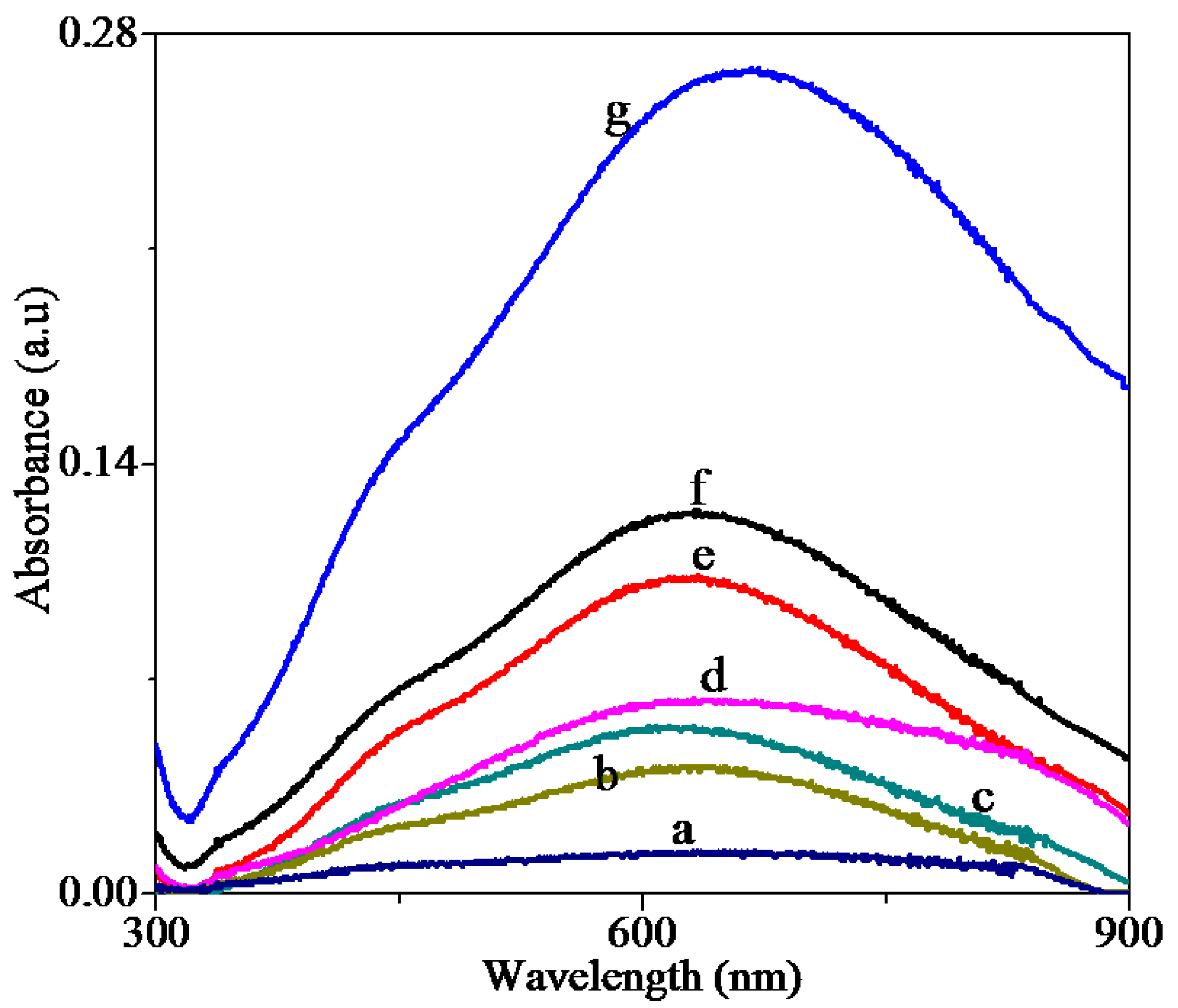
Figure 6.
Optical absorbance for silver nanoplate films formed after the various exposure times of (a) 15 min, (b) 30 min, (c) 1 h, (d) 2 h, (e) 8 h, (f) 14 h, and (g) 22 h.
Figure 6.
Optical absorbance for silver nanoplate films formed after the various exposure times of (a) 15 min, (b) 30 min, (c) 1 h, (d) 2 h, (e) 8 h, (f) 14 h, and (g) 22 h.
The size of the particles on the surface was analyzed using the same approach as the 15 min sample and the analysis results are plotted in
Figure 5 and given in
Table 1. Importantly, the average edge length of triangular nanoplates for different exposure times did not change. However, the count of triangular nanoplates increased with exposure time, showing the density of nanoplates on surface increases with exposure time.
The optical properties of the silver nanoplate films formed after different exposure times were studied by observing their absorbance spectra, which are presented in
Figure 6. In the suspension there were three clear peaks at 330 nm, 550 nm, and 660 nm (see
Figure 1). The signal from the substrate-bound nanoplate films is much weaker, as expected, as the optical characterization was done on the thin silver nanoplate films instead of the concentrated nanoplates in the suspension. However, for the sample with the highest coverage (produced after 22 h of substrate exposure), a broad surface plasmon at 660 nm was observed along with two shoulders at 330 nm and 550 nm. These peaks align closely with the spectrum of the suspension, further confirming the presence of the nanoplates on the substrate. Additionally, the intensity of the absorbance increased with increasing exposure time due to the increasing coverage of the nanoplates, as revealed by the SEM images.
Table 1.
The measurement of the edge length and the coverage of triangular nanoplates on the surface for 15 min to 22 h samples of exposure times.
Table 1.
The measurement of the edge length and the coverage of triangular nanoplates on the surface for 15 min to 22 h samples of exposure times.
| Exposure Times | Edge Length of Nanoplates (nm) | Coverage of Nanoplates on the Surfaces (Counts/µm) |
|---|
| 15 min | 25.05 ± 7.37 | 79 |
| 30 min | 25.59 ± 9.72 | 103 |
| 1 h | 26.21 ± 8.40 | 248 |
| 2 h | 24.37 ± 7.37 | 202 |
| 8 h | 27.47 ± 6.68 | 534 |
| 14 h | 25.24 ± 7.49 | 423 |
| 22 h | 25.11 ± 7.68 | 503 |
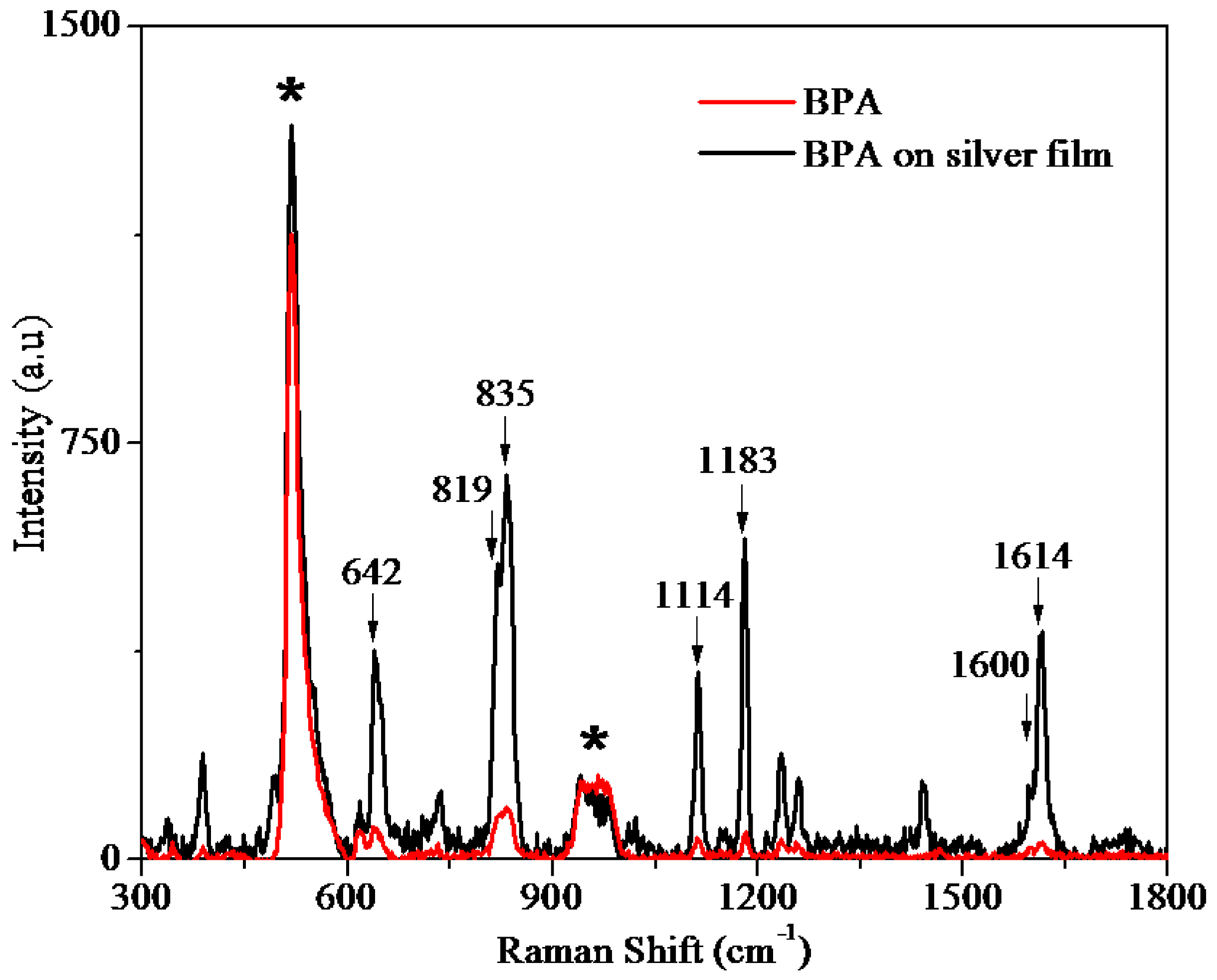
Figure 7.
The SERS spectrum of BPA on the silver nanoplate film (formed after 2 h exposure time) and Raman of BPA absorbed on a clean silicon surface. * are labels for the silicon peaks.
Figure 7.
The SERS spectrum of BPA on the silver nanoplate film (formed after 2 h exposure time) and Raman of BPA absorbed on a clean silicon surface. * are labels for the silicon peaks.
Figure 7 shows the SERS spectrum of 1 mM bisphenol A (BPA) on the silver nanoplate film formed after exposing the substrate to the nanoplate suspension for 2 h. The normal Raman spectrum for 1 mM BPA on the silicon surface is also provided in
Figure 7. As expected, BPA shows seven major Raman peaks at 642 cm
−1, 819 cm
−1, 835 cm
−1, 1114 cm
−1, 1183 cm
−1, 1600 cm
−1, and 1614 cm
−1, clearly indicating the presence of BPA on the substrate [
40]. These BPA peaks were strongly enhanced when the molecule was absorbed on the silver nanoplate film, clearly demonstrating that these nanoplate films are a good SERS substrate and suitable for further investigations for incorporation in a SERS-based sensor.


