Review

Toward Long-Term Implantable Glucose Biosensors for Clinical Use

1
Department of Mechanical Engineering, College of Engineering, Kyung Hee University, Gyeonggi-do 17104, Korea
2
LG Electronics, Seoul 06763, Korea
*
Author to whom correspondence should be addressed.
Appl. Sci. 2019, 9(10), 2158; https://doi.org/10.3390/app9102158
Submission received: 1 April 2019 / Revised: 30 April 2019 / Accepted: 9 May 2019 / Published: 27 May 2019
(This article belongs to the Special Issue Biomaterials and Biofabrication)

Abstract

:
Continuous glucose monitoring (CGM) sensors have led a paradigm shift to painless, continuous, zero-finger pricking measurement in blood glucose monitoring. Recent electrochemical CGM sensors have reached two-week lifespans and no calibration with clinically acceptable accuracy. The system with the recent CGM sensors is identified as an “integrated glucose monitoring system,” which can replace finger-pricking glucose-testing for diabetes treatment decisions. Although such innovation has brought CGM technology closer to realizing the artificial pancreas, discomfort and infection problems have arisen from short lifespans and open wounds. A fully implantable sensor with a longer-term lifespan (90 days) is considered as an alternative CGM sensor with high comfort and low running cost. However, it still has barriers, including surgery for applying and replacing and frequent calibration. If technical refinement is conducted (e.g., stability and reproducibility of sensor fabrication), fully implantable, long-term CGM sensors can open the new era of continuous glucose monitoring.

1. Introduction

Diabetes mellitus is a chronic disease that affects more than 400 million people worldwide [1]. Since high or low blood glucose concentration episodes and high glycemic variability can cause diabetic complications (e.g., diabetic retinopathy, kidney failure, heart disease, diabetic neuropathy, and diabetic foot disease) and even lead to death, it is crucial for diabetic patients to maintain their blood glucose concentration in a normal range and to decrease glycemic variability [2,3,4,5]. To maintain blood glucose concentration in the normal level and reduce blood glucose variability, people with diabetes must first understand their blood glucose concentrations. Blood glucose concentration measurement is accompanied with painful finger-pricking. Such intermittent monitoring is not only painful, but also ineffective to prevent abnormal blood glucose concentrations between measurement points [6]. Alternately, continuous glucose monitoring (CGM) is proposed. CGM can constantly measure blood glucose concentrations and give users alarm when abnormal blood glucose concentration occurs (Figure 1). Thus, CGM enables to reduce occurrence of hypo- or hyperglycemia and high glycemic variability [7,8]. An implantable sensor is a key component of continuous glucose monitoring system (CGMS). The implantable sensor decides comfort or pain during wearing CGMS, accuracy, and lifespan of CGMS. Since Minimed released the first CGMS in 1999, CGMS technologies have been innovated to open the new era of glucose monitoring: Zero-finger-pricking, continuous glucose monitoring [9,10,11,12]. In this review, we emphasize commercial CGMS technologies including sensor requirements and the state of the art. We also introduce regulation change in CGMS to help researchers develop commercial-quality CGMSs.

2. Materials and Methods

2.1. Subcutaneous Continuous Glucose Monitoring

An implantable sensor is a key component of a CGMS. Commercial CGMSs employ subcutaneous sensors that are implanted under the skin and continuously monitor subcutaneous interstitial fluid (ISF) glucose. Despite of the invasive concept of subcutaneous sensors, subcutaneous sensors have gained attention, mainly due to high reliability and easy accessibility. Subcutaneous ISF glucose is a well-known alternative sample to blood glucose [13,14]. Subcutaneous ISF glucose shows good correlation with time lag of 5–30 min [15]. This indirect sensing inevitably needs in vivo calibration. Most of CGM sensors are calibrated against “reference blood glucose reading” using home glucose monitoring devices. Frequent calibration can guarantee higher accuracy, but increase pain and discomfort. Recently, some CGM sensors have reached zero in vivo calibration [16,17,18]. Stable and reproducible sensor-production has revolutionize diabetes therapy from in vivo calibration to in vitro factory calibration, “zero finger pricking.” Details about calibration are described in Section 2.3.

2.2. Accuracy

The U.S. Food and Drug Administration (FDA) identified that CGMS were aids in the detection of episodes of hyperglycemia and hypoglycemia, not replacing standard home glucose monitoring devices. In the hearing of the FDA in 2016, the panel gave positive votes to the Dexcom G5® integrated continuous glucose monitoring system for use of treatment decision [19]. Such paradigm shift to clinical use is based on the accuracy of CGM sensors. The accuracy of subcutaneous CGM sensors depends on calibration and degree of biocompatibility. Since subcutaneous sensors do not measure blood glucose, but instead measure glucose in subcutaneous interstitial fluid (ISF), signals of subcutaneous sensors have to be calculated to blood glucose concentrations. Recent “zero finger-pricking” CGM sensors are realized due to a new sensor manufacturing including factory calibration. The factory calibration removes the need of user calibration and associated painful, finger-pricking blood glucose measurement. This is possible because of the reproducibility and stability in sensor production. Such factory calibration sensors can increase accuracy with lab-based calibration [20]. The fully-implantable sensor, Eversense® (Senseonics), still needs calibration twice per day to maintain 9% mean absolute relative difference (MARD) [21]. To apply subcutaneous CGM sensors into a body, subcutaneous sensors require skin incision, which makes immune responses inevitable. Thereby, subcutaneous sensors require high biocompatibility to guarantee sensor functionality and safety in vivo. Further details of calibration and biocompatibility will be discussed in the following sections. In this section, we describe the CGMS evaluation method and performance requirement of iCGM (integrated CGM).

2.2.1. Evaluation

Accuracy of systems for blood glucose monitoring has been widely evaluated based on the Clarke’s error grid analysis. The Clarke’s error grid analysis analyzes clinical implication of blood glucose monitoring system by comparing between system-generated glucose values and the reference blood glucose values. The grid is divided into five zones that explains accuracy of glucose sensors. Zone A indicates that sensor reading deviates from the reference blood glucose by less than 20% or sensor reading shows hypoglycemia (<70 mg/dL) when the reference is also in the hypoglycemia. If the estimated values of blood glucose fall into zone A, the value means clinically correct. Zone B explains that sensor reading deviate from the reference with benign error or no treatment. Zone C means that the determined values lead to overcorrect acceptable blood glucose concentrations. Such values would result the actual blood glucose concentration into hypoglycemia or hyperglycemia. Zone D represents “dangerous failure to detect and treat” errors. The determined values fall into the target range whereas the actual blood glucose is not in the target range. Zone E is an “erroneous treatment” zone. In zone E, the determined values would lead opposite results to the reference values. In this conventional analysis, the determined blood glucose values in zones A and B are clinically acceptable (Figure 2) [22].
A new evaluation method for CGMS is needed as research and industrial efforts on the development of CGMS increase. Since CGMS provides the time sequence of blood glucose data, error grid analysis has to reflect the temporal characteristics of blood glucose. Clarke et al. introduced a new concept of “rate-error grid analysis (R-EGA)” and a new “point-error grid analysis (P-EGA)” that is modified from the traditional the Clarke’s error grid analysis. In R-EGA, estimated rate (rate of change of sensor blood glucose (SBG)) is plotted against reference rate (rate of change of reference blood glucose (RBG)). R-EGA is divided into five zones: AR, BR, CR, DR, and ER. Clinical meaning of each error zone is similar to the conventional Clarke’s error grid analysis. Zone AR means that SBG rate perfectly fit to RBG rate or has errors within ±1 mg·dL−1·min−1. Zone AR is expanded to error within ±2 mg·dL−1 min−1 at extreme RBG rates of ±4 mg·dL−1·min−1. Zone BR indicates benign errors; they do not lead clinically-wrong decision or negative outcome. Zone CR causes overcorrection. Zone DR fails to detect significant change of RBG. In zone ER, SBG rate is opposite to RBG rate. In P-EGA, RBG is plotted against SBG. P-EGA is also divided into five zones: AP, BP, CP, DP, and EP. In this analysis, rate of blood glucose is also considered and the zones are expanded according to the rates of blood glucose. When RBG rate is within ±1 mg·dL−1 min−1, P-EGA zones are the same with the zones of the traditional EGA. In the case of RBG rate of −2 to −1 mg·dL−1 min−1, the upper limits of upper AP, BP, and DP zones are added by 10 mg·dL−1 compared to the traditional EGA. If RBG rate is faster than −2 mg·dL−1·min−1, the upper limits of upper AP, BP, and DP zones are expanded by 20 mg·dL−1 compared to the traditional EGA. With RBG rate if 1–2 mg· dL−1·min−1, the lower limits of lower AP, BP, and DP zones are added by 10 mg·dL−1. If RBG rate is faster than 2 mg·dL−1·min−1, the lower limits of lower AP, BP, and DP zones are added by 20 mg·dL−1. By combining R-EGA and P-EGA, sensor readings of CGMS can be estimated according to the error grid matrix [23].

2.2.2. Performance Requirement of iCGM

The FDA classifies the Dexcom G6 Continuous Monitoring System and equivalent devices into Class II and names the devices “Integrated continuous glucose monitoring system[s].” Considering that CGMS was classified into highly-invasive class III, the regulation change can facilitate the development of CGMS. Along this authorization, the FDA established criteria, which outline the performance requirements for assuring iCGM devices’ accuracy (Table 1). If CGMS can meet the requirements, the CGMS can work with different types of compatible devices including automated insulin dosing systems and insulin pumps [24]. Then, CGM data provides the conditional input for automated insulin devices. This closed-loop system realizes automation of insulin therapy that is crucial to people with type 1 diabetes [25,26,27].

2.3. Calibration

2.3.1. Calibration to Blood Glucose

Subcutaneous glucose sensors needs calibration of sensor signal-to-glucose concentration (in vitro) and glucose concentration-to-blood glucose (in vivo). Electrical current of amperometric sensors or fluorescence intensity of fluorescent sensors should be converted into information of glucose concentration. Assuming a linear relationship between sensor reading and blood glucose, blood glucose can be calculated from sensor readings. Thus, sensor sensitivity means the calibration factor. After in vitro calibration, in vivo calibration should be followed. Since subcutaneous glucose sensing is indirect sensing for blood glucose, we should have calibration factor to convert glucose concentration in subcutaneous interstitial fluid to blood glucose concentration. Until recent development of factory-calibrated CGM sensors, users conduct in vivo calibration by themselves using finger-pricking home blood glucose devices. The in vivo calibration is usually conducted two times a day, reducing errors from sensor and individual variation. Although finger-pricking calibration can increase accuracy [28], the process is painful, time-consuming, and sometimes leads to false readings. Therefore, there have been strong motivations to remove the user calibration process.

2.3.2. Factory Calibration

CGMS sensor technology of Abbot FreeStyle Libre and Dexcom G6® has reached factory calibration through consistent sensor manufacturing, high sensor stability, and consistent blood/subcutaneous-interstitial-fluid glucose relationship. Factory-calibrated CGM sensors remove burden of user in vivo calibration and include calibration-factor (i.e., sensor sensitivity) preprogramming into the sensor electronics during sensor manufacturing process. The first step of the factory calibration is manufacturing sensor lots with low sensor-to-sensor variation. The consistent sensor manufacturing process is crucial to such quality manufacturing. The consistency is able to obtain from reproducibility of glucose-oxidase (sensor) deposition on working electrode and uniformity of glucose-limiting membrane coating. Second, laboratory-based calibration to determine the in vitro sensor sensitivity is carried out using a number of sensors from each sensor lot. The in vitro calibration is conducted at the end of sensor manufacturing process. Then, the sensor manufacturer converts the lot glucose sensitivity into a sensor code and preprograms the sensor code into the sensor electronics memory. Since the sensors are not used soon after production, the sensor sensitivity should be consistent during shelf storage. Abbott FreeStyle Libre immobilizes the enzyme in a crosslinked polymer matrix to optimize enzyme stability that determines the sensor stability. The sensor sensitivity during wear is significantly affected from sensor biocompatibility. Since subcutaneous sensors breaks skin barrier, the foreign body responses can interfere sensor signal. If sensor biocompatibility is sufficiently high to minimize the interference, the sensor sensitivity can show small variation even during long-term wear time. Abbott FreeStyle Libre, the first factory-calibrated CGM sensor, shows 14 days of the consistent sensor sensitivity. Dexcom G6® guarantees 10 days of the sensor stability. The last requirement of the factory calibration is the consistent relationship of glucose concentration between blood and subcutaneous-interstitial-fluid. The manufacturers assume that relationship of glucose concentration between two compartments is not different within a person at different body sites [29].

2.4. Biocompatibility

Biocompatibility of invasive sensors is one of the most important parameters to make sensors survive in a body, resulting in less sensor failure, long lifespan, high accuracy, and good usability [30]. Since approved CGM sensors are implanted under the skin, the sensors cannot avoid the foreign body responses. If the sensors have good biocompatibility, inflammation is mild, reducing cellular biofouling and less sensor failure. Also, mild inflammation forms thin fibrous capsule surrounding sensors [31]. Then, glucose diffusion and perfusion at the implantation site can have less variation in time domain and concentration domain. Thus, biocompatibility is one of the main concerns when researchers develop CGM sensors, especially for long-term use. In addition, implanted sensors can induce skin irritation, tissue damage, and severe foreign body reaction. These symptoms contribute to dislike of having implantable sensors. Highly biocompatible CGM sensors can lower critical barriers to CGMS use: high running cost followed by short-term lifespan, low accuracy, and discomfort of having implanted sensors.
To increase biocompatibility, we can optimize shape, size, texture, material, surgery, and properties of biointerface of CGM sensors. These parameters determine the degree of biocompatibility. For example, small-sized sensors give less damage to subcutaneous tissues, consequently reducing the foreign body response. PEGylation is a promising way to form highly biocompatible sensor-tissue interface [32,33,34]. PEGylated biointerface reduces inflammation and facilitates wound healing process. Such biointerface increased survival ratio of implanted sensors. Surgery procedure is also carefully considered. Small wound opening and minimal tissue damage during surgery causes less foreign body responses, thereby increasing stability and accuracy of implanted sensors. An anti-inflammatory agent can mitigate inevitable reaction to the foreign body. When dexamethasone, a strong anti-inflammatory agent, was released for a month from the implanted polylatic-co-glycolic acid (PLGA) microspheres, the acute and chronic inflammation were inhibited [35]. The commercially available, fully implantable CGMS sensor, Eversense® (Senseonics), also employs dexamethasone to reduce inflammation for 90 days use [36].
Depending on a sensor scheme, biocompatibility issues can vary and efforts have to be optimized to deal with all potential matters in a particular sensor design. Adhesives used for needle-type CGM sensors can cause skin irritation that increase dislike of wearing sensors. Such discomfort is one of common reasons of stopping CGMS-based therapy. A recent study proposed process to attach the transmitter housing to the fabric patch using heat-staking with a specialized assembly apparatus. Skin under the heat-staked patch showed less skin irritation compared to skin under the patch made with the cyanoacrylate-based adhesive [37].

3. Continuous Glucose Monitoring Systems

3.1. History

Electrochemical glucose biosensors have led point-of-care technology. In 1962, Clark and Lyon proposed the first concept of biosensors that consisted of an oxygen electrode, an inner oxygen semipermeable membrane, a glucose-oxidase layer, and an outer dialysis membrane. Since then, efforts on electrochemical glucose biosensors brought glucose biosensor technology to a revolution in the health care of people with diabetes: self-monitoring of blood glucose [38]. Self-monitoring of blood glucose (SMBG) was realized in 1987 by Medisense Inc. The first system for SMBG was composed of the enzyme electrode test strip and the pen-sized glucose meter to display blood glucose concentrations. The current systems for SMBG are similar to the first system. Users drop blood sample on the enzyme electrode test strip and then inset the strip into the glucose meter. Although SMBG provides self-monitoring of blood glucose, SMBG cannot be an ideal tool to maintain blood glucose in the normal range because blood glucose is strongly affected by daily lifestyle and behavior (e.g., diet and exercise).
CGM is a powerful tool for managing blood glucose concentration. CGM can effectively prevent diabetic complications whose occurrence increases by repeating episodes of hypoglycemia and hyperglycemia. CGMS includes the implantable glucose sensors that measure glucose concentration every single time in a body. CGMS help diabetic patients recognize hypoglycemia and hyperglycemia by alarming when hypo- or hyperglycemia occurs. CGMS does not need finger-pricking process to obtain blood sample and user effort, thereby increasing quality of lives of people with diabetes [39]. Also, diabetic patients can effectively avoid hypo- and hyperglycemia through continuous monitoring of blood glucose. Hence, efforts on development of CGMS have been conducted by numbers of researchers and companies. The first concept of CGM was proposed by Shichiri et al. in 1982 [40]. Shichiri et al. succeeded to develop the artificial endocrine pancreas incorporating the needle-type glucose sensors that were implanted under the dog skin for up to seven days. After 17 years, Minimed succeeded to release the first CGMS. The details of commercial advancement in CGMS are describe in the following section.

3.2. Semi-Implantalbe CGMS with Electrochemical Sensors

In virtue of advancements in CGMS over the past decades, CGMSs are commercially available and even replace SMBG for clinical decision (Table 2). The first FDA-approved sensor for CGM was the Minimed Continuous Glucose Monitoring System, which was composed of the needle-type implantable sensor, the transmitter connected to the sensor, and the monitor to display. The sensor was implanted under the skin of abdomen or forearm and the measured data was sent to the monitor through the attached transmitter. The first CGM sensors required calibration at least four times a day. Although the composition of CGMS has not changed significantly, the current CGMSs (Dexcom G6® and Abbott FreeStyle Libre) do not require calibration anymore. Since revolutionary change in CGMS is due to advance in sensor manufacturing, Dexcom G6® and Abbott FreeStyle Libre do not need in vivo calibration, thus reaching zero-finger pricking glucose monitoring [41]. As a result, diabetic patients can now relieve from painful, daily, routine finger-pricking calibration. The zero finger-pricking glucose monitoring dramatically increases qualify of lives of diabetic patients. The other revolution in CGMS is that CGMS can replace standard home glucose monitoring devices to decide diabetic treatment. FDA limited CGM sensors as aid in the detection of episodes of hypo-and hyperglycemia, not replacing devices of standard home glucose monitoring devices. However, since Dexcom G5® has received the FDA approval to replace SMBG in 2016, Dexcom G6 and Abbott FreeStyle Liber now also can replace SMBG. Furthermore, the FDA classifies the new class of CGMS with implantable sensors as integrated continuous glucose monitoring system (iCGM) in “moderate-risk” class II in 2018 (Table 2) [42]. Considering that FDA classified the CGM sensors as “Sensor, Glucose, Invasive” in class III, such regulation change can accelerate advancement of commercial CGMS. The iCGM does not need to pass the most rigorous review for the “highest risk” medical devices anymore and now has an opportunity to reduce the regulatory burden. If any CGMS can meet the accuracy criteria of iCGM, the CGMS can transmit glucose monitoring data to digitally connected devices, including automated insulin dosing systems and any other medical devices for managing diseases or blood-glucose.

3.3. Fully-Implantalbe CGMS with Fluorescence Sensors

Another big step forward in CGMS is the FDA-approved, fully implantable, 90-day CGMS by Senseonics [43,44]. Although CGMSs with enzyme electrochemical sensors have reached to 14-day use with zero finger-pricking, efforts on long-term CGMS have been conducted, mainly due to the short lifespan of enzyme-based CGMS. A promising glucose-biosensor is boronic-acid based sensors because boronic acid has high-sensitivity, high-biocompatibility, solubility in aqueous solution, reversible-binding to glucose, and long-lifespan in vivo. Since the Shinkai group synthesized a saccharide-sensitive probe based on bis-boronic acid in 1994 [45], there have been efforts on developing CGMS with boronic-acid based glucose-sensors in academia and industry. The Takeuchi group immobilized fluorescence-conjugated diboronic-acids (Figure 3) into small-sized hydrogels [46,47]. Takeuchi et al. implanted the fluorescent hydrogels under the skin of mouse ears and constantly detected fluorescence intensity through skin. Using this sensing concept, Takeuchi et al. succeeded to continuous glucose monitoring for up to 140 days (Figure 4) [47]. Such results prove that boronic-acid based sensors are stable in vivo for a long period of over three months. Senseonics has been developed small-sized, all-integrated, fully implantable CGMS, and obtained FDA approval in 2018. Eversense® (Senseonics) is designed to implant under the skin of upper arms for 90 days [43,44]. The wearable transmitter wirelessly activates a light source in the system. Fluorescence-conjugated diboronic acids are excited by the light. The fluorescence intensity is measured by the integrated photodetectors. The integrated RF systems sends measured data to the wearable transmitter. Although Eversense® still needs two calibrations a day, this type of CGMS can remove weekly sensor self-insertions. Further technical improvement (e.g., enhancing sensor-biostability and production reproducibility) can reduce calibration number to maintain high accuracy (MARD < 9%).

4. Conclusions

The implantable glucose biosensor is the key component of continuous glucose monitoring that is a powerful tool to manage disease or blood glucose. The electrochemical biosensor technologies have led innovation in CGMS. The recent enzyme-based CGMSs have reached no calibration before use and even replace standard SMBG for making diabetic treatment decisions. Such innovation has led paradigm shift to “zero finger-pricking.” Despite the great progress in glucose monitoring, there have been efforts made toward the development of long-lasting CGMS that can dramatically reduce numbers of self-insertion and costs. Boronic acid-based glucose biosensors have proven their potential for long-term use. However, they still need several calibrations a day. If further technical refinement is required, e.g., calibration algorithm and long-term biostability, we envision that boronic acid-based CGMS also can reach long-term CGM with no calibration and low cost.
Since the recent low-cost, zero in vivo calibration CGMSs reduce barriers to CGMS use, we expect that CGMS market extends to even healthy people [48]. This easy access to CGMS makes it possible to collect personal health data at various progress of disease and analysis the data that will provide important information to design strategies for effective, proactive diabetic management.

Author Contributions

Y.J.H. and S.-H.K. surveyed the state of art in continuous glucose monitoring and wrote the paper.

Funding

This research was funded by NATIONAL RESEARCH FOUNDATION OF KOREA, grant number NRF-2017R1C1B5076947.

Conflicts of Interest

The authors declare no conflict of interest.

References

  1. Global Report on Diabetes; World Health Organization: Geneva, Switzerland, 2016.
  2. Hirsch, I.B. Glycemic variability and diabetes complications: does it matter? Of course it does! Diabetes Care 2015, 38, 1610–1614. [Google Scholar] [CrossRef]
  3. Heo, Y.J.; Takeuchi, S. Towards smart tattoos: implantable biosensors for continuous glucose monitoring. Adv. Healthc. Mater. 2013, 2, 43–56. [Google Scholar] [CrossRef]
  4. Rodbard, D. Interpretation of continuous glucose monitoring data: Glycemic variability and quality of glycemic control. Diabetes Technol. Ther. 2009, 11, S55–S67. [Google Scholar] [CrossRef] [PubMed]
  5. Rodbard, D. New and improved methods to characterize glycemic variability using continuous glucose monitoring. Diabetes Technol. Ther. 2009, 11, 551–565. [Google Scholar] [CrossRef] [PubMed]
  6. Wang, J. Glucose biosensors: 40 years of advances and challenges. Electroynalysis 2001, 13, 983–988. [Google Scholar] [CrossRef]
  7. Rodbard, D. Continuous glucose monitoring: a review of successes, challenges, and opportunities. Diabetes Technol. Ther. 2016, 18, S2–S3. [Google Scholar] [CrossRef] [PubMed]
  8. Rodbard, D. Continuous glucose monitoring: A review of recent studies demonstrating improved glycemic outcomes. Diabetes Technol. Ther. 2017, 19, S25–S37. [Google Scholar] [CrossRef]
  9. Heinemann, L. Continuous Glucose Monitoring (CGM) or Blood Glucose Monitoring (BGM): Interactions and Implications. J. Diabetes Sci. Technol. 2018, 12, 873–879. [Google Scholar] [CrossRef] [Green Version]
  10. Ajjan, R.A. How can we realize the clinical benefits of continuous glucose monitoring? Diabetes Technol. Ther. 2017, 19, S27–S36. [Google Scholar] [CrossRef]
  11. Murata, T.; Nirengi, S.; Kawaguchi, Y.; Sukino, S.; Watanabe, T.; Sakane, N. Accuracy of a novel “factory-calibrated” continuous glucose monitoring device in normal glucose levels: a pilot study. Biomed. Sci. 2017, 3, 109–113. [Google Scholar] [CrossRef]
  12. Garg, S.K.; Akturk, H.K. Flash glucose monitoring: the future is here. Diabetes Technol. Ther. 2017, 19, S1–S3. [Google Scholar] [CrossRef]
  13. Rebrin, K.; Steil, G.M. Can interstitial glucose assessment replace blood glucose measurements? Diabetes Technol. Ther. 2000, 2, 461–472. [Google Scholar] [CrossRef]
  14. Koschinsky, T.; Jungheim, K.; Heinemann, L. Glucose sensors and the alternate site testing-like phenomenon: relationship between rapid blood glucose changes and glucose sensor signals. Diabetes Technol. Ther. 2003, 5, 829–842. [Google Scholar] [CrossRef]
  15. Sinha, M.; McKeon, K.M.; Parker, S.; Goergen, L.G.; Zheng, H.; El-Khatib, F.H.; Russell, S.J. A comparison of time delay in three continuous glucose monitors for adolescents and adults. J. Diabetes Sci. Technol. 2017, 11, 1132–1137. [Google Scholar] [CrossRef]
  16. Bailey, T.; Bode, B.W.; Christiansen, M.P.; Klaff, L.J.; Alva, S. The performance and usability of a factory-calibrated flash glucose monitoring system. Diabetes Technol. Ther. 2015, 17, 787–794. [Google Scholar] [CrossRef]
  17. Shah, V.N.; Laffel, L.M.; Wadwa, R.P.; Garg, S.K. Performance of a factory-calibrated real-time continuous glucose monitoring system utilizing an automated sensor applicator. Diabetes Technol. Ther. 2018, 20, 428–433. [Google Scholar] [CrossRef]
  18. Schnell, O.; Barnard, K.; Bergenstal, R.; Bosi, E.; Garg, S.; Guerci, B.; Haak, T.; Hirsch, I.B.; Ji, L.; Joshi, S.R.; et al. Role of continuous glucose monitoring in clinical trials: recommendations on reporting. Diabetes Technol. Ther. 2017, 19, 391–399. [Google Scholar] [CrossRef]
  19. Freckmann, G.; Schlüter, S.; Heinemann, L.; Diabetes Technology Working Group of the German Diabetes Society. Replacement of Blood Glucose Measurements by Measurements With Systems for Real-Time Continuous Glucose Monitoring (rtCGM) or CGM With Intermittent Scanning (iscCGM): A German View. J. Diabetes Sci. Technol. 2017, 11, 653–656. [Google Scholar] [CrossRef] [Green Version]
  20. Hoss, U.; Budiman, E.S. Factory-calibrated continuous glucose sensors: the science behind the technology. Diabetes Technol. Ther. 2017, 19, S44–S50. [Google Scholar] [CrossRef]
  21. Senseonics, Inc. Senseonics Proposed Summary of Safety and Effectiveness Data (SSED); U S Food and Drug Administration: Silver Spring, MD, USA, 2018.
  22. Clarke, W.L.; Cox, D.; Gonder-Frederick, L.A.; Carter, W.; Pohl, S.L. Evaluating clinical accuracy of systems for self-monitoring of blood glucose. Diabetes Care 1987, 10, 622–628. [Google Scholar] [CrossRef]
  23. Kovatchev, B.P.; Gonder-Frederick, L.A.; Cox, D.J.; Clarke, W.L. Evaluating the accuracy of continuous glucose-monitoring sensors: continuous glucose–error grid analysis illustrated by TheraSense Freestyle Navigator data. Diabetes Care 2004, 27, 1922–1928. [Google Scholar] [CrossRef] [PubMed]
  24. Evaluation of Automatic Class III Designation for Dexcom G6 Continuous Glucose Monitoring System Decision Summary, DEN170088; U S Food and Drug Administration: Silver Spring, MD, USA, 2018.
  25. Welsh, J.B. Role of continuous glucose monitoring in insulin-requiring patients with diabetes. Diabetes Technol. Ther. 2018, 20, S2–S42. [Google Scholar] [CrossRef] [PubMed]
  26. Castle, J.R.; DeVries, J.H.; Kovatchev, B. Future of automated insulin delivery systems. Diabetes Technol. Ther. 2017, 19, S67–S72. [Google Scholar] [CrossRef] [PubMed]
  27. Kropff, J.; DeVries, J.H. Continuous glucose monitoring, future products, and update on worldwide artificial pancreas projects. Diabetes Technol. Ther. 2016, 18, S2–S53. [Google Scholar] [CrossRef]
  28. Approval for Adding the Upper Arm as An Alternate Insertion Site for the Guardian Sensor (3); U S Food and Drug Administration: Silver Spring, MD, USA, 2018.
  29. Wadwa, R.P.; Laffel, L.M.; Shah, V.N.; Garg, S.K. Accuracy of a factory-calibrated, real-time continuous glucose monitoring system during 10 days of use in youth and adults with diabetes. Diabetes Technol. Ther. 2018, 20, 395–402. [Google Scholar] [CrossRef] [PubMed]
  30. Cappon, G.; Acciaroli, G.; Vettoretti, M.; Facchinetti, A.; Sparacino, G. Wearable continuous glucose monitoring sensors: A revolution in diabetes treatment. Electronics 2017, 6, 65. [Google Scholar] [CrossRef]
  31. Heo, Y.J.; Takahashi, M.; Shibata, H.; Okitsu, T.; Kawanishi, T.; Takeuchi, S. Nanoreplica moulding of polyacrylamide hydrogels. Micro Nano Lett. 2012, 7, 1108–1111. [Google Scholar] [CrossRef]
  32. Xu, M.; Zhu, J.; Wang, F.; Xiong, Y.; Wu, Y.; Wang, Q.; Weng, J.; Zhang, Z.; Chen, W.; Liu, S. Improved in vitro and in vivo biocompatibility of graphene oxide through surface modification: poly (acrylic acid)-functionalization is superior to PEGylation. ACS Nano 2016, 10, 3267–3281. [Google Scholar] [CrossRef]
  33. Jeong, J.; Cho, H.J.; Choi, M.; Lee, W.S.; Chung, B.H.; Lee, J.S. In vivo toxicity assessment of angiogenesis and the live distribution of nano-graphene oxide and its PEGylated derivatives using the developing zebrafish embryo. Carbon 2015, 93, 431–440. [Google Scholar] [CrossRef]
  34. Chu, M.K.; Gordijo, C.R.; Li, J.; Abbasi, A.Z.; Giacca, A.; Plettenburg, O.; Wu, X.Y. In vivo performance and biocompatibility of a subcutaneous implant for real-time glucose-responsive insulin delivery. Diabetes Technol. Ther. 2015, 17, 255–267. [Google Scholar] [CrossRef]
  35. Onuki, Y.; Bhardwaj, U.; Papadimitrakopoulos, F.; Burgess, D.J. A review of the biocompatibility of implantable devices: current challenges to overcome foreign body response. J. Diabetes Sci. Technol. 2008, 2, 1003–1015. [Google Scholar] [CrossRef] [PubMed]
  36. Christiansen, M.P.; Klaff, L.J.; Brazg, R.; Chang, A.R.; Levy, C.J.; Lam, D.; Denham, D.S.; Atiee, G.; Bode, B.W.; Walters, S.J.; et al. A prospective multicenter evaluation of the accuracy of a novel implanted continuous glucose sensor: PRECISE II. Diabetes Technol. Ther. 2018, 20, 197–206. [Google Scholar] [CrossRef] [PubMed]
  37. Gisin, V.; Chan, A.; Welsh, J.B. Manufacturing process changes and reduced skin irritations of an adhesive patch used for continuous glucose monitoring devices. J. Diabetes Sci. Technol. 2018, 12, 725–726. [Google Scholar] [CrossRef] [PubMed]
  38. Wang, J. Electrochemical glucose biosensors. Chem. Rev. 2008, 108, 814–825. [Google Scholar] [CrossRef] [PubMed]
  39. Szadkowska, A.; Gawrecki, A.; Michalak, A.; Zozulińska-Ziółkiewicz, D.; Fendler, W.; Młynarski, W. Flash glucose measurements in children with type 1 diabetes in real-life settings: to trust or not to trust? Diabetes Technol. Ther. 2018, 20, 17–24. [Google Scholar] [CrossRef]
  40. Shichiri, M.; Yamasaki, Y.; Kawamori, R.; Hakui, N.; Abe, H. Wearable artificial endocrine pancreas with needle-type glucose sensor. Lancet 1982, 320, 1129–1131. [Google Scholar] [CrossRef]
  41. Forlenza, G.P.; Kushner, T.; Messer, L.H.; Wadwa, R.P.; Sankaranarayanan, S. Factory-Calibrated Continuous Glucose Monitoring: How and Why It Works, and the Dangers of Reuse Beyond Approved Duration of Wear. Diabetes Technol. Ther. 2019, 21, 1–7. [Google Scholar] [CrossRef] [PubMed]
  42. Akturk, H.K.; Garg, S. Technological advances shaping diabetes care. Curr. Opin. Endocrinol. Diabetes Obes. 2019, 26, 84–89. [Google Scholar] [CrossRef]
  43. Dehennis, A.; Mortellaro, M.A.; Ioacara, S. Multisite study of an implanted continuous glucose sensor over 90 days in patients with diabetes mellitus. J. Diabetes Sci. Technol. 2015, 9, 951–956. [Google Scholar] [CrossRef]
  44. Deiss, D.; Szadkowska, A.; Gordon, D.; Mallipedhi, A.; Schütz-Fuhrmann, I.; Aguilera, E.; Ringsell, C.; De Block, C.; Irace, C. Clinical Practice Recommendations on the Routine Use of Eversense, the First Long-Term Implantable Continuous Glucose Monitoring System. Diabetes Technol. Ther. 2019, 21, 254–264. [Google Scholar] [CrossRef] [Green Version]
  45. Murakami, H.; Nagasaki, T.; Hamachi, I.; Shinkai, S. Sugar sensing utilizing aggregation properties of boronic-acid-appended porphyrins and metalloporphyrins. J. Chem. Soc. Perkin 2 1994, 5, 975–981. [Google Scholar] [CrossRef]
  46. Shibata, H.; Heo, Y.J.; Okitsu, T.; Matsunaga, Y.; Kawanishi, T.; Takeuchi, S. Injectable hydrogel microbeads for fluorescence-based in vivo continuous glucose monitoring. Proc. Natl. Acad. Sci. USA 2010, 107, 17894–17898. [Google Scholar] [CrossRef] [PubMed] [Green Version]
  47. Heo, Y.J.; Shibata, H.; Okitsu, T.; Kawanishi, T.; Takeuchi, S. Long-term in vivo glucose monitoring using fluorescent hydrogel fibers. Proc. Natl. Acad. Sci. USA 2011, 108, 13399–13403. [Google Scholar] [CrossRef] [PubMed] [Green Version]
  48. Vettoretti, M.; Cappon, G.; Acciaroli, G.; Facchinetti, A.; Sparacino, G. Continuous Glucose Monitoring: Current Use in Diabetes Management and Possible Future Applications. J. Diabetes Sci. Technol. 2018, 12, 1064–1071. [Google Scholar] [CrossRef]
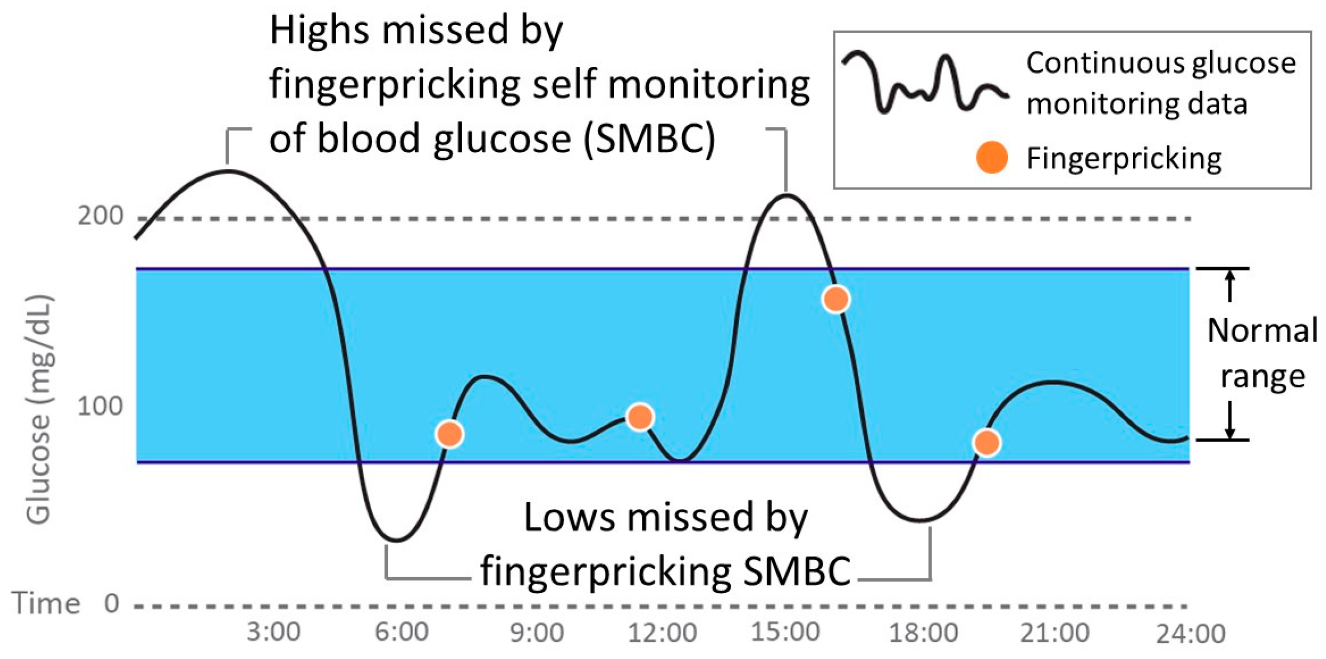
Figure 1. Comparison of finger-pricking self-monitoring of blood glucose (SMBC) and continuous glucose monitoring (CGM).
Applsci 09 02158 g001
Figure 2. Clarke error grid. Reproduced with permission [22]. Copyright 1988, Elsevier.
Applsci 09 02158 g002
Figure 3. Glucose sensing mechanism of diboronic-acid-based fluorescence. Modified figures from those in Ref. [46].
Applsci 09 02158 g003
Figure 4. (a) Fluorescence hydrogel with boronic acids under the skin of mouse ear. The hydrogel glows through skin. (b) The fluorescence intensity of hydrogels responded to blood glucose concentration continuously after 140 days from implantation. Modified figures from those in Ref. [47].
Applsci 09 02158 g004
Table 1. Performance requirements of integrated continuous glucose monitoring system (iCGM).
Overall Range<70 mg/dL70–180 mg/dL>180 mg/dL
within ±20% error in the lower one-sided 95%, must exceed 87%within ±15 mg/dL errors in the lower one-sided 95%, must exceed 85%within ±15% error in the lower one-sided 95%, must exceed 70%within ±15% error in the lower one-sided 95%, must exceed 80%
within ±40 mg/dL error in the lower one-sided 95%, must exceed 98%within ±40% error in the lower one-sided 95%, must exceed 99%within ±40% error in the lower one-sided 95%, must exceed 99%
no corresponding blood glucose value shall read above 180 mg/dL. no corresponding blood glucose value shall read less than 70 mg/dL.
Table 2. Comparison of commercial “continuous glucose monitoring” sensors.
CompanyMedtronicDexcomAbbott
ProductMiniMed 670G Guardian Sensor 3G4 PlatinumG4 Platinum with SW 505, G5 MobileG6 MobileFreeStyle Libre ProFreeStyle Libre
FDA approval September 2016October 2012October 2014March 2018September 2016September 2017
Accuracy (MARD %)10.55% (abdomen, age 14+)
February 2018 9.09% (upper arm, age 14+)
13.3%
(age 18+)
17.4%
(age 2+)
9% (age 18+)
10.4% (age 2+)
9% (age 18+)12.1% (age 18+)9.7% (age 18+)
FDA approval for non-adjunctive device No
(Class III)
Requires fingerstick test for diabetes treatment decisions
No
(Class III)
Requires fingerstick test for diabetes treatment decisions
No
(Class III)
Requires fingerstick test for diabetes treatment decisions
Yes
(Class II)
Replaces fingersticks for diabetes treatment decisions
No
(Class III)
Aids in the detection of glucose level excursions (Professional use only)
Yes
(Class III)
Replaces fingersticks for diabetes treatment decisions
Sensor size9.5 mm long (90 degree insertion)12 mm long (45 degree insertion)12 mm long (45 degree insertion)Not disclosed5 mm long (90 degree insertion)5 mm long (90 degree insertion)
Calibration frequency per dayMin: 2, (3–4 Recommended)2 (every 12 h)2 (every 12 h)0 (factory calibration)0 (factory calibration)0 (factory calibration)
SensorGlucose oxidaseGlucose oxidaseGlucose oxidaseGlucose oxidaseGlucose oxidaseGlucose oxidase
Sensor lifespan7 days (including 2 h warm-up)7 days (including 2 h warm-up)7 days (including 2 h warm-up)10 days (including 2 h warm-up)14 days10 days (including 12 h warm-up)