Article

Mechanochemical-Assisted Extraction and Pharmacological Study of Triterpenoids from Antrodia Camphorata

1
Department of Food Science and Technology, Zhejiang University of Technology, Hangzhou 310032, China
2
Zhejiang Suichang Limin Pharmaceutical Co., Ltd., Suichang 323300, China
*
Author to whom correspondence should be addressed.
Appl. Sci. 2019, 9(20), 4281; https://doi.org/10.3390/app9204281
Submission received: 9 September 2019 / Revised: 30 September 2019 / Accepted: 3 October 2019 / Published: 12 October 2019
(This article belongs to the Section Food Science and Technology)

Abstract

:

Featured Application

Antrodia camphorata treated by ball mill could increase the yield of triterpenoids, and the triterpenoids extracted by ball mill showed better pharmacological activity, it suggested that ball mill had the potential to be a pretreatment method for Antrodia camphorata and other natural plants.

Abstract

Antrodia camphorata (AC) is a precious medicinal mushroom native to Taiwan and famous for its excellent pharmacological activity. A ball mill assisted mechanochemical extraction method was applied in the extraction of triterpenoids from Antrodia camphorata. Compared with the ethanol hot thermal reflux method, mechanochemical-assisted extraction afforded an increased yield of triterpenoids to 1.82 ± 0.04% under conditions of mixing with 10 WT% NaHCO3, milling for 20 min, and extracting with water and chloroform. Triterpenoids from Antrodia camphorata extracted by the mechanochemical-assisted extraction method (TAEM) resulted in stronger pharmacological activity as compared to that extracted by ethanol (TAEE). HPLC and LC-MS/MS results showed that the mechanochemical method could extract triterpenoids which were barely extracted by ethanol extraction. The results of this study could provide valuable ideas and a basis for the application of the mechanochemical-assisted extraction method in the extraction of triterpenoids from AC.

1. Introduction

Antrodia camphorata (AC) is a medicinal fungus, a fungus of the genus Antrodia of the Fomitopsidaceae family, which is only native to Taiwan and has been used for treatment of liver cancer for many years [1]. There are abundant pharmacologically active ingredients in AC, and more than 80 components have been isolated and identified, including triterpenoids, polysaccharides, adenosine, superoxide dismutase (SOD), benzenoids, benzoquinones and maleic/succinic acid derivatives. Triterpenoids are the main active components of AC fruiting bodies, and they are similar in structure [2]. Antcins are the main triterpenoids in AC [3], including Antcin A, B, C, H, K and N [4]. At present, increasing attention has been paid to the research on their pharmacological activities, such as antioxidant, anti-inflammatory, anti-cancer and immunomodulation properties, etc., [5,6].
The mechanochemical-assisted extraction method (MAEM) is an innovative extraction technology, which has been widely used in the extraction of bioactive substances in recent years [7,8,9]. At present, the extraction of the bioactive substances from herbal medicine is mainly based on traditional solvent extraction in industrial processes [10]. However, the traditional solvent extraction method is time-consuming, requiring a high extraction temperature and low extraction rate, which decreases the quality and pharmacological activity of the target extracts [11]. MAEM is a technology that can destroy lignocellulose in order to obtain more target products [12]. Some studies have shown that the extraction of flavonoids from bamboo leaves by MAEM [13] improves the yield of flavonoids and that the obtained flavonoids have strong hydrophilicity, which is more conducive to subsequent experiments. Most of the active components in plants are found inside cells [14], which is the same for AC and its triterpenoids. MAEM can destroy plant tissues during grinding, and the addition of solid alkali can react with the components in the plant to increase the solubility of the original substances, which increases the yield of extracted components that are barely extracted by other extraction methods.
In this study, we aim to prove mechanochemical-assisted extraction method can achieve a higher yield and maintain the initial activity of triterpenoids in AC, as compared with the traditional ethanol thermal reflux extraction method.

2. Materials and Methods

2.1. Materials and Reagents

Fruiting bodies of AC in solid dish cultured were provided by Zhejiang Limin Pharmaceutical Co., Ltd. (Hangzhou, China), AC was cultured for 7 months, grinded into powder and sieved through 40 meshes. A BCA-100 protein quantitative kit and a NOS detection kit were purchased from Nanjing Jiancheng Biotechnology Co., Ltd. (Nanjing, China). The NO detection kit was purchased from Biyuntian Biotechnology Co., Ltd. (Shanghai, China). The IP cell lysate was purchased from Hangzhou Bori Science and Technology Co., Ltd. (Hangzhou, China). A549 cells (CBP-60084) and RAW264.7 cells (SCSP-5036) were purchased from the cell bank of the Chinese Academy of Sciences (Shanghai, China). Rats (clean grade) were provided by the Zhejiang Laboratory Animal Center (Hangzhou, China).

2.2. Preparation of Samples

2.2.1. Mechanochemical-Assisted Extraction Method (MAEM)

A total of 10.0 g AC powder and 1 g NaHCO3 were added into the PM-200 ball mill with steel balls (3 mm diameter, 150 g). The mixture was ground at 300 rpm for 20 min, then the powder was extracted by water at 50 °C for 30 min and centrifuged at 2650× g for 10 min. The supernatant was mixed with 95% ethanol solution (1:4 v/v) and placed in a refrigerator at 4 °C overnight. Then the mixture was centrifuged at 2650× g for 10 min and the pH was adjusted to <3.0 by 2 M HCl, after which the concentration was extracted by chloroform (1:1 v/v), three times. The chloroform layer was combined and evaporated under reduced pressure using a rotary evaporator. The scheme of MAEM was shown in Figure 1.

2.2.2. Ethanol Hot Reflux Extraction Method (EHRE)

A total of 10.0 g AC powder was added to 300 mL 95% ethanol, and refluxed at 90 °C for 3 h. The extract was centrifuged at 2650× g for 10 min, and the supernatant was dispersed with water. Then the mixed liquor was extracted by chloroform (1:1 v/v), three times, and the chloroform layer was extracted by saturated NaHCO3 solution. The saturated NaHCO3 solution layer was adjusted to pH < 3.0 by 2 M HCl, and extracted by chloroform (1:1 v/v) for three times after concentration. The filtrate was then combined and evaporated under reduced pressure using a rotary evaporator.
Triterpenoids from the AC extracted by mechanochemical-assisted extraction method (TAEM) and ethanol (TAEE) of the same mass were weighed, configured into 4 mg/mL and stored in the refrigerator for later use. The contents of triterpenoids in TAEM and TAEE were determined referring to the method described by Te-Wei Ma [15].

2.3. Analysis of TAEM and TAEE by HPLC and LC-MS/MS

TAEM and TAEE were analyzed by a Waters 1525 series HPLC system (Waters, Milford, MA, USA) coupled with a Waters 2487 Absorbance Detector (Waters, Milford, MA, USA) using a C18 column (4.6 × 250 mm, SymmetryShieldTM RP, Waters, Milford, MA, USA). The mobile phase consisted of 0.1% AcOH/H2O (A)–Acetonitrile (B), the gradient program was set as: 0–10 min, 10% B; 10–40 min, 10–50% B; 40–50 min, 50% B; 50–85 min, 50–100% B; 85–115 min, 100% B; 115–120 min, 100–10% B. The flowrate was 1.0 mL/min, the UV detection wavelength was set at 254 nm, the operating temperature was maintained at 25 °C. The concentration of the samples was 2 mg/mL.
A C18 column (5 μm 250 × 4.6 mm, Diamonsil C18 (2), Dikma, Beijing, China) was used and the samples were analyzed by Thermo Scientific Dionex Ultimate 3000 system (Thermo Scientific Dionex, Waltham, MA, USA) and Thermo Scientific LCQ Fleet (Thermo Scientific Dionex, Waltham, MA, USA) liquid chromatography-mass spectrometry. The gradient program was set as above. The system operated in the positive and negative ionization modes of electrospray ionization (ESI). The total ion chromatogram (TIC) and mass spectrogram were recorded. Based on the obtained molecular ions (m/z), and matching that with literature, the components could be inferred.

2.4. Antioxidant Experiment In Vitro

The free radicals scavenging capacity of TAEM and TAEE and the reduction capacity of ferrous ions were determined. The methods were previously described by our group [16] and slightly modified. Ascorbic acid (VC) was used as a positive control.

2.4.1. Scavenging Effect on DPPH Radicals

Briefly, 1 mL TAEM and TAEE solution (0~2 mg/mL) was mixed with 2 mL DPPH, reacted in darkness for 30 min. The absorbance at 517 nm was measured. The DPPH radical scavenging activity was calculated as follows:
S % = [ 1 ( A 1 A 2 ) / A 0 ] × 100 %
where A0 is the absorbance of control without sample solution; A1 and A2 are the absorbance of experimental sample with and without DPPH.

2.4.2. Scavenging Effect on ABTS Radicals

ABTS stock solution was prepared referring to the method described previously [17]. TAEM and TAEE sample solutions (0~2.0 mg/mL) were mixed with 2 mL ABTS stock solution, reacted at room temperature (25 °C) for 6 min. The absorbance at 734 nm was measured. The calculation formula of scavenging activity was the same as Formula (1), where A0 is the absorbance of control without sample solution; A1 and A2 are the absorbance of experimental sample with and without ABTS stock solution.

2.4.3. Scavenging Effect on Hydroxyl Radicals (·OH)

A total of 1 mL sample solution (0~2.0 mg/mL) was mixed with 0.5 mL of FeSO4 solution (1.5 mM), 0.35 mL H2O2 (6 mM) and 0.4 mL sodium salicylate solution (2 mM), then reacted at 37 °C for 1 h in a water bath. The absorbance was measured at a wavelength of 562 nm. Formula (1) was used to calculate the scavenging activity, where A0 is the absorbance of control without sample solution; A1 and A2 are the absorbance of experimental sample with and without sodium salicylate solution.

2.4.4. Reducing Power

A total of 2.5 mL of the sample solution (0~2.0 mg/mL) was mixed with 2.5 mL of phosphate buffer (0.2 M, pH = 6.6) and 2.5 mL of 1% (w/v) potassium ferricyanide (K3Fe(CN)6), then the reaction was carried out in a water bath at 50 °C for 20 min. A total of 2.5 mL of 10% (w/v) trichloroacetic acid (TCA) was added to complete the reaction, then centrifuged at 480× g for 15 min. A total of 5 mL of the supernatant was added to 5.0 mL of water and 1.2 mL (0.1% w/v) of ferric chloride (FeCl3). The absorbance was measured at 700 nm.

2.5. Anticancer Activity of TAEM and TAEE

2.5.1. Cell Culture

Cell viability measurement followed our previous experimental method [18] and has been modified. A549 cells were cultured in F12 medium which contained 10% FBS, 100 units/mL penicillin G and 100 µg/mL streptomycin sulfate. RAW264.7cells were cultured in DMEM medium containing 2 mM glutamine, 1% penicillin–streptomycin and 10% FBS. Cells were stored in a CO2 incubator (Thermo-3111, Waltham, MA, USA) with 5% CO2 at 37 °C.

2.5.2. Cell Viability Assay (MTT Assay)

A549 cells (5 × 10³ cells/well) were inoculated in a 96-well plate. After 24 h, the cells were incubated with tested drugs (TAEM or TAEE) (12.5, 25, 50, 100, 200, 400 μg/mL, 100 μL/well) for 24 h. Then, 10 μL of MTT reagent at a concentration of 5 mg/mL was added to each well. After 4 h of treatment, the culture solution was removed. The precipitate was dissolved in 50 μL of DMSO, the absorbance was measured at a wavelength of 570 nm, and the inhibition rates of A549 cells were calculated.

2.6. Anti-Inflammatory Activity and Mechanism of TAEM and TAEE

2.6.1. Toxicity Test

After the RAW264.7 cells (5 × 10⁴ cells/well) were inoculated in a 96-well plate for 24 h, cells were incubated with tested drugs (12.5, 25, 50, 100, 200 μg/mL, 100 μL/well) in the presence of 0.5 μg/mL Lipopolysaccharide (LPS)for 24 h. Then, 10 μL MTT reagent (0.5 mg/mL) was added to each well. After 4 h of incubation, the culture solution was removed. The precipitate was dissolved in 100 μL DMSO and the absorbance was determined at 570 nm. The cytotoxicity was calculated.

2.6.2. Measurement of Nitric Oxide (NO) Production

RAW 264.7 (2.5 × 10⁵ cells/mL) 2mL were inoculated in a 6-well plate for 24 h. Each well was replaced with serum-free 1.8 mL DMEM medium. After 4 h, 100 μL LPS (0.5 μg/mL) and tested drugs (6.25, 12.5, 25, 50, 100, 200 μg/mL) were added and incubated for 24 h. Then, 100 μL cell supernatant was inoculated into a 96-well plate, and Griess reagent (2% sulfonamide, 0.2% nylethanediamine in 5% H3PO4) was added and reacted in darkness at room temperature for 10 min. Absorbance at 540 nm was determined.

2.6.3. Measurement of iNOS Protein Induced by LPS in RAW 264.7 (BCA Assay)

RAW 264.7 cells (2 × 10⁵ cells/mL) were inoculated in a 6-well plate overnight. The serum was removed and 1.8mL DMEM medium was added, incubating for 4 h. Then LPS (0.5 μg/mL) and tested drugs (6.25, 12.5, 25, 50, 100, 200 μg/mL) were added. After being incubated for 24 h, the medium was removed and the cells were washed with PBS three times. The IP-cell lysate was added to lyse the cells for 1 min. Cell lysate was centrifuged at 10,142× g and supernatant was collected, protein content was measured by Bicinchoninic Acid (BCA) method, and stored at −70 °C for reserve.

2.7. Splenocyte Proliferation Assay

2.7.1. Preparation of Spleen Cell Suspension

Rats were sacrificed by cervical dislocation and soaked in 75% ethanol for 3 min. The spleens were quickly removed and placed in a dish containing Hank’s solution. Each spleen was ground to make a single cell suspension. The cell suspension was diluted in Hank’s solution, centrifuged at 120× g for 10 min, and washed twice. Finally, the cells were resuspended in 1 mL of RPMI 1640 cell complete medium, and the number of cells was counted.

2.7.2. Induction of Rat Spleen Lymphocyte Proliferation Response of TAEM and TAEE

The cells (2 × 10⁵ cells/mL) were inoculated in a 96-well plate at 0.1 mL per well. Tested drugs (12.5, 25, 50, 100, 200, 400 μg/mL) was added to the 96-well plate, 0.2 mL per well and incubated for 24 h, the splenocyte proliferation ability was evaluated by measuring cellular MTT reduction as described previously.

2.8. Statistical Analysis

All experiments were performed in three groups in parallel. Data processing statistics were analyzed by Student’s unpaired t test, and plotting was performed using Origin for Windows V. 8.5 (OriginLab, Inc., North Carolina, MA, USA), and Graphpad prism 5, SPSS for Windows V. 20.0 (SPSS, Inc., Chicago, IL, USA).

3. Results and Discussion

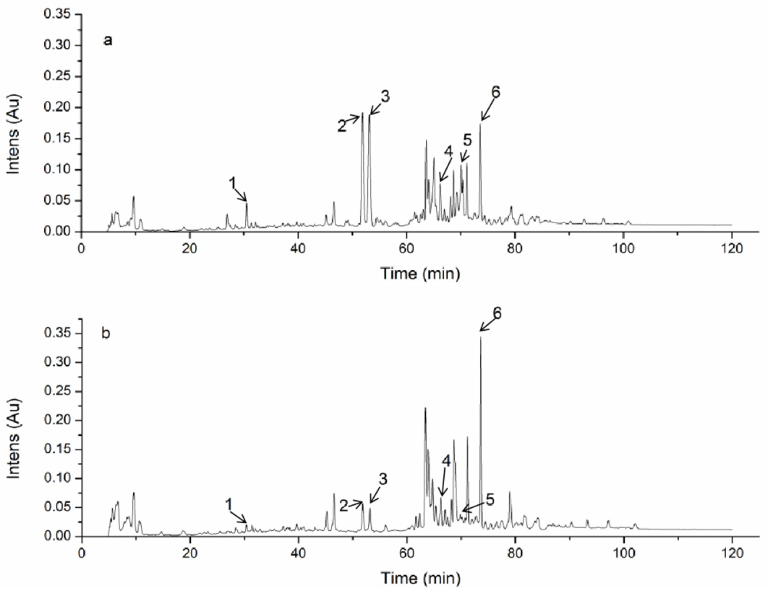
3.1. HPLC Analysis of TAEM and TAEE

Triterpenoids in Antrodia camphorata have always been a focus of research, and are also the most bioactive components in AC [19]. The contents of some substances in TAEM and TAEE are obviously different (Figure 2). The content of peaks 1, 2, 3 and 5 in TAEM was significantly higher than that in TAEE. We suggest that the components with polarity were much more easily extracted by MAEM.

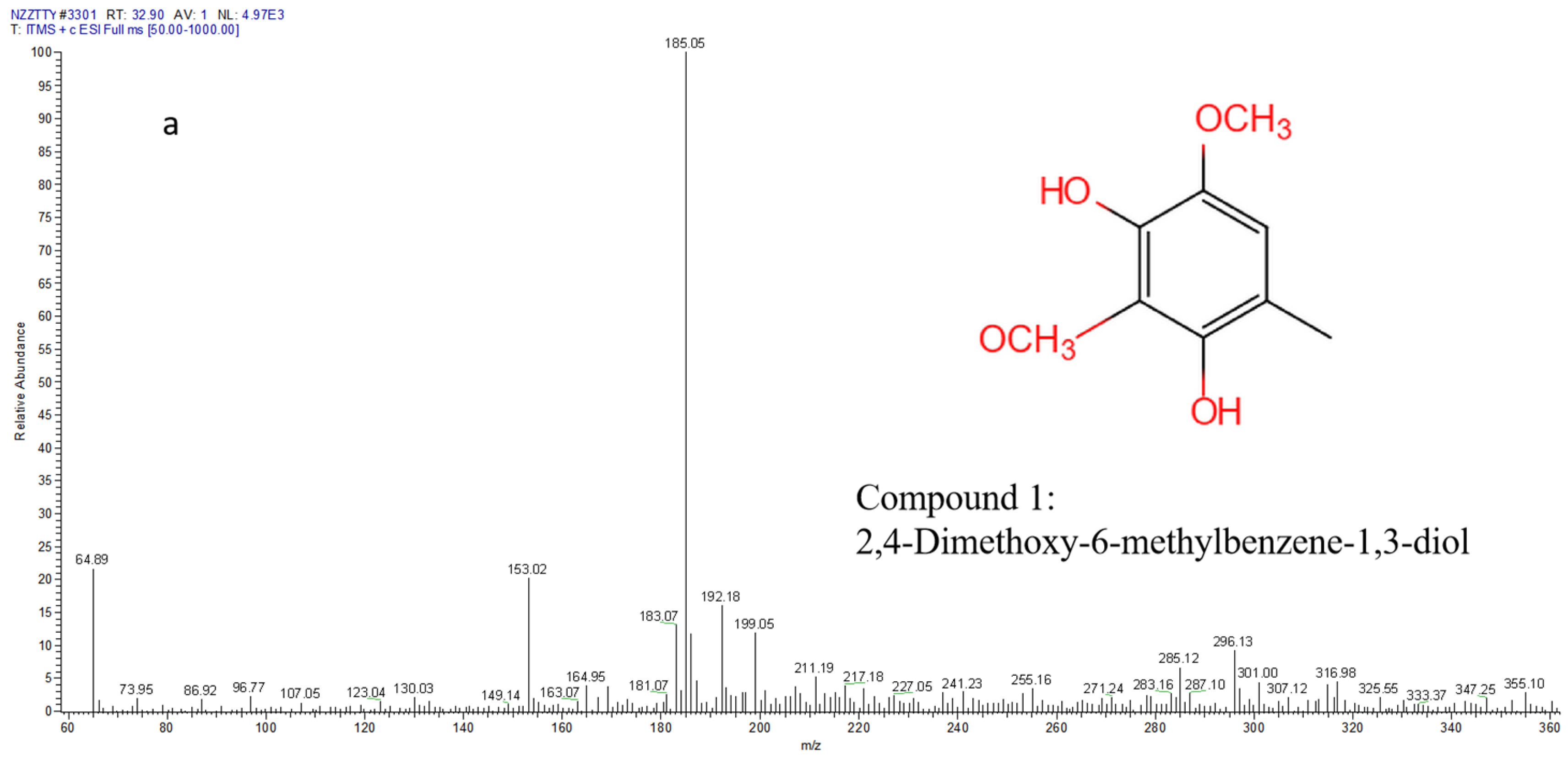
3.2. Chemical Analysis of Compounds by LC-MS

The analysis method was in reference to Mats Thulin et al. [20]. The compounds, including a benzenoid and four triterpenoids Antcin K, C, H and A, were identified in the extract of Antrodia camphorata by LC-MS (Figure 3). Their molecular ions (m/z) were matched with those of references [1,21] and the results were listed in Table 1, compound 1: 2, 4-Dimethoxy-6-methylbenzene-1,3 -diol was eluted at TR = 32.90 min at the molecular ion of m/z 185.05. Compound 2 and 3: Antcin K, eluted at TR = 66.86 min and 67.48 min, respectively, with the molecular ion of m/z 489.46, which were also the compounds with different R and S configurations. Compound 4: Antcin C, was eluted at TR = 74.67 min, with molecular ion of m/z 471.37. Compound 5: Antcin H, eluted at TR = 77.30 min, with a molecular ion of m/z 487.28. Compound 6 was Antcin A eluted at TR = 80.71 min, with a molecular ion of m/z 453.48.

3.3. Antioxidant Activity of TAEM and TAEE

Comparing the two extraction methods, the yield of triterpenoids extracted by MAEM and ethanol hot reflux extraction method (EHRE) were 1.82 ± 0.04% and 1.02 ± 0.13% (Table 2), respectively. Besides, MAEM had a lower extraction temperature (50 °C), shorter extraction time (30 min) and avoided the extensive use of organic reagents. As for EHRE, long-term (120 min) extraction at a high temperature (90 °C) might be damaging to active components in AC.
There have been many reports on the antioxidant activity of AC [22,23,24]. Four experiments related to antioxidation, scavenging ability on DPPH radicals, hydroxyl radicals, ABTS radicals and the reducing power of TAEM and TAEE were performed, and the results were shown in Figure 4. Both TAEM and TAEE exhibited strong scavenging capacity for DPPH free radicals (Figure 4a), with the EC50 of 0.46 mg/mL and 0.73 mg/mL, respectively.
When the concentration reached 1.6 mg/mL and 1.8 mg/mL, the scavenging effects were 94.31 ± 3.01% and 93.17 ± 2.87% while the positive control (ascorbic acid) was 97.53 ± 2.11% at the same concentration. Similarly, scavenging capacity of TAEM and TAEE on ABTS radicals increased rapidly from 0 to 0.4 mg/mL, which was similar to ascorbic acid (Figure 4b). As Figure 4c showed, the scavenging capacity on hydroxyl radicals was stronger than that of ascorbic acid at a pretty low concentration (0~0.4 mg/mL). However, it slowly increased at a concentration of 0.4~2.0 mg/mL, and the scavenging ability was estimated to be 62.62 ± 2.17%. At a concentration of 2.0 mg/mL, the scavenging ability was estimated to be 51.54 ± 1.57%, while ascorbic acid showed a significant scavenging ability of 98.32 ± 1.67% at 0.8 mg/mL. Reducing powers of the samples increased slowly at 0~1.2 mg/mL (Figure 4d), then the values rose in parallel at different levels of concentration until 2.0 mg/mL. The reducing powers hardly increased when the concentration was above 2.0 mg/mL.
Both TAEM and TAEE exhibited certain antioxidant activities and the antioxidant capacities were all concentration-dependent. It has been reported that the antioxidant activity of extracts from Antrodia camphorata is related to the crude triterpenoids [25]. The difference in antioxidant activity was probably due to the difference of triterpenoids between TAEM and TAEE.

3.4. Anticancer Activity of TAEM and TAEE

Inhibitory effect on A549 cells of various concentrations of TAEM and TAEE (12.5, 25, 50, 100, 200, 400 mg/mL) was measured. Results showed that both of them inhibited A549 cells in a concentration-dependent manner (Figure 5).
The inhibition rate of A549 cells reached 20~30%, even with TAEM and TAEE at concentrations of 12.5 μg/mL, and with the IC50 of TAEM and TAEE being 188.26 and 356.51 μg/mL. Moreover, at low concentrations (12.5~100 μg/mL), TAEM had only a slight advantage over TAEE (3%~5%), however, when the concentration increased to 200~400 μg/mL, TAEM possessed stronger cytotoxicity than TAEE (>12%). It was reported that ergostane triterpenoids of AC, including Antcin A, B, C, H and K exhibited certain anticancer activity [24,26], especially Antcin K, which had the strongest cytotoxicity effect on human hepatoma cells [26]. However, Pei-Li Zhu et al. found that the cytotoxicity of Antcin H against hepatocellular carcinoma cells was very low, and it probably played a role in anti-metastasis [27]. Therefore, the content of Antcin H in TAEM may not be the cause of its increased anticancer activity. Ying-ChiDu et al. [28] found that compounds with hydroxyl groups at C-3 had lower cytotoxicity to cancer cells, while carbonyl groups at C-3 were very important for their cytotoxicity. Antcin H and Antcin K both have a hydroxyl group at the C-3 position, so we speculated that the increase of their two contents is not related to the increase of TAEM anti-cancer cell activity. Antcin C and Antcin A both contain carbonyl groups at the C-3 position, which is the main factor affecting the cytotoxicity of extracts from AC to cancer cells, so this might explain why the difference of anticancer activity between TAEM and TAEE at the same concentration is not particularly large.

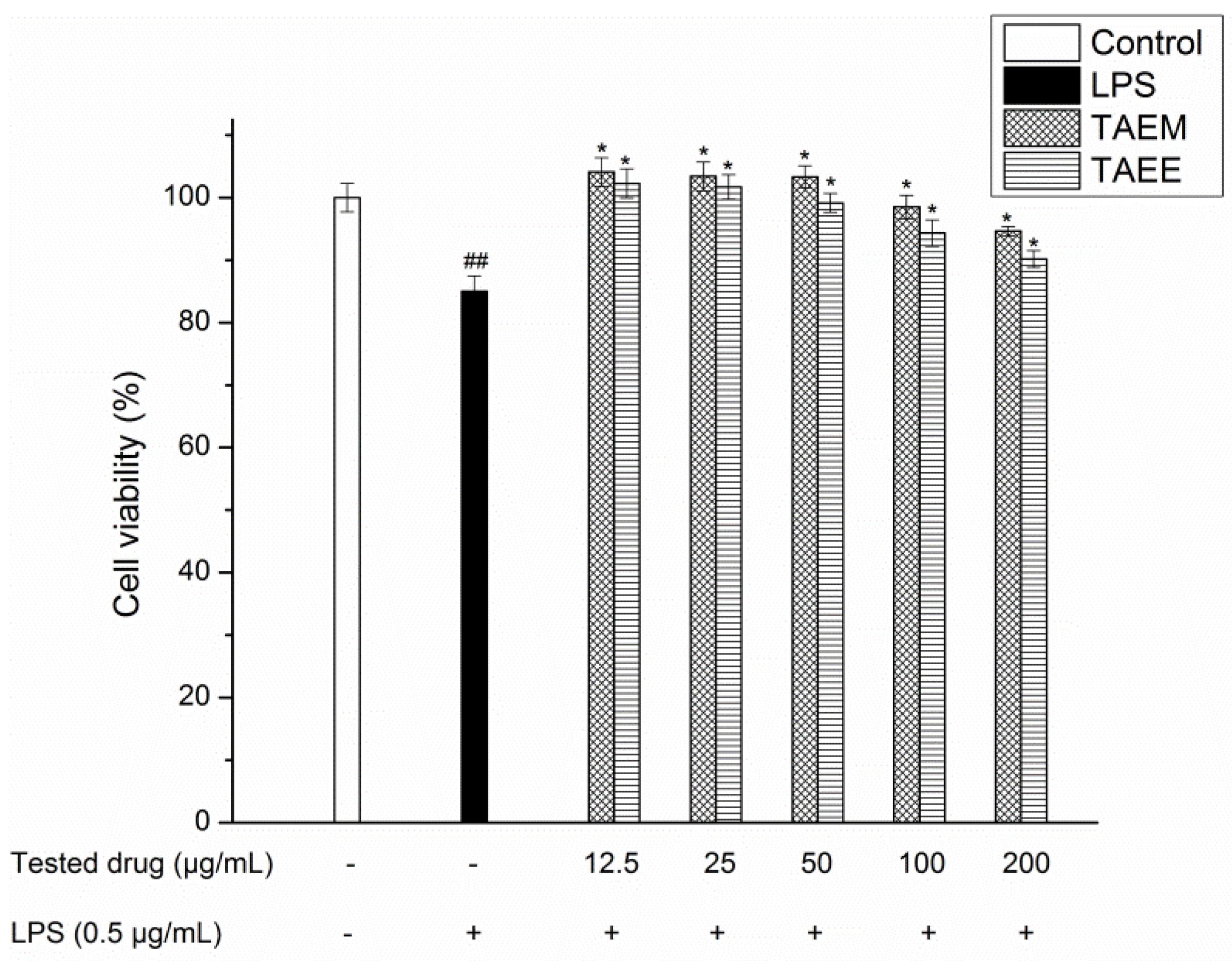
3.5. Anti-Inflammatory Activity of TAEM and TAEE

Prior to the anti-inflammatory investigation, the cytotoxicity of TAEM and TAEE on RAW264.7 cells were examined. Figure 6 showed they did not affect the viability of RAW2634.7 cells at a concentration of 12.5~200 μg/mL. The cytotoxicity on RAW264.7 was no more than 3%. The concentration of 100 μg/mL or less was selected to conduct an experimental study on the anti-inflammatory activity.
NO is a factor synthesized and released by vascular endothelial cells to activate the inflammatory response. The large amount of NO produced by the host in the inflammatory response may provide a defense against the invasion of cells, viruses and parasites, and the non-specific immunity of NO can prevent the invasion of pathogens. To determine the effect of TAEM and TAEE on anti-inflammatory activity, NO production was measured. As shown in Figure 7, a low NO expression (2.46 μM) was exhibited in the control group. After being stimulated by LPS, the NO content was significantly up-regulated to 5.89 μM. In the experimental group, when LPS was present with the co-treatment of different concentrations of tested drugs, the production of NO stimulated by LPS was significantly decreased. More specifically, production of NO was decreased to 4.76, 4.32, 3.98 and 3.26 μM by TAEM, and 4.87, 4.56, 4.31 and 4.07 μM by TAEE at 12.5, 25, 50, 100, 200 μg/mL, respectively. Obviously, TAEM possessed stronger inhibition ability on NO production than TAEE.
Inducible nitric oxide synthase (iNOS) is an indispensable enzyme for NO synthesis [29]. In activated macrophages, regulation of iNOS levels is critical for NO production. Regulation of NO production or iNOS expression may be an important target for the treatment of inflammation. As shown in Figure 8, the control group was treated without tested drugs or LPS. The basal expression of iNOS protein in RAW264.7 cells was very low. In the LPS group, the expression of iNOS protein in RAW264.7 cells increased significantly after stimulation by LPS, which was 5.24 ± 0.16-fold of the control. When TAEM and TAEE (12.5 μg/mL) were given, the expression of iNOS protein decreased significantly to 2.04 ± 0.11 and 3.02 ± 0.12-fold of the control, TAEM and TAEE both inhibited iNOS expression in a concentration-dependent manner. However, TAEM exhibited a stronger inhibition activity than TAEE especially at a high concentration (50~100 μg/mL).
The anti-inflammatory activity of components from AC has been reported in many studies. One of the most representative components, Antcin K, plays an important role in the hepatoprotective effect. The mechanism was that Antcin K inhibited the expression of iNOS, thereby exhibiting the anti-inflammatory effect [30]. Besides, the presence of 2,4-dimethoxy-6-methylbenzene-1,3-diol also contributed to its anti-inflammatory activity by reducing NO production [1]. Obviously, at any concentration, there was a big difference between TAEE and TAEM in inhibiting the expression of iNOS, and the difference of Antcin K content was the most critical. The content of Antcin K in TAEM is much higher than that in TAEE, so TAEM has a better anti-inflammatory activity.

3.6. Immunomodulatory Activity of TAEM and TAEE

One of the indicators for measuring the level of immune function is the amount of lymphocytes in the body. As shown in Figure 9, TAEM and TAEE directly stimulated the proliferation of rat spleen lymphocytes in the range of 25–400 ug/mL, and at 100 ug/mL, the stimulating effect reached its maximum. TAEM promoted the proliferation rate of spleen lymphocytes in rats more than TAEE at each concentration. There were few studies on the immunoregulation of triterpenoids from AC. It has been suggested that the immunomodulatory effect of Antrodia camphorata may be attributed to the improvement of cell tolerance [6]. The difference of immunomodulatory activity between TAEM and TAEE was mainly due to the fact that TAEM could extract triterpenoids, which, through TAEE, were difficult to extract.
It can be seen from the results that TAEM is superior to TAEE in the yield of triterpenoids and in the pharmacological activity at the same extract concentration. The reason for that is ball milling destroyed the cell wall of the plant cells, making the triterpenoids inside easier to extract. However, some triterpenoids were almost absent in TAEE. This could be because the triterpenoids in AC were sensitive to light and heat, and ethanol thermal reflux extraction with continuous light and high temperature treatment would destroy the triterpenoids.

4. Conclusions

In conclusion, application of the ball mill assisted mechanochemical extraction method in extract of triterpenoids from Antrodia camphorata significantly increased the yield of triterpenoids, as compared to the ethanol hot reflux extraction method. More importantly, at the same extract concentration, TAEM exhibited stronger pharmacological activity than TAEE. MAEM proceeded more efficiently at a lower temperature, and organic reagents were not used. This provides a basis for the application of ball mill assisted mechanochemical technology in the raw material treatment of AC. In order to better understand the mechanism of mechanochemistry in the extraction of natural products, further analysis and experiments are needed.

Author Contributions

Conceptualization, R.H. and A.Z.; Funding acquisition, R.H.; Methodology, R.H.; Project administration, R.H. and P.S.; Resources, Z.X.; Software, K.W.; Supervision, P.S.; Validation, P.S.; Writing—original draft, K.W.; Writing—review & editing, K.W.

Funding

This study was supported by Key Research and Developement Project of Zhejiang Province, China (No. 2018C02045).

Conflicts of Interest

The authors declare no conflict of interest.

References

  1. Yang, S.S.; Wang, S.Y.; Lin, Y.Y.; Kuo, Y.H.; Wang, G.J.; Lee, T.H. New Constituents with iNOS Inhibitory Activity from Mycelium of Antrodia camphorata. Planta Med. 2009, 75, 512–516. [Google Scholar] [CrossRef]
  2. Tzeng, Y.M.; Geethangili, M. Review of pharmacological effects of antrodia camphorata and its bioactive compounds. Evidence-based Complement. Altern. Med. 2011, 2011, 212641. [Google Scholar]
  3. Wang, Y.J.; Lee, S.C.; Hsu, C.H.; Kuo, Y.H.; Yang, C.C.; Lin, F.J. Antcins, triterpenoids from Antrodia cinnamomea, as new agonists for peroxisome proliferator-activated receptor α. J. Food Drug Anal. 2019, 27, 295–304. [Google Scholar] [CrossRef]
  4. Lu, M.C.; El-Shazly, M.; Wu, T.Y.; Du, Y.C.; Chang, T.T.; Chen, C.F.; Hsu, Y.M.; Lai, K.H.; Chiu, C.P.; Chang, F.R.; et al. Recent research and development of Antrodia cinnamomea. Pharmacol. Ther. 2013, 139, 124–156. [Google Scholar] [CrossRef]
  5. Lin, Y.S.; Lin, Y.Y.; Yang, Y.H.; Lin, C.L.; Kuan, F.C.; Lu, C.N.; Chang, G.H.; Tsai, M.S.; Hsu, C.M.; Yeh, R.A.; et al. Antrodia cinnamomea extract inhibits the proliferation of tamoxifen-resistant breast cancer cells through apoptosis and skp2/microRNAs pathway. BMC Complement. Altern. Med. 2018, 18, 152. [Google Scholar] [CrossRef]
  6. Lee, M.T.; Lin, W.C.; Wang, S.Y.; Lin, L.J.; Yu, B.; Lee, T.T. Evaluation of potential antioxidant and anti-inflammatory effects of Antrodia cinnamomea powder and the underlying molecular mechanisms via Nrf2- and NF-κB-dominated pathways in broiler chickens. Poult. Sci. 2018, 97, 2419–2434. [Google Scholar] [CrossRef]
  7. Wang, P.; Tang, J. Solvent-free mechanochemical extraction of chondroitin sulfate from shark cartilage. Chem. Eng. Process. Process. Intensif. 2009, 48, 1187–1191. [Google Scholar] [CrossRef]
  8. Su, W.; Zhu, X.; Chen, X.; Xie, J.; Wang, P. Mechanochemical-assisted extraction and antioxidant activity of polysaccharides from Ganoderma lucidum spores. Int. J. Food Sci. Technol. 2012, 47, 927–932. [Google Scholar]
  9. Wu, K.; Ju, T.; Deng, Y.; Xi, J. Mechanochemical assisted extraction: A novel, efficient, eco-friendly technology. Trends Food Sci. Technol. 2017, 66, 166–175. [Google Scholar] [CrossRef]
  10. Azmir, J.; Zaidul, I.; Rahman, M.M.; Sharif, K.; Mohamed, A.; Sahena, F.; Jahurul, M.; Ghafoor, K.; Norulaini, N.; Omar, A. Techniques for extraction of bioactive compounds from plant materials: A review. J. Food Eng. 2013, 117, 426–436. [Google Scholar] [CrossRef]
  11. Barba, F.J.; Zhu, Z.; Koubàa, M.; Sant’Ana, A.S.; Orlien, V.; Sant’Ana, A.D.S. Green alternative methods for the extraction of antioxidant bioactive compounds from winery wastes and by-products: A review. Trends Food Sci. Technol. 2016, 49, 96–109. [Google Scholar] [CrossRef]
  12. Li, H.; Wang, X.; Liu, C.; Ren, J.; Zhao, X.; Sun, R.; Wu, A. An efficient pretreatment for the selectively hydrothermal conversion of corncob into furfural: The combined mixed ball milling and ultrasonic pretreatments. Ind. Crop. Prod. 2016, 94, 721–728. [Google Scholar] [CrossRef]
  13. Xie, J.; Lin, Y.S.; Shi, X.J.; Zhu, X.Y.; Su, W.K.; Wang, P. Mechanochemical-assisted extraction of flavonoids from bamboo (Phyllostachys edulis) leaves. Ind. Crop. Prod. 2013, 43, 276–282. [Google Scholar] [CrossRef]
  14. Wang, M.; Bi, W.; Huang, X.; Chen, D.D.Y. Ball mill assisted rapid mechanochemical extraction method for natural products from plants. J. Chromatogr. A 2016, 1449, 8–16. [Google Scholar] [CrossRef]
  15. Ma, T.W.; Lai, Y.; Yang, F.C. Enhanced production of triterpenoid in submerged cultures of Antrodia cinnamomea with the addition of citrus peel extract. Bioprocess Biosyst. Eng. 2014, 37, 2251–2261. [Google Scholar] [CrossRef]
  16. He, R.; Zhao, Y.; Zhao, R.; Sun, P. Antioxidant and antitumor activities in vitro of polysaccharides from E. sipunculoides. Int. J. Boil. Macromol. 2015, 78, 56–61. [Google Scholar] [CrossRef]
  17. Tang, Y.Z.; Liu, Z.Q. Free-Radical-Scavenging Effect of Carbazole Derivatives on DPPH and ABTS Radicals. J. Am. Oil Chem. Soc. 2007, 84, 1095–1100. [Google Scholar] [CrossRef]
  18. He, R.; Ye, J.; Zhao, Y.; Su, W. Partial characterization, antioxidant and antitumor activities of polysaccharides from Philomycusbilineatus. Int. J. Boil. Macromol. 2014, 65, 573–580. [Google Scholar] [CrossRef]
  19. Zhang, Y.; Li, D.; Wang, Z.; Zang, W.; Rao, P.; Liang, Y.; Mei, Y. Alpha-terpineol affects synthesis and antitumor activity of triterpenoids from Antrodia cinnamomea mycelia in solid-state culture. Food Funct. 2018, 9, 6517–6525. [Google Scholar] [CrossRef]
  20. Thulin, M.; Moore, A.J.; El-Seedi, H.; Larsson, A.; Christin, P.A.; Edwards, E.J. Phylogeny and generic delimitation in Molluginaceae, new pigment data in Caryophyllales, and the new family Corbichoniaceae. Taxon 2016, 65, 775–793. [Google Scholar] [CrossRef]
  21. Huang, H.S.; Hsu, J.L.; Yu, C.C.; Chang, L.C.; Chen, C.R.; Huang, T.C.; Chang, C.I. Qualitative and quantitative analysis of seven signature components in the fruiting body of Antrodia cinnamomea by HPLC—ESI—MS/MS. Acta Chromatogr. 2015, 28, 387–402. [Google Scholar] [CrossRef]
  22. Hsieh, Y.L.; Wu, S.P.; Fang, L.W.; Hwang, T.S. Effects of Antrodia camphorata extracts on anti-oxidation, anti-mutagenesis and protection of DNA against hydroxyl radical damage. BMC Complement. Altern. Med. 2015, 15, 237. [Google Scholar] [CrossRef]
  23. Wang, J.J.; Wu, C.C.; Lee, C.L.; Hsieh, S.L.; Chen, J.B.; Lee, C.I. Antimelanogenic, Antioxidant and Antiproliferative Effects of Antrodia camphorata Fruiting Bodies on B16-F0 Melanoma Cells. PLoS ONE 2017, 12, 0170924. [Google Scholar] [CrossRef]
  24. Zhang, F.; Cai, H.; Liu, X.; Chen, F.; Yang, N.; Hou, Y.; Ma, Y.; Luo, Y.; Liu, J. Simultaneous determination of 19 fatty acids in Antrodia camphorata by derivatized GC–MS and evaluation of antioxidant activity of Antrodia camphorata crude oil. Arch. Pharmacal Res. 2015, 38, 1–8. [Google Scholar] [CrossRef]
  25. Song, T.Y.; Yen, G.C. Antioxidant properties of Antrodia camphorata in submerged culture. J. Agric. Food Chem. 2002, 50, 3322. [Google Scholar] [CrossRef]
  26. Lee, Y.P.; Tsai, W.C.; Ko, C.J.; Rao, Y.K.; Yang, C.R.; Chen, D.R.; Yang, M.H.; Yang, C.C.; Tzeng, Y.M. Anticancer effects of eleven triterpenoids derived from Antrodia camphorata. Anticancer. Res. 2012, 32, 2727–2734. [Google Scholar]
  27. Zhu, P.L.; Fu, X.Q.; Li, J.K.; Tse, A.K.W.; Guo, H.; Yin, C.L.; Chou, J.Y.; Wang, Y.P.; Liu, Y.X.; Chen, Y.J.; et al. Antrodia camphorata Mycelia Exert Anti-liver Cancer Effects and Inhibit STAT3 Signaling in vitro and in vivo. Front. Pharmacol. 2018, 9, 1449. [Google Scholar] [CrossRef]
  28. Du, Y.C.; Wu, T.Y.; Chang, F.R.; Lin, W.Y.; Hsu, Y.M.; Cheng, F.T.; Lu, C.Y.; Yen, M.H.; Tsui, Y.T.; Chen, H.L.; et al. Chemical profiling of the cytotoxic triterpenoid-concentrating fraction and characterization of ergostane stereo-isomer ingredients from Antrodia camphorata. J. Pharm. Biomed. Anal. 2012, 58, 182–192. [Google Scholar] [CrossRef]
  29. Huang, G.J.; Deng, J.S.; Huang, S.S.; Lee, C.Y.; Hou, W.C.; Wang, S.Y.; Sung, P.J.; Kuo, Y.H. Hepatoprotective effects of eburicoic acid and dehydroeburicoic acid from Antrodia camphorata in a mouse model of acute hepatic injury. Food Chem. 2013, 141, 3020–3027. [Google Scholar] [CrossRef]
  30. Li, Z.W.; Kuang, Y.; Tang, S.N.; Li, K.; Huang, Y.; Qiao, X.; Yu, S.W.; Tzeng, Y.M.; Lo, J.Y.; Ye, M. Hepatoprotective activities of Antrodia camphorata and its triterpenoid compounds against CCl 4 -induced liver injury in mice. J. Ethnopharmacol. 2017, 206, 31–39. [Google Scholar] [CrossRef]
Figure 1. Scheme of ball mill assisted triterpenoids from the Antrodia Camphorata extraction method.
Applsci 09 04281 g001
Figure 2. HPLC analysis of Triterpenoids from Antrodia camphorata extracted by the (a) mechanochemical-assisted extraction method (TAEM) and (b) ethanol reflux method (TAEE).
Applsci 09 04281 g002
Figure 3. MS spectra of compounds extracted from Antrodia camphorata. (a) MS spectra of 2,4-dimethoxy-6-methylbenzene-1,3-diol, [M + H]+ m/z 185, (b) MS spectra of Antcin K [M + H]+ m/z 489, (c) MS spectra of Antcin K [M + H]+ m/z 489, (d) MS spectra of Antcin C [M + H]+ m/z 471, (e) MS spectra of Antcin H [M + H]+ m/z 487, (f) MS spectra of Antcin A [M − H] m/z 455.
Applsci 09 04281 g003aApplsci 09 04281 g003bApplsci 09 04281 g003c
Figure 4. Antioxidant activity of TAEM and TAEE. Scavenging activity of DPPH radicals (a), ABTS radicals (b) and hydroxyl radicals (c), and reducing powers (d). Data are expressed as mean ± SD (n = 3).
Applsci 09 04281 g004
Figure 5. TAEM and TAEE on the average inhibition rate of A549 cancer cells. Data are expressed as mean ± SD (n = 3).
Applsci 09 04281 g005
Figure 6. Cytotoxicity of TAEM and TAEE in Lipopolysaccharide-stimulated RAW264.7 cells. Data are expressed as mean ± SD (n = 3). ## p < 0.05 compared with control group. * p < 0.05 compared with LPS treatment group.
Applsci 09 04281 g006
Figure 7. Effects of TAEM and TAEE on NO production in LPS-stimulated RAW264.7 cells. Data are expressed as mean ± SD (n = 3). ### p < 0.01 compared with control group (without TAEM or TAEE treatment); * p < 0.05, ** p < 0.01 compared with LPS treatment group.
Applsci 09 04281 g007
Figure 8. Effects of TAEM and TAEE on the vitality of iNOS. Data are expressed as mean ± SD (n = 3). ### p < 0.01 compared with control group (without TAEM or TAEE treatment); * p < 0.05, ** p < 0.01 compared with LPS treatment group.
Applsci 09 04281 g008
Figure 9. Effects of different concentrations TAEM and TAEE on the growth rate of splenic lymphocyte in rats. * p < 0.05, ** p < 0.01 compared with control group.
Applsci 09 04281 g009
Table 1. Chemical characterization of the main compounds detected in TAEM by LC-MS/MS.
Peak NumberCompoundMolecular FormulaTR (min)[M + H]+[M − H]Molecular WeightReference
12, 4-dimethoxy-6-methylbenzene-1,3-diolC9H12O432.90185183184[1]
2Antcin KC29H44O666.86489487488[21]
3Antcin KC29H44O667.48489487488[21]
4Antcin CC29H42O574.67471479470[21]
5Antcin HC29H42O677.30487485486[21]
6Antcin AC29H42O480.71455453454[21]
Table 2. Comparison of mechanochemical-assisted extraction method (MAEM) and ethanol hot reflux extraction method (EHRE).
MethodTemperature (°C)SolventTime (min)Yield c (%)
MAEM a50Water301.82 ± 0.04
EHRE b90Ethanol1201.02 ± 0.13
a MAEM is ball mill assisted mechanochemical extraction method. b EHRE is ethanol hot reflux extraction method. c Extracted from dried fruiting bodies (10 g). Each value is expressed as mean ± SD (n = 3).

