Article

A Neuro-Prosthetic Device for Substituting Sensory Functions during Stance Phase of the Gait

1
Department of Mechanical Engineering and Virtual Reality Applications Center, Iowa State University of Science and Technology, Ames, IA 50011, USA
2
Department of Veterinary Medicine & Biological Science, Texas A & M University, College Station, TX 77843, USA
*
Author to whom correspondence should be addressed.
Appl. Sci. 2019, 9(23), 5144; https://doi.org/10.3390/app9235144
Submission received: 23 October 2019 / Revised: 15 November 2019 / Accepted: 20 November 2019 / Published: 27 November 2019

Abstract

:
In this study, we present the experimental results demonstrating the functionality of our recently developed “balancing device” for walking restoration in patients with spinal cord injuries. Since we are preparing this device for testing on dogs, we program the analytical core of the device to recognize both stance and swing phases of the dog gait, the direction that the dog is falling, as well as selecting a suitable balancing strategy to prevent falling. The analytical core of the device is a commercial microcontroller, the Teensy, which is able to provide suitable stimulation commands and intensities as a voltage for delivery to the stimulation circuit and target muscles. We show the functional schematic of the device along with experimental results obtained by testing the device in a simulated robotic dog. Results show that the sensory system of the animal lost by spinal cord injury can be replaced by the sensing core of the device and the analytical core can provide appropriate stimulation control to balance the body of a dog. All test results are obtained using our robot test-bed and living animals are not involved in this study.

1. Introduction

1.1. A Background on Functional Electrical Stimulation Devices and Application of Sensors in Gait Event Detection and Control

Micro-electronics have recently emerged as the central part of many devices and lab equipment in biology and neuroscience [1,2,3]. One application is in electrical stimulation of nerve tissues and body organs which is important in neuroscience both in understanding the mechanisms of neuronal activity and animal behavior and as a tool for treatment of central and peripheral nervous system disease [4].
Functional Electrical Stimulation (FES) is a technique that applies electrical charge to muscles and organs for rehabilitation and achievement of dynamic movements [5,6,7]. Neural prostheses to deliver FES may have an analytical and stimulation core to gather data from sensors and actively control the electrical charge delivered to muscles using electrodes [8]. Timing control for stimulation is necessary and important for these devices and, therefore, the sensing core of the device must gather information about the gait phases [9,10,11]. Different FES systems utilize different sensors and different timing control methods depending on the moving function the device aims to assist or replace and the remaining abilities of patient after neural injuries. Usually, a combination of different sensors is used in an FES system to gather gait data in real time. Accelerometers, gyroscopes, and force-sensitive resistors are common sensors that are utilized in FES systems.
A common application of FES is in drop-foot stimulator devices where the peroneal nerve and tibialis anterior muscle are stimulated during the swing phase of the gait to raise the toes and prevent falling [12,13]. The force-sensitive resistor is the most common sensor used in such stimulators to control stimulation timing. This sensor is placed inside the shoe and under the heel. When patient lifts the heel from the ground, the force is removed from the sensor or heel switch. This indicates the start of swing phase and stimulation is initiated. Once the force is reapplied on the sensor, stimulation is terminated. Some researchers developed advanced stimulators for this purpose to detect more gait events and assist patients suffering from dropped foot. Melo et al. [14] invented a gait neuro-prosthesis to help these patients using FES. They selected Arduino MCU as their computational modulus to obtain data from Inertial Measurement Units (IMUs) and force-sensitive resistors and then calculate and control the actuation command to target muscles.
For some FES systems with simple application of stimulation and gait detection methods, a smaller number of sensors are used. Monaghan et al. [15] utilized a single gyroscope attached on the shank of the human patient to measure shank angular velocity and detect stance/swing phase of the foot. For complicated systems with FES applications, more accurate gait event detection is needed, which has encouraged extensive research in this field to propose different methods and algorithms. As an example, Zahradka et al. [16] designed a real-time system using two inertial measurement units to detect seven phases of the gait with very high reliability.
In this study, we develop a wearable neuro-prosthetic device to replace sensory functions of patients that suffer spinal cord injuries. We first design this device for dogs that we selected as our animal subject. We design an advance sensing core for this device to determine and calculate the stimulation command based on three timing controls. The first timing control is based on the hip orientation of the patient. The second is based on stance/swing phase gait detection, and the third is based on gait abnormalities during swing phase. In this paper, we omit discussion of the third timing control since it is the subject of another study, which will be reported elsewhere.
In our previous work [17], we designed and built a FES-based wearable device using an Arduino Uno board that could be used for stimulation of hindleg muscles of dogs suffering from spinal cord injury. The sensing core of this balancing device includes a single IMU attached to the pelvis of the dog to gain data about pelvis angle of rotation. Timing control for the stimulation was based solely on hip orientation. To study the healthy gait of the dog, we used VICON software (Vicon Industries, Inc, Hauppauge, NY, USA) [18] to track the gait of a healthy dog on the treadmill. Our test results showed that the pelvis of the healthy dog does not rotate much during normal walking. However, during abnormal gait, dramatic rotation of the pelvis occurs before falling. We concluded that falling occurs when the pelvis angle of rotation exceeds a critical value, which can be determined by the veterinarian for each individual dog. This critical value can be measured using an IMU attached to the hip and can be used as an indication of falling. The Arduino calculates the necessary neural stimulation command when data obtained from the IMU indicates falling and, in turn, provides the voltage or charge needed for muscle stimulation. To test this balancing device, we built a bionic test-bed to mimic stimulation responses of dog hindlimbs [19]. This robot had two legs designed based on canine anatomy, with motorized hip and hock joints. Using this test-bed, we programmed Arduino to provide stimulation and balance the robodog to prevent falling.
While our previous design showed excellent results regarding balancing of the robot using our proposed balancing strategies, the simple sensing core of the device made it difficult to use the real animal unless offline data was gathered from VICON for each individual animal patient. The previous device did not have a separate sensing core for gait detection of the robot/animal, and data identifying the robot gait were gathered from robot motion control sensors, not dedicated external sensors on the device. The device was not able to independently detect gait phases and anomalous gait events or measure the joint angles of the leg of the animal in real time.
In our current study, we replace the Arduino board with a more powerful Teensy 3.2 processor board [20] and add three additional external IMUs to the sensing core of the device in order to track all the gait events and gait abnormalities of the animal in real time. The Teensy 3.2 is physically smaller in comparison with Arduino UNO, which makes it more suitable for a lightweight wearable device. It also provides additional I/O pins for external components, a more powerful computing processor for performance of complex operations, and two I2C buses to allow communication with external sensor devices. Using this microcontroller, we are able to attach four IMUs simultaneously and test new balancing strategies in a more advanced bionic test-bed with three motorized hip, knee, and hock joints. Test results for this advanced sensing core of the device is reported in this paper, and the robot motion control sensors are only used as a reference to validate the accuracy of the sensing core of our system. We show that our device can recognize which limb is at the stance/swing phase and identify the active limb needed to receive stimulation based on a selected strategy. We program our microcontroller to provide several different stimulation commands, based on proposed strategies. The robotic test-bed is used to mimic the responses. Four IMUs attached on the hip, femur, tibia, and metatarsus of the patient can obtain global information about patient gait in real time. Unlike our previous study, in which we used offline data obtained from VICON on abnormal gait motion to balance the patient body, here we use real-time data from the advanced sensing core of our device to identify abnormalities in the natural gait. In the next section, we provide more information about our new device and the functions this novel neuro-prosthesis can perform to assist patients with disabilities.

1.2. Innovation and Motivation for Current Study

The central nervous system of quadrupeds, which includes the brain and spinal cord, plays a key role in controlling body movements [21,22]. Damage to these organs can cause severe permanent disabilities and any treatment is limited in efficacy because it is not possible to regenerate lost nerve cells [23]. To help patients, neural prostheses are used to assist, rehabilitate, reduce pain, and restore normal bodily functions [24,25,26,27,28]. Functional Electrical Stimulation (FES) as a useful rehabilitation method can apply electrical charge to stimulate peripheral nerves and restore motor functions lost by injury.
Dogs are excellent animal subjects in which to study spinal cord disease and rehabilitation because spinal cord injuries in dogs cause the same debilitating effects as in humans. The outcome of this injury for a dog can be weakness in hindlimb muscles and loss of sensation causing disability in walking and maintaining balance. While many dogs suffering such disease will completely lose their ambulatory functions in the hindlimbs, some of them are still able to make walking movements through muscle reflexes elicited when the toes touch the ground. However, due to lack of sensory feedback information ascending to the coordinating centers in the brain, such dogs will lose their balance after a few steps when the hindquarters tilt, and the animal will fall because of an inability to correct this process.
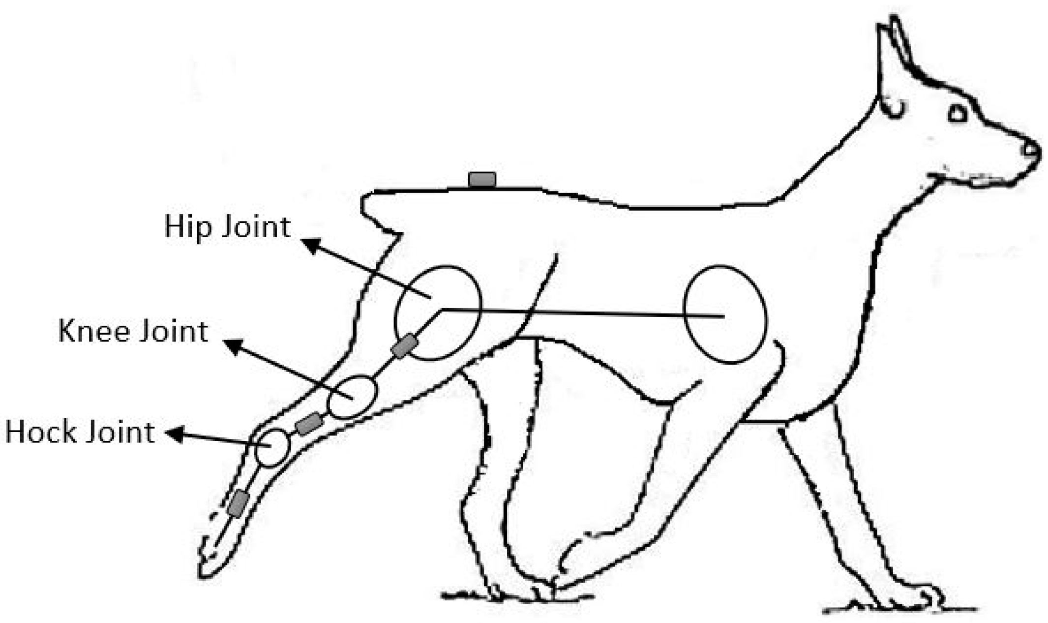
To restore walking, our idea is to attach four IMUs on the pelvic, femur, tibia, and metatarsus region of the dog to gain information about angle of rotation at the pelvis and hip, knee, and hock flexion angles (Figure 1). Using this information, we can track dog gait in real time.
We categorize the walking gait for each leg to two phases: Swing and stance phases (Figure 2). The swing phase commences when the dog lifts its leg from the ground and moves it forward. During the swing phase, the leg is not in contact with the ground, as shown in Figure 2. The stance phase is defined as the time when the leg is in contact with the ground, again shown in Figure 2. During stance phase, the toes of the dog are in contact with the ground. Because of lack of sensation, spinal cord injured-dogs are unable to move their hindlegs to follow normal trajectory lines during the swing phase. This causes them to place their toes in an unusual or unstable position at the start of the stance phase. Because the limb does not have the appropriate sensory feedback and control loop to permit weight-bearing, in this condition, the pelvis rotates and tilts laterally, causing falling.
To develop our new balancing device, we propose that both gait phases can be controlled by electrical stimulation to restore walking. During swing phase, IMUs should measure the joint angles and stimulate muscle nerves based on data from normal joint angles of the gait to force the leg to follow normal trajectory lines. This method is not the subject of the current study and will be explained in a separate paper. During the stance phase, our aim is to balance the pelvis to prevent falling. The IMU on the pelvis measures the pelvis rotational angle and, when it reaches a falling threshold condition, the appropriate muscles are actuated to correct pelvis orientation and maintain balance. Since the stimulation strategies during stance and swing phases are different, our balancing device should be programmed to swiftly recognize the stance and swing phase of the gait for each leg.
In our previous work [19], we extensively discussed the design of our robotic test-bed and showed the accuracy of the robot in mimicking dog walking gait and the joints’ control systems to stimulation commands that resemble the muscle response to stimulation. We also showed two strategies to maintain pelvic equilibrium during the stance phase while the knee joint was fixed. In addition, we relied on the robot dog simulator actuator potentiometers to tell us the joint angles for real-time gait and balance correction. This approach, while allowing us to develop and test algorithms on our robotic dog simulator, cannot be used on a live animal.
In the current work, we have advanced the sensing core of the device to measure joint angles using multiple IMU devices on the dog leg. These sensors allow us to quickly measure the leg postures and select the appropriate balancing strategy based on the body condition and orientation. We will explain the complete device structure and connections in the following sections. In summary, we can categorize the device functions into the following divisions:
Function 1. Device recognizes the stance and swing phase of each leg,
Function 2. Device recognizes if the body tilts from the side on which the leg is at stance or swing phase,
Function 3. Device selects the suitable balancing strategy at stance phase to maintain pelvic equilibrium and balance the body,
Function 4. Device stimulates muscle nerves at swing phase to force the leg to follow normal trajectory lines.
In this work, we explain our approach to Functions 1, 2 and 3 of our balancing device. Function 4 and a final validation test on live animals that we are currently working to implement are the subjects of separate papers.

2. Materials and Methods

2.1. Hardware Set-Up

Our balancing device includes three cores: sensing, analytical, and stimulation. The stimulation core includes electrical charge delivery. The sensing core consists of four IMUs connected to Teensy 3.2 using two I2C buses. These IMUs are attached to the pelvis, femur, tibia, and metatarsus of the dog hindlimbs to obtain rotation rates and acceleration data of these joints and bones as the dog walks. Using these data, the analytical core can calculate hip, knee and hock joint angles during walking and stimulation. The analytical core also calculates the stimulation command based on a selected strategy. The stimulation signal then is applied to Pulse Width Modulation(PWM) pins connected to H-bridge and stimulation circuit and, when fully implemented on an actual dog patient, electrodes. Each H-bridge (TB6612FNG dual motor driver carrier, Pololu Corporation, Las Vegas, NV, USA) has two outputs suitable to be connected to a separate stimulation circuit and a pair of electrodes (Figure 3). The stimulation circuit consists of an IC 7555 timer (NXP Semiconductors, Eindhoven, The Netherlands) to generate ~80 Hz pulses and a transformer with 220 v primary to 12 v, 100/150 mA secondary, which is reversely connected. This is a typical muscle stimulator circuit that can be found commercially [29]. A stimulation circuit and a pair of electrodes connected to a single H-bridge output is used to stimulate muscles for increasing a joint angle, and the other circuit and electrode pair apply stimulation to muscle, which pulls the bone to decrease the angle of the same joint. Figure 3 shows the device sensing and analytical core as well as the pin connection for only one motor driver carrier. It also shows the connection of this motor driver to stimulation circuit and pair of electrodes.
For the current device shown in Figure 3, we have used skin electrodes that are safely placed on the skin and stimulate underlying tissue. However, this device can be advanced to be compatible to wireless micro/nano implantable electrodes to activate nerves by applying smaller currents. The system with implantable electrodes is more complex since surgery and precise application of electrical charge are needed. During the last decade, scientists proposed designs for micro/nano switches [30,31] that can have applications as a structure of implantable electrodes. In addition, biocompatible materials that can be suitably implanted in body and work on body temperature have been investigated, which are suitable to be used as the material of stimulation electrodes [32,33].
The Teensy microcontroller uses PWM output to adjust the intensity of stimulation based on pelvis angle of rotation obtained from IMU #1 and the specific strategy. Electrodes deliver charges to the target muscles and make the stimulation core of our device. To test our device using our robotic dog test-bed, we removed the electrodes and stimulation circuits and connected the Teensy output to the robot joint motors using our two TB6612FNG dual motor driver carriers [34]. We also read the robot joint potentiometers and obtained data from these sensors as well. These potentiometers read the position of the motor shaft and report accurate joint angles. However, we used the data obtained from potentiometers only for comparison with and validation of the data that we derive from IMUs about joint angles. All the device cores and device to test-bed hardware connections have been listed in Table 1.
Our robotic test-bed is shown in Figure 4 with IMUs of the device attached to the right limb. Our robot dog is in a balanced posture at Figure 4a and the unbalanced posture with inclined pelvis tilted to the left has been shown in Figure 4b. We have designed this robot based on the anatomy of dog hindquarters with three motor-potentiometer joints for this robot to mimic hip, knee, and hock joint angle movements. We have simplified the dog hindquarters and modeled each limb as a three-link manipulator shown in Figure 5. In this model, ball and socket joints and motor-potentiometer joints can provide rotational movements while rod and bushing joints can provide both rotational and up and down movements. Our test-bed design, based on this mechanism model, provides hindquarter motion and rates that compare accurately with those of an actual dog [19].
In Figure 5, we have shown the orientation of the IMUs. Using IMUs, we can read the velocities and accelerations in three directions and we use ‘Direction Cosine Matrix Filter’ [35] to filter IMU noise and calculate Roll, Pitch, and Yaw angles of IMU simultaneously. IMUs are calibrated based on a multi-position optimization method. They need to be moved by hand in multiple directions and placed in a set of different static positions for calibration before they are used. An open access Arduino library is available to acquire a set of data from the IMU and calibrate Pololu IMU products [36]. For our application, the robot leg moves in a sagittal plane of the robot dog only, and the roll angles of the IMUs are used to calculate the hip, knee, and hock joint angles in real time. Our IMU type is AltIMU-10 v5 [37], and we get data with the resolution of 10 3 every 10 ms.
When mimicking gait, we controlled the position of test-bed motor shafts by Teensy using our potentiometers. However, during balancing tests, the motor position is controlled using IMUs attached to the robot limb and potentiometer readings act as references for data comparison.
We connected the motors and potentiometers of the left limb to a separate Teensy 3.2 and H-bridge board. This Teensy is programmed to change the joint angles of the left leg for a specific time duration in order to tilt the dog pelvis. This open-loop motion is similar to the reflexive motion of the actual dog leg in an injured patient. Using this method, we can mimic the motion of the dog falling away from the left side and can test the balancing device. The right robot limb can then show the response to stimulation from the balancing device. All the connections from second Teensy board to the robot left limb are shown in Table 2.

2.2. Function 1: Stance/Swing Phase Recognition

We program our Teensy microcontroller to determine gait stance and swing phases based on the angular velocity of the hip joint. The angular velocity can be easily obtained using rate gyroscopes on our second IMU attached to the femur.
In our previous study [17], we showed VICON software could be used to obtain various data about animal gait including changes in animal joint angles during each step. We used Fourier series to approximate VICON discrete time data about joint angles as continuous time functions.
Figure 6 shows the approximated hip joint angle and its angular velocity during the period of one step. As shown, during swing phase, when the dog lifts the limb from the ground and swings it forward, the angular velocity is less than zero. When the limb is at a stance phase and during the short period before and after stance phase when the toes are still in contact with the ground and forward motion still has not been started, the angular velocity is higher or close to zero. Even when the dog walks with an abnormal gait, the angular velocity during swing forward can be negative. Using the rate gyroscope, we can measure the angular velocity of the hip joint angle and identify that the leg is in swing forward condition and not suitable for receiving balancing stimulation.

2.3. Function 2: Recognition of Body Tilt Direction

Once the stance and swing phase of each leg is recognized, the tilt direction of the body from the side that the leg is in swing or stance phase can easily be determined using IMU attached on the pelvis of the dog. When IMU reads positive angles, it means that the dog is falling from the right side while negative angles mean the dog is falling from the left side. Assuming the right leg is in swing phase, to balance the body when dog is falling from the right side, the muscles of left leg at stance phase must be stimulated to bring the left side of the pelvis down to maintain equilibrium. If dog is falling from the left, the left leg muscles are stimulated to push the pelvis up.

2.4. Function 3: Software Programing and Balancing Strategies

In this study, the Teensy 3.2 microcontroller as our device analytical core is programmed to perform two tasks. The first is to run the test-bed. We need our robot to mimic normal dog walking for the right limb during stance/swing phase recognition test as well as falling during balancing tests. To program the walking, a control loop around the joint motors is used to control the position. The block diagram for each motor-potentiometer joint controlling has been shown in Figure 7. The gait motion for each leg follows the average, representative, open loop gait based on approximated functions of joints angles found in our previous studies [17]. The legs follow the predetermined gait trajectories, but, for mimicking falling, we used the same controlling strategy for the left limb and with a separate Teensy that calculates the desired joints angles based on a modified falling function. This falling function changes the left limb joint angles over a few seconds to bring down the pelvis and mimic falling from the left side.
The second microcontroller task is to serve as our device stimulation core in our tests, calculate the stimulation command required to actuate target muscles, send it to motors to correct the pelvic orientation, and gather data from IMUs to obtain information about test-bed responses to stimulation. Figure 8 shows the block diagram for this controlling function. The pelvis rotational angle, α , can be read by IMU #1, and Δ x is the downward hip motion required for leveling the pelvis. If h is the width of dog pelvis, Δ x can be found from following equation:
Δ x = h × tan ( α ) .
The desired Δ x is zero when the pelvis and consequently the dog’s body is in balance. Knowing the error between actual and desired Δ x , Teensy uses the selected strategy function, J ( Δ x ) , to calculate desired hip, knee and hock angles as follows:
ϕ = J ( Δ x ) ,
where
ϕ = ( β θ φ )
and β ,   θ and φ are hip, knee, and hock joint angles, respectively. The actual joint angles are obtained by IMUs attached to femur, tibia, and metatarsus. Based on errors between desired and actual joints angles, Teensy runs motors CW or CCW until α and, consequently, Δ x goes close to zero.
In the next sections, we discuss the strategies we use for balancing.

Balancing Strategies

Table 3 and Table 4 summarize our balancing strategies.   β 0 is the initial hip joint, θ 0 is the initial knee joint, and φ 0 is the initial hock joint angle. In Table 3, three strategies have been shown for the situation in which the falling occurs from the side with the leg in swing phase and the muscles of the dog in stance phase must be stimulated to pull the pelvis down to balance the dog body. In Table 4, we have shown two strategies that consider the dog falling from the side with the leg in stance phase. In this situation, dog muscles at stance phase are stimulated to push the pelvis up. The J function shown in Figure 8 has been calculated geometrically for each strategy and reported in Table 3 and Table 4.
As we have shown in our previous study [19], during the stance phase of normal dog walking, the knee joint remains almost fixed at 140 degrees. If a spinal cord-injured dog has the ability (through spinal cord reflexes that will not be impaired in the most common spinal cord injuries) to maintain this natural knee joint angle during stance phase of walking, the best strategy for balancing is to stimulate muscles attached to the hip and hock joint to level the pelvis and keep the knee joint unchanged. This condition can be fulfilled using strategy number 1.
Since the knee is a flexible joint, dogs will normally move it for several purposes and stimulating muscles attached to this joint for balancing is preferable for some circumstances. In the second strategy, we aimed to stabilize the body by changing knee and hock joint angles using charge stimulation and we kept the hip joint fixed.
In strategy number 3, we change all joint angles to balance the pelvis, but we keep the metatarsus vertical to the ground. This method is suitable when dog weight is an important factor for falling. The vertical metatarsus helps the dog resist the weight of body during falling.
In the Strategy Numbers 4 and 5, we assume that the dog falls from the side on which the leg is at stance phase, and we correct the pelvis orientation by stimulation of the leg muscles at stance phase to move the pelvis up. For the Strategy Number 4, we keep the hip joint angle fixed and balance the hip by changing knee and hock joint angles. In Strategy Number 5, we stimulate muscle nerves to change all joint angles and return the metatarsus back to the vertical.
The programming flowchart for stimulation technique at stance phase has been illustrated in Figure 9. As shown, the pelvis orientation is first determined using the angles calculated from data received from IMU #1. If the pelvis angle of rotation is higher than the critical value, we use the gyroscope reading of IMU #2 to determine the stance/swing phase of each leg. Once the leg at stance phase is recognized, the muscle of that leg is selected to be stimulated in order to correct the hip orientation. This is done by calculating the current hip, knee and hock joint angles based on the data received from IMU #2, #3, and #4 and determining the desired joint angles based on the strategies and Jacobean functions listed in Table 3 and Table 4. Stimulation of the muscles are continued until the pelvis of animal is leveled and the animal body gets balanced.

3. Results and Discussion

3.1. Stance/Swing Phase Recognition

In this section, we aimed to test our proposed method for stance/swing phase recognition using our test-bed. Having IMU #2 attached to the femur of the robot, we ran the test-bed to mimic dog walking gait and tracked both hip joint angles and angular velocities during one step. Figure 10 shows the hip joint angles read by the joint potentiometer and as calculated using IMU #2. As shown, our test-bed mimics the hip joint movements and the IMU readings can be used to determine angles. By comparing potentiometer and calculated angles from IMU, we can conclude that the IMU can suitably track the joint motion. This is important because a real animal does not have potentiometers on the leg joints, and the IMU will be used to measure the angles. In this figure, we have applied a moving average filter with a length of 10 to filter some noises in angles obtained from IMU readings and get smoother results.
Figure 11a shows the hip angular velocities read by our gyroscope. As seen, we experience some friction in our test-bed hip joint, which causes a disturbance in gyroscope readings. For example, we see in some parts of the swing phase when we expect the angular velocity to be negative that the gyroscope reads positive numbers. The same happens in some parts of the stance phase when expected positive gyroscopes are disturbed. Figure 11b shows the angular velocities we calculated from joint angles we showed in Figure 10. As expected, the results shown in Figure 11b are noisy since they are calculated from derivatives of hip angular movements; therefore, none of these results read by gyroscopes or calculated from tracked hip joint angle can be used for our device to recognize the Stance/Swing phase of the gait.
To resolve this problem, we applied moving average filter of length of 12 to our gyroscope readings in order to filter our calculated angular velocities. Results shown in Figure 12 demonstrate that window filtering can resolve the problem of negative angular velocities during swing phase and positive ones during stance phase of the gait. Gyroscope readings provided better results than calculated velocities and can be used for gait phase recognition.
If we use our device with a live dog, it is very possible to have this sort of problem in our readings, especially if the gait is abnormal. We modify the window filtering applied to our results in order to obtain accurate data for gait phase recognition.

3.2. Results of Balancing Strategies

To test the balancing device, we must first mimic the abnormal condition with our test-bed. We gradually change the hip, knee, and hock angles of the left robot limb in order to change the orientation of the pelvis. We assumed the critical value for pelvic orientation when a dog is at risk of falling is 10 degrees. We adjusted both legs at typical stance positions for a dog, with hip, knee and hock joint angles around 101, 140, and 129 degrees, respectively. We ran the left limb with Teensy #2 to produce abnormality in pelvic orientation that can be recognized by IMU #1 and Teensy #1 (device analytical core). Figure 13 shows the pelvic orientation changed from 0 to around 12 degrees in 5 s. This mimics the situation when a dog falls from the left.
When the left limb starts falling, it causes some noise at start-up which doesn’t affect the balancing test results. This noise is around three seconds in Figure 13.
Now that we can mimic falling, we used our proposed strategies to test the robot response to stimulation and balancing device functionality. Using the angle measurements from IMU #1, we monitor the hip angle. When the angle exceeded the threshold of 10 degrees, the balancing device produced a stimulation command based on the selected strategy. The device selected the maximum inclined angle read by IMU #1 as α in Equation (1), read IMU #2, 3 and 4 to find hip, knee, and hock joint angles including those before stimulation, and calculated the final angles based on each of our strategies. Finally, it applied stimulation voltage to joint motors to balance the pelvis. We used our joint potentiometers as our reference for IMUs’ data accuracy check.
Figure 14, Figure 15 and Figure 16 show the robot hip, knee, and hock joint responses to stimulation, respectively, based on strategy #1. In each of these figures, we have plotted the pelvis orientation at (a). As seen, when the pelvis declines more than 10 degrees, stimulation starts, and it gets balanced immediately. At (b), we have shown the stimulation command calculated by Teensy and output joint angles found from IMU readings. At (c), the joint angles found by IMUs are compared with potentiometers readings. Results show a good match between the two.
Based on strategy #1, the knee joint is kept fixed during stimulation. This has been shown in Figure 15b, c. The hip joint angle increases, and hock joint angle decreases to level the pelvis as shown in Figure 14 and Figure 16b,c.
The robot hip, knee, and hock joint responses to stimulation based on strategy #2 are shown in Figure 17, Figure 18 and Figure 19, respectively, with pelvic orientation changes in (a), joint response in (b), and potentiometer reading comparison in (c). Based on this second strategy, the hip joint is kept fixed. During the experiment on the test-bed, when the knee and hock joints move, they may cause a little motion in the hip joint, but the motor at this joint returns the femur to the initial position. This causes a little noise in our reading for both IMUs and potentiometer.
If the device is used for a live animal and it is not able to fix the movement at the hip joint, stimulation must be applied to surrounding muscles to prevent this motion. To balance the hip, both knee and hock joints angles decrease as shown in Figure 18 and Figure 19b,c.
Strategy #3 experimental results are shown in Figure 20, Figure 21 and Figure 22. All three joints angles are decreased during the stimulation based on this strategy. We see appropriate accuracy for IMUs to find joint angles by comparison of data from IMUs and potentiometers at robot joints.
To test strategies 4 and 5, we apply a falling command to the right leg of our robot using Teensy #1. As soon as the IMU reads numbers more than 10 degrees, Teensy calculates a new stimulation command based on our proposed strategies. Figure 23, Figure 24 and Figure 25 show experimental results for strategy #4 with pelvic orientation changes in (a) and joint response measured by IMUs in (b). We see that appropriate stimulation can push the pelvis up and balance the body. The same results for strategy #5 have been shown in Figure 26, Figure 27 and Figure 28.
The final pelvis orientation error for each strategy has been listed in Table 5. As shown, the maximum error from our system and algorithm is 2.69 degrees. This orientation error, which is related to strategy #3, is within the normal range for pelvis orientation of the healthy dog. The smallest error is about 1.75 degrees, related to strategy #4. From these results, we conclude that our system is able to successfully apply voltage/charge until it balances the dog body and returns the pelvis to normal and acceptable orientation.
In terms of the control system algorithm, none of these strategies show a clear advantage, since the final pelvis orientation error is small in all cases; however, for helping an actual disabled animal, the suitable strategy must be selected based on the remaining abilities of the dog after injury. For example, many of the injured individuals are still able to maintain 140 degrees for knee joint angles during walking. For these dogs, strategy #1 is the most suitable method of balancing since less electrical stimulation to muscle nerves is needed for balancing control. In fact, limited walking abilities of animal can play an important role in balancing. For dogs with more serious injuries, knee joint muscles may not be able to maintain a fixed angle at the knee joint. Strategy #2 or #3 should be selected for these dogs to help them stand and walk by applying electrical charges to suitable nerves of muscles supporting the knee joint.

4. Conclusions

In this study, we showed the functionality of our new balancing device, which has been designed to help spinal cord-injured dogs with the stepping ability to restore walking without falling. This device has a complete sensory system to recognize the critical condition when dog posture presents a risk of falling. We also demonstrated a technique to allow our device to swiftly select the correct limb at stance phase, which is in contact with the ground, as the target for stabilizing muscle stimulation. At the time of stimulation, the sensing core of the device provides all the required information about initial joint angles, and the final desired angles to balance the body are calculated by the device analytical core based on suitable balancing strategy. We proposed various balancing strategies for dogs with different needs, and our device can select the appropriate strategy based on body position in real time. We tested our device on a test-bed designed and built based on measured dog anatomy. This robot provides roll movements of the joints using DC motors and potentiometers to control the position of motor shafts. We show that these potentiometers could be used as reference sensors to provide joint angle data for comparison with data from IMUs.
We ran our test-bed to obtain hip gyroscope data and demonstrated that the device could recognize the limb at stance/swing phase when we filtered gyroscope readings with a moving average filter. We also tested three proposed balancing strategies with our robot and showed that our strategies worked suitably for leveling the pelvis and stabilizing the body. The good match between joints angles data received from IMUs and potentiometers in all experimental results demonstrates the accuracy of angles found from IMU readings and, consequently, the sensing core of our balancing device.
Although test results on the robot dog test bed show that our walking augmentation system works perfectly, the device must be tested on real animals. Some modifications of the control algorithm, balancing strategies, and stimulation timing may be needed based on the actual dog anatomy and adjusted for accurate muscle responses to electrical charges. Several sensors with wires may make this system difficult for a patient to wear and attach sensors to suitable places. To improve this system, we plan to look at methods to wirelessly connect IMUs to our system analytical core. Electrodes can be designed to be wireless and implantable as well. Implantable electrodes can be placed inside the body of the animal and stimulate nerves with smaller electrical charges.

Author Contributions

Data curation, N.T.; Formal analysis, N.T., G.R.L., and N.D.J.; Funding acquisition, G.R.L. and N.D.J.; Investigation, N.T.; Methodology, N.T.; Software, N.T.; Supervision, G.R.L. and N.D.J.; Validation, N.T., G.R.L., and N.D.J.; Writing—original draft, N.T.; Writing—review and editing, G.R.L. and N.D.J.

Funding

This research was founded by the McGee–Wagner Interdisciplinary Research Fund and the Virtual Reality Application Center at Iowa State University.

Acknowledgments

We would like to thank the technicians at Boyd lab at Iowa State University for their assistance in building our bionic test-bed.

Conflicts of Interest

The authors declare no conflict of interest. The founding sponsors had no role in the design of the study; in the collection, analyses, or interpretation of data; in the writing of the manuscript, and in the decision to publish the results.

References

  1. Besson, T.; Debayle, D.; Diochot, S.; Salinas, M.; Lingueglia, E. Low cost venom extractor based on arduino® board for electrical venom extraction from arthropods and other small animals. Toxicon 2016, 118, 156–161. [Google Scholar] [CrossRef] [PubMed]
  2. D’Ausilio, A. Arduino: A low-cost multipurpose lab equipment. Behav. Res. Methods 2012, 44, 305–313. [Google Scholar] [CrossRef] [PubMed]
  3. Kornuta, J.A.; Nipper, M.E.; Dixon, J.B. Low-cost microcontroller platform for studying lymphatic biomechanics in vitro. J. Biomech. 2013, 46, 183–186. [Google Scholar] [CrossRef] [PubMed]
  4. Bures, J. Electrophysiological Methods in Biological Research; Academic Press: New York, NY, USA, 1960. [Google Scholar]
  5. Melo, P.L.; Silva, M.T.; Martins, J.M.; Newman, D.J. Technical developments of functional electrical stimulation to correct drop foot: Sensing, actuation and control strategies. Clin. Biomech. 2015, 30, 101–113. [Google Scholar] [CrossRef]
  6. Pfurtscheller, G.; Müller, G.R.; Pfurtscheller, J.; Gerner, H.J.; Rupp, R. ‘Thought’—Control of functional electrical stimulation to restore hand grasp in a patient with tetraplegia. Neurosci. Lett. 2003, 351, 33–36. [Google Scholar] [CrossRef]
  7. Davis, G.M.; Hamzaid, N.A.; Hasnan, N. Functional electrical stimulation in clinical applications: Fitness and cardiovascular health. In Proceedings of the 2014 IEEE 19th International Functional Electrical Stimulation Society Annual Conference (IFESS), Kuala Lumpur, Malaysia, 17–19 September 2014; pp. 1–4. [Google Scholar]
  8. Yang, C.C.; Hsu, Y.L. A review of accelerometry-based wearable motion detectors for physical activity monitoring. Sensors 2010, 10, 7772–7788. [Google Scholar] [CrossRef]
  9. Kesar, T.M.; Perumal, R.; Jancosko, A.; Reisman, D.S.; Rudolph, K.S.; Higginson, J.S.; Binder-Macleod, S.A. Novel patterns of functional electrical stimulation have an immediate e_ect on dorsiflexor muscle function during gait for people poststroke. Phys. Ther. 2010, 90, 55–66. [Google Scholar] [CrossRef]
  10. Kesar, T.M.; Perumal, R.; Reisman, D.S.; Jancosko, A.; Rudolph, K.S.; Higginson, J.S.; Binder-Macleod, S.A. Functional electrical stimulation of ankle plantarflexor and dorsiflexor muscles: E_ects on poststroke gait. Stroke 2009, 40, 3821–3827. [Google Scholar] [CrossRef]
  11. Tan, Z.; Liu, H.; Yan, T.; Jin, D.; He, X.; Zheng, X.; Xu, S.; Tan, C. The e_ectiveness of functional electrical stimulation based on a normal gait pattern on subjects with early stroke: A randomized controlled trial. BioMed Res. Int. 2014, 2014, 545408. [Google Scholar] [CrossRef]
  12. Burridge, J.H.; Taylor, P.N.; Hagan, S.A.; Wood, D.E.; Swain, I.D. The effects of common peroneal stimulation on the effort and speed walking: A randomized controlled trial with chronic hemiplegic patients. Clin. Rehabil. 1997, 11, 201–210. [Google Scholar] [CrossRef]
  13. Kottink, A.I.R.; Oostendorp, L.J.M.; Buurke, J.H.; Nene, A.V.; Hermens, H.J.; IJzerman, M.J. The orthotic effect of functional electrical stimulation on the improvement of walking in stroke patient with a dropped foot: A systematic review. Artif. Organs 2004, 28, 577–586. [Google Scholar] [CrossRef] [PubMed]
  14. Luzio de Melo, P.; da Silva, M.T.; Martins, J.; Newman, D. A Microcontroller Platform for the Rapid Prototyping of Functional Electrical Stimulation-Based Gait Neuroprostheses. Artif. Organs 2015, 39, E56–E66. [Google Scholar] [CrossRef] [PubMed]
  15. Monaghan, C.C.; van Riel, W.J.; Veltink, P.H. Control of triceps surae stimulation based on shank orientation using a uniaxial gyroscope during gait. Med. Biol. Eng. Comput. 2009, 47, 1181–1188. [Google Scholar] [CrossRef] [PubMed]
  16. Zahradka, N.; Behboodi, A.; Wright, H.; Bodt, B.; Lee, S. Evaluation of Gait Phase Detection Delay Compensation Strategies to Control a Gyroscope-Controlled Functional Electrical Stimulation System During Walking. Sensors 2019, 19, 2471. [Google Scholar] [CrossRef] [PubMed]
  17. Taghavi, N.; Luecke, G.R.; Jeffery, N. A bionic test-bed for sensing and balance augmentation in biological applications. In Proceedings of the ASME International Mechanical Engineering Congress and Exposition(IMECE), Phoenix, AZ, USA, 11–17 November 2016. [Google Scholar]
  18. VICON. Available online: https://www.vicon.com/ (accessed on 26 November 2019).
  19. Taghavi, N.; Luecke, G.R.; Jeffery, N. A wearable body controlling device for application of functional electrical stimulation. Sensors 2018, 18, 1251. [Google Scholar] [CrossRef]
  20. Teensy. Available online: https://www.pjrc.com/teensy/ (accessed on 26 November 2019).
  21. Von Holst, E. Relations between the central nervous system and the peripheral organs. Br. J. Anim. Behav. 1954, 2, 89–94. [Google Scholar] [CrossRef]
  22. Edwards, F.A.; Gibb, A.J.; Colquhoun, D. ATP receptor-mediated synaptic currents in the central nervous system. Nature 1992, 59, 144–147. [Google Scholar] [CrossRef]
  23. Taghavi, N.; Luecke, G.R.; Jeffery, N.D. A Review on Electromechanical Methods and Devices for Neural Rehabilitation. Evol. Mech. Eng. 2018, 1, 1–4. [Google Scholar]
  24. Kraft, G.H.; Fitts, S.S.; Hammond, M.C. Techniques to improve function of the arm and hand in chronic hemiplegia. Arch. Phys. Med. Rehabil. 1992, 73, 220–227. [Google Scholar]
  25. Nudo, R.J. Remodeling of cortical motor representations after stroke: Implications for recovery from brain damage. Mol. Psychiatry 1997, 2, 188–191. [Google Scholar] [CrossRef]
  26. Taub, E. Constraint-induced movement therapy and massed practice. Stroke 2000, 31, 986–988. [Google Scholar] [CrossRef] [PubMed]
  27. Melzack, R.; Wall, P.D. Acupuncture and transcutaneous electrical nerve stimulation. Postgrad. Med. J. 1984, 60, 893–896. [Google Scholar] [CrossRef] [PubMed]
  28. Duchenne, G.B. Physiology of Motion Demonstrated by Means of Electrical Stimulation and Clinical Observation and Applied to the Study of Paralysis and Deformities; Kaplan, E., Ed.; BWB Saunders: Philadelphia, PA, USA, 1959. [Google Scholar]
  29. Electroschematics. Available online: https://www.electroschematics.com/2083/electronic-muscle-stimulator/ (accessed on 26 November 2019).
  30. Taghavi, N.; Nahvi, H. Pull-in instability of cantilever and fixed-fixed nano-switches. Eur. J Mech. Solids 2013, 41, 123–133. [Google Scholar] [CrossRef]
  31. Taghavi, N.; Nahvi, H. Stability analysis of arch shape carbon nanotubes modeled by nonlocal elasticity theory. J. Comput. Theor. Nanosci. 2013, 10, 719–727. [Google Scholar] [CrossRef]
  32. Abbasi-Shirsavar, M.; Baghani, M.; Taghavimehr, M.; Golzar, M.; Nikzad, M.; Ansari, M.; George, D. Anexperimental-numerical study on shape memory behavior of PU/PCL/ZnO ternary blend. J. Intell. Mater. Syst. Struct. 2019, 30, 116–126. [Google Scholar] [CrossRef]
  33. Ansari, M.; Golzar, M.; Baghani, M.; AbbasiShirsavar, M.; Taghavimehr, M. Force recovery evaluation of thermo-induced shape-memory polymer stent: Material, process and thermo-viscoelastic characterization. Smart Mater. Struct. 2019, 28, 095022. [Google Scholar] [CrossRef]
  34. Pololu TB6612FNG Dual Motor Driver Carrier. Available online: https://www.pololu.com/product/713 (accessed on 26 November 2019).
  35. Starlino_DCM_Tutorial. Available online: http://www.starlino.com/dcm_tutorial.html (accessed on 12 October 2019).
  36. Pololu MinIMU-9 v5. Available online: https://www.pololu.com/product/2738/resources (accessed on 12 October 2019).
  37. Pololu AltIMU-10 v5. Available online: https://www.pololu.com/product/2739 (accessed on 12 October 2019).
Figure 1. Position of IMUs on dog hindleg and pelvis.
Applsci 09 05144 g001
Figure 2. Swing and stance phase of dog walking gait: left leg at stance phase, muscles should be stimulated to maintain balance, right leg at swing phase, muscles should be stimulated to follow healthy trajectory line.
Applsci 09 05144 g002
Figure 3. Device hardware.
Applsci 09 05144 g003
Figure 4. (a) robot dog at balance; (b) robot dog is falling from the left.
Applsci 09 05144 g004
Figure 5. Hindquarter model with attached IMUs and their roll directions.
Applsci 09 05144 g005
Figure 6. Approximated hip joint angle and its angular velocity during the time of one step.
Applsci 09 05144 g006
Figure 7. Block diagram for mimicking normal and abnormal gait.
Applsci 09 05144 g007
Figure 8. Block diagram for balancing.
Applsci 09 05144 g008
Figure 9. Block diagram for balancing.
Applsci 09 05144 g009
Figure 10. Hip joint angle during the period of one step.
Applsci 09 05144 g010
Figure 11. Hip angular velocities (a) gyroscope readings; (b) calculations.
Applsci 09 05144 g011
Figure 12. Filtered hip angular velocities from gyroscope readings and from calculations.
Applsci 09 05144 g012
Figure 13. Pelvis orientation during falling.
Applsci 09 05144 g013
Figure 14. Stimulation results based on strategy #1 (a) pelvic orientation, (b) robot hip joint response, (c) comparison of angles found from IMU readings with potentiometers.
Applsci 09 05144 g014
Figure 15. Stimulation results based on strategy #1 (a) pelvis orientation, (b) robot knee joint response, (c) comparison of angles found from IMU readings with potentiometers.
Applsci 09 05144 g015
Figure 16. Stimulation results based on strategy #1 (a) pelvic orientation, (b) robot hock joint response, (c) comparison of angles found from IMU readings with potentiometers.
Applsci 09 05144 g016
Figure 17. Stimulation results based on strategy #2 (a) pelvic orientation, (b) robot hip joint response, (c) comparison of angles found from IMU readings with potentiometers.
Applsci 09 05144 g017
Figure 18. Stimulation results based on strategy #2 (a) pelvic orientation, (b) robot knee joint response, (c) comparison of angles found from IMU readings with potentiometers.
Applsci 09 05144 g018
Figure 19. Stimulation results based on strategy #2 (a) pelvic orientation, (b) robot hock joint response, (c) comparison of angles found from IMU readings with potentiometers.
Applsci 09 05144 g019
Figure 20. Stimulation results based on strategy #3 (a) pelvic orientation, (b) robot hip joint response, (c) comparison of angles found from IMU readings with potentiometers.
Applsci 09 05144 g020
Figure 21. Stimulation results based on strategy #3 (a) pelvic orientation, (b) robot knee joint response, (c) comparison of angles found from IMU readings with potentiometers.
Applsci 09 05144 g021
Figure 22. Stimulation results based on strategy #3 (a) pelvic orientation, (b) robot hock joint response, (c) comparison of angles found from IMU readings with potentiometers.
Applsci 09 05144 g022
Figure 23. Stimulation results based on strategy #4 (a) pelvic orientation, (b) robot hip joint response.
Applsci 09 05144 g023
Figure 24. Stimulation results based on strategy #4 (a) pelvic orientation, (b) robot knee joint response.
Applsci 09 05144 g024
Figure 25. Stimulation results based on strategy #4 (a) pelvic orientation, (b) robot hock joint response.
Applsci 09 05144 g025
Figure 26. Stimulation results based on strategy #5 (a) pelvic orientation, (b) robot hip joint response.
Applsci 09 05144 g026
Figure 27. Stimulation results based on strategy #5 (a) pelvic orientation, (b) robot knee joint response.
Applsci 09 05144 g027
Figure 28. Stimulation results based on strategy #5 (a) pelvic orientation, (b) robot hock joint response.
Applsci 09 05144 g028
Table 1. Device connections.
Teensy Pin NumberIMU #1 PinIMU #2 PinIMU #3 PinIMU #4 PinPot #1 PinPot #2 PinPot #3 PinCarrier #1 PinCarrier #2 Pin
1 AIN1
2 AIN2
3 PWMA
4 PWMA
5 PWMB
8 AIN2
9 AIN1
10 STBYSTBY
11 BIN1
12 BIN2
18SDASDA
19SCLSCL
25 PWMB
26 BIN1
27 BIN2
29 SCLSCL
30 SDASDA
A0 WIPER PIN
A1 WIPER PIN
A3 WIPER PIN
3.3VVINVINVINVINVINVINVIN
GNDGNDGNDGNDGNDGNDGNDGND
Table 2. Second Teensy connections.
Teensy Pin NumberPot #1 PinPot #2 PinPot #3 PinCarrier #1 PinCarrier #2 Pin
0 AIN1
5 PWMA
6 PWMA
7 AIN2
10 STBYSTBY
11 AIN1
12 AIN2
14 BIN2
20 PWMB
21 BIN1
22WIPER PIN
23 WIPER PIN
A1 WIPER PIN
3.3VVINVINVIN
GNDGNDGNDGND
Table 3. Strategies for falling from the side with the leg in swing phase.
Strategy Number 1 Applsci 09 05144 i001
Initial joint angles: β 0 , θ 0 , and φ 0
Final joint angles after stimulation: β , θ 0 , and φ
J 1 = ( sin 1 ( L m L sin ( γ ) + sin ( β 0 θ 0 2 ) + θ 0 2 θ 0   π 2 γ sin 1 ( L m L sin ( γ ) sin ( β 0 θ 0 2 ) + θ 0 2 )
Strategy Number 2 Applsci 09 05144 i002
Initial joint angles: β 0 , θ 0 , and φ 0
Final joint angles after stimulation: β 0 , θ , and φ
J 2 = ( β 0 2 tan 1 ( a   +   a 2   +   b 2     c 2 b + c ) π 2 sin 1 ( L sin ( β 0     θ 2 ) L m + L 0 sin ( β 0     θ 0 2 ) L m ) β 0 + 2 tan 1 ( a   +   a 2   +   b 2     c 2 b   +   c ) )
Strategy Number 3 Applsci 09 05144 i003
Initial joint angles: β 0 , θ 0 , and φ 0
Final joint angles after stimulation: β , θ , and φ
J 3 = ( β = θ 2 + sin 1 ( L sin ( β 0     θ 0 2 ) l 2 ( 1     cos ( θ ) ) ) θ = cos 1 ( 1 Δ x 2   +   L 2   +   2 L . Δ x cos ( β 0     θ 0 2 ) 2 l 2 ) φ =   π 2 β + θ )
Table 4. Strategies for falling from the side with the leg in stance phase.
Strategy Number 4 Applsci 09 05144 i004
Initial joint angles: β 0 , θ 0 , and φ 0
Final joint angles after stimulation: β 0 , θ , and φ
J 4 = ( β 0 2 tan 1 ( a   +   a 2   +   b 2     c 2 b   +   c ) π 2 + sin 1 ( L 0 sin ( β 0     θ 0 2 ) L m L sin ( β 0     θ 2 ) L m + sin ( γ 0 ) ) β 0 + 2 tan 1 ( a   +   a 2   +   b 2     c 2 b   +   c ) )
Strategy Number 5 Applsci 09 05144 i005
Initial joint angles: β 0 , θ 0 , and φ 0
Final joint angles after stimulation: β , θ , and φ
J 5 = ( tan 1 ( L 0 sin ( β 0     θ 0 2 )   +   L m sin ( γ 0 ) L m cos ( γ 0 )   +   L 0 cos ( β 0     θ 0 2 ) + Δ x     L m ) + θ 2 2 L 0 sin ( β 0     θ 0 2 )   +   L m sin ( γ 0 ) 2 l   ×   sin ( β     θ 2 ) π 2 β 0 + θ )
Table 5. Pelvis orientation errors for different strategies tested on the robodog.
StrategyFinal Pelvis Orientation Error, Degree
Strategy #11.96
Strategy #21.92
Strategy #32.69
Strategy #41.75
Strategy #52.51