Evaluation of Rhesus Macaque Models for Cerebral Palsy
Abstract
:Simple Summary
Abstract
1. Introduction
2. Materials and Methods
2.1. Subjects
2.2. Qualitative Evaluation
2.3. Quantitative Evaluation
2.4. Imaging Evaluation
2.5. Statistical Analysis
3. Results
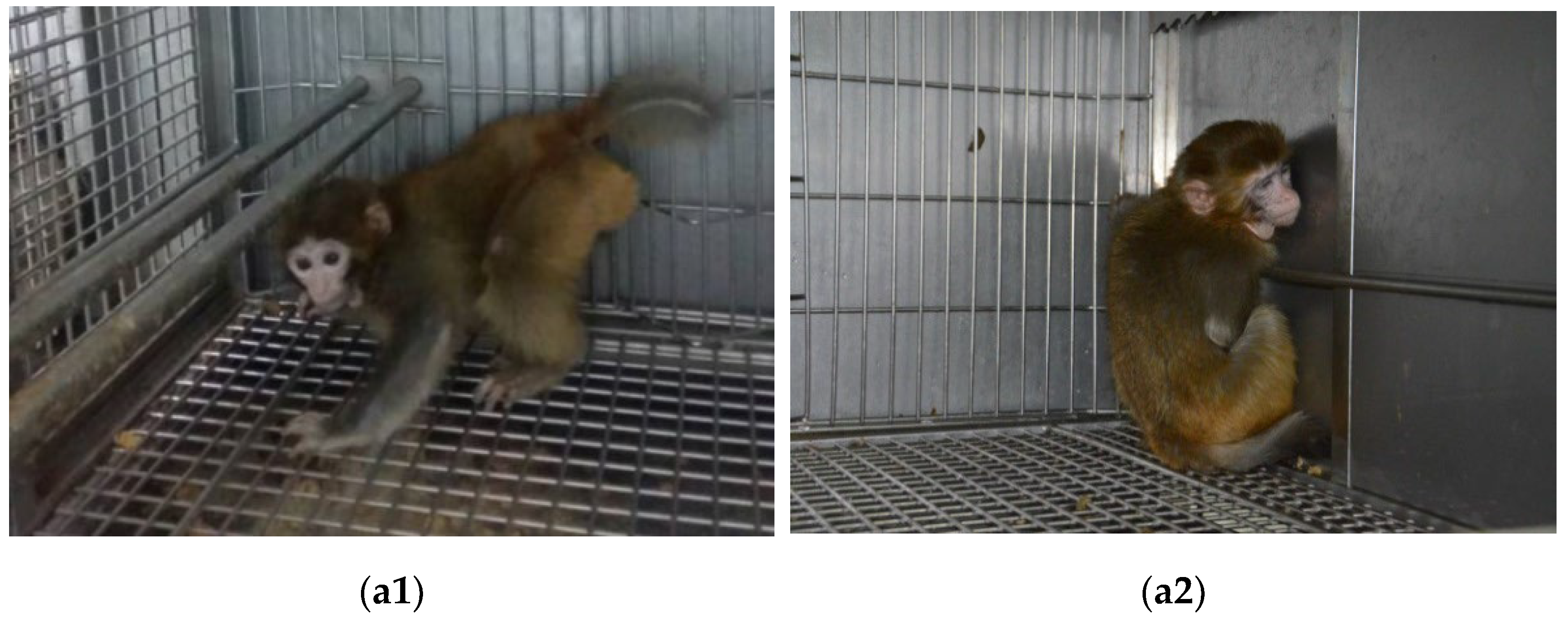
3.1. Abnormal Posture
3.2. Motor Dysfunction
3.3. Gross and Fine Motor Behavior
3.4. Muscular Tension
3.5. MRI
4. Discussion
5. Conclusions
Author Contributions
Funding
Institutional Review Board Statement
Informed Consent Statement
Data Availability Statement
Acknowledgments
Conflicts of Interest
References
- Nguyen, A.; Armstrong, E.A.; Yager, J.Y. Evidence for therapeutic intervention in the prevention of cerebral palsy: Hope from animal model research. Semin. Pediatr. Neurol. 2013, 20, 75–83. [Google Scholar] [CrossRef] [PubMed]
- Hadders-Algra, M.; Boxum, A.G.; Hielkema, T.; Hamer, E.G. Effect of early intervention in infants at very high risk of cerebral palsy: A systematic review. Dev. Med. Child Neurol. 2017, 59, 246–258. [Google Scholar] [CrossRef] [PubMed]
- Feather-Schussler, D.; Ferguson, T.S. Cerebral palsy: Underlying causes and animal models. eLS 2015, 1–12. [Google Scholar] [CrossRef]
- Santos, P.D.D.; Silva, F.C.D.; Ferreira, E.G.; Iop, R.D.R.; Bento, G.G.; Silva, R.D. Instruments that evaluate functional independence in children with cerebral palsy: A systematic review of observational studies. Fisioter. Pesqui. 2016, 23, 318–328. [Google Scholar] [CrossRef]
- Jacobson Misbe, E.N.; Richards, T.L.; Mcpherson, R.J.; Burbacher, T.M.; Juul, S.E. Perinatal asphyxia in a nonhuman primate model. Dev. Neurosci. 2011, 33, 210–221. [Google Scholar] [CrossRef] [PubMed]
- Ornoy, A. Animal models of cerebral palsy: What can we learn about cerebral palsy in humans. In Cerebral Palsy; Miller, F., Bachrach, S., Lennon, N., O’Neil, M., Eds.; Springer: Cham, Switzerland, 2018; pp. 1–12. [Google Scholar]
- Vanduffel, W. In-vivo connectivity in monkeys. In Micro-, Meso- and Macro-Connectomics of the Brain; Kennedy, H., Essen, D.C.V., Christen, Y., Eds.; Springer International Publishing: New York, NY, USA, 2016; pp. 75–87. [Google Scholar]
- Seidlitz, J.; Sponheim, C.; Glen, D.; Ye, F.Q.; Saleem, K.S.; Leopold, D.A.; Ungerleider, L.; Messinger, A. A population MRI brain template and analysis tools for the macaque. Neuroimage 2018, 170, 121–131. [Google Scholar] [CrossRef]
- Traudt, C.M.; Mcpherson, R.J.; Bauer, L.A.; Richards, T.L.; Burbacher, T.M.; Mcadams, R.M. Concurrent erythropoietin and hypothermia treatment improve outcomes in a term nonhuman primate model of perinatal asphyxia. Dev. Neurosci. 2013, 35, 491–503. [Google Scholar] [CrossRef]
- Fitch, H.; Coen, R.; Lindburg, D.; Robinson, P.; Hajek, P.; Hesselink, J.; Crutchfield, S. Cerebral palsy in a macaque monkey. Am. J. Primatol. 1988, 14, 181–187. [Google Scholar] [CrossRef]
- Mcadams, R.M.; Fleiss, B.; Traudt, C.; Schwendimann, L.; Snyder, J.M.; Haynes, R.L.; Natarajan, N.; Gressens, P.; Juul, S.E. Long-term neuropathological changes associated with cerebral palsy in a nonhuman primate model of hypoxic-ischemic encephalopathy. Dev. Neurosci. 2017, 39, 124–140. [Google Scholar] [CrossRef]
- Windle, W.F.; Bailey, C.J. Neurological deficits of asphyxia neonatorum in the monkey (Macaca mulatta). Anat. Rec. 1958, 130, 389–390. [Google Scholar] [CrossRef]
- Ranck, J.B.; Windle, W.F. Brain damage in the monkey, Macaca mulatta, by asphyxia neonatorum. Exp. Neurol. 1959, 1, 130–154. [Google Scholar] [CrossRef]
- Jacobson, H.N.; Windle, W.F. Responses of foetal and newborn monkeys to asphyxia. J. Physiol. 1960, 153, 447–465. [Google Scholar] [CrossRef] [PubMed]
- Myers, R.E. Two patterns of perinatal brain damage and their conditions of occurrence. Am. J. Obstet. Gynecol. 1972, 112, 246–276. [Google Scholar] [CrossRef]
- Loeliger, M.; Inder, T.P.; Cain, S.; Camm, E.; Yoder, B.; Mccurnin, D. Developmental and neuropathological consequences of ductal ligation in the preterm baboon. Pediatr. Res. 2009, 65, 209–214. [Google Scholar] [CrossRef]
- Griffith, J.L.; Shimony, J.S.; Cousins, S.A.; Rees, S.E.; Mccurnin, D.C.; Inder, T.E. MR imaging correlates of white-matter pathology in a preterm baboon model. Pediatr. Res. 2012, 71, 185–191. [Google Scholar] [CrossRef]
- Van der Staay, F.J.; Arndt, S.; Nordquist, R.E. Evaluation of animal models of neurobehavioral disorders. Behav. Brain Funct. 2009, 5, 11. [Google Scholar] [CrossRef] [PubMed]
- Josenby, A.; Jarnlo, G.C.; Nordmark, E. Longitudinal construct validity of the GMFM-88 total score and coal total score and the GMFM-66 score in a 5-year follow-up study. Phys. Ther. 2009, 89, 342. [Google Scholar] [CrossRef]
- Kenyon, L.K. Gross motor function measure (GMFM-66 and GMFM-88) users’ manual. Phys. Occup. Ther. Pediatr. 2014, 8, 111–112. [Google Scholar] [CrossRef]
- Zitella, L.M.; Xiao, Y.Z.; Teplitzky, B.A.; Kastl, D.J.; Duchin, Y.; Baker, K.B. In vivo 7T MRI of the non-human primate brainstem. PLoS ONE 2015, 10, e0127049. [Google Scholar] [CrossRef]
- Calabrese, E.; Badea, A.; Coe, C.L.; Lubach, G.R.; Shi, Y.; Styner, M.A. A diffusion tensor MRI atlas of the postmortem rhesus macaque brain. Neuroimage 2015, 117, 408–416. [Google Scholar] [CrossRef] [Green Version]
- Scott, J.A.; Grayson, D.; Fletcher, E.; Lee, A.; Bauman, M.D.; Schumann, C.M.; Buonocore, M.H.; Amaral, D.G. Longitudinal analysis of the developing rhesus monkey brain using magnetic resonance imaging: Birth to adulthood. Brain. Struct. Funct. 2016, 221, 2847–2871. [Google Scholar] [CrossRef] [PubMed] [Green Version]


| Behavior | Scoring Standards | Score |
|---|---|---|
| Standing | Unable to stand | 0 |
| Standing unstable | 1 | |
| Standing stable and sustainable | 2 | |
| Moving | Unable to move | 0 |
| Disturbance of gait and coordination | 1 | |
| Moving smoothly | 2 | |
| Running | Unable to run | 0 |
| Unstable running | 1 | |
| Fast complete movement | 2 | |
| Jumping | Unable to jump | 0 |
| Take-off posture | 1 | |
| Jumping | 2 | |
| Hanging | Unable to hang | 0 |
| Grasping but unable to hang | 1 | |
| Hanging and shaking the body | 2 | |
| Hanging upside down | Unable to hang upside down | 0 |
| Lasts less than 3 s | 1 | |
| Hanging upside down | 2 | |
| Climbing | Unable to climb | 0 |
| Relying on cage or short distance for climbing | 1 | |
| Climbing on the cage | 2 | |
| Fearful behavior | Body without reaction when stimulated | 0 |
| Body appearing to move when stimulated | 1 | |
| Body moving smoothly when stimulated | 2 |
| Behavior | Scoring Standards | Score |
|---|---|---|
| Lick | Unable to lick a 10 cm2 board with cream | 0 |
| Licking half the board | 1 | |
| Licking the board clean | 2 | |
| Pinch | Toe unable to bend | 0 |
| Unable to successfully pinch objects | 1 | |
| Complete pinch movement | 2 | |
| Feeding | Unable to finish feeding action | 0 |
| With feeding movement, without eating | 1 | |
| Complete feeding movement | 2 | |
| Handle | Unable to handle things | 0 |
| Unable to maintain handling position | 1 | |
| Complete handling movement | 2 | |
| Grooming | Unable to groom itself | 0 |
| Grooming actions not coherent | 1 | |
| Grooming coherent and sustained | 2 |
| Grade | Scoring Standards | Score |
|---|---|---|
| 0 | With no increased muscle tone, but could move freely | 0 |
| 1 | With slightly increased muscle tone, and no resistance in the affected limb for passive activity within the entire scope | 1 |
| 2 | With slightly increased muscle tone, the former 1/2 ROM * of the affected limb was slightly stuck in passive activity, while the latter 1/2 ROM showed slight resistance | 2 |
| 3 | With mildly increased muscle tone, the affected limb showed resistance within most of ROM in passive activity, but could still move | 3 |
| 4 | With moderately increased muscle tone, the affected limb showed resistance within the entire ROM in passive activity, and activity was difficult | 4 |
| 5 | With highly increased muscle tone, the affected limb was stiff, showing great resistance, and passive activity was extremely difficult | 5 |
Publisher’s Note: MDPI stays neutral with regard to jurisdictional claims in published maps and institutional affiliations. |
© 2022 by the authors. Licensee MDPI, Basel, Switzerland. This article is an open access article distributed under the terms and conditions of the Creative Commons Attribution (CC BY) license (https://creativecommons.org/licenses/by/4.0/).
Share and Cite
Zhu, Y.; Xiong, Y.; Zhang, J.; Tong, H.; Yang, H.; Zhu, Q.; Xu, X.; Wu, D.; Tang, J.; Li, J. Evaluation of Rhesus Macaque Models for Cerebral Palsy. Brain Sci. 2022, 12, 1243. https://doi.org/10.3390/brainsci12091243
Zhu Y, Xiong Y, Zhang J, Tong H, Yang H, Zhu Q, Xu X, Wu D, Tang J, Li J. Evaluation of Rhesus Macaque Models for Cerebral Palsy. Brain Sciences. 2022; 12(9):1243. https://doi.org/10.3390/brainsci12091243
Chicago/Turabian StyleZhu, Yong, Yanan Xiong, Jin Zhang, Haiyang Tong, Hongyi Yang, Qingjun Zhu, Xiaoyan Xu, De Wu, Jiulai Tang, and Jinhua Li. 2022. "Evaluation of Rhesus Macaque Models for Cerebral Palsy" Brain Sciences 12, no. 9: 1243. https://doi.org/10.3390/brainsci12091243
APA StyleZhu, Y., Xiong, Y., Zhang, J., Tong, H., Yang, H., Zhu, Q., Xu, X., Wu, D., Tang, J., & Li, J. (2022). Evaluation of Rhesus Macaque Models for Cerebral Palsy. Brain Sciences, 12(9), 1243. https://doi.org/10.3390/brainsci12091243


