Review

The Role of the NF-kB Pathway in Intracranial Aneurysms

by
Laurentiu Andrei Blaj
1,2,
Andrei Ionut Cucu
2,3,*,
Bogdan Ionel Tamba
4,5 and
Mihaela Dana Turliuc
1,2
1
Department of Neurosurgery, “Grigore T. Popa” University of Medicine and Pharmacy, 700115 Iasi, Romania
2
“Prof. Dr. N. Oblu” Emergency Clinical Hospital, 700309 Iasi, Romania
3
Faculty of Medicine and Biological Sciences, University Stefan cel Mare of Suceava, 720229 Suceava, Romania
4
Advanced Research and Development Center for Experimental Medicine (CEMEX), “Grigore T. Popa” University of Medicine and Pharmacy, 700115 Iasi, Romania
5
Department of Pharmacology, Clinical Pharmacology and Algesiology, “Grigore T. Popa” University of Medicine and Pharmacy, 700115 Iasi, Romania
*
Author to whom correspondence should be addressed.
Brain Sci. 2023, 13(12), 1660; https://doi.org/10.3390/brainsci13121660
Submission received: 30 October 2023 / Revised: 25 November 2023 / Accepted: 28 November 2023 / Published: 30 November 2023
(This article belongs to the Section Molecular and Cellular Neuroscience)

Abstract

:
The pathophysiology of intracranial aneurysms (IA) has been proven to be closely linked to hemodynamic stress and inflammatory pathways, most notably the NF-kB pathway. Therefore, it is a potential target for therapeutic intervention. In the present review, we investigated alterations in the vascular smooth muscle cells (VSMCs), extracellular matrix, and endothelial cells by the mediators implicated in the NF-kB pathway that lead to the formation, growth, and rupture of IAs. We also present an overview of the NF-kB pathway, focusing on stimuli and transcriptional targets specific to IAs, as well as a summary of the current strategies for inhibiting NF-kB activation in IAs. Our report adds to previously reported data and future research directions for treating IAs using compounds that can suppress inflammation in the vascular wall.

1. Introduction

Intracranial aneurysms (IA) are pathologically acquired dilatations of the arterial wall. Their pathophysiology involves several complex mechanisms, including genetic predisposition, hemodynamic stress, inflammation, and vascular remodeling. The prevalence of IA is estimated to be 2–5% in the general population, with an equal distribution among sexes and an annual risk of rupture of 1.4%. Ruptured IA has a 30–50% morbidity and a 50% mortality rate, posing a serious strain to the healthcare system. The pathophysiological mechanisms underlying aneurysm formation, growth, and rupture are still not completely understood, although significant advances have been made in recent years [1]. Aneurysm formation begins with endothelial and hemodynamic dysfunction precipitated by independent risk factors such as smoking, hypertension, and atherosclerosis [2,3]. It is followed by a coordinated inflammatory response to oxidative stress involving macrophages, mast cells, T cells, complement system, and cytokines [4]. These processes will lead to phenotypic modulation of the vascular smooth muscle cells (VSMCs), inducing apoptosis, inflammatory mediators’ upregulation, and the recruitment of other inflammatory cells, resulting in weakening of the arterial wall [5].
The nuclear factor-kappa B (NF-kB) pathway is a key inflammatory signaling pathway that regulates the expressions of various genes involved in cytokine production and cell survival. In VSMCs, NF-kB activation regulates cell survival and apoptosis, contributing to vascular remodeling and the regulation of vascular tone [6]. Also, it regulates the extracellular matrix (ECM) proteins like matrix metalloproteinases (MMPs) and the expressions of inflammatory markers like interleukin 1β and 6 (IL-1β and IL-6) promoting endothelial dysfunction [7,8]. Overall, the NF-kB pathway plays a critical role in the regulation of VSMC function, thus becoming an important pathway in the development and progression of IA by contributing to chronic inflammation and hemodynamic stress.
In this review, we looked at the relationship between the NF-kB pathway and IA in terms of its role in development, progression, and subsequent rupture. This relationship was explored by assessing how the NF-kB pathway impacts endothelial dysfunction, the regulation of VSMCs, and the control of the extracellular matrix, all of which collectively contribute to the formation of IAs.

2. Methods

We conducted a literature search of the MEDLINE/PubMed and EMBASE databases to identify studies related to the role of the NF-kB pathway in intracranial aneurysms. The search was performed on September 2023 with the keywords “intracranial aneurysm”, “NF-kB pathway”, “vascular smooth muscle cell”, “extracellular matrix”, and “endothelial dysfunction”. Two investigators (LAB and AIC) independently screened titles and abstracts. Discrepancies in the study selection were discussed with all authors and a consensus was reached.
The eligibility criteria consisted of: (1) studies reporting the relationship between the NF-kB pathway and intracranial aneurysms; (2) studies reporting the role of the NF-kB pathway in vascular smooth muscle cells, the extracellular matrix, and endothelial dysfunction; (3) studies in the English language; and (4) original research articles and reviews (editorials and letters were excluded).

3. Results

We screened 654 articles published between 1980 and September 2023. After the first screening by abstract, we excluded 12 duplicate studies, 224 unrelated to the topic, and 148 not relevant. A total of 193 articles were excluded after the full-text screening identified as irrelevant. We included 77 articles based on the eligibility criteria.
Most authors have employed similar models for IA induction, which was first described by Hashimoto et al. in rats [9]. The basic concept involves inducing hemodynamic stress to the arterial walls through unilateral common carotid ligation, unilateral renal artery ligation, and a high-salt diet. The results consisted of IA induction at the anterior cerebral artery/olfactory artery bifurcation. To this end, we can conclude that the results are specific to anterior circulation aneurysms; however, no differences were found between potential patient populations or subtypes of IA in the available research.
Regarding the timing of the intervention, most studies chose to start treatment immediately after aneurysm induction, the day after, or one month after. No explanation was provided regarding the optimal stage for implementing therapeutic strategies.

4. Discussion

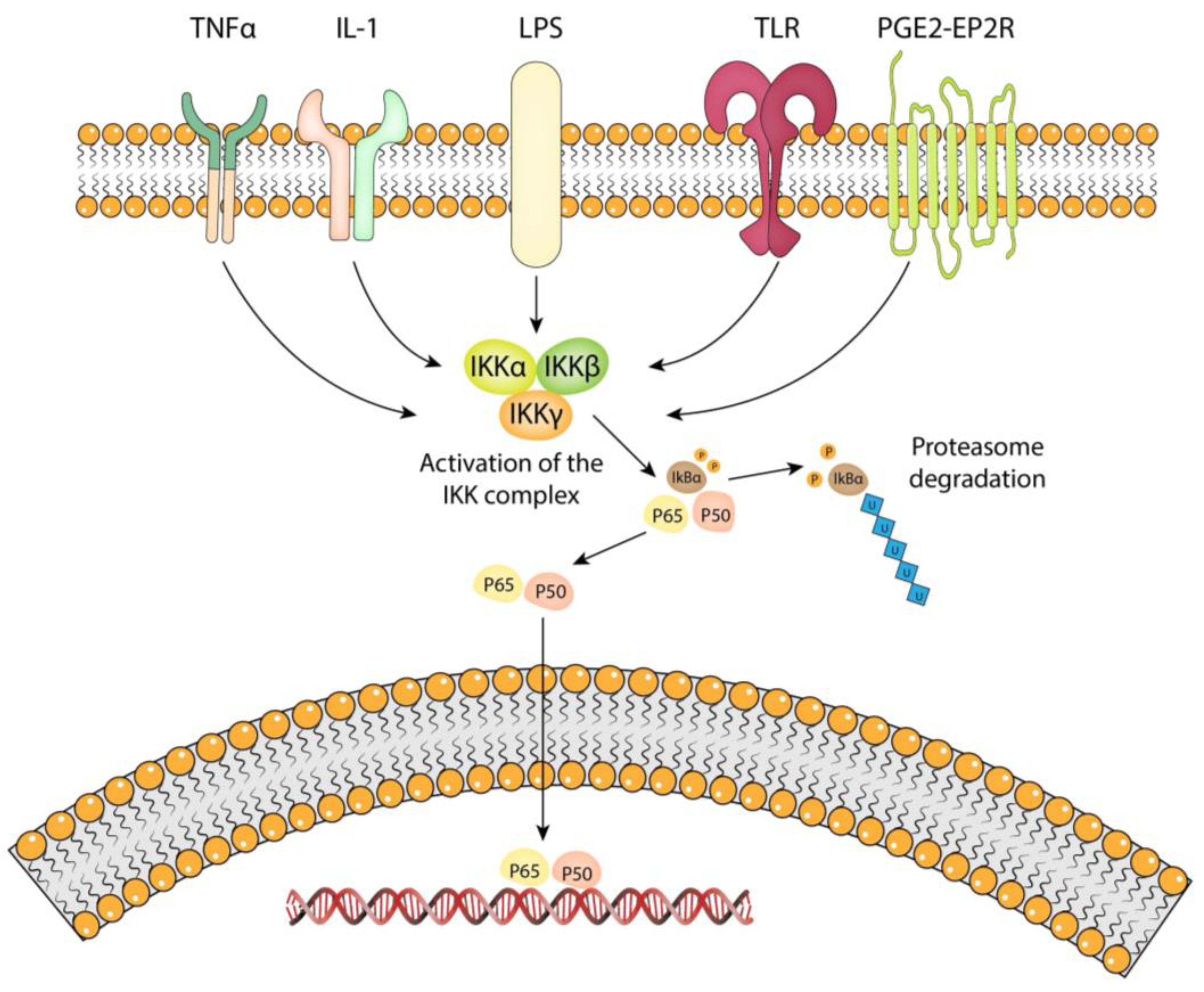
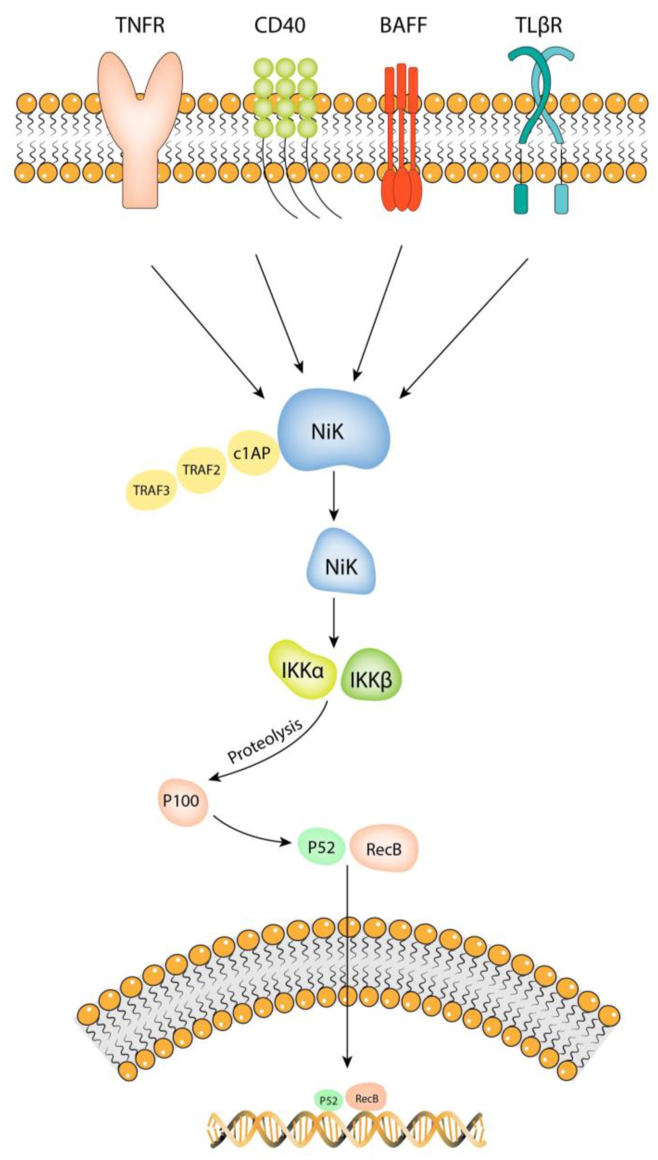
4.1. Overview of the NF-kB Pathway

The NF-kB pathway represents a family of regulated transcription factors that are activated concurrently, leading to gene transcription [10]. It was first discovered in 1986 by Baltimore et al. [11]. The transcriptional factors involved are homodimers or heterodimers of Rel proteins like p65/p50, p52, and its precursor p100 [12]. The intermediate proteins involved in the NF-kB activation pathway are represented by tumor necrosis factor receptor type 1-associated death domain (TRADD), tumor necrosis factor receptor-associated factor 2 (TRAF2), NF-kB-inducing kinase (NIK), mitogen-activated protein kinase/ERK kinase-1 (MEKK), and IκB kinase (IKK) [13]. Since its discovery, the NF-kB pathway has been shown to play a critical role in regulating the expressions of genes involved in immune function, inflammation, and cellular growth control [14].
The NF-kb pathway can be activated by various stimuli, including tumor necrosis factor α (TNF-α), interleukin 1 (IL-1), oxidative stress, lipoproteins from Gram-negative bacteria, activated B-cells, viral RNA, and hemodynamic pressure [15,16,17]. Given its responsiveness to a wide range of stimuli, it is a significant and intricate process. This complexity has led to its division into canonical and non-canonical pathways.
The canonical NF-κB pathway (Figure 1) is well characterized and is mediated by the IKK complex in response to pro-inflammatory signals [18]. The IKK complex consists of two catalytic subunits, IKKα and IKKβ, and a regulatory subunit, NEMO (also known as IKKγ) [19]. IKKα is structurally related to IKKβ and both contain a serine/threonine kinase domain at their N-terminus and protein–protein interaction motifs, including a leucine zipper (LZ) and a helix–loop–helix (HLH) at their C-terminal portion. IKKγ lacks catalytic activity and contains several coiled-coil motifs that mediate oligomerization [20]. In the absence of an external stimulus, NF-kB dimers remain in an inactivated state in the cytoplasm. IKKα/IKKβ dimers combine with IKKγ to activate the IKK complex. Once activated, IKKβ phosphorylates IkBα and associates with βTrCP. This initiates ubiquitination of IkBα and its subsequent degradation by the cytosolic proteasome. The inactivated form of the NF-kB dimer is composed of p50 and p65 proteins, coupled with IκBα. Once the latter is removed, the p50/p65 dimer is free to enter the nucleus and bind to its target genes [20]. The p50 subunit is known to interact specifically with Bcl-3 protein and influences the apoptotic behavior of the cell [21]. The p65 subunit plays a crucial role in inflammation and is known to interact with protein kinase C-δ (PKCδ) in vascular smooth muscle cells, regulating proinflammatory chemokine expression and modulating inflammation [22]. In addition, it binds to the Ccl2 and Ptgs2 genes which encode MCP-1 and COX-2, respectively [23].
The NF-kB non-canonical pathway (Figure 2) is mediated by NF-kB-inducing kinase (NIK) and is activated when ligands bind to TNFR, CD40, BAFF, and TLβR receptors [24]. NIK was first identified as a mitogen-activated kinase (MAP3K) and is the first component of the non-canonical pathway [25]. It remains in an inactivated form when bonded to the TRAF3/TRAF2/cIAP complex. Once the ligand–receptor complex is formed, the TRAF3/TRAF2/cIAP complex is recruited and NIK is activated. NIK directly phosphorylates IKKα but does not influence IKKβ [24]. IKKα induces the proteolysis of p100 to p52, generating p52/RelB dimers in order to activate NF-kB regulated anti-apoptotic bcl-2 and bcl-xl genes [26]. In the end, the transcription of Bcl-2 and Bcl-xl genes will lead to an increased cell survival [27].

4.2. Stimuli That Activate the NF-kB Pathway in IA Pathology

The NF-kB pathway can be activated in endothelial cells by a variety of stimuli, which are presumed to be causative factors in intracranial aneurysm pathology. Some of these stimuli include inflammatory cytokines (TNF-α and IL-1β), Toll-like receptor agonists, and hemodynamic stress. All of them have a role in vascular smooth muscle motility, extracellular matrix remodeling, and endothelial dysfunction.
In an experimental study, Aoki et al. revealed that the increased TNF-α content in IA lesions is caused by an increase in hemodynamic stress. They also proved that the incidence of IA was significantly suppressed in TNFR1-heterozygous and TNFR1-deficinet mice compared to that in wild-type mice, thus clarifying the crucial role of TNF-alpha-TNFR1 signaling in IA formation [28].
Procollagen types I and III expressions are decreased in cerebral aneurysm walls, possibly due to the activation of the p65 subunit of the NF-kB pathway by IL-1β. This, in turn, leads to the downregulation of procollagen types I and III and lysyl oxidase (LOX). This process can persist for up to three months following aneurysm induction, resulting in a loss of structural support [29]. A loss of collagen fibers and increased elastic capacity in the aneurysm wall leads to the formation of a ruptured zone, thus initiating the development and rupture of IA [30].
Toll-like receptors (TLR) are involved in another pathway for NF-kB activation. The expression of TLR4 was present in the endothelial cells and adventitia in human unruptured cerebral aneurysms, whereas rats presented it only in endothelial cells. The experimental data show that TLR4 expression reached its peak 1 month after aneurysm induction and decreased to the level before aneurysm induction 3 months after [31]. The expressions of TLR2, MyD88, and NF-kB p65 in patients with intracranial aneurysms are significantly higher compared to normal controls, which is why Zhang et al. proposed the TLR2/4-MyD88-NF-kB signaling pathway to be involved in the pathogenesis of intracranial aneurysms [32].
The link between hemodynamic stress, cerebral aneurysm formation, and the NF-kB pathway is the PGE2-EP2 signaling function. Shear-stress-induced PGE2-EP2 receptor signaling activates NF-kB in endothelial cells at an early stage and contributes to chronic inflammation in the arterial walls for cerebral aneurysm formation [33]. Another mechanism is based on variable mechanosensory activation. In human intracranial aneurysms, low shear stress clearly identified regions with high NF-kB activity, whereas disturbed flow was associated with randomly aligned cells and high NF-kB activation [34].

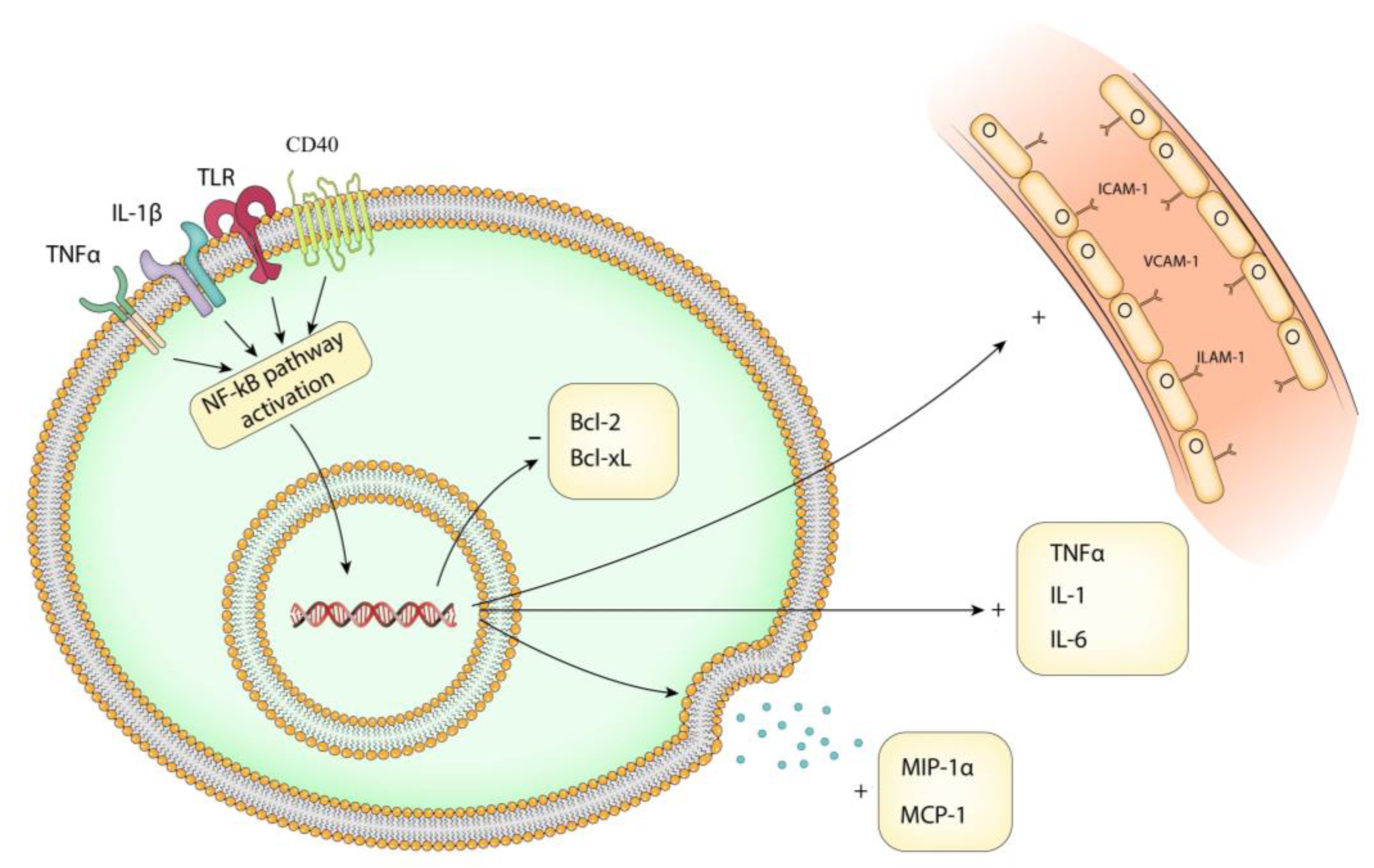
4.3. Transcriptional Targets of NF-kB Pathway in IA Pathology

The NF-kB pathway controls the expression of a variety of genes involved in the immune response, cell survival, proliferation, and inflammation. Transcriptional targets (Figure 3) depend on the signaling molecules involved and are tightly regulated. Cytokine release is a major consequence of this pathway. Activated NF-kB regulates the expression levels of IL-1β, IL-6, and TNF-α via the canonical pathway and phosphorylation of IκB. Mezzasoma et al. found that the NF-kB/ERK1/2 pathway is involved in the processing and release of IL-1β [35]. This creates a positive feedback loop that promotes chronic inflammation [36,37].
The NF-kB pathway also regulates the expression of adhesion molecules. NF-κB activation leads to the expression of adhesion molecules such as E-selectin, vascular cell adhesion molecule-1 (VCAM-1), and intracellular adhesion molecule-1 (ICAM-1) [38]. This close relationship is due to the fact that the promoter regions of ICAM-1, VCAM-1, and ELAM-1 contain NF-kB binding sites. The treatment of endothelial cells with TNF increases the adhesion of monocytes to the endothelial surface, thus amplifying the inflammatory response [39].
Regarding chemokines, the NF-kB pathway plays a crucial role in the recruitment and shaping of the cellular microenvironment. Also, this could explain the active recruitment of inflammatory cells in the aneurysm wall. Monocyte chemoattractant protein 1 (MCP-1) and macrophage inflammatory protein 1α (MIP-1α) can be regulated by protein kinase C-δ (PKCδ) through cytosolic interactions with the NF-kB subunit p65 in vascular smooth muscle [22]. In addition, MCP-1 expression can be influenced by reducing the upstream phosphorylation of IκB, as well as the activation of the NF-kB promoter [40]. Chalouhi et al. found high plasma concentrations of chemokines and chemoattractant cytokines in the lumen of 18 human IA, whereas Aoki et al. found that MCP-1 expression was upregulated in the aneurysm walls in the early stages of aneurysm formation [41,42].
One of the major roles of the NF-kB pathway is the regulation of anti-apoptotic proteins Bcl-2, Bcl-xL, and apoptotic protein A20. Bcl-2 is regulated by p53 protein binding to its p1 promoter [27,43]. A20 regulates the immune response and controls the apoptotic pathway induced by TNF-α. Its function is mediated via the NF-kB dependent alteration of cIAP1/2 in the non-canonical signaling [44]. In IA, the upregulation of the Bcl-2 gene by MiR-140 knock-out alleviates its development and progression [45]. Therefore, a regulated Bcl-2 by the NF-kB pathway could represent a potential therapeutic target for IA.

4.4. Role of NF-kB Pathway in Vascular Smooth Muscle Cell

The NF-kB pathway plays an important role in the vascular smooth muscle cell (VSMC) by being involved in the pathogenesis of vascular diseases, such as atherosclerosis, restenosis, and hypertension. It regulates VSMC by inducing proliferation, apoptosis, and differentiation (Table 1). A vascular smooth muscle cell is a highly specialized cell that has a crucial role in regulating vascular tone and blood pressure. Its primary function is contraction and relaxation, which is regulated by contractile proteins, ion channels, and signaling molecules [6]. IA growth is marked by the ongoing structural weakness of nearby smooth muscle cells, although the exact process remains uncertain. Liu et al. demonstrated that consistent cyclic mechanical stretching prompts alterations in the aneurysm tissue by exhibiting fewer VSMCs and decreased collagen type IV and VI. Preserving the well-being of VSMC offers an additional avenue for therapeutic intervention aimed at preventing the progression of IA [46].
In VSMCs, both the canonical (p65) and non-canonical (p52) NF-kB signaling pathways can be activated. Exposure to the receptor activator of nuclear factor kB-ligand (RANKL) can increase both NF-kB/p52 and NF-kB/phosphor-p65 levels, whereas treatment with tumor necrosis factor-related apoptosis-inducing ligand (TRAIL) has been proven to significantly increase NF-kB/Phospho-p65 levels [47]. Another way to activate the NF-kB pathway in vascular smooth muscle cells is through Chlamydophila pneumoniae-induced ICAM-1 expression. Vielma et al. proved that, in human aortic endothelial cells, NF-kB activation is protein kinase C (PKC)-dependent when subjected to C. pneumoniae-infected endothelial cells [48]. An activated NF-kB pathway leads to a pro-inflammatory status in the vascular wall. In a study on cell cultures, Currie et al. demonstrated that heat shock factor-1 (HSF-1) exacerbates Ang-II-induced inflammation via the NF-kB pathway. A countermeasure to it is heat shock protein 27 (Hsp27) that regulates the phosphorylation of p65 subunit of NF-kB in the Ang II-induced signaling pathway of NF-kB by suppressing the IKK complex [49,50].
The calcification of VSMCs, a byproduct of chronic inflammation, leads to a loss of elasticity and increased stiffness of the vessel wall. Excessive glucose concentrations can activate NF-kB and trigger pro-calcific effects in VSMCs, which may actively augment vascular calcification [51]. One way to prevent such a development is zinc supplementation. In a study by Voelkl et al., zinc supplementation upregulated the endogenous inhibitor of NF-kB, zinc-finger protein TNFAIP3, and decreased IkBα phosphorylation, upregulated IkBα protein abundance, and triggered the p65 subunit with decreased NF-κB-dependent transcriptional activity [52].
The survival of VSMCs depends on the NF-κB/Rel transcription factors. They regulate the expression of Bcl-xL and Bfl-1/A1, which protect the cells against death. Romano et al. proved that TNF-α enhanced the basal expression of Bcl-xL twofold via the NF-kB pathway and it induced the expression of Bfl-1/A1 [53]. TNF-α-induced NF-kB pathway activation can be managed by A20 protein (also known as tumor necrosis factor α-induced protein 3–TNFAIP3). Utilizing its deubiquitinating activity, A20 removes Lys63 polyubiquitin chains from the NF-kB essential modulator (NEMO), TNF receptor-associated factor 6 (TRAF6), and receptor-interacting protein ½ (RIP1/2) to suppress NF-kB activation in vascular smooth muscle cells [54].
Table 1. Overview of NF-kB pathway in vascular smooth muscle cells.
TriggerMechanismEffectTreatmentReferences
RANKLp52 pathwayPro-calcific effectTRAIL[47]
C. pneumoniaePKC dependent pathwayIncreased expression of ICAM-1 Not specified[48]
HSF-1Ang-II-induced inflammationActivation of proinflammatory transcription factors Hsp27[49,50]
HyperglycemiaActivation of SGK 1Vascular calcificationZinc supplementation[51,52]
TNF-αp65 pathwayIncreased expression of Bfl-1/A1 and Bcl-xLA20 protein[54]
RANKL, receptor activator of nuclear factor kB-ligand; TRAIL, tumor necrosis factor-related apoptosis-inducing ligand; C. pneumoniae, Chlamydophila pneumoniae; PKC, protein kinase C; ICAM-1, intracellular adhesion molecule-1; HSF-1, heat shock factor-1; Ang-II, angiotensin II; Hsp27, heat shock protein 27; SGK 1, glucocorticoid-inducible kinase 1; and TNF-α, tumor necrosis factor α.

4.5. Role of NF-kB Pathway in Extracellular Matrix Remodeling

Extracellular matrix (ECM) remodeling is a complex process involving the synthesis, deposition, and degradation of ECM proteins. The ECM provides structural support and plays a critical role in cell signaling, migration, proliferation, and differentiation. ECMs’ enzymes are a group of proteolytic enzymes that are involved in the degradation and remodeling of EMCs. Examples of such enzymes are matrix metalloproteinases (MMPs), serine proteases, and cysteine proteases. MMPs are regulated by various signaling pathways, including the NF-kB pathway.
The NF-κB-binding site is present in the promoter of the MMP-9 gene and an NF-κB-like element in the promoter of the MMP-1 gene. Using an over-expression of IkBα, Bond et al. proved that the NF-kB pathway is required for the cytokine upregulation of MMP-1, -3, and -9 in VSMCs [7,55]. In cardiac myoblast H9c2 cells, TNF-α increased the mRNA expression and protein activity of MMP-9 [56]. Also, MMP-2, MMP-9, VEGF, ROS, IL-1β, and IL-6 were significantly increased in TNF-α-induced endothelial cells via the NF-kB pathway [8]. In a study on fibroblasts, Xu et al. demonstrated that MMP-1 expression was decreased when the NF-kB pathway was inhibited by the peptide inhibitor SN50 [57]. TNF-α exposed cells showed increased NF-κB activity and increased ratios of MMP-2/TIMP-2, and MMP-9/TIMP-1 in airway smooth muscle cells (ASM). In turn, extracellular matrix deposition can be reduced by suppressing the NF-kB pathway via the estrogen receptor β (ER-β) [58]. In IA, MMP-2 and -9 are two important factors that promote progression; therefore, they can become candidates for treatment targets via the NF-kB pathway [59].
A connection between TIMP-2 and the NF-kB signaling pathway has been established in melanoma cell lines and lung epithelial cells. Sun et al. proved that tissue inhibitor of metalloproteinase-2 (TIMP-2) overexpression increased NF-kB transcriptional activity, via decreasing the inhibitory molecule IkBα [60]. A more in-depth analysis was conducted by Lizarraga et al. In their study, they concluded that NF-kB activity was increased by exposure to TIMP-2, with maximal activity being achieved after 24 h and rapidly decreasing by 48 h. Also, IkB1α was downregulated 24 h after exposure to TIMP-2, while IkBβ levels were only slightly decreased. Bcl-3 expression diminished 24 h after TIMP-2 exposure and increased after 48 h [61]. It can be inferred from these findings that the primary factor is the inhibition of IkB1α.
Regarding MMP-3, Sanchavanakit et al. observed that the pretreatment of murine cementoblast cell lines with pyrrolidine dithiocarbamate (PDTC), an inhibitor of NF-kB or p38 MAPK, resulted in MMP-3 suppression [62]. Also, in a study on glioma cells, the upregulation of the Bmi-1 gene promoted cell migration and invasion via the NF-kB-mediated upregulation of MMP-3. Another NF-kB inhibition drug, Resveratrol, significantly reduced the level of MMP-3 [63,64].

4.6. The NF-kB Pathway in Endothelial Dysfunction

Endothelial dysfunction is a condition in which the endothelium loses its normal function and is unable to regulate vascular tone and permeability. Under normal conditions, the NF-kB pathway is tightly regulated in endothelial cells. However, under various conditions, endothelial dysfunction takes over and can lead to an uneven expression of adhesion proteins, loss of permeability, and vasodilatory impairment. Also, damage to the endothelial cell layer is cited as the first event in IA formation [65].
The activation of the NF-kB pathway in endothelial cells has been shown to upregulate hypoxia inducible factor-1 (HIF-1) via a positive feedback loop. This, in turn, causes endothelial cell dysfunction, cell proliferation, angiogenesis, and inflammation [66]. In addition, dermal microvascular endothelial cell (HMECs-1) permeability is influenced by the hemoglobin (Hb)-induced NF-kB pathway, which is under the control of the TLR signaling pathway [67].
In a randomized, double-blind, placebo-controlled crossover study of 14 non-diabetic overweight or obese adults, Pierce et al. demonstrated that activating the NF-kB pathway via the stimulation of oxidative stress led to marked impairments in baseline vascular function by influencing endothelium-dependent dilatation [68]. Vascular tone can be influenced by the NF-kB-mediated COX-2/iNOS pathway. Using an androgen receptor agonist, Gonzales et al. found that dihydrotestosterone enhanced NF-kB activation and increased cyclooxygenase-2 protein levels in rats. These data suggest an increase in the basal production of vasodilatory factors [69]. Vasodilatation will, in turn, create a low endothelial wall shear stress that may facilitate the growing phase and initiate the rupture of IA [70].
The NF-kB pathway can modulate endothelial cell death and increase cerebral vessel permeability. The selective deletion of the NF-kB essential modulator (NEMO) or the upstream kinase Tak1 in brain endothelial cells resulted in cerebral hypoperfusion and disruption of the blood–brain barrier (BBB). This proved that TAK1-NEMO signaling has a protective role in cerebral vessels via the NF-kB pathway [71]. Another pathway to disrupt the BBB is via the NOTCH3/NF-kB signaling pathway. IL-1β induced the nuclear translocation of NF-kB/p65, upregulated MMP-9 expression in pericytes, and increased permeability in the BBB [72].
Regarding the influence of the NF-kB pathway on adhesion molecules, the inhibition of NF-kB pathway using amino acids like glycine, histidine, and cysteine has been proven to reduce E-selectin expression and IL-6 production in human coronary arterial endothelial cells (HCAECs) stimulated with TNF-α [73]. Also, increased lipid peroxides in preeclamptic plasma stimulate ICAM-1 expression through a NF-kB-mediated mechanism. This hypothesis is supported by the findings that vitamin E and N-acetyl cysteine not only inhibit endothelial cell NF-kB activation by preeclamptic plasma, but also ICAM-1 expression, leading to endothelial dysfunction [74]. A similar relationship between ICAM-1 and NF-kB activation has been found in cells subjected to angiogenic basic fibroblast growth factor (bFGF/FGF-2). bFGF inhibits the TNF-mediated activation of NF-kB by blocking the phosphorylation and degradation of IkBα and decreases ICAM-1 expression. The end result is a decrease in cell adhesion and increased permeability [75]. The latter has also been observed in the wall of the IA compared with paired healthy cerebral arteries [76].
TNF-α-activated endothelial cells stimulate VSMCs and immune cell proliferation and promote the early and late apoptosis of VSMCs. This leads to an increase in inflammation in the vascular wall and promotes aneurysm formation [77].

4.7. Current Strategies for Inhibiting NF-kB Activation

The management of IA remains challenging due to the limited understanding of its pathogenesis. The current working hypothesis has been reduced to an inflammatory injury coupled with hemodynamic stress, placing the NF-kB pathway at the forefront of the discussion. As such, the inhibition of the NF-kB pathway has emerged as a promising strategy for the treatment and prevention of IA (Table 2). Attempts have been made with both pharmaceutical and natural extracts, but so far, the results have only been in the experimental phase.
Research results suggest that statins can inhibit the NF-kB pathway. The anti-inflammatory action of statins derives from suppressing TLR4/MyD88/NF-kB signaling by decreasing the expression of TLRs 2 and 4 [78]. Pitavastatin suppresses IA progression by reducing the number of DNA-binding forms of the NF-kB p65 subunit. Moreover, pitavastatin treatment can cause the regression of degenerative changes in preexisting IA walls [79]. Also, treatment with simvastin suppresses the development of IAs and has a preventive effect on the progression of preexisting IAs [80].
Another commonly used drug is aspirin. It has been proven that aspirin significantly reduced the degeneration of aneurysm walls by inhibiting macrophages-mediated chronic inflammation and dramatically inhibiting the expression of NF-kB and monocyte chemoattractant protein-1 (MCP-1) in the aneurysm wall [81]. However, treatment with aspirin did not appear to alter the histopathological findings after aneurysm formation, suggesting that it would have its greatest effect during the acute inflammatory phase [82].
Nifedipine is a widely used calcium antagonist and it has been proven to inhibit TNF-α induced reactive oxygen species (ROS) generation, as well as decrease the NF-kB activity in cultured fibroblasts. In rats, nifedipine inhibited DNA binding NF-kB in aneurysmal walls, preventing the enlargement and degeneration of the aneurysmal walls of preexisting IAs [83].
Regarding the prevention of IA formation, zinc supplementation significantly increased the expression of the anti-inflammatory signaling protein A20, an inhibitor of the nuclear factor kB (NF-kB) pathway, in rat IAs. Zinc administration may prevent the growth of rat IAs by inducing the A20-attributed inactivation of NF-kB signaling [84]. However, long-term treatment with zinc (more than 3 months) can develop resistance and enhance the activity of the NF-kB pathway, which can lead to the activation of metastatic mechanisms [85].
The TNF-α inhibitor, Etanercept, significantly reduced aneurysm formation in rats. It suppressed the expressions of NF-kB, iNOS, MMP-9 mRNA, and phospho-IKKα and phospho-IKKβ mRNAs. There was no significant difference at 1 month or 3 months after aneurysm induction between the control group, low-dose etanercept group, and high-dose etanercept group [86]. Side effects from using TNF-α inhibitors have been observed in individuals with rheumatoid arthritis and include potential cancer risks, however, the study results remain controversial [87]. Also, the STAT3 inhibitor, BP-1-102, can reduce the expression of inflammatory factors and MMPs bound to NF-kB by inhibiting the activation of the JAK/STAT3/NF-kB pathway, and then restore the vascular wall elastin to reduce blood pressure, thereby treating aneurysms in mice [88]. Another compound that has proven its efficacy, although still experimental, is represented by bone marrow mesenchymal stem cells (BMSCs). Human BMSC-derived exosomes inhibited the activation of the phosphatidylinositol-3 kinase (PI3K)/protein kinase B (Akt)/ nuclear factor-kappa B (NF-kB) signaling pathway and maintained Th17/Treg balance, which, in turn, suppress aneurysm formation [89].
Natural extracts have been shown to possess anti-inflammatory properties. Resveratrol, a polyphenol compound widely found in plants, such as Polygonum cuspidatum, Cassia tora, grapes, and peanuts, can inhibit IA formation in mice subjected to induced hypertension by downregulating the NF-kB pathway [90]. Also, Tanashione IIA (Tan IIA) is an anti-inflammatory component isolated from a traditional Chinese medicine called Danshen (Salvia miltiorrhiza Bunge). Tan IIA treatment prevented the process of macrophage infiltration and the degeneration of aneurysmal walls due to the inhibition of NF-kB and MCP-1 expression in rats [91].
Table 2. Therapeutic targets for NF-kB pathway inhibition.
CompoundAnimal Model/Cell CultureMechanism of ActionEffectYearReferences
PitvastinRatsReduced binding to DNA for p56 subunitSmaller aneurysm size, lower expression of MCP-1, VCAM-1, MMP-9, IL-1β, and iNOS2009[79]
SimvastinRatsNot specifiedReduced expression of MCP-1, VCAM-1, MMP-2, and MMP-92008[80]
AspirinRatsNot specifiedReduced expression of MCP-1, VCAM-12015[81]
NifedipineRatsInhibited translocation of p65 subunit into the nucleusReduced expression of MCP-1 and MMP-22008[83]
ZincRatsIncreased production of A20 proteinSmaller aneurysm size2021[84]
EtanecerptRatsTNF-α inhibitorSmaller aneurysm size and thicker internal elastic membrane2014[86]
BP-1-102MiceSTAT3 inhibitorReduced expression of TNF-α, IL-1β, IL-6, MCP-1, IL-10, and MMP2020[88]
BMSCsCell culture/rabbitsInhibition of KLF5 and suppression of PI3k/Akt/NF-kB pathwayIncreased eNOS mRNA expression, decreased iNOS mRNA, MMP-2, and MMP-9 expression2020[89]
ResveratrolCell cultureMMP-3 inhibitorInhibits cell apoptosis by increasing the expression of Bcl-2 protein, decreased iNOS and NO expression2014[64]
MiceNot specifiedLower IA incidence, reduced expression of MMP-2, and MMP-92022[90]
Tanshione IIARatsTNF-α inhibitorReduced expression of MCP-1, MMP-2 and MMP-92019[91]
STAT3, signal transducer and activator of transcription 3; KLF5, kruppel-like factor 5; PI3k, phosphoinositide-3-kinase; Akt, protein kinase B; NF-kB, nuclear factor kB; MCP-1, monocyte chemoattractant protein-1; VCAM-1, vascular cell adhesion molecule 1; MMP, matrix metalloproteinase; TNF-α, tumor necrosis factor α; IL-1β, interleukin 1β; IL-10, interleukin 10; iNOS, inducible nitric oxide synthase; eNOS, endothelial nitric oxide synthase; and NO, nitric oxide.
To our knowledge, there are no studies in which a combination of therapeutic strategies has been employed, although Aoki et al. proved that the synergic activation of the NF-kB pathway with prostaglandin E2 and TNF-α increases the expression of inflammatory markers [23]. We believe that the next step in the inhibition of the NF-kB pathway is a combination of current strategies to prevent the progression and rupture of IA. The inhibition of the NF-kB pathway has proven advantageous in diverse neurovascular disorders, such as intracranial stenosis, moyamoya disease, and ischemic stroke. Consequently, the therapeutic approaches outlined above hold potential for applications in managing these specific conditions [92].

4.8. Clinical Trials Targeting Inflammation for Treating IA

As of the writing of this review, we identified several clinical trials targeting the reduction of inflammation to treat IA. Of great interest is the ongoing Statin Treatment for Unruptured Intracranial Aneurysms Study (STUDIES), where researchers are looking to identify whether oral atorvastatin administered daily for six months will reduce the inflammatory signs in the wall of saccular aneurysms on high-resolution magnetic resonance imaging (HRMRI) [93]. The same protocol was used in the ATREAT-VBD trial that focused on unruptured intracranial vertebrobasilar dissecting aneurysms that did not benefit from surgery [94]. Aspirin has been used in two clinical trials so far. Hassan et al. conducted a prospective study of 11 patients using low-dose aspirin (81 mg daily for 3 months) and showed promising results [95]. A high dose of aspirin (300 mg daily for 3 months) is currently ongoing in the Unruptured Intracranial Aneurysm Aspirin Trial (UIAAT) [96]. Regarding ruptured IA, the ongoing FINISHER trial is investigating the use of dexamethasone in aneurysmal subarachnoid hemorrhage to reduce delayed cerebral ischemia [97].

4.9. Future Research Directions for Treating IA

Although recent studies have investigated the potential of targeting the NF-kB pathway for IA treatment, there is still much to be learned regarding the specific mechanisms involved. Potential avenues for future investigation already discussed in this review may consist of the peptide inhibitor SN50, pyrrolidine dithiocarbamate (PDTC), vitamin E, and N-acetyl cysteine. Further research may focus on anti-cold induced RNA binding protein (CIRP) antibody to suppress vascular wall inflammation via the TLR4/NF-kB pathway. Promising results have been obtained for abdominal aortic aneurysms in rats [98]. Also, microRNA(miR)-195 suppressed abdominal aortic aneurysms through the TNF-α/NF-kB and VEGF/PI3K/Akt pathway [99]. Prostaglandin E receptor subtype 2 (EP2) antagonists can be another potential therapeutic target for aneurysm suppression by inhibiting the prostaglandin E2-EP2-NF-kB signaling in macrophages [23].
Traditional Chinese medicine has offered anti-inflammatory extracts that are rumored to inhibit the NF-kB pathway. Paeonol, a major phenolic ingredient extracted from peony bark, has been proven to alleviate inflammatory disorders and aneurysm progression, while Daidzein attenuates abdominal aortic aneurysm in angiotensin II-induced mice via NF-kB, p38MAPK, and TFG-β1 [100,101].
Future research should focus on whether the inhibition of the NF-kB pathway might be beneficial for preventing IA recurrence after endovascular coiling or surgical clipping. To this end, imaging modalities such as the auto-segmentation of tomography images or hybrid machine learning systems may represent future directions for the better selection of patients that benefit from targeting the NF-kB pathway [102,103].

4.10. Limitations

Current therapeutic strategies have only been employed in preclinical settings, with the exception of statins and aspirin. Potential challenges could arise from translating them to clinical treatments, mainly because there is little variability in the preclinical models used, the sample sizes are small, and the research is conducted under standard conditions, unlike in a clinical scenario. Regarding efficacy and safety, aspirin was the only drug with preliminary results in patients presenting with IA. In addition, no known interactions or contraindications between NF-kB-pathway-targeted therapies and other medications commonly used in IA treatment have been reported. Another limitation is the limited number of clinical trials investigating the effect of NF-kB pathway inhibition; therefore, to our knowledge, no long-term outcomes have been reported in patients with IA.

5. Conclusions

The NF-kB signaling pathway is an important mediator of the pathophysiology of intracranial aneurysms through its role in vascular smooth muscle cell proliferation, extracellular matrix remodeling, and endothelial dysfunction. The identification of various stimuli and transcriptional targets has led to the development of various strategies to inhibit their activation. Most notably, statins and aspirin are currently used in human trials and hold great promise. However, there is still much to be learned about the precise role of NF-kB in IA pathology. Overall, the NF-kB pathway is a promising therapeutic target for aneurysm formation and prevention and is a reliable source for identifying novel therapeutic targets.

Author Contributions

Conceptualization, L.A.B. and A.I.C.; methodology, B.I.T. and M.D.T.; data curation, L.A.B. and B.I.T.; writing—original draft preparation, L.A.B. and A.I.C.; writing—review and editing, M.D.T. and B.I.T.; visualization, M.D.T.; supervision, M.D.T. All authors have read and agreed to the published version of the manuscript.

Funding

This research received no external funding.

Institutional Review Board Statement

Not applicable.

Informed Consent Statement

Not applicable.

Data Availability Statement

No new data were created or analyzed in this study. Data sharing is not applicable to this article.

Conflicts of Interest

The authors declare no conflict of interest.

References

  1. Texakalidis, P.; Sweid, A.; Mouchtouris, N.; Peterson, E.C.; Sioka, C.; Rangel-Castilla, L.; Reavey-Cantwell, J.; Jabbour, P. Aneurysm Formation, Growth, and Rupture: The Biology and Physics of Cerebral Aneurysms. World Neurosurg. 2019, 130, 277–284. [Google Scholar] [CrossRef] [PubMed]
  2. Chalouhi, N.; Ali, M.S.; Jabbour, P.M.; Tjoumakaris, S.I.; Gonzalez, L.F.; Rosenwasser, R.H.; Koch, W.J.; Dumont, A.S. Biology of Intracranial Aneurysms: Role of Inflammation. J. Cereb. Blood Flow Metab. 2012, 32, 1659–1676. [Google Scholar] [CrossRef]
  3. Kono, K.; Fujimoto, T.; Shintani, A.; Terada, T. Hemodynamic Characteristics at the Rupture Site of Cerebral Aneurysms: A Case Study. Neurosurgery 2012, 71, E1202–E1209. [Google Scholar] [CrossRef] [PubMed]
  4. Signorelli, F.; Sela, S.; Gesualdo, L.; Chevrel, S.; Tollet, F.; Pailler-Mattei, C.; Tacconi, L.; Turjman, F.; Vacca, A.; Schul, D.B. Hemodynamic Stress, Inflammation, and Intracranial Aneurysm Development and Rupture: A Systematic Review. World Neurosurg. 2018, 115, 234–244. [Google Scholar] [CrossRef] [PubMed]
  5. Etminan, N.; Buchholz, B.A.; Dreier, R.; Bruckner, P.; Torner, J.C.; Steiger, H.-J.; Hänggi, D.; Macdonald, R.L. Cerebral Aneurysms: Formation, Progression, and Developmental Chronology. Transl. Stroke Res. 2014, 5, 167–173. [Google Scholar] [CrossRef] [PubMed]
  6. Hilgers, R.H.P.; Webb, R.C. Molecular Aspects of Arterial Smooth Muscle Contraction: Focus on Rho. Exp. Biol. Med. 2005, 230, 829–835. [Google Scholar] [CrossRef] [PubMed]
  7. Bond, M.; Chase, A.J.; Baker, A.H.; Newby, A.C. Inhibition of Transcription Factor NF-kB Reduces Matrix Metalloproteinase-1, -3 and -9 Production by Vascular Smooth Muscle Cells. Cardiovasc. Res. 2001, 50, 556–565. [Google Scholar] [CrossRef] [PubMed]
  8. Zhou, P.; Lu, S.; Luo, Y.; Wang, S.; Yang, K.; Zhai, Y.; Sun, G.; Sun, X. Attenuation of TNF-α-Induced Inflammatory Injury in Endothelial Cells by Ginsenoside Rb1 via Inhibiting NF-κB, JNK and P38 Signaling Pathways. Front. Pharmacol. 2017, 8, 464. [Google Scholar] [CrossRef]
  9. Hashimoto, N.; Handa, H.; Hazama, F. Experimentally Induced Cerebral Aneurysms in Rats. Surg. Neurol. 1978, 10, 3–8. [Google Scholar]
  10. Nishikori, M. Classical and Alternative NF-.KAPPA.B Activation Pathways and Their Roles in Lymphoid Malignancies. J. Clin. Exp. Hematop. 2005, 45, 15–24. [Google Scholar] [CrossRef]
  11. Sen, R.; Baltimore, D. Multiple Nuclear Factors Interact with the Immunoglobulin Enhancer Sequences. Cell 1986, 46, 705–716. [Google Scholar] [CrossRef] [PubMed]
  12. Manna, P.; Ghosh, M.; Ghosh, J.; Das, J.; Sil, P.C. Contribution of Nano-Copper Particles to in Vivo Liver Dysfunction and Cellular Damage: Role of IκBα/NF-κB, MAPKs and Mitochondrial Signal. Nanotoxicology 2011, 6, 1–21. [Google Scholar] [CrossRef] [PubMed]
  13. Lee, J.; Jung, E.; Park, J.; Jung, K.; Lee, S.; Hong, S.; Park, J.; Park, E.-K.; Kim, J.-E.; Park, S.-H.K.; et al. Anti-Inflammatory Effects of Magnolol and Honokiol Are Mediated through Inhibition of the Downstream Pathway of MEKK-1 in NF-κB Activation Signaling. Planta Medica 2005, 71, 338–343. [Google Scholar] [CrossRef] [PubMed]
  14. Scheinman, R.I.; Beg, A.A.; Baldwin, A.S. NF-Kappa B P100 (Lyt-10) Is a Component of H2TF1 and Can Function as an I Kappa B-like Molecule. Mol. Cell. Biol. 1993, 13, 6089–6101. [Google Scholar] [CrossRef] [PubMed]
  15. Pallett, M.A.; Lu, Y.; Smith, G.L. DDX50 Is a Viral Restriction Factor That Enhances IRF3 Activation. Viruses 2022, 14, 316. [Google Scholar] [CrossRef]
  16. Jiang, S.; Liu, C.; Zhang, X.; Xu, X.; Zou, J.; Fang, Y.; Ding, X. Renal Protection by Delayed Ischaemic Preconditioning Is Associated with Inhibition of the Inflammatory Response and NF-κB Activation. Cell Biochem. Funct. 2007, 25, 335–343. [Google Scholar] [CrossRef]
  17. Lu, X.; Chen, Q.; Liu, H.; Zhang, X. Interplay Between Non-Canonical NF-κB Signaling and Hepatitis B Virus Infection. Front. Immunol. 2021, 12, 730684. [Google Scholar] [CrossRef]
  18. Lawrence, T. The Nuclear Factor NF-B Pathway in Inflammation. Cold Spring Harb. Perspect. Biol. 2009, 1, a001651. [Google Scholar] [CrossRef]
  19. Hinz, M.; Scheidereit, C. The IκB Kinase Complex in NF-κB Regulation and Beyond. EMBO Rep. 2013, 15, 46–61. [Google Scholar] [CrossRef]
  20. Karin, M. The IκB Kinase (IKK) Complex as a Critical Regulator of Immune Responses. Int. Congr. Ser. 2005, 1285, 97–103. [Google Scholar] [CrossRef]
  21. Schwarz, E.M.; Krimpenfort, P.; Berns, A.; Verma, I.M. Immunological Defects in Mice with a Targeted Disruption in Bcl-3. Genes Dev. 1997, 11, 187–197. [Google Scholar] [CrossRef] [PubMed]
  22. Ren, J.; Wang, Q.; Morgan, S.; Si, Y.; Ravichander, A.; Dou, C.; Kent, K.C.; Liu, B. Protein Kinase C-δ (PKCδ) Regulates Proinflammatory Chemokine Expression through Cytosolic Interaction with the NF-κB Subunit P65 in Vascular Smooth Muscle Cells. J. Biol. Chem. 2014, 289, 9013–9026. [Google Scholar] [CrossRef] [PubMed]
  23. Aoki, T.; Frȍsen, J.; Fukuda, M.; Bando, K.; Shioi, G.; Tsuji, K.; Ollikainen, E.; Nozaki, K.; Laakkonen, J.; Narumiya, S. Prostaglandin E2-EP2-NF-κB Signaling in Macrophages as a Potential Therapeutic Target for Intracranial Aneurysms. Sci. Signal 2017, 10, eaah6037. [Google Scholar] [CrossRef] [PubMed]
  24. Thu, Y.M.; Richmond, A. NF-κB Inducing Kinase: A Key Regulator in the Immune System and in Cancer. Cytokine Growth Factor Rev. 2010, 21, 213–226. [Google Scholar] [CrossRef] [PubMed]
  25. Malinin, N.L.; Boldin, M.P.; Kovalenko, A.V.; Wallach, D. MAP3K-Related Kinase Involved in NF-kappaB Induction by TNF, CD95 and IL-1. Nature 1997, 385, 540–544. [Google Scholar] [CrossRef] [PubMed]
  26. Yang, C.H.; Murti, A.; Pfeffer, L.M. Interferon Induces NF-κB-Inducing Kinase/Tumor Necrosis Factor Receptor-Associated Factor-Dependent NF-κB Activation to Promote Cell Survival*. J. Biol. Chem. 2005, 280, 31530–31536. [Google Scholar] [CrossRef] [PubMed]
  27. Catz, S.D.; Johnson, J.L. Transcriptional Regulation of Bcl-2 by Nuclear Factor Kappa B and Its Significance in Prostate Cancer. Oncogene 2001, 20, 7342–7351. [Google Scholar] [CrossRef]
  28. Aoki, T.; Fukuda, M.; Nishimura, M.; Nozaki, K.; Narumiya, S. Critical Role of TNF-Alpha-TNFR1 Signaling in Intracranial Aneurysm Formation. Acta Neuropathol. Commun. 2014, 2, 34. [Google Scholar] [CrossRef]
  29. Aoki, T.; Kataoka, H.; Ishibashi, R.; Nozaki, K.; Morishita, R.; Hashimoto, N. Reduced Collagen Biosynthesis Is the Hallmark of Cerebral Aneurysm: Contribution of Interleukin-1beta and Nuclear Factor-kappaB. Arterioscler. Thromb. Vasc. Biol. 2009, 29, 1080–1086. [Google Scholar] [CrossRef]
  30. Signorelli, F.; Pailler-Mattei, C.; Gory, B.; Larquet, P.; Robinson, P.; Vargiolu, R.; Zahouani, H.; Labeyrie, P.-E.; Guyotat, J.; Pelissou-Guyotat, I.; et al. Biomechanical Characterization of Intracranial Aneurysm Wall: A Multiscale Study. World Neurosurg. 2018, 119, e882–e889. [Google Scholar] [CrossRef]
  31. Aoki, T.; Nishimura, M.; Ishibashi, R.; Kataoka, H.; Takagi, Y.; Hashimoto, N. Toll-like Receptor 4 Expression during Cerebral Aneurysm Formation. Laboratory Investigation. J. Neurosurg. 2010, 113, 851–858. [Google Scholar] [CrossRef] [PubMed]
  32. Zhang, X.; Wan, Y.; Feng, J.; Li, M.; Jiang, Z. Involvement of TLR2/4-MyD88-NF-κB Signaling Pathway in the Pathogenesis of Intracranial Aneurysm. Mol. Med. Rep. 2021, 23, 230. [Google Scholar] [CrossRef] [PubMed]
  33. Aoki, T.; Nishimura, M.; Matsuoka, T.; Yamamoto, K.; Furuyashiki, T.; Kataoka, H.; Kitaoka, S.; Ishibashi, R.; Ishibazawa, A.; Miyamoto, S.; et al. PGE(2) -EP(2) Signalling in Endothelium Is Activated by Haemodynamic Stress and Induces Cerebral Aneurysm through an Amplifying Loop via NF-κB. Br. J. Pharmacol. 2011, 163, 1237–1249. [Google Scholar] [CrossRef] [PubMed]
  34. Baeriswyl, D.C.; Prionisti, I.; Peach, T.; Tsolkas, G.; Chooi, K.Y.; Vardakis, J.; Morel, S.; Diagbouga, M.R.; Bijlenga, P.; Cuhlmann, S.; et al. Disturbed Flow Induces a Sustained, Stochastic NF-κB Activation Which May Support Intracranial Aneurysm Growth in Vivo. Sci. Rep. 2019, 9, 4738. [Google Scholar] [CrossRef] [PubMed]
  35. Mezzasoma, L.; Antognelli, C.; Talesa, V.N. A Novel Role for Brain Natriuretic Peptide: Inhibition of IL-1 β Secretion via Downregulation of NF-kB/Erk 1/2 and NALP3/ASC/Caspase-1 Activation in Human THP-1 Monocyte. Mediat. Inflamm. 2017, 2017, 5858315. [Google Scholar] [CrossRef]
  36. St-Jacques, B.; Ma, W. Role of Prostaglandin E2 in the Synthesis of the Pro-Inflammatory Cytokine Interleukin-6 in Primary Sensory Neurons: An in Vivo and in Vitro Study: PGE2 Induces IL-6 in DRG Neurons. J. Neurochem. 2011, 118, 841–854. [Google Scholar] [CrossRef] [PubMed]
  37. Yang, B.; Zhang, Z.; Yang, Z.; Ruan, J.; Luo, L.; Long, F.; Tang, D. Chanling Gao Attenuates Bone Cancer Pain in Rats by the IKKβ/NF-κB Signaling Pathway. Front. Pharmacol. 2020, 11, 525. [Google Scholar] [CrossRef]
  38. Weber, C.; Erl, W.; Pietsch, A.; Strobel, M.; Ziegler-Heitbrock, H.W.L.; Weber, P.C. Antioxidants Inhibit Monocyte Adhesion by Suppressing Nuclear Factor-icB Mobilization and Induction of Vascular Cell Adhesion Molecule-1 in Endothelial Cells Stimulated to Generate Radicals. Arter. Thromb. 1994, 14, 1665–1673. [Google Scholar] [CrossRef]
  39. Kumar, A.; Dhawan, S.; Aggarwal, B.B. Emodin (3-Methyl-1,6,8-Trihydroxyanthraquinone) Inhibits TNF-Induced NF-kB Activation, IkB Degradation, and Expression of Cell Surface Adhesion Proteins in Human Vascular Endothelial Cells. Oncogene 1998, 17, 913–918. [Google Scholar] [CrossRef]
  40. Fagiani, F.; Catanzaro, M.; Buoso, E.; Basagni, F.; Di Marino, D.; Raniolo, S.; Amadio, M.; Frost, E.H.; Corsini, E.; Racchi, M.; et al. Targeting Cytokine Release through the Differential Modulation of Nrf2 and NF-κB Pathways by Electrophilic/Non-Electrophilic Compounds. Front. Pharmacol. 2020, 11, 1256. [Google Scholar] [CrossRef]
  41. Chalouhi, N.; Points, L.; Pierce, G.L.; Ballas, Z.; Jabbour, P.; Hasan, D. Localized Increase of Chemokines in the Lumen of Human Cerebral Aneurysms. Stroke 2013, 44, 2594–2597. [Google Scholar] [CrossRef] [PubMed]
  42. Aoki, T.; Kataoka, H.; Ishibashi, R.; Nozaki, K.; Egashira, K.; Hashimoto, N. Impact of Monocyte Chemoattractant Protein-1 Deficiency on Cerebral Aneurysm Formation. Stroke 2009, 40, 942–951. [Google Scholar] [CrossRef] [PubMed]
  43. Toshiyuki, M.; Reed, J.C. Tumor Suppressor P53 Is a Direct Transcriptional Activator of the Human Bax Gene. Cell 1995, 80, 293–299. [Google Scholar] [CrossRef] [PubMed]
  44. Feoktistova, M.; Makarov, R.; Brenji, S.; Schneider, A.T.; Hooiveld, G.J.; Luedde, T.; Leverkus, M.; Yazdi, A.S.; Panayotova-Dimitrova, D. A20 Promotes Ripoptosome Formation and TNF-Induced Apoptosis via cIAPs Regulation and NIK Stabilization in Keratinocytes. Cells 2020, 9, 351. [Google Scholar] [CrossRef] [PubMed]
  45. Deng, J.; Ning, K.; Liu, D.; Wu, D.; Wan, R.; Ge, J. MiR-140 Promotes the Progression of Intracranial Aneurysms by Targeting BCL2L2. NeuroReport 2023, 34, 38. [Google Scholar] [CrossRef]
  46. Liu, P.; Song, Y.; Zhou, Y.; Liu, Y.; Qiu, T.; An, Q.; Song, J.; Li, P.; Shi, Y.; Li, S.; et al. Cyclic Mechanical Stretch Induced Smooth Muscle Cell Changes in Cerebral Aneurysm Progress by Reducing Collagen Type IV and Collagen Type VI Levels. Cell. Physiol. Biochem. 2018, 45, 1051–1060. [Google Scholar] [CrossRef]
  47. Harper, E.; Rochfort, K.D.; Forde, H.; Davenport, C.; Smith, D.; Cummins, P.M. Activation of the Non-Canonical NF-κB/P52 Pathway in Vascular Endothelial Cells by RANKL Elicits pro-Calcific Signalling in Co-Cultured Smooth Muscle Cells. Cell. Signal. 2018, 47, 142–150. [Google Scholar] [CrossRef]
  48. Vielma, S.A.; Krings, G.; Lopes-Virella, M.F. Chlamydophila pneumoniae Induces ICAM-1 Expression in Human Aortic Endothelial Cells via Protein Kinase C–Dependent Activation of Nuclear Factor-κB. Circ. Res. 2003, 92, 1130–1137. [Google Scholar] [CrossRef]
  49. Chen, Y.; Currie, R.W. Small Interfering RNA Knocks down Heat Shock Factor-1 (HSF-1) and Exacerbates pro-Inflammatory Activation of NF-kappaB and AP-1 in Vascular Smooth Muscle Cells. Cardiovasc. Res. 2006, 69, 66–75. [Google Scholar] [CrossRef]
  50. Voegeli, T.S.; Currie, R.W. siRNA Knocks down Hsp27 and Increases Angiotensin II-Induced Phosphorylated NF-κB P65 Levels in Aortic Smooth Muscle Cells. Inflamm. Res. 2009, 58, 336–343. [Google Scholar] [CrossRef]
  51. Henze, L.A.; Estepa, M.; Pieske, B.; Lang, F.; Eckardt, K.-U.; Alesutan, I.; Voelkl, J. Zinc Ameliorates the Osteogenic Effects of High Glucose in Vascular Smooth Muscle Cells. Cells 2021, 10, 3083. [Google Scholar] [CrossRef] [PubMed]
  52. Voelkl, J.; Tuffaha, R.; Luong, T.T.D.; Zickler, D.; Masyout, J.; Feger, M.; Verheyen, N.; Blaschke, F.; Kuro-o, M.; Tomaschitz, A.; et al. Zinc Inhibits Phosphate-Induced Vascular Calcification through TNFAIP3-Mediated Suppression of NF-κB. J. Am. Soc. Nephrol. 2018, 29, 1636–1648. [Google Scholar] [CrossRef] [PubMed]
  53. Giordano, A.; Avellino, R.; Ferraro, P.; Romano, S.; Corcione, N.; Romano, M.F. Rapamycin Antagonizes NF-κB Nuclear Translocation Activated by TNF-α in Primary Vascular Smooth Muscle Cells and Enhances Apoptosis. Am. J. Physiol.-Heart Circ. Physiol. 2006, 290, H2459–H2465. [Google Scholar] [CrossRef] [PubMed]
  54. Yao, D.; Xu, L.; Xu, O.; Li, R.; Chen, M.; Shen, H.; Zhu, H.; Zhang, F.; Yao, D.; Chen, Y.-F.; et al. O-Linked β-N-Acetylglucosamine Modification of A20 Enhances the Inhibition of NF-κB (Nuclear Factor-κB) Activation and Elicits Vascular Protection after Acute Endoluminal Arterial Injury. ATVB 2018, 38, 1309–1320. [Google Scholar] [CrossRef] [PubMed]
  55. Vincenti, M.P.; Coon, C.I.; Brinckerhoff, C.E. Nuclear Factor kappaB/P50 Activates an Element in the Distal Matrix Metalloproteinase 1 Promoter in Interleukin-1beta-Stimulated Synovial Fibroblasts. Arthritis Rheum. 1998, 41, 1987–1994. [Google Scholar] [CrossRef]
  56. Sun, W.-T.; Yang, C.L.H.; Or, T.C.T.; Luo, D.; Li, J.C.B. Ginsenoside Rb1 from Panax Notoginseng Suppressed TNF-α-Induced Matrix Metalloproteinase-9 via the Suppression of Double-Strand RNA-Dependent Protein Kinase (PKR)/NF-κB Pathway. Molecules 2022, 27, 8050. [Google Scholar] [CrossRef]
  57. Sirum-Connolly, K.; Brinckerhoff, C.E. Lnterleukin-1 or Phorbol Induction of the Stromelysin Promoter Requires an Element That Cooperates with AP-1. Nuclic Acids Res. 1991, 19, 335–341. [Google Scholar] [CrossRef]
  58. Ambhore, N.S.; Kalidhindi, R.S.R.; Pabelick, C.M.; Hawse, J.R.; Prakash, Y.S.; Sathish, V. Differential Estrogen-Receptor Activation Regulates Extracellular Matrix Deposition in Human Airway Smooth Muscle Remodeling via NF-κB Pathway. FASEB J. 2019, 33, 13935–13950. [Google Scholar] [CrossRef]
  59. Aoki, T.; Kataoka, H.; Morimoto, M.; Nozaki, K.; Hashimoto, N. Macrophage-Derived Matrix Metalloproteinase-2 and -9 Promote the Progression of Cerebral Aneurysms in Rats. Stroke 2007, 38, 162–169. [Google Scholar] [CrossRef]
  60. Sun, J.; Stetler-Stevenson, W.G. Overexpression of Tissue Inhibitors of Metalloproteinase 2 Up-Regulates NF-κB Activity in Melanoma Cells. JMS 2009, 4, 4. [Google Scholar] [CrossRef]
  61. Lizárraga, F.; Maldonado, V.; Meléndez-Zajgla, J. Tissue Inhibitor of Metalloproteinases-2 Growth-Stimulatory Activity Is Mediated by Nuclear Factor-Kappa B in A549 Lung Epithelial Cells. Int. J. Biochem. Cell Biol. 2004, 36, 1655–1663. [Google Scholar] [CrossRef] [PubMed]
  62. Sanchavanakit, N.; Saengtong, W.; Manokawinchoke, J.; Pavasant, P. TNF-α Stimulates MMP-3 Production via PGE2 Signalling through the NF-kB and P38 MAPK Pathway in a Murine Cementoblast Cell Line. Arch. Oral Biol. 2015, 60, 1066–1074. [Google Scholar] [CrossRef] [PubMed]
  63. Sun, P.; Mu, Y.; Zhang, S. A Novel NF-κB/MMP-3 Signal Pathway Involves in the Aggressivity of Glioma Promoted by Bmi-1. Tumor Biol. 2014, 35, 12721–12727. [Google Scholar] [CrossRef] [PubMed]
  64. Huang, T.; Gao, D.; Jiang, X.; Hu, S.; Zhang, L.; Fei, Z. Resveratrol Inhibits Oxygen-Glucose Deprivation-Induced MMP-3 Expression and Cell Apoptosis in Primary Cortical Cells via the NF-κB Pathway. Mol. Med. Rep. 2014, 10, 1065–1071. [Google Scholar] [CrossRef]
  65. Sheinberg, D.L.; McCarthy, D.J.; Elwardany, O.; Bryant, J.-P.; Luther, E.; Chen, S.H.; Thompson, J.W.; Starke, R.M. Endothelial Dysfunction in Cerebral Aneurysms. Neurosurg. Focus 2019, 47, E3. [Google Scholar] [CrossRef] [PubMed]
  66. Jain, T.; Nikolopoulou, E.A.; Xu, Q.; Qu, A. Hypoxia Inducible Factor as a Therapeutic Target for Atherosclerosis. Pharmacol. Ther. 2018, 183, 22–33. [Google Scholar] [CrossRef] [PubMed]
  67. Lisk, C.; Kominsky, D.; Ehrentraut, S.; Bonaventura, J.; Nuss, R.; Hassell, K.; Nozik-Grayck, E.; Irwin, D.C. Hemoglobin-Induced Endothelial Cell Permeability Is Controlled, in Part, via a Myeloid Differentiation Primary Response Gene–88–Dependent Signaling Mechanism. Am. J. Respir. Cell Mol. Biol. 2013, 49, 619–626. [Google Scholar] [CrossRef] [PubMed]
  68. Pierce, G.L.; Lesniewski, L.A.; Lawson, B.R.; Beske, S.D.; Seals, D.R. Nuclear Factor-κB Activation Contributes to Vascular Endothelial Dysfunction via Oxidative Stress in Overweight/Obese Middle-Aged and Older Humans. Circulation 2009, 119, 1284–1292. [Google Scholar] [CrossRef]
  69. Gonzales, R.J.; Duckles, S.P.; Krause, D.N. Dihydrotestosterone Stimulates Cerebrovascular Inflammation through NFjB, Modulating Contractile Function. J. Cereb. Blood Flow Metab. 2009, 29, 244–253. [Google Scholar] [CrossRef]
  70. Shojima, M.; Oshima, M.; Takagi, K.; Torii, R.; Hayakawa, M.; Katada, K.; Morita, A.; Kirino, T. Magnitude and Role of Wall Shear Stress on Cerebral Aneurysm: Computational Fluid Dynamic Study of 20 Middle Cerebral Artery Aneurysms. Stroke 2004, 35, 2500–2505. [Google Scholar] [CrossRef]
  71. Ridder, D.A.; Wenzel, J.; Müller, K.; Töllner, K.; Tong, X.-K.; Assmann, J.C.; Stroobants, S.; Weber, T.; Niturad, C.; Fischer, L.; et al. Brain Endothelial TAK1 and NEMO Safeguard the Neurovascular Unit. J. Exp. Med. 2015, 212, 1529–1549. [Google Scholar] [CrossRef] [PubMed]
  72. Qin, W.; Li, J.; Zhu, R.; Gao, S.; Fan, J.; Xia, M.; Zhao, R.C.; Zhang, J. Melatonin Protects Blood-Brain Barrier Integrity and Permeability by Inhibiting Matrix Metalloproteinase-9 via the NOTCH3/NF-κB Pathway. Aging 2019, 11, 11391–11415. [Google Scholar] [CrossRef] [PubMed]
  73. Hasegawa, S.; Ichiyama, T.; Sonaka, I.; Ohsaki, A.; Okada, S.; Wakiguchi, H.; Kudo, K.; Kittaka, S.; Hara, M.; Furukawa, S. Cysteine, Histidine and Glycine Exhibit Anti-Inflammatory Effects in Human Coronary Arterial Endothelial Cells. Clin. Exp. Immunol. 2012, 167, 269–274. [Google Scholar] [CrossRef] [PubMed]
  74. Takacs, P.; Kauma, S.W.; Sholley, M.M.; Walsh, S.W.; Dinsmoor, M.J.; Green, K. Increased Circulating Lipid Peroxides in Severe Preeclampsia Activate NF-κΒ and Upregulate ICAM-1 in Vascular Endothelial Cells. FASEB J. 2001, 15, 279–281. [Google Scholar] [CrossRef] [PubMed]
  75. Flati, V.; Pastore, L.I.; Griffioen, A.W.; Satijn, S.; Toniat, E.; D’Alimonte, I.; Marchetti, P.; Gulin, A.; Martinottp, S. Endothelial Cell Anergy Is Mediated by bFGF through the Sustained Activation of P38-MAPK and NF-kappaB Inhibition. Int. J. Immunopathol. Pharmacol. 2006, 19, 761–773. [Google Scholar] [CrossRef]
  76. Vakil, P.; Ansari, S.A.; Cantrell, C.G.; Eddleman, C.S.; Dehkordi, F.H.; Vranic, J.; Hurley, M.C.; Batjer, H.H.; Bendok, B.R.; Carroll, T.J. Quantifying Intracranial Aneurysm Wall Permeability for Risk Assessment Using Dynamic Contrast-Enhanced MRI: A Pilot Study. AJNR Am. J. Neuroradiol. 2015, 36, 953–959. [Google Scholar] [CrossRef] [PubMed]
  77. Chu, F.; Wang, M.; Ma, H.; Zhu, J. Simvastatin Modulates Interaction between Vascular Smooth Muscle Cell/Macrophage and TNF-α–Activated Endothelial Cell. J. Cardiovasc. Pharmacol. 2018, 71, 268–274. [Google Scholar] [CrossRef]
  78. Koushki, K.; Shahbaz, S.K.; Mashayekhi, K.; Sadeghi, M.; Zayeri, Z.D.; Taba, M.Y.; Banach, M.; Al-Rasadi, K.; Johnston, T.P.; Sahebkar, A. Anti-Inflammatory Action of Statins in Cardiovascular Disease: The Role of Inflammasome and Toll-like Receptor Pathways. Clin. Rev. Allerg. Immunol. 2021, 60, 175–199. [Google Scholar] [CrossRef]
  79. Aoki, T.; Kataoka, H.; Ishibashi, R.; Nakagami, H.; Nozaki, K.; Morishita, R.; Hashimoto, N. Pitavastatin Suppresses Formation and Progression of Cerebral Aneurysms through Inhibition of the Nuclear Factor kappaB Pathway. Neurosurgery 2009, 64, 357–365; discussion 365–366. [Google Scholar] [CrossRef]
  80. Aoki, T.; Kataoka, H.; Ishibashi, R.; Nozaki, K.; Hashimoto, N. Simvastatin Suppresses the Progression of Experimentally Induced Cerebral Aneurysms in Rats. Stroke 2008, 39, 1276–1285. [Google Scholar] [CrossRef]
  81. Li, S.; Wang, D.; Tian, Y.; Wei, H.; Zhou, Z.; Liu, L.; Wang, D.; Dong, J.-F.; Jiang, R.; Zhang, J. Aspirin Inhibits Degenerative Changes of Aneurysmal Wall in a Rat Model. Neurochem. Res. 2015, 40, 1537–1545. [Google Scholar] [CrossRef] [PubMed]
  82. Ho, J.P.; Galex, I.A.; Sadeghi, N.B.; Weledji, N.; Cabello Bermudez, S.I.; Mitchell, B.A.; Bush, D.M.; Yap, E.; Davis, N.C.; Catalino, M.P.; et al. Rabbit Elastase Aneurysm: Imaging and Histology Correlates for Inflammation and Healing. World Neurosurg. 2021, 148, e242–e251. [Google Scholar] [CrossRef] [PubMed]
  83. Aoki, T.; Kataoka, H.; Ishibashi, R.; Nozaki, K.; Hashimoto, N. Nifedipine Inhibits the Progression of an Experimentally Induced Cerebral Aneurysm in Rats with Associated Down-Regulation of NF-Kappa B Transcriptional Activity. Curr. Neurovasc. Res. 2008, 5, 37–45. [Google Scholar] [CrossRef]
  84. Hayashi, K.; Kataoka, H.; Minami, M.; Ikedo, T.; Miyata, T.; Shimizu, K.; Nagata, M.; Yang, T.; Yamamoto, Y.; Yokode, M.; et al. Association of Zinc Administration with Growth Suppression of Intracranial Aneurysms via Induction of A20. J. Neurosurg. 2020, 134, 992–998. [Google Scholar] [CrossRef] [PubMed]
  85. Holubova, M.; Axmanova, M.; Gumulec, J.; Raudenska, M.; Sztalmachova, M.; Babula, P.; Adam, V.; Kizek, R.; Masarik, M. KRAS NF-κB Is Involved in the Development of Zinc Resistance and Reduced Curability in Prostate Cancer. Metallomics 2014, 6, 1240. [Google Scholar] [CrossRef] [PubMed]
  86. Yokoi, T.; Isono, T.; Saitoh, M.; Yoshimura, Y.; Nozaki, K. Suppression of Cerebral Aneurysm Formation in Rats by a Tumor Necrosis Factor-α Inhibitor. J. Neurosurg. 2014, 120, 1193–1200. [Google Scholar] [CrossRef] [PubMed]
  87. Choi, B.; Park, H.J.; Song, Y.-K.; Oh, Y.-J.; Kim, I.-W.; Oh, J.M. The Risk of Newly Diagnosed Cancer in Patients with Rheumatoid Arthritis by TNF Inhibitor Use: A Nationwide Cohort Study. Arthritis Res. Ther. 2022, 24, 191. [Google Scholar] [CrossRef]
  88. Jiang, Z.; Huang, J.; You, L.; Zhang, J.; Li, B. Pharmacological Inhibition of STAT3 by BP-1-102 Inhibits Intracranial Aneurysm Formation and Rupture in Mice through Modulating Inflammatory Response. Pharmacol. Res. Perspect. 2021, 9, e00704. [Google Scholar] [CrossRef]
  89. Sun, X.; Zheng, X.; Zhang, X.; Zhang, Y.; Luo, G. Exosomal microRNA-23b-3p from Bone Marrow Mesenchymal Stem Cells Maintains T Helper/Treg Balance by Downregulating the PI3k/Akt/NF-κB Signaling Pathway in Intracranial Aneurysm. Brain Res. Bull. 2020, 165, 305–315. [Google Scholar] [CrossRef]
  90. Jin, T.; An, Q.; Qin, X.; Qin, X.; Hu, Y.; Hu, J.; Zhou, B.; Leng, B. Resveratrol Inhibits Cerebral Aneurysms in Mice via Downregulating the NF-κB Pathway. Acta Biochim. Pol. 2022, 69, 613–618. [Google Scholar] [CrossRef]
  91. Ma, J.; Hou, D.; Wei, Z.; Zhu, J.; Lu, H.; Li, Z.; Wang, X.; Li, Y.; Qiao, G.; Liu, N. Tanshinone IIA Attenuates Cerebral Aneurysm Formation by Inhibiting the NF-κB-mediated Inflammatory Response. Mol. Med. Rep. 2019, 20, 1621–1628. [Google Scholar] [CrossRef] [PubMed]
  92. Mejia-Munne, J.C.; Ellis, J.A.; Feldstein, N.A.; Meyers, P.M.; Connolly, E.S. Moyamoya and Inflammation. World Neurosurg. 2017, 100, 575–578. [Google Scholar] [CrossRef] [PubMed]
  93. Statin Treatment for Unruptured Intracranial Aneurysms Study (STUDIES). Available online: https://clinicaltrials.gov/study/NCT04149483 (accessed on 25 November 2023).
  94. Atorvastatin in the Treatment of Intracranial Unruptured VertebroBasilar Dissecting Aneurysms (ATREAT-VBD). Available online: https://classic.clinicaltrials.gov/ct2/show/NCT04943783 (accessed on 25 November 2023).
  95. Hasan, D.M.; Chalouhi, N.; Jabbour, P.; Dumont, A.S.; Kung, D.K.; Magnotta, V.A.; Young, W.L.; Hashimoto, T.; Richard Winn, H.; Heistad, D. Evidence That Acetylsalicylic Acid Attenuates Inflammation in the Walls of Human Cerebral Aneurysms: Preliminary Results. JAHA 2013, 2, e000019. [Google Scholar] [CrossRef]
  96. Unruptured Intracranial Aneurysm Aspirin Trial (UIAAT). Available online: https://clinicaltrials.gov/study/NCT03661463 (accessed on 25 November 2023).
  97. Güresir, E. Fight INflammation to Improve Outcome after Aneurysmal Subarachnoid HEmorRhage. 2023. Available online: http://clinicaltrials.gov (accessed on 25 November 2023).
  98. Wang, Y.; Lu, L.; Li, W.; Gu, S. Effect of Anti-CIRP Antibody on Inflammatory Response, Tumor Formation and Abdominal Aortic Aneurysm in Rats. Trop. J. Pharm Res. 2020, 19, 1215–1219. [Google Scholar] [CrossRef]
  99. Ma, X.; Yao, H.; Yang, Y.; Jin, L.; Wang, Y.; Wu, L.; Yang, S.; Cheng, K. miR-195 Suppresses Abdominal Aortic Aneurysm through the TNF-α/NF-κB and VEGF/PI3K/Akt Pathway. Int. J. Mol. Med. 2018, 41, 2350–2358. [Google Scholar] [CrossRef] [PubMed]
  100. Chen, S.; Luo, K.; Bian, S.; Chen, J.; Qiu, R.; Wu, X.; Li, G. Paeonol Ameliorates Abdominal Aortic Aneurysm Progression by the NF-κB Pathway. Ann. Vasc. Surg. 2021, 77, 255–262. [Google Scholar] [CrossRef]
  101. Liu, Y.-F.; Bai, Y.-Q.; Qi, M. Daidzein Attenuates Abdominal Aortic Aneurysm through NF-κB, p38MAPK and TGF-Β1 Pathways. Mol. Med. Rep. 2016, 14, 955–962. [Google Scholar] [CrossRef]
  102. Heydarheydari, S.; Tahmasebi Birgani, M.; Rezaeijo, S.M. Auto-Segmentation of Head and Neck Tumors in Positron Emission Tomography Images Using Non-Local Means and Morphological Frameworks. Pol. J. Radiol. 2023, 88, 365–370. [Google Scholar] [CrossRef]
  103. Hosseinzadeh, M.; Gorji, A.; Fathi Jouzdani, A.; Rezaeijo, S.M.; Rahmim, A.; Salmanpour, M.R. Prediction of Cognitive Decline in Parkinson’s Disease Using Clinical and DAT SPECT Imaging Features, and Hybrid Machine Learning Systems. Diagnostics 2023, 13, 1691. [Google Scholar] [CrossRef]
Figure 1. Canonical NF-kB pathway. Activation of the NF-kB pathway through the TNF-α, IL-1, LPS, TLR, and PGE2-EP2 receptors induces the activation of the IKK complex, which is composed of two catalytic subunits, IKKα and IKKβ, and a regulatory subunit IKKγ. Once activated, IKKβ phosphorylates IkBα and initiates its subsequent degradation, leaving the p50/p65 dimer free for entry into the nucleus. The illustration is created by the authors using Adobe Illustrator v2020.
Brainsci 13 01660 g001
Figure 2. Non-canonical NF-kB pathway. Activation of the non-canonical pathway via the ligand binding to TNFR, CD40, BAFF, and TLβR receptors will lead to recruitment of the TRAF3/TRAF2/cIAP complex, leaving the NIK unrestricted. Once activated, NIK will directly phosphorylate IKKα. This causes IKKα to induce the proteolysis of p100 to p52, generating p52/RelB dimers. The P52/RelB dimer enters the nucleus and regulates the bcl-2 and bcl-xl genes. The illustration is created by the authors using Adobe Illustrator v2020.
Brainsci 13 01660 g002
Figure 3. Transcriptional targets of NF-kB pathway. Activated canonical (p65) and non-canonical (p52) NF-kB signaling pathways increase the release of cytokines IL-1β, IL-6, and TNF-α. Adhesion molecules like ICAM-1, VCAM-1, and ECAM-1 have an increased expression in endothelial cells and chemokines (MCP-1, MIP-1α) are upregulated. Anti-apoptotic proteins Bcl-2 and Bcl-xL are downregulated by TNF-α. The illustration is created by the authors using Adobe Illustrator v2020.
Brainsci 13 01660 g003
Disclaimer/Publisher’s Note: The statements, opinions and data contained in all publications are solely those of the individual author(s) and contributor(s) and not of MDPI and/or the editor(s). MDPI and/or the editor(s) disclaim responsibility for any injury to people or property resulting from any ideas, methods, instructions or products referred to in the content.

